lab exercises week 1: #3 microscope #5 simple staining #1 ubiquity of microorganisms pre lab due:...
TRANSCRIPT

Lab Exercises Week 1:
#3 Microscope#5 Simple Staining#1 Ubiquity of Microorganisms
Pre lab due: NonePost lab due: 1/17/15 & 1/24/15

• Light passes through specimen and then series of magnifying lenses
• Bright-field microscope is most common type• Three key concepts
– Magnification: apparent increase in size• Modern compound microscopes have two lens types:
objective and ocular
Principles of Light Microscopy
• Magnification is product of objective (4x, 10x, 40x, and 100x) and ocular lens (10x)
• Condenser lens (betweenlight source and specimen) focuses light on specimen, does not magnify
Ocular lens(eyepiece)Magnifies theimage, usually10-fold (10×).
Specimenstage
CondenserFocusesthe light.
Iris diaphragmControls theamount of lightthat enters theobjective lens.
Objective lensA selection of lensoptions providedifferentmagnifications. Thetotal magnification isthe product of themagnifying power ofthe ocular lens andthe objective lens.
Light source
RheostatControls thebrightness of thelight.
Courtesy of Leica, Inc., Deerfield, FL
Copyright © The McGraw-Hill Companies, Inc. Permission required for reproduction or display.

Microscopy-Brightfield

1.5. Size in the Microbial World
Copyright © The McGraw-Hill Companies, Inc. Permission required for reproduction or display.
1 micron (µ) = 10–6 metermicrometer (µm) = 10–6 meter = .000001 meter
Nucleus
MitochondriaProteinsSmallmolecules
Atoms Lipids Ribosomes Smallestbacteria
Mostbacteria
Most eukaryotic cells Adult roundworm
Human height
10 m1 m0.1 m1 cm1 mm100 µm10 µm0.1 nm 1 nm 10 nm 100 nm 1 µm
The basic unit of length is the meter (m), and allother units are fractions of a meter.
These units of measurement correspond to unitsin an older but still widely used convention.
1 angstrom (Å) = 10–10 meternanometer (nm) = 10–9 meter = .000000001 meter
1 meter = 39.4 inchesmillimeter (mm) = 10–3 meter = .001 meter
Prion fibril
Viruses
Electron microscope
Light microscope
Unaided human eye

Oil has same refractive index as glass

Samples can be immobilized, stained to visualize Basic dyes (positive charge)
• Attracted to negatively charged cellular components
Acidic dyes (negative charge)• Negative staining: cells repel, so colors background• Can be done as wet mount
3.2. Microscopic Techniques: Dyes and Staining
Spread thin film ofspecimen over slide.
Allow to air dry. Flood the smear withstain, rinse, and dry.
Examine with microscope.
Copyright © The McGraw-Hill Companies, Inc. Permission required for reproduction or display.
Pass slidethroughflame toheat-fixspecimen.

Goldilocks technique
Using a sterile loop place three drops of water onto one slide.
Re-sterilize your loop and get some bacteria on your loop (enough that you can see it with your eye).
Mix with water to create a smear. 1st one you will be able to see the bacteria (very
cloudy). 2nd one you will only barely be able to see the
bacteria (slightly cloudy). 3rd one you should not be able to see bacteria in
water drop (clear).
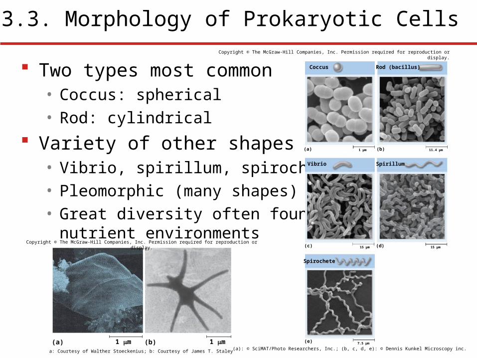
Two types most common• Coccus: spherical• Rod: cylindrical
Variety of other shapes• Vibrio, spirillum, spirochete• Pleomorphic (many shapes)• Great diversity often found in low
nutrient environments
3.3. Morphology of Prokaryotic Cells
(e)
(c)
(a)
Vibrio
Spirochete
Coccus Rod (bacillus)
Spirillum
11.4 µm1 µm
15 µm15 µm
7.5 µm
(b)
(d)
Copyright © The McGraw-Hill Companies, Inc. Permission required for reproduction or display.
(a): © SciMAT/Photo Researchers, Inc.; (b, c, d, e): © Dennis Kunkel Microscopy inc.1 m(a) 1 m (b)
a: Courtesy of Walther Stoeckenius; b: Courtesy of James T. Staley
Copyright © The McGraw-Hill Companies, Inc. Permission required for reproduction or display.

Most prokaryotes divide by binary fission• Cells often stick together
following division• Form characteristic groupings
Groupings
Cell dividesin one plane.
Diplococcus
Chains
Chain of cocci
Packets
(a)
Packet
Clusters
(b)
Cell dividesin two or more planesperpendicular to oneanother.
Cluster
Cell dividesin several planes atrandom.
(c)
Copyright © The McGraw-Hill Companies, Inc. Permission required for reproduction or display.
(a): (top): © George Musil/Visuals Unlimited; (bottom): © David M. Phillips/Visuals Unlimited; (b): © R. Kessel & C. Shih/Visuals Unlimited; (c): © Oliver Mecks/Photo Researchers, Inc.

1. Algae
2. Fungi
3. Protozoa
4. Bacteria
5. Viruses
Which microbes are eukaryotes? Which are prokaryotes? Which can perform
photosynthesis? Which are classified based on
locomotion? Which have cell walls? Which have some type of
nucleic acid?

Major Groups of Microbial World
Copyright © The McGraw-Hill Companies, Inc. Permission required for reproduction or display.
Microbial World
Domain Bacteria EucaryaArchaea
Prokaryotes (unicellular) Eukaryotes
Algae(unicellular ormulticellular)
Protists
Helminths(multicellular
parasites)
Fungi(unicellular ormulticellular)
Protozoa(unicellular)
Viruses Viroids Prions
Organisms(living)
Infectious agents(non-living)

Prokaryotes found growing in severe conditions• Ocean depths, volcanic vents, polar regions all harbor
thriving prokaryotic species• Many scientists believe that if life exists on other
planets, it may resemble these microbes
Individual species have limited set of conditions Important to grow microbes in culture
• Medical significance• Nutritional, industrial uses
Microbial Ubiquity

Prokaryotes inhabit nearly all environments• Some live in comfortable habitats favored by humans• Some live in harsh environments
• Termed extremophiles; most are Archaea
Major conditions that influence growth
• Nutrient availability
• Temperature
• Atmosphere- Oxygen
• pH
• Water availability
4.5. Environmental Factors That Influence Microbial Growth