labeling fluorescence in situ hybridization probes for genomic
TRANSCRIPT

Labeling FISH Probes for DNA Targets 21
21
From: Methods in Molecular Biology, Vol. 204: Molecular Cytogenetics: Protocols and ApplicationsEdited by: Y. S. Fan © Humana Press Inc., Totowa, NJ
2
Labeling Fluorescence In Situ HybridizationProbes for Genomic Targets
Larry E. Morrison, Ramesh Ramakrishnan, Teresa M. Ruffalo,and Kim A. Wilber
1. IntroductionFluorescence in situ hybridization (FISH) requires nucleic acid probes, including
deoxyribonucleic acid (DNA), ribonucleic acid (RNA), or nucleic acid analogs, labeleddirectly with fluorophores, or capable of indirect association with fluorophores. Thenucleic acid provides the FISH assay with its specificity through complementarypairing of the probe nucleotides with nucleotides of the target nucleic acid. Theappended fluorophores provide the ability to visually detect the homologous regionswithin the cellular structure using a fluorescence microscope. Photographic or elec-tronic cameras can also be used to provide permanent images of the fluorescence stain-ing patterns, and the latter can be used to provide quantitative measurements of theprobe fluorescence.
This chapter describes a variety of methods by which DNA can be coupled tofluorophores to form FISH probes directed toward genomic targets. Following a briefdiscussion of labeling methodologies, fluorophore selection, and sources of probeDNA, a number of detailed protocols are provided that describe both enzymatic andchemical labeling of FISH probes.
1.1. Direct and Indirect Fluorophore LabelingFluorophores can be associated with nucleic acid probes by chemical conjugation
to the nucleic acid, or by chemical conjugation of the nucleic acid with a nonfluorescentmolecule that can bind fluorescent material after hybridization. The former method iscalled “direct labeling” and the latter method is called “indirect labeling.” In indirectlabeling, the molecule directly attached to the nucleic acid probe is typically eitherbiotin or a hapten, such as dinitrophenol (DNP) or digoxigenin. The in situ hybridiza-tion is performed with the hapten- or biotin-labeled probe, after which the specimen isincubated with fluorophore-labeled antibody or avidin. Because a number of
02_Morrison_21-40F[7.10.2] 7/10/2002, 12:58 PM21

22 Morrison et al.
fluorophores can be attached to each antibody or avidin molecule, the indirect methodallows for the association of multiple fluorophores with each directly attached bindingmoiety. Furthermore, additional rounds of antibody binding, sometimes referred to as“sandwiching,” can be utilized to further increase the number of bound fluorophores.For example, if goat IgG anti-DNP was used to bind to DNP-labeled probes, thenfluorophore-labeled anti-goat IgG can be used to amplify the signal in a second roundof indirect labeling.
In addition to binding multiple rounds of avidin and/or antibody secondary reagents,the amount of fluorescence staining can be increased using enzyme conjugates of avi-din or antibodies. For enzyme conjugates to be effective in FISH, fluorescent productsof the enzymatic reaction must remain localized near the site of probe binding. Twoapproaches to dye localization include the generation of a precipitating fluorescentproduct (ELF reagent, Molecular Probes, Inc., Eugene, OR) (1–3), and generation ofhighly reactive fluorescent compounds that covalently attach to neighboring cellularmaterial (CARD/TSA system, NEN, Boston, MA) (4–6).
While indirect labeling has the potential for generating greater fluorescence signal,it also has the disadvantage of requiring additional incubation steps to bind theantibody and avidin reagents. The introduction of fluorescent antibodies also canincrease the background fluorescence owing to nonspecific binding of the antibodiesand avidin proteins to extraneous cellular material on the microscope slide, and theslide surface itself. Furthermore, when multicolor FISH is utilized to simultaneouslyidentify several different genomic targets, a different, spectrally distinct fluorophoremust be used to unambiguously identify each of the targets. For direct-labeled probes,this means finding N spectrally distinct fluorescent labels to identify N different targets.For indirect-labeled probes this means not only selecting N different labels, but alsofinding N different binding pairs (hapten-antibody or biotin-avidin pairs) for bindingeach of the N fluorescent labels. For very small genomic targets, for example, targetsless than 70 kilobases (kb), indirect labeling may be required to achieve visuallyinterpretable staining. However, larger targets are usually detectable using directlabeling alone. For research applications where probes or particular targets may beused infrequently or are under initial investigation, individual laboratories mayopt for small target probes, such as plasmid or cosmid clones, for which indirectlabeling may be a necessity. However, with the availability of bacterial artificial chro-mosome (BAC) libraries generated in connection with the Human Genome Project,large target probes can be easily generated and discerned with little difficulty whendirectly labeled.
1.2. Survey of Nucleic Acid Labeling ChemistryFor either direct- or indirect-labeling, the probe nucleic acid must be modified to
attach a fluorophore, biotin, or hapten. Both chemical and enzymatic reactions havebeen used for this purpose. Early fluorescence in situ hybridization was performedwith a chemically modified probe, using periodate oxidation of a 3'-terminal ribo-nucleotide to form the dialdehyde, coupled in turn with a hydrazine derivative offluorescein (7). Biotin or a hapten could presumably be added by this same chemistry,
02_Morrison_21-40F[7.10.2] 7/10/2002, 12:58 PM22

Labeling FISH Probes for DNA Targets 23
however, the chemistry is restricted to RNA probes or DNA probes to which a 3'-terminalribonucleotide has been added, using terminal transferase, for example. Other chemicalmodifications reported for in situ hybridization probes include a reaction to introducethe hapten aminoacetylfluorene (AAF) (8–12), and mercuration (13). Mercuratedprobes are reacted post hybridization with a bifunctional molecule containing thedetection moiety and a thiol group (14,15).
A convenient method of chemical labeling that is described in more detail belowuses platinum complexes (16). In this method, the detection moiety is derivatized toform a coordinating ligand of a platinum complex. The labeled complex is furtherreacted with nucleic acid resulting in the formation of a coordinate covalent bondbetween the platinum and primarily guanine residues of the nucleic acid. Otherchemistries employed in labeling hybridization probes include, bisulfite mediatedtransamination of cytosine (17,18), photochemical reaction with photobiotin (19),bromination of thymine, guanine, and cytosine with N-bromosuccinimide; followedby reaction with amine-containing detection moieties (20), and condensation ofterminal phosphate groups with diamines, followed by coupling with amine reactivedetection moieties (21,22).
Enzymatic reactions, especially those using polymerases to incorporate labelednucleoside triphosphates, have been the most popular means of labeling nucleic acidsby far. Of these, nick translation to incorporate biotinylated nucleoside triphosphatesis the oldest and most frequently used method (23–25). Other haptens incorporated bythis method include dinitrophenol (26), digoxigenin (27), and fluorescein (28). In addi-tion to being used as an indirect label with anti-fluorescein antibodies, fluoresceinincorporated by nick translation has been used for directly detected probes (26,28). Avariety of fluorophores are now commercially available that can be incorporated bypolymerases for directly detected FISH (e.g., from Molecular Probes Inc., Eugene,OR; New England Nuclear, Boston, MA; or Vysis, Inc., Downers Grove, IL).
In addition to nick translation, DNA polymerases have been used to incorporatelabeled nucleoside triphosphates into FISH probes by PCR. This has included PCRwith flanking primers that amplify DNA inserts within plasmids (29), as well as PCRwith random and degenerate (30) primers. Examples of these important enzymaticlabeling protocols are provided below. (See Subheadings 3.1.–3.3.).
Note that in situ hybridization probes perform best when the probe lengths are <1 kb.Published procedures often call for probe lengths in the range of 200–600 base pairs(bp). Methods for fragmenting probes have included sonication, alkali treatment, heat,or enzymatic degradation. Smaller probe lengths can also be generated as a consequenceof certain enzymatic labeling methods, such as the polymerase chain reaction (PCR).
1.3. Fluorophore SelectionA wide variety of fluorophores are available for labeling in situ hybridization
probes, with emission extending from the ultraviolet end of the spectrum to the nearinfrared. The most frequently used fluorophores belong to several common chemicalclasses—the coumarins, fluoresceins, rhodamines, and cyanines. The structures ofthese compounds are shown in Fig. 1, together with two frequently used indirect-
02_Morrison_21-40F[7.10.2] 7/10/2002, 12:58 PM23

24 Morrison et al.
labels. Changing the substituents (Rn) on the basic structures modifies the chemicaland spectral properties, including the extinction coefficient for absorption of light, thefluorescence quantum yield, the fluorescence lifetime, and the fluorescence excitationand emission spectra. For example, 7-amino-4-methylcoumarin-3-acetic acid (AMCA;R6 = amino, R3 = methyl, R2 = acetate, in Structure D, Fig. 1) has an excitationmax at 354 nm and an emission max of 441 nm. Changing the substituents to form7-diethylaminocoumarin-3-carboxylic acid (R6 = diethylamino, R2 = carboxylate, instructure D, Fig. 1) shifts the excitation max to 432 nm and the emission max to 472 nm.In the case of rhodamines, Rhodamine Green™ (R7 = R8 = R9 = R10 = hydrogen, R11
or R12 = carboxylate, in Structure B., Fig. 1) has an excitation max at 504 nm and
Fig. 1. Chemical structures of four common fluorophore classes (A–D) and two commonindirect labels (E and F). A. fluoresceins, B. rhodamines, C. cyanines (Cy 3, Cy 5, and Cy 7only), D. coumarins, E. biotin, F. digoxigenin. Specific compounds in each class differ by theirchemical substituents, indicated as R’s in the chemical structures.
02_Morrison_21-40F[7.10.2] 7/10/2002, 12:58 PM24

Labeling FISH Probes for DNA Targets 25
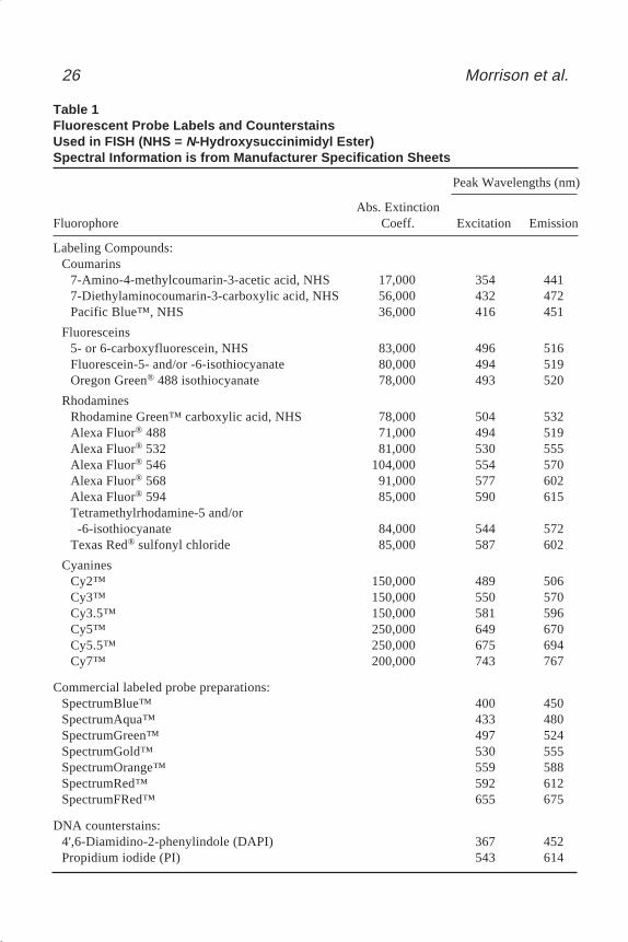
an emission max of 532 nm, while tetramethylrhodamine isothiocyanate (TRITC;R7 = R8 = R9 = R10 = methyl, R11 or R12 = isothiocyanate) has an excitation max at 544 nmand an emission max of 572 nm. The spectral characteristics of the cyanines arestrongly affected by changing the number of carbons separating the two indole rings.For example, Cy 3 (m = 1 in structure C, Fig. 1) has an excitation maximum at 550 nmand an emission maximum of 570 nm, while Cy 5 (m = 2 in Structure C., Fig. 1) has anexcitation max at 649 nm and an emission max of 670 nm. The excitation and emis-sion maxima for a number of fluorescent labels used on in situ hybridization probesare listed in Table 1. Also included are two common nucleic acid counterstains, 4',6-diamidino-2-phenylindole (DAPI) and propidium iodide (PI).
For in situ hybridization, the most desirable properties are high absorption extinc-tion coefficient (preferably greater than 10,000/M·cm) and high fluorescence quantumyield (preferably >0.2). The excitation and emission spectra are also very importantand must be selected with regard to the spectral distribution of the excitation source,the microscope optical system, the fluorescence detector, and the filter sets available.In the case of multitarget hybridization, the spectral distributions of each fluorophorepresent in the assay must be chosen carefully to allow the fluorescence of each to beindividually distinguished (for a review of multitarget hybridization, see ref. 31).
Spectral properties are not all that must be considered in label selection. In particu-lar, the different chemical structures of the fluorescent labels lead to different levels ofinteraction with various cellular components, cellular debris, extracellular matrix, andthe slide surface. Background staining of a specimen is highly dependent, therefore,upon the chemical structure of the fluorescent label. While highly hydrophobic labelscan reduce probe solubility and increase adsorption to some cellular components, oftenthe only way to determine how a label will perform in an in situ hybridization is byactually preparing the labeled probe and hybridizing it to the target tissue.
1.4. Sources of Probe DNAFor most FISH applications, probes are prepared by culturing bacteria or yeast that
contains the desired cloned sequence. The cells are harvested, lysed, and the cloneDNA is purified from the host chromosomal DNA and cellular material. Bacterialcells containing the clone of interest are typically grown in media that selects for theclone by use of an antibiotic, or in the case of yeast, in media which lacks a particularnutrient. Bacterial clone DNA can be isolated by several common methods such asalkaline lysis or boiling (32).*,** Alternatively, extraction kits are available commer-cially from several suppliers, including Qiagen, Inc. (Chatsworth, CA), Stratagene (LaJolla, CA), and Gentra Systems, Inc. (Minneapolis, MN). The preferred method forgenerating FISH probes from YACs involves amplification of the insert by Alu-PCR**.
Vectors which maintain large inserts such as cosmids, P1s, PACs, or BACs, arebest suited for generating FISH probes homologous to unique sequence DNA, as the
*See various volumes in the series Current Protocols in Molecular Biology, Janssen, K. (serieseditor), John Wiley and Sons, New York.
**See various volumes in the series Current Protocols in Human Genetics, Boyle, A. L..(series editor), John Wiley and Sons, New York.
02_Morrison_21-40F[7.10.2] 7/10/2002, 12:58 PM25
26 Morrison et al.
Table 1Fluorescent Probe Labels and CounterstainsUsed in FISH (NHS = N-Hydroxysuccinimidyl Ester)Spectral Information is from Manufacturer Specification Sheets
Peak Wavelengths (nm)
Abs. ExtinctionFluorophore Coeff. Excitation Emission
Labeling Compounds:Coumarins
7-Amino-4-methylcoumarin-3-acetic acid, NHS 17,000 354 4417-Diethylaminocoumarin-3-carboxylic acid, NHS 56,000 432 472Pacific Blue™, NHS 36,000 416 451
Fluoresceins5- or 6-carboxyfluorescein, NHS 83,000 496 516Fluorescein-5- and/or -6-isothiocyanate 80,000 494 519Oregon Green® 488 isothiocyanate 78,000 493 520
RhodaminesRhodamine Green™ carboxylic acid, NHS 78,000 504 532Alexa Fluor® 488 71,000 494 519Alexa Fluor® 532 81,000 530 555Alexa Fluor® 546 104,000 554 570Alexa Fluor® 568 91,000 577 602Alexa Fluor® 594 85,000 590 615Tetramethylrhodamine-5 and/or -6-isothiocyanate 84,000 544 572Texas Red® sulfonyl chloride 85,000 587 602
CyaninesCy2™ 150,000 489 506Cy3™ 150,000 550 570Cy3.5™ 150,000 581 596Cy5™ 250,000 649 670Cy5.5™ 250,000 675 694Cy7™ 200,000 743 767
Commercial labeled probe preparations:SpectrumBlue™ 400 450SpectrumAqua™ 433 480SpectrumGreen™ 497 524SpectrumGold™ 530 555SpectrumOrange™ 559 588SpectrumRed™ 592 612SpectrumFRed™ 655 675
DNA counterstains:4',6-Diamidino-2-phenylindole (DAPI) 367 452Propidium iodide (PI) 543 614
02_Morrison_21-40F[7.10.2] 7/10/2002, 12:58 PM26
Labeling FISH Probes for DNA Targets 27
probe should span at least 40 kb of contiguous sequence. Probes that represent highlyrepetitive sequences, such as that found near centromeres or telomeres, can be madefrom a single plasmid containing an insert of approximately 300–10,000 bp.
Two of the most common means for identifying a clone containing the desiredtarget sequence are: (1) screening the appropriate clone libraries, or (2) searchingonline databases, such as Genbank at the National Center for Biotechnology Informa-tion, with a known sequence such as mRNA from a particular gene, or anonymoussequence from the end of a clone insert or a Sequence Tagged Site (STS).
Whole chromosome painting probes, which contain sequences spread across thebreadth of a specific chromosome, were originally prepared from whole chromosomephage and bacterial libraries (33,34). These libraries were ultimately obtained fromchromosomes that were isolated by flow sorting. More recently, whole chromosomeprobes are prepared from flow sorted chromosomes (35,36) or microdissected chro-mosomes (37) that are amplified by DOP-PCR, without the intervening steps requiredto make bacterial libraries (30,38).
2. Materials2.1. Nick-Translation
1. Sample DNA, typically 1 µg/50 µL labeling reaction.2. Fluorophore labeled dUTP at a working concentration of 0.2 mM.3. 0.3 mM dATP.4. 0.3 mM dCTP.5. 0.3 mM dGTP.6. 0.3 mM dTTP.7. Nick-translation enzyme: mix of DNA polymerase I (10,000 U/mL, DNase I (3 U/mL,
such as Promega (Madison, WI) enzyme mix) .8. 10X Nick-translation buffer: 500 mM Tris-HCl, pH 7.2, 100 mM MgSO4, 1 mM
dithiothreitol (DTT).9. Nuclease-free water.
10. 15°C incubator.11. Stop solution: 0.25 mM EDTA.12. 3M sodium acetate, pH 5.5.13. 70% ethanol.14. 100% ethanol.15. TE solution: 10 mM Tris-HCl, pH 7.5–8, 1 mM EDTA.16. Sephadex G-50 type spin column (e.g., ProbeQuant™ G-50 spin columns, Amersham
Pharmacia, Piscataway, NJ). Other methods can be used for removal of unincorporatednucleotides.
17. Microcentrifuge tubes.18. Microcentrifuge.19. Speed-vac lyophilizer.20. Pipetors.21. Pipet tips, preferably sterilized.22. Tris-acetate buffer (TAE): 40 mM Tris-acetate, pH 7.5–7.8, 1 mM EDTA, or Tris-borate
buffer (TBE): 45 mM Tris-borate, pH 8.0, 1 mM EDTA.23. Agarose.
02_Morrison_21-40F[7.10.2] 7/10/2002, 12:58 PM27

28 Morrison et al.
24. Hot plate or microwave oven.25. 55°C water bath.26. Mini horizontal gel electrophoresis apparatus with casting tray and combs, power supply.27. DNA molecular weight markers, size range should cover 50–1000 bp.28. 10 mg/mL ethidium bromide (EtBr).29. 10X gel loading buffer: 50% (v/v) glycerol, 100 mM EDTA, 0.25% bromphenol blue.30. UV transilluminator and UV protective visor.31. Polaroid camera with Kodak Wratten red filter and type 667 Polaroid film.
2.2. Random Priming1. Sample DNA, typically 10 ng to 3 µg/50 µL labeling reaction.2. Fluorophore labeled dUTP at a working concentration of 1 mM.3. 10 mM dATP.4. 10 mM dCTP.5. 10 mM dGTP.6. 10 mM dTTP.7. 40 U/mL Klenow fragment, (such as Life Technologies (Gaithersburg, MD) enzyme) .8. 2.5X Random primer/buffer solution: 125 mM Tris-HCl, pH 6.8, 12.5 mM MgCl2, 25 mM
2-mercaptoethanol, 750 µg/mL random octamer primers (Life Technologies).9. Nuclease-free water.
10. 37°C incubator.11. Boiling water bath.12. Ice.13. Stop solution, 0.25 mM EDTA.14. 3M sodium acetate, pH 5.5.15. 70% ethanol.16. 100% ethanol.17. Sephadex G-50 type spin column (optional, depending on method preferred for removal
of unincorporated nucleotides).18. Microcentrifuge tubes.19. Microcentrifuge.20. Speed-vac lyophilizer.21. Pipetors.22. Pipet tips, preferably sterilized.
2.3. DOP-PCR1. Flow sorted chromosomes (500–1000/PCR)2. 10X PCR buffer I: Applied Biosystems (Foster City, CA).3. PCR assay buffer: 10 µL of 10X PCR buffer I, 2 µL dNTPs (10 mM each), 1 µL of primer
(100 ng), 1 µL Taq polymerase, nuclease-free water to 100 µL.4. PCR labeling buffer: Same as PCR assay buffer, with addition of 1 µL of SpectrumOrange™
or SpectrumGreen™ dUTP, except with final vol of 99 µL.5. 100 ng/µL primer.6. 50 nmol SpectrumOrange™, SpectrumGreen™ 2'-deoxyuridine- 5'-triphosphate (Vysis,
Inc., Downers Grove, IL) or other labeled nucleoside triphosphate–Reconstitute with 50-µLof nuclease-free water to give a 1 mM solution. Stable for up to 3 mo if stored at –20°C.
7. Taq polymerase: any commercial variety available can be used, but optimal concentra-tion should be first determined by titrating with appropriate substrate.
02_Morrison_21-40F[7.10.2] 7/10/2002, 12:58 PM28

Labeling FISH Probes for DNA Targets 29
8. Nuclease-free water.9. Mineral oil (if necessary), light white (Sigma Chemical Co., St. Louis, MO).
10. NuSieve GTG agarose (FMC BioProducts, Rockland, ME).11. 1X TBE buffer for gel electrophoresis (BioRad, Hercules, CA).12. Primers: The primer used was designated 6 MW (30), with sequence 5' CGA CTC GAG
NNN NNN ATG TGG 3'.
2.4. Labeling Probes with Aliphatic Amines1. Amine-modified DNA to be labeled.2. A suitable reaction buffer (50 mM sodium tetraborate at pH 9.3 for isothiocyanates and
sulfonic acid chlorides, 0.2 M 3-[N-morpholino]-propanesulfonic acid (MOPS; SigmaChemical Co.) at pH 7.4 or 50 mM sodium tetraborate at pH 8.5 for N-hydroxysuccinimidylesters).
3. Amine-reactive derivative of the desired label.4. Dimethyl sulfoxide, dimethyl formamide, or acetone (whichever is capable of dissolving
the desired amine-reactive label in a 10–20 mM solution).5. A prepacked Sephadex G-25 column (Amersham Pharmacia, Piscataway, NJ).6. TE solution: 10 mM Tris-HCl, pH 7.5–8, 1 mM EDTA).
2.5. ULS Labeling1. TE buffer 10 mM Tris-HCl, 0.3 mM EDTA, pH 8.0.2. DNA for labeling.3. DNase I (Roche Biochemicals, Nutley, New Jersey) cat. no. 104 159, approx 2000 Kunitz
U/mg.4. 5 mM sodium acetate, 1 mM CaCl2, 50% glycerol, pH 5.25. 10X nicking buffer: 50 mM Tris-HCl, pH 7.5, 10 mM CaCl2, 10 mM MgCl2.6. 10 M ammonium acetate.7. 100% ethanol.8. Kreatech kit components: Vial 1: code LK1101 (60 µL rhodamine-ULS®, 0.5 U/µL), or
code LK1301 (60 µL dGreen-ULS®, 0.5 U/µL), Vial 2: code LK006, Labeling Solution, 2 mL.9. Spin columns: e.g., ProbeQuant™ G-50 spin columns (Amersham Pharmacia Biotech,
Piscataway, NJ) or QIAquick™ spin columns (Qiagen, Valencia, CA).10. Ultrasonic disruptor (e.g., Branson Ultrasonics, Danbury, CT).
2.6. Labeling Proteins for Indirect Detection1. N-hydroxysuccinimidyl derivative of desired fluorophore.2. Dimethyl sulfoxide, dimethyl formamide, or acetone (whichever is capable of dissolving
the desired fluorophore in a 20 mM solution).3. A prepacked Sephadex G-25 column (Amersham Pharmacia).4. 50 mM boric acid, pH 8.5 to 9.3.5. TBS: 25 mM Tris-HCl, 140 mM NaCl, 51 mM KCl, pH 7.4.6. PBS: 10 mM Na2HPO4, 1.8 mM KH2PO4, 140 mM NaCl, 2.7 mM KCl, pH 7.4.
3. Methods3.1. Nick-Translation
Nick translation is a method for incorporating labeled nucleotides into DNA suchas an isolated fragment or an intact clone (39,40). The method uses a combination of
02_Morrison_21-40F[7.10.2] 7/10/2002, 12:58 PM29

30 Morrison et al.
two enzymes, deoxyribonuclease I (DNase I) which nicks the DNA creating free 3'hydroxyls, and DNA polymerase I, which processively adds nucleotides to the 3' ter-minal hydroxyl. The 5' to 3' exonuclease activity of the DNA polymerase removesnucleotides from the 5' terminus of the nick as the polymerization proceeds. Bothlabeled and unlabeled nucleotides are substituted during the reaction and varying sizedfragments are generated; however, there is no net synthesis of DNA. The resultantdouble-stranded fragments must be denatured prior to hybridization.
1. Prepare 0.1 mM dTTP by adding 100 µL of 0.3 mM dTTP to 200 µL nuclease-free water.2. Prepare 0.1 mM dNTP mix by combining 100 µL each of 0.3 mM dATP, 0.3 mM dCTP,
0.3 mM dGTP. Excess nucleotide mixtures can be stored at –20°or –80°C.3. For each labeling reaction, on ice, prepare a tube containing 1 µg DNA , 2.5 µL of 0.2 mM
fluorophore-labeled dUTP, 5 µL of 0.1 mM dTTP, 10 µL of 0.1 mM dNTP mix, 5 µL of10X nick translation buffer and make up to a final vol of 40 µL with nuclease-free water(see Note 1).
4. To each tube add 10 µL nick-translation enzyme mix.5. Mix and briefly centrifuge.6. Incubate at 15°C for 8–16 h.7. Add 5 µL stop solution.8. To remove unincorporated nucleotides, add 5 µL 3 M sodium acetate, 125 µL 100% etha-
nol; centrifuge at 12K for 20–30 min. Carefully pour off supernatant, or draw off with apipettor. Add 100 µL 70% ethanol per tube, briefly vortex pellet; centrifuge at 15,000gfor 5 min. Carefully pour off supernatant, or draw off with pipettor (see Note 2).
9. Dry pellet under vacuum 10–20 min. Resuspend in 10 µL TE, which yields an approx100 ng/µL final concentration (see Notes 3 and 4). Alternatively, unincorporated nucle-otides can be removed using a Sephadex G-50 type spin column according to themanufacturer’s instructions. Labeled DNA will be in 50–100 µL after the column andwill need to be concentrated by ethanol precipitation as described in step 8.
10. To determine size of the labeled DNA fragments, add 0.5 g agarose to 50 mL of TAEbuffer or TBE buffer, carefully heat to boiling in microwave, or on a hot plate. After allagarose is melted, cool to 55°C in a water bath.
11. Add 2.5 µL EtBr to the agarose and mix. Pour molten agarose into casting tray using a12 or 16 well comb.
12. For each labeled DNA, mix 2 µL DNA (~200 ng) plus 7 µL of water, or 1X TAE or TBE,plus 1 µL loading buffer.
13. Run labeled DNAs plus molecular weight marker at 70–100 V until leading dye hasmigrated approx 5 cm into the gel.
14. View DNA with UV transilluminator and take a Polaroid picture. Majority of fragmentsshould be in the range of 200–600 bp.
3.2. Random PrimingRandom priming is a means of labeling DNA fragments whereby, a mixture of all
possible combinations of hexamers, octamers, or nonamers are annealed to denaturedDNA (41,42). These small oligonucleotides then act as primers that allow for synthesisof the complementary DNA strand by the Klenow enzyme and incorporation of bothlabeled and unlabeled nucleotides. The labeled material will be a combination of bothdouble- and single stranded fragments that must be denatured prior to hybridization.
02_Morrison_21-40F[7.10.2] 7/10/2002, 12:58 PM30

Labeling FISH Probes for DNA Targets 31
1. Prepare a 10X dNTP mixture such that final concentrations are 1 mM dATP, 1 mM dCTP,1 mM dGTP, 0.3 mM fluorophore-labeled dUTP, 0.7 mM dTTP. Excess mixture can bestored at –20°or –80°.
2. Dissolve DNA (400–500 ng preferred) in 20 µL nuclease-free water (see Notes 5 and 6).3. Add 20 µL 2.5X random primer/buffer solution.4. Heat denature for 5 min in boiling water; rapidly cool on ice.5. On ice, add 5 µL 10X dNTP mix and 4 µL nuclease-free water to a final vol of 49 µL.6. Mix and briefly centrifuge.7. Add 1 µL Klenow enzyme and mix. Briefly centrifuge.8. Incubate at 37°C for 1–6 h. Longer incubation times usually increase product yield (see
Note 7).9. Add 5 µL stop solution.
10. Unincorporated nucleotides and primers can be removed by adding 5 µL of 3 M sodiumacetate, 125 µL of 100% ethanol; centrifuge at 15,000g for 20–30 min. Carefully pour offsupernatant, or draw off with a pipetor. Add 100 µL of 70% ethanol per tube, brieflyvortex pellet; centrifuge at 15,000g for 5 min. Carefully pour off supernatant, or draw offwith pipetor.
11. Dry pellet under vacuum 10–20 min. Resuspend in 20 µL TE, which yields an approx100 ng/µL final concentration. Alternatively, unincorporated nucleotides can be removedusing a Sephadex G-50 type spin column according to the manufacturer’s instructions.Labeled DNA will be in 50–100 µL after the column, and will need to be concentrated byethanol precipitation as described in step 10.
12. Optimally, fragments should range from 200–600 bp, and can be visualized by gel elec-trophoresis. Refer to steps 10–14 (see Subheading 3.1.) for electrophoresis protocol.
3.3. DOP-PCRPCR generated whole chromosome DNA, either from microdissected or flow sorted
chromosomes, is currently viewed as the preferred route to high quality probes suitablefor whole chromosome staining of individual chromosomes. This is true for stainingsingle chromosomes as well as for staining all 24 human chromosomes combinatoriallyin a multiplex FISH (M-FISH) (43) or spectral karyotyping (44) assay. The procedureprovided below (45) is a modification of the original Degenerative-Oligonucleotide-Primed-PCR (DOP-PCR) protocol (38), and is optimized to amplify chromosomeDNA in the presence of SpectrumOrange™ or SpectrumGreen™ dUTP (Vysis, Inc.).The protocol also should permit labeling with a variety of other labeled dUTP’s, withlittle or no modification. The reaction involves the use of an oligonucleotide with anXho-I restriction endonuclease site at its 5' end, a defined six-nucleotide sequence atthe 3' end, and a set of degenerate nucleotides (a random mix of all 4 nucleotides) inbetween. Theoretical calculations indicate that the defined 3' sequence occurs every4 kb along the genome. Under suitable conditions, an amplification using this primercould be primed off the specific 3' sequence, probably stabilized by annealing of oneor more of the degenerate nucleotides (38). The specific 5' sequence permits theannealing of this primer at a higher temperature to previously amplified DNA. In prac-tice, the initial cycles in the DOP-PCR protocol include a low temperature annealingstep (low fidelity PCR), followed by multiple cycles at a higher annealing temperature(higher fidelity PCR), resulting in a population of randomly amplified DNA.
02_Morrison_21-40F[7.10.2] 7/10/2002, 12:58 PM31

32 Morrison et al.
The flow sorted chromosomes are normally amplified through two rounds of DOP-PCR, and the resulting product is then labeled by a third round of amplification in thepresence of fluorescently labeled nucleoside triphosphates. Although nick-translationprotocols could be used to label probes for chromosomal analysis, this additional stepis usually unnecessary to obtain high quality probes.
3.3.1. Amplification of DNATypically, flow sorted chromosomes are resuspended and amplified in PCR assay
buffer, using the following conditions: 95°C for 5 min, followed by 9 cycles of 94°Cfor 1 min, 30°C for 1.5 min, and 72°C for 3 min, with a ramp time of 2 min, 5 s,followed by 35 cycles at 94°C for 15 s, 62°C for 15 s, and 72°C for 15 s (see Notes 8–10).This is followed by a single extension at 72°C for 10 min, and then holding the tem-perature at 4°C. Using the same procedure, 1 µL of the PCR product is then reamplifiedin a final vol of 100 µL. 10 µL of this reaction should then be electrophoresed on a 1%agarose gel, resulting in a smear, ranging in size from approx 300–1000 bp. The yieldof DNA from this reaction varies from 1.5–3.0 µg (see Note 11).
3.3.2. Labeling of DNA1 µL from the previous round of PCR is labeled in PCR labeling buffer, using the
same conditions described for DNA amplification. 1–2 µL of the labeled DNA canthen be used directly for hybridization without purification.
3.4. Labeling of DNA Probes with Aliphatic AminesIn addition to the one-step labeling of probes using polymerases to incorporate
labeled nucleotides, a two-step labeling procedure can be utilized involving: (1) incor-porating nucleoside triphosphates with aliphatic amine substituents into the probeDNA using the polymerase-based labeling protocols described above (see Subhead-ing 3.1.–3.3.), and (2) reacting the modified DNA with amine reactive fluorophores.The two-step procedure offers the advantage that any amine reactive fluorophore canbe attached to the modified DNA, instead of only fluorophores that are commerciallyavailable already attached to nucleoside triphosphates. Additionally, all polymerase-based labeling reactions can use the same nucleoside triphosphate–allylamine-dUTP.Therefore, a single set of polymerase reaction conditions can be used for all fluores-cent labels, instead of having to optimize for each different fluorescent nucleosidetriphosphate. Also, incorporation of allylamine-dUTP may be more efficient than in-corporation of other labeled nucleoside triphosphates. Labeling kits for two-step labelincorporation via allylamine-dUTP are commercially available (ARES labeling kits,Molecular Probes, Inc., Eugene, OR).
Aliphatic amines introduced into DNA enzymatically, using allylamine-dUTP inplace of labeled nucleoside triphosphates, can be conjugated with amine-reactivefluorophores, biotins, or haptens by the following procedure. The procedure can alsobe used to label probes containing aliphatic amines introduced by a number of otherchemistries (22,46). This includes synthetic DNA, RNA, and PNA oligomers contain-ing aliphatic amine modified bases, or 3'- or 5'-amine-modified termini, introducedvia phosphoramidite chemistry.
02_Morrison_21-40F[7.10.2] 7/10/2002, 12:58 PM32

Labeling FISH Probes for DNA Targets 33
1. Dissolve amine-modified DNA in a suitable reaction buffer at a concentration of 10 nmolaliphatic amines/0.6–1.0 mL reaction buffer.
2. Dissolve the amine reactive labeling compound in a suitable solvent to a final concentra-tion of 10–20 mM.
3. Add a 50-fold (isothiocyanates) or 100- to 200-fold (N-hydroxysuccinimidyl esters orsulfonic acid chlorides) molar excess of the amine-reactive dye to the amine modifiedDNA (see Note 12).
4. Allow the reaction solution to stir at room temperature overnight.5. Separate the probe from unconjugated label by gel permeation chromatography using a
Sephadex G-25 column equilibrated and eluted with water or TE buffer. The labeledprobe will elute in the excluded volume (see Note 13).
6. Store the labeled probe at 4°C or lower until ready for use.
3.5. ULS LabelingThe Universal Linkage System (ULS®; KREATECH Biotechnology BV, Amsterdam,
The Netherlands) is a labeling methodology that uses a platinum dye complex to reactwith the N7 position of the guanine nucleotides. This reaction results in the formationof a stable bond between the nucleic acid and the platinum fluorophore complex.Depending on reaction conditions the ULS compound, to a lesser extent, will alsoform a complex with the adenine bases. This methodology has been used to label DNA(including plasmids, cosmids, molecular weight markers, DNA in low melting pointagarose, BACs, PACs, YACs, whole chromosome libraries, DOP-PCR products andhighly repetitive sequences such as satellite, centromeric, and telomeric DNA), RNA,PNA, oligonucleotides, and amplified nucleic acid products (16,47). ULS reagentsand kits are also offered by Molecular Probes (Eugene, OR).
Prior to any labeling with the ULS compounds the template to be labeled must be of a sizerange <1000 bp. Template larger than 1000 bp will result in a substantial amount of spottedbackground. PCR products are usually <1000 bp and therefore can be labeled directly. Thetwo methods of size reduction described will work with both the Kreatech and the MolecularProbes labeling protocols–sonication and enzymatic cleavage. Molecular Probes recommendsan alternative DNase I protocol for use with the Alexa-ULS reagents. This protocol and thecorresponding reagents are part of the ULS labeling kits provided by Molecular Probes.
3.5.1. DNA Fragmentation3.5.1.1. SONICATION
1. Prepare a DNA solution at a concentration of 20 ng/µL in TE buffer. Using a minimumvolume of 100 µL for sonication is best.
2. Sonicate the DNA solution in a small conical bottom plastic tube for 3 cycles of 1 mineach, while keeping the sample on ice. Select ultrasonic disruptor power level and dutycycle to deliver the highest power possible while minimizing cavitation. Allow the DNAsolution to cool on the ice for 1 min prior to the start of sonication, and after each cycle.In a microcentrifuge, centrifuge the DNA solution for 5 s at max speed before the secondand third sonication steps to force all of the solution to the bottom of the tube (see Note 14).
3. Confirm adequate DNA size by electrophoresis on an aliquot of the DNA solution usinga 1% agarose gel. Refer to steps 10–14 of Subheading 3.1. for electrophoresis protocol.
4. Proceed to labeling protocols.
02_Morrison_21-40F[7.10.2] 7/10/2002, 12:58 PM33

34 Morrison et al.
3.5.1.2. DNASE TREATMENT
1. Prepare a stock solution of DNase I by dissolving 1 mg of DNase I (Roche cat. no. 104159, approx 2000 Kunitz U/mg) in 1 mL of 5 mM sodium acetate, 1 mM CaCl2, 50%glycerol, pH 5.2. Keep the buffer on ice prior to and during the addition of the lyophilizedDNase. Invert this solution until completely mixed. Do not vortex. The stock solutionshould be stored at –20°C avoiding freeze thaw cycles.
2. Dilute the DNase I stock 1:5000 in 1X nicking buffer (see Note 15).3. Add the following components to a microcentrifuge tube on ice: 1 µg template DNA, 2.5 µL
10X nicking buffer, 3–5 µL diluted DNase I, H2O to 25 µL.4. Incubate this reaction at 37°C for 10 min.5. Stop the reaction by placing on ice.6. Precipitate the reaction mixture with 1/4 vol of 10 M ammonium acetate and 2.5 vol of
100% ethanol.7. Resuspend the pellet in the appropriate amount of labeling solution (see Subheading 3.5.2.).8. Confirm adequate DNA size by electrophoresis on an aliquot of the DNA solution using
a 1% agarose gel.
3.5.2. Labeling3.5.2.1 PROTOCOL RECOMMENDED BY KREATECH
Optimal labeling efficiencies are achieved when the ULS reagent and nucleic acidare combined at a 1:1 ratio (i.e. 1 U ULS reagent:1 µg DNA). For labeling amounts ofnucleic acid other than the standard 1 µg, the ratio of nucleic acid to ULS reagentshould be kept at 1:1. When labeling small amounts, a minimum of 100 ng of nucleicacid in a 20 µL vol should be used. Alternatively, larger amounts should not exceed10 µg of nucleic acid in a 20 µL vol. Labeling in a larger vol is possible as long as thetemplate concentration is not lower than 5 ng/µL, and the amount of ULS reagentadded is adjusted to the amount of input template. In the case of very dilute template,the labeling solution can be omitted.
1. Add 1 U (2 µL) ULS reagent to 1 µg of input template (see Notes 16 and 17).2. Adjust volume with labeling solution to 20 µL and mix well.3. Incubate for 15 min at 65°C.4. Centrifuge briefly.5. Purify probe on a spin column.
3.5.2.2. PROTOCOL RECOMMENDED BY MOLECULAR PROBES
Molecular Probes provides a kit that utilizes the ULS labeling system coupled to avariety of their dyes. The kit contains reagents both for fragmentation of the DNA andlabeling protocols. The labeling protocols and precautions are very similar with excep-tion of the following:
1. Molecular Probes ULS dye reagents are dissolved in different buffers prior to labeling,depending upon the dye selected. A chart is provided with all specific information relat-ing to each available dye.
2. Immediately prior to labeling, DNA is denatured at 95°C for 5 min, and then snap cooledon ice. It is noted that denaturation is not a necessity but can improve the labeling effi-ciency by 20–40%.
02_Morrison_21-40F[7.10.2] 7/10/2002, 12:58 PM34

Labeling FISH Probes for DNA Targets 35
3. The labeling reaction total volume is 25 µL.4. Incubation is at 80°C for 15 min.5. Molecular Probes states that the modifications to the previously stated Kreatech proto-
cols have been made owing to the specifications of their own dyes that are coupled to theULS reagents.
3.6. Labeling Proteins for Indirect DetectionSecondary detection reagents for indirectly labeled FISH probes are prepared by
conjugating fluorophores to proteins such as avidin, streptavidin, or antibodies. A largevariety of fluorophore labeled avidins and antibodies are commercially available (e.g.,Accurate Chemical and Scientific Co., Westbury, NY; Calbiochem, San Diego, CA;Cappel Organon Teknika, Durham, NC; DAKO Corp., Carpinteria, CA; JacksonImmunoResearch Laboratories, West Grove, PA; Kirkegaard and Perry Laboratories,West Grove, PA; Molecular Probes, Inc., Eugene, OR; Pierce Chemical Co., Rock-ford, IL; Sigma Chemical Co., St. Louis, MO). Labeled proteins can also be preparedfairly easily in the laboratory. Unlabeled avidins and antibodies can be obtained fromthe same suppliers listed above.
Streptavidin is often preferred over avidin as a secondary reagent because of itsnear-neutral pH isoelectric point, which is believed to reduce nonspecific binding.Similarly, while whole antibodies can be used in FISH, the more hydrophobic Fc regionis often removed to reduce nonspecific binding. The Fc region can be cleaved from theantigen-binding regions of antibodies by digestion with papain, to produce Fab par-ticles (single binding arms), or pepsin, to produce F(ab')2 with the two binding armsstill connected. Fab' particles can be prepared from F(ab')2 preparations by selectivereduction of disulfide bonds that joint the two binding arms. Methods for preparingF(ab')2, Fab, and Fab' antibody fragments can be found in the literature (48,49).
1. Dissolve the protein to a final concentration of 1–10 mg/mL in 50 mM boric acid atpH 8.5–9.3.
2. Dissolve the N-hydroxysuccinimidyl derivative of the desired fluorophore in a solventsuch as dimethyl sulfoxide, dimethyl formamide, or acetone, to a final concentration of20 mM.
3. Add a sufficient volume of the fluorophore solution to the protein solution to provide a1- to 20-fold molar excess of fluorophore to protein (see Notes 18 and 19).
4. Allow the reaction to proceed with gentle stirring at room temperature for 2 h.5. Separate the protein from unconjugated fluorophore by gel permeation chromatography
using a Sephadex G-25 column equilibrated and eluted with TBS or PBS. The labeledprotein will elute in the excluded volume (see Note 20).
6. Store the labeled protein at 4°C until ready for use (see Note 21).
3.7. Characterization of Labeled ProbesTo calculate accurate probe concentrations, enough DNA must be available to gen-
erate an absorbance value of at least approx 0.05 in the minimal volume of liquidrequired to fill the spectrophotometer cuvet. For conventional absorbance spectropho-tometer cuvets with 1 cm pathlengths, the minimal volumes are several hundred micro-liters (semimicro cuvets). Spectrophotometers specifically designed for DNA workcan utilize cuvets with volumes near 10 µL. The following equation can be used to
02_Morrison_21-40F[7.10.2] 7/10/2002, 12:58 PM35
36 Morrison et al.
calculate the nucleic acid concentration, [nucl], from the measured absorbance at260 nm, A260:
[nucl] = [A260 – (εF,260/εF,MAX)AF,MAX]/εnucl,260
where εnucl,260 is the absorbance extinction coefficient of the nucleic acid at 260 nm,and εF,260/εF,MAX is the ratio of the absorbance extinction coefficients of the label at260 nm, εF,260, and at the peak wavelength of the longest wavelength absorbance band,εF,MAX. This ratio for the labeled probe is approximated by the ratio of absorbancevalues of the unconjugated label at these two wavelengths. For single-stranded DNA,εnucl,260 = 10,000 M–1·cm–1, or 0.0286 (µg/mL) mL·ng–1·cm–1. The former value givesthe concentration in nucleotide molarity, while the latter value gives the concentrationin micrograms nucleic acid per milliliter. The label concentration, [F], is calculatedfrom the probe absorbance at the long wavelength absorbance maximum of the label:
[F] = AF,MAX/εF,MAX
at which the nucleic acid absorbance is assumed to be negligible. Values of εF,MAX canbe obtained from the suppliers of the labeling reagents. The percentage of nucleotideslabeled is then equal to 100[F]/[nucl].
When the amount of probe is too small to obtain accurate absorbance measurements,or contains some contaminating RNA, fluorometry with bisbenzimide, commonly knownas Hoescht 33258 dye, can be used to determine nucleic acid concentration. The Hoeschtdye has little affinity for RNA but binds to the minor groove of double stranded DNA.Hoescht 33258 dye bound to DNA can be excited at 365 nm, and has peak emission at458 nm. Fluorometry is very sensitive and can be used for DNA concentrations rangingfrom 0.01–5 mg/mL, with an optimum DNA concentration range of 0.05–0.3 mg/mL.
The relative fluorescence intensities are measured on the sample DNA solution, FS,and the same solution minus the DNA (blank solution), FB. In addition, the fluores-cence intensity of a DNA standard solution, FSTD, and corresponding blank solution,FSTD,B are also measured (typically the sample and standard blank solutions are thesame). The DNA standard solution should have a DNA concentration, [DNA]STD, closeto that expected for the DNA sample solution. The sample DNA concentration,[DNA]S, is then calculated as follows:
[DNA]S = [DNA]STD(FS - FB)/(FSTD – FSTD,B)
Accurate pipetting and thorough mixing of solutions is critical for reproducible results.The concentrations and labeling percentages of proteins can be determined by
absorbance spectroscopy using the same equations as for nucleic acid probes, exceptthat measurements are recorded at 280 nm instead of 260 nm. The absorbance extinc-tion coefficients for goat IgG and Fab' have been reported to be 198,000/M–1·cm–1 and61,200/M–1·cm–1 (49), respectively, and the extinction coefficient for F(ab')2 shouldbe twice that for Fab'.
4. Notes1. Fluorophores will photobleach if exposed to light for extended periods of time. Labeled
DNAs and dUTPs can be handled for short periods of time in light but should not beexposed any longer than required to set up an experiment.
02_Morrison_21-40F[7.10.2] 7/10/2002, 12:58 PM36

Labeling FISH Probes for DNA Targets 37
2. It is easier to assess range of fragment sizes if unincorporated nucleotides are removedprior to gel electrophoresis.
3. After ethanol precipitation, DNA pellets labeled with a red or orange fluorophore areusually readily seen by eye, whereas, those labeled with a green fluorophore may appearwhite, or very pale green.
4. 50–100 ng of a red or orange labeled unique sequence probe, and 200–400 ng of a greenlabeled probe per 10 µL FISH mix typically yield sufficiently bright signals on the major-ity of sample types.
5. For optimal results, template DNA should be linear, preferably by digestion using arestriction enzyme with a six base pair recognition site and purified by phenol/chloroformextraction followed by ethanol precipitation.
6. Starting amounts of DNA can range from 10 ng to 3 µg.7. Labeling at 37°C can range from 1 h to overnight. Optimal conditions should be deter-
mined for individual applications.8. Contamination is a very serious issue in PCR. To avoid contamination of PCR reactions,
use the best quality reagents possible, and use them exclusively for PCR. It is best toroutinely aliquot all solutions and store them frozen at –20° C until ready for use. Also,use aerosol-resistant tips for pipetting to minimize cross-contamination. Designate onearea in the lab exclusively for PCR work. When possible, prepare DNA samples in aseparate room. In addition, it is absolutely essential to run suitable negative controls (i.e.,without target sequence) each time an experimental PCR is performed.
9. Titrate the Taq polymerase with the specific target. Excess enzyme can result in nonspe-cific background.
10. Vary the number of cycles to find the appropriate number of cycles which gives the bestsignal to noise ratio.
11. The presence of high molecular weight DNA (visible during electrophoresis as a smearextending from the well to approx 2.0 kb size) indicates unacceptable labeled probe. Suchprobe results in very high nonspecific, noncellular background that can sometimes bereduced by sonication. As mentioned in Note 9, titrate the Taq polymerase, and find theappropriate number of PCR cycles.
12. The organic solvent: aqueous buffer ratio should not exceed 1:4 unless the DNA is knownto be soluble at a higher ratio.
13. As an alternative to gel permeation chromatography, the unconjugated fluorophore canbe separated from the protein by dialyzing in a storage buffer (e.g., TE), changing thebuffer solution at several hour intervals until the unconjugated label is completelyremoved (2 or more buffer changes).
14. When sonicating, the volume of the sample, the type of vessel in which the sample iscontained, the length of the sonication period, the number of sonication cycles, the ultra-sonic disruptor power level, and the ultrasonic disruptor duty cycle, are all dependentupon the make and model of the ultrasonic disruptor instrument and the sonication probeused. Some experimentation may be required to obtain properly sized nucleic acidfragments.
15. When performing the DNase I digestion, all solutions should be kept on ice. Preparesolutions immediately before use.
16. DNA should be purified to remove proteins, RNA, and free nucleotides before labeling.17. High tris(hydroxymethyl)aminomethane (Tris) concentrations (>40 mM) or EDTA
(>5 mM), Mg acetate (>100 mM), NaCl (>100 mM), and restriction enzyme digestionbuffers should be avoided because of their rate-limiting effect on the labeling reaction.
18. The molar excess of reactive fluorophore-to-protein, and the protein concentration willdetermine the extent of protein labeling. The labeling ratio required for optimal perfor-
02_Morrison_21-40F[7.10.2] 7/10/2002, 12:58 PM37

38 Morrison et al.
mance of the labeled protein reagent will depend upon which fluorophore and protein areused, and will need to be determined experimentally. Too low a labeling ratio results inweak fluorescence signals, while too high a ratio can inhibit the specific protein bindingreaction and increase nonspecific binding.
19. The amount of 20 mM fluorophore solution added to the protein should not result in theorganic solvent concentration exceeding 20% of the total reaction volume, necessary toprevent denaturation of the protein.
20. As an alternative to gel permeation chromatography, the unconjugated fluorophore canbe separated from the protein by dialyzing in TBS or PBS, changing the buffer solution atseveral hour intervals until the dye color is no longer imparted to the buffer.
21. Alternative protein labeling protocols abound in the literature and are available from sup-pliers of labeling reagents (e.g., see the “Amine-Reactive Probes” information sheet fromMolecular Probes, Inc., or “Procedure for Labeling Proteins with Fluorochromes” byResearch Organics, Cleveland, OH).
References1. Bueno, D., Skinner, J., Abud, H., and Heath, J. K. (1996) Double in situ hybridization on
mouse embryos for detection of overlapping regions of gene expression. Trends Genet.12, 385–387.
2. Diwu, Z., Klaubert, D. H., and Haugland, R. P. (1999) Spectral properties and biologicalapplications of ELF enzyme substrates that generate bright fluorescent precipitates at theenzymatic activity sites. Proc. SPIE-Intl. Soc. Opt. Eng. 3602, 265.
3. Jowett, T. and Yan, Y. L. (1996) Double fluorescent in situ hybridization to zebrafishembryos. Trends Genet. 12, 387–389.
4. Bobrow, M. N., Harris, T. D., Shaughnessy, K. J., and Litt, G. J. (1989) Catalyzed reporterdeposition, a novel method of signal amplification. Application to immunoassays.J. Immunol. Methods 125, 279–285.
5. Raap, A. K., Van De Corput, M. P., Vervenne, R. A., Van Gijlswijk, R. P., Tanke, H. J.,and Wiegant, J. (1995) Ultra-sensitive FISH using peroxidase-mediated deposition ofbiotin- or fluorochrome tyramides. Hum. Mol. Genet. 4, 529–534.
6. Speel, E. J., Ramaekers, F. C., and Hopman, A. H. (1997) Sensitive multicolor fluorescencein situ hybridization using catalyzed reporter deposition (CARD) amplification.J. Histochem. Cytochem. 45, 1439–1446.
7. Bauman, J. G., and Van Duijn, P. (1981) Hybrido-cytochemical localization of specificDNA sequences by fluorescence microscopy. Histochem. J. 13, 723–733.
8. Cremer, T., Landegent, J., Bruckner, A., et al. (1986) Detection of chromosome aberra-tions in the human interphase nucleus by visualization of specific target DNAs with radio-active and nonradioactive in situ hybridization techniques: Diagnosis of trisomy 18 withprobe L1.84. Hum. Genet. 74, 346–352.
9. Cremer, T., Tesin, D., Hopman, A. H., and Manuelidis, L. (1988) Rapid interphase andmetaphase assessment of specific chromosomal changes in neuroectodermal tumor cells byin situ hybridization with chemically modified DNA probes. Exp. Cell Res. 176, 199–220.
10. Landegent, J. E., Jasen in De Wal, N., Baan, R. A., Hoeijmakers, J. H., and Van Der Ploeg,M. (1984) 2-Acetylaminofluorene-modified probes for the indirect hybridocytochemicaldetection of specific nucleic acid sequences. Exp. Cell Res. 153, 61–72.
11. Nederlof, P. M., Robinson, D., Abuknesha, R., et al. (1989) Three-color fluorescencein situ hybridization for the simultaneous detection of multiple nucleic acid sequences.Cytometry 10, 20–27.
02_Morrison_21-40F[7.10.2] 7/10/2002, 12:58 PM38

Labeling FISH Probes for DNA Targets 39
12. Nederlof, P. M., Van Der Flier, S., Wiegant, J., et al. (1990) Multiple fluorescence in situhybridization. Cytometry 11, 126–131.
13. Dale, R. M. and Ward, D. C. (1975) Mercurated polynucleotides: New probes for hybrid-ization and selective polymer fractionation. Biochemistry 14, 2458–2469.
14. Hopman, A. H., Wiegant, J., Tesser, G. I., and Van Duijn, P. (1986) A nonradioactive insitu hybridization method based on mercurated nucleic acid probes and sulfhydryl-haptenligands. Nucl. Acids Res. 14, 6471–6488.
15. Hopman, A. H., Wiegant, J., and Van Duijn, P. (1987) Mercurated nucleic acid probes, anew principle for nonradioactive in situ hybridization. Exp. Cell Res. 169, 357–368.
16. Van Belkum, A., Linkels, E., Jelsma, T., Van Den Berg, F. M., and Quint, W. (1994)Nonisotopic labeling of DNA by newly developed hapten-containing platinum compounds.Biotechniques 16, 148–153.
17. Draper, D. E. (1984) Attachment of reporter groups to specific, selected cytidine resi-dues in RNA using a bisulfite-catalyzed transamination reaction. Nucl. Acids Res. 12,989–1002.
18. Reisfeld, A., Rothenberg, J. M., Bayer, E. A., and Wilchek, M. (1987) Nonradioactivehybridization probes prepared by the reaction of biotin hydrazide with DNA. Biochem.Biophys. Res. Commun. 142, 519–526.
19. Keller, G. H., Huang, D. P., and Manak, M. M. (1989) Labeling of DNA probes with aphotoactivatable hapten. Anal. Biochem. 177, 392–395.
20. Keller, G. H., Cumming, C. U., Huang, D. P., Manak, M. M., and Ting, R. (1988) Achemical method for introducing haptens onto DNA probes. Anal. Biochem. 170, 441–450.
21. Chu, B. C., Wahl, G. M., and Orgel, L. E. (1983) Derivatization of unprotected polynucle-otides. Nucl. Acids Res. 11, 6513–6529.
22. Morrison, L. E., Halder, T. C., and Stols, L. M. (1989) Solution-phase detection of poly-nucleotides using interacting fluorescent labels and competitive hybridization. Anal.Biochem. 183, 231–244.
23. Langer, P. R., Waldrop, A. A., and Ward, D. C. (1981) Enzymatic synthesis of biotin-labeled polynucleotides: novel nucleic acid affinity probes. Proc. Natl. Acad. Sci. USA 78,6633–6637.
24. Langer-Safer, P. R., Levine, M., and Ward, D. C. (1982) Immunological method formapping genes on Drosophila polytene chromosomes. Proc. Natl. Acad. Sci. USA 79,4381–4385.
25. Manuelidis, L., Langer-Safer, P. R., and Ward, D. C. (1982) High-resolution mapping ofsatellite DNA using biotin-labeled DNA probes. J. Cell. Biol. 95, 619–625.
26. Ried, T., Landes, G., Dackowski, W., Klinger, K., and Ward, D. C. (1992) Multicolor fluo-rescence in situ hybridization for the simultaneous detection of probe sets for chromosomes13, 18, 21, X and Y in uncultured amniotic fluid cells. Hum. Mol. Genet. 1, 307–313.
27. Arnoldus, E. P., Wiegant, J., Noordermeer, I. A., et al. (1990) Detection of the Philadel-phia chromosome in interphase nuclei. Cytogenet. Cell. Genet. 54, 108–111.
28. Wiegant, J., Ried, T., Nederlof, P. M., Van Der Ploeg, M., Tanke, H. J., and Raap, A. K.(1991) In situ hybridization with fluoresceinated DNA. Nucl. Acids Res. 19, 3237–3241.
29. Ried, T., Baldini, A., Rand, T. C., and Ward, D. C. (1992) Simultaneous visualization ofseven different DNA probes by in situ hybridization using combinatorial fluorescence anddigital imaging microscopy. Proc. Natl. Acad. Sci. USA 89, 1388–1392.
30. Telenius, H., Pelmear, A. H., Tunnacliffe, A., et al. (1992) Cytogenetic analysis by chro-mosome painting using DOP-PCR amplified flow-sorted chromosomes. Genes Chromo-somes Cancer 4, 257–263.
02_Morrison_21-40F[7.10.2] 7/10/2002, 12:58 PM39

40 Morrison et al.
31. Morrison, L. E. and Legator, M. S. (1999) Multi-color fluorescence in situ hybridizationstechniques, in An Introduction to Fluorescence in situ Hybridization: Principles and Clini-cal Applications (Andreeff, M. and Pinkel, D., eds.), Wiley-Liss, New York, pp. 77–118.
32. Sambrook, J., Fritsch, E. F., and Maniatis, T. (1989) Molecular Cloning, A LaboratoryManual. 2nd ed. Cold Spring Harbor Laboratory Press, Cold Spring Harbor.
33. Collins, C., Kuo, W. L., Segraves, R., Fuscoe, J., Pinkel, D., and Gray, J. W. (1991) Con-struction and characterization of plasmid libraries enriched in sequences from singlehuman chromosomes. Genomics 11, 997–1006.
34. Deaven, L. L., Van Dilla, M. A., Bartholdi, M. F., et al. (1986) Construction of humanchromosome-specific DNA libraries from flow-sorted chromosomes. Cold Spring Harb.Symp. Quant. Biol. 51, 159–167.
35. Carter, N. P., Ferguson-Smith, M. A., Perryman, M. T., et al. (1992) Reverse chromosomepainting: A method for the rapid analysis of aberrant chromosomes in clinical cytogenet-ics. J. Med. Genet. 29, 299–307.
36. Telenius, H., De Vos, D., Blennow, E., Willat, L. R., Ponder, B. A., and Carter, N. P.(1993) Chromatid contamination can impair the purity of flow sorted metaphase chromo-somes. Cytometry 14, 97–101.
37. Guan, X. Y., Meltzer, P. S., and Trent, J. M. (1994) Rapid generation of whole chromo-some painting probes (WCPs) by chromosome microdissection. Genomics 22, 101–107.
38. Telenius, H., Carter, N. P., Bebb, C. E., Nordenskjold, M., Ponder, B. A., and Tunnacliffe,A. (1992) Degenerate oligonucleotide-primed PCR: General amplification of target DNAby a single degenerate primer. Genomics 13, 718–725.
39. Cherif, D., Bernard, O., and Berger, R. (1989) Detection of single-copy genes bynonisotopic in situ hybridization on human chromosomes. Hum. Genet. 81, 358–362.
40. Kelly, R. B., Cozzarelli, N. R., Deutscher, M. P., Lehman, I. R., and Kornberg, A. (1970)Enzymatic synthesis of deoxyribonucleic acid. XXXII. Replication of duplex deoxyribo-nucleic acid by polymerase at a single strand break. J. Biol. Chem. 245, 39–45.
41. Feinberg, A. P. and Vogelstein, B. (1983) A technique for radiolabeling DNA restrictionendonuclease fragments to high specific activity. Anal. Biochem. 132, 6–13.
42. Feinberg, A. P. and Vogelstein, B. (1984) A technique for radiolabeling DNA restrictionendonuclease fragments to high specific activity. Addendum. Anal. Biochem. 137, 266–267.
43. Speicher, M. R., Gwyn Ballard, S., and Ward, D. C. (1996) Karyotyping human chromo-somes by combinatorial multi-fluor FISH. Nat. Genet. 12, 368–375.
44. Schrock, E., Du Manoir, S., Veldman, T., et al. (1996) Multicolor spectral karyotyping ofhuman chromosomes. Science 273, 494–497.
45. Morrison, L., Legator, M., Ramakrishnan, R., Zimmerman, D., Volling-Kwasneski, M.,and Koeppen, P. (1998) FISH karyotyping with combinations of whole chromosomeprobes directly labeled with five visible fluorescent labels. Cytometry Supplement 9, 150.
46. Morrison, L. E. (1995) Detection of energy transfer and fluorescence quenching, inNonisotopic probing, blotting, and sequencing (Kricka, L. J., ed.), Academic Press, SanDiego, pp. 429–471.
47. Van Belkum, A., Linkels, E., Jelsma, T., Houthoff, H. J., Van Den Berg, F., and Quint, W.(1993) Application of a new, universal DNA labeling system in the PCR mediated diag-noses of Chlamydia trachomatis and human papillomavirus type 16 infection in cervicalsmears. J. Virol. Methods 45, 189–200.
48. Garvey, J. S., Cremer, N. E., and Sussdorf, D. H. (1977) Methods in Immunology: A Labo-ratory Text for Instruction and Research. 3rd ed. W. A. Benjamin, Inc., Reading, PA.
49. Morrison, L. E. (1988) Time-resolved detection of energy transfer: Theory and applica-tion to immunoassays. Anal. Biochem. 174, 101–120.
02_Morrison_21-40F[7.10.2] 7/10/2002, 12:58 PM40

http://www.springer.com/978-1-58829-006-9