lactococcus garvieae infection in rainbow trout ... · pdf fileamplification were performed...
TRANSCRIPT

Revue Méd. Vét., 2014, 165, 1-2, 12-19
DIDINEN (B.I.) AND COLLABORATORS12
Introduction
Lactococcus garvieae, the etiological agent of lactococcosis, causes significant economic losses both in marine and freshwater aquaculture all over the World [20,37]. Lactococcosis in cultured fish have been reported in several countries, such as United States, Australia, South Africa, Japan, Korea, Taiwan, Iran, England, Italy, Spain, Israel, France, Bulgaria, Greece and Portugal [8,9,10,13,14,18,20,21,25,30,32,34,37] and was regularly observed in
rainbow trout since 2001 in Turkey [3,6,12,16,24,29,35]. The streptococcal/lactococcal infections usually cause highmorbidity and mortality and last a long period of time in the farmed fish [5]. The losses from lactococcosis produced can exceed approximately 50–80% of the total production in rainbow trout [38]. Lactococosis regularly reoccurred, especiallyduring the warm summer months. Therefore, L. garvieae is now considered as one of the most important pathogens at in the rainbow trout industry in Turkey [3]. The isolation of the disease in human showing its zoonotic character is also important. The studies were showed the
SUMMARY
The disease outbreak occurred in rainbow trout (15-107g) at an average water temperature of 13.5°C during september-december 2012. The cumulative mortality rate was approximately 15 % within 4 months. The most typical symptoms in infected fish were marked mono and bilateral exophthalmos, opacity and thickening, along with pseudomembrane formation in swim-bladder and bulbus arteriosus walls described for the first time during an epizootic outbreak of lactococcosis, petechial and focal haemorrhages in viscera, enlarged liver and spleen. 25 bacterial isolates from sick fish were identified as Lactococcus garvieae by morphological, physiological, and biochemical features and confirmed by PCR using species-specific primers. Further more, 16S rRNA gene of one isolate (KC4) was partially sequenced and showed 100 % identity with the Genbank sequences of L. garvieae. According to the histopathological examination, severe panophthalmitis, peri and myocarditis and gastritis were described. Pathological findings were also observed in kidney and liver. Subcapsular and sinuzoidal mononuclear cell infiltration and necrosis of the hepatocytes has not been described previously for lactococcosis. Moreover, this is the first report of a focal mononuclear cell infiltration in the lamina propria of stomach in some fish. In conclusion, this study presents interesting and original data on the caracterization of L. garvieae isolated from rainbow trout, particularly new histopathological observations.
Key words : Lactococcus garvieae, rainbow trout histopathology, molecular identification
RESUME
Infection naturelle de truites arc en ciel (Oncorhynchus mykiss Walbaum, 1792) par Lactococcus garvieae: Nouvelles observations histopathologiques, phénotypiques et identification moléculaire
L’épidémie est survenue chez la truite arc en ciel (15-107g) à une température d’eau moyenne de 13,5 ° C en septembre-décembre 2012. Le taux de mortalité cumulée a été d’environ 15% en 4 mois. Les symptômes les plus typiques des poissons infectés ont été des exophthalmies mono et bilatérales, une opacité et un épaississement, avec la formation de fausses membranes, au niveau de la vessie natatoire et des parois du bulbe artériel, des pétéchies et hémorragies localisées dans les viscères, le foie et la rate. Il s’agit de la première description de la formation de pseudomembranes sur la vessie natatoire et les parois du bulbe artériel au cours d’une épizootie de lactococcose. 25 isolats bactériens obtenu à partir des poissons malades ont été identifiés d’après leurs caractéristiques morphologiques, physiologiques et biochimiques, comme étant des souches de Lactococcus garvieae ce qui a été confirmé par PCR en utilisant des amorces spécifiques de cette espèce. Le gène de l’ARNr 16S d’un iisolat (KC4), qui a été partiellementséquencé, partage 100% d’identité avec les séquences de L. garvieae disponibles dans GenBank. Selon l’examen histopathologique, des panophtalmies sévères, des péricardites, des myocardites et des gastrites ont été décrites. Des anomalies ont également été observées dans les reins et le foie. L’infiltration sous-capsulaire et sinuzoïdale de cellules mononucléaires et la nécrose des hépatocytes n’ont pas été décrites précédemment pour la lactococcose. En outre, l’infiltration focale de cellules mononucléaires a été signalée dans la lamina propria de l’estomac chez certains poissons.En conclusion, l’identification moléculaire, phénotypique, et la sensibilité aux antimicrobiens de L. garvieae isolé de la truite arc en ciel et de nouvelles observations histopathologiques au cours épidémie de lactococcosis en Turquie a été réalisée dans cette étude.
Mots clés : Lactococcus garvieae, truite arc en ciel, histopathologie, identification moléculaire
Naturally Lactococcus garvieae infection in rainbow trout (Oncorhyncus mykiss Walbaum, 1792): new histopathological observations, phenotypic and molecular identification
B.I. DIDINEN1, B. YARDIMCI2*, E.E. ONUK2, S. METIN1*, P. YILDIRIM1
1Suleyman Demirel University, Egirdir Fisheries Faculty, Isparta, Turkey 2Ondokuz Mayıs University, Faculty of Veterinary Medicine, Department of Diseases of Aquatic Animals, Samsun, Turkey
*Corresponding author: [email protected] or [email protected]

Revue Méd. Vét., 2014, 165, 1-2, 12-19
HISTOPATHOLOGY OF LACTOCOCCUS GARVIEAE INFECTION 13
ability of L. garvieae to cause late prosthetic, heart bypass grafting or joint prothesis, infection in patient [4,22,27] .
L. garvieae causes hemorrhagic septicaemia, enteritis, ascites, bilateral exophthalmus with haemorrhage; darkening of the skin; congestion of the intestine, liver, kidney, spleen, and brain; and hemorrhagic enteritis in farmed trout [11,16,17,31]. There is a few published data on the histopathological lesions associated with lactococcosis. The main histopathological findings in infected fish are epicarditis, peritonitis, enteritis, meningitis and panophthalmitis [6,14,15,19].
The identification of L. garvieae can be performed by conventionally microbiological methods and API diagnostic kits [2,11,13]. PCR technique in identification of L. garvieae strains comes to the forefront [1,3,19,33,34,36,39].
The disease can be treated with chemotherapeutics, but selection of efficient antimicrobial agents is sometimes complex due to resistant strains. L. garvieae isolates in Turkey are totally resistant to gentamycin, neomycin, lincomycin, sulfamethoxazole-trimethoprim, which are very common in Turkey; moreover, resistance is developed against oxytetracycline, erythromycin, amoxicillin, florfenicol and doxycycline to a great extent [3].
In this article, we describe the lactococcosis of rainbow trout raised during september-december 2012 in Turkey. L. garvieae was isolated as the causative agent of the disease and identified. The morphological, biochemical and cultural characteristics of L. garvieae, its identification by 16 S rRNA sequence, and its histopathology are described.
Materials and Methods
FISH SAMPLING
A total of 20 rainbow trout (body weight 15-107g ) suspected of showing the clinical signs of the disease were collected from a farm in the Mediterranean region of Turkey during september-december 2012. The land-based farm has hatchery unit and concrete ponds with a total production capacity of 450 tons per year.
ISOLATION AND IDENTIFICATION OF BACTERIA
For bacterial isolation, samples were obtained from the kidney, liver, spleen, brain and eye of each fish, streaked on trypticase-soy agar plates (TSA, Merck), and incubated at 25°C for 48 h. Single colonies were restreaked on the same media to obtain pure isolates. 25 isolates obtained from ten sick fish were identified by morphological, physiological, biochemical, and enzymatic characterization. For long-term preservation, cultures were frozen at -70 oC in trypticase-soy broth (TSB, Merck) with 15 % (v/v) glycerol.
A presumptive identification of the isolates was performed using the following tests: Gram staining and
cell morphology, motility, oxidase and catalase activities, oxidative and fermentative degradation of glucose with O/F basal medium (Merck) supplemented with 1% glucose, H2S production on triple-sugar iron medium (TSI, Merck), gas from glucose in TSI, production of hemolysin on Columbia sheep blood agar (bioMerieux), caseinase on TSA with skim milk, amylase on TSA with starch and lipase on Tributyrin Agar (Merck) with Glycerol tributyrate (Merck), citrat utilization on Simmon Sitrat Agar (Merck), methyl red/ VP on MR-VP broth, growth at different temperatures (4, 15, 37, 42 and 45°C) on TSB and growth on TSB broth containing 0,3,5 and 8 % NaCl (Merck) and MacConkey agar (Merck). Additional tests were performed using API 20 E and API 50 CH systems (bioMerieux) [5,23].
DNA EXTRACTION, PCR AMPLIFICATION AND SEQUENCING
Genomic DNA of isolates was extracted from pure subcultures using DNeasy Blood & Tissue kit (Qiagen, GmbH) according to the manufacturer’s instructions. Molecular identification of isolates was carried out using the L. garvieae specific oligonucleotide primers pLG-1 (5’-CATAACAATGAGAATCGC-3’) and pLG-2 (5’-GCACCCTCGCGGGTTG-3’) [39]. The The PCR reaction mixture containing, 1xPCR Buffer, 1.5 mM of MgCl2, 0.2 mM each dNTP, 1.0 U Taq polymerase (Fermentas), 1µM each primer and 5 µl template DNA in DEPC-treated water (Sigma) to a final volume of 25 μl. Thirty five cycles of amplification were performed in a Thermo PxE 0.2 thermal cycler (Thermo Scientific) after an initial denaturation step at 95°C for 3 min. Each cycle consisted of a denaturation step at 94°C for 1 min, an annealing step at 55°C for 1 min, and an extension step at 72°C for 1,5 min, with a final extension at 72°C for 10 min. The expected size of the PCR product was 1100 bp. All the PCR products were electrophoresed in 1,5 % agarose gel containing ethidium bromide and then visualized using an UV transilluminator. L. garvieae ATCC 43921 and V. salmoninarum NCIMB 13133 were used as a positive and negative controls, respectively.
The partial 16S rRNA gene of the isolate KC4 was amplified using the universal primers 518F (5’-CCAGCAGCCGCGGTAATACG-3’) and 800R (5’-TACCAGGGTATCTAATCC-3’). The PCR products were purified and sequencing on single strand using 518F primers by a commercial sequencing company (Iontek Istanbul, Turkey). The sequences were compared with reference sequences published in the GenBank database using the Basic Local Alignment Search Tool (BLAST).
ANTIMICROBIAL SENSITIVITY
Antimicrobial tests were performed on the kidney isolate using disc diffusion assay on Mueller–Hinton agar (Oxoid). National committee for clinical laboratory standards (NCCLS) were used for the evaluation of the results [28].

Revue Méd. Vét., 2014, 165, 1-2, 12-19
DIDINEN (B.I.) AND COLLABORATORS14
HISTOPATHOLOGICAL EXAMINATION
Following the necropsy, tissue samples from skin, gill, muscle, liver, kidney, spleen, heart, stomach, intestine, brain, eye and gonads were taken for histopathological examination and fixed in 10% neutral formaldehyde solution. Tissue samples taken are routinely processed and embedded in paraffin. Tissue sections of 4-6 µ in width were stained with haematoxyline-eosin (HE) and examined under light microscope (Eclipse E600, Nikon).
Results and Discussion
The disease outbreak occurred in rainbow trout weighing 53.75±40.77g in a farm at an average water temperature of 13.5°C during September-December 2012. Cumulative mortality attributed to this pathogen was 20 % within 4 months. Monthly mortality rates were almost equal. The outbreak was associated with under stressful conditions, including overcrowding and poor water quality.
Infected trout exhibited lethargy, anorexia, dark coloration, marked mono and bilateral exophthalmos with periocular haemorrhages (figure 1.A) and eyeball disruption. The lesions of eyes were similar with findings of Avcı et al. 2013 in experimental immersion administration study.
In the macroscopic examination, transparent fluid accumulation in the body cavity were found. Skeletal muscle and liver were seen to be anemic and revealed some linear congestion (figure 1.B). Opacity and thickening were observed along with pseudomembrane formation on swim-bladder and bulbus arteriosus walls (figure 1.C,D). Liver and spleen were enlarged. Petechial and focal haemorrhages were seen in liver, adipose tissue, pyloric seca and muscle. Bloody gelatinous exudate in the lumens of intestine were observed. These clinical findings were similar to those
previously described by other studies [14,16,18,26,32,34]. Pseudomembrane like formation of the intestines, kidney and spleen for lactococcosis was reported by Eldar and Ghittino (1999) and Chen et al. (2002). However, to our knowledge, this is the first observation of a pseudomembrane formation on swim-bladder and bulbus arteriosus walls during an epizootic outbreak of lactococcosis.
Phenotypic characteristics of isolated L. garvieae strains were found highly homogeneous. However, differences were observed in hippurate hydrolysis and acid production from melibiose, ribose and mannitol among L. garvieae isolates (Table 1). Different results were reported about utilization of hippurate, β-glucuronidase, citrat, VP, pyrrolidonyl arylamidase, urease, acid from D-mannose, D-lactose, D-ribose, sorbitol and raffinose by L. garvieae strains [3,12,34].
In PCR assay targeting the 16S rRNA gene, PCR products of the expected sized (1100 bp) were observed in all L. garvieae isolates obtained from diseased fish but also for the reference strain (figure 2).
The partial 16S rRNA gene region sequence of isolate KC4 showed 100 % identity with various L. garvieae isolates avalaible in Genbank (GenBank accession number KC333889.1, HF562964.1 and JX975402.1).
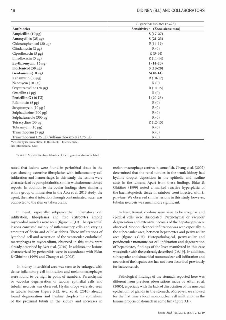
Altun et al., (2012) noted that L. garvieae strains in Turkey developed resistance to oxytetracycline, erythromycin, amoxicillin and florfenicol. Similary, some of our isolated L. garvieae strains harbored resistance to oxytetracycline but displayed sensitivity to amoxicillin, florfenicol and moderate sensitivity to erythromycin (Table 2).
According to the histopathological examination major lesions were found to be located in eyes, kidney, heart
Figure 1: Rainbow trout naturally infected with L. garvieae A. Marked exophthalmos. B. Focal haemorrhages in liver. C,D. Opacity and thickening, along with pseudomembrane formation in swim-bladder and bulbus arterious walls
Figure 2: L. garvieae specific PCR, 1100 bp. M; Marker (100-3000 bp), 1; L. garvieae ATCC 43921, 2-11; L. garvieae field isolates, 12; V. salmoninarum NCIMB 13133.

Revue Méd. Vét., 2014, 165, 1-2, 12-19
HISTOPATHOLOGY OF LACTOCOCCUS GARVIEAE INFECTION 15
and liver. A significant fibroplasia with inflamatory cell infiltration and vascularization were found in oculer area. Panophthalmitis was observed in which posterior chamber (the back section of the eye’s interior), iris and optic nerve papilla were more significantly involved. Cornea and ciliary body were thickened more distinctly (figure 3.A,B). Eldar &
Ghittino (1999) reported that ocular alterations resulted from a severe haemorrhagic panophthalmitis with destruction of the anterior and posterior chambers, the optic nerve papilla was heavily affected and inflammation progressed into the retrobulbar fat and striated muscle in rainbow trout infected with L. garvieae and Streptococcus iniae. Chang et al. (2002)
Characteristics CharacteristicsColony size <2 mm Acid production from:Gram + Lactose -Shape coccus Trehalose +Motility - Inulin -Hemolysis α Raffinose -Oxidase - Starch -Catalase - Glycogen -O/F F Glycerol -Urease - Erythritol -Gelatinase - d-Arabinose -Caseinase + l-Arabinose -Lipase - d-Xylose -Hippurate hydrolysis v l-Xylose -Pyrrolidonyl arylamidase + Adonitol -Lysine decarboxylase - β-Methylxylidose -Ornithine decarboxylase - Galactose +α-Galactosidase - d-Glucose +β-Galactosidase - d-Fructose +β-Glucuronidase - d-Mannose +Alkaline phosphatase - l-Sorbose -Leucine arylamidase + Rhamnose -Arginine dihydrolase + Dulcitol -Citrat utilization - Inositol -Indole - α-Methylxy-d-mannoside -Voges-Proskauer + α-Methylxy-d-glucoside -Methyl Red + N-Acetylglucosamine +Gas from glucose - Amygladin +NO3 reduction - Arbutin +Aesculin hydrolysis + Salicin +H2S production on TSI - Cellobiose +Growth in: Maltose +
at +4˚C + Melibiose vat 15˚C + Saccharose +at 37˚C + Melezitose -at 42˚C + Xylitol -at 45 ˚C + β-Gentiobiose +
Growth at: d-Turanose -% 1 NaCl + d-Lyxose -% 2.5 NaCl + d-Tagatose +% 6.5 NaCl + d-Fucose -% 8 NaCl - l-Fucose -
Growth on McConkey agar - d-Arabitol -Acid production from: l-Arabitol -
Ribose v Potassium gluconate +Mannitol v Potassium 2-ketogluconate -Sorbitol - Potassium 5-ketogluconate -
v, variable character
Table I: Phenotypic characteristics of L. garvieae isolates (n=25) from rainbow trout
Revue Méd. Vét., 2014, 165, 1-2, 12-19
DIDINEN (B.I.) AND COLLABORATORS16
noted that lesions were found in periorbital tissue in the eyes showing extensive fibroplasias with inflammatory cell infiltration and hemorrhage. In this study, the lesions were characterized by panophtalmitis, similar with aforementioned reports. In addition to the ocular findings show similarity with a group of immersion in the Avcı et al. 2013 study, the agent, the natural infection through contaminated water was connected to the skin or taken orally.
In heart, especially subpericardial inflamatory cell infiltration, fibroplasias and free eritrocytes among myocardial muscles were seen (figure 3.C,D). The epicardial lesions consisted mainly of inflammatory cells and varying amounts of fibrin and cellular debris. These infiltrations of lymphoid cell and activation of the ventricular endothelial macrophages in myocardium, observed in this study, were already described by Avcı et al. (2010). In additon, the lesions characterized by pericarditis were in accordance with Eldar & Ghittino (1999) and Chang et al. (2002).
In kidney, intersititial area was seen to be enlarged with dense inflamatory cell infiltration and melanomacrophages were found to be high in point of numbers. Parenchymal or vacuolar degeneration of tubular epithelial cells and tubular necrosis was observed. Hyalin drops were also seen in tubular lumens (figure 3.E). Avcı et al. (2010) already found degeneration and hyaline droplets in epithelium of the proximal tubuli in the kidney and increases in
melanomacrophage centres in some fish. Chang et al. (2002) determined that the renal tubules in the trunk kidney had hyaline droplet deposition in the epithelia and hyaline casts in the lumens. Apart from these findings, Eldar & Ghittino (1999) noted a marked reactive hyperplasia of the haematopoietic tissue in rainbow trout infected with L. garvieae. We observed similar lesions in this study, however, tubular necrosis was much more significant.
In liver, Remak cordons were seen to be irregular and epitelial cells were dissociated. Parenchymal or vacuolar degeneration and extensive necrosis of the hepatocytes were observed. Mononuclear cell infiltration was seen especially in the subcapsular area, between hepatocytes and perivascular area (figure 3.G,H). Histopathological, perivascular and periductular mononuclear cell infiltration and degeneration of hepatocytes, findings of the liver manifested in this case was similar with these already described [2,6,19]. In addition, subcapsular and sinuzoidal mononuclear cell infiltration and necrosis of the hepatocytes has not been described previously for lactococcosis.
Pathological findings of the stomach reported here was different from previous observations made by Altun et al. (2005), especially with the lack of dissociation of the mucosal epithelium of glands in the stomach. Moreover, we showed for the first time a focal mononuclear cell infiltration in the lamina propria of stomach in some fish (figure 3.F.).
L. garvieae isolates (n=25)Antibiotics Sensitivity * (Zone sizes: mm)Ampicillin (10 µg) S (17-27)Amoxycillin (25 µg) S (21-23)Chloramphenicol (30 µg) R(14-19)Clindamycin (2 µg) R (0)Ciprofloxacin (5 µg) R (5-14)Enrofloxacin (5 µg) R (11-14)Erythromycin (15 µg) I (14-20)Florfenicol (30 µg) S (10-20)Gentamycin(10 µg) S(10-14)Kanamycin (30 µg) R (10-12)Neomycin (10 µg ) R (0)Oxytetracycline (30 µg) R (14-15)Oxacillin (1 µg) R (0)Penicillin G (10 IU) I (20-25)Rifampicin (5 µg) R (0)Streptomycin (10 µg ) R (0)Sulphadiazine (300 µg) R (0)Sulphafurazole (300 µg) R (0)Tetracycline (30 µg) R (12-15)Tobramycin (10 µg) R (0)Trimethoprim (5 µg) R (0)Trimethoprim(1.25 µg) /sulfamethoxazole(23.75 µg) R (0)
*Sensitivity (S: susceptible; R: Resistant; I: Intermediate) IU: International Unit
Table II: Sensitivities to antibiotics of the L. garvieae strains isolated

Revue Méd. Vét., 2014, 165, 1-2, 12-19
HISTOPATHOLOGY OF LACTOCOCCUS GARVIEAE INFECTION 17
A few focal necrotic area was also observed in spleen Altun et al. (2005) also found necrosis and haemorrhagic foci in the spleen. In contrast, Chang et al. (2002) determined that diseased fish showing serositis consisting of fibroplasias, and macrophage and lymphocyte infiltrations in spleen.
Fibrinous meningitis associated with the lactococcosis have also been described in fish [6,14,19]. Altun et al. (2005) noted epithelial hypertrophy in the gills with partialy fusion of the gill lamellae. Avcı et al. (2010) determined vascular changes including edema, hemorrhage and telangiectasia, swelling of epithelial cells in gills. Severe hyperemia, hemorrhages in the villus of the intestines and infiltration of inflammatory cells in the lamina propria were
determined for lactococosis by Avcı et al. (2010). In contrast, histopathological findings of skin, gill, muscle, intestine, brain and gonads were not significant in this case.
In conclusion, this work presents phenotypic and molecular identification, antimicrobial susceptibility of L. garvieae isolated from rainbow trout and new histopathological observations during lactococcosis outbreak in Turkey.
References
1. ALTUN S, DILER O AND ADILOGLU AK: Genotyping of Lactococcus garvieae strains from rainbow trout (Oncorhynchus mykiss) by 16S rDNA sequencing. Bulletin of the European Association of Fish Pathologists, 2004, 24, 119-124
2. ALTUN S, DILER A, DILER O, BASAK K AND ISIKLI BI: Histopathology of streptococcosis in rainbow trout (Oncorhynchus mykiss, Walbaum). Bulletin of the European Association of Fish Pathologists, 2005, 25, 131-135.
3. ALTUN S, ONUK EE, CIFTCI A, BUYUKEKIZ AG AND DUMAN M: Phenotypic, genotypic characterisation and antimicrobial susceptibility determination of Lactococcus garvieae strains. Journal of the Faculty of Veterinary Medicine, Kafkas University, 2012, 19, 375-381.
4. AUBIN GG, BÉMER P, GUILLOUZOUIC A, CRÉMET L, TOUCHAIS S, FRAQUET, N, BOUTOILLE D, REYNAUD A, LEPELLETIER D AND CORVEC S: First report of a hip prosthetic and joint infection caused by Lactococcus garvieae in a woman fishmonger. Journal of Clinical Microbiology, 2011, 49, 2074-2076.
5. AUSTIN B AND AUSTIN DA: Bacterial Fish Pathogens: Diseases of Farmed and Wild Fish, 4th ed, Springer-Praxis, Chichester, UK. ISBN 1402060688, 2007.
6. AVCI H AYDOĞAN A TANRIKUL TT AND BIRINCIOĞLU S: Pathological and Microbiological ınvestigations in rainbow trout (Oncorhynchus mykiss, Walbaum, 1792) naturally infected with Lactococcus garvieae. Journal of the Faculty of Veterinary Medicine, Kafkas University, 2010, 16, 313-318.
7. AVCI H, BIRINCIOGLU SS, TANRIKUL TT, EPIKMEN ET, METIN N AND AVSEVER ML: Experimental Lactococcus garvieae infection in rainbow trout, Oncorhynchus mykiss, Walbaum 1792: a comparative histopathological and immunohistochemical study. Journal of Fish Diseases, 2013 Aug 20. doi: 10.1111/jfd.12132.
8. BAECK GW, KIM JH, GOMEZ DK AND PARK SC: Isolation and characterization of Streptococcus sp. from diseased flounder (Paralichthys olivaceus) in Jeju Island. Journal of Veterinary Science, 2006, 7, 53-58.
9. BARK S AND MCGREGOR D: The first occurrence of lactococcosis in farmed trout in England. Trout News, 2001, 31, 9-11.
Figure 3: A. Corneal thickening (asterisk) and general appearance of eye (HE, 10x). B. New vascular formation (black arrow), dense inflamatory cell infiltration (white arrows) in the eye and exudation in the anterior chamber of eye (asterisk); (HE, 30x). C. Subpericardial inflamatory cell infiltration in heart (black arrow); (HE, 40x). D. Haemorrhages among myocardial muscles of heart (black arrows); (HE, 40x). E. Tubular necrosis (black arrows), melanomacrophages (white arrow) and mononuclear cell infiltration (asterisks) in kidney (HE, 100x). F. Focal mononuclear cell infiltration in the lamina propria of stomach (black arrows); (HE, 100x). G. Subcapsular haemorrhage and mono-nuclear cell infiltration (black arrow) and necrosis of the hepatocytes (arrow heads); (HE, 100x). H. Mononuclear cell infiltration between hepatocytes, perivascular and periductular area (black arrows) and necrosis of the hepatocytes (arrow heads) in liver (HE, 100x).

Revue Méd. Vét., 2014, 165, 1-2, 12-19
DIDINEN (B.I.) AND COLLABORATORS18
10. BRAGG R AND BROERE JSE: Streptococcosis in rainbow trout in South Africa. Bulletin of the European Association of Fish Pathologists, 1986, 6, 89-91.
11. CAGIRGAN H AND TANRIKUL TT: A new problem Enterococcus-like Infection in Rainbow Trout (Oncorhynchus mykiss) Farms in Turkey (in Turkish). Veteriner Kontrol ve Arastirma Enstitusu Dergisi, 1995, 33, 9-19.
12. CAGIRGAN H: Biotyping of Lactococcus garvieae Isolated from Turkey. EU Journal of Fisheries Aquatic Science, 2004, 21, 267-269.
13. CARSON J, GUDKOVS N AND AUSTIN B: Characteristics of an Enterococcus-like bacterium from Australia and South Africa, pathogenic for rainbow trout (Oncorhynchus mykiss, Walbaum). Journal of Fish Diseases, 1993, 16, 381-388.
14. CHANG PH, LIN CW AND LEE YC: Lactococcus garvieae infection of cultured rainbow trout, O. mykiss in Taiwan and associated biophysical characteristics and histopathology. Bulletin of the European Association of Fish Pathologists, 2002, 22, 319-327.
15. CHEN SC, LIAW LL, SU HY, KO SC, WU CY, CHAUNG HC, TSAI YH, YANG KL, CHEN YC, CHEN TH, LIN GR, CHENG SY, LIN YD, LEE JL, LAI CC, WENG YJ AND CHU SY: Lactococcus garvieae, a cause of disease in grey mullet, Mugil cephalus L. in Taiwan. Journal of Fish Diseases, 2002, 25, 727-732.
16. DILER O, ALTUN S, ADILOGLU AK, KUBILAY A AND ISIKLI B: First occurence of Streptococcosis affecting farmed rainbow trout (O. mykiss) in Turkey. Bulletin of the European Association of Fish Pathologists, 2002, 22, 21- 26.
17. DOMENECH A, PRIETA J, FERNANDEZ-GARAYZABAL JF, COLLINS MD, JONES D AND DOMINGUEZ L: Phenotypic and phylogenetic evidence for a close relationship between Lactococcus garvieae and Enterococcus seriolicida. Microbiologia, 1993, 9, 63-68.
18. ELDAR A, GHITTINO C, ASANTA L, BOZZETTA E, GORIA M, PREARO M AND BERCOVIER H: Enterococcus seriolicida is a junior synonym of Lactococcus garvieae, a causative agent of septicemia and meningoencephalitis in fish. Current Microbiology, 1996, 32, 85-88.
19. ELDAR A, AND GHITTINO C: Lactococcus garvieae and Streptococcus iniae infections in rainbow trout Oncorhynchus mykiss: Similar, but different diseases. Diseases of Aquatic Organisms, 1999, 36, 227- 231.
20. EVANS JJ, KLESIUS PH AND SHOEMAKER CA: First isolation and characterization of Lactococcus garvieae from Brazilian Nile tilapia, Oreochromis niloticus (L.), and pintado, Pseudoplathystoma corruscans (Spix & Agassiz). Journal of Fish Diseases, 2009, 32, 943–951.
21. EYNGOR M, ZLOTKIN A, GHITTINO C, PREARO M, DOUET DG, CHILMONCZYK S AND ELDAR A: Clonality and diversity of the fish pathogen Lactococcus garvieae in Mediterranean countries. Applied and Environmental Microbiology, 2004, 70, 5132-5137.
22. FEFER JJ, RATZAN KR, SHARP SE AND SAIZ E: Lactococcus garvieae endocarditis: report of case and reviewof the literatüre. Diagnostic Microbiology and Diagnostic Disease, 1998, 32, 127-130.
23. HOLT JG, KRIEG NR, SNEATH PHA, STALEY JT AND WILLIAMS ST: Bergey’s manual of determinative bacteriology, 9th ed. Williams & Williams, Baltimore. ISBN 0683006037, 1994.
24. KAV K AND ERGANIS O: Antibiotic susceptibility of Lactococcus garvieae in rainbow trout (Oncorhynchus mykiss) farm. Bulletin of The Veterinary Institute in Pulawy, 2008, 52, 223-226.
25. KUSUDA R, KAWAI K, SALATI F, BANNER CR AND FRYER L: Enterococcus seriolicida sp. Nov., a Fish Pathogen. International Journal of Systematic Bacteriology, 1991, 41, 406-409.
26. MUZQUIZ JL, ROYO FM., ORTEGA C, DEBLAS I, RUIZ I AND ALONSO JL: Pathogenicity of streptococcosis in rainbow trout (Onchorhynchus mykiss): dependence on age of diseased fish. Bulletin of the European Association of Fish Pathologists, 1999, 19, 114-119.
27. NAVAS ME, HALL G AND EL BEJJANI D: A Case of endocarditis caused by Lactococcus garvieae and suggested methods for identification. Journal of Clinical Microbiology, 2013, 51, 1990-1992.
28. NCCLS (National Committee for Clinical Laboratory Standards of Antimicrobial Susceptibility Testing): Performance Standards for Antimicrobial Susceptibility Testing; Eleventh Information Supplement. NCCLS document M100-S11 NCCLS,West Walley Road, Suite 1400, Wayne, Pennsylvania 19087-1898 USA, 2001.
29. OZER S, BULDUKLU PS AND DONMEZ E: Streptococcocis occurence at rainbow trout (Oncorhynchus mykiss, Walbaum) cultivated in province Mersin-Turkey. Fisheriesscienses.com, 2008, 2, 272-283.
30. PEREIRA F, RAVELO C, TORANZO AE AND ROMALDE JL: Lactococcus garvieae, an emerging pathogen for the Portguese trout culture. Bulletin of the European Association of Fish Pathologists, 2004, 24, 274-279.
31. SALATI F, TASSI P AND BRONZI, P: Isolation of Enterococcus like bacterium from Diseased Adriatic Sturgeon Acipenser naccarii, Farmed Italy. Bulletin of the European Association of Fish Pathologists, 1996, 16, 96-99.
32. SAVVIDIS GK, ANATOLITIS C, KANAKI Z AND VAFEAS G: Epizootic outbreaks of lactococcosis disease in rainbow trout (O. mykiss Walbaum) culture in Greece. Bulletin of the European Association of Fish Pathologists, 2007, 27, 223-228.
33. SHARIFIYAZDI H, AKHLAGHI M, TABATABAEI M AND MOSTAFAVI ZADEH S: Isolation and characterization of Lactococcus garvieae from diseased rainbow trout (Oncorhynchus mykiss, Walbaum) cultured in Iran. Iranian Journal of Veterinary Research, Shiraz University, 2010, 11, 342-350.
34. SOLTANI M, NIKBATHT GH, MOUSAVI HAE, AHMADZADEH N: Epizootic outbreak of lactococcosis

Revue Méd. Vét., 2014, 165, 1-2, 12-19
HISTOPATHOLOGY OF LACTOCOCCUS GARVIEAE INFECTION 19
caused by Lactococcus garvieae in farmed rainbow trout (Oncorhynchus mykiss) in Iran. Bulletin of the European Association of Fish Pathologists, 2008, 28, 207-212.
35. TANRIKUL T AND GULTEPE N: Mix Infections in rainbow trout (Oncorhynchus mykiss, Walbaum): Lactococcus garvieae and Vibrio anguillarum O1. Journal of Animal and Veterinary Advances, 2011, 10, 1019-1023.
36. TSAI MA, WANG PC, YOSHIDA T, LIAW LL, CHEN SC: Development of a sensitive and specific LAMP PCR assay for detection of fish pathogen Lactococcus garvieae. Diseases of Aquatic Organism, 2013, 102, 225-235
37. VELA AI, VAZQUEZ C, GIBELLO A, BLANCO M M, MORENO M A, LIEBANA P, ALBENDEA C, ALCALA B, MENDEZ A, DOMINGUEZ L AND
FERNANDEZ-GARAYZABAL JF: Phenotypic and genetic characterization of Lactococcus garvieae isolated in Spain from Lactococcosis outbreaks and comparison with isolates of other countries and sources. Journal of Clinical Microbiology, 2000, 38, 10: 3791-3795.
38. VENDRELL D, JL BALCAZAR, RUIZ-ZARZUELA I, DE BLAS I, GIRONES O AND MUZQUIZ JL: Lactococcus garvieae in fish: A review. Comparative Immunology, Microbiology and Infectious Diseases, 2006, 29, 177-198.
39. ZLOTKIN A, ELDAR A, GHITTINO C AND BERCOVIER H: Identification of Lactococcus garvieae by PCR. Journal od Clinical Microbiology, 1998, 36, 983-985.