the isolation of lactococcus lactis subsp. cremoris from nature with
TRANSCRIPT

AN ABSTRACT OF THE THESIS OF
May soon Salama for the degree of Doctor of Philosophy in
Microbiology presented on May 3, 1993
Title: The Isolation of Lactococcus lactis subsp. cremoris From
Nature With Probes for 16S Ribosomal RNAs
Abstract Approved:
Stephen J. lovannoni
William E. Sandine
Lactococcus lactis subsp. cremoris is of considerable interest to
the dairy industry, which relies upon the limited number of strains
available the manufacture of Cheddar cheese free of fermented and
fruity flavors. Our purpose was to identify unique ribosomal RNA
sequences that could be used to discriminate L. lactis subsp. cremoris
from related subspecies lactis. The 16S rRNAs from 13 Lactococcus
strains were partially sequenced using reverse transcriptase in order
to identify domains unique to L. lactis subsp. cremoris.
Oligonucleotide probes specific for the species Lactococcus lactis
(212RLa) and the subspecies cremoris (68RCa) were designed,
synthesized and evaluated for ability to discriminate lactococci and L.
lactis subsp. cremoris from closely related strains.
These probes were used in colony hybridizations to rapidly
screen large numbers of colonies for L. lactis subsp. cremoris.
Thirty-eight plant and vegetable species, twelve other samples from
Redacted for Privacy
Redacted for Privacy

dairy farms, twenty-one individual raw milk and milk product
samples from the United States, China, Morocco, and Yugoslavia, were
examined for lactic acid bacteria by the colony hybridization method
using the 68RCa and 212RLa probes.
Lactococcus lactis subsp. lactis was found to occur on potato,
cucumber, sweet peas, beans, cantaloupe, corn, cow's body and tail
and in colostrum, goat and cow raw milk, cottage cheese and cream.
Lactococcus lactis subsp. diacetylactis was isolated from cow raw
milk obtained from Morocco, as well as goat raw milk and cottage
cheese from Yugoslavia. Lactococcus lactis subsp. cremoris was
isolated from raw milk obtained from Morocco, Yugoslavia and China
and from cottage cheese obtained from Yugoslavia. The
phenotypical, morphological, and physiological characteristics of the
newly isolated lactococcal strains generally agreed with the standard
description for the genus Lactococcus.
The isolation of L. lactis from different plant sources confirmed
that plants are a natural source of this bacterium. The fact that a
few strains of L. lactis subsp. cremoris were isolated from raw milk
and cottage cheese from Morocco and Yugoslavia, but not from
plants, suggests that a natural habitat of the subspecies cremoris
could be raw milk and milk products and prepared by traditional
dairy practices.
The biochemical and physiological characteristics of the new L.
lactis isolates, their resistance to bacteriophage preparations
obtained from cheese factories, and their acid producing capabilities,
indicate the potential usefulness of these strains as dairy starter
cultures.

The Isolation of Lactococcus lactis subsp. cremoris From Nature WithProbes for 16S Ribosomal RNAs
by
Maysoon Salama
A THESIS
submitted toOregon State University
in partial fulfillment ofthe requirements for the
degree ofDoctor of Philosophy in Microbiology
Completed May 3, 1993Commencement June, 1993
APPROVED:
Professor of Microbiology in charge of major
Professor of Microbiology in charge of major
Chairman of the Department of Microbiology
Dean of Gradua dSchool
Date thesis is presented May 1 1993
Typed by author Maysoon Salama
Redacted for Privacy
Redacted for Privacy
Redacted for Privacy
Redacted for Privacy
Dedicated with love ....
To my dear husband Mohammad A. Alayan, my beloved son
Atta M. Alayan, my parents Qadriya Odeh and Subhi Salama
(God bless his soul), and to both dear families, the Salamas
and the Alayans.

ACKNOWLEDGEMENTS
Foremost, my deepest gratitude to Almighty Allah, the most
beneficent and the most merciful, for giving me the strength and
endurance to overcome all the emotional and physical challenges
during my years as a graduate student.
A special debt of gratitude to Dr. S. J. Giovannoni and Dr. W. E.
Sandine for the insightful advice, guidance, continual encouragement,
and deep interest in the progress of my work. I treasure their
friendship and appreciate their sensitivity and unique fatherly/
brotherly care. I would like also to extend my special thanks to the
other committee members, Dr. Mark A. Daeschel, Dr. Henry Schaup,
Dr. Gary L. Taghon, and Dr. D. Mattson.
I wish to express my sincere gratitude to Dr. J. L. Fryer for
allowing me the opportunity to join Oregon State University as a
Teaching Assistant Graduate Student and for the Tartar Graduate
Student Fellowship I was awarded in 1988. A special word of
thanks and appreciation to Dr. Sandine for giving me the privilege
and opportunity to work as a Research Assistant Graduate Student
with Dr. Giovannoni.
Thanks to all the faculty and staff of the Department of
Microbiology with whom I have been associated over the years, and
a special thanks to the Dairy and Molecular Evolution groups who
provided me with friendship and encouragement. I wish to
acknowledge Dr. K. Field in particular, for her help during the early
stages of my work and during the preparation of the manuscripts
and the thesis, for continual encouragement, and for her sincere

friendship and sensitivity. She has been my model as a dedicated
scientist, wife and mother. Special thanks go to my friend Theresa
Britschgi for being a close friend and for teaching me how to draw
the secondary structures of rRNAs using the computer. Thanks to
Kirsti Ritalati and Kelley Nathman who helped me with a lot of
enthusiasm during the later stages of my work for a short, yet
fruitful, while.
Most of all, a very unique thanks and appreciation to my
beloved husband, Mohammad A. Alayan, and my wonderful son, Atta
M. Alayan, who provided me with their constant love, care, support,
encouragement, and understanding. Atta has been a special source
of inspiration and fun for the family. His smiling face and shiny
eyes, understanding, patience, and support for his graduate student
parents were unique. My deepest thanks to my parents who
nourished me with their endless compassion and love and who gave
me an unswerving joy of learning and a sense of confidence and
independence. Special thanks go to my sisters and brothers for their
moral support. I would like also to thank all my special friends in
Corvallis for the joyful time we spent together and for their moral
support.

CONTRIBUTION OF AUTHORS
The contribution of Dr. W. E. Sandine and Dr. S. J. Giovannoni is
much appreciated. Their guidance, expertise, advice, and interest in
the work was behind the success of this project.
The proposal, which was submitted to The National Dairy
Promotion and Research Board and subsequently approved and
funded, was a joint effort of Dr. Giovannoni and Dr. Sandine.

CHAPTER 1:
CHAPTER 2:
TABLE OF CONTENTS
Introduction
Development and Application ofOligonucleotide Probes forIdentification of Lactococcus lactissubsp. cremoris
AbstractIntroductionMaterials and MethodsResultsDiscussionAcknowledgementsReferences
CHAPTER 3: Isolation of lactococci From Natureby Colony Hybridization WithRibosomal RNA Probes
AbstractIntroductionMaterials and MethodsResults and DiscussionAcknowledgementsReferences
CHAPTER 4: Isolation of New Strains ofLactococcus lactis subspeciescremoris
AbstractIntroductionMaterials and MethodsResults and DiscussionAcknowledgementsReferences
PAGE
1
28
29303237464950
55
565759637273
77
78808291106107

CHAPTER 5: Characterization of Novel Strains ofLactococcus lactis using the BiologCarbon Source Utilization Systemand Phage Typing 1 1 3
Abstract 1 14Introduction 1 15Materials and Methods 1 1 7
Results and Discussion 12 5Acknowledgements 13 6References 13 7
BIBLIOGRAPHY 13 9

LIST OF FIGURES
FIGURES
CHAPTER 2
Figure 1: Secondary structure model for 5' region oflactic acid bacterium 16S rRNAs. 38
PAGE
Figure 2: Nucleotide sequences of 5' regions of lacticacid bacteria 16S rRNAs. 41
Figure 3: Autoradiogram of a dot blot hybridization tobulk cellular RNAs from lactic acid bacteriaand control strains.
Figure 4: Autoradiogram of a dot blot hybridization tofixed whole cells of lactic acid bacteria andcontrol strains.
CHAPTER 3
43
45
Figure 1: Colony hybridization to bacteria on glassfiber filters. 64
Figure 2: Colony hybridization to L. lactis subsp. lactis,L. lactis subsp. cremoris, and unknownenvironmental isolates from a plant sample(Prunus laurocerasus). 67
Figure 3: Colony hybridization of environmental florafrom a fresh corn sample. 68
CHAPTER 5
Figure 1: Dendogram showing relationship betweenlactococcal strains as recovered from theevaluation of carbon-source utilization usingthe Biolog GP microtiter plate test system. 126

TABLES
CHAPTER 2
Table 1:
CHAPTER 3
Table 1:
Table 2:
CHAPTER 4
Table 1:
Table 2:
Table 3:
LIST OF TABLES
Primers used for sequencing of 16S rRNAs orfor hybridization experiments.
List of oligonucleotides used.
Genotypic and phenotypic characteristics ofsome of the environmental strains isolatedfrom a fresh corn sample using the colonyhybridization procedure with 32P -labeled16S rRNA probes.
The occurrence of L. lactis subsp. cremoris andL. lactis subsp. lactis on green plant material
The occurence of Lactococcus lactis subsp.cremoris and Lactococcus lactis subsp. lactison vegetables from local produce and othertypes of environmental samples from adairy farm location and in raw milk samplesfrom the United States.
The occurrence of Lactococcus lactis subsp.cremoris and Lactococcus lactis subsp. lactisin milk and milk products from Yugoslavia,China, and Morocco.
PAGE
34
65
70
83
84
86

Table 4:
Table 5:
Table 6:
Table 7:
Genotypic and phenotypic characteristics ofsome of the environmental strains isolatedfrom vegetables, plants, raw milk, colostrum,and other samples from a dairy farm (U.S)using the colony hybridization procedure with32P-labeled 16S rRNA probes. All strains areGram positive cocci and acidify litmus milkbefore coagulation.
Genotypic and phenotypic characteristics ofsome of the environmental strains isolatedfrom five Chinese raw milk samples usingthe colony hybridization procedure with 32Plabeled 16S rRNA probes. All strains areGram positive cocci and acidify litmus milkbefore coagulation.
Genotypic and phenotypic characteristics ofsome of the environmental strains isolatedfrom one Moroccan raw milk sample usingthe colony hybridization procedure with 32Plabeled 16S rRNA probes. All strains areGram positive cocci and acidify litmus milkbefore coagulation.
Genotypic and phenotypic characteristics ofsome of the environmental strains isolatedfrom several Yugoslavian raw milk and milkproduct samples using the colonyhybridization procedure with 32P- labeled16S rRNA probes. All strains are Grampositive cocci and acidify litmus milk beforecoagulation.
92
96
98
99
Table 8: Flavor evaluation of some newly isolatedlactic acid bacteria. 1 03

CHAPTER 5
Table 1:
Table 2:
Table 3:
Table 4:
Table 5:
Environmental strains isolated from differentsources using the colony hybridizationprocedure with 32P- labeled 16S rRNA probes.All strains are fast acid producers and werechosen for bilog testing.
Environmental strains isolated from differentsources using the colony hybridizationprocedure with 32P- labeled 16S rRNA probes.All strains are fast acid producers and werechosen for phage testing.
Characteristics useful for differentiatingL. lactis srains for cluster groups.
Challenging the environmental lactococcalstrains with Galloway West company phages.Twenty-three strains were testedagainst 32 phage preparations which werenumbered from 1 through 32.
Challenging the environmental lactococcalstrains with Marschall products companyphages. Twenty three strains were testedagainst 72 positive whey samples for phage(whey samples 1-72), 30 negative wheysamples (whey samples 73-102), and singleplaque isolates (SPI 1-38). The phage testreading was based on a scale of 0 (no lysis)to 3 (total lysis) based on spot testevaluation. Only those strains with readingsof 1 or greater were reported.
1 1 8
1 2 1
130
132
134

The Isolation of Lactococcus lactis subsp. cremoris From Nature WithProbes for 16S Ribosomal RNAs
CHAPTER 1
Introduction
Ecology of lactic acid bacteria.
In the early years of the science of dairy bacteriology it was
thought that freshly drawn milk was sterile. It was not until 1891
that Schultz (as cited by Harding (21) ) found that milk contained a
large number of bacteria. At that time it was thought that this was
an indication of udder disease. Later, Moore confirmed the findings
of Schulz, and concluded that bacteria do occur in normal milk. Once
this idea was firmly established, the species of bacteria occurring in
milk were brought under examination. Walker (67) conducted one of
the earliest studies in this regard. He found that Streptococcus lactis
(now known as Lactococcus lactis subsp. lactis and herein referred to
as L. lactis) constituted at least 95% of the organisms present in all
the milk samples he studied. In more recent times Sherman and
Hastings (58) found streptococci in the milk of 31.1% of 48 cows and
in 15.1% of the samples from 161 cows. Streptococcus cremoris (now
known as Lactococcus lactis subsp. cremoris is found in even lower
numbers in milk. Of 3,000 isolates from 59 samples of commercial
raw milk, only 4% were L. lactis subsp. cremoris according to a study
conducted by Nelson and Thornton (38). In that same year 35
strains of Lactococcus were isolated from raw milk samples in a

2
remote area of the Jura mountains in France (25). Only 2 of the
isolates were L. lactis subsp. cremoris. This supported the idea that
this organism occurs naturally and is only an environmental variety
of L. lactis subsp. lactis.
Since lactic acid bacteria (LAB) are found most often in milk it
was only natural that the body of cows, including the udder and the
mouth, would be suspected as being a natural habitat for these
microorganisms. LAB have been reported to occur on milking
utensils (4), and in the udders (15, 46), surfaces, mouths, and feces of
cows (14). However, these reports were made without the
identification of lactococci to species and subspecies. Later
investigators, who had more refined differential tests for the
identification of the LAB, failed in many cases to isolate the
organisms from these sources. Plant material, not cattle, was then
suggested to be the natural habitat for lactococci (14, 58, 61). One of
the most fruitful studies carried out to investigate the presence of
LAB on plant material was that done by Stark and Sherman (61).
Two hundred cultures were isolated from different plant samples
and identified as L. lactis subsp. lactis. Samples of fresh corn and
corn silks tested were found in every instance to contain only L.
lactis subsp. lactis. Esten (14) tried to isolate these organisms from
various plant materials but none were found. Several types of grain
feeds were also tested. Only one culture of L. lactis subsp. lactis was
isolated from corn meal. Pinter (43) was able to find L. lactis subsp.
lactis on clover, beans, and grass. Out of 50 samples of plant
materials studied, 20% showed the presence of various streptococci.
Out of the 20%, 70% were Streptococcus faecalis (now known as

3
Enterococcus faecalis) and 30% were L. lactis subsp. lactis. Attempts
to isolate L. lactis subsp. cremoris from plant material have been
made by a few investigators, with no success. Yawger (73) screened
60 samples of plant material for L. lactis subsp. cremoris. He
managed to isolate 16 cultures of L. lactis subsp. lactis, but no strains
of L. lactis subsp. cremoris. Even though the results of his
experiments were negative, he felt that plant materials still
represented the most logical source for L. lactis subsp. cremoris.
Much of the early work on the ecology of LAB had little value,
because reliable methods of distinguishing these organisms from
fecal streptococci did not exist (25). In an attempt to determine the
natural habitat of Lactococcus organisms, Radich (44) examined 27
different species of vegetables, 18 species of fruits, and many
individual cow raw milk samples. L. lactis subsp. lactis was found
on potatoes, corn, cucumber, peas, beans, and cantaloupe. In each
case the organism was isolated in low numbers. All fruits examined
failed to yield any lactococci with the exception of cantaloupe. From
31 individual cow raw milk samples, 4 isolates of L. lactis subsp.
cremoris were obtained, but all proved to be slow acid producers.
Three of the remaining strains were L. lactis subsp. lactis biovar.
diacetylactis, and 28 were L. lactis subsp. lactis. The isolation of L.
lactis subsp. lactis from several plant sources confirmed the belief
that plants are the natural habitat for this bacterium. Failure to
isolate L. lactis subsp. cremoris from plant material suggested that
this species may be a variant of L. lactis subsp. lactis, with milk as its
natural habitat. King and Koburger (27) characterized Group N
streptococci isolated from meats, frozen vegetables, dairy products,

4
bran-trough water, and poultry feed. From 18 samples, 184 isolates
of L. lactis were obtained. These were generally more resistant
(94.6%) to 20 bacteriophages than dairy starter culture isolates (77%
resistant). No L. lactis subsp. lactis biovar. diacetylactis strains were
isolated and the subspecies cremoris was recovered from only
cottage cheese and raw milk. Unfortunately, the L. lactis subsp.
cremoris strains found in milk were lost because they failed to
survive freezing.
Even more recently, Fenton (16) studied the role of farm
machinery in harboring LAB that were present on grass. Sixteen
different groupings or species of LAB were isolated from grass, but
no L. lactis subsp. cremoris was found. Pediococcus acidilactici and
Streptococcus faecium were the predominant organisms. Lactococcus
lactis subsp. cremoris was isolated from two items of farm
machinery, especially the forest harvester. Fourteen percent of the
isolates were identified as L. lactis subsp. cremoris based on the their
ability to grow at 100C but not 450C and their inability to produce
ammonia from arginine. It is questionable whether or not that
represents sufficient testing to identify an organism as L. lactis
subsp. cremoris. In another study L. lactis subsp. cremoris w a s
reported to have been isolated from frozen peas (7), but subsequent
work (8) showed that these cultures had been incorrectly designated
and were in fact unusual Group N streptococci with properties
different from those of both lactis and cremoris subspecies. The
natural habitat of subsp. cremoris thus remains unknown. More
studies similar to that of Hirsch (25) are needed to understand its
ecological relationship to other lactococci. Several scientists believe

5
that L. lactis subsp. cremoris may only be isolated from nature very
infrequently (1, 25, 27, 35, 37, 39, 52, 72 ). Lawrence and co-
workers (35) also emphasized the great need that exists for more
strains of L. lactis subsp. cremoris for use in starter cultures. These
authors further emphasized that most strains of L. lactis subsp.
cremoris in use in starter cultures these days are related because
they are descendants of strains that were originally isolated 70 years
ago from cream in Denmark and the United States. Thus it is of
utmost importance to isolate from nature new strains of this
bacterium that are suitable for use in fermented milk products.
Economic importance.
LAB are of great economic importance to the dairy and other
fermented food industries. Their application makes possible the
manufacture of thousands of fermented foods, especially when used
in mixed cultures with other types of bacteria, yeast and molds (49).
They have been shown also to enhance the nutritional quality of
certain foods. They are also beneficial in inhibiting pathogens (11)
and spoilage bacteria in foods, food products, and animal feed (12,
13, 68). The importance of LAB in the health of newborn human
infants is generally accepted (6, 74). In addition, much of research
attention is now given to the possible usefulness of these organisms
in intestinal health (50), reduction of blood cholesterol levels (20,
24), cancer prevention (3, 19), elevating immunocompetence (59),
and antibiotic production (50, 57).

6
Dairy lactococci have been used for centuries in the production
of fermented dairy products. The need to isolate Lactococcus starter
culture strains has been emphasized by cheese makers, industry
consultants, and research workers (51). Undesirable flavors
encountered in cultured dairy products, insufficient development of
acid during fermentation and frequent culture failures resulting fromvirus infection of existing strains are some factors which have
contributed to this need for new strains of lactococci.
Over the past several decades, cultures of L. lactis subsp.
cremoris were found to be the most suitable for producing high
quality Cheddar cheese. The temperature tolerance, proteolytic ,
lypolytic, and acid producing properties of this organism allow its use
to manufacture aged Cheddar cheese free of flavor and body defects
(35, 64). The dairy industries in the United States, Australia and
Canada have suffered heavy economic losses during the past decade,due to the occurrence of slit openness and fruity flavor in Cheddar
cheese (63). Perry (42) reported the occurrence of fruity flavor in
Cheddar cheese when certain strains of L. lactis subsp. lactis wereused as starters in New Zealand. Since flavor and body defects result
in a large economic loss to the dairy industry each year, the isolation
and selection of starter cultures, especially L. lactis subsp. cremoris,is important.
Bacteriophage infection of starter cultures represents the mostchallenging problem to the cheese industry, as well as the greatestsource of economic loss. There are no known cultures in use in thedairy industry which are "permanently" resistant to lysis bybacteriophage. Some LAB are lysed by several known races of

7
phage, while others are susceptible to only one race (51). The
intensive use of the same cultures has increased the phage problem,
and therefore, newly isolated cultures resistant to existing
bacteriophages are in great demand.
Taxonomy and phylogenetic position of Lactic acid bacteria.
Lactic acid fermentation was already known to humans when
they invented writing. However, it took several thousand years from
these early applications before Louis Pasteur recognized, in 1857, the
microbial nature of lactic fermentations, as described by
Stackebrandt and Teuber (60). Fortuitously, the first bacterial pure
culture (on earth) obtained by Joseph Lister in 1878, turned out to
be Bacterium lactis which is now called L. lactis subsp. lactis (60).
In 1942, S. Or la Jensen (41) established a systematic order for
LAB on the basis of morphological and cultural features, source of
energy and nutritional needs, agglutination, and growth response
toward different temperatures. In his classification, genera of LAB
were placed in 3 groups. The first group included rod and sphere
forms without catalase, which produced only traces of by-products in
addition to lactic acid. Thermobacterium, Streptobacterium, and
Streptococcus were members of this group. The second group
included rod and sphere forms without catalase; detectable amounts
of gas and other by-products in addition to lactic acid were produced.
Betabacterium and Betacoccus belonged to this group. The third
group included rod and sphere forms with catalase. Microbacterium
and Tetracoccus belonged to this group.

8
With the development and application of modern biochemical
and molecular methods, evidence was provided that the traditional
identification scheme for LAB only partially correlated with their
natural phylogenetic relationships (60). Schleifer (55) reported,
based on extensive nucleic acid hybridization studies (29, 30, 54) and
comparative oligonucletide cataloguing of 16S rRNA (36), catalase
negative, facultatively anaerobic Gram positive cocci could be
classified into 3 genetically distinct groups. The majority of
streptococci including pyogenic (29), the 'mutans-like' (55), 'milleri-
like' (31) and 'viridans' (54) streptococci were placed in the first
group. The second group is composed of fecal streptococci and has
been described as a new genus, Enterococcus (53, 9). The third group
is formed by a few representatives of the lactic streptococci such as
S. lactis and S. raffinolactis (30). DNA/23S rRNA hybridization and
superoxide dismutase studies have shown that lactic streptococci
form a group distinct from the pyogenic streptococci and enterococci
(56). Nucleic acid hybridization studies and immunological
relationships of superoxide dismutase showed that Streptococcus
lactis (and its subspecies), Lactobacillus (Lb.) xylosus, Lb. hordniae, S.
gravieae, S. plantarum and S. raffinolactis are closely related to each
other but not to other streptococci. Therefore Schleifer and co-
workers (56) proposed a new genus, Lactococcus, to accomodate the
lactic or Group N species. Currently four species, Lactococcus lactis, L.
gravieae, L. plantarum, and L. raffinolactis, are recognized. Similarity
in lipoteichoic acid structures, lipid pattern, fatty acid and
menaquinone composition also demonstrated the relatedness of these
organisms (56).

9
Lactococcus picium was recently described as a new
Lactococcus species (70). This was based on chemical and taxonomic
studies performed on a representative strain of LAB of unknown
taxonomic position isolated from Salmonid fish. However, not all
Group N strains are members of the genus Lactococcus. Some motile
L. lactis-like strains from chicken feces and river water (22, 23)
which react with Group N antiserum, have been shown to be
unrelated to lactococci (56). Of the many molecular properties tested
(e.g, cell wall composition and structure, immunological relationships
of lactic dehydrogenase and enzymes of the glycolytic pathway), DNA
and ribosomal RNAs have been the molecules most useful in
phylogeny (60, 62). Construction of meaningful phylogenetic trees is
usually the outcome of the analysis of these nucleic acids. Such trees
offer the opportunity to establish a phylogenetically comprehensive
classification scheme for bacteria (60). Collins and co-workers (10)
examined the phylogenetic status of members of the genus
Lactococcus and similar motile strains which reacted with Group N
antiserum using reverse transcriptase sequencing of 16S rRNA (RT
sequencing). Their data clearly demonstrated that lactococci
represent a distinct phylogenetic group equivalent in rank to the
genera Enterococcus and Streptococcus. This was in agreement with
earlier nucleic acid hybridization and immunological studies of
superoxide dismutase (56). Within the genus Lactococcus it was
evident that L. plantarum is closely related to L. raffinolactis
whereas L. lactis shows a closer affinity with L. gravieae (13).
Results of DNA-DNA and DNA-RNA hybridization studies (17, 56) are
in agreement with these intrageneric relationships. Also, Lactococcus

10
and Streptococcus proved to be more closely related to each other
than to Enterococcus (10). The motile Group N strains from chicken
feces and river water, however, were found to be phylogenetically
unrelated to lactococci but were closely related to members of the
genus Enterococcus. Based on RT sequencing and DNA-rRNA
hybridization, together with phenotypic criteria, it was proposed that
these motile strains be classified in a new genus Vagococcus
Vagococcus fluvialis sp.nov. (10)
Nucleic acid sequencing and hybridization techniques.
The identification of microorganisms is essential in basic as
well as applied research. The classification of organisms traditionally
has been based on similarities in their morphological, physiological,
and biochemical characteristics (5, 33, 40). It is now clear that
classification based on these criteria does not necessarily correlate
well with natural (i.e., evolutionary) relationships as defined by
macromolecular sequence comparisons (33). Several molecular
methods for evaluating phylogenetic relationships are available (e.g.,
genomic DNA/DNA and genomic DNA/rRNA hybridization, 5S rRNA
and protein sequencing, 16S rRNA oligonucleotide cataloguing,
enzymological patterning, etc.). All of these methods have
advantages as well as limitations (40). Macromolecular sequencing
seems preferred because it permits quantitative analysis of
relationships (18, 33). Until recently 16S rRNA cataloguing was the
most powerful technique for determining the phylogenetic
relationships of microorganisms. Sequencing of 16S rRNA by reverse

11
transcriptase (RT-sequencing) has recently been introduced as animproved alternative to cataloguing (69). Of the macromolecules
used for phylogenetic analysis, the ribosomal RNAs, particularly 16S
rRNA, have proved the most useful for establishing distant
relationships because of their universal distribution, high
information content (their size is about 1,500 nucleotides), and
conservative nature (18, 33, 40, 62). In recent years the use of rRNA
sequences for identification and phylogenetic analyses has been
generally accepted (39). RT sequencing now supersedes
oligonucleotide cataloguing as the most rapid and powerful technique
for determining the phylogenetic relationships of microorganisms
(10, 69, 71). In contrast to generation and comparison of rather
short oligonucleotides (e.g. 6-20 bases in length), this method
produces long stretches of sequence (> 95% of the total sequence)
which facilitates more precise phylogenetic determinations (66, 69).
Within the last decade, the application of nucleic acid
sequencing techniques to microbial systematics has led to significant
practical and theoretical advances. It is bringing a much-needed
phylogenetic perspective into microbiology (40). There is no more
fundamental and straightforward way to classify and relate
organisms than by appropriate nucleic acid sequence comparisons
(40). Recent chemical and molecular systematic studies, for example,
have done much to clarify the phylogeny of 'lactic' or group N
streptococci (10).
The 16S rRNAs vary in their nucleotide sequence but they also
contain regions that are conserved among all organisms so far
investigated (34, 40, 45, 71). These conserved sequences represent

12
broadly applicable initiation sites for primer elongation sequencing
techniques. By analysing partial 16S rRNA sequences, it is possible
to design specific probes directed against rRNA or the genes that
encode them (rDNA). These probes can be designed specifically for
different levels of phenotypic groups ranging from kingdom to
species (5, 18, 45, 47, 62) and even to subspecies (48).
Diagnostic bacteriology is entering a new era marked by the
application of gene probes in addition to other classical identification
methods (2). Current methods of detecting microorganisms by
nucleic acid hybridization with DNA probes have been used as rapid,
sensitive, specific, and powerful diagnostic techniques for infectious
diseases (18, 28, 32, 65). DNA probes based on highly variable rRNA
regions have been applied successfully for the identification,
detection, and quantification of microorganisms in soil, intestinal
tract, and rumen (18, 28, 62). Nucleic acid hybridization probes have
broad applications for detection of genetically altered
microorganisms in the environment and the study of population
structure and dynamics in microbial ecosystems (18). In general,
hybridization probes used for microbial identification are highly
specific synthetic oligonucleotides, or cloned genes from particular
organisms, usually used after radioactive labeling (32).
DNA colony hybridization allows rapid and reliable
identification of microorganisms directly on the primary plate
without the need for classical identification protocols (26). The
probes thus offer an alternative to traditional isolation and
identification methods. Non-radioactive labeling of suitable probes

13
would make the application of this technology more convenient and
routine in laboratory studies.
Because LAB have similar nutritional and growth requirements,
it is very difficult to identify them by classical methods. With the
development of such new genetic techniques, it is now possible to
isolate L. lactis subsp. cremoris. Thus, the main objectives of this
study were:
1. To isolate 16S rRNA from lactococci and study their nucleotide
sequences in comparison to those of each other and other lactococci.
The hypothesis being tested here is that there are specific sequences
of ribosomal RNA that are conserved and unique to each of the
organisms being studied.
2. To develop a replica plating technique for screening large
numbers of bacterial colonies for isolates of L. lactis subsp. cremoris.
The hypothesis being tested is that these organisms are present in
nature and can be isolated, provided rapid screening methods are
developed.
3. To extend the phenotypic characterization of the newly isolated
strains, including screening for carbon source utilization and phage
sensitivity.
4. To examine the newly isolated lactococcal strains to insure that
they possess suitable acid-producing and flavor properties for their
successful use in fermented milk product manufacture.
The following chapters describe our effort to achieve these
objectives. Chapters 2, 3, 4, and 5 of this thesis have all been either
published, sent for publication or are being prepared for publication.

14
Chapter 2, was published in Vol. 57, p.1313-1318 (1990) of Applied
and Environmental Microbiology.

15
References
1. Abd-El-Malek, Y. and T. Gibson. 1948. Studies in the
bacteriology of milk. J. Dairy Res. 15:233.
2. Alberto, J. L. M., and E. C. de Macario. (Eds.). 1990.
Gene probes for bacteria. Academic Press.
3. Ayebo, A. D., K. M. Shahani, and R. Dam. 1981. Antitumor
component(s) of yogurt: Fractionation. J. Dairy Sci. 64:2318-
23 23.
4. Bergey, D. H. 1904. The source and nature of bacteria in
milk. Harrisburg. 40 p. (Pennsylvania Department of
Agriculture Bulletin 125).
5. Betzl, D., W. Ludwing, and K. H. Schleifer. 1990.
Identification of lactococci and enterococci by colony
hybridization with 23S rRNA-targeted oligonucleotide probes.
App. Environ. Microbiol. 56:2927-2929.
6. Brown, J. P. 1977. Role of gut flora in nutrition and health: a
review of recent advances in bacteriological techniques,
metabolism and factors affecting flora composition. Crit. Rev.
Food Sci. Nutr. 8:229-336.

16
7. Cavett, J. J., G. J. Dring, and A. W. Knight. 1965.
Bacterial spoilage of thawed frozen peas. J. Appl. Bact. 28:241-
245.
8. Cavett, J. J., and E. I. Garvie. 1967. Biochemical and
serological characters of three strains of streptococci reported
as Streptococcus cremoris isolated from deep- frozen peas after
thawing. J. Appl. Bacteriol. 30:377-381.
9. Collins, M. D., D. Jones, J. A. E. Farrow, R. Klipper-Balz,
and K. H. Schleifer. 1984 a. Enterococcus avium sp. nov.,
nom. rev.; E. casseliflavus sp. nov., nom. rev.; E. durans sp. nov.,
nom. rev.; E. gallinarum comb. nov. and E. malodoratus sp. nov.
Int. J. System. Bact. 34:220-223.
10. Collins, M. D., C. Ash, J. A. E. Farrow, S. Wallbanks, and
A. M. Williams. 1989. 16S ribosomal ribonucleic acid
sequence analysis of lactococci and related taxa. Description of
Vagococcus fluvialis gen. nov., sp. nov. J. Appl. Bacteriol.
67:453-460.
11. Daly, C., W. E. Sandine, and P. R. Elliker. 1972.
Interaction of food starter cultures and food-borne pathogens:
Streptococcus diacetylactis versus food pathogens. J.
Milk Food Technol. 35:349-357.
17
12. Dellaglio, F. and E. Santi. 1984. Role of lactic acid bacteria
during the forages preservation. Microbiol. Alim. Nutr. 2:109-
121 .
13. Elliker, P. R., W. E. Sandine, B. A. Hauser, and W. K.
Moseley. 1964. Influence of culturing cottage cheese dressing
with different organisms on flavor and keeping quality. J.
Dairy Sci. 47:680.
14. Esten, W. M. 1909. Bacterium lactis acidi and its sources.
Storrs. 27p. (Connecticut. Agricultural Experiment Station.
Bulletin 59).
15. Evans, Alice C. 1916. The bacteria of milk freshly drawn
from normal udders. J. Infectious Diseases. 18:437 -476.
16. Fenton, M. P. 1987. An investigation into the sources of
lactic acid bacteria in grass silage. J. Appl. Bacteriol. 62:181-
188 .
17. Garvie, E. I. 1978. Streptococcus raffinolactis (Orla Jensen
and Hansen); a group N Streptococcus found in raw milk. Int. J.
Syst. Bact. 28:190-193.
18. Giovannoni, S. J., E. F. Delong, G. J. Olsen, and N. R.
Pace. 1988. Phylogenetic genus-specific oligonucleotide

18
probes for identification of single microbial cells. J. Bact.
170:720-72.
19. Golden, B. R., and S. L. Gorbach. 1983. The effect of oral
administration of Lactobacillus and antibiotics on intestinal
bacterial activity and chemical induction of large bowel tumor.
Develop. Ind. Microbiol. 25:139-150.
20. Grunewald, K. K. 1982. Serum cholesterol levels in rats fed
skim milk fermented with Lactobacillus acidophilus. J. Food
Sci. 47:2708-2709.
21. Harding, H. A. and J. K. Wilson. 1909. A study of the
udder flora of cows. Geneva, 40 p. (New York. Agricultural
Experiment Station. Technical Bulletin 27).
22. Hashimoto, H., R. Noborisaka, R. Yanagawa. 1974.
Distribution of motile streptococci in feces of man and
animal and in river and sea water. Jap. J. Bact. 29 :387-393 .
23. Hashimoto, H., H. Kawakami, K. Tomakane, Z. Yoshii, G.
Hahn, and A. Tolle. 1979. Isolation and characterization of
motile group N streptococci. J. Fac. Appl. Biol. Sci. Hiroshima
Univ. 18:197-216.
19
24. Hepner, G., R. Fried, S. S. Jeor, and L. Fusetti. 1977.
Hypochloresterolemic effect of yogurt and milk. Am. J. Clin.
Nutr. 32 :19 -24 .
25. Hirsch, A. 1952. The evolution of lactic streptococci. J. Dairy
Res. 19:290-293.
26. Kapperud, G., K. Dommarsnes, M. Skurnik, and E.
Hornes. 1990. A synthetic oligonucleotide probe and a cloned
polynucleotide probe based on the yopA gene for detection and
enumeration of virulent Yersinia enterocolitica. Appl. Environ.
Microbiol. 56 (1) :17-23.
27. King, N. S. and J. A. Koburger. 1970. Characterization of
some Group N streptococci. J. Dairy Sci. 53:402-409.
28. Klijn, N., A. H. Weerkamp, and W. E. De Vos. 1991.
Identification of mesophilic lactic acid bacteria by using
polymerase chain reaction-amplified variable regions of 16S
rRNA and specific DNA probes. App. Environ. Microbiol.
57(11):3390-3393.
29. Klipper-Balz, R. and K. H. Schleifer. 1984. Nucleic acid
hybridization and cell wall composition studies of pyogenic
streptococci. FEMS Microbiol. Lett. 24:355-364.
20
30. Klipper-Balz, R., G. Fisher, and K. H. Schleifer. 1982.
Nucleic acid hybridization of group N and group D streptococci.
Curr. Microbiol. 7:245-250.
31. Klipper-Balz, R., B. L. Williams, R. Lutticken and K. H.
Schleifer. 1984. Relatedness of 'Streptococcus milleri' with
Streptococcus anginosus and Streptococcus constellatus.
System. Appl. Microbiol. 5:494-500.
32. Kumar, A., P. Tchen, F. Roullet, and J. Cohen. 1987.
Nonradioactive labeling of synthetic oligonucleotide probes
with terminal deoxynucleotidyl transferase. Analyt. Biochem.
169:376-382.
33. Lane, D. L., B. Pace, G. J. Olsen, D. A. Stahl, M. L. Sogin,
and N. R. Pace. 1985. Rapid determination of 16S ribosomal
sequences for phylogenetic analyses. Proc. Nat. Acad. Sci.
82:6955-6959.
34. Lane, D. L., K. G. Field, B. Pace, G. J. Olsen, and N. R.
Pace. 1988. Reverse transcriptase of ribosomal RNA for
phylogenetic analysis. Methods in Enzymology. 167:138-145.
35. Lawrence, R. C., T. D. Thomas, and B. E. Terzaghi. 1976.
Reviews of the progress of dairy science: Cheese starters. J.
Dairy Res. 43:141-193.

21
36. Ludwig, W., E. Seewaldt, R. Klipper-Balz, K. H.
Schleifer, C. R. Magrum, C. R. Woese, G. E. Fox, and E.
Stackebrandt. 1985. The phylogenetic position of
Streptococcus and Enterococcus. J. Gen. Microbiol. 131:543-
55 1.
37. Mundt, J. 0., W. F. Graham, and I. E. McCarty. 1967.Spherical lactic acid producing bacteria on southern grown raw
and processed vegetables. Appl. Microbiol. 15:1303 -1308.
3 8. Nelson, G. A. and H. R. Thornton. 1952. The lactic
streptococci in Edmonton milks and creams. Canadian Journal
of Technology. 30:130-135.
39. Nichols, A. A. and M. Hoyle. 1949. Bacteriophages in
typing lactic streptococci. J. Dairy Res. 16 :167 -173.
40. Olsen, G. J., D. J. Lane, S. J. Giovannoni, and N. R. Pace.
1986. Microbial ecology and evolution: A ribosomal RNA
approach. Ann. Rev. Microbiol. 40:337 -365.
41. Orla-Jensen, S. 1942. The lactic acid bacteria, 2nd ed., E.
Munksgaard, Copenhagen, pp. 106-107.
42. Perry, K. D. 1961. A comparison of the influence of
Streptococcus lactis and Streptococcus cremoris starters on the
flavor of Cheddar cheese. J. Dairy Research. 28:221.

22
43. Pinter, I. J. 1940. The occurrence of streptococci on
plant material. Master's thesis. Ithaca, Cornell University. 17
numb. leaves.
44. Radich, P. C. 1968. An ecological study of the lactic
streptococci. M.S. Thesis, Oregon State University, Corvallis.
51 p.
45. Rehnstam, A., A. Norqvist, H. Wolf-Watz, and A.Hagstrom. 1989. Identification of Vibrio anguillarum in fish
by using partial 16S rRNA sequences and a specific 16S rRNA
oligonucleotide probe. Appl. Environ. Microbiol. 55(8): 1907
1 910 .
46. Rogers, L. A. and A. 0. Dahlberg. 1914. The origin of
some of the streptococci found in milk. Journal of Agricultural
Research. 1:491-511.
47. Romaniuk, P. J., and T. J. Trust. 1987. Identification of
Campylobacter species by Southern hybridization of genomic
DNA using oligonucletide probes for 16S rRNA genes. FEMS
Microbiol. Lett. 43:331 -335.
48. Salama, M. S., W. E. Sandine, and S. J. Giovannoni. 1991.
Development and application of oligonucleotide probes for

23
identification of Lactococcus lactis subsp. cremoris . Appl.
Environ. Microbiol. 57:1313-1318.
49. Sandine, W. E. 1987. Looking backward and forward at the
practical applications of genetic researches on lactic acid
bacteria. FEMS Microbiol. Rev. 46:205-220.
50. Sandine, W. E., K. S. Muralidhara, P. R. Elliker, and D. C.
England. 1972. Lactic acid in food and health: a review with
special reference to enteropathogenic Escherichia coli as well as
enteric diseases and their treatment with antibiotics and
lactobacilli. J. Milk Food Technol. 35:691-702.
51. Sandine, W. E., Radich, P. C., and P. R. Elliker. 1972.
Ecology of the lactic streptocci: A review. J. Milk Food Technol.
35:176-185.
52. Schieblich, M. and W. Schonherr. 1957. Streptococci in
acid fodders and their distribution in the raw material.
Tierernahr. Futtermittelk 11:232. Dairy Sci. Abstr. 19:231.
53. Schleifer, K. H. and R. Klipper-Balz. 1984. Transfer of
Streptococcus faecalis and Streptococcus faecium to the genus
Enterococcus nom. rev. as Enterococcus faecalis comb. nov. and
Enterococcus faecium comb. nov. Int. J. System. Bact. 34:31-34.
24
54. Schleifer, K. H. and R. Klipper-Balz. 1987. Molecular and
chemotaxonomic approaches to the classification of streptococci,
enterococci and lactococci: a review. System. and Appl.
Microbiol. 10:1-19.
55. Schleifer, K. H., R. Klipper-Balz, J. Kraus, and R.Gebring. 1984. Relatedness and classification of
Streptococcus mutans and 'mutans-like' streptococci. J. Dent.
Res. 63:1046 -1050.
56. Schleifer, K. H., J. Kraus, C. Dvorak, R. Klipper-Balz, M.
D. Collins, and W. Fisher. 1985. Transfer of Streptococcus
lactis and related streptococci to the genus Lactococcus gen.
nov. System. Appl. Microbiol. 6:183-195.
57. Shahani, K. M., J. R. Vakil, and A. Kilara. 1976. Natural
antibiotic activity of Lactobacillus acidophilus and bulgaricus,
I. Cultural conditions for the production of antibiotics. Cult.
Dairy Prod. J. 11:14-17.
5 8. Sherman, J. M. and E. G. Hastings. 1915. The presence of
Streptococci in the milk of normal animals. Creamery and Milk
Plant Monthly, no. 6, 11-12.
5 9. Simone, C. De., S. B. Bianchi, R. Negri, M. Ferrazzi, L.
Baldinelli, and R. Vasely. 1986. The adjuvant effect of
yogurt on production of gamma interferon by Con A-stimulated

25
human peripheral blood lymphocytes. Nutr. Rep. Int. 33:419-433.
60. Stackebrandt, E. and M. Teuber. 1988. Molecular
taxonomy and phylogenetic position of lactic acid bacteria.
Biochimie 70:317-324.
61. Stark, Pauline and J. M. Sherman. 1935. Concerning the
habitat of Streptococcus lactis. J. Bacteriol.
30:639-646.
62. Tsien, H. C., B. J. Bratina, K. Tsuji, and R. S. Hanson.
1990. Use of oligonucleotide signature probes for identification
of physiological groups of methylotrophic bacteria. Appl.
Environ. Microbiol. 56:2858-2865.
63. Vedamuthu, E. R., W. E. Sandine, and P. R. Elliker. 1969.
Flavor and texture in Cheddar cheese. I. Role of mixed strain
lactic starter cultures. J. Dairy Sci. 49:144-150.
64. Vedamuthu, E. R., W. E. Sandine, and P. R. Elliker. 1969.
Flavor and texture in Cheddar cheese. II. Carbonyl compounds
produced by mixed strain lactic starter cultures. J. Dairy Sci.
49 :151 -157.
65. Walla, S., A. Khan, and N. Rosenthal. 1990. Construction
and applications of DNA probes for detection of polychlorinated

26
biphenyl-degrading genotypes in toxic organic contaminated
soil environments. Appl. Environ. Microbiol. 56(1):254-259.
66. Walkbanks, S., A. J. Martinez-Murcia, J. L. Phillips, andM. D. Collins. 1990. 16S rRNA sequence determination for
members of the genus Carnobacterium and related lactic acid
bacteria and description of Vagococcus salmoninarum sp. nov.
Int. J. Syst. Bact. 40(3):224-230.
67. Walker, E. E. 1902. The bacterial flora of freshly drawn
milk. Journal of Applied Microscopy and Laboratory Methods.
5:2029-2044.
68. Weber, G. H. and W. A. Broich. 1986. Shelf life extension
of cultured dairy foods. Cult. Dairy Prod. J. 21 (4) :19-23.
69. Williams, A. M., J. A. E. Farrow, and M. D. Collins. 1989.
Reverse transcriptase sequencing of 16S rRNA from
Streptococcus cecorum. Lett. App. Microbiol. 8:185-189.
70. Williams, A. M., J. L. Fryer, and M. D. Collins. 1990.
Lactococcus piscium sp. nov. a new Lactoccoccus species from
salmonid fish. FEMS Microbiol. Let. 68:109-114.
71. Woese, C. R., E. Stackebrandt, T. J. Macke, and G. E. Fox.1985. A phylogenetic definition of the major eubacterial taxa.
Syst. Appl. Microbiol. 6:143-151.

27
72. Yawger, E. S. and J. M. Sherman. 1937 a. Variants of
Streptococcus lactis which do not ferment lactose. J. Dairy Sci.
20:83.
73. Yawger, E. S. and J. M. Sherman. 1937 b. Streptococcus
cremoris. J. Dairy Sci. 20:205.
74. Yoshioka, H., K-i. Iseki, and K. Fujita. 1983. Development
and differences of intestinal flora in the neonatal period in
breast-fed and bottle-fed infants. Pediatrics 72 :3 1 7-3 21 .

28
CHAPTER 2
Development and Application of Oligonucleotide Probes for
Identification of Lactococcus lactis subsp. cremoris
Maysoon Salama, William Sandine, and Stephen Giovannoni
Department of Microbiology, Oregon State University,
Corvallis, OR 97331-3804
Published in Applied and Environmental Microbiology
1991, 57:1313-1318.

29
ABSTRACT
Lactococcus lactis subsp. cremoris is of considerable interest tothe dairy industry, which relies upon the few available strains for
the manufacture of Cheddar cheese free of fermented and fruityflavors. The subspecies cremoris differs from related subspecies bythe lack of a few phenotypic traits. Our purpose was to identify
unique ribosomal RNA sequences that could be used to discriminate
L. lactis subsp. cremoris from related subspecies. The 16S rRNAs
from 13 Lactococcus strains were partially sequenced using reverse
transcriptase in order to identify domains unique to L. lactis subsp.cremoris. All 5 strains of subspecies cremoris had a unique base
sequence in a hypervariable region located 70 to 100 bases from the5' terminus. In this region, all L. lactis subsp. lactis biovar.
diacetylactis strains examined had an identical sequence to that of L.lactis subsp. lactis 7962, which was different from other strains of
the subspecies lactis by only one nucleotide at position 90 (E. coli 16S
rRNA structural model; 1). Oligonucleotide probes specific for the
genus Lactococcus (212RLa) and the subspecies cremoris (68RCa)
were synthesized and evaluated by hybridization to known rRNAs as
well as fixed whole cells. Efficient and specific hybridization to the
genus-specific probe was observed for the 13 Lactococcus strainstested. No hybridization was seen with the control species. All five
strains of subspecies cremoris hybridized to the subspecies-specificprobe.

30
INTRODUCTION
Dairy lactococci have been used for centuries in the production
of fermented dairy products. Since the work of Vedamuthu and
colleagues (23, 24), Lactococcus lactis subsp. cremoris (previously
known as Streptococcus cremoris) has been the organism of choice
for use in manufacturing fermented milk products, particularly
Cheddar cheese. All of the strains of this subspecies now in use are
believed to be descendants of original isolates taken from cream in
Denmark and the United States. The intensive use of these strains
has led to problems with bacteriophage infections. Consequently, it
is important to the dairy industry to identify new strains of L. lactis
subsp. cremoris suitable for the manufacture of Cheddar cheese.
Lawrence and coworkers (12) emphasized the great need that exists
for more strains of the subspecies cremoris for use in starter
cultures. Attempts to isolate new strains from nature using
traditional microbiological approaches have not been fruitful (4, 17,
18), possibly because subsp. cremoris occurs naturally in very small
numbers. Alternatively, the cremoris phenotype may not occur
naturally, but rather may have evolved in association with dairy-
related practices. With the availability of molecular methods for the
study of systematics and microbial ecology (15), molecular probes
can now be employed to methodically screen natural isolates of L.
lactis for the subsp. cremoris genotype.
In recent years ribosomal RNA (rRNA) sequences, particularly
16S rRNAs, have been used widely to characterize microorganisms
(6, 11, 14, 18). The 16S rRNAs vary in their nucleotide sequences,

31
but they contain some segments that are invariant in all organisms
(13). These conserved sequences provide binding sites for primer
elongation sequencing protocols (5, 14). Other regions of the 16S
rRNA are unique to particular organisms or groups of related
organisms. This offers the opportunity to design specific
hybridization probes to identify an organism or a group of organisms
(3, 5, 14). Such probes have potential for use in screening large
numbers of natural isolates for commercially significant strains.
On the basis of comparative analysis of 16S rRNA catalogs, 16S
rRNA sequences and nucleic acid hybridization studies, the
mesophilic coccus-shaped lactic acid bacteria are considered to be amonophyletic microbial group. They are now placed in the genus
Lactococcus (2, 17, 21). This suggested that it might be possible to
design phylogenetic genus-specific rRNA probes for the detection of
these organisms.
The aim of this study was to design and synthesize two classes
of phylogenetic probes: a subspecies-specific rRNA probe for L. lactis
subsp. cremoris, and a species-specific rRNA probe for the lactococci.

32
MATERIALS AND METHODS
Organisms and growth conditions. Thirteen strains of
lactococci were grown in litmus milk (0.75 gm litmus powder/L skim
milk) and stored at -700C in litmus milk containing 15% glycerol.
The strains used in this study were Lactococcus lactis subsp. lactis
ATCC 11955, ATCC 11454, 7962, C2, and f2d2, L. lactis subsp.
cremoris BK5, 107/6, 205, P2, and HP, L. lactis subsp. lactis biovar.
diacetylactis DRC-1, 18-16, and 26-2. The first two were obtained
from the American Type Culture Collection in Rockville, MD; and the
remaining strains were from the Department of Microbiology culture
collection, Oregon State University. Active cultures were usually
prepared in M-17 broth (22).
Extraction of RNA. Cells from five hundred ml of a log phase
culture were harvested at 8000 x g for 15 min and resuspended in
15 ml ice cold STE buffer (100 mM NaC1, 50 mM Tris -HCI, pH 7.4, 1.0
mM sodium ethylenediaminetetraacetic acid, [Na EDTA], pH 7.4). The
suspension was passed twice through a French pressure cell at
20,000 psi to disrupt the cells. Cell debris was removed by
centrifugation at 8000 x g for 15 min. The nucleic acid was purified
from the supernatant fluid by repeated extraction with phenol
saturated with STE buffer (pH 6.5), followed by one chloroform/
isoamyl alcohol (24:1, w:v) extraction, and precipitation with 1/10t h
volume of 2.0 M sodium acetate and 2.0 volumes of ethanol. The
precipitated nucleic acid was collected by centrifugation at 13,000 x
g for 10 min, washed with 70% ethanol and resuspended in TE buffer

33
(10 mM Tris-HC1, pH 7.4, 1.0 mM Na EDTA, pH 7.4). The bulk cellular
RNA was adjusted to a concentration of 2 mg/ml and stored at -700Cin TE buffer. The bulk cellular RNAs prepared by this technique
were found to be predominantly 16S and 23S rRNAs when examined
by agarose gel electrophoresis and ethidium bromide staining (data
not shown). The control 16S rRNAs from Dermocarpa PCC 7437,
Myxosarcina PCC 7312, Strongylocentrotus purpureus, Halobacterium
volcanii, and Pseudomonas aeruginosa IUCC SXI were prepared by
isopycnic centrifugation in cesium trifluoroacetate density gradients
(5)
Reverse transcription reactions. The sequencing protocol
used was the base-specific dideoxynucleotide-terminated chain
elongation method of Lane et al. (10, 11) with the following minor
changes: the denaturation temperature was 650C, and microtiter
plates were used rather than microfuge tubes.
Oligonucleotide probes and primers. Table 1 lists the
primer sequences that were used either for sequencing or
hybridization purposes. The subspecies-specific rRNA probe for
L. lactis subsp. cremoris (68RCa), and the species-specific rRNA probe
for the lactococci (212RLa), were synthesized on an Applied
Biosystems DNA synthesizer. The oligonucleotides were purified by
electrophoresis on 20% polyacrylamide gels and then recovered by
elution as described by Lane et al. (10). Oligonucleotides were end-
labeled with a32P according to the protocol of Saramella and
colleagues (18). Labeled probes were purified on C18 reverse-phase

TABLE 1. primers used for sequencing of 16S rRNAs or for the hybridization experiments.
Primer* Hits E. coli No. Sequence (5'to 3')
1406F# negative control 1391-1406 lUYACACACCGCCCGT1406R# universal 1406-1392 ACGGGCGGTGTGIRC519R# universal 536-519 GWATTACCGCGGCMCIU343aR@ eubacteria 357-343 CMCIUCCMCCOGTA212RLa lactococci 233-212 CTITGAGTGATGCAATPGCATC68RCa L. cremoris 87-68 'IUCAAGCACCAA'PCITCATC
* R, Reverse; F, Forward# See reference 11.@ Provided courtesy of C. Woese.

35
Sep-Pak columns (Millipore Corporation, Milford, Massachusetts) as
described previously (10).
Nylon membrane hybridization. The bulk cellular RNAs
were dot-blotted on nylon membranes and hybridized to
radiolabeled probes as described previously (5), with minor
modifications. An S&S manifold apparatus (Schleicher & Schuell,
Keene, NH 03431) was used to dot blot appropriate rRNA target
molecules (50 ng) onto Nytran nylon membranes (0.45 mm;
Schleicher & Schuell). The filters were dried in a vacuum oven at
800C for 15-20 min, and then cross-linked by exposure to UV light(200 J/m2). After this treatment, about 5-10 ml of prehybridizationbuffer (6x SSPE [1.08 M NaC1, 60 mM NaPO4, and 60 mM EDTA, pH
7.5], 5x Denhardt's solution [0.1% Ficol, 0.1% polyvinylpyrolidone, and
0.1% BSA], and 0.1% SDS) were added to the blots in a microseal bag
and prehybridization was carried out for 20-30 min at roomtemperature. The prehybridization buffer was then replaced with
3-5 ml of hybridization buffer (6x SSPE, lx Denhardt's solution, 0.1%
SDS, and approximately 106 cpm of 32P-labeled probe). The bags
were sealed and incubated at room temperature overnight. Filters
were washed 3 times for 15-20 min at room temperature in 6x SSPE,
0.1% SDS, then one time at the predetermined stringency
temperature (450C for both 212RLa and 68RCa probes and 370C for
both 1406R and 1406F probes). After drying, filters were exposed to
X-ray film for 6 to 24 h.

36
Whole cell dot blot hybridization. Whole cells were
hybridized to oligonucleotide probes as described previously (5),
with minor modifications. Briefly, the cells were grown in M-17
broth and counted using a Petroff Hausser counting chamber. A cell
pellet was obtained by centrifugation at 800 rpm for 10-15 min. The
pellet was suspended in 5 ml 145 mM NaCl, 100 mM sodium
phosphate, pH 7.5 (PBS). Formaldehyde was then added at a
concentration of 1%. The suspended cells were left on ice for 30 min.
with occasional shaking. Then the cells were washed twice in PBS,
suspended in 5 ml of 145 mM NaC1, 10 mM Tris-HC1, pH 7.5 and 5 ml
of 100% ethanol while stirring on ice, and held at -200C. Glass fiber
filters (GFC Whatman No. 934-AH) were prepared for blotting by
soaking in poly L-lysine (50 mg/ml in 10 mM Tris, pH 8), following
which they were air dried and sprayed on the back with a thin layer
of acrylic spray before being used for blotting. About 5 x 10⁷ fixed
cells were directly blotted on the pretreated GFC filters using the S&S
manifold apparatus. Filters were air dried and hybridized as for
rRNAs.

37
RESULTS
Sequencing of lactococcal 16S rRNAs. The rRNAs from 13
closely related lactococcus strains were sequenced by reverse
transcription in the presence of dideoxynucleotides. A conserved site
at positions 357-343 (here and throughout the manuscript we refer
to nucleotide positions relative to the structural model of E. coli 16S
rRNA; 1) was used to sequence the 5' region of the 16S rRNAs from
the 13 Lactococcus strains. About 260-280 nucleotides of sequence
were obtained with this primer. However, for most of the strains it
was not possible with this primer to sequence accurately through the
variable region located at positions 70-100. The 212RLa probe,
which binds specifically to lactococcal 16S rRNAs at positions 212-
233 (Fig. 1 or Table 1), enabled us to sequence through the
remaining 5' region of the molecule, which included several variable
regions of interest. No sequence differences were seen between the
five L. lactis subsp. cremoris strains tested. However, L. lactis subsp.
lactis 7962 differed from the other L. lactis subsp. lactis strains
tested by one base at position 90. Also, the sequence of L. lactis
subsp. lactis C2 was identical to that of the subspecies cremoris over
182 nucleotides with the exception of two uncertainties at positions
71 and 80. A complete sequence for the subspecies lactis ATCC
11955 at the hypervariable region between positions 98-68 could
not be obtained accurately and still is being investigated. All of the
L. lactis subsp. lactis biovar. diacetylactis strains had exactly the
same sequences as L. lactis subsp. lactis 7962 over about 300-320
nucleotides. Figure 1 illustrates a secondary structural model for the

38
Fig. 1. Secondary structure model for 5' region of lactic acid
bacteria 16S rRNAs. The positions marked by *, +, and #
are the sites of variations within the lactic acid bacteria.
The shadowed lines indicate the sites of the species-
specific and subspecies-specific probes. Numbering
corresponds to the Escherichia coil 16S rRNA structural
model (1).
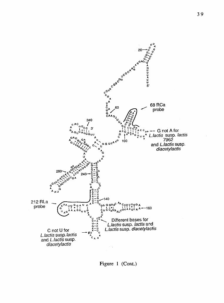
212 RLaprobe
c20& 0a. u.u,
G AC,AG A
Ar4cA -
GUC
GG
GC'
cGUG
CuA
UA
CGAA A G
60U
340 uU ii4, GA o
GAGuCA,., I
A GG,..G/I 1 ,
CC 3.GC 0I CU u,*
+ ""-- G not A forCAC
GuuA C G of Ai Al T AI C C
GAG AcG A a
A G, GA c A AG 100
G ,te U UA Llactis susp. lactis.Gu tpu U A
c //GA A
diacetylactisA G
U u
C ..... AAA 0 u GC
280---, GG Au G UA cG C-GGr..k u 240---c-G
C'"- c- GCA GUU G A
Li AAAUGGCU:AGUG U U
GCG
GA CA G -C
A AU A- UA- uA LIcG,-140UA
A C - G,c UGCAU C AA
'''GG G GGAc cauuuGG AG I I 1 1 I
uAGAA AGUuuG CG CCAUA eGUI I I I I I I I I
A AGc A k"--160A A AU
UUUAUU
5'
68 RCaprobe
7962cc,;, % c GG GC
G C u and L.lactis susp.
C not U forLlactis susp. lactisand Llactis susp.
diacetylactis
A
Ut AUUAC '"\ Different bases forLlactis susp. lactis and
',;=8 Llactis susp. diacetylactisA
UA
C A A
Figure 1 (Cont.)
39

40
5' domain of the Lactococcus lactis subsp. cremoris 16S rRNA. Sites
of variations within the Lactococcus genus are indicated. The partial
sequences of lactic acid bacteria are shown aligned in Figure 2.
Construction of probes and hybridization experiments.
Based on the analysis of the partial sequence information, two
phylogenetic probes were designed and synthesized, a subspecies-
specific rRNA probe for L. lactis subsp. cremoris, and a species-
specific probe for the lactococci. The sequences of both probes are
indicated in Table 1. The species-specific probe is 22 nucleotides in
length and is located at positions 212 to 233 of the 16S rRNA. This
probe was used to identify members of the lactococci by
hybridization of the probe to bulk cellular RNA. Strong, specific
hybridization to the probe was noted for all the lactococci examined
(Fig. 3). On the other hand, no cross reactivity was seen when the
probe was tested against other eubacterial (Dermocarpa PCC 7437,
Myxosarcina PCC 7312, and Pseudomonas aeruginosa strain IUCC
SXI), archaebacterial (Halobacterium volcanii), and eukaryotic
(Strongylocentrotus purpureus) RNAs (Fig. 3). Identical results were
obtained for whole cell hybridizations. Specific hybridization of the
212RLa probe was observed to all lactococcal bacterial strains (Fig.
4). However, L. lactis subsp. lactis 7962 hybridized to the probe
weakly. The number of cells was increased 4-fold for L. lactis subsp.
lactis 7962 to give a signal approximately equivalent to the other
strains. The 212RLa probe did not bind to any of the control strains,
which included Enterococcus pyogenes, Enterococcus faecalis,

41
Fig. 2. Nucleotide sequences of 5' regions of lactic acid bacteria
16S rRNAs. Points indicate nucleotide identity with L.
lactis subsp. cremoris 205. The accumulated positions are
given in the right margins. Lowercase letters indicate
uncertainty in the determination.
Lc: L. lactis subsp. cremoris, Ll: L. lactis subsp. lactis, Ld:
L. lactis subsp. lactis biovar. diacetylactis.

LcLcLcLcLc
205BK5107/6P2HP
UUAUUUGAGAGUUUGAUCCU GGCUCAGGACGAACOCUGGC GGCGUGCCUAAUACAUOCAA GUUGAGCGAUGAAGAUUOGU OCUUGCACCAAUUUGAAGAG 100100100100100
LI 11454 C 0 A....0 ..... C.0..U... 100Ll 7962 C 0 100LI C2 R K 100LI f2d2 C 0 A. .0 ... C.0..U 100Ld DRC-1 C 0 100Ld 18-16 C 0 100Ld 26-2 C 0 100
Lc 205 CAGCgAACGGGUGAGUAAC0 COUgGGGAAUCUGCCUUUGA GCGGOGGACAACAUUUGGAA ACOAAUGCUAAUACCOCAUA ACAACUUUAAACAUAAGUUtt 200Lc BK5 200Lc 107/6 200Lc P2 200Lc HP 200LI 11454 144LI 7962 A C 200LI C2 N 161LI f2d2 A C 200Ld DRC-1 A C 200Ld 18-16 A C 200Ld 26-2 A C 200
Lc 205 UAAGUUUGAAAGAUGCAAUU OCAUCACUCaAAGAUgAuCC CGCGUUGuaUUAGCUAGUUG GUGAGGUaAAGGCUCACCaA GOCOAUGAuACAUAGCCGACLc BK5 300Lc 107/6 300Lc P2 300Lc HP 300LI 7962 300LI f2d2 300Ld DRC-1 300Ld 18-16 300Ld 26-2 300
Lc 205 CUGAGAGGGUgAUcGGCCAC auuGGOACuGAGACACGOCC 340Lc BK5 312Lc 107/6 312Lc P2 312Lc HP 324LILi
7962f2d2
312340
Ld DRC-1 326Ld 18-16 326Ld 26-2 326

1406Ra b
1
2345678 ak9
212RLaa b
68RCa 1406Fa b
43
a b
Fig. 3. Autoradiogram of a dot blot hybridization to bulk cellular
RNAs from lactic acid bacteria and control strains. The
universal (1406R), species-specific (212RLa), subspecies-
specific (68RCa) and a negative control (1406F) probe
were used. The order of the blotted RNAs is: la-5a (L.
lactis subsp. cremoris BK5, 107/6, 205, p2, and HP); 6a-9a
and lb (L. lactis subsp. lactis 11955, 11454, 7962, C2, and
f2d2); 2b-4b (L. lactis subsp lactis biovar. diacetylactis
DRC, 18-16, and 26-2); 5b (Dermocarpa PCC 7437); 6b
(Myxosarcina PCC 7312); 7b (Strongylocentrotus
purpureus); 8b (Halobacterium volcanii); and 9b
(Pseudomonas aeruginosa IUCC SXI).

44
Staphylococcus epidermidis, Salmonella pullorum, and Bacillus
subtilis. The binding of the 1406R universal probe was used as a
positive control for the presence of detectable target sequence. An
oligonucleotide that is not complementary to the rRNA (1406F)
served as a control for non-specific binding (Fig. 3 and 4).
The subspecies-specific probe (68RCa) was complementary to a20-base pair region located at positions 68 to 87 of a highly variable
domain. This probe was designed to discriminate L. lactis subsp.
cremoris from other lactococci. In RNA-DNA hybridization
experiments, this probe bound specifically and efficiently to the
RNAs (Fig. 3), as well as to fixed whole cells (Fig. 4) of the five L.
lactis subsp. cremoris strains. All of the control strains, including the
other lactococci related to the subspecies cremoris, failed to hybridize
to the 68RCa probe. The only exception was L. lactis subsp. lactis C2,
which hybridized to the 68RCa probe on all occasions, as predicted
from sequencing studies. A different source of this strain confirmed
these results, indicating that strain C2 has the same sequence as the
subsp. cremoris at the homologous positions. L. lactis subspecies
lactis strain ATCC 11955 hybridized weakly to the 68RCa probe. This
might be attributed to non-specific binding. The sequence of the
16S rRNA of this strain at the probe site has not yet been
determined.

1406Ra b
1
23456789 We
212RLaa b a b
.1
.41:
45
68RCa 1406Fa b
Fig. 4. Autoradiogram of a dot blot hybridization to fixed whole
cells of lactic acid bacteria and control strains. The order
of the blotted cells was same as in Fig. 2 for the lactic acid
bacteria. The control strains, Enterococcus pyogenes,
Enterococcus faecalis, Staphylococcus epidermidis,
Salmonella pullorum, and Bacillus subtilis, were blotted in
wells 5b-9b respectively. All control strains were
obtained from the Department of Microbiology culture
collection, Oregon State University. The number of cells
was increased 4-fold for L. lactis subsp. lactis 7962 to
give a signal approximately equivalent to the other
strains.

46
DISCUSSION
Because of its rapidity and technical simplicity, the reverse
transcriptase sequencing method was helpful for determining 16S
rRNA partial sequences from the 13 lactococcal strains. The 16S
rRNAs of the lactococcal strains showed a high degree of similarity.
However, among the eight L. lactis subsp. lactis and diacetylactis
biovar. strains studied, only the subsp. lactis C2 had the same
nucleotide sequence as that of the subspecies cremoris at the position
of the probe target, and thus hybridized strongly. The two strains of
L. lactis subsp. lactis C2 originated in Australia; from there they have
been dispersed to other laboratories. Phenotypically, the strain
behaves like the subspecies lactis. However, the 16S rRNA sequence
of the strain resembles that of the subspecies cremoris. It is possible
that the cremoris phenotype could have evolved naturally from the
subspecies lactis, in association with dairy-related practices, by the
loss of certain phenotypic traits. Alternatively, there is a possibility
that strain C2 originally had the phenotype of the subspecies
cremoris, but has acquired certain traits of the subspecies lactis,
perhaps by means of a transducing phage. In this regard, a
temperate bacteriophage has been found in the C2 strain which
converts lactose-, maltose-, or mannose-negative recipient cells of
this strain to the respective carbohydrate-positive phenotype (13).
The instability of "pure" cultures of lactic acid bacteria, which would
ordinarily be regarded as being constant in properties, has been
reported by Hunter et al. (7). This issue could be resolved in the

47
near future if we succeed in obtaining natural isolates of the
cremoris genotype and study their phenotypic properties in detail.
Nucleic acid hybridization recently was introduced as a rapid
tool for the identification of microorganisms (8, 9, 14). Ribosomal
RNAs are attractive candidates as targets for hybridization probes
due to their unique organization, the presence of highly conserved
and variable regions, and their presence in high copy number.
The small differences between the 16S rRNA sequences of the
lactic acid bacteria were sufficient to allow differentiation between
closely related subspecies. Wallace et al. (25) indicated that
oligonucleotides that differ in sequence at only one position are
potentially useful as sequence-specific probes. The nucleotide
sequence that we selected as target site for the species-specific probe
(212RLa) was unique to the lactococci, as indicated by comparisons to
a data base of more than 200 known eubacterial 16S rRNA
sequences. Furthermore, this was verified by the specific
hybridization of the probe to all 13 lactic acid strains investigated,
but none of the control organisms. A 3-base-pair mismatch in the
oligonucleotide probe (68RCa) of 20 base pairs was sufficient to
discriminate the subspecies cremoris from the closely related
subspecies lactis and its diacetylactis biovar.
The relatively small size of the oligonucleotide hybridization
probes used in our study minimizes problems of cellular
permeability and access to binding sites. However, the amount of
probe that is specifically bound may be influenced by many
variables, including the permeability of fixed cells and the
accessibility of the rRNAs in fixed cell preparations (5). One or more
48
of such variables could account for the weak hybridization between
L. lactis subsp. lactis 7962 fixed whole cells and the genus- specific
probe (212RLa), as opposed to a much stronger signal of the same
strain when bulk cellular RNA was hybridized to the probe.
The hybridization probes described here provide a highly
sensitive and specific means for the rapid detection and
identification of lactic acid bacteria in general and L. lactis subsp.
cremoris in particular. The use of these probes may contribute
substantially to the isolation and study of new strains of the
subspecies cremoris from natural habitats.

49
ACKNOWLEDGEMENTS
We thank Dr. Katharine Field for her advice and efforts during
the early stages of this work. This work was supported by a grant
from the National Dairy Board, and the OSU Agricultural
Experimental Station, of which this is technical report number 9342.

50
REFERENCES
1. Brosius, J., J. L. Palmer, J. P. Kennedy, and H. F. No ller.
1978. Complete nucleotide sequence of a 16S rRNA gene from
Escherichia. Proceedings of the Natl. Acad Sci., USA
75:4801-4805.
2. Collins, M. D., C. Ash, J. A. E. Farrow, S. Walbanks, and
A. M. Williams. 1989. 16S Ribosomal ribonucleic acid
sequence analyses of lactococci and related taxa. Description of
Vagococcus flavialis gen. nov., sp. nov. J. Appl. Bacteriol.
67:453-460.
3. De Long, E. F., G. S. Wickham, and N. R. Pace. 1989.
Phylogenetic stains: Ribosomal RNA-based probes for the
identification of single cells. Science 243:1360-1362.
4. Fenton, M. P. 1987. An investigation into the source of
lactic acid bacteria in grass silage. J. Appl. Bacteriol.
62:181-188.
5. Giovannoni, S. J., E. F. De long, G. J. Olsen, and N. R.
Pace. 1988. Phylogenetic genus-specific oligonucleotide
probes for identification of single microbial cells. J. Bact.
170:720-72.

51
6. Giovannoni, S. J., S. Turner, G. J. Olsen, S. Barns, D.
Lane, and N. R. Pace. 1988. Evolutionary relationships
among cyanobacteria and green chloroplasts. J. Bacteriol.
170:3584-3592.
7. Hunter, G. J. E. 1939. Examples of variation within pure
cultures of Streptococcus cremoris. J. Dairy Research
10:46-470.
8. Izat, A. L., C. D. Driggers, M. Colberg, M. A. Reiber, andM. H. Adams. 1989. Comparison of the DNA probe to culture
methods for the detection of Salmonella on poultry carcasses
and processing waters. J. Food Protection. 52(8):564-57.
9. Kapperud, G., K. Dommarsnes, M. Skurnik, and E.Hornes. 1990. A synthetic oligonucleotide probe and a cloned
polynucleotide probe based on the Yop A gene for detection
and enumeration of virulent Yersinia enterolitica. J. Appl.
Environ. Microbiol. 56(1):17-23.
10. Lane, D. L. B. Pace, G. J. Olsen, D. Stahl, M. L. Sogin, and
N. R. Pace. 1985. Rapid determination of 16S ribosomal RNA
sequence for phylogenetic analysis. Proc. Natl. Acad. Sci. USA
82:6955-6959.

52
11. Lane, D. L., K. G. Field, G. J. Olsen, and N. R. Pace. 1988 .
Reverse transcriptase sequencing of ribosomal RNA for
phylogenetic analysis. Meth. Enz. 167:138-144.
12 . Lawrence, R. C., T. D. Thomas and B. E. Terzaghi. 1976 .
Reviews of the progress of dairy science: Cheese starters. J.
Dairy Res. 43 :141-193.
13. McKay, L. L. 1982. Regulation of lactose metabolism in
dairy Streptococci. Developments in Food Microbiology-1. ed.
R. Davies. Applied Science Publisher LTD. pp. 153-182.
14. Olsen, G. J., D. J. Lane, S. J. Giovannoni, and N. R. Pace.1986. Microbial ecology and evolution: a ribosomal RNA
approach. Ann. Rev. Microbiol. 40:337 -365.
15. Pace, N. R., R. J. Stahl, D. J. Lane, and G. J. Olsen. 1986.
The analysis of natural microbial populations by ribosomal RNA
sequences. Adv. Microb. Ecol. 9:1-55.
16. Rhenstam, A., A. Norqvist, H. Wolf-watz, and A.
Hagstorm. 1989. Identification of Vibrio anguillarum in fish
by using partial 16S rRNA sequences and a specific 16S rRNA
oligonucleotide probe. Microbiol. 55:1907-1910.

53
17. Sandine, W. E., P. C. Radich, and P. R. Elliker. 1972.
Ecology of the lactic streptococci: A review. J. Milk Food
Technol. 35:176-185.
18. Schieblich, M. and W. Schonherr. 1957. Streptococci in
acid fodders and their distribution in the raw material.
Tierernahr. Futtermittelk 11:232 (Dairy Sci. Abstr. 19: 231).
19. Schleifer, K. H. and R. Klipper-Blaz. 1987. Molecular and
chemotaxonomic approaches to the classification of streptococci,
enterococci, and lactococci: A review. System. Appl. Microbiol.
10:1-19.
20. Sgaramella, V., and H. G. Khorana. 1972. Total synthesis
of the structural gene for an alanine transfer RNA from yeast.
Enzymatic joining of the chemically synthesized
polynucleotides to form the DNA duplex representing
nucleotide sequence 1 to 20. J. Mol. Biol. 72:427-444.
21. Stackebrandt, E. and M. Teuber. 1988. Molecular
taxonomy and phylogenetic position of lactic acid bacteria.
Biochimie. 70:317-324.
22. Terzaghi, B. E. and W. E. Sandine. 1975. Improved
medium for lactic streptococci and their bacteriophage. Appl.
Microbiol. 29:807-813.

54
23. Vedamuthu, E. R., W. E. Sandine and P. R. Elliker. 1966.
Flavor and texture in Cheddar cheese. I. Role of mixed strain
lactic starter cultures. J. Dairy Sci. 49(2):144-150.
24. Vedamuthu, E. R., W. E. Sandine and P. R. Elliker. 1966.Flavor and texture in Cheddar cheese. II. Carbonyl compounds
produced by mixed strain lactic starter cultures. J. Dairy Sci.
49(2):151-157.
25. Wallace, R. B., J. Shaffer, R. F. Murphy, J. Bonner, T.Hirose, and K. Itakura. 1979. Hybridization of synthetic
oligodioxyribonucleotides to 0X174 DNA: the effect of a single
base pair mismatch. Nucleic Acids Res. 6:3543-3557.
55
CHAPTER 3
Isolation of Lactococci From Nature by ColonyHybridization With Ribosomal RNA Probes
Maysoon S. Salama, William E. Sandine, and Stephen J. Giovannoni
Department of Microbiology, Oregon State University
Corvallis OR 97330-3804

56
ABSTRACT
Previously we described oligonucleotide probes based on
unique sequence domains in 16S rRNAs which could be used to
discriminate L. lactis subsp. cremoris from related strains. These
probes were used in colony hybridizations to rapidly screen large
numbers of colonies. Inocula from green plant surfaces and raw milk
samples were enriched in skim milk and plated on PMP agar (19).
Colonies were lifted from plates with poly-l-lysine coated glass micro
fiber filters, treated with sodium dodecyl sulfate, and hybridized to
four oligonucleotide probes: 68RCa, specific for L. lactis subsp.
cremoris; 212RLa, specific for L. lactis species; 1406R, universal
probe used as positive control; and 1406F, negative control probe.
The method discriminated Lactococcus lactis subsp. cremoris from
other closely related Gram positive organisms, such as L. lactis subsp.
lactis, L. lactis subsp. lactis biovar. diacetylactis, and Enterococcus
faecalis, in mixed culture. The colony hybridization approach
described here generally will be applicable to screening large
numbers of colonies from natural environments for specific rRNA
genotypes.

57
INTRODUCTION
Dairy lactococci have been used for centuries in the production
of fermented dairy products. The need to isolate new Lactococcus
lactis subsp. cremoris starter culture strains has been emphasized by
cheese makers, industry consultants, and research workers (10, 15).
Undesirable flavors encountered in cultured dairy products,
insufficient development of acid during fermentation, and frequent
culture failures resulting from virus infection are some factors which
have contributed to the need for new strains of Lactococcus lactis
subsp. cremoris.
The isolation of bacteria from nature traditionally has been
dependent on the design of selective media and the screening of
isolates using arrays of tests for phenotypic characteristics. Among
the commercially significant subspecies of Lactococcus lactis,
traditional methods for isolation and identification limit the number
of new strains which can be evaluated. Various studies have
emphasized the virtual impossibility of isolating new strains of L.
lactis subsp. cremoris from nature with conventional approaches (7,
15, 16). Two alternative explanations for the failure to isolate new
strains of L. lactis subsp. cremoris by these approaches are: 1) that
Lactococcus lactis subsp. cremoris is very rare in nature, or 2) that
these strains do not occur in nature at all but co-evolved in
association with dairy practice.
In a previous study (13) we described a 16S rRNA-targeted
species-specific probe (212RLa) for L. lactis and a subspecies-specific
58
probe (68RCa) for L. lactis subsp. cremoris. Lactococcus strains
hybridized to these probes as predicted, with the exception of an
atypical strain, L. lactis subsp. lactis C2, which had the same
nucleotide sequence as L. lactis subsp. cremoris at the positions of
the 68RCa probe target, and thus hybridized strongly. Since then, we
have sought to develop a colony hybridization technique which
would allow the direct identification of the L. lactis subsp. cremoris
rRNA genotype in a mixed environmental population.
Here we describe a colony hybridization method which permits
large numbers of isolates from natural environments to be screenedefficiently. The method is analogous to approaches used routinely in
molecular biology to screen libraries of molecular clones, and offers
similar advantages to microbiologists seeking to identify rare cellularclones in large populations.

59
MATERIALS AND METHODS
Bacterial strains and environmental samples. The
bacterial strains used as controls for colony hybridization
experiments and whole cell dot blot hybridizations were obtained
from the Oregon State University (OSU) culture collection. The non-
starter lactococcal strains, Lactococcus gravieae, Lactococcus
plantarum, and Lactococcus raffinolactis, were ATCC strains.
Lactococcus lactis subsp. hordniae was obtained from Dr. R. E.
Kunkee, College of Agricultural and Environmental Sciences,
University of California, Davis. Lactococcus piscium was obtained
from Dr. R. L. Holt, Microbiology Department, OSU. All strains were
either patched on M17 agar plates (19) or grown in M17 broth at300C. Environmental enrichments were obtained from a plantgrowing on the OSU campus (Prunus laurocerasus) and a fresh corn
sample from local produce. Weighed portions of collected material
were added to stomacher bags (Lab-Blender 400, Tekmar company,
Cincinnati, Ohio) containing 99 ml of sterile 11% skim milk and
stomached for 1 min. The bags were then held at 210C for 2 days to
enrich for lactococci. Dilutions of the enrichments were spread on
the surfaces of PMP agar plates, a modified formulation of M17 (19)
using the insoluble trimagnesium phosphate instead of disodium-B-
glycerophosphate. Plates were incubated anaerobically at 210C for 2to 3 days. For the plant samples, several bright yellow colonies
surrounded by halos of clearing contrasted against the purple
medium were picked, streaked for purification, and patched on PMP

60
plates. Dilutions from the corn sample enrichment were directly
spread onto large (150 X 15 mm) PMP agar plates and the plates
were incubated anaerobically at 210C for 2 to 3 days. The large
plates allowed the use of lower dilutions of the sample with less
crowded plates, thus facilitating return to those colonies needed for
further investigation following the colony hybridizations. Colonies
from the corn sample enrichment were lifted directly from PMP agar
plates with glass microfibre filters, and screened by colony
hybridization. After colony lifting, the plates were reincubated for
an additional 2 days to allow regrowth.
RNA-DNA colony hybridization. Colonies were lifted from
M17 or PMP agar plates onto glass microfibre filters (Whatman no.
934-AH). The glass fiber filters were previously soaked in poly-L-
lysine (50 mg /ml in 10 mM Tris-HC1, pH 8), air dried and sprayed on
the back with a thin layer of acrylic spray (Krylon 1301, Crystal Clear
Acrylic) for mechanical support during subsequent treatments.
Colony hybridizations were performed using a modified combination
of the Anderson and McKay (2) and Kapperud (8) methods. In the
modified procedure we describe, the glass fiber filters with the liftedcolonies were placed, colony side up, on Whatman no. 1 paper soaked
with 5% SDS and left for 3 to 5 min. The filters were then dried at
800C in a vacuum oven for 10 to 15 min. After this treatment, 5-10ml of prehybridization buffer (6x SSPE [1.08 M NaC1, 60 mM NaPO4,
and 60 mM EDTA, pH 7.5], 5x Denhardt's solution [0.1% Ficol, 0.1%
polyvinylpyrolidone, and 0.1% BSA], and 0.1% SDS) were added to the
filters in a Petri plate or HB-1 hybridizer (Techne Ltd., Duxford,

61
Cambridge CBZ 4PZ. U. K). Prehybridization was carried out for 45 to
60 min at 650C. The prehybridization buffer was then replaced with
5-7 ml of hybridization buffer (6x SSPE, lx Denhardt's solution, 0.1%
SDS, and approximately 106 cpm of 32P-labeled probe). The
hybridization was carried out at room temperature overnight. Filters
were washed 3 times for 20-30 min at room temperature in 6x SSPE,
0.1% SDS, then one time at the predetermined stringency
temperature (450C for 212RLa probe, 500C for 68RCa probe and 370C
for both 1406R and 1406F probes). Filters were then exposed to X-
ray film for 6 to 24 h.
Glass fiber filters with lifted colonies were hybridized three
times to three different probes in the following order: 68RCa,
212RLa, 1406R. After each hybridization and X-ray film exposure,
the filter was washed at 650C for 4 to 5 h, then exposed to X-ray film
to insure that all of the hybridized probe was washed off completely
before performing the next hybridization.
Whole-cell dot blot hybridization. After regrowth of the
lifted colonies on the PMP plates, those cells which hybridized to the
212RLa probe or both 68RCa and 212RLa probes were picked and
streaked for purification on PMP plates. The pure colonies were
grown in M17 broth for whole-cell dot blot hybridization to confirm
their relatedness to lactococci. Whole cells were hybridized to
oligonucleotide probes as described previously (13).
Phenotypic characterization. The environmental isolates
which were proved to belong to the Lactococcus genus by genotypic

62
testing were further tested phenotypically for arginine hydrolysis
and gas production from citrate utilization using the differential
broth described by R. S. Reddy and co-workers (11), growth at pH
9.2, growth in 4% NaC1 and growth at 400C. These criteria are very
important for distinguishing lactococci from other related
microorganisms as well as differentiating between L. lactis subsp.
lactis and subsp. cremoris.
63
RESULTS AND DISCUSSION
Different approaches were considered (2, 6, 8, 12) in attempts
to render bacterial cells permeable to nucleic acid probes and to
develop and optimize a method of rRNA-DNA colony hybridization.
From the results obtained with modifications of the methods of Beitz
and co-workers (3), Anderson and McKay (2) and Kapperud (8), we
arrived at an approach which used glass fiber filters to detect
colonies of Gram positive bacteria, particularly the dairy lactococci.
The resolution of the autoradiograms with glass fiber filters was
greater than with nitrocellulose, Nytran, or Zeta probe membranes
using two different procedures for colony hybridizations (data notpresented).
The results of a colony hybridization experiment using the method
we developed are shown in Fig. 1. The cells were patched on PMP
agar plates, incubated, and then lifted onto glass fiber filters as
described earlier. Filters were hybridized to 4 different probes(Table 1). The 1406R, a universal probe complementary to a highlyconserved region of 16S rRNAs, was used as positive control. All
strains hybridized to this probe, indicating that the cells were
efficiently lifted and permeabilized by the treatment (Fig. la). The
1406F, a universal probe complementary to the 1406R probe, wasused as a negative control for non-specific binding (Fig. lb).
Hybridization of the 212RLa probe, specific for the L. lactis species, isshown in panel lc. This probe hybridized to L. lactis subsp. lactis

S. pyogenes
E. faecalis
S. epidermidis
L. lactis
L. cremoris
S. pullorum
B. subtilis
S. pyogenes
E. faecalis
S. epidermidis
L. I act i s
L. cremoris
S. pullorum
B. subtilis
lal 1406R
lc] 212RLa
(131 406F
64
Id) 68RCa
Is bomr
Fig. 1. Colony hybridization to bacteria on glass fiber
filters. (a) Cells hybridized to the universal probe
1406R; (b) cells hybridized to the negative control
1406F; (c) cells hybridized to the species-specific probe
212RLa; (d) cells hybridized to the subsp.-specific probe
68RCa. The bacterial strains included: Streptococcus
pyogenes, Enterococcus faecalis, Staphylococcus
epidermidis, Lactococcus lactis subsp. lactis, Lactococcus
lactis subsp. cremoris, Salmonella pullorum, and Bacillus
subtilis. The dots in each row are replicates of one strain
of the species indicated.

TABLE 1. List of oligonucleotides used.
Oligo.# Target Position* Sequence
1406F negative control 1391-1406 1GYACACACCGCCCGT1406R universal 1406-1392 ACGCGCGGTGTGIRC212RLa lactococci 233-212 CITTGAGTGATGCAATTGCATC68RCa L. cremoris 87-68 TGCAAGCACCAATCTTCATC
R ReverseF: Forward# Oligonucleotide* Position numbers refer to E. coli 16S rRNA, Brosius et. al. (5).

66
and L. lactis subsp. cremoris, but not to Streptococcus pyogenes,
Enterococcus faecalis, Staphylococcus epidermidis, Salmonella
pullorum, or Bacillus subtilis. The specificity of the 68RCa probe for
the subsp. cremoris is illustrated in Fig. ld. Of the known strains
tested, only L. lactis subsp. cremoris hybridized to this probe.
To ascertain whether the probes were able to differentiate
lactococci and subsp. cremoris from other strains occurring in
natural populations, several environmental isolates from a plantsample enrichment (Prunus laurocerasus) were patched on PMP agar
together with known L. lactis subsp. lactis and L. lactis subsp.
cremoris strains. After incubation, colonies were lifted onto glass
fiber filters and hybridized to three probes: 1406R, 212RLa, and
68RCa. As documented in Fig. 2, the environmental isolates
hybridized to the 1406R universal probe, as did L. lactis subsp. lactisand L. lactis subsp cremoris. (Fig. 2a). However, in this experiment
the environmental isolates did not hybridize with either the 212RLa
or 68RCa probes, while the subsp. cremoris showed specific binding
to both probes and subsp. lactis hybridized to the 212RLa probe, but
not to the 68RCa probe (Fig. 2b and 2c). This indicated that none of
the environmental isolates from this sample were L. lactis. Furthertesting of these isolates supported this finding. Unlike lactococci,
these isolates were able to grow at 450C. Thus, the probes were able
to discriminate between L. lactis and other organisms appearing in
these enrichment cultures.
The first environmental sample harboring microbial flora that
would hybridize to both probes (212RLa and 68RCa probes) was afresh corn sample (Fig. 3). Thirty percent of the colonies which

la I 1406R_ bl 212RLa
.45
67
1 c 1 68RCa
Fig. 2. Colony hybridization of L. lactis subsp. lactis, L. lactis
subsp. cremoris, and unknown environmental isolates
from a plant sample (Prunus laurocerasus). (a)
Hybridization to the universal probe 1406R; (b)
hybridization to the species-specific probe 212RLa; (c)
hybridization to the subsp.-specific probe 68RCa. Lane 1,
L. lactis subsp. lactis (the four dots represent one strain);
lane 2, L. lactis subsp. cremoris (the four dots represent
one strain); lanes labeled 3, environmental isolates (each
dot represents a different isolate).

la11406R
. .
. .
1bl 212RLa
. I,.0.
68
Fig. 3. Colony hybridization of environmental flora from a
fresh corn sample. The sample was plated on a PMP agar
plate after enrichment, incubated anaerobically, lifted
onto a sterile glass fiber filter, and hybridized three times
to three different probes. (a) Hybridization to the
universal probe 1406R; (b) hybridization to the species-
specific probe 212RLa; (c) hybridization to the subsp.-
specific probe 68RCa. The glass fiber filter was washed at
650C after each hybridization to prepare it to be
hybridized to the next probe. Arrows indicate filter
orientation marks.

69
hybridized to the universal 1406R probe (Fig. 3a), also hybridized to
the 212RLa probe. Six (about 60%) of the colonies which hybridized
to the 212RLa probe also hybridized to the 68RCa probe, indicating
that these were lactic acid bacteria of the L. lactis subsp. cremoris
rRNA genotype (Fig. 3c). All of the colonies (Co-1 through Co-6)
which hybridized to the 68RCa probe were picked from the
reincubated original agar plate, streaked for purification and
confirmed for positive hybridization to this probe by means of
whole-cell dot blot hybridization (13, Table 2). These strains had the
phenotypic characteristics of L. lactis subsp. lactis. They were
positive for arginine dihydrolase, growth at pH 9.2, growth in 4%
NaC1, and growth at 400C (Table 2). In this respect they resemble L.
lactis subsp. lactis strain C2, which has the rRNA genotype of L. lactis
subsp. cremoris, but resembles L. lactis subsp. lactis in phenotype
(13). In a recent report (1), it was found that lithium ion inhibited
the growth of L. lactis subsp. cremoris strains and L. lactis subsp.
lactis strain C2, but not other L. lactis subsp. lactis strains. We have
found that natural isolates from the corn sample which hybridized to
the 68RCa probe were not sensitive to lithium inhibition. Further
screening of environmental samples will be needed to fully
understand the relationships among the subspecies of L. lactis.
While the cremoris and lactis subspecies are the only lactococci
used as starters to manufacture fermented milk products, other
lactococci are recognized (14). To gain further knowledge of the
molecular systematics of all members of the Lactococcus genus,
additional rRNA probing of these non-starter species was carried out.
Our findings support the recent classification of the

TABLE 2. Genotypic and phenotypic characteristics of some of the environmental strains isolated froma fresh corn sample using the colony hybridization procedure with 32P-labeled 16S rRNA probes.All strains are Gram positive cocci and acidify litmus milk before coagulation.
Isolate #Hybridization to:68RCa 212RLa
Source:100C
Growth at400C 450C NaCl pH
Hydrolysisof Arginine
4% 9.2
L. lactis + Laboratory strain + + + + +L. crem* + + Laboratory strain +Col + + corn + + + + +Co2 + + corn + + + + +Co3 + + corn + + + + +Co4 + + corn + + + + +Co5 + + corn + + + + +Cob + + corn + + + + +
* L. lactis subsp. cremoris# Whole-cell dot blot hybridization
0

71
lactococci (17). When hybridized to the 68RCa probe, L. lactis
subsp. hordniae, L. gravieae, L. plantarum, and L. raffinolactis (all
non-starter lactococci) failed to hybridize to this cremoris
subspecies-specific probe. However, only L. lactis subsp. hordniae
was able to hybridize to the 212RLa probe which is specific to the
Lactococcus lactis species. Lactococcus piscium was recently
described as a new Lactococcus species (20). This organism did not
hybridize to either the 68RCa or the 212RLa probes as expected.
We have described a colony hybridization approach which may
provide a general method for the isolation of bacteria from natural
environments using glass microfibre filters and ribosomal RNA
probes. We have presented a specific use of this technique for the
general identification and isolation of the L. lactis species from mixed
cultures. The technique may also be applicable to the isolation of
novel species from nature using rRNA genetic markers cloned from
natural microbial populations (4, 9, 18).

72
ACKNOWLEDGEMENTS
We thank Dr. R. E. Kunkee, College of Agricultural and
Environmental Sciences, University of California, Davis, for sending us
a L. hordniae strain. We are grateful to Dr. R. L. Holt, Microbiology
Department, OSU, for providing a Lactococcus piscium strain.
This work was supported by a grant from the National Dairy
Promotion and Research Board, and the OSU Agricultural Experiment
Station.

73
REFERENCES
1. Al-Zoreky, N., and W. E. Sandine. 1991. The Lactococcus
genus: A selective and differential agar medium. J. Food Sci.
56(6):1729-1730 & 1734.
2. Anderson, D. G., and L. L. McKay. 1983. Simple and rapid
method for isolating large plasmid DNA from lactic streptococci.
Appl. Environ. Microbiol. 46:549-552.
3. Betzl, D., W. Ludwig, and K. H. Schleifer. 1990.
Identification of lactococci and enterococci by colony
hybridization with 23S rRNA-targeted oligonucleotide probes.
Appl. Environ. Microbiol. 56(6):2927-2929.
4. Britschgi, T. B., and S. J. Giovannoni. 1991. Phylogenetic
analysis of a natural marine bacterioplankton population by
rRNA gene cloning and sequencing. Appl. Environ. Microbiol.
57:1707-1713.
5. Brosius, J., J. L. Palmer, J. P. Kennedy, and H. F.No ller. 1978. Complete nucleotide sequence of a 16S rRNA
gene from Escherichia. Proceedings of the Natl. Acad Sci., USA
75:4801-4805.

74
6. Buluwela, L., A. Foster, T. Boehm, and T. H. Rabbits.1989. A rapid procedure for colony screening using nylon
filters. Nucleic Acid Res. 17:452.
7. Fenton, M. P. 1987. An investigation into the source of
lactic acid bacteria in grass silage. J. Appl. Bacteriol.
62:181 -188.
8. Kapperud, G., K. Dommarsnes, M. Skurnik, and E.Hornes. 1990. A synthetic oligonucleotide probe and a
cloned polynucleotide probe based on the YopA gene for
detection and enumeration of virulent Yersinia
enterolitica. J. Appl. Environ. Microbiol. 56(1):17-23.
9. Liesack, W. and E. Stackebrandt. 1992. Occurrence
of novel groups of the domain bacteria as revealed by
analysis of genetic material isolated from an Australian
terrestrial environment. J. Bacteriol. 174(15):5 07 2-5078.
10. Radich, P. C. and W. E. Sandine. 1968. An ecological
study of the lactic acid streptococci. M. S. thesis. Oregon State
University.
11. Reddy, M. S., E. R. Vedamuthu, and G. W. Reinbold.1971. A differential broth for separating the lactic
streptococci. J. Milk and Food Tech. 34(1):43-45.

75
12. Rhenstam, A., A. Norqvist, H. Wolf-watz, and A.Hagstorm. 1989. Identification of Vibrio anguillarum in
fish by using partial 16S rRNA sequences and a specific 16S
rRNA oligonucleotide probe. Microbiol. 55:1907-1910.
13. Salama, M. S., W. E. Sandine, and S. J. Giovannoni.1991. Development and application of oligonucleotide probes
for identification of Lactococcus lactis subsp. cremoris. Appl.
Environ. Microbiol. 57:1313-1318.
14. Sandine, W. E. 1988. New nomenclature of the non-rod
shaped lactic acid bacteria. Biochemie 70:519-522.
15. Sandine, W. E., P. C. Radich, and P. R. Elliker. 1972.
Ecology of the lactic streptococci: a review. J. Milk Food
Technol. 35:176-185.
16. Schieblich, M. and W. Schonherr. 1957. Streptococci in
acid fodders and their distribution in the raw material.
Tierernaehr. Fuetter 11:232.
17. Schleifer, K. H. and R. Kilpper-Balz. 1987. Molecular and
chemotaxonomic approaches to the classification of streptococci,
enterococci, and lactococci: A review. System. Appl. Microbiol.
10:1-19.

76
18. Schmidt, T. M., E. F. De long, and N. R. Pace. 1991.
Analysis of a marine picoplankton community by 16S rRNA
gene cloning and sequencing. J. Bacteriol. 173 (14 ) : 4371
4378 .
19. Terzaghi, B. E., and W. E. Sandine. 1975. Improved
medium for lactic streptococci and their bacteriophage. Appl.
Microbiol. 29:807-813.
20. Williams, A. M., J. L. Fryer, and M. D. Collins. 1990.
Lactococcus piscium sp. nov. a new Lactoccoccus species from
salmonid fish. FEMS Microbiol. let. 68:109-114.

77
CHAPTER 4
Isolation of New Strains of Lactococcus lactis
subspecies cremoris
Maysoon S. Salama, Tamara Musafija-Jeknic,
William E. Sandine, and Stephen J. Giovannoni
Department of Microbiology, Oregon State University,
Corvallis OR 97330-3804.

78
ABSTRACT
Previously (25) we described a colony hybridization method
for isolation of bacteria from natural environments using rRNA
probes. Here we report the successful use of the method in the
isolation of dairy lactococci.
Thirty-eight plant species and vegetables, twenty-one
individual raw milk and milk product samples from the United
States, China, Morocco, and Yugoslavia, and twelve other samples
from dairy farms were examined for lactic acid bacteria by thecolony hybridization method. Two types of probes were used for
hybridization, the 68RCa (specific for L. lactis subsp. cremoris ) and
212RLa (specific for L. lactis species).
Lactococcus lactis subsp. lactis was found to occur on potato,
cucumber, sweet peas, beans, cantaloupe, corn, cow's body and tail,
colostrum, goat and cow raw milk, cottage cheese and cream.
Lactococcus lactis subsp. lactis biovar. diacetylactis was isolated from
cow raw milk obtained from Morocco and goat raw milk as well ascottage cheese from Yugoslavia. Many strains which hybridized to
the 68RCa probe, and therefore suspected to be L. lactis subsp.
cremoris, surprisingly were phenotypically like L. lactis subsp. lactis.These were isolated from fresh corn, Chinese raw milk, and
Yugoslavian cottage cheese and cream. Some isolates, however, were
typical of L. lactis subsp. cremoris.
The phenotypical, morphological, and physiological
characteristics of the newly isolated lactococcal strains generally

79
agreed with the standard description for the genus. They were able
to grow at 100C and at 400C, but not at 450C; litmus milk was
acidified and completely reduced before curding, no gas production
was observed in the coagulated milk. However, some of the strains
of the subspecies lactis, cremoris, and L. lactis subsp. lactis biovar.
diacetylactis had a slightly different phenotype in regard to ability to
grow at 400C, in 4% NaC1, at pH 9.2, and ability to hydrolyze arginine.
Acid producing properties of the culture collection were
variable. Many of the strains were fast acid producers, most were
acceptable for flavor when grown in milk; and a few were with malty
flavor which is considered a defect.
The isolation of L. lactis subsp. lactis from different plant
sources confirmed that plants are a natural source of this bacterium.
Strains of L. lactis subsp. cremoris were isolated from raw milk and
cottage cheese from Morocco and Yugoslavia, but not from plants.
This suggests that subspecies cremoris strains occur in association
with dairy environments.

80
INTRODUCTION
Lactococcus bacteria are employed as starter cultures for a
variety of industrial dairy fermentations (34). Lactococcus lactis
subsp. lactis and subsp. cremoris are relied upon for acid production
and subsp. lactis biovar. diacetylactis for flavor in cottage cheese,
cultured sour cream and cultured buttermilk.
Lactococci occur in milk (27, 33). The literature provides
evidence that the natural reservoir for the lactococci, especially L.
lactis subsp. lactis and L. lactis subsp. lactis biovar. diacetylactis, is
green plant material. However, the natural habitat of L. lactis subsp.
cremoris is not yet confirmed. In one study of 3000 isolates from 59
samples of commercial raw milk only 4% were L. lactis subsp.
cremoris (17). In another study by Hirch (9), 2 of 35 raw milk
isolates were L. lactis subsp. cremoris. Radich (23) reported
isolation of three L. lactis subsp. cremoris strains from 31 producer
raw milk samples. Unfortunately, most of the reported cremoris
isolates were either lost because they failed to survive frozen storage
or produced acid too slowly for usage in dairy industry. Later
investigators, who had refined differential tests for the identification
of the lactococci, failed in many cases to isolate the organisms from
similar sources. Attempts to isolate L. lactis subsp. cremoris from
plant material have been made by several investigators with no
success (6, 22, 23, 36). Lactococcus lactis subsp. cremoris strains
were reported to have been isolated from frozen peas (4, 5) but
subsequent work (5) showed that these cultures had been incorrectly
identified and were in fact unusual Group N streptococci with

81
properties different from those of both subsp. lactis and cremoris.
The natural habitat of L. lactis subsp. cremoris thus remains
unknown. Therefore studies of their distribution in nature and
relationship to other lactococci are needed.
Several scientists have suggested that L. lactis subsp. cremoris
may only be isolated from nature infrequently (1, 9, 13, 14, 15, 16,
18, 23, 29, 35, 36). Lawrence and co-workers (14) also emphasized
the great need that exists for more strains of L. lactis subsp. cremoris
for use as starter cultures. The limited number of strains of this
subspecies available for the dairy industry have been used
extensively, resulting in intensified problems with bacteriophage
infections. In a previous study (25) we described a 16S rRNA-
targeted species-specific probe (212RLa) for L. lactis and a
subspecies-specific probe (68RCa) for L. lactis subsp. cremoris.
Lactococcus strains hybridized to these probes as predicted, with the
exception of an atypical strain, L. lactis subsp. lactis C2, which had
the same nucleotide sequence as L. lactis subsp. cremoris at the
positions of the 68RCa probe target, and thus hybridized strongly. In
another study (26) we described a colony hybridization method
which permits the direct identification of the L. lactis subsp. cremorisrRNA genotype in a mixed environmental population. This study was
undertaken to: (1) isolate these bacteria from nature with newly
developed screening methods, and (2) characterize the newly
isolated lactococcal strains to determine if they possess suitable acid
producing, flavor, and phage resistance properties for their potential
use in fermented milk product manufacture.
82
MATERIALS AND METHODS
Collection and preparation of samples. Twenty three different
plant species were examined for the presence of lactic acid bacteriaas listed in Table 1. Plant materials were collected from trees,
shrubs, grass, or weeds around the Oregon State University campus
and the Gibson farm. The occurrence of lactococci on vegetables from
local produce markets in the Corvallis area and other types of
environmental samples from the Gibson farm located at Junction City,Oregon, also were studied. The various samples examined are listedin Table 2. Weighed portions of collected material were added to
stomacher bags (Lab-Blender 400, Tekmar company, Cincinnati, Ohio)
containing 99 ml of sterile 11% skim milk and stomached for 1 min.
The bags were then held at 210C for 2 days to enrich for lactococci.
Dilutions of the enrichments were spread on the surfaces of PMP
agar plates (32) and incubated anaerobically at 210C for 2 to 3 days.
In addition, several raw milk samples obtained from China and
Yugoslavia and several other milk products from Yugoslavia were
tested; these are shown in Table 3. Cow raw milk samples and
colostrum were procured from the Gibson and Bricker farms. These
samples were incubated for 2 days at 210C as an enrichment forlactococci. Several other cow and goat raw milk samples were
obtained from China, Morocco and Yugoslavia. Other dairy product
samples such as homemade hard cheese, cottage cheese, cream, and
yogurt originated from Yugoslavia. Four of the milk samples
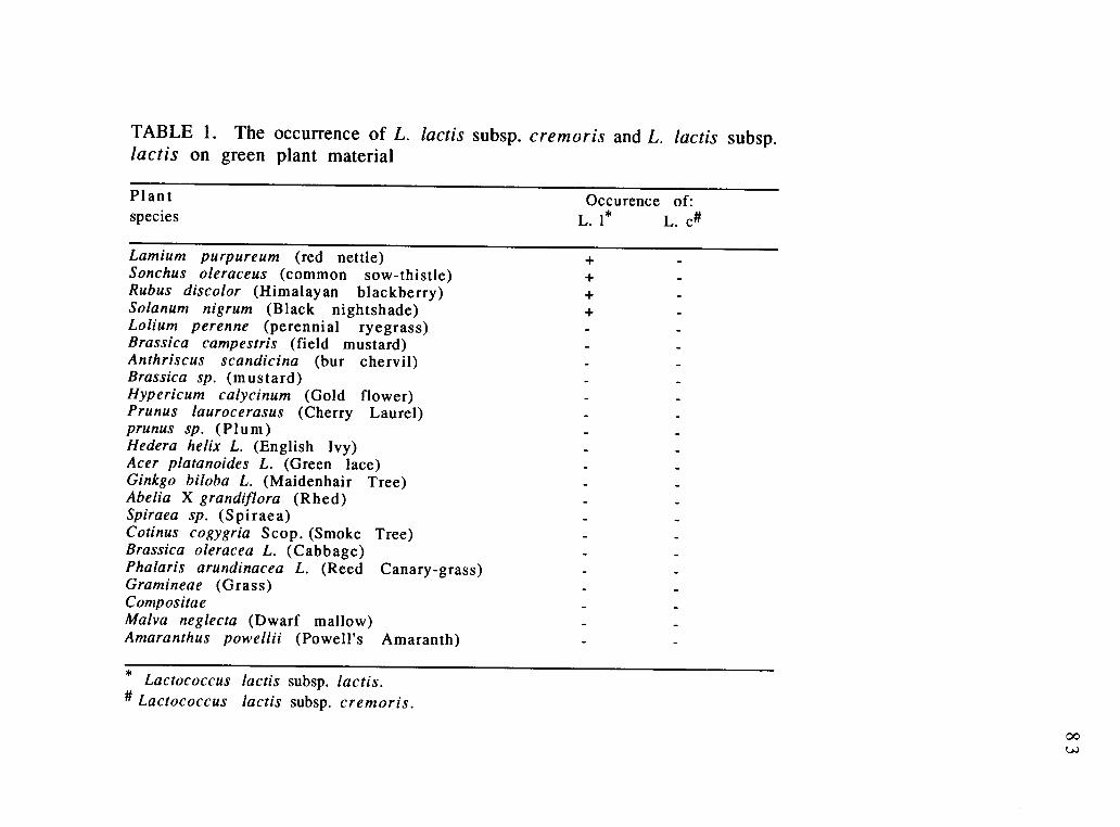
TABLE 1. The occurrence of L. lactis subsp. cremoris and L. lactis subsp.lactis on green plant material
Plant Occurence of:species L. 1* L. c#
Lamium purpureum (red nettle)Sonchus oleraceus (common sow-thistle)Rubus discolor (Himalayan blackberry)Solanum nigrum (Black nightshade)Lolium perenne (perennial ryegrass)Brassica campestris (field mustard)Anthriscus scandicina (bur chervil)Brassica sp. (mustard)Hypericum calycinum (Gold flower)Prunus laurocerasus (Cherry Laurel)prunus sp. (Plum)Hedera helix L. (English Ivy)Acer platanoides L. (Green lace)Ginkgo biloba L. (Maidenhair Tree)Abelia X grandiflora (Rhed)Spiraea sp. (Spiraea)Cotinus cogygria Scop. (Smoke Tree)Brassica oleracea L. (Cabbage)Phalaris arundinacea L. (Reed Canary-grass)Gramineae (Grass)CompositaeMa lva neglecta (Dwarf mallow)Amaranthus powellii (Powell's Amaranth)
* Lactococcus lactis subsp. lactis.Lactococcus lactis subsp. cremoris.

TABLE 2. The occurrence of Lactococcus lactis subsp. cremoris and Lactococcus lactis subsp. lactis onvegetables from local produce and other types of environmental samples from a dairy farmlocation and in raw milk samples from the United States.
Source: Occurence of: Source: Occurence of:Vegetables L. 1* L. c# raw milk & L. 1 L. c
others
Potato + colostrum-1 +Cucumber + colostrum-2 +Sweet peas + raw milk (6)@Beans + cow's tail-1 +Cantilope + cow's tail-2 +Corn + cow's body +Broccoli + cow's salivaSquash cow's fecesParsley Alfalfa feedMushroom dry feedTomato salt lickCauliflower salt lick boxRed cabbage drinking waterAsparagus
* Lactococcus lactis subsp. lactis.Lactococcus lactis subsp. cremoris.
@ Six samples tested.

85
obtained from China (CM-1 to CM-4) came from Behai Dairy Plant,
Palian Liaoning Province. Each sample was hand-milked from ten
different cows. The fifth Chinese milk sample (CM-5) was provided
by Dr. Paul Kindstedt, University of Vermont, College of Agriculture,
Department of Microbiology. This sample was commingled milk from
a large storage tank at a processing dairy plant in Harbin, China. The
milk sample from Morocco was provided by Dr. Noreddine
Benkerroum, Institut Agronomique et Veterinaire Hassan II,
Department de Microbiologie Alimentaire et de Biotechnologie, B.P
6202 Rabat Instituts, Morocco. The sample was obtained from a
farmer in a local market. All Chinese and Moroccan milk samples
were naturally enriched upon arrival from China and Morocco, and
were screened for lactococci by colony hybridization without further
incubation. Samples from Yugoslavia were collected during the
period of October, 1991 through March, 1992. Samples were either
obtained from private farms or bought from local markets. All
samples originated from farms around Sarajevo, Bosnia except for
two, the goat raw milk-2 and cream-13 which were obtained from
Fojnica and Uzice respectively (Table 3). All samples were kept
frozen whenever possible and were enriched upon arrival for
lactococci as described earlier in the text for raw milk and othertypes of samples. After enrichment, samples were frozen again until
tested. However, some samples were plated and few colonies were
picked at random before the samples were frozen again for furtherstudy.
For colony lifts, dilutions from each of these samples were
directly spread onto large (150 X 15 mm) PMP agar plates. The large

TABLE 3. The occurrence of Lactococcus lactis subsp. cremoris and Lactococcuslactis subsp. lactis in milk and milk products from Yugoslavia China and Morocco.
Source
Cottage cheese-9Cottage cheese-11Cow'sraw milk-3Cottage cheese-7Cottage cheese-6Cream (Uzice)-13Cow's raw milk-1Cows raw milk-4Goats raw milk-2Cottage cheese-4aCottage cheese-8Cottage cheese-10Cottage cheese-11Cream-10aYogurt-6ahard cheese-3aCottage cheese-12China milk-1China milk-2China milk-3China milk-4China milk-5Morocco
Locality Occurence of:L. I* L. clt
Kiseljak-market + +Ilijas-market + +Barice + +Kobilja Glava-farm +Gornja Vogosca-market +Uzice +Zuc +Petrovici +Fojnica +Vogosca-market +Pazaric-farm +Kiseljak-market +Ilijas +SjerogosteNahorevo-farmVrelo BosneVogosca-marketBehai-China, dairy plant + +Behai-China, dairy plant +Behai-China, dairy plant +Behai-China, dairy plant + +Harbin-China, dairy plant +Farmer, local market + +
*Lactococcus lactis subspecies lactis.
# Lactococcus lactis subspecies cremoris.
87
plates allowed the use of lower dilutions of samples with less
crowded plates, thus facilitating return to those colonies needed for
further investigation following colony hybridizations The PMP agarplates were incubated anaerobically at 210C for 2 to 3 days prior tocolony hybridization. After colony lifting, the plates were re-incubated for an additional 2 days to allow re-growth.
RNA-DNA colony hybridization. Colonies were lifted from M17
or PMP agar plates onto glass microfibre filters (Whatman no. 934-AH). The glass fiber filters were previously soaked in poly-L-lysine
(50 mg /ml in 10 mM Tris-HC1, pH 8), air dried and sprayed on theback with a thin layer of acrylic spray (Krylon 1301, Crystal Clear
Acrylic) for mechanical support during subsequent treatments.
Colony hybridizations were performed using a method we developedearlier (26) which is a modified combination of the Anderson andMcKay (2) and Kapperud (11) methods. In the modified procedure,
the glass fiber filters with the lifted colonies were placed, colony side
up, on Whatman no. 1 paper soaked with 5% SDS and left for 3 to 5min. The filters were then dried at 800C in a vacuum oven for 10 to15 min. After this treatment, 5-10 ml of prehybridization buffer(6x SSPE [1.08 M NaC1, 60 mM NaPO4, and 60 mM EDTA, pH 7.5], 5x
Denhardt's solution [0.1% Ficol, 0.1% polyvinylpyrolidone, and 0.1%
BSA], and 0.1% SDS) were added to the filters in a Petri plate or HB-1
hybridizer (Techne Ltd., Duxford, Cambridge CBZ 4PZ. U. K).
Prehybridization was carried out for 45 to 60 min at 650C. Theprehybridization buffer was then replaced with 5-7 ml ofhybridization buffer (6x SSPE, lx Denhardt's solution, 0.1% SDS, and

8 8
approximately 106 cpm of 32P-labeled probe). The hybridization
was carried out at room temperature overnight. Filters were washed
3 times for 20-30 min at room temperature in 6x SSPE, 0.1% SDS,
then one time at the predetermined stringency temperature (450C
for 212RLa probe, 500C for 68RCa probe and 370C for both 1406R
and 1406F probes). Filters were then exposed to X-ray film for 6 to
24 h.
Glass fiber filters with lifted colonies were hybridized two
times to two different probes in the following order: 68RCa, then
212RLa. After each hybridization and X-ray film exposure, the filter
was washed at 650C for 4 to 5 h, then exposed to X-ray film to insure
that all of the hybridized probe was washed off completely before
performing the next hybridization.
Whole-cell dot blot hybridization. After re-growth of the lifted
colonies on the PMP plates, cells which hybridized to either 212RLA
or 68RCa or both probes were picked and streaked for purification on
PMP plates. The pure colonies were grown in M17 broth for whole-
cell dot blot hybridization to confirm their relatedness to lactococci.
Whole cells were hybridized to oligonucleotide probes as described
previously (25).
Phenotypic and physiologic characterization. The
environmental isolates which were suspected to belong to theLactococcus lactis species by genotypic testing were further tested
phenotypically for arginine hydrolysis and gas production from
citrate using the differential broth described by Reddy and co-

89
workers (24), growth at pH 9.2, growth in 4% NaC1 and growth at
400C. Arginine hydrolysis was also tested by means of Niven's
broth(19). Reactions to these tests distinguish lactococci from other
related microorganisms as well as differentiate between L. lactis
subsp. lactis and L. lactis subsp. cremoris. Litmus reduction before
milk curding also is considered an important diagnostic aid for
lactococci. All strains were inoculated into litmus milk and observed
frequently until the time of curding. Also all strains were confirmed
as Gram positive cocci.
Fast or slow acid production. About 120 isolates werechosen for further analysis. These were subcultured in 5 ml ofsterile 11% nonfat milk. Cultures were incubated at 210C until they
clotted the milk. The coagulated cultures were then used as
inoculum at the rate of one percent into a second tube of sterile milk.
These tubes were incubated at 210C for 16-18 hours. Cultures which
coagulated milk within 16-18 hours were considered to be fast acid
producing strains and those which did not do so were streaked on
fast-slow differential agar (FSDA II) plates (21, 27), and incubated at
300C for 48 hours to screen for fast cells. Fast Lac+ Prot+ cells
formed yellow colonies against an opaque light-blue background.
Lac+ Prot- colonies were less brightly colored and resembled yellow
doughnuts. Colonies of Lac- Prot+ and Lac- Prot- were
indistinguishable from each other but were clearly differentiated
from the other two types; they appeared as colorless translucent
colonies. Fast colonies were tested again for coagulating 11% milk

90
within 16-18 hours. Cultures which coagulated milk after more than
18 hours were considered slow strains.
Organoleptic and flavor evaluation. A one percent
inoculum was added to 100 ml of sterile 11% nonfat milk in a Pyrex
flask and incubated at 210C for 18 hours. Samples of the coagulated
milk culture were poured into drinking cups and flavor evaluations
made noting in particular any bitter, malty, or salty flavors thatmight be present.
91
RESULTS AND DISCUSSION
Representative strains of the lactococci isolated from plants,
vegetables, colostrum, and other types of environmental samples
from the United States are listed in Table 4. From plant material and
vegetables, only L. lactis subsp. lactis was isolated. Neither L. lactis
subsp. cremoris nor L. lactis subsp. lactis biovar. diacetylactis was
found on any of the plant species or vegetables examined. Colonies
were lifted onto glass fiber filters and hybridized to two probes,
68RCa and 212RLa, as described in Materials and Methods.
Lactococcus lactis subsp. lactis was found on Lamium purpureum,
Sonchus oleraceus, Rubus discolor, and Solanum nigrum (Table 1 and
4). It was also found on sweet peas, potato, cucumber, beans,
cantaloupe, broccoli, and corn (Table 2 and 4). Most of the colonies
which hybridized to the 68RCa probe and some of the colonies which
hybridized only to the 212RLa probe were selected from the original
agar plate after reincubation, streaked for purification, and
confirmed for positive hybridization to both probes by means of
whole-cell dot blot hybridization (25). All of the colonies confirmed
for positive hybridization to the probes were further tested
phenotypically for L. lactis characteristics. Table 4 lists the genotypic
and phenotypic characteristics of some of the environmental strains
isolated from vegetables, plants, raw milk, colostrum, and other
samples from a dairy farm using the colony hybridization method.
According to our results, L. lactis subsp. lactis seems to occur only on
a few plants. This finding has been substantiated by other workers

TABLE 4. Genotypic and phenotypic characteristics of some of the environmental strains isolated fromvegetables, plants, raw milk, colostrum, and other samples from a dairy farm (U.S) using the colonyhybridization procedure with 32P-labeled 16S rRNA probes. All strains are Gram positive cocci andacidify litmus milk before coagulation.
Isolate *Hybridization to: Source:68RCa 212RLa 10°C
Growth at :
40°C 45°C NaCI4 %
pH9.2
Hydrolysisof Arginine
# no. ofisolates
FB50 + L. purpureum + + + + + 5FB55 + L. purpureum + - + + + 1FB58 + S. oleraceus + + + + + 1FB61 + R. discolor + + + + + 6SPS 1 + Sweet peas + + + + + 16POT53 + Potato + + + + + 11CUC80 + Cucumber + + + + + 4COR105 + Corn-2 + + + + + 9BEN121 + Beans + + + + + 4Col + + corn-1 + + + + + 6F2 + cow's tail-1 + + + + 14F8 + cow's tail-1 + + + + + 5F 1 1 + cow's tail-1 + + - + 4FB11 + cow's tail-2 + + + + + 18FB19 + cow's tail-2 + + + + 4FB33 + cow's tail-2 + + + 1FB34 + cow's body + + + + 3FB35 + cow's body + + + + + 13F36 + colostrum-1 + + + + + 1FB1 + colostrum-2 + + + + + 9FB9 + colostrum-2 + + + + 1
*Whole-cell dot blot hybridization#Number of isolates with the same genotypic and phenotypic characteristics.

93
such as Stark (30) who isolated L. lactis subsp. lactis from only six
plant sources, by Pinter (22) who was able to find this organism only
on three different plants, and by Radich (23) who reported only six
vegetables to be a source of L. lactis subsp. lactis. Although we were
able to find L. lactis subsp. lactis on the above mentioned plant
species and vegetables, it was only isolated from Lamium
purpureum, Sonchus oleraceus, Rubus discolor, sweet peas, potato,
cucumber, beans, and corn. These isolated strains are represented in
table 4 by FB50, FB55, FB58, FB61, SPS1, POT53, CUC8, COR105, and
BEN121. Lactococcus lactis subsp. lactis was also isolated from
potatoes, corn, cucumber, peas, beans, and cantaloupe by Radich (23).
Radich failed to isolate L. lactis subsp. cremoris or L. lactis subsp.
lactis biovar. diacetylactis from any of the vegetable or fruit samples
he tested (23). All L. lactis subsp. lactis strains that we isolated from
plant material and vegetables had the same phenotype except for
one strain, FB55, isolated from Lamium purpureum, which failed to
grow at 400C (Table 4). Six strains of L. lactis subsp. lactis isolated
from a fresh corn sample and represented by Col hybridized to both
probes (68RCa and 212RLa). These strains had the phenotypic
characteristics of L. lactis subsp. lactis. They were positive for
arginine dihydrolase, growth at pH 9.2, growth in 4% NaCl and
growth at 400C (Table 4). In this respect they resemble L. lactis
subsp. lactis strain C2 which has the rRNA genotype of L. lactis subsp.
cremoris but resembles L. lactis subsp. lactis in phenotype(26).
Three types of L. lactis subsp. lactis phenotypes were isolated
from a cow's tail, and another three types from a different cow's tail.
These phenotypes are represented by strains F2, F8, F11, FB11, FB19,

94
and FB33 (Table 4). Two different L. lactis subsp. lactis phenotypes
were isolated from a cow's body. These are represented by FB34 andFB35 (Table 4). Strains F8, FB11, and FB35 represent the same
phenotype; strains FB19 and FB34 represent another similar
phenotype. Lactococcus. lactis subsp. lactis was also isolated from
two samples of colostrum. Only one phenotype was isolated from the
first sample (colostrum-1). This is represented by strain F36 (Table4). Strains FB1 and FB9 represent two L. lactis subsp. lactis
phenotypes isolated from another colostrum sample (colostrum-2,
Table 4).
Table 5 lists representative strains of confirmed lactococci
isolated from the milk samples obtained from China. All samples
contained L. lactis subsp. lactis strains as represented by CM1-1,
CM2-63, CM3-90, CM4-7, and CM5-7 (Table 5). Other isolates were
phenotypically similar to L. lactis subsp. lactis, but with minordifferences. These are represented by CM1-31, 33, 39, 42; CM4-7;
CM5-3, and CM5-4 (Table 5). Three of the five milk samples (CM1,
CM2, and CM5) contained organisms of the L. lactis subsp. lactis C2
group, similar to those found previously in the corn sample (L. lactis
subsp. cremoris genotype with L. lactis subsp. lactis phenotype, Table3). These strains are represented by CM1-54, CM2-86, and CM5-5
(Table 5).
Isolates of L. lactis subsp. cremoris (CM1-3 and CM4-27; Table
3) were found in two of the five milk samples tested. RNA was
extracted from these two strains and hybridized to both 212RLa and68RCa probes by means of dot blot hybridization (25). Both strains
showed specific hybridization to both probes (data not presented).

95
These results, together with the phenotypic characteristics presented
in Table 5, suggest that both strains are L. lactis subsp. cremoris.
However when re-tested for biochemical characteristics using the
differential broth after storage at -800C (24), these strains were
both no longer negative for arginine dihydrolase. However, they
remained negative when Niven's broth (19) was used for testing.
Seasonal variations in phage sensitivity of some L. lactis subsp.
cremoris starter cultures have been reported by Heap and Lawrence
(8). Their explanation for these seasonal differences was the
existence of variant cells within the individual strains which differed
markedly in phage sensitivity. They suggested that the proportion of
these variants may change upon subculturing. It is possible,
therefore, that the two strains (CM1-3 and CM4-27; Table 3) were
not exactly the same cultures when subcultured in different kinds of
media such as Niven's or differential broth since variant cells of
different types could have emerged in one medium but not the other.

TABLE 5. Genotypic and phenotypic characteristics of some of the environmental strains isolated fromfive Chinese raw milk samples using the colony hybridization procedure with 32P-labeled 16S rRNAprobes. All strains are Gram positive cocci and acidify litmus milk before coagulation.
Isolate **Hybridization to: Source: Growth at : Hydrolysis #No. of68RCa 212RLa 10 °C 40 °C 45°C NaCI pH of Arginine isolates
4% 9.2
CM1-1 + China milk-1 + + + + + 33CM1-3@ + + China milk-1 +
1CM1-31 + China milk-1 + + + + / + / - 1CM1-33 + China milk-1 + + + + + / - 1CM1-39 + China milk-1 + + + + 3CM1-42 + China milk-1 + + + + 1CM1-54 + + China milk-1 + + + + + 20CM2-63 + China milk-2 + + + + + 3CM2-86 + + China milk-2 + + + + + 3CM3-90 + China milk-3 + + + + + 3CM4-5 + China milk-4 + + + + + 10CM4-7 + China milk-4 + + +1- + + 1CM4-27* + + China milk-4 + + / - 1CM5-3 + + China milk-5 + + +/- +/- + 1CM5-4 + + China milk-5 + + + / - + 1CM5-5 + + China milk-5 + + + + + 1CM5-6 + + China milk-5 + + - + 1CM5-7 + China milk-5 + + + + + 5
**Whole-cell dot blot hybridization#Number of isolates with the same genotypic and phenotypic characteristics.@ Changed into arginine hydrolase and growth at 4% NaCI positive after storage at -80oC.*Changed into arginine hydrolase and growth at 40°C, and at pH 9.2 positive after storage at -80 °C.
97
Strain CM1-3 also became able to grow at 4% NaC1, and strain CM4-
27 developed the capacity to grow at 400C and at pH 9.2. Instances
of variability in certain pure cultures of lactic acid bacteria have
been described in the literature. Substrains of L. lactis subsp.
cremoris and L. lactis subsp. lactis varying in their ability to attack
maltose and sucrose were described by Sheman and Hussong as cited
in Hunter (10). Orla Jensen (20) encountered variable characteristics
for certain lactococci. Yawger and Sherman (36) reported the
isolation of variants of L. lactis subsp. lactis from milk which did not
ferment lactose. In a study done by Hunter (10) it was reported that
variation was observed within some strains of L. lactis subsp.
cremoris, especially with regard to acid production (or viability),
response to high temperature conditions, ability to produce ropiness
in milk, and degree of resistance to phage attack. Variation in acid
production also has been observed for some of our environmental
isolates of lactococcal strains.
For the first time in years, we recently succeeded in isolating
strains of the L. lactis subsp.cremoris from two raw milk samples
obtained from Morocco and Yugoslavia and from two cottage cheese
samples originating in Yugoslavia. Tables 6 and 7 list representative
strains of the confirmed lactococi isolated from the Moroccan and
Yugoslavian samples. Genotypically and phenotypically, 20 of the
strains isolated from the Moroccan milk (represented by MS-22, and
MS-19) were L. lactis subsp. lactis, as indicated by their reaction with
the 212RLa probe and phenotypic characteristics (Table 6). Four
strains, represented by MS-19, produced gas from citrate

TABLE 6. Genotypic and phenotypic characteristics of some of the environmental strains isolated fromone Morocco raw milk sample using the colony hybridization procedure with 32P-labeled 16S rRNAprobes. All strains are Gram positive cocci and acidify litmus milk before coagulation.
Isolate *Hybridization to: Source: Growth at : Hydrolysis #no. of68RCa 212RLa 100C 400C 450C NaCl pH of Arginine isolates
4% 9.2
MS-5@ + + Morocco milk + 2 4MS-6 + Morocco milk + + NR 15MS-21 + Morocco milk + NR 2MS-19 + Morocco milk + + + + +(G) 4MS-22 + Morocco milk + + + + + 16MS-46 + Morocco milk + + + + +(S) 1MS-70 + Morocco milk + + + + 1
* Whole-cell hybridization# Number of isolates with the same genotypic and phenotypic characteristics.@ L. lactis subsp. cremoris(G): Gas production from citrate(S): Slow acid production

TABLE 7. Genotypic and phenotypic characteristics of some of the environmental strains isolatedfrom several Yugoslavian raw milk and milk product samples using the colony hybridizationprocedure with 32P-labeled 16S rRNA probes. All strains are Gram positive cocci and acidify litmusmilk before coagulation.
Isolate *Hybridization to:68RCa 212RLa
Source:10°C
Growth at :
40°C 45°C NaCI4 %
pH9.2
Hydrolysisof Arginine
#No. ofisolates
K1 + + Cottage cheese-7 + + + + 3K2 + + Cottage cheese-7 + + + + 1A3 + + Cream (Uzice)-13 + + + + + 7C8 + + Cream (Uzice)-13 + + + + 2112 + + Cottage cheese-11 + + + 11102 + + Cottage cheese-11 + + + + + 21117M + + Cottage cheese-11 + 1AM1 + Cottage cheese-9 + +/ - + 2AM4 + + Cottage cheese-9 + + / - 1AM12 + + Cottage cheese-9 + + + / 1AM13 + Cottage cheese-9 + + + + 1032 + + Raw milk-3 + + + + 1011 + Raw milk-1 + + + + + 2022 + Goat raw milk-2 + + + + + 2023 + Goat raw milk-2 + + + + +(G) 1
* Whole- cell dot blot hybridization.# Number of isolates with the same genotypic and phenotypic characteristics.(G): Gas production from citrate.

TABLE 7 (continued). Genotypic and phenotypic characteristics of some of the environmental strainsisolated from several Yugoslavian raw milk and milk product samples using the colony hybridizationprocedure with 32P-labeled 16S rRNA probes. All strains are Gram positive cocci and acidify litmusmilk before coagulation.
Isolate *Hybridization to: Source:68RCa 212RLa 10°C
Growth at :
40°C 45°C NaCI4 %
pH9.2
Hydrolysis #No. ofof Arginine isolates
41 + Raw milk-4 + + + + + 161 + Cottage cheese-6 + + + + + 1072 + Cottage cheese-7 + + + + + 1073 + Cottage cheese-7 + + + +(G) 182 + Cottage cheese-8 + + + + 183 + Cottage cheese-8 + + + + + 191 + Cottage cheese-9 + + + + + 192 + Cottage cheese-9 + + + + +(G) I102 + Cottage cheese-10 + + + + + 4111 + Cottage cheese-11 + + + + + 11105 + Cottage cheese-11 + + + 11113 + Cottage cheese-11 + + +/- +/- + 1404 + Cottage cheese-4a + + + +(G) 1408 + Cottage cheese-4a + + + + + 1
* Whole- cell dot blot hybridization.4 Number of isolates with the same genotypic and phenotypic characteristics.(G): Gas production from citrate.

1 0 1
in differential broth (Table 6). Also, these four strains tested
positive for acetylmethylcarbinol and diacetyl production when
checked by the king's test (12) and the Voges-Proskauer test (7).
These are characteristics of L. lactis subsp. lactis biovar. diacetylactis.
Another 19 strains reacted with the 212RLa probe, proving their
relation to the Lactococcus genus, but showed variability in their
phenotypic characteristics These strains are represented by MS-6,
21, 46, and 70 (Table 4). Unlike the Chinese milk sample strains,
none of the strains isolated from the Moroccan milk sample belonged
to the C2 group. Twenty-four isolates, represented by MS-5,
hybridized to both probes (212RLa and 68RCa), and had the
phenotypic characteristics of L. lactis subsp. cremoris.
Out of the seventeen samples tested from Yugoslavia, thirteen
tested positive for lactococci (Table 3). Cream-10a, yogurt-6a, hard
cheese-3a, and cottage cheese-12 were the four samples that did not
show any hybridization to any of our probes (68RCa, 212RLa). All
samples from Yugoslavia which tested positive as lactococci
contained L. lactis subsp. lactis with variable phenotypes as
represented by AM1 and AM13 (cottage cheese-9); 011 (raw milk-
1); 022 (goat raw milk-2); 41 (raw milk-4); 61 (cottage cheese-6),
072 (cottage cheese-7); 82 and 83 (cottage cheese-8); 91 (cottage
cheese-9); 102 (cottage cheese-10); 111, 1105, and 1113 (cottage
cheese-11); and 408 (cottage cheese-4a). Some samples contained
organisms of the L. lactis subsp. lactis C2 group. These strains are
represented by K1 and K2 (cottage cheese-7); A3 and C8 (cream,
Kajmak-13), and 1102 (cottage-11). All of these strains are listed inTable 7. Four strains of L. lactis subsp. lactis biovar. diacetylactis

102
(023, 073, 92, and 404) were found in goat raw milk-2, cottage
cheese-7, cottage cheese-9, and cottage cheese-4a, respectively
(Table 7). So L. lactis subsp. lactis biovar. diacetylactis was only
isolated from raw milk and cottage cheese but not from plants. Other
investigators have likewise found this to be the case. For example
Radich (23) was able to isolate this organism only from individual
producer milk samples and Swarthing (31) obtained his strains of L.
lactis subsp. lactis biovar. diacetylactis from raw milk or starter
cultures. The only person who isolated this bacterium from plant
material was Matuszewski as cited by Briggs (3). Isolates of the
subspecies cremoris (112, 1117, and AM4) were found in cottage
cheese-11, cottage cheese-9, and raw milk-3 (Table 7). King and
Koburger (10) characterized Group N streptococci isolated from
meats, vegetables, dairy products, barn-trough water, and poultry
feed. No L. lactis subsp. lactis biovar. diacetylactis strains were
isolated and L. lactis subsp. cremoris was recovered from only
cottage cheese and raw milk. Unfortunately, the L. lactis subsp.
cremoris strains found in milk were lost because they failed to
survive freezing.
Of 120 strains chosen for further testing, 64 were fast acid
producers, either when tested directly for coagulating milk within
16-18 h at 210C, or after being streaked on FSDA II. Almost all of
these strains tested acceptable when evaluated for flavor (Table 8).
Only two strains of L. lactis susp. lactis from the Moroccan milk (MS-
22 and 70) tested malty, and another strain of L. lactis susp. cremoris
from Yugoslavia tasted bitter. These findings suggest that some of
these strains may be useful to the dairy product industry. These

TABLE 8. Flavor evaluation of some newly isolated lactic acid bacteria.
Isolate flavor pH
L. 1 C2* clean, acid flavor 4.3L. c 205# clean, acid flavor 4.4L. c 107/6# flat-low acid 4.9L. 1 Col clean, low acid flavor 4.6L. 1 Co2 clean, acid flavor 4.5L. 1 Co3 clean, acid flavor 4.5L. 1 Co4 clean, acid flavor 4.5L. 1 Co5 clean, low acid flavor 4.7L. 1 Co6 clean, acid flavor 4.5L. c MS3 clean, acid flavor 4.2L. c MS5 clean, acid flavor 4.15L. c MS7 clean, acid flavor 4.15L. d MS19 (slow) clean, low acid flavor 4.9L. d MS38 clean, acid flavor 4.25L. 1 MS22 malty 4.25L. 1 MS41 clean, acid flavor 4.15L. 1 MS46 (slow) clean, not sour 4.5L. 1 MS58 clean, acid flavor 4.2L. 1 MS70 malty 4.25L. 1 CM1-2 clean, acid flavor 4.4L. 1 CM3-90 clean, acid flavor 4.4L. 1 CM3-93 (slow) clean, acid flavor 4.4L. c CM4-27 clean, acid flavor 4.5L. 1 CM5-6 clean, acid flavor 4.45L. 1 CM5-7 clean, acid flavor 4.35
*Control Lactococcus lactis ssp. lactis strain C2
# Control Lactococcus lactis ssp. cremoris strains 107/6 and 205

TABLE 8 (continued). Flavor evaluation of some newly isolated lacticacid bacteria.
Isolate flavor pH
L. 1 MS17L. c CM1-3L. 1 CM2-63 (slow)L. 1 CM5-8L. 1 CM1-54L. 1 FB1L. 1 FB10L. 1 FB12L. 1 FB33L. 1 FB34L. 1 FB35L. 1 FB58L. 1 FB61L. 1 FB62L. 1 BEN121L. 1 MS39L. 1 MS71 (slow)L. 1 AM5L. c 112L. 1 85L. 1 011
clean, acid flavorclean, acid flavor
clean, acid flavorclean, acid flavorclean, acid flavor
clean, acid flavor
clean, low acid flavorclean, acid flavorclean, acid flavor
clean, acid flavorclean, acid flavor
clean, acid flavor
clean, low acid flavorclean, acid flavorclean, acid flavor
clean, acid flavor
clean, low acid flavor
clean, acid flavor
clean, acid flavor
slightly different.
clean, acid flavor
4.4
4.5
4.4
4.4
4.4
4.4
4.6
4.5
4.3
4.4
4.5
4.6
4.6
4.55
4.35
4.4
4.7
4.4
4.4
4.4
4.2
* Control Lactococcus lactis ssp. lactis strain C2# Control Lactococcus lactis ssp. cremoris strains 107/6 and 205
0-P-

105
new isolates, especially L. lactis subsp. cremoris, will provide a
source of natural genetic variation which can be used to provide
greater protection against phage infection in cheese plants.

106
ACKNOWLEDGEMENTS
We thank Dr. Paul Kindstedt, University of Vermont, College of
Agriculture, Dept. of Microbiology, for providing a milk sample from
China. We are also thankful to Dr. Noreddine Benkerroum, Institut
Agronomique et Veterinaire Hassan II, Department de Microbiologie
Alimentaire et de Biotechnologie, B.P 6202 Rabat Instituts, Morocco
for providing a milk sample from Morocco. We are grateful to Dr. R.
E. Kunkee, College of Agricultural and Environmental Sciences,
University of California, Davis, for sending us a L. hordniae strain.
The technical help of Ms. Kelley Nathman is highly appreciated.
This work was supported by a grant from the National Dairy
Promotion and Research Board, the OSU Agricultural Experiment
Station and International Research Exchange, IREX.
107
REFERENCES
1. Abd-El-Malek, Y. and T. Gibson. 1948. Studies in the
bacteriology of milk. J. Dairy Res. 15:233.
2. Anderson, D. G., and L. L. McKay. 1983. Simple and rapid
method for isolating large plasmid DNA from lactic streptococci.
Appl. Environ. Microbiol. 46:549-552.
3. Briggs, C. A. E. 1952. A note on serological classification of
Streptococcus diacetylactis (Matuszewski et al.). J. Dairy Res.
19:167.
4. Cavett, J. J., G. J. Dring, and A. W. Knight. 1965.
Bacterial spoilage of thawed frozen peas. J. Appl. Bact.
28:241-245.
5. Cavett, J. J., and E. I. Garvie. 1967. Biochemical and
serological characters of three strains of streptococci reported
as Streptococcus cremoris isolated from deep- frozen peas after
thawing. J. Appl. Bacteriol. 30:377 -381.
6. Fenton, M. P. 1987. An investigation into the sources of
lactic acid bacteria in grass silage. J. Appl. Bacteriol. 62:181-
188 .
108
7. Harrigan, W. F., and M. E. McCance. 1976.
Laboratory methods in food and dairy Microbiology.
revised edn., W. E. Harrigan, Academic press, pp. 74.
8. Heap, H. A., and R. C. Lawrence. 1977. The contribution of
strarter strains to the level of phage infection in a commercial
cheese factory. N. Z. J. Dairy sci. Technol. 12:213-218.
9. Hirsch, A. 1952. The evolution of lactic streptococci. J. Dairy
Res. 19:290-293.
10. Hunter, G. J. E. 1939. Examples of variation within pure
cultures of Streptococcus cremoris. J. Dairy Research 10:46-
470.
11. Kapperud, G., K. Dommarsnes, M. Skurnik, and E.Hornes. 1990. A synthetic oligonucleotide probe and a cloned
polynucleotide probe based on the yopA gene for detection and
enumeration of virulent Yersinia enterocolitica. Appl. Environ.
Microbiol. 56(1):17-23.
12. King, N. S. 1948. A modification of the Voges-
Proskauer test forrapid colorimetric determination of
acetylmethylcarbinol plus diacetyl in the buttercultures.
Dairy Ind. 13:860.

109
13. King, N. S. and J. A. Koburger. 1970. Characterization of
some Group N streptococci. J. Dairy Sci. 53:402-409.
14. Lawrence, R. C., T. D. Thomas, and B. E. Terzaghi. 1 976 .
Reviews of the progress of dairy science: Cheese starters. J.
Dairy Res. 43:141-193.
15. Mundt, J. 0., and A. H. Johnson. 1959. Physiological
properties of group D streptococci isolated from plants.
Food Res. 24:218.
16. Mundt, J. 0., W. F. Graham, and I. E. McCarty. 1967.
Spherical lactic acid producing bacteria on southern grown raw
and processed vegetables. Appl. Microbiol. 15:1303 -1308.
17. Nelson, G. A. and, H. R. Thornton. 1952. The lactic
streptococci in Edmonton milks and creams. Canadian Journal
of Technology. 30:130-135.
18. Nichols, A. A. and M. Hoyle. 1949. Bacteriophages in
typing lactic streptococci. J. Dairy Res. 16:167-173.
19. Nivens, C. J. Jr., K. L. Smiley, and J. M. Sherman. 1942.
The hydrolysis of arginine by streptococci. J. Bacteriol. 43:651
660 .

1 1 0
2 0 . Orla-Jensen, S. 1942. The lactic acid bacteria, 2nd ed., E.
Munksgaard, Copenhagen, pp. 106-107.
21. Orberg, P. K. 1985. Antimicrobial resistance and plasmid
properties of Leuconostoc and group N Streptococcus. Ph.D.
Thesis. Oregon State Univ., Corvallis.
22. Pinter, I. J. 1940. The occurrence of streptococci on plant
material. Master's thesis. Ithaca, Cornell University. 17 numb.
leaves.
23. Radich, P. C. 1968. An ecological study of the lactic
streptococci. M.S. Thesis, Oregon State University, Corvallis.
51 P.
2 4 . Reddy, M. S., E. R. Vedamuthu, and G. W. Reinbold.
1971. A differential broth for separating the lactic streptococci.
J. Milk and Food Tech. 34(1):43 -45 .
25. Salama, M. S., W. E. Sandine, and S. J. Giovannoni. 1991.
Development and application of oligonucleotide probes for
identification of Lactococcus lactis subsp. cremoris. Appl.
Environ. Microbiol. 57:1313 -1318.
26. Salama, M. S., W. E. Sandine, and S. J. Giovannoni. 1993.
Isolation of lactococci and Lactococcus lactis subsp. cremoris

1 1 1
from nature by colony hybridization with ribosomal RNA. Appl.
Environ. Microbiol. (under review)
27. Sandine, W. E. 1985. The streptococci: Milk products.
In Bacterial Starter Cultures for Foods. Ed. Stanley E. Gilliland.
CRC press, Inc. Boca Raton, Florida. pp. 5-23.
28. Sandine, W. E. 1987. Looking backward and forward at the
practical applications of genetic researches on lactic acid
bacteria. FEMS Microbiol. Rev. 46:205-220.
29. Schieblich, M. and W. Schonherr. 1957. Streptococci in
acid fodders and their distribution in the raw material.
Tierernahr. Futtermittelk 11:232. Dairy Sci. Abstr. 19:231.
30. Stark, P. and J. M. Sherman. 1935. Concerning the
habitat of Streptococcus lactis. Journal of Bacteriology.
30:639-646.
31. Swarthing, P. F. 1951. Biochemical and serological
properties of some citric acid fermenting streptococci from milk
and dairy products. J. Dairy Res. 18:256.
32. Terzaghi, B. E. and W. E. Sandine. 1975. Improved
medium for lactic streptococci and their bacteriophage. Appl.
Microbiol. 29: 807-813.

1 1 2
33. Walker, E. E. 1902. The bacterial flora of freshly drawn
milk. Journal of Applied Microscopy and Laboratory Methods.
5:2029-2044.
34. Willem, M., and G. Simons. 1988. Molecular cloning of
lactose genes in dairy lactic streptococci: the phospho-B-
galactosidase and B-galactosidase genes and their expression
products. Biochemie 70:461.
35. Yawger, E. S., and J. M. Sherman. 1937 a. Variants of
Streptococcus lactis which do not ferment lactose. J. Dairy Sci.
20:83.
36. Yawger, E. S., and J. M. Sherman. 1937 b. Streptococcuscremoris. J. Dairy Sci. 20:205.

1 1 3
CHAPTER 5
Characterization of Novel Strains of Lactococcus lactis
Using the Biolog Carbon Source Utilization System andPhage Typing
Maysoon S. Salama, William E. Sandine, and Stephen J. Giovannoni
Department of Microbiology, Oregon State University
Corvallis OR 97330-3804

1 1 4
ABSTRACT
Two types of probes (the 212RLa, specific for Lactococcus lactisspecies and the 68RCa, specific for Lactococcus lactis subspecies
cremoris) were used for the isolation of lactococcal strains fromnatural environments. Thirty-two new strains of Lactococcus lactis
subspecies cremoris were recovered from raw milk samples from
Morocco and Yugoslavia and from home style cottage cheese obtainedfrom Yugoslavia. Dairy and vegetation samples collected from theU.S.A as well as dairy samples from Morocco, China, and Yugoslavia
have yielded 346 L. lactis subspecies lactis strains. Out of the 120new strains tested for acid production 64 were fast acid producersAlmost all of these strains were judged to be acceptable when
evaluated for flavor. Also, the majority of the strains were resistantto a wide range of Lactococcus bacteriophages.
The biochemical and physiological characteristics of theseenvironmental isolates and their resistance to many bacteriophages
encountered by the dairy industry indicate the potential usefulnessof these strains as dairy starter cultures for use in making fermentedmilk products.

1 1 5
INTRODUCTION
The isolation of new Lactococcus strains has been achieved by
using oligonucleotide probes complementary to unique sequences in
ribosomal RNAs, to rapidly screen a large number of colonies from
green plant surfaces, enriched raw milk samples, and other types of
environmental samples. The availability of these new strains,
particularly L. lactis subsp. cremoris, is highly significant to the dairy
industry. The anticipation that wild-type strains might be less
susceptible to phage attack, and could have improved proteolytic and
flavor properties for cheese making has enhanced demand for these
strains within the dairy industry. Phage problems in particular have
had a negative commercial impact. Practices such as phage typing
and starter culture rotation, which circumvent some of the effects of
phage attack, rely on the maintenance of genetically diverse starter
culture strains.
It is evident that L. lactis strains with novel genetic and
phenotypic properties are of great interest to the dairy industry. It
is also evident that significant genetic diversity exists among
subspecies of L. lactis. Natural phylogenetic relationships among
bacterial strains are an important basis for classification and
commercial use. In the past few years genetic criteria have provided
significant insight into the natural relationships among lactococci and
have led to important changes in their taxonomy and nomenclature.
However, at present, there is no phenotypic or genotypic data base
available that characterizes these strains sufficiently well to

1 1 6
distinguish among ecotypes. Information of this type is available for
some bacterial species, though much is based on allozyme variability.
This investigation was conducted to study the nature of wild strainsof L. lactis and provide genetic and phenotypic bases for identifying
lactococci.
Recently, a technique to identify gram-negative or gram-
positive bacteria on the basis of metabolic fingerprinting was
developed by Biolog Inc. (Hayward, Calif.) (2, 3). Several Scientists
have applied this system for microbiological studies (1, 4, 5, 6, ).
Trial tests with strains sent to Biolog indicated that Lactococcusstrains could effectively reduce the indicator dyes used by thissystem, permitting the rapid screening of strains for utilization of 95carbon sources. We consider the ability to acquire this extensive
biochemical information to be an important and functional adjunct to
genetic measures of relatedness.

1 1 7
MATERIALS AND METHODS
Bacterial strains and environmental samples. Strains
used for this study are listed in Tables 1 and 2. The bacterial strains
used as controls for Biolog carbon source utilization system tests (L.
lactis subsp. cremoris BK5, Lactococcus lactis subsp. lactis f2d2 and
C2, and Lactococcus lactis subsp. lactis. biovar diacetylactis 26-2.)
were obtained from Oregon State University (OSU) culture collection.
The environmental strains were isolated from various sources by
means of a colony hybridization method (8) described in Chapter 3 .
These strains were verified by phenotypic tests as lactococci. All
strains were streaked on PMP medium (9) and incubated at 300C for
24h. One colony of each strain was subcultured on M17 agar plates
(9) for Biolog analysis.
Metabolic characterization of Lactococcus strains. The
aerobic utilization of 95 carbon sources in the presence of a redox
indicator (tetrazolium violet) was determined for the individual
environmental isolates and reference strains using GP microtiter
plates (Biolog, Inc., Hyward, Calif.). Although the manufacturer's
intended application for these plates is the identification of bacteria,
they were used in this study to obtain a metabolic profile for a group
of environmental lactococcal strains as a measure of their
relatedness. Cell suspensions of the environmental and control
strains of lactococci in saline (0.85%) were used to inoculate the
Biolog GP microplates (15µ1 per well). The optical densities of the

TABLE 1. Environmental strains isolated from different sources using the colony hybridizationprocedure with 32P-labeled 16S rRNA probes. These strains were chosen for biolog testing.Strains with a * designation coagulated milk within 16-18 h.
Isolate Hybridization to:68RCa 212RLa
Source100C
Growth at40°C 45°C NaCI
4%p H9.2
Hydrolysisof Arginine
Col + + corn + + + + +*Co3 + + corn + + + + +F2 + cow's tail + + + +*FBI + colostrum + + + + +*FB10 + colostrum + + + + +*FB 12 + cow's tail + + + + +*FB33 + cow's tail + + - +*FB34 + cow's body + + + +FB51 + Lamium puppureum + + + + +*FB58 + Sonchus oleraceus + + + + +FB61 + Rubus discolor + + + + +*BEN121 + Beans + + + + +*CMP1-3 + + China milk-1 + + +*FB 62 + Rubus discolor + + + + +CM1-39 + China milk-1 + + + +CM1-42 + China milk-1 + + + +CM1-54 + + China milk-1 + + + + +CM2-63 + China milk-2 + + + + +CM2-64 + China milk-2 + + + + +*CM2-87 + + China milk-2 + + + + +*CM3-90 + China milk-3 + + + + +

TABLE 1 (continued). Environmental strains isolated from different sources using the colonyhybridization procedure with 32P-labeled 16S rRNA probes. These strains were chosen for biologtesting. Strains with a * designation coagulated milk within 16-18h.
Isolate Hybridization to:68RCa 212RLa
Source10°C
Growth at40°C 45°C NaCI
4%pH9.2
Hydrolysisof Arginine
CM5-4 - + China milk-4 + + +/- +*CM4-27 + + China milk-4 + +/-*CM5-6 + + China milk-5 + + +CM5-7 + China milk-5 + + + + +
*CM5-8 + China milk-5 + + + + +*MS-17 + + Morocco milk +*MS-39 + Morocco milk + + + + +(G)
*MS-22 + Morocco milk + + + + +*MS-41 + Morocco milk + + + + +MS-44 + + Morocco milk +MS-46 + Morocco milk + + + + +MS-52 + + Morocco milk +MS-70 + Morocco milk + + + +MS-71 + Morocco milk + + + + +(G)K2 + + Cottage cheese-7 + + + +K9 + + Cottage cheese-7 + + + + +A3 + + Cream (Uzice)-13 + + + + +C8 + + Cream (Uzice)-13 + + + +M1 + + Cream (Uzice)-13 + + + + +1102 + + Cottage cheese-11 + + + + +

TABLE 1 (continued). Environmental strains isolated from different sources using the colonyhybridization procedure with 32P-labeled 16S rRNA probes. These strains were chosen for biologtesting. Strains with a * designation coagulated milk within 16-18 h.
Isolate Hybridization to:68RCa 212RLa
Source10°C
Growth at40°C 45°C NaC1
4%p H9.2
Hydrolysisof Arginine
1117M + + Cottage cheese-11 +AM4 + + Cottage cheese-9 + + / -AM5 + Cottage cheese-9 +AM12 + + Cottage cheese-9 + + + / -032 + Raw milk-3 + + + + -*0 1 1 + Raw milk-1 + + + + +*0 2 2 + Goat's raw milk-2 + + + + +*023 - + Goat's raw milk-2 + + + + +(G)41 - + Raw milk-4 + + + + +*6 1 + Cottage cheese-6 + + + + +073 + Cottage cheese-7 + + + +(G)*8 2 + Cottage cheese-8 + + + +*8 3 + Cottage cheese-8 + + + + +91 + Cottage cheese-9 + + + + +92 + Cottage cheese-9 + + + + +(G)102 + Cottage cheese-10 + + + + +*1 1 1 + Cottage cheese + + + + +1105 + Cottage cheese + + +404 + Cottage cheese-4a + + + +(G)408 + Cottage cheese-4a + + + + +

TABLE 2. Environmental strains isolated from different sources using the colony hybridizationprocedure with 32P-labeled 16S rRNA probes. All strains are fast acid producers and werechosen for phage testing.
Isolate Hybridization to:68RCa 212RLa
Source100C
Growth at400C 450C NaCI
4%pH9.2
Hydrolysisof Arginine
Co3 + + corn + + + + +FB 1 + colostrum + + + + +FB12 + cow's tail + + + + +FB62 + Rubus discolor + + + + +BEN121 + Beans + + + + +CM1-3 + + China milk-1 + + +CM4-27 + + China milk-4 + + + +CM5-6 + + China milk-5 + + +MS-5 + + Morocco milk +MS -11 + + Morocco milk +MS -17 + + Morocco milk +MS -22 + Morocco milk + + + + +MS -39 + Morocco milk + + + + +(G)
(G): Gas production from citrate

TABLE 2 (continued). Environmental strains isolated from different sources using the colonyhybridization procedure with 32P-labeled 16S rRNA probes. All strains are fast acid producersand were chosen for phage testing.
Isolate Hybridization to:68RCa 212RLa
Source100C
Growth at400C 450C NaC1
4%pH9.2
Hydrolysisof Arginine
MS-52 + + Morocco milk +MS-70 + Morocco milk + + + +AM4 + + Cottage cheese-9 + +/-011 + Raw milk-1 + + + + +022 + Goat's raw milk-2 + + + + +023 + Goat's raw milk-2 + + + + +(G)61 + Cottage cheese-6 + + + + +82 + Cottage cheese-8 + + + +83 + Cottage cheese-8 + + + + +111 + Cottage cheese-11 + + + + +408 + Cottage cheese-4a + + + + +
(G): gas production from citrate

123
cell suspensions were 0.4 +/- 0.05 at A=600 nm. A computer
controlled Thermo Max Microplate Reader (Molecular Devices) (X=
590 nm) was used to measure the reduced form of tetrazolium dye.
The results were stored automatically in a computer file to beevaluated for metabolic profiles. Data compiled from the analysis of
the metabolic fingerprint were evaluated with microlog ML2
software, which performs a cluster analysis based on principles ofnumerical taxonomy.
Phage typing. The 23 environmental isolates listed in Table 2
were tested for sensitivity to a variety of phages from three industryphage banks. These tests were performed by The Bioproducts Group
of Quest International, Sarasota, Florida, The Cultsure Division of
Galloway West Company, Millville, Utah and Marschal Division of
Rhone-Poulenc, Madison, Wisconsin. Each company maintains
independent phage banks isolated from whey samples obtained from
commercial Cheddar cheese making factories. Quest International
and Galloway West followed the following procedure: Milk cultures
of the strains were prepared. Cultures were then transferred to M17
lactose broth and after overnight growth at 320C were tested withphages. The individual isolates were overlaid on M17 lactose agarplates as M17 semi-solid agar infusion. The semi-solid agar
contained 0.2% CaC12 (9). Different phage preparations were spotted
on the overlaid plates with a multiple inoculation device. Plates
were incubated overnight and examined for clearing. Phage samples
used by Quest International were: Composite Quest phage Lab
sample 1990-1992, three fresh buttermilk whey samples 1993, and

124
composite cheese plant phages from Canada (7). Galloway West Co.
used 32 phage preparations from their own bank of phages.
Marschall Products performed a different and more complete phage
testing. A total of 72 positive whey samples for phage, representing
one month of testing (whey samples 1-72), and 30 negative whey
samples, representing two weeks, were tested (whey samples 73-
102), along with 38 of single plaque isolates (SPI 1-38) from their
phage bank. The cells were grown in M17 broth overnight at 320C.
Put into 7 ml overlay tubes of M17 top agar were 0.1 ml of the
overnight culture and 50 1.1.1 of 1.0 M CaC12. The tubes were then
gently vortexed and poured onto large, square, grid plates (100
mm2) of M17 bottom agar. The plates were allowed to dry. The
whey samples were spun down and the supernatant was transferred
to a sterile Eppendorf tube. The purified phage was filter sterilized.
Spots were made on the dry plates with 3 1.11 of either the whey
sample supernatant or the purified phage stock. Plates were allowed
to absorb the spot. They were then incubated at 370C for one hour
followed by 320C ovrnight. The plates were read at approximately
24 hours. The phage test reading was based on a scale of 0 (no lysis)
to 3 (total lysis) based on spot test evaluation. Only those strains
with readings of 1 or greater were reported.

125
RESULTS AND DISCUSSION
Metabolic characterization of Lactococcus strains. A
total of 61 environmental strains (Table 1) and 4 reference strains
were tested for carbon-source utilization. Carbon sources utilized byall of the lactococcal strains tested were N-acetyl mannosamine, D-
fructose, a-D-glucose, a-D-lactose, lactulose, maltose, D-mannitol, and
D-mannose. Carbon sources utilized by none of the lactococci tested
were inulin, arabitol, L-fucose, m-inositol, D-mannitol, D-melibiose,
D-raffinose, L-mamnose, sedoheptulosan, stachyoseacetic acid, xylitol,B-hydroxybutyric acid, Y- hydroxybutyric acid, p-hydroxyphenyl
acetic acid, a-ketovaleric acid, propunic acid, succinamic acid, succinic
acid, alaninamide, D-alanine, L-alanine, L-alanyl-glycine, L-
asparagine, L-glutamic acid, glycyl-L-glutamic acid, L-pyroglutamic
acid, L-serine, putresiene, thymidine-5'-monophosphate, and D-L-a-glycerol phosphate. The following sugars were utilized only by a
limited number of lactococcal strains. Mannan was only utilized by
strain CM-5-4, D-melezitose was utilized only by strain 61, methyl
succinate was utilized only by strain 073, N-acetyl L-glutamic acid
was utilized only by MS71, 2,3-butanediol was only utilized by strain
K9, glucose-l-phosphate and glucose-6-phosphate were utilized onlyby strain 1102, D-ribose was utilized by only strains BEN 121 and
CM5-7, D-sorbitol was utilized only by strains BEN121 and 61, tween80 was utilized only by strains FB62, CM1-2, and CM5-4, a-
ketovaleric acid was utilized only by strains FB62, CM2-87, and CM2-

126
Fig. 1. Dendogram Showing relationship between lactococcal strains
as recovered from the evaluation of carbon-source utilization using
the Biolog GP microtiter plate test system. L. c: Lactococcus lactis
subsp. cremoris. L. 1: Lactococcus lactis subsp. lactis. L. d: Lactococcus
lactis subsp. lactis. biovar. diacetylactis. L. 1*: Lactococcus lactis
subsp. lactis. which hybridized to the 68RCa probe (specific for
Lactococcus lactis subsp. cremoris). I, II, and III: cluster groups.

n t Lt 011-27 A13 2 Lc 1117r 83 Lc 03281 L11105C 5 LI CHI-54
0 6 LI 40807 L.1 MIE 8 LI 41E 9 Ll 61EIO LI 1831Ell LI 1862Ell LI F1361
n El3 1833EH LI F8S8E15 Ld 11571r- E16 LI au-207 LI 02208 LA 015-8
120 L.I 111 FF19 LI tIS22
G21 Ld 073
1 4
H 2 3 L C12-87.124 L.1 R12-63
22 LI 1102v
126 LI 12 I125 LI MI
127 LI 83128 1.1 t91 2 9 L 1 8 2
1 K30 LI 015-7101 LI 0111(32 LI 015-6+K33 LI C111-421(31 Ll 011-3.K35 Li 015-4K36 Ld 1011:37 Ld 0231(38 L.1 CO3K39 L.1 11539K10 L.1 11316
KK11 Li 181012 LI COIN
K13 L. 1 11570K41 Li 011-39
-D
L4S Ll 102L46 LI cru-so147 LI C2118 LI A3L49 Lc MNL50 LI F202L51 LI 11541L52 Ld 26-2L53 LI F81154 L.1 11112155 Ld 92
1156 LI 91
157 LI /111.
058 Lc 11552059 Lc 11511060 Lc 11517061 Lc 81(5
I 062 Lc NU063 1.c PII15
..... . !L.A.60 80
% Similarity
P61 L.1 t2
065 L.1 801121
100
Figure 1 (Cont.)
K
L
MN
0
0
I
1 ll
127

1 2 8
63, and fructose-6-phoshate was utilized only by strains C8, 41,CM2-87,1102, and CM2-63. The following sugars were utilized by
greater than 97% of the lactococcal strains; dextrin except for strains
M1 and AM5, cellobiose with the exception of strains AM12 and
AM5, D-galactose except for strain MS22, D-psicose except for strainC01, salicin except for strain K2, adinosine except for strain MS52,
deoxy adinosine except for strains MS52, and AM5, innosine except
for MS17 and MS52, and N-acetyl mannosamine with the exception
of strains CM1-39, CM2-87, CM4-27, CM5-6, MS70, AM5, and K2.
The rest of the reactions were variable.
The data compiled from the evaluation of metabolic test
battery were used to generate dendograms which showed metabolic
relatedness of the lactococcal strains. A dendogram derived from the
carbon-source utilization analysis is shown in Fig. 1. A total of 10clusters (containing two or more strains) and 7 unclustered strains
were obtained at similarity levels above 88%. They were given thealphabetical ordering from A to Q. Three cluster groups (I, II, andIII) could be defined at similarity levels above 78%. Cluster group I
(clusters A to L) contained all of the L. lactis subsp. lactis reference
strains f2d2 and C2 and the biovar. diacetylactis 26-2. It also
contained most of the environmental strains identified as L. lactis
subsp. lactis. Also, all of the environmental strains identified as L.lactis subsp. lactis, which hybridized to the probe specific to the
subspecies cremoris (68RCa probe), came in this cluster group. One
of these strains (L. 1 A3, Fig. 1) clustered near the L. lactis subsp.lactis C2 reference strain, which also hybridized to the 68RCa probe.
Only two of the L. lactis subsp. cremoris strains occurred in this

129
cluster group close to the top (Fig. 1). One of these strains (L. c 032)
was positive for growth at 400C and pH 9.2, which are characteristics
of L. lactis subsp. lactis, but not of L. lactis subsp. cremoris. However,this strain was negative for arginine deiminase and growth at 4%
NaC1, and hybridized to the 68 RCa probe, characteristics of L. lactissubsp. cremoris. The other strain (L. c 1117, Fig.1) was typical L.
lactis subsp. cremoris both phenotypically and genotypically. Cluster
group II (clusters M to P) contained the reference L. lactis subsp.
cremoris strain BK5 together with most of the other environmental L.
lactis subsp. cremoris strains. Only three of the environmental L.
lactis subsp. lactis strains occurred in this cluster group (L. 1 91, L. 1
Ml, and L. 1 K2; Fig.1). Both L. 1 M1 and L. 1 K2 shared some
characteristics with L. lactis subsp. cremoris, such as hybridization tothe 68RCa probe and inability to grow at pH 9.2. Also, strain M1
failed to grow at 400C. However, strain L. 1 91 had typical L. lactissubsp. lactis characteristics. Cluster III contained only one strain(BEN 121, Fig. 1).
The differential utilization of certain carbon sources (Table 3)
correlated with the clustering of the lactococcal strains into the threegroup clusters (I, II, and III; Fig.1). Strains of cluster group I were
mostly capable of utilizing a-cyclodextrin, B-cyclodextin, amygdalin,
arbutin, gentobiose, maltose, palatinose, turanose, pyruvic acid, andglycerol. Some strains of this cluster group were capable of utilizing
adinosine monophosphate an uridine monophosphate. However,
strains of cluster group II were not able to utilize any of the above
mentioned carbon sources with the exception of few strains whichwere able to utilize gentobiose or maltotriose. This may be utilized

130
TABLE 3. Characteristics useful for differentiating L. lactis strains forcluster groups.
Carbon source Test for cluster groupI I I III
a-Cyclodextrin +/- +13-Cyclodextrin +/- +Amygdalin +/- +Gentobiose + -/+ +Mallotriose + -/+ +Palatinose +/- +Turanose +/- +/-Pyruvic acid +/- +Glycerol +/- +Adenosine monophosphate -/+ +Uridine monophosphate -/+ +
+/-: Most strains can utilize the carbon source indicated- /+: Most strains can not utilize the carbon source indicated+ : All strains can utilize the carbon source indicated.
: All strains can not utilize the carbon source indicated.

1 3 1
as a biochemical characterization test to differentiate between for L.
lactis subsp. lactis and L. lactis subsp. cremoris. The only strain (L. 1
BEN 121) in cluster group III was capable of utilizing all of the
carbon sources listed in table 3 with the exception of glycerol.
Phage typing. When the 23 environmental lactococcal strains
were challenged by phages from Quest International, strains 011,
CM5-6, and 111 exhibited very faint clearing. Strain CO3 showed
turbid clearing. Strains 011, CM5-6, and 111 were affected by the
same sample, which was one of the Quest 1993 Buttermilk whey
samples. Strain CM1-3 was affected by another of Quest fresh
buttermilk whey samples. CO3 was hit by the Canadian composite.
All the affected strains were inoculated into 10 ml of M17 lactose
broth containing calcium and 0.5 ml inoculum of the representative
whey samples were added to the tubes. After 6 h at 320C, the tubes
were centrifuged, and filter-sterilized. The suspected phage lysates
and the original phage suspensions were spotted in 5 microliter
volumes on agar overlays containing the supposedly susceptible
strains. After incubation, only CO3 showed similar turbid clearing .
Other strains were unaffected. It was concluded that only strain CO3
is susceptible to phages in the Canadian preparation. The other
observations were probably artifacts or caused by low amounts oflysin. Because the effect was not generalized (lysis reactions are
usually non-specific) they were most likely artifacts.
Table 4 summarizes the results of phage sensitivities of some
environmental strains when challenged against a bank of 32 phagesfrom Galloway West Company. Out of the 23 strains, the following
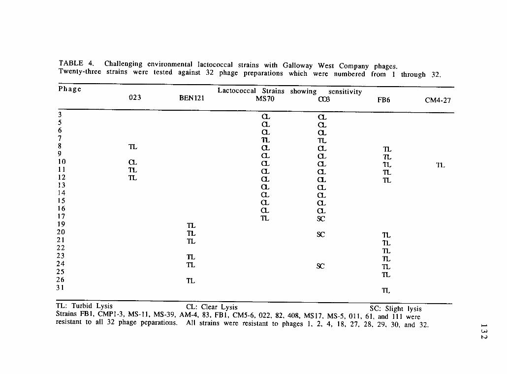
TABLE 4. Challenging environmental lactococcal strains with Galloway West Company phages.Twenty-three strains were tested against 32 phage preparations which were numbered from 1 through 32.
Phage Lactococcal Strains showing sensitivity023 BEN121 MS70 CO3 FB6 CM4-27
3
5678 TL910 CL11 it12 TL131415161719 IL20 TL21 TL2223 TL24 TL2526 TL31
SC
SC
11TLItTLIt
TLit11it11TL
it
TL
TL: Turbid Lysis CL: Clear Lysis SC: Slight lysisStrains FBI, CMP1-3, MS-11, MS-39, AM-4, 83, FBI, CM5-6, 022, 82, 408, MS17, MS-5, 011, 61, and 111 wereresistant to all 32 phage peparations. All strains were resistant to phages 1, 2, 4, 18, 27, 28, 29, 30, and 32.
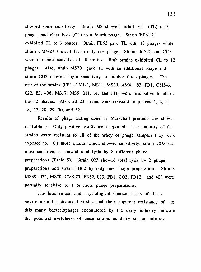
1 3 3
showed some sensitivity. Strain 023 showed turbid lysis (TL) to 3
phages and clear lysis (CL) to a fourth phage. Strain BEN121
exhibited TL to 6 phages. Strain FB62 gave TL with 12 phages while
strain CM4-27 showed TL to only one phage. Strains MS70 and CO3
were the most sensitive of all strains. Both strains exhibited CL to 12
phages. Also, strain MS70 gave TL with an additional phage and
strain CO3 showed slight sensitivity to another three phages. The
rest of the strains (FB1, CM1-3, MS11, MS39, AM4, 83, FB1, CM5-6,
022, 82, 408, MS17, MS5, 011, 61, and 111) were insensitive to all of
the 32 phages. Also, all 23 strains were resistant to phages 1, 2, 4,
18, 27, 28, 29, 30, and 32.
Results of phage testing done by Marschall products are shown
in Table 5. Only positive results were reported. The majority of the
strains wetre resistant to all of the whey or phage samples they were
exposed to. Of those strains which showed sensitivity, strain CO3 was
most sensitive; it showed total lysis by 8 different phage
preparations (Table 5). Strain 023 showed total lysis by 2 phage
preparations and strain FB62 by only one phage preparation. Strains
MS39, 022, MS70, CM4-27, FB62, 023, FB1, CO3, FB12, and 408 were
partially sensitive to 1 or more phage preparations.
The biochemical and physiological characteristics of these
environmental lactococcal strains and their apparent resistance of to
this many bacteriophages encountered by the dairy industry indicate
the potential usefulness of these strains as dairy starter cultures.

TABLE 5. Challenging the environmental lactococcal strains with Marschall productsCompany phages. Twenty-three strains were tested against 72 positive whey samples forphage (whey samples 1-72), 30 negative whey samples (whey samles 73-102), and 38single plaque isolates (SPI 1-38). The phage test reading was based on a scale of 0 (no lysis)to 3 (total lysis) based on spot test evaluation. Only those strains with readings of 1 orgreater were reported.
Whey sampleNo. CM4-27 FB62
Lactococcal Strains showing sensitivity023 FBI CO3 FB12
1 I10 311 1 212 213 1
14 1
18 221 1
26 227 1 2 2 3 228 229 231 1 I38 139 1 1 1 1 34 1
40 1 341 144 245 246 2

TABLE 5 (continued). Challenging the environmental lactococcal strains with Marschall productsCompany phages. Twenty-three strains were tested against 72 positive whey samples for phage(whey samples 1-72), 30 negative whey samples (whey samples 73-102), and 38 single plaque isolates(SPI 1-38). The phage test reading was based on a scale of 0 (no lysis) to 3 (total lysis) based on spottest evaluation. Only those strains with readings of 1 or greater were reported.
Whey sampleNo. MS39 022
Lactococcal StrainsMS70 FB62
showing023
sensitivityFB1 CO3 408
47 349 251 1
52 1
55 1
57 26 3 2 1
60 1 1
63 1
64 368 37 1
SPI 11 1
SPI 13 1 1 1
SPI 18 1 3 1
SPI 23 1
SPI 24 1 1 3SPI 34 1 2SPI 38 1
SPI 17 3

136
ACKNOWLEDGMENTS
We are thankful to Mr. Ibrahim Saleh and Mr. Peter Gadzinski
for the technical help in using the Biolog ML2 program. We thank
Mr. Jeff Fowles for facilitating the use of the Thermo Max Microplate
reader at the EPA, Corvallis, Oregon. We are grateful to Dr. E. R.
Vedamuthu, Quest International, and Dr. Randall Thunell, Galloway
West Company for the phage typing analyses.

1 3 7
REFERENCES
1. Alterthum, F. and L. 0. Ingram. 1989. Efficient ethanol
production from glucose, lactose, and xylose by recombinant
Escherichia coli. J. Appl. Environ. Microbiol. 55:1943-1948.
2. Bochner, B. 1989. Sleuthing out bacterial identities. Nature.
339(6220): 1 5 7 .
3. Bochner, B. 1989. "Breathprints" at the microbial level. ASM
News. 55(10):536.
4. Carnahan, A. M., S. W.Joseph, and J. M. Janda. 1989.
Species Identification of Aeromonas strains based on carbon
substrate oxidation profiles. J. clinic. Microbiol. 27:2128 -2129 .
5. Doering, C., P. Kampfer, S. Manulis, G. Kritzman, J.
Schneider, J. Zakrazewska, H. Schrempf, and I. Barash.
1992. Diversity among Streptomyces strains causing potato
scab. J. Appl. Environ. Microbiol. 58:3932 -3940.
6. Fredrickson, J. K., D. L. Balkwill, J. M. Zachara, S. W. Li,
F. J. Brockman, and M.A. Simmons. 1991. Physiological
diversity and distribution of heterotrophic bacteria in deep
cretaceous sediments of the Atlantic coastal plain. J. Appl.
Environ. Microbiol. 57:402 -411.

138
7. Moineau, S., J. Fortier, H. W. Ackermann, and S.Pandian. 1992. Characterization of lactococcal bacteriophages
from Quebec cheese plants. Can. J. Microbiol. 38:875 -882.
8. Salama, M. S., W. E. Sandine, and S. J. Giovannoni. 19 93 .
Isolation of lactococci and Lactococcus lactis subsp. cremoris
from nature by colony hybridization with ribosomal RNA.
Appl. Environ. Microbiol. (In review)
9. Terzaghi, B. E. and W. E. Sandine. 1975. Improved
medium for lactic streptococci and their bacteriophage. Appl.
Microbiol. 29:807-813.

139
BIBLIOGRAPHY
1. Abd-El-Malek, Y. and T. Gibson. 1948. Studies in the
bacteriology of milk. J. Dairy Res. 15:233.
2. Alberto, J. L. M., and E. C. de Macario. (Eds.). 1990.
Gene probes for bacteria. Academic Press.
3. Alterthum, F. and L. 0. Ingram. 1989. Efficient ethanol
production from glucose, lactose, and xylose by recombinant
Escherichia coli. J. Appl. Environ. Microbiol. 55:1943-1948.
4. Al-Zoreky, N., and W. E. Sandine. 1991. The Lactococcus
genus: A selective and differential agar medium. J. Food Sci.
56(6):1729-1730 & 1734.
5. Anderson, D. G., and L. L. McKay. 1983. Simple and
rapid method for isolating large plasmid DNA from lactic
streptococci. Appl. Environ. Microbiol. 46:549-552.
6. Ayebo, A. D., K. M. Shahani, and R. Dam. 1981.
Antitumor component(s) of yogurt: Fractionation. J. Dairy Sci.64:2318-2323.
7. Bergey, D. H. 1904. The source and nature of bacteria in
milk. Harrisburg. 40 p. (Pennsylvania. Department of
Agriculture. Bulletin 125).

140
8. Betzl, D., W. Ludwing, and K. H. Schleifer. 1990.
Identification of lactococci and enterococci by colony
hybridization with 23S rRNA-targeted oligonucleotide probes.
App. Environ. Microbiol. 56:2927-2929.
9. Bochner, B. 1989. Sleuthing out bacterial identities. Nature.
339(6220):157.
10. Bochner, B. 1989. "Breathprints" at the microbial level. ASM
News. 55(10):536.
11. Britschgi, T. B., and S. J. Giovannoni. 1991. Phylogenetic
analysis of a natural marine bacterioplankton population by
rRNA gene cloning and sequencing. Appl. Environ. Microbiol.
57:1707-1713.
12. Briggs, C. A. E. 1952. A note on serological classification of
Streptococcus diacetylactis (Matuszewski et al.). J. Dairy
Res. 19:167.
13. Brosius, J., J. L. Palmer, J. P. Kennedy, and H. F. Noller.
1978. Complete nucleotide sequence of a 16S rRNA gene fromEscherichia. Proceedings of the Natl. Acad Sci., USA 75:4801-
4805.
1 4 1
14. Brown, J. P. 1977. Role of gut flora in nutrition and health: a
review of recent advances in bacteriological techniques,
metabolism and factors affecting flora composition. Crit. Rev.
Food Sci. Nutr. 8:229-336.
15. Buluwela, L., A. Foster, T. Boehm, and T. H. Rabbits.1989. A rapid procedure for colony screening using nylon
filters. Nucleic Acid Res. 17:452.
16. Cavett, J. J., G. J. Dring, and A. W. Knight. 1965.
Bacterial spoilage of thawed frozen peas. J. Appl. Bact. 28:241-
245.
17. Carnahan, A. M., S. W.Joseph, and J. M. Janda. 1989.
Species Identification of Aeromonas strains based on carbon
substrate oxidation profiles. J. clinic. Microbiol. 27:2128-2129.
18. Cavett, J. J., and E. I. Garvie. 1967. Biochemical and
serological characters of three strains of streptococci reported
as Streptococcus cremoris isolated from deep- frozen peas after
thawing. J. Appl. Bacteriol. 30:377-381.
19. Collins, M. D., D. Jones, J. A. E. Farrow, R. Klipper-Balz,and K. H. Schleifer. 1984 a. Enterococcus avium sp. nov.,
nom. rev.; E. casseliflavus sp. nov., nom. rev.; E. durans sp. nov.,
nom. rev.; E. gallinarum comb. nov. and E. malodoratus sp. nov.
Int. J. System. Bact. 34:220-223.

142
20. Collins, M. D., C. Ash, J. A. E. Farrow, S. Walibanks, andA. M. Williams. 1989. 16S ribosomal ribonucleic acid
sequence analysis of lactococci and related taxa. Description of
Vagococcus fluvialis gen. nov., sp. nov. J. Appl. Bacteriol.
67:453-460.
21. Daly, C., W. E. Sandine, and P. R. Elliker. 1972.
Interaction of food starter cultures and food-borne pathogens:
Streptococcus diacetylactis versus food pathogens. J. Milk Food
Technol. 35:349-357.
22. Dellaglio, F. and E. Santi. 1984. Role of lactic acid bacteria
during the forages preservation. Microbiol. Alim. Nutr. 2:109-
121 .
23. DeLong, E. F., G. S. Wickham, and N. R. Pace. 1989.
Phylogenetic stains: Ribosomal RNA-based probes for the
identification of single cells. Science 243:1360-1362.
24. Doering, C., P. Kampfer, S. Manulis, G. Kritzman, J.
Schneider, J. Zakrazewska, H. Schrempf, and 1. Barash.
1992. Diversity among Streptomyces strains causing potato
scab. J. Appl. Environ. Microbiol. 58:3932-3940.
25. Elliker, P. R., W. E. Sandine, B. A. Hauser, and W. K.
Moseley. 1964. Influence of culturing cottage cheese dressing
143
with different organisms on flavor and keeping quality. J.
Dairy Sci. 47:680.
2 6 . Esten, W. M. 1909. Bacterium lactis acidi and its sources.
Storrs. 27p. (Connecticut. Agricultural Experiment Station.
Bulletin 59).
2 7 . Evans, A. C. 1916. The bacteria of milk freshly drawn
from normal udders. J. Infectious Diseases. 18:43 7
47 6.
2 8 . Fenton, M. P. 1987. An investigation into the sources of
lactic acid bacteria in grass silage. J. Appl. Bacteriol. 62:181
18 8.
2 9 . Fredrickson, J. K., D. L. Balkwill, J. M. Zachara, S. W. Li,
F. J. Brockman, and M.A. Simmons. 1991. Physiological
diversity and distribution of heterotrophic bacteria in deep
cretaceous sediments of the Atlantic coastal plain. J. Appl.
Environ. Microbiol. 57:402 -411.
3 0 . Garvie, E. I. 1978. Streptococcus raffinolactis (Orla Jensen
and Hansen); a group N Streptococcus found in raw milk. Int. J.
Syst. Bact. 28:190-193.
3 1 . Giovannoni, S. J., E. F. Delong, G. J. Olsen, and N. R.
Pace. 1988. Phylogenetic genus-specific oligonucleotide

144
probes for identification of single microbial cells. J. Bact.
170:720-72.
32. Giovannoni, S. J., S. Turner, G. J. Olsen, S. Barns, D.
Lane, and N. R. Pace. 1988. Evolutionary relationships
among cyanobacteria and green chloroplasts. J. Bacteriol.
170:3584-3592.
33. Golden, B. R., and S. L. Gorbach. 1983. The effect of oral
administration of Lactobacillus and antibiotics on intestinal
bacterial activity and chemical induction of large bowel tumor.
Develop. Ind. Microbiol. 25:139-150.
34. Grunewald, K. K. 1982. Serum cholesterol levels in rats fed
skim milk fermented with Lactobacillus acidophilus. J. Food
Sci. 47:2708-2709.
35. Harding, H. A. and J. K. Wilson. 1909. A study of the
udder flora of cows. Geneva, 40 p. (New York. Agricultural
Experiment station. Technical Bulletin 27).
36. Harrigan, W. F., and M. E. McCance. 1976.
Laboratory methods in food and dairy Microbiology.
revised edn., W. E. Harrigan, Academic press, pp. 74.

145
37. Hashimoto, H., R. Noborisaka, R. Yanagawa. 1974.
Distribution of motile streptococci in faeces of man and animal
and in river and sea water. Jap. J. Bact. 29:387-393.
38. Hashimoto, H., H. Kawakami, K. Tomakane, Z. Yoshii, G.Hahn, and A. Tolle. 1979. Isolation and characterization of
motile group N streptococci. J. Fac. Appl. Biol. Sci. Hiroshima
Univ. 18:197-216.
39. Heap, H. A., and R. C. Lawrence. 1977. The contribution of
strarter strains to the level of phage infection in a commercial
cheese factory. N. Z. J. Dairy sci. Technol. 12:213-218.
40. Hepner, G., R. Fried, S. S. Jeor, and L. Fusetti. 1977.
Hypochloresterolemic effect of yogurt and milk. Am. J. Clin.
Nutr. 32:19-24.
41. Hirsch, A. 1952. The evolution of lactic streptococci. J.
Dairy Res. 19:290-293.
42. Hunter, G. J. E. 1939. Examples of variation within pure
cultures of Streptococcus cremoris. J. Dairy Research
10:46-470.
43. hat, A. L., C. D. Driggers, M. Colberg, M. A. Reiber, andM. H. Adams. 1989. Comparison of the DNA probe to culture

146
methods for the detection of Salmonella on poultry carcasses
and processing waters. J. Food Protection. 52(8):564-57.
44. Kapperud, G., K. Dommarsnes, M. Skurnik, and E.Hornes. 1990. A synthetic oligonucleotide probe and a cloned
polynucleotide probe based on the yopA gene for detection and
enumeration of virulent Yersinia enterocolitica. Appl. Environ.
Microbiol. 56(1): 17-23.
45. King, N. S. 1948. A modification of the Voges-
Proskauer test forrapid colorimetric determination of
acetylmethylcarbinol plus diacetyl in the buttercultures.
Dairy Ind. 13:860.
46. King, N. S. and J. A. Koburger. 1970. Characterization of
some Group N streptococci. J. Dairy Sci. 53:402-409.
47. Klijn, N., A. H. Weerkamp, and W. E. De Vos. 1991.
Identification of mesophilic lactic acid bacteria by using
polymerase chain reaction-amplified variable regions of 16S
rRNA and specific DNA probes. App. Environ. Microbiol.
57 (11 ) : 3390-3393.
48. Klipper-Balz, R. and K. H. Schleifer. 1984. Nucleic acid
hybridization and cell wall composition studies of pyogenic
streptococci. FEMS Microbiol. Lett 24:355-364.

147
49. Klipper-Balz, R., G. Fisher, and K. H. Schleifer. 1982.
Nucleic acid hybridization of group N and group D streptococci.
Curr. Microbiol. 7:245-250.
50. Klipper-Balz, R., B. L. Williams, R. Lutticken and K. H.
Schleifer. 1984. Relatedness of 'Streptococcus milleri' with
Streptococcus anginosus and Streptococcus constellatus.
System. Appl. Microbiol. 5:494-500.
51. Kumar, A., P. Tchen, F. Roullet, and J. Cohen. 1987.
Nonradioactive labeling of synthetic oligonucleotide probes
with terminal deoxynucleotidyl transferase. Analyt. Biochem.
169:376-382.
52. Lane, D. L., B. Pace, G. J. Olsen, D. A. Stahl, M. L. Sogin,
and N. R. Pace. 1985. Rapid determination of 16S ribosomal
sequences for phylogenetic analyses. Proceedings of the Natl.
Acad. Sci., USA. 82:6955-6959.
53. Lane, D. L., K. G. Field, B. Pace, G. J. Olsen, and N. R.
Pace. 1988. Reverse transcriptase of ribosomal RNA for
phylogenetic analysis. Meth. Enz. 167:138-144.
5 4. Lawrence, R. C., T. D. Thomas, and B. E. Terzaghi. 1976.
Reviews of the progress of dairy science: Cheese starters. J.
Dairy Res. 43:141 -193.

148
55. Liesack, W. and E. Stackebrandt. 1992. Occurrence of
novel groups of the domain Bacteria as revealed by analysis of
genetic material isolated from an Australian terrestrial
environment. J. Bacteriol. 174(15):5072-5078.
56. Ludwig W., E. Seewaldt, R. Klipper-Balz, K. H. Schleifer,
C. R. Magrum, C. R. Woese, G. E. Fox, and E.
Stackebrandt. 1985. The phylogenetic position of
Streptococcus and Enterococcus. J. Gen. Microbiol. 131:543-
551.
57. McKay, L. L. 1982. Regulation of lactose metabolism in dairy
Streptococci. Developments in Food Microbiology-1. ed. R.
Davies. Applied Science Publisher LTD. pp. 153-182.
58. Moineau, S., J. Fortier, H. W. Ackermann, and S.
Pandian. 1992. Characterization of lactococcal bacteriophages
from Quebec cheese plants. Can. J. Microbiol. 38:875-882.
59. Mundt, J. 0., and A. H. Johnson. 1959. Physiological
properties of group D streptococci isolated from plants. Food
Res. 24:218.
60. Mundt, J. 0., W. F. Graham, and I. E. McCarty. 1967.
Spherical lactic acid producing bacteria on southern grown raw
and processed vegetables. Appl. Microbiol. 15 :1303 -1308.

149
61. Nelson, G. A. and H. R. Thornton. 1952. The lactic
streptococci in Edmonton milks and creams. Canadian Journal
of Technology. 30:130-135.
62. Nichols, A. A. and M. Hoyle. 1949. Bacteriophages in
typing lactic streptococci. J. Dairy Res. 16 :167-173.
63. Nivens, C. J. Jr., K. L. Smiley, and J. M. Sherman. 1942.
The hydrolysis of arginine by streptococci. J. Bacteriol. 43:65 1
6 6 O.
64. Olsen, G. J., D. J. Lane, S. J. Giovannoni, and N. R. Pace.1986. Microbial ecology and evolution: A ribosomal RNA
approach. Ann. Rev. Microbiol. 40:337-365.
65. Orla-Jensen, S. 1942. The lactic acid bacteria, 2nd ed., E.
Munksgaard, Copenhagen, pp. 106-107.
66. Orberg, P. K. 1985. Antimicrobial resistance and plasmid
properties of Leuconostoc and group N Streptococcus. Ph.D.
Thesis. Oregon State Univ., Corvallis.
67. Pace, N. R., R. J. Stahl, D. J. Lane, and G. J. Olsen. 1986.
The analysis of natural microbial populations by ribosomal
RNA sequences. Adv. Microb. Ecol. 9:1-55.

150
68 . Perry, K. D. 1961. A comparison of the influence of
Streptococcus lactis and Streptococcus cremoris starters on the
flavor of Cheddar cheese. J. Dairy Research. 28:221.
69. Pinter, I. J. 1940. The occurrence of streptococci on
plant material. Master's thesis. Ithaca, Cornell University. 17
numb. leaves.
70. Radich, P. C. 1968. An ecological study of the lactic
streptococci. M.S. Thesis, Oregon State University, Corvallis.
PP 51.
71. Reddy, M. S., E. R. Vedamuthu, and G. W. Reinbold.1971. A differential broth for separating the lactic
streptococci. J. Milk and Food Tech. 34(1):43 -45 .
72. Rehnstam, A., A. Norqvist, H. Wolf-Watz, and A.
Hagstrom. 1989. Identification of Vibrio anguillarum in fish
by using partial 16S rRNA sequences and a specific 16S rRNA
oligonucleotide probe. Appl. Environ. Microbiol. 55(8):1907-
1910 .
73. Rogers, L. A. and Arnold 0. Dahlberg. 1914. The origin of
some of the streptococci found in milk. J. Agricul. Res. 1:491
511 .

1 5 1
74. Romaniuk, P. J., and T. J. Trust. 1987. Identification of
Campylobacter species by southern hybridization of genomic
DNA using oligonucletide probes for 16S rRNA genes. FEMS
Microbiol. Lett. 43:331-335.
75. Salama, M. S., W. E. Sandine, and S. J. Giovannoni. 1991.
Development and application of oligonucleotide probes for
identification of Lactococcus lactis subsp. cremoris. Appl.
Environ. Microbiol. 57:1313 -1318.
76. Salama, M. S., W. E. Sandine, and S. J. Giovannoni. 1993.
Isolation of lactococci and Lactococcus lactis subsp. cremoris
from nature by colony hybridization with ribosomal RNA. Appl.
Environ. Microbiol. (under review)
77. Sandine, W. E. 1985. The streptococci: Milk products.
In Bacterial Starter Cultures for Foods. Ed. Stanley E. Gilliland.
CRC press, Inc. Boca Raton, Florida. pp. 5-23.
78. Sandine, W. E. 1987. Looking backward and forward at the
practical applications of genetic researches on lactic acid
bacteria. FEMS Microbiol. Rev. 46:205-220.
79. Sandine, W. E. 1988. New nomenclature of the non-rod
shaped lactic acid bacteria. Biochemie 70:519 -522.

152
80. Sandine, W. E., K. S. Muralidhara, P. R. Elliker, and D. C.England. 1972. Lactic acid in food and health: a review with
special reference to enteropathogenic Escherichia coli as well as
enteric diseasea and their treatment with antibiotics and
lactobacilli. J. Milk Food Technol. 35:691-702.
81. Sandine, W. E., Radich, P. C., and P. R. Elliker. 1972.
Ecology of the lactic streptocci: A review. J. Milk Food Technol.
35:176-185.
82. Schieblich, M. and W. Schonherr. 1957. Streptococci in
acid fodders and their distribution in the raw material.
Tierernahr. Futtermittelk 11:232. Dairy Sci. Abstr. 19:231.
83. Schleifer, K. H. and R. Klipper-Balz. 1984. Transfer of
Streptococcus faecalis and Streptococcus faecium to the genus
Enterococcus nom. rev. as Enterococcus faecalis comb. nov. and
Enterococcus faecium comb. nov.. Int. J. System. Bact. 34:31-
34.
84. Schleifer, K. H. and R. Klipper-Balz. 1987. Molecular andchemotaxonomic approaches to the classification of
streptococci, enterococci and lactococci: a review. System. and
Appl. Microbiol. 10:1-19.
85. Schleifer, K. H., R. Klipper-Balz, J. Kraus, R. Gebring.

1 5 3
1984. Relatedness and classification of Streptococcus mutans
and 'mutans-like' streptococci. J. Dent. Res. 63:1046 -1050.
86. Schleifer, K. H., J. Kraus, C. Dvorak, R. Klipper-Balz, M.D. Collins, and W. Fisher. 1985. Transfer of Streptococcus
lactis and related streptococci to the genus Lactococcus gen.
nov. System. Appl. Microbiol. 6:183 -195 .
87. Schmidt, T. M., E. F. Delong, and N. R. Pace. 1991.
Analysis of a marine picoplankton community by 16S rRNA
gene cloning and sequencing. J. Bacteriol. 173(14 ):4371
4378 .
88 . Sgaramella, V., and H. G. Khorana. 1972. Total synthesis
of the structural gene for an alanine transfer RNA from yeast.
Enzymatic joining of the chemically synthesized
polynucleotides to form the DNA duplex representing
nucleotide sequence 1 to 20. J. Mol. Biol. 72:427 -444.
89. Shahani, K. M., J. R. Vakil, and A. Kilara. 1976. Natural
antibiotic activity of Lactobacillus acidophilus and bulgaricus,
I. Cultural conditions for the production of antibiotics. Cult.
Dairy Prod. J. 11(4): 14 - 17 .
90. Sherman, J. M. and E. G. Hastings. 1915. The presence of
Streptococci in the milk of normal animals. Creamery and Milk
Plant Monthly, no. 6, 11-12.

154
91. Simone, C. De., S. B. Bianchi, R. Negri, M. Ferrazzi, L.
Baldinelli, and R. Vase ly. 1986. The adjuvant effect of
yogurt on production of gamma interferon by Con A-
stimulated human peripheral blood lymphocytes. Nutr. Rep.
Int. 33:419-433.
92. Stackebrandt, E. and M. Teuber. 1988. Molecular
taxonomy and phylogenetic position of lactic acid bacteria.
Biochimie 70:317-324.
93. Stark, P. and J. M. Sherman. 1935. Concerning the
habitat of Streptococcus lactis. Journal of Bacteriology.
30:639-646.
94. Swarthing, P. F. 1951. Biochemical and serological
properties of some citric acid fermenting streptococci from
milk and dairy products. J. Dairy Res. 18:256.
95. Terzaghi, B. E. and W. E. Sandine. 1975. Improved
medium for lactic streptococci and their bacteriophage. Appl.
Microbiol. 29:807-813.
96. Tsien, H. C., B. J. Bratina, K. Tsuji, and R. S. Hanson.1990. Use of oligonucleotide signature probes for identification
of physiological groups of methylotrophic bacteria. Appl.
Environ. Microbiol. 56:2858-2865.
155
97. Vedamuthu, E. R., W. E. Sandine, and P. R. Elliker.1969. Flavor and texture in Cheddar cheese. I. Role of mixed
strain lactic starter cultures. J. Dairy Sci. 49:144 -150.
98. Vedamuthu, E. R., W. E. Sandine, and P. R. Elliker.1969. Flavor and texture in Cheddar cheese. II. Carbonyl
compounds produced by mixed strain lactic starter cultures. J.
Dairy Sci. 49:151-157.
99. Walia, S., A. Khan, and N. Rosenthal. 1990. Construction
and applications of DNA probes for detection of polychlorinated
biphenyl-degrading genotypes in toxic organic contaminated
soil environments. Appl. Environ. Microbiol. 56(1):254 -259.
100. Walkbanks, S., A. J. Martinez-Murcia, J. L. Phillips, andM. D. Collins. 1990. 16S rRNA sequence determination for
members of the genus Carnobacterium and related lactic acid
bacteria and description of Vagococcus salmoninarum sp. nov.
Int. J. Syst. Bact. 40(3):224-230.
101. Walker, E. E. 1902. The bacterial flora of freshly drawn
milk. Journal of Applied Microscopy and Laboratory Methods.
5 :2029 -2044.
102. Wallace, R. B., J. Shaffer, R. F. Murphy, J. Bonner, T.
Hirose, and K. Itakura. 1979. Hybridization of synthetic
1 5 6
oligodioxyribonucleotides to AX174 DNA: the effect of a single
base pair mismatch. Nucleic Acids Res. 6:3543-3557.
103. Weber, G. H. and W. A. Broich. 1986. Shelf life extension
of cultured dairy foods. Cult. Dairy Prod. J. 21 (4):19 -23 .
104. Williams, A. M., J. A. E. Farrow, and M. D. Collins. 1989.
Reverse transcriptase sequencing of 16S rRNA from
Streptococcus cecorum. Lett. App. Microbiol. 8:185 -189.
105. Williams, A. M., J. L. Fryer, and M. D. Collins. 1990.
Lactococcus piscium sp. nov., a new Lactoccoccus species from
salmonid fish. FEMS Microbiol. Let. 68:109-114.
106. Willem, M., and G. Simons. 1988. Molecular cloning of
lactose genes in dairy lactic streptococci: the phospho-B-
galactosidase and B-galactosidase genes and their expression
products. Biochemie 70:461.
107. Woese, C. R., E. Stackebrandt, T. J. Macke, and G. E. Fox.1985. A phylogenetic definition of the major eubacterial taxa.
Syst. Appl. Microbiol. 6:143 -151.
108. Yawger, E. S., and J. M. Sherman. 1937 a. Variants of
Streptococcus lactis which do not ferment lactose. J. Dairy Sci.
20:83.

1 5 7
109. Yawger, E. S., and J. M. Sherman. 1937 b. Streptococcuscremoris. J. Dairy Sci. 20:205.
110. Yoshioka, H., K-i. Iseki, and K. Fujita. 1983. Development
and differences of intestinal flora in the neonatal period in
breast-fed and bottle-fed infants. Pediatrics.72:317-321.