large cell morphology, cmyc+ tumour cells, and pd-1
TRANSCRIPT
RESEARCH Open Access
Large cell morphology, CMYC+ tumourcells, and PD-1+ tumour cell/intense PD-L1+ cell reactions are important prognosticfactors in nodal peripheral T-celllymphomas with T follicular helper markersYasuhito Mihashi1,2, Shoichi Kimura1,2, Hiromi Iwasaki3, Yumi Oshiro4, Yasushi Takamatsu5, Shigeto Kawauchi6,Shohei Shimajiri7, Kenji Ishizuka8 and Morishige Takeshita1*
Abstract
Background: The clinicopathological characteristics and prognostic factors in nodal peripheral T-cell lymphomas(PTCLs) with two or more T follicular helper markers (TFH+) are not adequately investigated.
Methods: Immunohistologically, we selected 22 patients with TFH+ lymphoma (PTCL-TFH) in 47 of PTCL-nototherwise specified (NOS), and subclassified into large and small cell groups. We compared the two groups with 39angioimmunoblastic T-cell lymphoma (AITL) and seven follicular T-cell lymphoma (F-TCL) patients. Prognosticfactors were analysed by overall survival in patients with three types of TFH+ PTCLs.
Results: Thirteen large cell and nine small cell PTCL-TFH patients had more than two TFH markers includingprogrammed cell death-1 (PD-1). Large cell PTCL-TFH showed frequent CMYC expression in 10 patients (77%), and fourof 11 large cell group (36%) had somatic RHOA G17V gene mutation by Sanger sequencing. Large cell PTCL-TFHpatients showed significantly worse prognosis than those of the small cell group, AITL, and F-TCL (p < 0.05). In TFH+PTCLs, CMYC+ tumour cells, and combined PD-1 ligand 1 (PD-L1) + tumour cells and intense reaction of PD-L1+ non-neoplastic cells (high PD-L1+ cell group) were significantly poor prognostic factors (p < 0.05). Combinations of CMYC+or PD-1+ tumour cells and high PD-L1+ cell group indicated significantly poor prognosis (p < 0.01).
Conclusion: Large cell PTCL-TFH indicated poor prognosis in TFH+ PTCLs. These data suggested that CMYC+ tumourcells and intense PD-L1+ cell reaction influenced tumour cell progression in TFH+ PTCLs, and PD-1+ tumour cell/intense PD-L1+ cell reactions may play a role in immune evasion.
Keywords: AITL, CMYC, PD-1, PD-L1, Peripheral T-cell lymphoma, T follicular helper cell
© The Author(s). 2021 Open Access This article is licensed under a Creative Commons Attribution 4.0 International License,which permits use, sharing, adaptation, distribution and reproduction in any medium or format, as long as you giveappropriate credit to the original author(s) and the source, provide a link to the Creative Commons licence, and indicate ifchanges were made. The images or other third party material in this article are included in the article's Creative Commonslicence, unless indicated otherwise in a credit line to the material. If material is not included in the article's Creative Commonslicence and your intended use is not permitted by statutory regulation or exceeds the permitted use, you will need to obtainpermission directly from the copyright holder. To view a copy of this licence, visit http://creativecommons.org/licenses/by/4.0/.The Creative Commons Public Domain Dedication waiver (http://creativecommons.org/publicdomain/zero/1.0/) applies to thedata made available in this article, unless otherwise stated in a credit line to the data.
* Correspondence: [email protected] of Pathology, Faculty of Medicine, Fukuoka University, 7-45-1Nanakuma, Jonan-ku, Fukuoka 814-0180, JapanFull list of author information is available at the end of the article
Mihashi et al. Diagnostic Pathology (2021) 16:101 https://doi.org/10.1186/s13000-021-01163-7
BackgroundT follicular helper (TFH) cells are mainly located in ger-minal centres and frequently express CD4, programmedcell death-1 (PD-1), CD10, chemokine (C-X-C motif)ligand (CXCL) 13, BCL6 and inducible T-cell co-stimulator [1]. In peripheral T-cell lymphomas (PTCLs),angioimmunoblastic T-cell lymphoma (AITL) and follicu-lar T-cell lymphoma (F-TCL) are derived from TFH cellsand defined by expression of two or more TFH markers(TFH+) [2, 3]. Furthermore, less than half of patients withPTCL-not otherwise specified (NOS) (41%) exhibit morethan two TFH markers (PTCL-TFH) [4], and the PTCL-TFH patients show similar incidences of gene mutationsof Ras homolog family member A (RHOA) G17V and tetmethylcytosine dioxygenase (TET)2 to those of AITL andF-TCL [5]. No prognostic differences have been reportedamong PTCL-TFH and TFH− PTCL-NOS patients andAITL. In PTCL-NOS, more than 70% large tumour cellsand international prognostic index were significant poorprognostic factors (p = 0.008, p < 0.001, respectively) [6].Transcription factor CMYC plays a role in tumour cell
proliferation and progression in high grade B-celllymphoma, T-acute lymphoblastic leukaemia (T-ALL)and adult T-cell leukaemia/lymphoma (ATLL) [7–11].More than 30% CMYC expression in lymphoma cellswas a significant prognostic factor in AITL patients (p =0.008), but not in PTCL-NOS [12]. CMYC controls thefunction of PD-1 ligand 1 (PD-L1), which has immuno-suppressive effects and promotes tumour cell growth inmouse and human T-ALL, and in solid tumours [13].CMYC expression in non-small cell lung cancer signifi-cantly correlated with PD-L1, and patients with CMYC+and PD-L1+ tumour cells had a worse prognosis thanother subgroups (p < 0.05) [14].Interactions between PD-1 and PD-L1 play a role in
immune suppression during inflammatory processes,and PD-L1 expression induces an immune evasionmechanism exploited by various malignancies [1, 15]. InT/natural killer (NK) cell neoplasia, PD-L1+ tumourcells were frequently found in anaplastic lymphomakinase (ALK) + and ALK− systemic anaplastic large celllymphoma (sALCL), occasionally in PTCL-NOS, andrarely in AITL [16–19]. Patients demonstrating com-bined PD-L1+ tumour cells and intense reaction of PD-L1+ non-neoplastic cells (high PD-L1+ group) showedsignificantly poorer prognosis compared with the lowPD-L1+ group in the above four types of PTCLs [18].The combination of PD-1+ tumour cells and high PD-L1+ group was related to shorter survival in AITLpatients (p = 0.051), but not PTCL-NOS [20].In the current study, we initially selected PTCL-TFH
from PTCL-NOS by immunohistology, and subclassifiedpatients into large and small cell groups. We then com-pared clinicopathological findings of the two groups of
PTCL-TFH with those of AITL and F-TCL. The largecell PTCL-TFH patients sometimes had the RHOAG17V mutation, which indicated a group with poorprognosis in TFH+ PTCLs [21]. Furthermore, CMYC+tumour cells and the combination of PD-1+ tumourcells and high PD-L1+ group indicated significantly poorprognostic factors in patients with three types of TFH+PTCLs by uni- and multivariate analyses. It was highlysuggested that histology, CMYC+ or PD-1+ tumour cell/intense PD-L1+ cell reactions were significantly influen-tial on tumour progression and patient prognosis inTFH+ PTCLs.
MethodsPatient selection, histological classification and clinical findingsRegistered patients were retrieved retrospectively fromthe Department of Pathology, Fukuoka University, from1990 to 2019. Histological classification was performedaccording to the WHO classification in 2017 [2, 22].Four TFH markers (PD-1, BCL6, CXCL13 and CD10)were examined by immunohistochemistry. There was nodifference in overall survival (OS) between patients withtwo (n = 26) and more than three (n = 38) TFH markers(p = 0.188), and the five-year survivals were 46 and 51%,respectively. Therefore, more than two TFH markerswas decided as TFH phenotype. Diffuse infiltrate of atyp-ical CD4+ lymphocytes with more than two TFHmarkers, neoplastic clear cell nests, prominent prolifera-tion of high endothelial venules and CD21+ dendriticcell nests were main criteria of AITL. Scattered andpatchy infiltrates of plasma cells, histiocytes and eosino-phils were reference findings of AITL. PTCL-TFH wasdefined by lacking the typical histological features ofAITL and having more than two TFH markers. FollicularTCL (F-TCL) was definite by nodular proliferation ofatypical TFH+ lymphocytes and lacing AITL features. 22patients with nodal PTCL-TFH, 25 nodal TFH− PTCL-NOS, 39 AITL and 7 F-TCL patients were examined inthis study. Criteria of small, medium and large tumour cellsizes were in accordance with those of mantle cells, cen-trocytes and centroblasts in lymphoid follicles. AmongPTCLs, the large cell group was characterised by diffuselynon-cohesive proliferation of ≥50% large lymphoma cellswith distinct nucleoli. The small cell group consisted ofpredominantly medium-sized (n = 7) and small cell (n =20) lymphomas. The small cell group included 10 cases ofLennert lymphoma. Corresponding medical records werereviewed to obtain clinical information, including AnnArbor stage, treatments and overall survival.
Histology, immunohistology, and detection of EBV-encoded RNAExcised tissue specimens were fixed in 10% formalin togenerate formalin-fixed and paraffin embedded (FFPE)
Mihashi et al. Diagnostic Pathology (2021) 16:101 Page 2 of 10

wax samples and stained with haematoxylin and eosin.Immunohistology was performed on the tumour tissuesusing the Leica Bond III automated stainer (LeicaBiosystems, Buffalo Grove, IL, USA). Antibodies againstthe following proteins were used: CD3 (PS1, Leica,Newcastle, UK), CD4 (4B12, Leica), CD8 (C81/44B, Leica),CD10 (56C6, Leica), CD25 (interleukin 2 receptor [IL2R],4C9, Leica), CD30 (BerH2, DakoCytomation, Glostrup,Denmark), PD-1 (NAT105, Abcam, Cambridge, MA),BCL6 (LN22, Leica), CXCL13 (BLC, R&D, Minneapolis,MN), CMYC (Y69, Abcam), MIB1 (MIB1, Dako), PD-L1(E1L3N, Cell Signaling, Danvers, MA), CD20 (L26,Nichirei, Tokyo), and CD21 (1F8, Dako). Tumour cellcounts were semi-quantitatively calculated by two pathol-ogists and percentages of antibody-positive cells were de-termined (0, 5, and 10%–100% in 10% increments) in overfive high power fields [11]. For the four TFH markers,samples with ≥20% labelling of the tumour cells were con-sidered positive [4]. Expression of CMYC, MIB1 and PD-L1 in ≥50% atypical lymphoid cells was estimated as posi-tive (n+) [16], and amount of PD-L1+ histiocytes and den-dritic cells in the entire cell populations was scored asfollows: R0 (no staining), R1+ (a few cells to < 5%), R2+ (≥5% – < 20%) and R3+ (≥ 20%). For the other antibodies,samples with ≥30% labelling of the tumour cells wereconsidered positive. The presence of EBV infection wasdetermined by in situ hybridisation of EBV-encoded RNA(EBERs) + nuclear signals (BOND EBER probe, Leica).
Quantitative real time polymerase chain reactionTotal RNAs were extracted from FFPE tumour speci-mens of 42 patients using the NucleoSpin total RNAFFPEXS (Macherey-Nagel, Duren, Germany), accordingto the manufacturer’s instructions, on a real-time PCRmachine (Mini OpticonTM, BioRad, Hercules, CA,USA). All samples were tested for expression of CMYC(assay ID: Hs00905030_m1, amplicon size 87 bp) [11]. Inaddition, samples were analysed for expression of GUSB(Hs99999908_m1), TBP (Hs00427620_m1), and ABL1(Hs00245443_m1), which were used for normalisation inthe final analysis.
Detection of RHOA G17V mutation by Sanger sequencingDNA samples from FFPE tumour tissue were extractedusing a GenElute™ Mammalian DNA Miniprep Kit(Sigma-Aldrich, St. Louis, MO, USA). Detection ofRHOA G17V mutation and wild type were assessed byallele-specific PCR. For RHOA amplification, PCR wasperformed with AmpliTaq gold (Thermo Fisher Scien-tific, Waltham, MA, USA) using 40 ng genomic DNA,0.3 μM primers, and 2 μL AmpliTaq gold master mix. APCR-amplified product of 244 bp, including the codonfor the 17th amino-acid, was obtained in 53 patients,and direct sequencing of these products was performed.
The coding DNA position 50G > T mutation of theRHOA gene predicted change of the wild-type G (Gly)to the mutant type V (Val) [21].
Statistical analysisAll pairwise comparisons of categorised variables be-tween the histological groups and types of PTCLs wereperformed using the χ2 or Fisher’s exact test. Of the 93recruited patients, 87 PTCL patients were examined forclinical outcome. Outcome was determined by calculat-ing cumulative survival from time of diagnosis to date ofthe last follow up or death. Overall survival (OS) curveswere generated using the Kaplan–Meier method withlog-rank tests, and analysed by the proportional Hazardmodel. A p value < 0.05 was considered statisticallysignificant. Analyses were performed using software JMP10 (SAS Institute, Cary, NC, USA).
ResultsClinical featuresThe clinical features and immunohistological findings of22 patients with PTCL-TFH and 25 with PTCL-NOS, 39AITL and 7 F-TCL are shown in Table 1. Thirteen of 22PTCL-TFH patients (59%) were composed of large celllymphoma and the remaining nine (41%) were small cell.Six of 11 large cell PTCL-TFH patients (55%) showed≥5000 U/ml sIL2R, which was significantly higher thanthat observed in small cell PTCL-NOS (13%) and F-TCL(0%) (p = 0.008, p = 0.039, respectively). Patients in thefour groups of TFH+ PTCLs frequently showed ad-vanced clinical stages III and IV.
Histological, immunohistological, and genetic findings,and EBV infectionLarge cell PTCL-TFH patients had significantly lowerpopulations of clear neoplastic cells and fewer reactionsof eosinophils and plasma cells than those of AITL(Fig. 1a, b; both p < 0.01). Large cell PTCL-TFHexpressed more than two TFH markers; among themPD-1 was positive in 12 patients (92%; Fig. 2a), BCL6 in12 (92%), CXCL13 in seven (54%; Fig. 2b) and CD10 inseven (54%). Small cell PTCL-TFH showed frequent ex-pressions of PD-1 (100%) and BCL6 (89%), but rarely ofCD10 (11%). Four of 10 Lennert lymphoma patientsshowed more than two TFH markers (Fig. 1c, f). Largecell PTCL-TFH showed frequent expression of CD25 ineight patients (62%) and CD30 in five (39%; Fig. 1d, e),which were significantly higher than one (11%) and 0(0%), respectively, in small cell PTCL-TFH, and eight(21%) and one (4%), respectively, in AITL (p < 0.05, p <0.01, respectively). Scattered and patchy infiltrates ofCD30+ lymphoma cells were detected in the five largecell PTCL-TFH patients. Large cell PTCL-TFH was fre-quently positive for CMYC in 10 patients (77%, Fig. 2c)
Mihashi et al. Diagnostic Pathology (2021) 16:101 Page 3 of 10

and MIB1 in 13 (100%), which were significantly higherthan 0 (0%) and seven (78%), respectively, in small cellPTCL-TFH, and 12 (31%) and 29 (74%), respectively, inAITL (p < 0.05, p < 0.01, respectively). Mean CMYCmRNA was 4.1 in tumour specimens of 42 patients,and ≥ 4.1 CMYC mRNA was detected in six of sevenlarge cell PTCL-TFH patients (86%), which was signifi-cantly higher than in small cell PTCL-TFH (0/5, 0%)and F-TCL (0/5, 0%) (both p < 0.05). The combination ofPD-L1+ tumour cells and intense reaction (R3+) of PD-L1+ non-neoplastic cells (high PD-L1+ group) wasfound in eight large cell PTCL-TFH patients (62%),which was significantly higher than in F-TCL (0%) (p =0.036; Fig. 2d). Scattered EBERs+ lymphocytes in thebackground were found in large cell (92%) and small cell(89%) PTCL-TFH patients, compared with large cell(29%) and small cell (56%) PTCL-NOS groups (p < 0.01,p < 0.05, respectively).
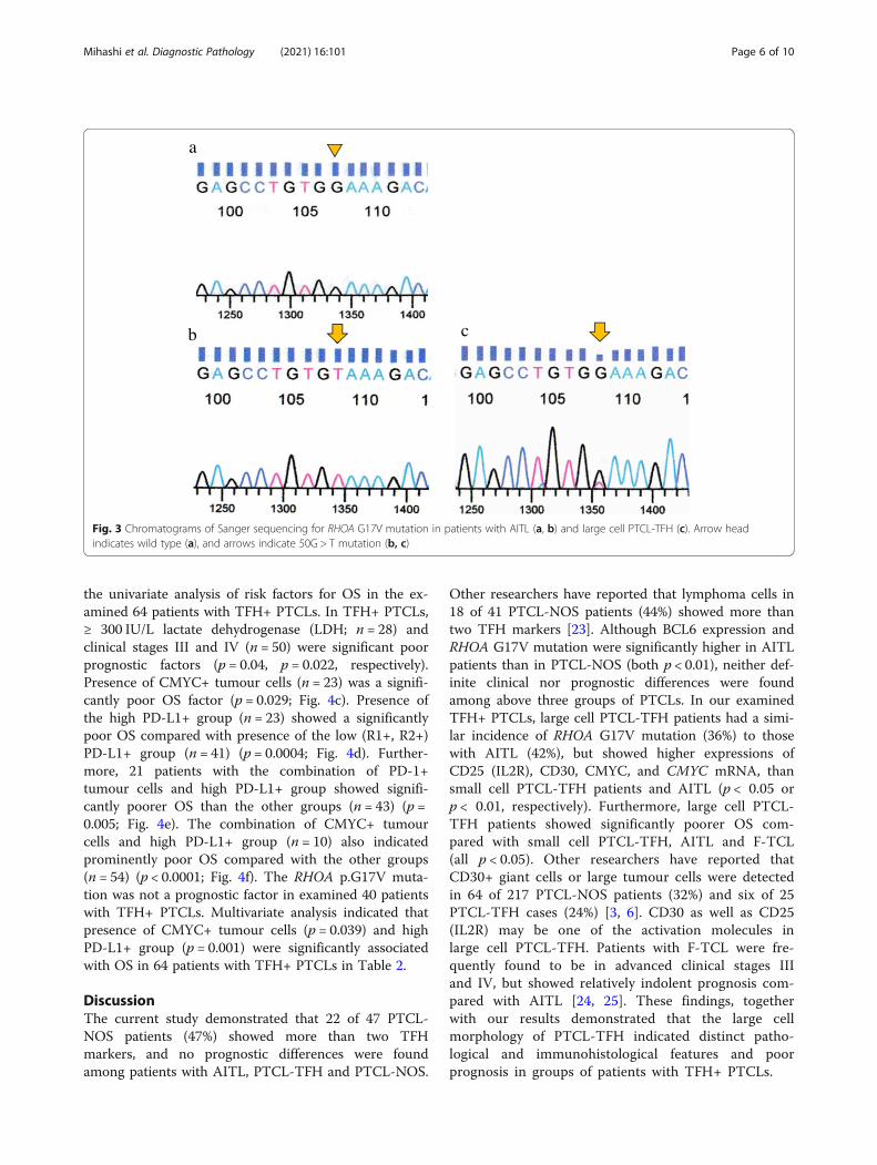
RHOA p.G17V mutation by Sanger sequencingBy Sanger sequencing, four of 11 large cell PTCL-TFHpatients (36%), one of six small cell PTCL-TFH (17%),
eight of 19 of AITL (42%), two of four F-TCL (50%) andone of 13 PTCL-NOS (8%) patients showed the RHOAp.G17V mutation (Fig. 3), but no significant differenceswere found among the groups.
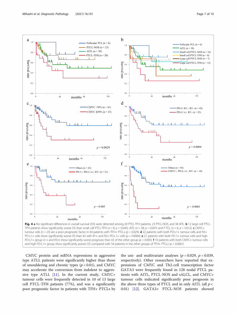
Treatments, prognosis and prognostic factors in TFH+PTCLsOf the total cohort, we examined treatments and out-come in 87 patients. Chemotherapies including cyclo-phosphamide, doxorubicin, vincristine, and prednisone(CHOP) or pirarubicin, cyclophosphamide, vincristine,and prednisolone (THP-COP) were mainly adminis-trated in the all examined patients. Nine of 20 PTCL-TFH patients (45%), seven of 23 PTCL-NOS (30%), 15of 38 AITL (40%) and one of six F-TCL (17%) died ofdisease. The examined PTCL-TFH, PTCL-NOS, AITLand F-TCL patients showed no prognostic differencesbetween groups (Fig. 4a). Twelve patients with large cellPTCL-TFH showed significantly poorer OS than eightsmall cell PTCL-TFH, 16 small cell PTCL-NOS, 38AITL and six F-TCL patients (p = 0.045, p = 0.014, p =0.047 and p = 0.012, respectively; Fig. 4b). Table 2 shows
Table 1 Clinical, histological, immunohistological and genetic findings of 93 patients with nodal peripheral T-cell lymphoma,angioimmunoblastic T-cell lymphoma and follicular T-cell lymphoma
Mihashi et al. Diagnostic Pathology (2021) 16:101 Page 4 of 10

Fig. 1 Histological findings and immunohistology in patients with large and small cell PTCL-TFH. Many atypical large lymphocytes are distributedthroughout (a, b), and small lymphocytes and histiocytes are intermingled in the background (a). c Diffuse infiltrate of small lymphocytes andnests of epithelioid histiocytes, indicating Lennert lymphoma. d Scattered large atypical lymphocytes are positive for CD30, and (e) only someCD30+ cells can be seen. f Diffuse infiltration of PD-1+ small lymphocytes in Lennert lymphoma. Magnification, × 400
Fig. 2 Immunohistological findings of patients with large cell PTCL-TFH. Atypical large lymphoid cells are diffusely positive for PD-1 (a), CXCL13(b), CMYC (c) and PD-L1 (d). PD-L1+ tumour cells, dendritic and histiocytic cells are admixed. Magnification, × 400
Mihashi et al. Diagnostic Pathology (2021) 16:101 Page 5 of 10
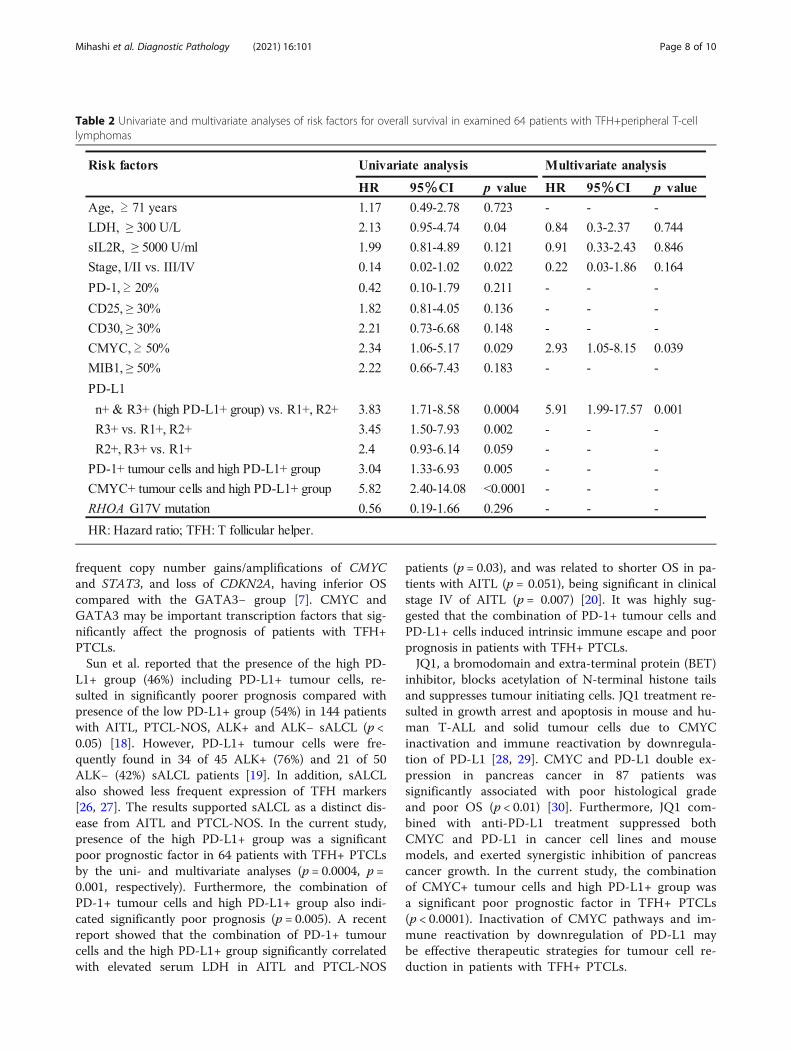
the univariate analysis of risk factors for OS in the ex-amined 64 patients with TFH+ PTCLs. In TFH+ PTCLs,≥ 300 IU/L lactate dehydrogenase (LDH; n = 28) andclinical stages III and IV (n = 50) were significant poorprognostic factors (p = 0.04, p = 0.022, respectively).Presence of CMYC+ tumour cells (n = 23) was a signifi-cantly poor OS factor (p = 0.029; Fig. 4c). Presence ofthe high PD-L1+ group (n = 23) showed a significantlypoor OS compared with presence of the low (R1+, R2+)PD-L1+ group (n = 41) (p = 0.0004; Fig. 4d). Further-more, 21 patients with the combination of PD-1+tumour cells and high PD-L1+ group showed signifi-cantly poorer OS than the other groups (n = 43) (p =0.005; Fig. 4e). The combination of CMYC+ tumourcells and high PD-L1+ group (n = 10) also indicatedprominently poor OS compared with the other groups(n = 54) (p < 0.0001; Fig. 4f). The RHOA p.G17V muta-tion was not a prognostic factor in examined 40 patientswith TFH+ PTCLs. Multivariate analysis indicated thatpresence of CMYC+ tumour cells (p = 0.039) and highPD-L1+ group (p = 0.001) were significantly associatedwith OS in 64 patients with TFH+ PTCLs in Table 2.
DiscussionThe current study demonstrated that 22 of 47 PTCL-NOS patients (47%) showed more than two TFHmarkers, and no prognostic differences were foundamong patients with AITL, PTCL-TFH and PTCL-NOS.
Other researchers have reported that lymphoma cells in18 of 41 PTCL-NOS patients (44%) showed more thantwo TFH markers [23]. Although BCL6 expression andRHOA G17V mutation were significantly higher in AITLpatients than in PTCL-NOS (both p < 0.01), neither def-inite clinical nor prognostic differences were foundamong above three groups of PTCLs. In our examinedTFH+ PTCLs, large cell PTCL-TFH patients had a simi-lar incidence of RHOA G17V mutation (36%) to thosewith AITL (42%), but showed higher expressions ofCD25 (IL2R), CD30, CMYC, and CMYC mRNA, thansmall cell PTCL-TFH patients and AITL (p < 0.05 orp < 0.01, respectively). Furthermore, large cell PTCL-TFH patients showed significantly poorer OS com-pared with small cell PTCL-TFH, AITL and F-TCL(all p < 0.05). Other researchers have reported thatCD30+ giant cells or large tumour cells were detectedin 64 of 217 PTCL-NOS patients (32%) and six of 25PTCL-TFH cases (24%) [3, 6]. CD30 as well as CD25(IL2R) may be one of the activation molecules inlarge cell PTCL-TFH. Patients with F-TCL were fre-quently found to be in advanced clinical stages IIIand IV, but showed relatively indolent prognosis com-pared with AITL [24, 25]. These findings, togetherwith our results demonstrated that the large cellmorphology of PTCL-TFH indicated distinct patho-logical and immunohistological features and poorprognosis in groups of patients with TFH+ PTCLs.
b c
a
Fig. 3 Chromatograms of Sanger sequencing for RHOA G17V mutation in patients with AITL (a, b) and large cell PTCL-TFH (c). Arrow headindicates wild type (a), and arrows indicate 50G > T mutation (b, c)
Mihashi et al. Diagnostic Pathology (2021) 16:101 Page 6 of 10
CMYC protein and mRNA expressions in aggressivetype ATLL patients were significantly higher than thoseof smouldering and chronic types (p < 0.01), and CMYCmay accelerate the conversion from indolent to aggres-sive type ATLL [11]. In the current study, CMYC+tumour cells were frequently detected in 10 of 13 largecell PTCL-TFH patients (77%), and was a significantlypoor prognostic factor in patients with TFH+ PTCLs by
the uni- and multivariate analyses (p = 0.029, p = 0.039,respectively). Other researchers have reported that ex-pressions of CMYC and Th2-cell transcription factorGATA3 were frequently found in 128 nodal PTCL pa-tients with AITL, PTCL-NOS and sALCL, and CMYC+tumour cells indicated significantly poor prognosis inthe above three types of PTCL and in only AITL (all p <0.01) [12]. GATA3+ PTCL-NOS patients showed
a b
c d
e f
Fig. 4 a No significant differences in overall survival (OS) were detected among 20 PTCL-TFH patients, 23 PTCL-NOS, and 38 AITL. b 12 large cell PTCL-TFH patients show significantly worse OS than small cell PTCL-TFH (n = 8; p = 0.045), AITL (n = 38; p = 0.047) and F-TCL (n = 6; p = 0.012). c CMYC+tumour cells (n = 23) are a poor prognostic factor in 64 patients with TFH+ PTCLs (p = 0.029). d 22 patients with both PD-L1+ tumour cells and R3+PD-L1+ cells show significantly worse OS than 42 with R1+ and R2+ PD-L1+ cells (p = 0.0004). e 21 patients with both PD-1+ tumour cells and highPD-L1+ group (n + and R3+) show significantly worse prognosis than 43 of the other group (p = 0.005). f 10 patients with both CMYC+ tumour cellsand high PD-L1+ group show significantly poorer OS compared with 54 patients in the other groups of TFH+ PTCLs (p < 0.0001)
Mihashi et al. Diagnostic Pathology (2021) 16:101 Page 7 of 10
frequent copy number gains/amplifications of CMYCand STAT3, and loss of CDKN2A, having inferior OScompared with the GATA3− group [7]. CMYC andGATA3 may be important transcription factors that sig-nificantly affect the prognosis of patients with TFH+PTCLs.Sun et al. reported that the presence of the high PD-
L1+ group (46%) including PD-L1+ tumour cells, re-sulted in significantly poorer prognosis compared withpresence of the low PD-L1+ group (54%) in 144 patientswith AITL, PTCL-NOS, ALK+ and ALK− sALCL (p <0.05) [18]. However, PD-L1+ tumour cells were fre-quently found in 34 of 45 ALK+ (76%) and 21 of 50ALK− (42%) sALCL patients [19]. In addition, sALCLalso showed less frequent expression of TFH markers[26, 27]. The results supported sALCL as a distinct dis-ease from AITL and PTCL-NOS. In the current study,presence of the high PD-L1+ group was a significantpoor prognostic factor in 64 patients with TFH+ PTCLsby the uni- and multivariate analyses (p = 0.0004, p =0.001, respectively). Furthermore, the combination ofPD-1+ tumour cells and high PD-L1+ group also indi-cated significantly poor prognosis (p = 0.005). A recentreport showed that the combination of PD-1+ tumourcells and the high PD-L1+ group significantly correlatedwith elevated serum LDH in AITL and PTCL-NOS
patients (p = 0.03), and was related to shorter OS in pa-tients with AITL (p = 0.051), being significant in clinicalstage IV of AITL (p = 0.007) [20]. It was highly sug-gested that the combination of PD-1+ tumour cells andPD-L1+ cells induced intrinsic immune escape and poorprognosis in patients with TFH+ PTCLs.JQ1, a bromodomain and extra-terminal protein (BET)
inhibitor, blocks acetylation of N-terminal histone tailsand suppresses tumour initiating cells. JQ1 treatment re-sulted in growth arrest and apoptosis in mouse and hu-man T-ALL and solid tumour cells due to CMYCinactivation and immune reactivation by downregula-tion of PD-L1 [28, 29]. CMYC and PD-L1 double ex-pression in pancreas cancer in 87 patients wassignificantly associated with poor histological gradeand poor OS (p < 0.01) [30]. Furthermore, JQ1 com-bined with anti-PD-L1 treatment suppressed bothCMYC and PD-L1 in cancer cell lines and mousemodels, and exerted synergistic inhibition of pancreascancer growth. In the current study, the combinationof CMYC+ tumour cells and high PD-L1+ group wasa significant poor prognostic factor in TFH+ PTCLs(p < 0.0001). Inactivation of CMYC pathways and im-mune reactivation by downregulation of PD-L1 maybe effective therapeutic strategies for tumour cell re-duction in patients with TFH+ PTCLs.
Table 2 Univariate and multivariate analyses of risk factors for overall survival in examined 64 patients with TFH+peripheral T-celllymphomas
Mihashi et al. Diagnostic Pathology (2021) 16:101 Page 8 of 10

In conclusion, differentiating large and small cellPTCL-TFH is necessary. Large cell PTCL-TFH patientsshowed frequent expression of activation molecules andsometimes RHOA G17V mutation, and pursued a pro-gressive clinical course in groups of TFH+ PTCLs. Pres-ence of CMYC+ tumour cells or the high PD-L1+group, and the combination of these two were signifi-cantly poor prognostic factors in patients with TFH+PTCLs (p = 0.029, p = 0.0004, or p < 0.0001, respectively).Furthermore, the combination of PD-1+ tumour cellsand high PD-L1+ group induced significantly poorprognosis (p = 0.005). CMYC inactivation and immunecheckpoint inhibitors might improve patient prognosisin TFH+ PTCLs. Further study is necessary to confirmthe clinicopathological characteristics of PTCL-TFH be-cause of the small number of patients in the currentstudy.
AbbreviationsAITL: Angioimmunoblastic T-cell lymphoma; ALK: Anaplastic lymphomakinase; ALL: Acute lymphoblastic leukaemia; ATLL: Adult T-cell leukaemia/lymphoma; EBV: Epstein-Barr virus; EBER: EBV-encoded RNA; F-TCL: FollicularT-cell lymphoma; NOS: Not otherwise specified; PD-1: Programmed celldeath-1; PD-L1: Programmed cell death-1 ligand 1; PTCL: Peripheral T-celllymphoma; TFH: T follicular helper
AcknowledgmentsWe are grateful to Tomomi Okabe and Tomoko Fukushige for their technicalassistance with the immunohistochemistry and genetic study. We thankGillian Campbell, PhD, from Edanz Group (https://en-author-services.edanz.com/ac) for editing a draft of this manuscript.
Authors’ contributionsY. M., S. K. and M. T. designed and performed the research. Y. M., Y. O., S. K.,and S. S. collected and summarised pathological data, and conducted thestatistical analysis. H. I., Y. T. and K. I. conducted treatment and collectedclinical data. Y. M., S. K., and M. T. wrote the manuscript. The author(s) readand approved the final manuscript.
FundingThis work was supported in part by a Grant-in-Aid for Scientific Research©(No.17 K08732) from the Ministry of Education, Science and Culture of Japan.
Availability of data and materialsThe datasets used and analysed in the current study are available from thecorresponding author on reasonable request.
Declarations
Ethics approval and consent to participateAll procedures performed in studies involving human participants were inaccordance with the ethical standards of the Institutional Ethics Committeeof Fukuoka University and the 1964 Helsinki Declaration and its lateramendments or comparable ethical standards. This study was approved bythe Institutional Review Board of Fukuoka University, Faculty of Medicine.
Consent for publicationNot applicable.
Competing interestsThe authors declare that they have no competing interests.
Author details1Departments of Pathology, Faculty of Medicine, Fukuoka University, 7-45-1Nanakuma, Jonan-ku, Fukuoka 814-0180, Japan. 2Departments ofOtolaryngology, Faculty of Medicine, Fukuoka University, 7-45-1 Nanakuma,
Jonan-ku, Fukuoka 814-0180, Japan. 3Departments of Haematology, ClinicalResearch Centre, National Hospital Organisation Kyushu Medical Centre, 1-8-1Jigyohama, Chuo-ku, Fukuoka 810-8563, Japan. 4Department of Pathology,Matsuyama Red Cross Hospital, 1 Bunkyo-cho, Matsuyama 791-0000, Japan.5Departments of Internal Medicine, Division of Medical Oncology,Haematology and Infectious Disease, Faculty of Medicine, Fukuoka University,7-45-1 Nanakuma, Jonan-ku, Fukuoka 814-0180, Japan. 6Departments ofPathology, Clinical Research Centre, National Hospital Organisation KyushuMedical Centre, 1-8-1 Jigyohama, Chuo-ku, Fukuoka 810-8563, Japan.7Department of Pathology, University of Occupational and EnvironmentalHealth, Iseigaoko Yahata Nishi-ku, Kitakyushu, Japan. 8Department of InternalMedicine, Faculty of Medicine, Kagoshima University, Sakuragaoka 8-35-1,Kagoshima 890-8544, Japan.
Received: 27 August 2021 Accepted: 19 October 2021
References1. Ueno H. T follicular helper cells in human autoimmunity. Curr Opin
Immunol. 2016;43:24–31. https://doi.org/10.1016/j.coi.2016.08.003.2. Dogan A, Gaulard P, Jaffe ES, Müller-Hermelink HK, de Leval L.
Angioimmunoblastic T-cell lymphoma and other nodal lymphomas of Tfollicular helper cell origin. In: Swerdlow SH, Campo E, Harris NL, et al.,editors. WHO Classification of Tumours of Haematopoietic and LymphoidTissues. Lyon: International Agency for Research on Cancer; 2017. p. 407–12.
3. Rodrıguez-Pinilla SM, Atienza L, Murillo C, Pérez-Rodríguez A, Montes-Moreno S, et al. Peripheral T-cell lymphoma with follicular T-cell markers.Am J Surg Pathol. 2008;32(12):1787–99. https://doi.org/10.1097/PAS.0b013e31817f123e.
4. Basha BM, Bryant SC, Rech KL, Feldman AL, Vrana JA, Shi M, et al.Application of a 5 marker panel to the routine diagnosis of peripheral T-celllymphoma with T-follicular helper phenotype. Am J Surg Pathol. 2019;43(9):1282–90. https://doi.org/10.1097/PAS.0000000000001315.
5. Dobay MP, Lemonnier F, Missiaglia E, Bastard C, Vallois D, Jais JP, et al.Integrative clinicopathological and molecular analyses ofangioimmunoblastic T-cell lymphoma and other nodal lymphomas offollicular helper T-cell origin. Haematologica. 2017;102(4):e148–51. https://doi.org/10.3324/haematol.2016.158428.
6. Weisenburger DD, Savage KJ, Harris NL, Gascoyne RD, Jaffe ES, MacLennanKA, et al. Peripheral T-cell lymphoma, not otherwise specified: are port of340 cases from the international peripheral T-cell lymphoma project. Blood.2011;117(12):3402–8. https://doi.org/10.1182/blood-2010-09-310342.
7. Heavican TB, Bouska A, Yu J, Lone W, Amador C, Gong Q, et al. Genetic driversof oncogenic pathways in molecular subgroups of peripheral T-cell lymphoma.Blood. 2019;133(15):1664–76. https://doi.org/10.1182/blood-2018-09-872549.
8. Chisholm KM, Bangs CD, Bacchi CE, Molina-Kirsch H, Cherry A, Natkunam Y.Expression profiles of MYC protein and MYC gene rearrangement inlymphomas. Am J Surg Pathol. 2015;39(3):294–303. https://doi.org/10.1097/PAS.0000000000000365.
9. Green TM, Nielsen O, de Stricker K, Xu-Monette ZY, Young KH, Møller MB.High levels of nuclear MYC protein predict the presence of MYCrearrangement in diffuse large B-cell lymphoma. Am J Surg Pathol. 2012;36(4):612–9. https://doi.org/10.1097/PAS.0b013e318244e2ba.
10. Starza RL, Borga C, Barba G, Pierini V, Schwab C, et al. Genetic profile of T-cell acute lymphoblastic leukemias with MYC translocations. Blood. 2014;124(24):3577–82. https://doi.org/10.1182/blood-2014-06-578856.
11. Mihashi Y, Mizoguchi M, Takamatsu Y, Ishitsuka K, Iwasaki H, Koga M, et al.C-MYC and its main ubiquitin ligase, FBXW7, influence cell proliferation andprognosis in adult T-cell leukaemia/lymphoma. Am J Surg Pathol. 2017;41(8):1139–49. https://doi.org/10.1097/PAS.0000000000000871.
12. Manso R, Bellas C, Martin-Acosta P, Mollejo M, Menarguez J, Rojo F, et al. C-MYC is related to GATA3 expression and associated with poor prognosis innodal peripheral T-cell lymphomas. Haematologica. 2016;101(8):e336–8.https://doi.org/10.3324/haematol.2016.143768.
13. Casey SC, Tong L, Li Y, Do R, Walz S, Fitzgerald KN, et al. MYC regulates theantitumour immune response through CD47 and PD-L1. Science. 2016;352(6282):227–31. https://doi.org/10.1126/science.aac9935.
14. Zhou C, Che G, Zheng X, Qiu J, Xie Z, Cong Y, et al. Expression andclinical significance of PD-L1 and c-Myc in non-small cell lung cancer. JCancer Res Clin Oncol. 2019;145(11):2663–74. https://doi.org/10.1007/s00432-019-03025-8.
Mihashi et al. Diagnostic Pathology (2021) 16:101 Page 9 of 10
15. Kythreotou A, Siddique A, Mauri FA, Bower M, Pinato DJ. PD-L1. J ClinPathol. 2018;71(3):189–94. https://doi.org/10.1136/jclinpath-2017-204853.
16. Sakakibara A, Kohno K, Eladl AE, Klaisuwan T, Ishikawa E, Suzuki Y, et al.Immunohistological assessment of the diagnostic utility of PD-L1: apreliminary analysis of anti-PD-L1 antibody (SP142) for lymphoproliferativediseases with tumour and non-malignant Hodgkin-reed Sternberg (HRS)-likecells. Histopathol. 2018;72(7):1156–63. https://doi.org/10.1111/his.13475.
17. Tiemann M, Samoilova V, Atiakshin D, Buchwalow I. Immunophenotyping of thePD-L1-positiv cells in angioimmunoblastic T cell lymphoma and Hodgkin disease.BMC Res Notes. 2020;13:139. https://doi.org/10.1186/s13104-020-04975-w.
18. Sun M, Su W, Qian J, Meng H, Ji H, Liu Y, et al. The prognostic value of toll-like receptor5 and programmed cell death-ligand1 in patients withperipheral T-cell non-Hodgkin lymphoma. Leuk Lymphoma. 2019;60(11):2646–57. https://doi.org/10.1080/10428194.2019.1602266.
19. Shen J, Li S, Medeiros LJ, Lin P, Wang SA, Tang G, et al. PD-L1 expression isassociated with ALK positivity and STAT3 activity, but not outcome inpatients with systemic anaplastic large cell lymphoma. Mod Pathol. 2020;33(3):324–33. https://doi.org/10.1038/s41379-019-0336-3.
20. Kim S, Kwon D, Koh J, Nam SJ, Kim YA, Kim TM, et al. Clinicopathologicalfeatures of programmed cell death-1 and programmed cell death-ligand-1expression in the tumor cells and tumor microenvironment ofangioimmunoblastic T cell lymphoma and peripheral T cell lymphoma nototherwise specified. Virch Arch. 2020;447(1):131–42. https://doi.org/10.1007/s00428-020-02790-z.
21. Nagao R, Kikuchi YY, Carreras J, Kikuchi T, Miyaoka M, et al.Clinicopathologic analysis of angioimmunoblastic T-cell lymphoma with orwithout RHOA G17V mutation using formalin-fixed paraffin-embeddedsections. Am J Surg Pathol. 2016;40(8):1041–50. https://doi.org/10.1097/PAS.0000000000000651.
22. Pileri SA, Weisenburger DD, Sng I, Nakamura S, Müller-Hermelink HK, ChanWC, et al. Peripheral T-cell lymphoma, NOS. In: Swerdlow SH, Campo E,Harris NL, et al., editors. WHO Classification of Tumours of Haematopoieticand Lymphoid Tissues. Lyon: International Agency for Research on Cancer;2017. p. 403–6.
23. Manso R, Gonzalez-Rincon J, Rodriguez-Justo M, Roncador G, Gómez S, et al.Overlap at the molecular and immunohistochemical levels betweenangioimmunoblastic T-cell lymphoma and a subgroup of peripheral T-celllymphomas without specific morphological features. Oncotarget. 2018;9:16124–33. https://doi.org/10.18632/oncotarget.24592.
24. Huang Y, Moreau A, Dupuis J, Petit B, Le Gouill S, et al. Peripheral T-celllymphomas with a follicular growth pattern are derived from follicularhelper T cells (TFH) and may show overlapping features withangioimmunoblastic T-cell lymphoma. Am J Surg Pathol. 2009;33(5):682–90.https://doi.org/10.1097/PAS.0b013e3181971591.
25. Hartmann S, Goncharova O, Portyanko A, Sabattini E, Meinel J, Küppers R,et al. CD30 expression in neoplastic T cells of follicular T cell lymphoma is ahelpful diagnostic tool in the differential diagnosis of Hodgkin lymphoma.Mod Pathol. 2019;32(1):37–47. https://doi.org/10.1038/s41379-018-0108-5.
26. Krishnan C, Warnke RA, Arber DA, Natkunam Y. PD-1 expression in T-celllymphomas and reactive lymphoid entities: potential overlap in stainingpatterns between lymphoma and viral lymphadenitis. Am J Surg Pathol.2010;34(2):178–89. https://doi.org/10.1097/PAS.0b013e3181cc7e79.
27. Manso R, Rodríguez-Perales S, Torres-Ruiz R, Santonja C, Rodríguez-PinillaSM. PD-L1 expression in peripheral T-cell lymphomas is not related to eitherPD-L1 gene amplification or rearrangements. Leuk Lymph. 2021;62(7):1648–56. https://doi.org/10.1080/10428194.2021.1881511.
28. Schubbert S, Cardenas A, Chen H, Garcia C, Guo W, Bradner J, et al.Targeting the MYC and PI3K pathways eliminates leukaemia-initiating cellsin T-cell acute lymphoblastic leukaemia. Cancer Res. 2014;74(23):7048–59.https://doi.org/10.1158/0008-5472.
29. Casey S, Baylot V, Felsher DW. The MYC oncogene is a global regulator ofimmune response. Blood. 2018;131(18):2007–15. https://doi.org/10.1182/blood-2017-11-742577.
30. Pan Y, Feia Q, Xiong P, Yang J, Zhang Z, et al. Synergistic inhibition ofpancreatic cancer with anti-PD-L1 and c-Myc inhibitor JQ1.Oncoimmunology. 2019;8(5):e1581529. https://doi.org/10.1080/2162402X.2019.1581529.
Publisher’s NoteSpringer Nature remains neutral with regard to jurisdictional claims inpublished maps and institutional affiliations.
Mihashi et al. Diagnostic Pathology (2021) 16:101 Page 10 of 10