leaf structure in linderbergia l. (scrophulariaceae 4 no 2 july 13/sangeeta...leaf structure in...
TRANSCRIPT
http://biosciencediscovery.com 208 ISSN: 2231-024X (Online)
Bioscience Discovery, 4(2):208-210, July 2013 ISSN: 2229-3469 (Print)
Leaf structure in Linderbergia L. (Scrophulariaceae)
Sangeeta S Sutar
Dept. of Botany, Sir Sayyed College of Arts, Commerce and Science, Aurangabad (MS) India [email protected]
ABSTRACT
Leaves of Linderbergia are dorsiventral and amphistomatic, stomata anomocytic, cuticular striations were present on adaxial epidermis of L. indica. Trichomes were glandular and mesophyll is differentiated into palisade and spongy tissue. Venation was pinnate; it is simple craspedodromous in L. grandiflora and semi craspedodromous in L. indica. The lamina margin was serrate in L. grandiflora and crenate in L.indica. Midvein and lateral veins of tooth lie close together in L. indica whereas these veins only enlarge at their tips in L. grandiflora. The margin of teeth develops characteristic spinules in L. indica and accumulation of tracheids was a constant feature in L. grandiflora.
Key words: Leaf, Linderbergia, Scrophulariaceae
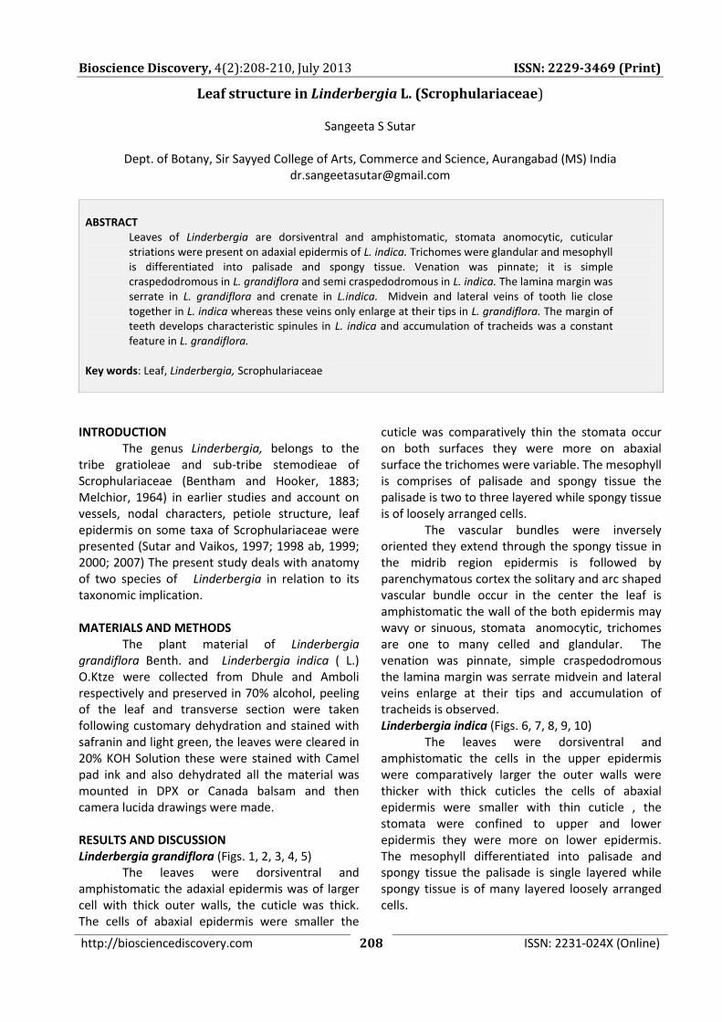
INTRODUCTION The genus Linderbergia, belongs to the tribe gratioleae and sub-tribe stemodieae of Scrophulariaceae (Bentham and Hooker, 1883; Melchior, 1964) in earlier studies and account on vessels, nodal characters, petiole structure, leaf epidermis on some taxa of Scrophulariaceae were presented (Sutar and Vaikos, 1997; 1998 ab, 1999; 2000; 2007) The present study deals with anatomy of two species of Linderbergia in relation to its taxonomic implication. MATERIALS AND METHODS The plant material of Linderbergia grandiflora Benth. and Linderbergia indica ( L.) O.Ktze were collected from Dhule and Amboli respectively and preserved in 70% alcohol, peeling of the leaf and transverse section were taken following customary dehydration and stained with safranin and light green, the leaves were cleared in 20% KOH Solution these were stained with Camel pad ink and also dehydrated all the material was mounted in DPX or Canada balsam and then camera lucida drawings were made. RESULTS AND DISCUSSION Linderbergia grandiflora (Figs. 1, 2, 3, 4, 5)
The leaves were dorsiventral and amphistomatic the adaxial epidermis was of larger cell with thick outer walls, the cuticle was thick. The cells of abaxial epidermis were smaller the
cuticle was comparatively thin the stomata occur on both surfaces they were more on abaxial surface the trichomes were variable. The mesophyll is comprises of palisade and spongy tissue the palisade is two to three layered while spongy tissue is of loosely arranged cells.
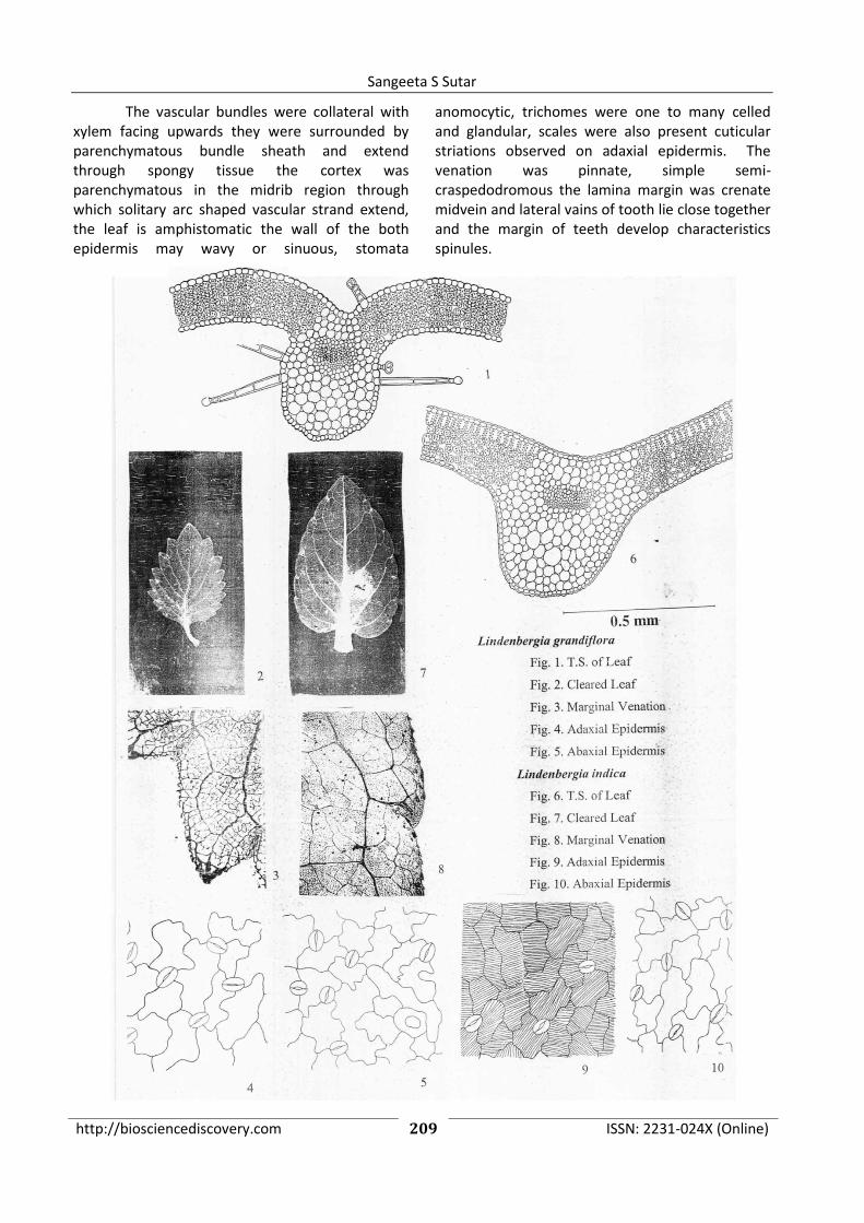
The vascular bundles were inversely oriented they extend through the spongy tissue in the midrib region epidermis is followed by parenchymatous cortex the solitary and arc shaped vascular bundle occur in the center the leaf is amphistomatic the wall of the both epidermis may wavy or sinuous, stomata anomocytic, trichomes are one to many celled and glandular. The venation was pinnate, simple craspedodromous the lamina margin was serrate midvein and lateral veins enlarge at their tips and accumulation of tracheids is observed. Linderbergia indica (Figs. 6, 7, 8, 9, 10)
The leaves were dorsiventral and amphistomatic the cells in the upper epidermis were comparatively larger the outer walls were thicker with thick cuticles the cells of abaxial epidermis were smaller with thin cuticle , the stomata were confined to upper and lower epidermis they were more on lower epidermis. The mesophyll differentiated into palisade and spongy tissue the palisade is single layered while spongy tissue is of many layered loosely arranged cells.
http://biosciencediscovery.com 209 ISSN: 2231-024X (Online)
Sangeeta S Sutar
The vascular bundles were collateral with xylem facing upwards they were surrounded by parenchymatous bundle sheath and extend through spongy tissue the cortex was parenchymatous in the midrib region through which solitary arc shaped vascular strand extend, the leaf is amphistomatic the wall of the both epidermis may wavy or sinuous, stomata
anomocytic, trichomes were one to many celled and glandular, scales were also present cuticular striations observed on adaxial epidermis. The venation was pinnate, simple semi-craspedodromous the lamina margin was crenate midvein and lateral vains of tooth lie close together and the margin of teeth develop characteristics spinules.
http://biosciencediscovery.com 210 ISSN: 2231-024X (Online)
Bioscience Discovery, 4(2):208-210, July 2013 ISSN: 2229-3469 (Print)
Species of Lindenbergia presently investigated reveals many features of common occurrence in leaf structure i.e. epidermal parameters and mesophyll differentiation. Venation was pinnate but it is simple craspedodromous in L. grandiflora and semi-craspedodromous L. indica. Hickey and Wolfe (1975), Hufford (19992) describe craspedodromous and Varghese (1969) and Canne Hillikar Kampny (1991) recorded craspedodromous including brochidodromus venation in scrophulariaceae. The
lamina margin was serrate in L. grandiflora and crenate in L. indica, midvein and lateral veins of tooth lie close in L. indica, where as these veins on enlarge at their tips in L. grandiflora. The margin of teeth develops characteristic spinules in L. indica and accumulation of tracheids was observed in L. grandiflora. Although both the species of Lindenbergia share common character in leaf structure i.e., epidermis and mesophyll they stand apart from each other in venation pattern.
LITERATURE CITED Bentham G and JD Hooker, 1873. “Genera Plantarum” L. Reeve & Co. London. Canne-Hilliker JM. and kamphy CM, 1991. Taxonomic significance of Agalinis (Scrophulariaceae). Can. J. Bot., 69:1935-1950 Hickey L J and wolfe JA, 1975.The bases of angiosperm phyllogeny: Vegetative morphology. Ann. Missouri Bot. Grad., 62:538-589. Hufford L, 1992. Leaf structure in Besseya and Synthyris (Scrophulariaceae). Can. J. Bot., 70:921-932. Melchior H, 1964. Engler’s syllabus der Plazenfamilien: II Ed. Geloruder Traeger, Berlin. Sutar SS and Vaikos NP, 1997. Anatomy of petiole in some Scrophulariaceae. Int. J. Mendel., 14 : 75-76. Sutar SS and Vaikos NP, 1998a. Vessel elementsin the Scrophulariaceae. Dr.B.A.M. Univ. J. Sci. 28:37-42. Sutar SS and Vaikos NP, 1998b. Application of nodal characters in the studies of Scrophulariaceae. J. Indian Bot. Soc., 77:39-42. Sutar SS and Vaikos NP, 1999. Foliar Epidermal studies in Lindernia All. (Scrophulariaceae) Dr. B. A. M. Univ. J. Sci., 29:107-110. Sutar SS and Vaikos NP, 2000. Leaf Structure in Lindernia All. (Scrophulariaceae) Dr. B. A. M. Univ. J. Sci., 30:107-110. Sutar SS and Vaikos NP, 2007.Leaf structure in Stemodia L. (Scrophulariaceae). Bioinfolet, 4:336-338. Vargese TM, 1969. In Recent advances in the antomy to tropical seed plants. XX Ed. K.A.H.P. Corporation, Delhi.
How to Cite this Article: Sutar Sangeeta S, 2013. Leaf structure in Linderbergia L. (Scrophulariaceae). Biosci. Disc., 4(2):208-210.