learning the optimal control of coordinated eye and head...
TRANSCRIPT

Learning the Optimal Control of Coordinated Eye andHead MovementsSohrab Saeb1*, Cornelius Weber2, Jochen Triesch1
1 Frankfurt Institute for Advanced Studies (FIAS), Goethe University Frankfurt, Frankfurt am Main, Germany, 2 Faculty of Mathematics, Informatics and Natural Sciences,
University of Hamburg, Hamburg, Germany
Abstract
Various optimality principles have been proposed to explain the characteristics of coordinated eye and head movementsduring visual orienting behavior. At the same time, researchers have suggested several neural models to underly thegeneration of saccades, but these do not include online learning as a mechanism of optimization. Here, we suggest anopen-loop neural controller with a local adaptation mechanism that minimizes a proposed cost function. Simulations showthat the characteristics of coordinated eye and head movements generated by this model match the experimental data inmany aspects, including the relationship between amplitude, duration and peak velocity in head-restrained and the relativecontribution of eye and head to the total gaze shift in head-free conditions. Our model is a first step towards bringingtogether an optimality principle and an incremental local learning mechanism into a unified control scheme for coordinatedeye and head movements.
Citation: Saeb S, Weber C, Triesch J (2011) Learning the Optimal Control of Coordinated Eye and Head Movements. PLoS Comput Biol 7(11): e1002253.doi:10.1371/journal.pcbi.1002253
Editor: Jorn Diedrichsen, University College London, United Kingdom
Received August 1, 2010; Accepted September 13, 2011; Published November 3, 2011
Copyright: � 2011 Saeb et al. This is an open-access article distributed under the terms of the Creative Commons Attribution License, which permitsunrestricted use, distribution, and reproduction in any medium, provided the original author and source are credited.
Funding: This work was supported by the German Federal Ministry of Education and Research within the ‘Bernstein Focus: Neurotechnology’ through researchgrant 01GQ0840, by EU projects ‘Plasticity and Learning in Cortical Networks’ (PLICON) and ‘Intrinsically Motivated Cumulative Learning Robots’ (IM-CLeVeR), andby the Hertie Foundation. The funders had no role in study design, data collection and analysis, decision to publish, or preparation of the manuscript.
Competing Interests: The authors have declared that no competing interests exist.
* E-mail: [email protected]
Introduction
Active perception of the visual world necessitates frequent
redirection of our gaze. Such visual orientation behavior comprises
multi-segment control of different motor systems, i.e. the
coordinated movement of several parts of the body including the
eyes, the head, and the torso. The coordinated movements of the
eyes and the head during fast gaze shifts are called saccadic eye
and head movements and are usually investigated in two
conditions: head-restrained and head-free.
In the head-restrained condition, head movement is limited so
that the gaze shifts rely only on eye movements. These eye
movements, known also as eye-only saccades, possess certain
physical properties. The relationship between the duration, peak
velocity and the amplitude of saccades is known as the main sequence
[1]. This relationship is stereotyped: the duration increases linearly
with the saccadic amplitude, while the peak velocity increases
linearly for low amplitudes and undergoes a soft saturation for
larger amplitudes [2–4]. The velocity profiles of saccadic eye
movements are smooth and symmetric for small amplitudes, while
they become skewed for larger amplitudes [5,6].
In the head-free condition, the head is allowed to accompany
the eye in visual orienting. These movements are usually
composed of two phases: in the first phase, the gaze is rapidly
shifted to the target using both the eyes and the head. Once the
gaze reaches the target, the second phase starts. In the second
phase, the head continues moving in the same direction as in the
first phase, but the eyes move backwards with the same velocity as
the head. As a result, the gaze remains stabilized on the target.
The general belief is that the vestibulo-ocular reflex (VOR) has a
fundamental role in generating the coordination of eye and head
during the second phase (see [7] for a review).
When the head is free to move, the kinematic characteristics of
saccadic eye movements change dramatically compared to the
head-restrained condition. As the gaze shift amplitude increases,
the eye movement amplitude approaches its limits, and the head
contribution becomes more prominent. Therefore, the eye’s
position and velocity is not determined only based on the current
gaze error, but it also depends on concurrent head position and
velocity. Furthermore, the eye’s peak velocity declines in the head-
free condition, its duration increases and its velocity profiles
change [8–14].
Previous StudiesPrevious computational studies on saccadic eye and head
movements revolve around two questions. The first question
concerns the optimality principles underlying the kinematic charac-
teristics, and the second one is about the neural architecture that
generates appropriate control signals for driving eye and head
muscles. Researchers dealing with both questions consider linear
eye and head plants, which are equivalent to linear differential
equations describing the mechanical properties of eye and head
motor systems. Such models are considered sufficient for modeling
the oculomotor and the head motor system dynamics [15,16].
Optimality principles. During saccadic eye and head
movements, visual information is not properly transmitted to the
brain either due to motion blur or because of neural suppression
induced by higher regions [17]. Therefore, saccadic gaze shifts
PLoS Computational Biology | www.ploscompbiol.org 1 November 2011 | Volume 7 | Issue 11 | e1002253
should be as fast as possible in order to increase the amount of
time the image is stabilized on the retina. This has been a
fundamental assumption of many studies that were aiming at
finding the optimality principles underlying the kinematic
characteristics of saccades.
Early studies proposed that the saccade trajectories are
optimized in such a way that they minimize the time to reach
the target [18]. This assumption, known as the minimum-time
principle, leads to a bang-bang control solution [19], for which the
resulting velocity profiles are not biologically plausible [20].
Therefore, additional assumptions are necessary.
A key assumption suggested by Harris and Wolpert was that
there exists additive white noise in the neural command, whose
instantaneous power (variance) is proportional to that of the
command signal [21]. Due to this assumption, the variance of the
final eye position increases as one tries to decrease the saccadic
duration by recruiting larger command signals. Therefore, in
addition to the saccadic duration, the variance of the eye position
should also be minimized. Because of this property, this principle is
also called the minimum-variance principle. As a result of these two
assumptions, a trade-off emerges between the speed and the
accuracy of saccades, and the optimal solution to this trade-off is a
trajectory that is biologically realistic [22].
Kardamakis and Moschovakis suggested another optimality
principle based on the minimum-effort rule and optimal control
theory [23]. This principle was used to obtain optimal control
signals for both oculomotor and head motor systems in
coordinated eye and head movements. The minimum-effort rule
obliges that the squared sum of the eye and the head torque signals
integrated over the movement period be minimized in order to
obtain the optimal control signal. The optimization process uses
boundary conditions for the gaze position and is only applied to
the first phase of coordinated eye and head movements. In the
head-restrained condition, this method achieves unimodal velocity
profiles with shorter acceleration and longer deceleration phases
compatible with many experimental findings [5,6]. For the head-
free condition, the contribution of eye and head to total gaze shift
obtained by this method is biologically realistic, and the eye-
velocity profiles become double-peaked as in the experiments [24].
Architectures. The functional architectures suggested for the
control of saccadic eye and head movements can be categorized
into two groups: architectures for head-restrained and head-free
control problems. Since the goal of our study is to solve these two
control problems using a single architecture, we review the existing
models based on their structure and the optimization method they
use rather than the control problem they are supposed to solve.
From this perspective, models can be categorized into feedback
models, which use gaze feedback to control saccades, and independent
control schemes which do not need a gaze feedback.
The first gaze feedback model was suggested by Laurutis and
Robinson [25] that was an extension of position control models of
the saccadic system [26,27] to incorporate gaze feedback signals.
This model was thereafter used and extended by others [9,28,29].
The gaze error signal used in feedback models is internally
estimated, such that no visual feedback is necessary. This is mainly
due to two reasons. First, vision is impaired during fast gaze shifts,
and second, there is a retinal processing delay of about 40–50 ms
which can make the controller unstable [30]. Therefore, the gaze
feedback signal can be regarded as an internal feedback. A more
recent model by Chen-Harris and colleagues estimates the gaze
feedback in a more elaborate way [31]. The internal feedback in
this model consists of two forward models: a forward model of the
oculomotor plant that predicts the state of the eye, and a forward
model of the target motion that predicts the state of the target.
This feedback used together with the absolute target position
provides a signal to drive an optimized feedback controller which
is based on the minimum-variance principle of Harris and Wolpert
[22], and requires re-optimization for each saccadic duration.
The independent eye and head control models rely on the
dynamics of their burst generator (BG) units rather than a gaze
feedback in order to generate the control signals [12,32,33]. These
BG units are per se closed loop controllers that use efference copies
of eye and head motor command signals. Although the eye and the
head control circuits have independent dynamics, there are ways
through which they can influence each other. Independent control
models usually assume that the relative contribution of eye and
head components to the gaze shift is known beforehand. However,
a recent neural model suggested by Kardamakis and colleagues
[33] is able to reproduce realistic contribution of eye and head
using the communication between the two circuits, and without
such an assumption. The parameters of this model are either set
according to experimental findings or optimized using a genetic
algorithm.
Our ContributionThe optimality principle studies by Harris and Wolpert [21] and
Kardamakis and Moschovakis [23] have not provided any
incremental learning mechanism for their optimization procedure.
In fact, the optimization procedures used in these studies are based
on Pontryagin’s extremum principle [34], which requires bound-
ary conditions at the initial and the final time of the saccadic
movement, and provides a global analytical solution rather than a
local adaptation mechanism. In the model suggested by Karda-
makis and Moschovakis, the cost was evaluated for several values
of gaze shift duration and the model parameters were eventually
set to satisfy the trade-off between the effort and the duration. It
may be speculated that such a solution can be a result of evolution,
nevertheless, numerous experimental results indicate that saccadic
eye and head movements are constantly adapted [35–37].
The neural control architectures that have been proposed to
generate the eye and the head control signals do not use any
optimality criteria to tune their parameters. The parameters of
such models are usually hand-tuned or adjusted by a global
optimization algorithm in a way that the model’s response fits to
the experimental data. The only exception we found is the model
Author Summary
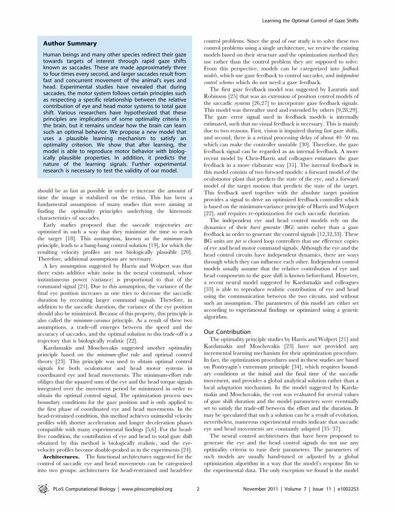
Human beings and many other species redirect their gazetowards targets of interest through rapid gaze shiftsknown as saccades. These are made approximately threeto four times every second, and larger saccades result fromfast and concurrent movement of the animal’s eyes andhead. Experimental studies have revealed that duringsaccades, the motor system follows certain principles suchas respecting a specific relationship between the relativecontribution of eye and head motor systems to total gazeshift. Various researchers have hypothesized that theseprinciples are implications of some optimality criteria inthe brain, but it remains unclear how the brain can learnsuch an optimal behavior. We propose a new model thatuses a plausible learning mechanism to satisfy anoptimality criterion. We show that after learning, themodel is able to reproduce motor behavior with biolog-ically plausible properties. In addition, it predicts thenature of the learning signals. Further experimentalresearch is necessary to test the validity of our model.
Learning the Optimal Control of Gaze Shifts
PLoS Computational Biology | www.ploscompbiol.org 2 November 2011 | Volume 7 | Issue 11 | e1002253

by Chen-Herris and colleagues [31] that relies on its internal
feedback process to generate neural command signals.
As an alternative control scheme, we introduce an open-loop
neural architecture. We try to obtain an adaptation mechanism
that on the one hand can be implemented by the brain circuitry
(see Discussion), and on the other hand minimizes a cost
function. To this end, we suggest a cost function that does not
directly depend on the saccadic duration, and therefore allows
for a gradient descent based solution without any need to define
boundary conditions. The control pathway of our model is
feedforward, and is constantly calibrated by an adaptation
mechanism that implicitly evaluates the optimality of the
controller with respect to the cost function and induces
parameter changes via a local learning rule. Therefore, our
model can be regarded as a first step towards bringing together
an optimality principle and an incremental local learning
mechanism into a unified control scheme. It extends our
previous model of eye-only saccade generation [38] to coordi-
nated eye and head movements.
Methods
The model architecture consists of two pathways: feedforward
control and adaptation. The feedforward control pathway comprises
a spatiotemporal map that performs spatial-to-temporal transforma-
tion as shown in Figure 1. The adaptation pathway is based on the
learning rules derived from a cost function (see Adaptation).
Spatial-to-Temporal TransformationSaccades are produced by a precisely timed pattern of activity
within the motor neurons innervating the eye and the head muscle
systems. However, the desired gaze shift is represented spatially in
areas such as the superior colliculus [39,40]. This is called the
spatial-to-temporal transformation problem (STTP) [41].
Here, we suggest a spatiotemporal map to perform such a
transformation. This map comprises several columns (delay lines),
each one including a number of neurons as shown in Figure 1.
There is one column per oculocentric position of the target, i.e. the
desired gaze shift amplitude. Only one visual dimension (e.g. the
horizontal position) is modeled. The activity of the neurons in the
columns is only dependent on the desired gaze shift amplitude and
the progress of time. The spatial-to-temporal transformation is
accomplished when these activities are integrated by two read-out
neurons (gray units in Figure 1) to create the neural control signals
that drive the eye and the head plants.
When an object triggers the initiation of a saccade, a single
column corresponding to the desired gaze shift amplitude is
activated. The activation of a column means that a wave of activity
propagates through the neurons of that column, starting from the
first neuron. The firing rate of each neuron changes as a Gaussian
function. Given column j is activated at time t~0, this
propagation can be formulated as:
sij(t)~A exp {(i{
t
Dt)2
2s2
0B@
1CA, ð1Þ
where sij(t) represents the instantaneous firing rate of the neuron i
in column j, Dt is the sampling period, s2 is the variance, and A
scales the height of the activity peak.
The two linear read-out neurons integrate the activity of the
spatiotemporal map by means of weighted connections. This
linear combination forms the neural command signals, ue(t) and
uh(t), needed to drive the eye and head plants:
ue(t)~XN
j~1
XMi~1
weijsij(t), ð2Þ
Figure 1. Model Architecture. The input, left, consists of one column of delay units per oculocentric position of the target. The read-out neurons(gray units; one for eye and one for head control) are linear and each weight parameter wij is adapted locally by the corresponding adaptation unit.The solid lines indicate the control signal pathway and the dashed lines represent the adaptation signal pathway.doi:10.1371/journal.pcbi.1002253.g001
Learning the Optimal Control of Gaze Shifts
PLoS Computational Biology | www.ploscompbiol.org 3 November 2011 | Volume 7 | Issue 11 | e1002253

uh(t)~XN
j~1
XMi~1
whijsij(t): ð3Þ
weij and wh
ij represent the weighted connections between neuron
i in column j and the eye and head read-out neurons, respectively.
N is the total number of columns and M is the number of neurons
in each column. Since we allow the neural command signals, ue(t)and uh(t), to become negative, we consider them as the difference
between the firing rates of the agonist and the antagonist
motoneurons [42] driving each plant.
The response of the eye plant is the eye position in head
coordinates, re(t), and the response of the head plant is the head
position in body coordinates, rh(t). The details of these plant
models as well as their corresponding responses are given in Text
S1.
AdaptationThe adaptation mechanism modifies the connection weights of
the neural controller through several trials, such that it approaches
an optimal behavior. Since the optimal behavior is determined by
a cost function, adaptation implies the minimization of that cost
function.
Before introducing the cost function, let us define the gaze error
as:
rg(t)~ro{re(t){rh(t), ð4Þ
where ro is the target object position in body coordinates, and re(t)and rh(t) as defined before. For simplicity, we have assumed that
the axes of eye and head rotation are perfectly aligned.
We define a cost function that addresses the following
objectives:
1. The gaze should reach the target as soon as possible and then
stand still on the target position. Therefore, the cost function
should depend on the absolute value of the gaze error, jrg(t)j.This dependency can be established via any arbitrary function
of jrg(t)j, three examples shown in Figure 2. Convex functions
such as a quadratic function do not seem a good choice since
they do not penalize small gaze errors. We will proceed with
the absolute value function because it results in more
compatibility with neurophysiological observations, as we will
see in the Discussion.
2. The power of the neural control signal should be constrained.
This assumption may be viewed as a regularization [43]. It also
addresses the problem of signal-dependent noise [21], as it
reduces the variability of the neural control signal by
preventing its power from becoming too large. Since the
neural control signal is linearly dependent on the weight values,
the cost function should depend on the absolute values of the
weight parameters. Thus, large values of these parameters will
be penalized regardless of their sign.
Accounting for these objectives, we formulate the cost function
as:
E~
ðT
0
jrg(t)jdtzXN
j~1
XMi~1
(aejweij j
nzahjwh
ij jn), ð5Þ
The time integral starts at saccade onset t~0, and T has a
sufficiently large value so that the integral covers the whole
movement duration. ae and ah are positive coefficients determin-
ing the contribution of the eye and head weight limiting terms to
the total cost, respectively. We set n~4 since this value leads to the
results which have the most similarity to the experimental data.
It is worth noting that the integration time T also covers part of
the fixation period. This property of the proposed cost function
facilitates the derivation of weight adaptation rules in case of
delayed visual error, as studied on humans [44] and on macaque
monkeys [45]. These studies show that a delayed visual error
signal, up to several hundred milliseconds, is still able to induce
saccadic adaptation.
The adaptable parameters of our model are the weights
projecting from the spatiotemporal map to the two read-out
neurons. We use a gradient descent method for minimizing the
cost function. Using this method, the weight update rules are
obtained as (see Text S1):
weij/we
ijzdeij
ðT
0
sgn(rg(t))feij(t)dt{4de
ijae(weij)
3, ð6Þ
whij/wh
ijzdhij
ðT
0
sgn(rg(t))fhij(t)dt{4dh
ijah(whij)
3, ð7Þ
where deij and dh
ij are adaptation rates, sgn(x) is the signum
function, and:
Figure 2. The first term of the cost function as a function of jrg(t)j. Three example functions (quadratic, absolute value and square root) areshown here.doi:10.1371/journal.pcbi.1002253.g002
Learning the Optimal Control of Gaze Shifts
PLoS Computational Biology | www.ploscompbiol.org 4 November 2011 | Volume 7 | Issue 11 | e1002253

feij(t)~
ðt
0
sij(t)he(t{t)dt, ð8Þ
fhij(t)~
ðt
0
sij(t)hh(t{t)dt: ð9Þ
The functions he(t) and hh(t) represent the impulse responses of
eye and head plants, respectively.
The block diagram representation of the adaptation mechanism
is shown in Figure 1 (gray area). This representation is inspired by
Equations 6–9 in the following way: the signals feij(t) and fh
ij(t) can
be regarded as the responses of the forward models of the eye and the
head plants, respectively. These forward models basically have the
same impulse response as the eye and head plants while receiving a
copy of the neural activity in the columns as input. The responses
of these forward models are multiplied by the sign of the gaze error
and then integrated over T (see Equations 6 and 7). The resulting
signals act on the same connection of the neuron that has
stimulated the adaptation units. This influence is shown by a
dashed arrow in Figure 1.
Results
We consider two conditions: the head-restrained condition,
where we set the head plant gain gh (see Text S1) to zero; and the
head-free condition where we set gh to its normal value, 1:719. In
biology, it is hypothesized that a neural gate prevents a common
gaze shift command from reaching the neck circuitry when head-
restrained saccades are desired [46].
For each condition, the learning procedure continues until the
model reached a stable response. The simulation time step was
1 ms and s was set to 0.002.
We used ae and ah as the free parameters of our model to find
the best match between the model behavior and experimental
data. To this end, we used a genetic algorithm (GA) as described in
Text S1. For the head-restrained condition, the GA fitness
function was defined as the sum of squared errors (SSE) between
the main sequence plots of the model and of the experiments [4].
The highest fitness value was found for ae~0:014. One should
note that the value of ah has no effect in the head-restrained
condition since gh~0 in this case. For the head-free condition, the
fitness function was set as the SSE between the relative eye/head
contribution of the simulated and of the experimental results, with
eye position initialized at zero. The best parameters found in this
case were ae~1:0 and ah~0:014.
Head-Restrained ConditionWith the best model parameters found by the GA, we simulated
the learning procedure (Equation 6) for different target object
positions. The integration time was set to T~300 ms, which was
enough for learning saccadic eye movements for all amplitudes.
We compared the simulation results to the experimental data
obtained by Harwood and colleagues on human subjects
performing horizontal eye movements [4]. This comparison was
made between the main sequence plots, as shown in Figure 3.
The resulting neural control signals and their corresponding
plant responses for three target object positions, 100, 200, and 300,are depicted in Figure 4. These signals comprise two main phases:
the saccadic phase during which the control signal is strong; and
the fixation phase when it has a roughly constant but slightly
oscillating positive value. The mean value of the neural control
signal in the fixation period is proportional to the target position,
and the small oscillations lead to slight eye drifts that are negligible
because of their low contribution to the cost function. In fact, the
eye plant filters out the high frequency inputs so that the eyes do
not follow these oscillations. The decrease of the firing rate at the
end of the plot is a boundary effect. No matter how long the
integration time T is, this effect is always observed at the final
time.
The general form of the optimized neural control signals shown
in Figure 4 resembles the firing patterns of abducens nucleus
Figure 3. Comparing the main sequence plots of the proposedmodel to experimental data, in head-restrained condition. (A)Peak velocity and (B) duration of saccades versus their amplitudes. Thesolid lines represent the model results after learning, and the crossesare experimental data taken from an experiment on human subjects [4].doi:10.1371/journal.pcbi.1002253.g003
Learning the Optimal Control of Gaze Shifts
PLoS Computational Biology | www.ploscompbiol.org 5 November 2011 | Volume 7 | Issue 11 | e1002253

motoneurons in monkey responsible for saccadic eye movements,
shown in Figure 5: a fast increase in the firing rate is followed by a
slow decrease (the burst phase), then follows an oscillatory steady
state that maintains the fixation (the tonic phase). During fixation,
in both model and experimental data, the sustained tonic firing
rate is proportional to the eye position. However, one should note
that the firing rate patterns shown in Figure 4 are differential, i.e.
they are obtained as the difference between the activity of agonist
and antagonist motor neurons, while the ones shown in Figure 5
are not.
Without changing the model parameters we tested if the model
is capable of reproducing realistic velocity profiles. For this, we
simulated the learning process for the amplitudes ranging from 100
to 800. The velocity profiles corresponding to different saccadic
amplitudes are shown in Figure 6. For small amplitudes, the
profiles are smooth and almost symmetric, while for larger
amplitudes they become skewed. The main reason for the former
symmetry is that the effect of weight updating mechanisms
(Equations 6 and 7) on the saccadic velocity is symmetric when the
second term of the cost function (Equation 5) is small enough. This
effect becomes biased against large weights when the second
weight regularization term grows as a result of an increase in target
eccentricity. The same trend is observed in experimental results,
an example is presented in a study by Collewijn and colleagues [6]
(see Figure 2 of this paper). It is worth noting that to make these
large eye-only saccades possible in such experiments, for each
saccadic amplitude A the saccade is made from 2A/2 to +A/2
relative to the central fixation point on the horizontal meridian.
For instance, a 800 saccade is made by moving the eyes from {400
to 400 in head coordinates.
Head-Free ConditionFor the head-free condition, we again used the parameter values
obtained by the GA and compared our results to experimental
data from a study on rhesus monkeys [10]. The integration time Twas set to 2 seconds for allowing the model to learn slow head
movements. We let the model learn the gaze shifts for object
positions ranging from 100 to 900 with a step size of 100, and for
different initial eye positions.
Experimental studies have revealed that the relative contribu-
tion of eye and head to total gaze shift varies depending on the
gaze shift amplitude [10]. To see if our model is able to reproduce
these observations, we have defined two quantities in compliance
with the mentioned studies: first, the eye contribution to the gaze shift,
which is defined as the amplitude of the eye movement that occurs
between the eye movement onset and gaze movement end.
Second, the head contribution to the gaze shift that indicates the head
movement amplitude within this period. These two quantities are
sketched in Figure 7 for object positions ranging from 100 to 900
and initial eye position equal to zero. In both model and
experimental data, the head contribution keeps increasing while
the eye contribution undergoes a soft saturation as a function of
Figure 4. Model behavior after learning for saccades to targetsat 100, 200, and 300 in head-restrained condition. (A) Optimizedneural command signals of the eye defined as the difference betweenagonist and antagonist neural commands. (B) Eye position (eccentricity)in head coordinates. Target positions are shown by dashed lines.doi:10.1371/journal.pcbi.1002253.g004
Figure 5. Experimental data from concurrent recording ofmotor neurons activities and eye position during head-restrained gaze shifts. (A) Firing pattern of an abducens nucleus(ABN) motor neuron during saccades with different amplitudes, codedby different colors. (B) The resulting change in the eye position. Bothneural activity and eye position signals are vertically shifted such thatthey have zero initial values. Dashed lines show target (final) eyepositions. Data are obtained from an experiment on rhesus monkeys[70], and are provided by M. Van Horn and K. Cullen.doi:10.1371/journal.pcbi.1002253.g005
Learning the Optimal Control of Gaze Shifts
PLoS Computational Biology | www.ploscompbiol.org 6 November 2011 | Volume 7 | Issue 11 | e1002253

gaze shift amplitude. This behavior is also evident in the eye and
head velocity profiles in Figure 8. While the head peak velocity
increases proportionally with the gaze shift amplitude, the eye
peak velocity saturates for very large gaze shifts (rgazew500).
The two phases of coordinated eye and head movements, i.e.
the rapid gaze shift phase and the VOR-like behavior, are evident
in the position plots shown in Figure 9. These two phases can also
be observed in the eye velocity profiles (Figure 8A), where the eye
velocity is positive during the first phase and negative during the
second.
According to our model, the main reason for the observed
increase of the head contribution compared to that of the eye is the
existence of slower poles in the head plant that require more time
to produce a considerable response. For very low gaze shift
amplitudes (rgazeƒ200) the eyes rapidly catch the target before the
head plant accelerates; therefore the head contribution is almost
zero. For larger gaze shift amplitudes, the head has enough time to
accelerate since the eye plant saturates due to the cost on its neural
command signal. Thus, the increase in the head contribution
gradually dominates the increase of the eye contribution, leading
to the results shown in Figure 7. Compared to the head-restrained
condition, the gaze shift duration is longer for the same gaze shift
amplitudes. This difference increases almost linearly by increasing
the amplitude (Figure 10; also compare Figure 8C with Figure 6),
which is compatible with experimental results [47].
Experimental studies have also shown that the relative
contribution of eye and head to total gaze shift depends on the
initial eye position in head coordinates [10,29,30,48]. In fact, gaze
shifts with identical amplitudes can be constructed of eye and head
movements having a variety of amplitudes. To check for the ability
of our model to reproduce this behavior with the same set of free
parameter values, we ran a second set of simulations. In these
simulations, three gaze shift amplitudes (250, 450, and 700) were
learned for different initial eye positions ({300, {150, 00 and 150).We compared the results of our simulation to experimental data
obtained from a study on rhesus monkeys [10] in Figure 11. In
both model and experimental results, when the eyes are initially
deviated away from the movement direction (negative initial eye
positions), the head contributes less - and consequently the eyes
contribute more - compared to the situation where the initial eye
position is deviated in the direction of the gaze shift (positive initial
eye positions). In terms of our model, this behavior can be
explained by looking at the neural command signals that are
necessary in each situation: if we consider no contribution from the
head, the final eye position will only depend on the initial eye
position, such that the eye movements starting from more positive
initial positions end up with higher final positions. This requires an
overall larger neural command signal compared to negative initial
eye positions, and according to the proposed cost function, larger
command signals impose higher costs. To decrease this cost, the
head should contribute more when the initial eye position is more
positive.
Discussion
Using the architecture shown in Figure 1 and considering a
simple cost function defined by Equation 5, we were able to
Figure 6. Adapted eye velocity profiles during the head-restrained condition for target positions from 50 to 800. Forcomparison to experimental results, see for example Figure 2 of [6].doi:10.1371/journal.pcbi.1002253.g006
Figure 7. Relative contribution of eye and head to total gazeshift for different gaze shift amplitudes. (A) Eye contributioncalculated as the relative displacement of the eye from the beginninguntil the end of gaze shift. (B) Head contribution calculated in the sameway. Dots are experimental data from a study on rhesus monkeysmaking horizontal gaze shifts [10]; and green circles are modelsimulation results.doi:10.1371/journal.pcbi.1002253.g007
Learning the Optimal Control of Gaze Shifts
PLoS Computational Biology | www.ploscompbiol.org 7 November 2011 | Volume 7 | Issue 11 | e1002253

reproduce the fundamental characteristics of coordinated eye and
head movements in both head-restrained and head-free condi-
tions. The proposed optimality principle has some similarities, as
well as differences, to existing principles [22,23]. A point-by-point
comparison between our model and other principles including the
minimum-time, minimum-variance, and minimum-effort is given
in Table 1.
A substantial difference of the new cost function from previous
ones is that it does not directly penalize the gaze shift duration.
Instead, it punishes the total gaze error integrated over an
arbitrary time interval (T) that is large enough to encompass the
gaze shift period. This has two benefits: first, it allows for the
application of the gradient descent method, since the total gaze
error can be expressed directly in terms of an unknown neural
command signal (see Text S1). This is not possible for the other
principles due to the fact that there exists no closed-form
expression of the gaze shift duration in terms of the neural
command. The incorporation of gradient descent into optimiza-
tion means that the optimization process turns into an incremental
learning process, which can be regarded as a step forward in the
direction of a biologically realistic implementation.
The second advantage of the arbitrary integration time in
Equation 5 is that it also covers part of the post-saccadic response.
This implies that our model is also able to generate the motor
commands needed immediately after the gaze shift, whereas the
previous models have only attempted to explain the gaze shift
phase. In the head-restrained condition, this is just before the
visual feedback from the target is re-established to keep the eye
position still on the target. For the head-free condition, the model
is able to reproduce a VOR-like behavior, where the eyes move
back toward their central position in head and at the same time
the head continues moving such that the gaze remains stabilized
on the target.
As pointed out in Table 1, the proposed optimality principle,
along with minimum-effort, is able to reproduce not only the main
sequence behavior in the head-restrained condition, but also the
coordination of eye and head during head-free gaze shifts, whereas
Figure 8. Velocity profiles generated by the proposed model,for different gaze shift amplitudes in head-free condition. (A)Eye, (B) head, and (C) gaze velocity profiles. The color codes for differentgaze shift amplitudes in degrees (see the legends).doi:10.1371/journal.pcbi.1002253.g008
Figure 9. Eye, head and gaze positions for a 800 gaze shift. The two phases of coordinated eye and head movements, rapid gaze shift andVOR-like behavior, are evident.doi:10.1371/journal.pcbi.1002253.g009
Learning the Optimal Control of Gaze Shifts
PLoS Computational Biology | www.ploscompbiol.org 8 November 2011 | Volume 7 | Issue 11 | e1002253

minimum-variance is not. Nevertheless, the minimum-variance
principle has been successfully generalized to other motor control
tasks such as arm movements [21]. Apart from minimum-time, all
of the models are able to generate biologically realistic velocity
profiles for eye-only saccades, but only the minimum-effort model
is capable of reproducing double-peaked eye-velocity profiles [23]
that are observed experimentally during head-free gaze shifts [24].
In the simulation results, the value of ae in the head-free
condition (1:0) is considerably larger than in the head-restrained
condition (0:014). This implies that the resulting eye controller
weights weij and consequently the average amplitude of the eye
command signal ue(t) should be smaller in the head-free condition.
This effect might be related to the hypothesis that the head
velocity signal inhibits the gain of the saccadic BG units [12].
The learning mechanism introduced by Equations 6 and 7
necessitates the existence of eye and head internal models that
provide feij(t) and fh
ij(t). These forward models respond to the
activity of individual neurons in the spatiotemporal map. In
addition, since vision is impaired during saccades, there should
exist another internal forward model which provides the sign of
the gaze error to the adaptation mechanism (see the adaptation
unit in Figure 1) using efference copies of the current neural
control signals, ue(t) and uh(t), as input.
The cerebellum is widely regarded as a neural substrate where
internal models of the motor system are located (see [49] for a
review), and the most convincing neurophysiological data for
internal models has been obtained for eye movements [50].
Bastian suggests that the cerebellum performs feedforward
correction on the movement based on the error assigned to the
previous movement [51]. Interestingly, an experimental study by
Soetedjo and Fuchs indicates that the complex spike activity of
Purkinje cells (P-cells) in the vermis of the oculomotor cerebellum
signals the sign (direction) but not the magnitude of the gaze error
during saccade adaptation [52], a finding which is consistent with
the adaptation mechanisms of our model. Furthermore, several
studies have revealed that cerebellar lesions permanently annihi-
late the adaptive capabilities of saccadic eye movements [36,53–
55], which suggest that the saccadic system is constantly calibrated
Figure 10. Gaze shift duration in the proposed model, forhead-restrained compared to head-free conditions. The dashedline shows the head restrained and the solid line the head-freecondition. The duration is in general higher in the head-free condition,and the difference increases by increasing the gaze shift amplitude.Circles show sampled data, and the lines are fitted by linear regressionusing the least squares approach. The correlation coefficients (r) and theslopes (s) are r~0:991 and s~0:007 for the solid line and r~0:998 ands~0:003 for the dashed line.doi:10.1371/journal.pcbi.1002253.g010
Figure 11. Eye and head contribution to the gaze shift as afunction of initial eye position. (A) Eye and (B) head contributionobtained for the gaze shift amplitudes of 250 (triangles), 450 (circles) and700 (squares). Main plots show model results after learning, and insetsillustrate the mean value of experimental data extracted from a study onrhesus monkeys [10]. The linear fits conducted both on the model and onthe experimental data are obtained by linear regression using the leastsquares approach. The correlation coefficient (r) and the slope (s) of eachline is as follows. For the model data, Panel A: 250 : r~{0:93, s~{0:19;450 : r~{0:94, s~{0:50; 700 : r~{0:99, s~{0:78. Panel B: 250 : r~0:96, s~0:08; 450 : r~0:95, s~0:43; 700 : r~0:98, s~0:54. For the ex-perimental data, Panel A: 250 : r~{0:56, s~{0:06; 450 : r~{0:88, s~{0:37; 700 : r~{0:97, s~{0:51. Panel B: 250 : r~0:44, s~0:04; 450 : r~0:79, s~0:34; 700 : r~0:94, s~0:53.doi:10.1371/journal.pcbi.1002253.g011
Learning the Optimal Control of Gaze Shifts
PLoS Computational Biology | www.ploscompbiol.org 9 November 2011 | Volume 7 | Issue 11 | e1002253

by the cerebellum. Specifically, the study of Buettner and Straube
showed that bilateral lesions in the cerebellar vermis lead to
hypometric saccades [54]. This effect can be reproduced in our
model by eliminating the adaptation signal (the first term in
Equation 6). In such a situation, the weight decay term will
decrease the weight values, leading to saccades that are smaller
than the desired gaze shift.
Based on the mentioned studies about the cerebellum, we
speculate that the adaptation signals affecting the feedforward
controller are likely to be produced by the cerebellar vermis. This
assumption, however, requires several parallel implementations of
the eye and head forward models for each weighted connection in
the feedforward control pathway. The existence of several parallel
microzones in the cerebellum that receive inputs via different sets
of mossy fibers and project their outputs via distinct P-cells [56]
offers a possible neural basis for that, but further investigations on
the exact functionality of these microzones are necessary.
So far we speculated on possible neural substrates responsible
for the adaptation. Now we look for possible neural substrates that
are maintaining the open-loop (feedforward) control of saccadic
gaze shifts. Takemura and colleagues analyzed the relationship
between the firing patterns of the P-cells in the ventral
paraflocculus (VPFL) area of the cerebellum and the following
ocular responses [57]. They used a second-order linear regression
method to reconstruct these signals based on three aspects of the
eye movement: position, velocity and acceleration. This second-
order linear method was able to reproduce temporal firing
patterns of VPFL neurons. When a single set of coefficients was
used for different visual stimuli in order to reconstruct the firing
pattern of the cells in the VPFL, the best fits were found for P-cells
in this area. This observation implies that there is a linear
relationship between the firing pattern of P-cells in the cerebellar
VPFL and the eye kinematics. Hence, the cerebellar VPFL is a
possible candidate for the neural controller of our model. More
specifically, we can consider the neural delay line structure as a
model of the granular layer and the read-out neuron as a P-cell, in
accordance with the cerebellar models which assume the granular
layer as a basis for the spatiotemporal representation of the input
signals and the P-cell layer as a layer that receives weighted
projections from the granular layer [58,59].
Another possible candidate for the open-loop neural controller
is the superior colliculus (SC). It has been assumed that the caudo-
rostral spread of activation emerging among the build-up cells of
the SC is caused by an internal feedback signal during saccadic eye
movements [28,60,61]. One of the most important predictions of
these models is that interrupting this spread should delay the
arrival of the activity at the rostral SC, and the eye should reach
the target with delay. However, a lesion experiment performed on
the SC does not support this idea: Aizawa and Wurtz observed
that instead of delaying the reach time, the lesion results in a
curved trajectory that does not end at the target position [62].
Motivated by this observation, Nakahara and colleagues suggested
a computational model of the SC in which the spread of activity is
a mere epiphenomenon of the asymmetric connections within the
SC [63]. This suggestion supports our assumption that the neural
activity propagation in the delay lines is a self-reliant process which
does not depend on any external feedback, and makes the SC a
strong candidate for the spatiotemporal map in our model.
Furthermore, experiments have revealed that within the projec-
tions from the SC to BG neurons, stronger connections are
correlated with larger saccade amplitudes [64]. This supports the
assumption that the spatiotemporal transformation in the saccadic
systems relies on the SC-BG projections. Nevertheless, in a model
of the saccade generating system suggested by Optican and Quaia,
the neuronal activity does not spread on the SC. Instead, together
with a saccade velocity feedback signal, it causes a wave of activity
on the cerebellar fastigial nucleus (FOR) that drives the BG
neurons [65,66].
When comparing model simulation results to experimental
data, the reader should note that part of the data is obtained from
monkeys while the other part is captured from human subjects (see
the figure captions). This discrepancy is primarily due to the fact
that appropriate data were not available for either monkeys or
human subjects in order to make precise comparisons between the
model and the primates behavior. In fact, the monkeys gaze shift
behavior is very similar to that of humans, and there are only slight
differences which are due to different mechanical properties of the
oculomotor system between the two species [67]. These differenc-
es, however, are negligible in our study as they do not have a
severe impact on the proposed computational principles.
The aim of this study was to keep the proposed computational
model as simple as possible, and to extend it only if there was some
aspect which could not be addressed by the simple model.
Therefore, the brainstem BGs and the motoneurons are not
distinguished in our model, and the read-out neurons in Figure 1
are a simplified representation of the brainstem-motoneuron
circuitry. Figure 12 illustrates a summary of our speculation on
possible neural substrates responsible for the control and the
learning of saccades. Indeed, more experimental investigations are
needed to clarify the contribution of the cerebellum or the SC to
Table 1. Comparing the proposed model to other models in various aspects.
Feature Minimum-Time [18] Minimum-Variance [21] Minimum-Effort [23] Our Model
Main Sequence 3 3 3 3
Realistic Velocity Profiles in Head-Fixed Condition 7 3 3 3
Eye Fixation in Head-Fixed Condition 7 3 7 3
Eye-Head Coordination 7 7 3 3
Neural Implementation 7 7 3 3
VOR-like behavior in Head-Free Condition 7 7 7 3
No Boundary Conditions 7 7 7 3
Incremental Learning 7 7 7 3
Generalized to other tasks 3 3 7 7
Double-Peaked Eye Velocity Profiles in Head-Free Condition 7 7 3 7
doi:10.1371/journal.pcbi.1002253.t001
Learning the Optimal Control of Gaze Shifts
PLoS Computational Biology | www.ploscompbiol.org 10 November 2011 | Volume 7 | Issue 11 | e1002253
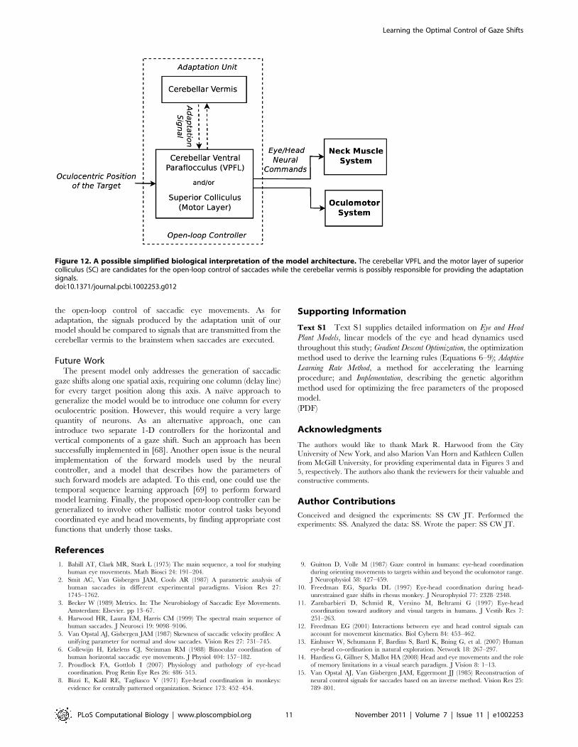
the open-loop control of saccadic eye movements. As for
adaptation, the signals produced by the adaptation unit of our
model should be compared to signals that are transmitted from the
cerebellar vermis to the brainstem when saccades are executed.
Future WorkThe present model only addresses the generation of saccadic
gaze shifts along one spatial axis, requiring one column (delay line)
for every target position along this axis. A naıve approach to
generalize the model would be to introduce one column for every
oculocentric position. However, this would require a very large
quantity of neurons. As an alternative approach, one can
introduce two separate 1-D controllers for the horizontal and
vertical components of a gaze shift. Such an approach has been
successfully implemented in [68]. Another open issue is the neural
implementation of the forward models used by the neural
controller, and a model that describes how the parameters of
such forward models are adapted. To this end, one could use the
temporal sequence learning approach [69] to perform forward
model learning. Finally, the proposed open-loop controller can be
generalized to involve other ballistic motor control tasks beyond
coordinated eye and head movements, by finding appropriate cost
functions that underly those tasks.
Supporting Information
Text S1 Text S1 supplies detailed information on Eye and Head
Plant Models, linear models of the eye and head dynamics used
throughout this study; Gradient Descent Optimization, the optimization
method used to derive the learning rules (Equations 6–9); Adaptive
Learning Rate Method, a method for accelerating the learning
procedure; and Implementation, describing the genetic algorithm
method used for optimizing the free parameters of the proposed
model.
(PDF)
Acknowledgments
The authors would like to thank Mark R. Harwood from the City
University of New York, and also Marion Van Horn and Kathleen Cullen
from McGill University, for providing experimental data in Figures 3 and
5, respectively. The authors also thank the reviewers for their valuable and
constructive comments.
Author Contributions
Conceived and designed the experiments: SS CW JT. Performed the
experiments: SS. Analyzed the data: SS. Wrote the paper: SS CW JT.
References
1. Bahill AT, Clark MR, Stark L (1975) The main sequence, a tool for studying
human eye movements. Math Biosci 24: 191–204.
2. Smit AC, Van Gisbergen JAM, Cools AR (1987) A parametric analysis of
human saccades in different experimental paradigms. Vision Res 27:
1745–1762.
3. Becker W (1989) Metrics. In: The Neurobiology of Saccadic Eye Movements.
Amsterdam: Elsevier. pp 13–67.
4. Harwood HR, Laura EM, Harris CM (1999) The spectral main sequence of
human saccades. J Neurosci 19: 9098–9106.
5. Van Opstal AJ, Gisbergen JAM (1987) Skewness of saccadic velocity profiles: A
unifying parameter for normal and slow saccades. Vision Res 27: 731–745.
6. Collewijn H, Erkelens CJ, Steinman RM (1988) Binocular coordination of
human horizontal saccadic eye movements. J Physiol 404: 157–182.
7. Proudlock FA, Gottlob I (2007) Physiology and pathology of eye-head
coordination. Prog Retin Eye Res 26: 486–515.
8. Bizzi E, Kalil RE, Tagliasco V (1971) Eye-head coordination in monkeys:
evidence for centrally patterned organization. Science 173: 452–454.
9. Guitton D, Volle M (1987) Gaze control in humans: eye-head coordination
during orienting movements to targets within and beyond the oculomotor range.
J Neurophysiol 58: 427–459.
10. Freedman EG, Sparks DL (1997) Eye-head coordination during head-
unrestrained gaze shifts in rhesus monkey. J Neurophysiol 77: 2328–2348.
11. Zambarbieri D, Schmid R, Versino M, Beltrami G (1997) Eye-head
coordination toward auditory and visual targets in humans. J Vestib Res 7:
251–263.
12. Freedman EG (2001) Interactions between eye and head control signals can
account for movement kinematics. Biol Cybern 84: 453–462.
13. Einhuser W, Schumann F, Bardins S, Bartl K, Bning G, et al. (2007) Human
eye-head co-ordination in natural exploration. Network 18: 267–297.
14. Hardiess G, Gillner S, Mallot HA (2008) Head and eye movements and the role
of memory limitations in a visual search paradigm. J Vision 8: 1–13.
15. Van Opstal AJ, Van Gisbergen JAM, Eggermont JJ (1985) Reconstruction of
neural control signals for saccades based on an inverse method. Vision Res 25:
789–801.
Figure 12. A possible simplified biological interpretation of the model architecture. The cerebellar VPFL and the motor layer of superiorcolliculus (SC) are candidates for the open-loop control of saccades while the cerebellar vermis is possibly responsible for providing the adaptationsignals.doi:10.1371/journal.pcbi.1002253.g012
Learning the Optimal Control of Gaze Shifts
PLoS Computational Biology | www.ploscompbiol.org 11 November 2011 | Volume 7 | Issue 11 | e1002253

16. Bizzi E, Dev P, Morasso P, Polit A (1978) E_ect of load disturbances during
centrally initiated movements. J Neurophysiol 41: 542–556.
17. Land MF, Tatler BW (2009) Looking and Acting: Vision and Eye Movements inNatural Behaviour. New York: Oxford University Press. 320 p.
18. Enderle JD, Wolfe JW (1987) Time-optimal control of saccadic eye movements.
IEEE Trans Biomed Eng 34: 43–55.
19. Sonneborn LM, Van Vleck FS (1964) The bang-bang principle for linear control
systems. J Soc Ind Appl Math A 2: 151–159.
20. Harris CM (1998) On the optimal control of behaviour: A stochastic perspective.J Neurosci Meth 83: 73–88.
21. Harris CM, Wolpert DM (1998) Signal-dependent noise determines motor
planning. Nature 394: 780–784.
22. Harris CM, Wolpert DM (2006) The main sequence of saccades optimizes
speed-accuracy trade-off. Biol Cybern 95: 21–29.
23. Kardamakis AA, Moschovakis AK (2009) Optimal control of gaze shifts.J Neurosci 29: 7723–7730.
24. Freedman EG, Sparks DL (2000) Coordination of the eyes and head: Movement
kinematics. Exp Brain Res 131: 22–32.
25. Laurutis VP, Robinson DA (1986) The vestibulo-ocular reex during humansaccadic eye movements. J Physiol 373: 209–233.
26. Zee DS, Optican LM, Cook JD, Robinson DA, Engel WK (1976) Slow saccades
in spinocerebellar degeneration. Arch Neurol 33: 243–251.
27. Van Gisbergen JAM, Robinson DA, Gielen S (1981) A quantitative analysis of
generation of saccadic eye movements by burst neurons. J Neurophysiol 45:417–442.
28. Guitton D, Munoz DP, Galiana HL (1990) Gaze control in the cat: studies and
modeling of the coupling between orienting eye and head movements indifferent behavioral tasks. J Neurophysiol 64: 509–531.
29. Goossens HHLM, Van Opstal AJ (1997) Human eye-head coordination in two
dimensions under different sensorimotor conditions. Exp Brain Res 114:542–560.
30. Freedman EG (2008) Coordination of the eyes and head during visual orienting.
Exp Brain Res 190: 369–387.
31. Chen-Harris H, Joiner WM, Ethier V, Zee DS, Shadmehr R (2008) Adaptivecontrol of saccades via internal feedback. J Neurosci 28: 2804–2813.
32. Phillips JO, Ling L, Fuchs AF, Siebold C, Plorde JJ (1995) Rapid horizontal gaze
movement in the monkey. J Neurophysiol 73: 1632–1652.
33. Kardamakis AA, Grantyn A, Moschovakis AK (2010) Neural network
simulations of the primate oculomotor system. V. Eye-head gaze shifts. BiolCybern 102: 209–225.
34. Pontryagin LS, Boltyanskii VG, Gamkrelidze RV, Mishchenko EF (1962) The
Maximum Principle. In: The Mathematical Theory of Optimal Processes. NewYork: John Wiley and Sons.
35. Dichgans J, Bizzi E, Morasso P, Tagliasco V (1973) Mechanisms underlying
recovery of eye-head coordination following bilateral labyrinthectomy inmonkeys. Exp Brain Res 18: 548–562.
36. Barash S, Melikyan A, Sivakov A, Zhang M, GlicksteinMea (1999) Saccadic
dysmetria and adaptation after lesions of the cerebellar cortex. J Neurosci 19:10931–10939.
37. Hopp J, Fuchs A (2004) The characteristics and neuronal substrate of saccadic
eye movement plasticity. Prog Neurobiol 72: 27–53.
38. Saeb S, Weber C, Triesch J (2009) A neural model for the adaptive control of
saccadic eye movements. In: Proceedings of International Joint Conference on
Neural Networks; 14–19 June 2009; Atlanta, Georgia, United States.
39. Freedman EG, Sparks DL (1997) Activity of cells in the deeper layers of the
superior colliculus of the rhesus monkey: Evidence for a gaze displacement
command. J Neurophysiol 78: 1669–1690.
40. Klier EM, Wang H, Crawford JD (2001) The superior colliculus encodes gaze
commands in retinal coordinates. Nat Neurosci 4: 627–632.
41. Kalesnykas RP, Sparks DL (1996) The primate superior colliculus and thecontrol of saccadic eye movements. Neuroscientist 2: 284–292.
42. Patestas MA, Gartner LP (2006) A Textbook of Neuroanatomy. MaldenMA:
Blackwell Publishing. pp 282–303.
43. Wang L, Gordon MD, Zhu J (2006) Regularized least absolute deviationsregression and an efficient algorithm for parameter tuning. In: Sixth IEEE
International Conference on Data Mining; 18–22 December 2006; Hong Kong,China. pp 690–700.
44. Fujita M, Amagai A, Minakawa F, Aoki M (2002) Selective and delay adaptation
of human saccades. Cognitive Brain Res 13: 41–52.45. Shafer JL, Noto CT, Fuchs AF (2000) Temporal characteristics of error signals
driving saccadic gain adaptation in the macaque monkey. J Neurophysiol 84:
88–95.46. Oommen BS, Stahl JS (2005) Overlapping gaze shifts reveal timing of an eye-
head gate. Exp Brain Res 167: 276–286.47. Tomlinson RD, Bahra PS (1986) Combined eye-head gaze shifts in the primate.
I. Metrics. J Neurophysiol 56: 1542–1557.
48. Populin LC, Tollin DJ, Weinstein JM (2002) Human gaze shifts to acoustic andvisual targets. Ann NY Acad Sci 956: 468473.
49. Wolpert DM, Miall RC, Kawato M (1998) Internal models in the cerebellum.Trends Cogn Sci 2: 338–347.
50. Kawato M (1999) Internal models for motor control and trajectory planning.Curr Opin Neurobiol 9: 718–727.
51. Bastian AJ (2008) Learning to predict the future: the cerebellum adapts
feedforward movement control. Curr Opin Neurobiol 16: 645–649.52. Soetedjo R, Fuchs AF (2006) Complex spike activity of Purkinje cells in the
oculomotor vermis during behavioral adaptation of monkey saccades. J Neurosci26: 7741–7755.
53. Optican LM, Robinson DA (1980) Cerebellar-dependent adaptive control of
primate saccadic system. J Neurophysiol 44: 1058–1076.54. Buettner U, Straube A (1995) The effect of cerebellar midline lesions on eye
movements. Neuroophthalmology 15: 7582.55. Winograd-Gurvich CT, Georgiou-Karistianis N, Evans A, Millist L,
Bradshaw JL, et al. (2003) Hypometric primary saccades and increasedvariability in visually-guided saccades in huntingtons disease. Neuropsychologia
41: 1683–1692.
56. Dean P, Porrill J, Ekerot CF, Jrntell H (2010) The cerebellar microcircuit as anadaptive _lter: experimental and computational evidence. Nat Rev Neurosci 11:
30–43.57. Takemura A, Inoue Y, Gomi H, Kawato M, Kawano K (2001) Change in
neuronal _ring patterns in the process of motor command generation for the
ocular following response. J Neurophysiol 86: 1750–1763.58. Medina JF, Garcia KS, Nores WL, Taylor NM, Mauk MD (2000) Timing
mechanisms in the cerebellum: testing predictions of a large-scale computersimulation. J Neurosci 20: 5516–5525.
59. Yamazaki T, Tanaka S (2007) A spiking network model for passage-of-timerepresentation in the cerebellum. Eur J Neurosci 26: 2279–2292.
60. Wurtz RH, Optican LM (1994) Superior colliculus cell types and models of
saccade generation. Curr Opin Neurobiol 4: 857–861.61. Grossberg S, Roberts K, Aguilar M, Bullock D (1997) A neural model of
multimodal adaptive saccadic eye movement control by superior colliculus.J Neurosci 17: 9706–9725.
62. Aizawa H, Wurtz RH (1998) Reversible inactivation of monkey superior
colliculus. I. Curvature of saccadic trajectory. J Neurophysiol 79: 2082–2096.63. Nakahara H, Morita K, Wurtz RH, Optican LM (2006) Saccade-related spread
of activity across superior colliculus may arise from asymmetry of internalconnections. J Neurophysiol 96: 765–774.
64. Moschovakis AK, Kitama T, Dalezios Y, Petit J, Brandi AM, et al. (1998) Ananatomical substrate for the spatiotemporal transformation. J Neurosci 18:
10219–10229.
65. Lefvre P, Quaia C, Optican LM (1998) Distributed model of control of saccadesby superior colliculus and cerebellum. Neural Netw 11: 1175–1190.
66. Optican LM, Quaia C (2002) Distributed model of collicular and cerebellarfunction during saccades. Ann NY Acad Sci 956: 164–177.
67. Shadmehr R, Orban de Xivry JJ, Xu-Wilson M, Shih TY (2010) Temporal
discounting of reward and the cost of time in motor control. J Neurosci 30:10507–10516.
68. Kuniharu A, Keller EL (2005) A model of the saccade-generating system thataccounts for trajectory variations produced by competing visual stimuli. Biol
Cybern 92: 21–37.
69. Porr B, Von Ferber C, Worgotter F (2003) ISO learning approximates a solutionto the inversecontroller problem in an unsupervised behavioral paradigm.
Neural Comput 15: 865–884.70. Sylvestre PA, Cullen KE (1999) Quantitative analysis of abducens neuron
discharge dynamics during saccadic and slow eye movements. J Neurophysiol82: 2612–2632.
Learning the Optimal Control of Gaze Shifts
PLoS Computational Biology | www.ploscompbiol.org 12 November 2011 | Volume 7 | Issue 11 | e1002253