lecture 3 chromosome · banding pattern. g-banding generates a characteristic lateral series of...
TRANSCRIPT

1
MBioS 503:Chromosomes
Joy Winuthayanon, PhDSchool of Molecular Biosciences
Biotech/Life Sciences [email protected]
https://labs.wsu.edu/winuthayanon
2
• Describe different states (appearances) of chromosomes and the function of each chromosome state
Objectives: Chromosomes
1
3
What is chromosome?• A discrete unit of the
genome carrying many genes.
• Each consists of a very long molecule of duplex DNA and an approximately equal mass of proteins.
• It is visible as a morphological entity only during cell division.
4
Viral genomes are packaged into their coats• Capsid – The external protein coat of a virus particle.• The length of DNA that can be incorporated into a virus is
limited by the structure of the head shell.• Nucleic acid within the head shell is extremely condensed.
A helical path for TMV RNA is created by the stacking of protein subunits in the virion (the entire virus particle).
helix

5
• Icosahedral symmetry – A structure with 60 rotational symmetries (such as a soccer ball).
• Filamentous RNA viruses condense the RNA genome as they assemble the head shell around it.
• nucleation center – A duplex hairpin in TMV (tobacco mosaic virus) in which assembly of coat protein with RNA is initiated.
Viral genomes are packaged into their coats
Maturation of phage lambda passes through several stages.
Top photo reproduced from D. Cue and M. Feiss, Proc. Natl. Acad. Sci. USA 90 (1993): 9240-9294. Copyright © 2004 National Academy of Sciences, U.S.A. Photo courtesy of Michael G. Feiss, University of Iowa. Bottom photo courtesy of Robert Duda, University of Pittsburgh.
6
• Spherical DNA viruses insert the DNA into a preassembled protein shell.• terminase – An enzyme that
cleaves multimers of a viral genome and then uses hydrolysis of ATP to provide the energy to translocate the DNA into an empty viral capsid starting with the cleaved end.
Viral genomes are packaged into their coats
Terminase protein binds to specific sites on a multimer of virus genomes
generated by rolling circle replication.
7
Nucleoid = structure in a prokaryotic cell that contains the genome.
o The DNA is bound to proteins and is not enclosed by a membrane.
The bacterial genome is a nucleoid with dynamic structural properties
Transcription and translation take place in the same compartment in bacteria.
8
The bacterial genome is a nucleoid with dynamic structural properties• Bacterial nucleoid: organized as
multiple loops compacted by nucleoid-associated proteins (NAPs) such as H-NS and HU.
• Nucleoid-associated proteins are typically small, abundant DNA-binding proteins that function in nucleoid architecture, domain topology, and gene regulation. A thin section
shows the bacterial nucleoid as a compact mass in the center of the cell.
The nucleoid spills out of a lysed E. coli cell in the form of loops of a fiber.
1 & 2

9
Growth phase and elements that affect nucleoid structure. A typical growth curve for E. coli growing in batch culture begins with a lag phase followed by the log phase of exponential growth and, finally, stationary phase (when the cells stop growing).
Nucleoid-associated proteins are expressed at different times during the growth curve
Different NAPs bind different sites on DNA
• IHF (integration host factor)
• H-NS (histone-like, nucleoid-structuring protein)
10
Bacterial genome is supercoiled
• The nucleoid has about 400 independent negatively supercoiled domains.
• The average density of supercoiling is approximately 1 turn/100 bp.
Clockwise(left-handed)
Counter-clockwise(right-handed)
11
Why does DNA supercoil?
• Economic way for DNA packaging within cells (less space)
Koster, Daniel A., et al. "Cellular strategies for regulating DNA supercoiling: a single-molecule perspective." Cell 142.4 (2010): 519-530.
12
Eukaryotic DNA has loops and domains attached to a protein scaffold
• DNA of interphase chromatin is negatively supercoiled into independent domains averaging 85 kb.
• Metaphase chromosomes have a protein scaffold to which the loops of supercoiled DNA are attached. Histone-depleted chromosomes
consist of a protein scaffold to which loops of DNA are anchored.
metaphase scaffold resembles the general form of a mitotic chromosome, surrounded by a halo of DNA

13
Specific sequences attach DNA to an interphase matrix
• DNA is attached to the nuclear matrix at specific sequences called matrix attachment regions (MARs).
• The MARs are A-T-rich but do not have any specific consensus sequence.
• chromosome scaffold – A proteinaceous structure in the shape of a sister chromatid pair, generated when chromosomes are depleted of histones.
Matrix-associated regions (MARs). 14
Chromatin is divided into euchromatin and heterochromatin
The sister chromatids of a mitotic pair each consist of a fiber (~30 nm in diameter) compactly folded into the chromosome
15
Chromatin is divided into euchromatin and heterochromatin
• We can see individual chromosomes only during mitosis.
• In most regions, the chromatin is less densely packed than in the mitotic chromosome. This material, called euchromatin, is relatively dispersed and occupies most of the nucleoplasm.
• During interphase, the general mass of chromatin is in the form of euchromatin, which is slightly less tightly packed than mitotic chromosomes.
• Some regions of chromatin are very densely packed (heterochromatin), displaying a condition comparable to that of the chromosome at mitosis.
It forms a series of discrete clumps
a tendency to be found at the nuclear peripheryand at the nucleous
16
• Regions of heterochromatin remain densely packed throughout interphase.
• Common form of heterochromatin that always remains heterochromatic is called constitutive heterochromatin.
• In contrast, another category of heterochromatin, called facultative heterochromatin, is regions of euchromatin that are converted to a heterochromatic state.
Chromosomes occupy chromosome territories in the nucleus and are not
entangled with one another.
Chromatin is divided into euchromatin and heterochromatin
3-5

17
Chromosome banding patterns
18
Chromosomes have banding patterns• Certain staining techniques cause the
chromosomes to have the appearance of a series of striations, which are called G-bands.
• The bands (red) are lower in G-C content than the interbands.
• Genes are concentrated in the G-C-rich interbands (grey).
The human X chromosome can be divided into distinct regions by its banding pattern.
G-banding generates a characteristic lateral series of bands in each member of the chromosome set.
19
Lampbrush chromosomes are extended
• chromomeres – “beads on a string” densely staining granules visible in chromosomes under certain conditions, especially early in meiosis.
• Sites of gene expression on lampbrush chromosomes show loops that are extended from the chromosomal axis.
A lampbrush chromosome is a meiotic bivalent in which the two pairs of sister chromatids are held together at chiasmata.
Photo courtesy of Joseph G. Gall, Carnegie Institution.
• A stretched-out-form of chromosome
• Best characterized in amphibians and birds
• Formed during usually extended meiosis (up to several months)
20
Polytene chromosomes form bands
• Polytene (=large) chromosomes of dipterans (two-wing insects) have a series of bands that can be used as a cytological map.
The polytene chromosomes of D. melanogaster form an alternating series of bands and
interbands. Individual bands containing particular genes can be identified by
in situ hybridization.
21
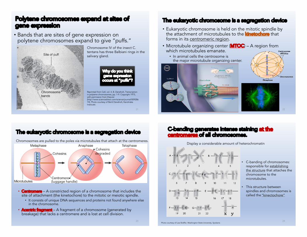
Polytene chromosomes expand at sites of gene expression
• Bands that are sites of gene expression on polytene chromosomes expand to give “puffs.”
Chromosome IV of the insect C. tentans has three Balbiani rings in the salivary gland.
Reprinted from Cell, vol. 4, B. Daneholt, Transcription in polytene chromosomes, pp. 1-9. Copyright 1975, with permission from Elsevier [http://www.sciencedirect.com/science/journal/00928674]. Photo courtesy of Bertil Daneholt, KarolinskaInstitutet.
Why do you think gene expression
occurs at “puffs”?
22
The eukaryotic chromosome is a segregation device
• Eukaryotic chromosome is held on the mitotic spindle by the attachment of microtubules to the kinetochore that forms in its centromeric region.• Microtubule organizing center (MTOC) – A region from
which microtubules emanate.• In animal cells the centrosome is
the major microtubule organizing center.
23
Chromosomes are pulled to the poles via microtubules that attach at the centromeres.
The eukaryotic chromosome is a segregation device
• Centromere – A constricted region of a chromosome that includes the site of attachment (the kinetochore) to the mitotic or meiotic spindle.• It consists of unique DNA sequences and proteins not found anywhere else
in the chromosome.
• Acentric fragment – A fragment of a chromosome (generated by breakage) that lacks a centromere and is lost at cell division.
Cohesinsdegraded
(luggage handle)
24
C-banding generates intense staining at the centromeres of all chromosomes.
Photo courtesy of Lisa Shaffer, Washington State University, Spokane.
Display a considerable amount of heterochromatin
• C-banding of chromosomes: responsible for establishing the structure that attaches the chromosome to the microtubules.
• This structure between spindles and chromosomes is called the“kinectochore”

25
Regional centromeres contain a centromerichistone H3 variant and repetitive DNA
• Installation of the centromere-specific histone H3 is an epigenetic (Dr. Hee’s section) and primary determinant that specifies a functional centromere.• Centromeres in higher eukaryotic chromosomes contain
large amounts of repetitive DNA and unique histone variants.• The function of the ever-present repetitive centromeric DNA
is not known.
Centromeres are characterized by a centromere-specific histone H3 variant and often have heterochromatin that is rich in satellite DNA sequences.
26
Organization of CENP-A and H3 nucleosomes in centromeres.
(a) Centromeres are ~40 kb long in chicken,corresponding to 200 nucleosomes per centromere. Of these, 30 are predicted to contain CENP-A (roughly 1 in 6–8 centromericnucleosomes). Thus, centromeric chromatin is largely composed of nucleosomes containing histone H3.
(b and c) The CENP-A chromatin was originally suggested to form an amphipathic organization, with CENP-A on the exterior facing the kinetochore, and H3 largely on the interior. This chromatin was proposed to form either a helix or loop structure. (d) The boustrophedon model of centromeric CENP-A-containing chromatin was proposed based on super-resolution microscopy.
Data from Fukagawa, T., et al. (2014). Dev Cell 30: 496–508doi: (10.1016/j.devcel.2014.08.016.
Regional centromeres contain a centromeric histone H3 variant and repetitive DNA
CENP = centromere protein
27
Point centromeres in S. cerevisiae contain short, essential DNA sequences
• CEN elements are identified in S. cerevisiae by the ability to allow a plasmid to segregate accurately at mitosis.
• CEN elements consist of the short, conserved sequences CDE-I (Centromere DNA Element) and CDE-III that flank the A-T-rich region CDE-II.
Three conserved regions can be identified by the sequence homologies between yeast CEN elements.
28
The S. cerevisiae centromere binds a protein complex• A specialized protein complex that is an alternative to the
usual chromatin structure is formed at CDE-II.• The histone H3 variant Cse4 is incorporated in the centromeric
nucleosome.• The CBF3 protein complex that binds to CDE-III is essential for
centromeric function.• The proteins that bind CEN serve
as an assembly platform for thekinetochore and provide theconnection to microtubules.
The DNA at CDE-II is wound around an
alternative nucleosome.

29
Telomeres
30
Telomeres have simple repeating sequences• The telomere is required for the stability of the chromosome
end.• A telomere consists of a simple repeat where a G-rich strand
at the 3ʹ terminus typically has a sequence of (T/A)1–4 G>2.
A typical telomere has a simple repeating structure with a G-T-rich strand that extends beyond the C-A-rich strand.
31
Telomeres seal the chromosome ends and function in meiotic chromosome pairing• The protein TRF2 catalyzes a
reaction in which the 3ʹ repeating unit of the G+T-rich strand forms a loop by displacing its homolog in an upstream region of the telomere.
A loop forms at the end of chromosomal DNA.
© Dr. Gopal Murti/Science Source.
The meiotic telomere cluster is visualized by telomere FISH.
Photo courtesy of S. P. Murphy and H. W. Bass, Florida State University. 32
• Telomeres promote pairing, synapsis, and recombination during meiosis via links to the cytoskeleton through nuclear envelope proteins.
Telomeres seal the chromosome ends and function in meiotic chromosome pairing

33
Telomeres are synthesized by a ribonucleoprotein enzyme• Telomerase uses the 3ʹ–OH of the G+T telomeric strand
and its own RNA template to iteratively add tandem repeats (5ʹ-TTAGGG-3ʹ in human) to the 3ʹ end at each chromosomal terminus.
• Telomerase uses a reverse transcriptase to extend the very ends of the chromosomes and solve the so-called end replication problem.
34
Telomerase positions itself by base pairing between the RNA template and the protruding single-stranded DNA primer.
Telomeres are synthesized by a ribonucleoprotein enzyme
35
Telomeres are essential for survival
• Telomerase is expressed in actively dividing cells and is not expressed in quiescent cells.• Loss of telomeres results in
senescence.• Escape from senescence can
occur if telomerase is reactivated, or via unequal homologous recombination to restore telomeres.
Mutation in telomerase causes telomeres to shorten in each cell
division.
36
Crossing-over in telomeric regions is usually suppressed by mismatch-repair systems, but can occur when they are mutated.
Telomeres are essential for survival

37
List 2 new things you have learnt today
6-8