lecture 9 - kau 9.pdf · the golgi apparatus the golgi apparatus (ga), also called golgi body or...
TRANSCRIPT

Lecture 9
٢٢٢ cell Biology ١
The Golgi Apparatus
The Golgi apparatus (GA), also called Golgi body or Golgi complex and found universally in both plant and animal cellsIs typically comprised of a series of five to eight cup-shaped, membrane-covered sacs called cisternae that look something like a stack of deflated balloons. In some unicellular flagellates, however, as many as 60 cisternaemay combine to make up the Golgi apparatus. Similarly, the number of Golgi bodies in a cell varies according to its function. Animal cells generally contain between ten and twenty Golgi stacks per cell, which are linked into a single complex by tubular connections between cisternae. This complex is usually located close to the cell nucleus.
٢٢٢ cell Biology ٢
٢٢٢ cell Biology ٣
Golgi Apparatus Structure
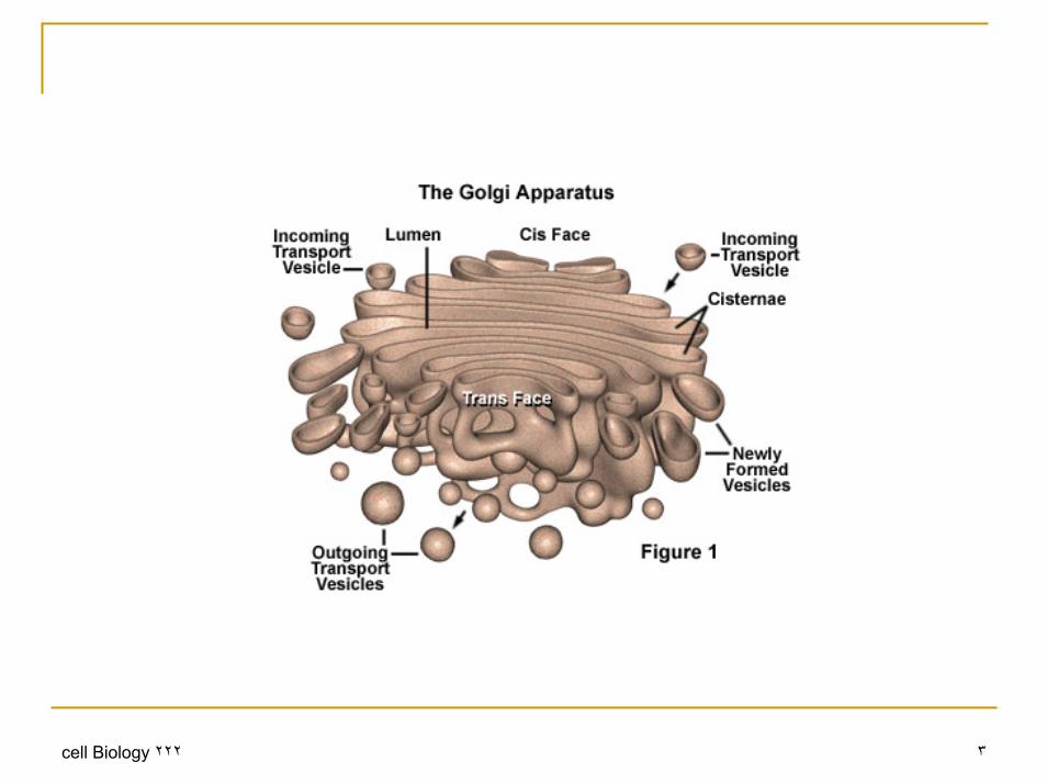
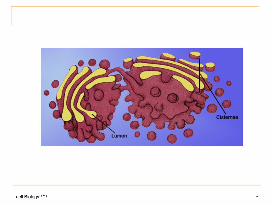
The Golgi is composed of membrane-bound stacks known as cisternae (singular: cisterna). Between four and eight are usually present; however, in some protists as many as sixty have been observed.Each cisterna comprises a flattened membrane disk, and carries Golgi enzymes to help or to modify cargo proteins that travel through them. The cisternae stack has five functional regions: the cis-Golgi network, cis-Golgi, medial-Golgi, trans-Golgi, and trans-Golgi network.
٢٢٢ cell Biology ٤
Golgi Apparatus StructureVesicles from the endoplasmic reticulum (via the vesicular-tubular cluster) fuse with the cis-Golgi network and subsequently progress through the stack to the trans Golgi network, where they are packaged and sent to the required destination Each region contains different enzymes which selectively modify the contents depending on where they reside.The cisternae also carry structural proteins important for their maintenance as flattened membranes which stack upon each other. The trans face of the trans-Golgi network is the face from which vesicles leave the Golgi. These vesicles then proceed to later compartments such as the cell membrane, secretory vesicles or late endosomes
٢٢٢ cell Biology ٥
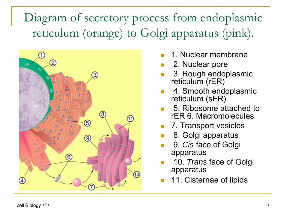
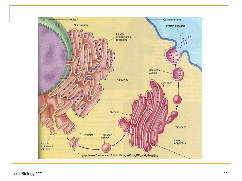
Diagram of secretory process from endoplasmic reticulum (orange) to Golgi apparatus (pink).
1. Nuclear membrane2. Nuclear pore3. Rough endoplasmic
reticulum (rER)4. Smooth endoplasmic
reticulum (sER)5. Ribosome attached to
rER 6. Macromolecules 7. Transport vesicles8. Golgi apparatus9. Cis face of Golgi
apparatus10. Trans face of Golgi
apparatus 11. Cisternae of lipids
٢٢٢ cell Biology ٦

Golgi Apparatus Structure
The modifications to molecules that take place in the Golgi apparatus occur in an orderly fashion. Each Golgi stack has two distinct ends, or faces. The cis face of a Golgi stack is the end of the organelle where substances enter from the endoplasmic reticulum for processing, while the trans face is where they exit in the form of smaller detached vesicles. Consequently, the cis face is found near the endoplasmic reticulum, from whence most of the material it receives comes, and the trans face is positioned near the plasma membrane of the cell, to where many of the substances it modifies are shipped. The chemical make-up of each face is different and the enzymes contained in the lumens (inner open spaces) of the cisternaebetween the faces are distinctive .
٢٢٢ cell Biology ٧
٢٢٢ cell Biology ٨

FunctionCells synthesise a large number of different macromolecules. The Golgi apparatus is integral in modifying, sorting, and packaging these macromolecules for cell secretion (exocytosis) or use within the cell. It primarily modifies proteins delivered from the rough endoplasmic reticulum but is also involved in the transport of lipids around the cell, and the creation of lysosomes. In this respect it can be thought of as similar to a post office; it packages and labels items which it then sends to different parts of the cell.Enzymes within the cisternae are able to modify substances by the addition of carbohydrates (glycosylation) and phosphates (phosphorylation).The Golgi plays an important role in the synthesis of proteoglycans, which are molecules present in the extracellular matrix of animals. It is also a major site of carbohydrate synthesisThe Golgi has a putative role in apoptosis
٢٢٢ cell Biology ٩

٢٢٢ cell Biology ١٠

Endoplasmic reticulumThe endoplasmic reticulum (ER) is an eukaryotic organelle that forms an interconnected network of tubules, vesicles, and cisternaewithin cells.The general structure of the endoplasmic reticulum is an extensive membrane network of cisternae held together by the cytoskeleton. The phospholipid membrane encloses a space, the cisternal space (or lumen), from the cytosol, which is continuous with the perinuclearspace. The functions of the endoplasmic reticulum vary greatly depending on the exact type of endoplasmic reticulum and the type of cell in which it resides. The three varieties are called rough endoplasmic reticulum, smooth endoplasmic reticulum and sarcoplasmic reticulum.The quantity of RER and SER in a cell can quickly interchange from one type to the other, depending on changing metabolic needs
٢٢٢ cell Biology ١١

Rough endoplasmic reticulumThe surface of the rough endoplasmic reticulum (RER) is studded with protein-manufacturing ribosomes giving it a "rough" appearance (hence its name). However, the ribosomes bound to the RER at any one time are not a stable part of this organelle's structure as ribosomes are constantly being bound and released from the membraneA ribosome only binds to the ER once it begins to synthesize a protein destined for the secretory pathway.Here, a ribosome in the cytosol begins synthesizing a protein until a signal recognition particle recognizes the pre-piece of 5-15 hydrophobic amino acids preceded by a positively charged amino acid
٢٢٢ cell Biology ١٢

Rough endoplasmic reticulum
This signal sequence allows the recognition particle to bind to the ribosome, causing the ribosome to bind to the RER and pass the new protein through the ER membrane. The pre-piece is then cleaved off within the lumen of the ER and the ribosome released back into the cytosol.The membrane of the RER is continuous with the outer layer of the nuclear envelopeAlthough there is no continuous membrane between the RER and the Golgi apparatus, membrane-bound vesicles shuttle proteins between these two compartments.Vesicles are surrounded by coating proteins called COPI and COPII. COPII targets vesicles to the golgi and COPI marks them to be brought back to the RER.
٢٢٢ cell Biology ١٣

Smooth endoplasmic reticulum
The smooth endoplasmic reticulum (SER) has functions in several metabolic processes, including synthesis of lipids and steroids, metabolism of carbohydrates, regulation of calcium concentration, drug detoxification, attachment of receptors on cell membrane proteins, and steroid metabolism.It is connected to the nuclear envelope. Smooth endoplasmic reticulum is found in a variety of cell types(both animal and plant) and it serves different functions in each.
٢٢٢ cell Biology ١٤
Smooth endoplasmic reticulum
The Smooth ER also contains the enzyme glucose-6-phosphatase which converts glucose-6-phosphate to glucose, a step in gluconeogenesisThe SER consists of tubules and vesicles that branch forming a network. In some cells there are dilated areas like the sacs of RER. The network of SER allows increased surface area for the action or storage of key enzymes and the products of these enzymes.
٢٢٢ cell Biology ١٥
٢٢٢ cell Biology ١٦
Sarcoplasmic reticulum
The sarcoplasmic reticulum (SR), from the Greek sarx, "flesh", is a special type of smooth ER found in smooth and striated muscle. The only structural difference between this organelle and the SER is the medley of proteins they have, both bound to their membranes and drifting within the confines of their lumens. This fundamental difference is indicative of their functions: the SER synthesizes molecules while the SR stores and pumps calcium ions.The SR contains large stores of calcium, which it sequesters andthen releases when the muscle cell is stimulated. The SR's release of calcium upon electrical stimulation of the cell plays a major role in excitation-contraction coupling.
٢٢٢ cell Biology ١٧