unit a: cells - gandha.weebly.com€¦ · golgi apparatus transport vesicle from er new vesicle...
TRANSCRIPT
Unit A: CellsCh. 4
A Tour of the Cell

Standards
By the end of this unit you should be able to:
Recognize and explain the function of each organelle
Look at micrographs/diagrams/pictures and correctly ID each organelle
Write/work with/explain the balanced chemical equation for cellular respiration
Relate the role of an organelle to a specific part of the body
Explain how the endomembrane system functions to compartmentalize the cell and move materials through it
Cell Intro
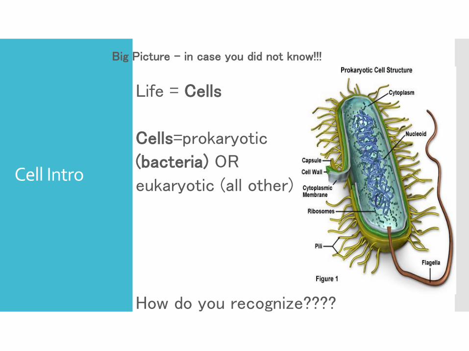
Big Picture – in case you did not know!!!
Life = Cells
Cells=prokaryotic
(bacteria) OR
eukaryotic (all other)
How do you recognize????

Cells EVERYWHERE!!!
Apoptosis in a Leukemia
Cell – Cell Suicide
Staphylococcus
aureus
Human Red Blood
Cells
Escherichia coli
Cells Everywhere!!!
How big is a cell????
How many cells in your body???
50 million million (50 trillion) cells – stretched end to end they would stretch around the Earth 47 times – if you could count one cell per second it would take you 2600 years !!!
Inner Life of Cell
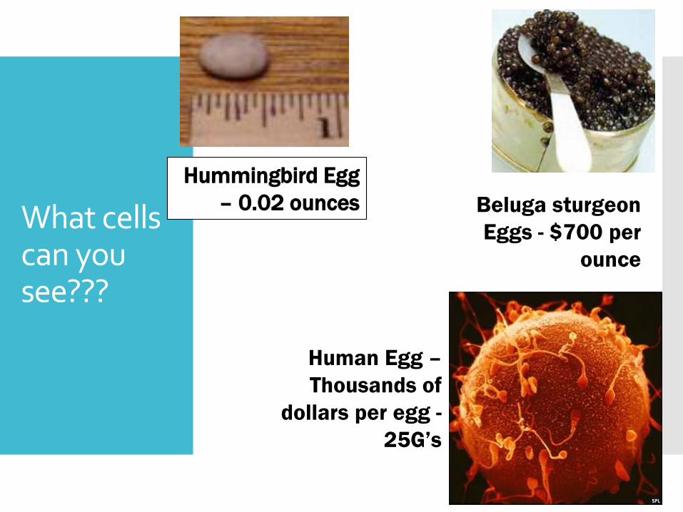
What cells can you see???
Hummingbird Egg
– 0.02 ounces Beluga sturgeon
Eggs - $700 per
ounce
Human Egg –
Thousands of
dollars per egg -
25G’s
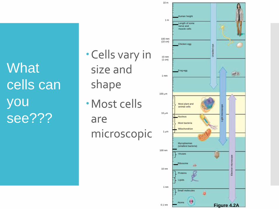
Cells vary in size and shape
Most cells are microscopic
Human height
Length of some
nerve and
muscle cells
Chicken egg
Frog egg
Una
ide
d e
ye
Lig
ht m
icro
sco
pe
Ele
ctr
on
mic
rosco
pe
10 m
1 m
100 mm
(10 cm)
10 mm
(1 cm)
1 mm
100 m
10 m
1 m
100 nm
10 nm
1 nm
0.1 nmAtoms
Proteins
Small molecules
Lipids
Viruses
Ribosome
Nucleus
Mycoplasmas
(smallest bacteria)
Most plant and
animal cells
Most bacteria
Mitochondrion
Figure 4.2A
What
cells can
you
see???

30 m10 m
30 m 10 m
Surface area
of one large cube
5,400 m2
Total surface area
of 27 small cubes
16,200 m2Figure 4.2B
The microscopic size of most cells ensures a sufficient sur face area
Across which nutrients and wastes can move to service the cell volume
A small cell has a greater ratio of sur face area to volume than a large cell of the same shapeSurface
area to
volume
ratio
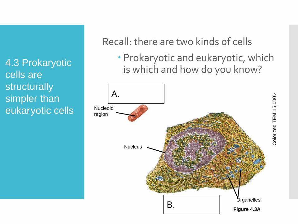
Recall: there are two kinds of cells
Prokaryotic and eukaryotic, which is which and how do you know?
Prokaryotic cell
Nucleoid
region
Nucleus
Eukaryotic cell Organelles
Colo
rize
d T
EM
15
,00
0
Figure 4.3A
4.3 Prokaryotic
cells are
structurally
simpler than
eukaryotic cells
A.
B.
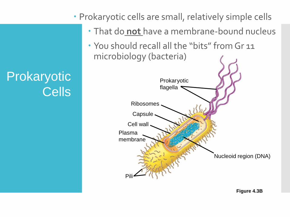
Prokaryotic
flagella
Ribosomes
Capsule
Cell wall
Plasma
membrane
Nucleoid region (DNA)
Pili
Prokaryotic cells are small, relatively simple cells
That do not have a membrane-bound nucleus
You should recall all the “bits” from Gr 11 microbiology (bacteria)
Figure 4.3B
Prokaryotic
Cells
4.4 Eukaryotic cells are partitioned into functional compartments (organelles)
All other forms of life (anything not bacteria) are composed of more complex eukaryotic cells
Distinguished by the presence of a true nucleus
Membranes form the boundaries of and within many eukaryotic cells
Compartmentalizing the interior of the cell into organelles and facilitating a variety of metabolic activities
Eukaryotic
Cells
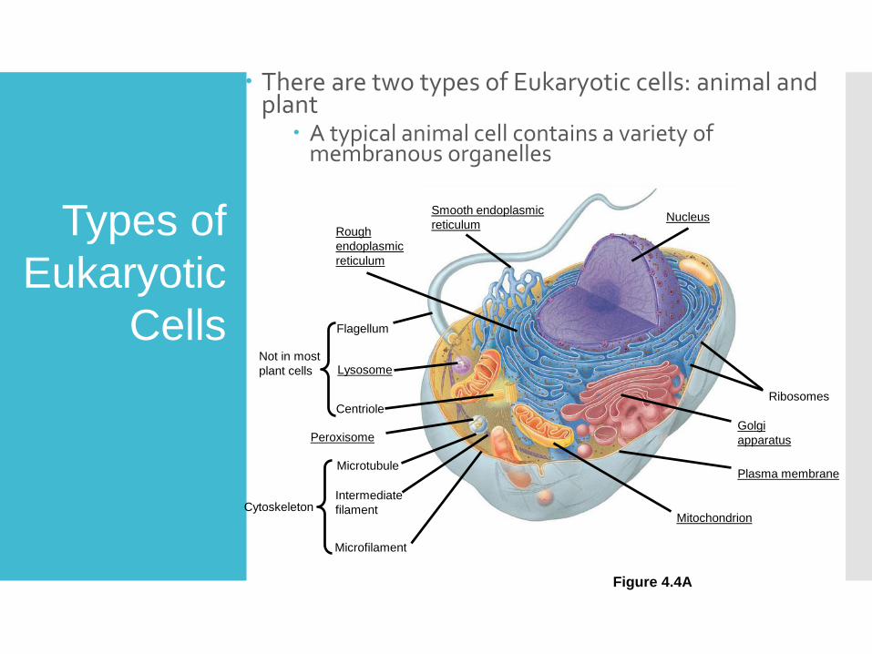
There are two types of Eukaryotic cells: animal and plant
A typical animal cell contains a variety of membranous organelles
NucleusSmooth endoplasmic
reticulumRough
endoplasmic
reticulum
Ribosomes
Golgi
apparatus
Plasma membrane
Mitochondrion
Flagellum
Not in most
plant cells Lysosome
Centriole
Microtubule
CytoskeletonIntermediate
filament
Microfilament
Peroxisome
Figure 4.4A
Types of
Eukaryotic
Cells

A typical plant cell has some structures that an animal cell lacks
Such as chloroplasts, a rigid cell wall and a central vacuole
Central
vacuoleNot in
animal
cellsChloroplast
Cell wall
Golgi
apparatus
Nucleus
Microtubule
CytoskeletonIntermediate
filament
Microfilament
Ribosomes
Smooth
endoplasmic
reticulum
Mitochondrion
Peroxisome
Plasma membrane
Rough
endoplasmic
reticulum
Figure 4.4B
Types of
Eukaryotic
Cells
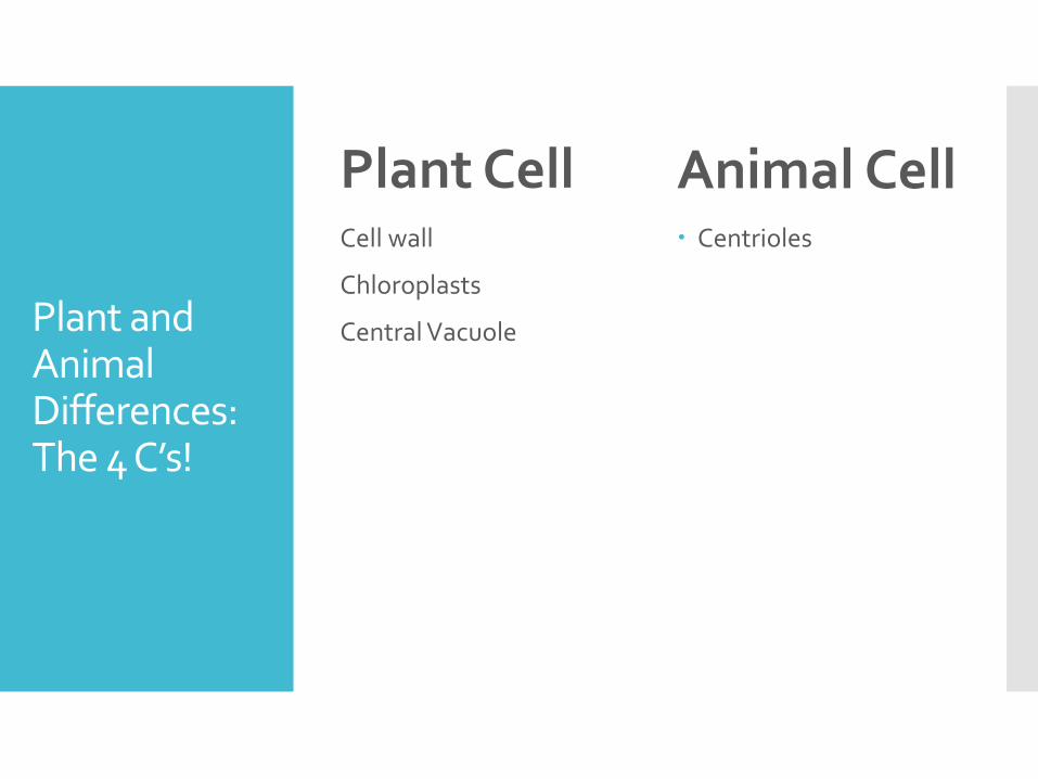
Plant and Animal Differences:The 4 C’s!
Plant CellCell wall
Chloroplasts
Central Vacuole
Animal Cell Centrioles
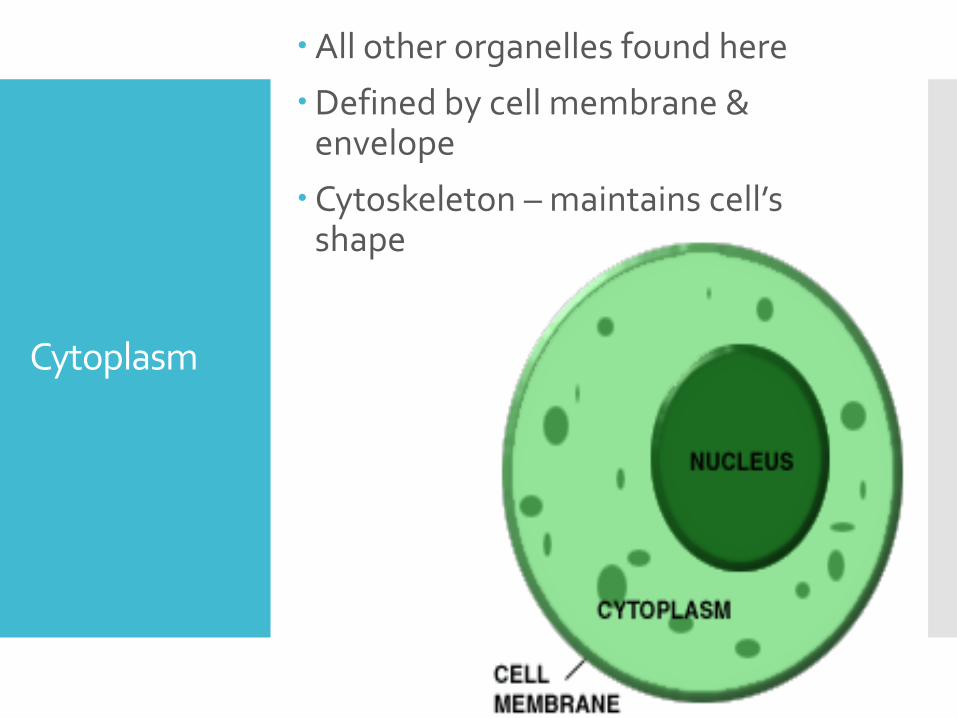
Cytoplasm
All other organelles found here
Defined by cell membrane & envelope
Cytoskeleton – maintains cell’s shape
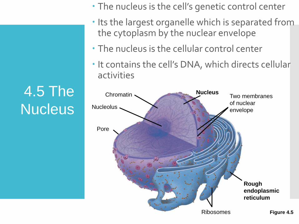
The nucleus is the cell’s genetic control center
Its the largest organelle which is separated from the cytoplasm by the nuclear envelope
The nucleus is the cellular control center
It contains the cell’s DNA, which directs cellular activities
NucleusChromatin
Nucleolus
Pore
Ribosomes
Rough
endoplasmic
reticulum
Two membranes
of nuclear
envelope
Figure 4.5
4.5 The
Nucleus

NuclearStructure
i) Nuclear Envelope – 2 membranes with pores
ii) Chromatin – thread likeBecomes chromosomes* (condensed) during cell division*Contains genetic material (DNA):
Meaningful parts = genesMade of - DNA & proteins
iii) Nucleolus – dark centerMakes rRNA (ribosomal RNA)
Many cell organelles are connected through the endomembrane system
a collection of membranous organelles that manufactures and distributes cell products
Nucleus, RER, SER, Golgi, Vesicles, Vacuoles
4.6
Endomembrane
System
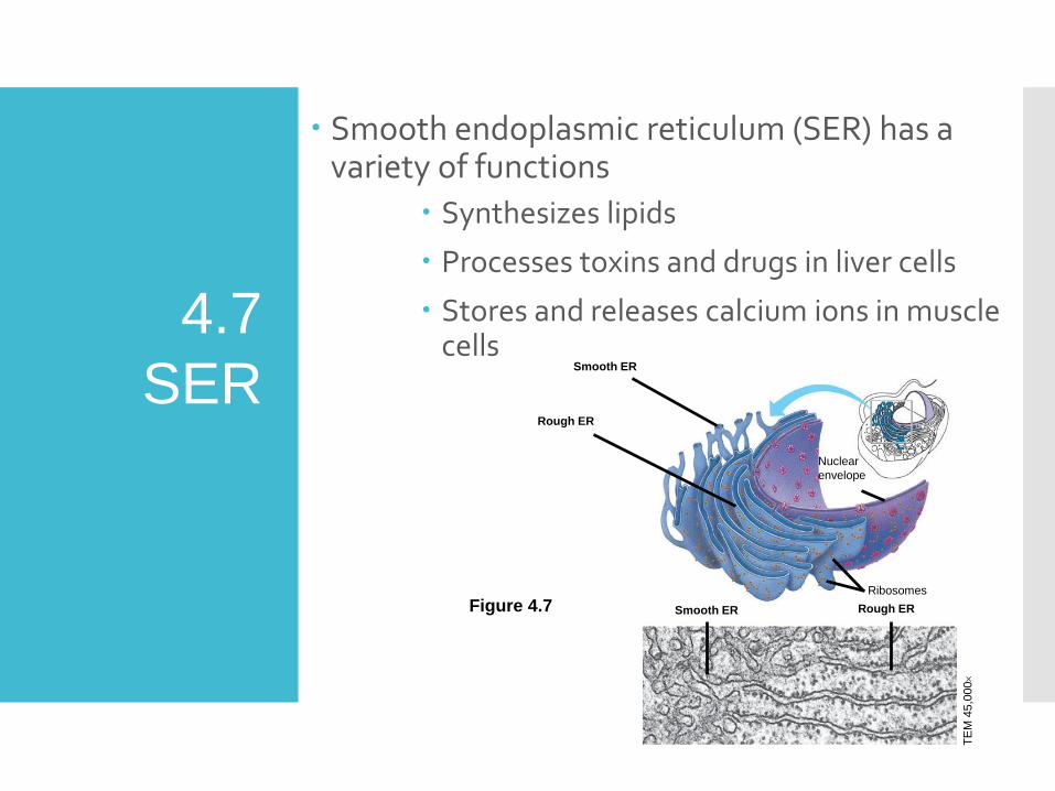
Smooth endoplasmic reticulum (SER) has a variety of functions
Synthesizes lipids
Processes toxins and drugs in liver cells
Stores and releases calcium ions in muscle cells
Smooth ER
Rough ER
Nuclear
envelope
Rough ER
Ribosomes
Smooth ER
TE
M 4
5,0
00
Figure 4.7
4.7
SER
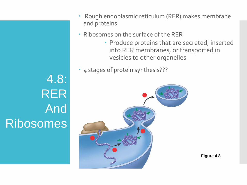
Rough endoplasmic reticulum (RER) makes membrane and proteins
Ribosomes on the surface of the RER
Produce proteins that are secreted, inserted into RER membranes, or transported in vesicles to other organelles
4 stages of protein synthesis???
4.8:
RER
And
Ribosomes
Figure 4.8
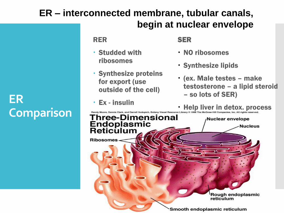
ER Comparison
RER
Studded with ribosomes
Synthesize proteins for export (use outside of the cell)
Ex - insulin
SER
NO ribosomes
Synthesize lipids
(ex. Male testes – make testosterone – a lipid steroid – so lots of SER)
Help liver in detox. process
ER – interconnected membrane, tubular canals,
begin at nuclear envelope

2 Types of Ribosomes
Free Floating
Make proteins for cell use (internal)
Embedded in RER
Make proteins for export out of cell (external)

The Golgi apparatus finishes, sorts, and ships cell products
Stacks of membranous sacs receive and modify ER products then ships them to other organelles or the cell surface
Figure 4.9
Golgi apparatus
TE
M 1
30
,000
Transport
vesicle from
the Golgi“Shipping” side
of Golgi apparatus
Golgi
apparatus
“Receiving” side of
Golgi apparatus
Transport
vesicle
from ER
New vesicle
forming
4.9 Golgi
Apparatus
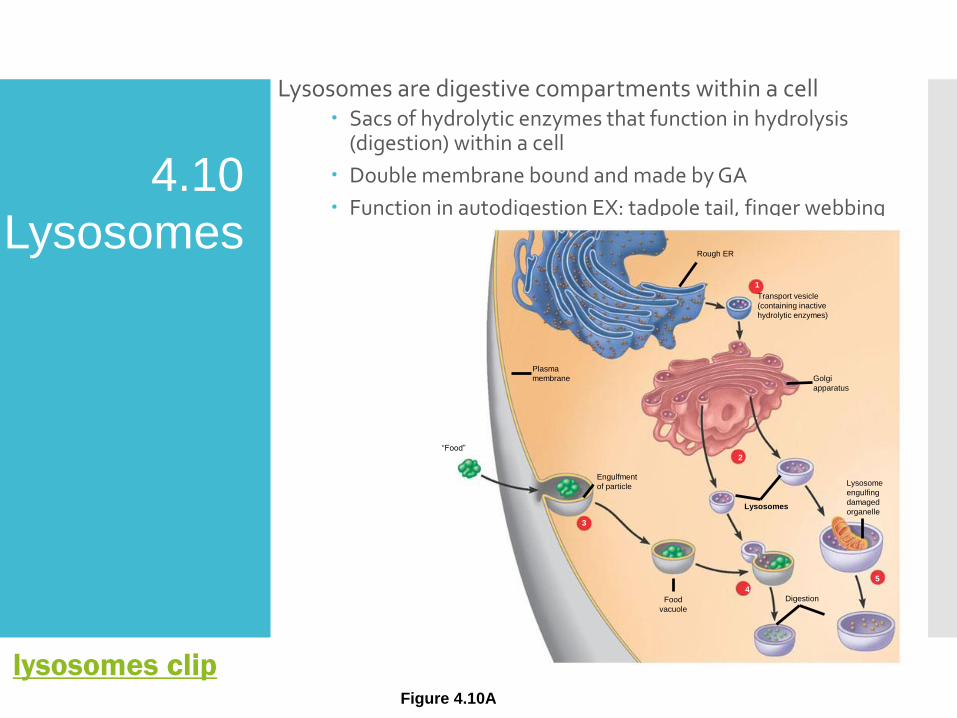
Lysosomes are digestive compartments within a cell Sacs of hydrolytic enzymes that function in hydrolysis
(digestion) within a cell
Double membrane bound and made by GA
Function in autodigestion EX: tadpole tail, finger webbing
Figure 4.10A
Golgi
apparatus
Plasma
membrane
“Food”
Food
vacuole
Lysosomes
2
Lysosome
engulfing
damaged
organelle
5
Digestion4
3
Engulfment
of particle
Transport vesicle
(containing inactive
hydrolytic enzymes)
1
Rough ER
4.10
Lysosomes
lysosomes clip
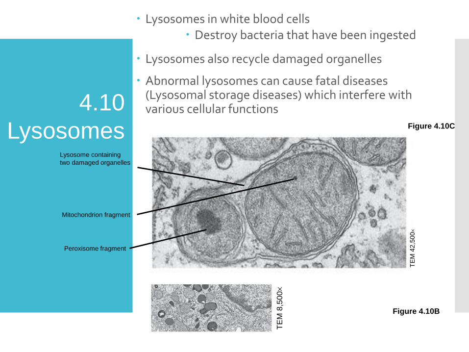
Lysosomes in white blood cells Destroy bacteria that have been ingested
Lysosomes also recycle damaged organelles
Abnormal lysosomes can cause fatal diseases (Lysosomal storage diseases) which interfere with various cellular functions
Figure 4.10B
Lysosome
Nucleus
TE
M 8
,50
0
4.10
Lysosomes
TE
M 4
2,5
00
Lysosome containing
two damaged organelles
Mitochondrion fragment
Peroxisome fragment
Figure 4.10C
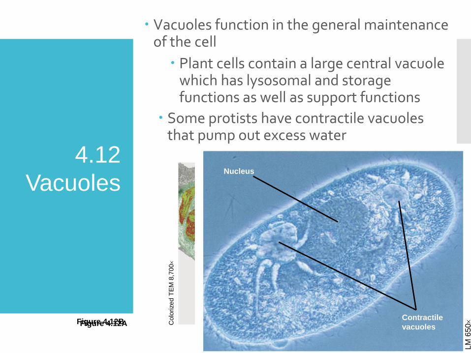
Vacuoles function in the general maintenance of the cell
Plant cells contain a large central vacuole which has lysosomal and storage functions as well as support functions
Some protists have contractile vacuoles that pump out excess water
Chloroplast
Central
vacuole
NucleusC
olo
rized T
EM
8,7
00
Figure 4.12A
4.12
Vacuoles
LM
65
0
Nucleus
Contractile
vacuolesFigure 4.12B

The various organelles of the endomembrane system are interconnected structurally and functionally
Nucleus
Smooth ER Nuclear envelope Golgi apparatus
Lysosome
Vacuole
Plasma
membrane
Rough ER
Transport vesicle
from ER to Golgi
Transport vesicle from
Golgi to plasma membrane
Figure 4.13
A review of the
endomembrane
system

ENERGY-CONVERTING ORGANELLES
4.14 Chloroplasts convert solar energy to chemical energy
Chloroplasts, found in plants and some protists, convert solar energy to chemical energy in sugars (glucose)
Contains chlorophyll
TE
M 9
,750
Chloroplast
Stroma
Intermembrane
space
Inner and outer
membranes
Granum
Figure 4.14

4.15 Mitochondria harvest chemical energy from food
Mitochondria carry out cellular respiration which uses the chemical energy in food (glucose) to make ATP for cellular work
OXYGEN + GLUCOSE ---- CARBON DIOXIDE + ATP + WATER
Double membrane bound, has own DNA
Figure 4.15
Mitochondrion
Outer
membrane
Intermembrane
space
Matrix
Inner
membrane
Cristae
TE
M 4
4,8
80
ENERGY-
CONVERTING
ORGANELLES
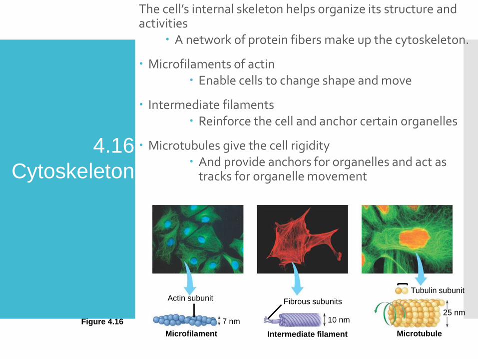
The cell’s internal skeleton helps organize its structure and activities
A network of protein fibers make up the cytoskeleton.
Microfilaments of actin Enable cells to change shape and move
Intermediate filaments Reinforce the cell and anchor certain organelles
Microtubules give the cell rigidity And provide anchors for organelles and act as
tracks for organelle movement
Actin subunit
Microfilament
7 nm
Fibrous subunits
10 nm
Intermediate filament Microtubule
25 nm
Tubulin subunit
Figure 4.16
4.16
Cytoskeleton
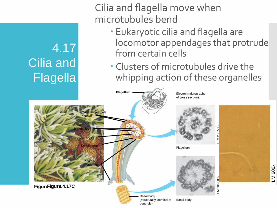
Cilia and flagella move when microtubules bend
Eukaryotic cilia and flagella are locomotor appendages that protrude from certain cells
Clusters of microtubules drive the whipping action of these organelles
LM
60
0
Colo
rize
d S
EM
4,1
00
Figure 4.17A Figure 4.17B
4.17
Cilia and
FlagellaFlagellum Electron micrographs
of cross sections:
Flagellum
Basal body
Basal body
(structurally identical to
centriole)
TE
M 2
06,5
00
TE
M 2
06,5
00
Plasma
membrane
Dynein arms
Radial spoke
Central
microtubules
Outer microtubule
doublet
Figure 4.17C

4.19 CELL SURFACES AND JUNCTIONS
Cell surfaces protect, support, and join cells Cells interact with their environments and each other via their
surfaces.
Plant cells
Are supported by rigid cell walls made largely of cellulose
Connect by plasmodesmata, which are connecting channels
Plasma membrane
Cytoplasm
Plasmodesmata
Vacuole
Layers of one
plant cell wall
Walls of two
adjacent plant
cells
Figure 4.18A
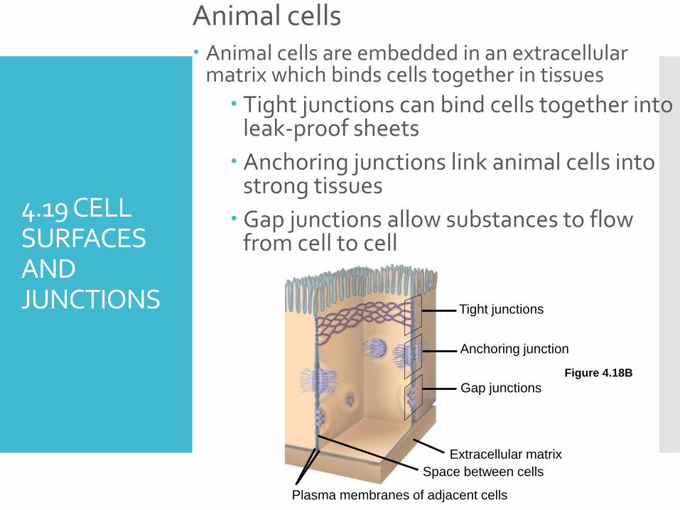
Animal cells Animal cells are embedded in an extracellular
matrix which binds cells together in tissues
Tight junctions can bind cells together into leak-proof sheets
Anchoring junctions link animal cells into strong tissues
Gap junctions allow substances to flow from cell to cell
4.19 CELL SURFACES AND JUNCTIONS
Anchoring junction
Tight junctions
Gap junctions
Extracellular matrix
Space between cells
Plasma membranes of adjacent cells
Figure 4.18B
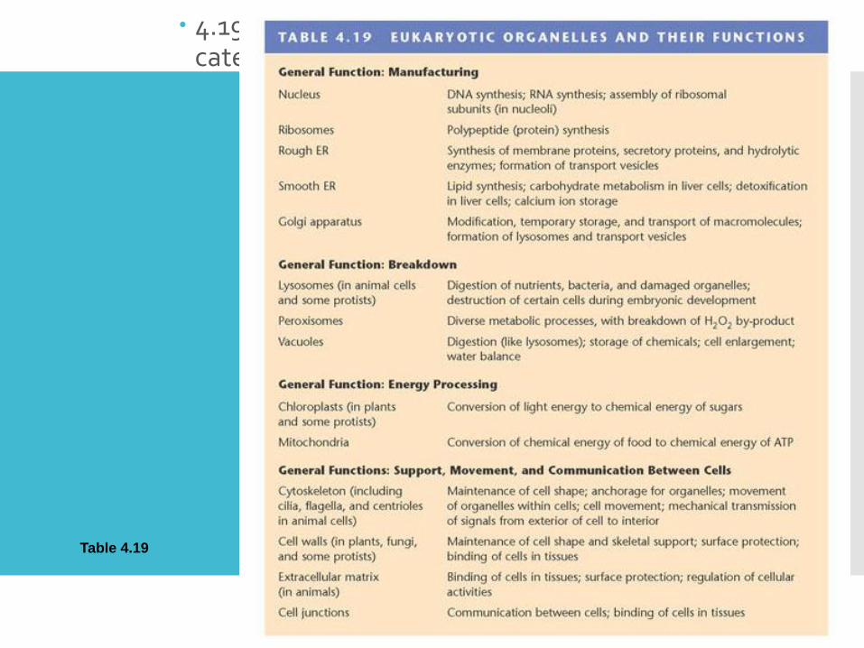
FUNCTIONAL CATEGORIES OF ORGANELLES 4.19 Eukaryotic organelles comprise four functional categories
Eukar yotic organelles fall into four functional groups
Manufacturing
Breakdown
Energy processing
Suppor t, movement, and communication between cells
Table 4.19