lincoln university common health concerns of small ruminants charlotte clifford-rathert, dvm...
TRANSCRIPT

Lincoln University
Common Health Concerns of Small Ruminants
Charlotte Clifford-Rathert, DVMCooperative Extension and Research
Assistant ProfessorState Extension Specialist, Small Ruminants

General diseases
1) Hoof diseases2) Caseous lymphadenitis3) Enterotoxemia4) Ketosis5) OPP and CAE6) Urinary Calculi7) Pink eye8) Polio9) Tetanus10) Mastitis11) Zoonotic Diseases
SoremouthRingwormToxoplasmosis

Foot rotOne of the most devastating sheep and goat diseases.
Caused by an interaction involving two types of anaerobic bacteria.– Bacteroides nodosus
• Can live only in animal’s foot• Can be eradicated from flock• Foot rot cannot occur without
it.• Does not survive longer than
two weeks in soil, but may remain in hooves of infected sheep for extended periods.
– Fusobacterium necrophorum• Aids in penetration of B.
nodosus into the skin and tissues
• Normal inhabitant of soil and sheep manure and is always present where sheep are raised

Foot Rot• EXTREMELY CONTAGIOUS
• Ideal conditions:– 40 to 70 degrees F– Wet environment– Dirty (manure) environment– Overgrown hooves
• Organism is very virulent
• Odiferous, can cause hoof wall to separate

Foot rot• Lameness
• One or more feet
• Starts with moist reddened area between toes Foot Scald
• Infection spreads under the sole and wall of the hoof.
• Different from -- – Foot abscess (bumble foot)– Founder (laminitis)– Injuries– Foreign bodies

Foot rot• Spread from infected sheep
to moist soil and back to non-infected sheep.
• Most commonly introduced to clean flock by purchase of infected sheep. Also mixing with infected flock and use of contaminated facilities.

Treatment
• Isolate infected animals• Penicillin• Tetracycline
• Intradigital (LA-200)• Crumbles top dress
feed• 10% copper sulfate foot bath• 10% zinc sulfate foot bath• Hydrated Lime / drylot• Therapeutic foot trimming

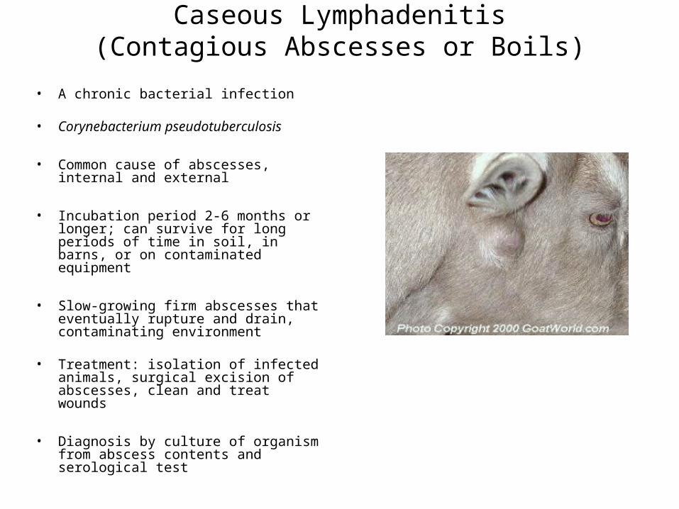
Caseous Lymphadenitis(Contagious Abscesses or Boils)
• A chronic bacterial infection
• Corynebacterium pseudotuberculosis
• Common cause of abscesses, internal and external
• Incubation period 2-6 months or longer; can survive for long periods of time in soil, in barns, or on contaminated equipment
• Slow-growing firm abscesses that eventually rupture and drain, contaminating environment
• Treatment: isolation of infected animals, surgical excision of abscesses, clean and treat wounds
• Diagnosis by culture of organism from abscess contents and serological test

• The contents of these abscesses is pale green in the early stages which turns cream as the abscess hardens and becomes “cheesy” or “caseous” or looks like an onion (surgical removal).
• Bacteria gain entry into the animal through a wound; (Skin wounds or mucous membranes)
• Localizing in one or more lymph nodes (secondary: inhalation, ingestion, penetration through intact skin)
Caseous lymphadenitisCLA, boils, abscesses, cheesy gland disorder
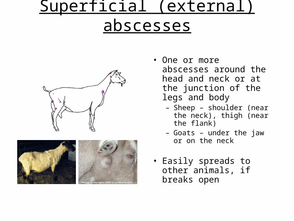
Superficial (external) abscesses
• One or more abscesses around the head and neck or at the junction of the legs and body– Sheep – shoulder (near
the neck), thigh (near the flank)
– Goats – under the jaw or on the neck
• Easily spreads to other animals, if breaks open

Visceral (internal) abscessesMore common with sheep
• Involves the lymph nodes and other organs (kidney and liver)
• Causes weight loss, poor health, reduced wool and milk production– “Thin ewe syndrome”
• Third most important cause of carcass condemnation

Treatment of CLAExternal abscesses
• Sample abscess – not all are CLA
• Antibiotics not effective
• Lance and flush superficial
abscesses with iodine
• Keep animal(s) isolated until wound heels
• Dispose of material carefully
Control and Prevention• Isolate infected animals
• Purchase CLA negative animals, serological testing available (ELISA, AGID, PCR)
• Facilities should be free of nails, wire, and other objects that might induce breaks in the skin
• Control of external parasites, prevent goats from rubbing against sharp objects
• DO NOT REUSE NEEDLES
• Clean all tattooers, shears, surgical instruments between animals
• Treat all wounds promptly
• Vaccination?? may help to control disease if already on farm, vaccine will cause abscess at site of vaccination

EnterotoxemiaOvereating disease, pulpy kidney disease
• Clostridium perfringins type C or D
C - baby lambs 2-3 weeks
D – older lambs
• May be most common bacterial pathogen
• Normal flora in warm blooded animals
• Flora disturbed by dietary change

• Affected animals are usually found dead!
• Common in lambs/kids consuming concentrates.
• Symptoms in live animal– Depression – Abdominal pain– Teeth grinding– Twitching or convulsions– Can be similar to other
diseases (E. coli scours, encephalitis)
• Treatment (not rewarding)
Enterotoxemia

Preventing enterotoxemia
• Vaccinate– Ewes – 2-3 wks before
lambing– Does – every 6 months– Lambs/Kids – at 4-6
weeks, then 2-3 weeks later.
– Vaccine may be combined with other clostridial diseases: CD-T (type C & D overeating, tetanus)
• Avoid sudden changes in feed

Pregnancy ToxemiaKetosis, Pregnancy disease, Twin lamb disease, Lambing paralysis
• Occurs during last trimester of pregnancy when fetus(es) are growing rapidly.
• Caused by low glucose concentrations in the blood and excessive breakdown of body fat to compensate.– Ketones – toxic by-product
• Caused by inadequate nutrition during late pregnancy.

Females most susceptible
• Females carrying multiple fetuses
• Thin ewes• Fat ewes • Timid ewes • Granny ewes
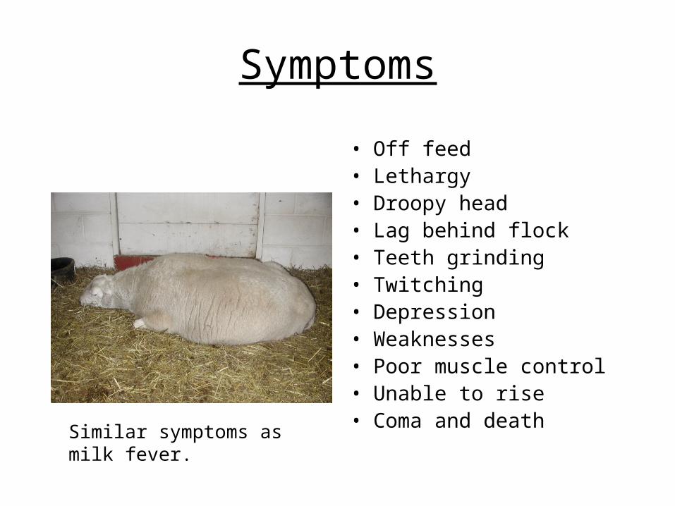
Symptoms
• Off feed• Lethargy• Droopy head• Lag behind flock• Teeth grinding• Twitching • Depression• Weaknesses • Poor muscle control• Unable to rise• Coma and deathSimilar symptoms as milk
fever.

Treatment
• Early detection• Quickly meet energy
needs of ewe– Glucose orally (e.g.
propylene glycol)– Glucose intravenously
• Force feed and vitamin injections to stimulate appetite
• Antibiotics to prevent pneumonia
• C-Section in advanced stages
Differentiated from milk fever by response to treatment(s).

Prevention
• Proper body condition (3+/5) during mid-pregnancy.
• Adequate nutrition during late pregnancy (especially energy). Good quality hay + grain supplementation.
• Adequate feeder space• Exercise• Avoid abrupt changes in
feed.

Caprine Arthritis Encephalitis(CAE)
• More common in dairy goats
• Virus shed in colostrum, milk, aerosol droplets, and other body secretions.
• Transmitted via colostrum/milk, and blood-contaminated tools
• Not a problem in range raised meat goats
• Neurological form in kids 2-6 months of age to adult
• Respiratory and arthritic form common in adults
• Diagnosis by ELISA, AGID, and PCR testing
• NO treatment
• Stress and poor management will influence clinical appearance

Prevention and Control
• Remove Kids at birth
• Feed heat treated colostrum/ milk • Isolate infected animals and cull
• Milk the infected does last
• Do not share needles/ equipment
• Test kids at 6 month intervals

Ovine Progressive Pneumonia
• Atypical pneumonia of sheep caused by a virus • Ovine Lentivirus• Numerous strains• Closely related to CAE virus in goats• Long incubation period (2-4 years) therefore seen in
older ewes• Once infected, remain infected for life• Transmission by ingestion of milk or colostrum from
infected ewe, or direct contact with nasal discharge droplets in overcrowded conditions.
• Virus is short-lived in the environment• Diagnosis by ELISA, AGID, and PCR

Clinical signs
• “thin ewe syndrome”,• hard bag, • increased effort to breath, coughing,• lethargy• nasal discharge, • fever, • arthritis, unsteady gait progressing to hind limb
paralysis

Urinary Calculi water belly, urolithiasis, and calculosis
• Metabolic disease of wethers and rams characterized by the formation of calculi (stones) within the urinary tract
• Blockage of the urethra by calculi causes retention of urine,
abdominal pain, distention and rupture of the urethra or bladder • The most common cause of urinary calculi is feeding rations
with high phosphorus levels. The ratio of calcium to phosphorus in the ration should be at least 2:1
• Addition of ammonium chloride to the ration will aid in
preventing urinary calculi.

Urinary Calculi
• It is also important that animals have an ample supply of clean, potable water.
• The addition of salt to the ration will increase water intake and decrease stone formation
• Correlation to hard water areas and increased frequency

Pinkeye Infectious Keratoconjunctivitis
• Highly contagious disease affecting the eyes of sheep and goats
• Bacteria: Chlamydia and Mycoplasma
• The disease will usually complete its course in three weeks in individuals
• The use of eye medications containing antibiotics may be helpful in individual cases
• There are no effective vaccines available
(The agent that causes pinkeye in sheep and goats is different from the one that causes it in cattle)

Polioencephalomylacia
• Also called polio
• Caused by a vitamin B1 (thiamine) deficiency
• Since the rumen manufactures B vitamins, polio is not caused by insufficient thiamine, but rather the ability to utilize it
• Most common symptom is blindness and star gazing
• Common scenario to overdose of Corid (treatment for Coccidiosis)

Tetanus(Clostridium tetani)
• A soil inhabitant that is a prolific spore producer • Usually related to docking and castrating by elastrator bands,
though any wound can harbor the tetanus organism• Tetanus occur from about four days to three weeks or longer
after infection is established in a wound • Symptoms include stiff gait, "lockjaw“, and third eyelid may
protrude across the eye. Animal will usually go down or stand with all four legs held out straight and stiff and the head drawn back. Convulsions may occur and animal dies
• Treatment consists of the tetanus anti-serum and antibiotics. It is usually unrewarding
• Prevented by vaccinating
* If you have horses or had them on your property you need to vaccinate against tetanus

Mastitis Hard bag , Blue bag
• Staphylococcus aureus and Pasteurella hemolytica • Two types of mastitis: acute and chronic• Affected ewe/doe may be reluctant to walk• May hold up one rear foot• May not permit her lambs/kids to nurse
– Resist milking
• Ewes with chronic mastitis often go undetected. • Mastitis is treated with antibiotics• Prevented by good management and sanitation.• Observe and handle udders

Zoonotic Diseases• *Ringworm• Campylobacter• Chlamydiosis• *Contagious ecthyma (“Orf” or sore mouth)• *Cryptosporidiosis• Leptospirosis• *Listeriosis• *Rabies• Salmonellosis• *Toxoplasmosis• Q Fever• Tuberculosis• Brucellosis

What is a zoonotic disease?
A disease that is transmissible between
animals and humans.

SoremouthContagious ecthyma, contagious pustular dermatitis, scabby mouth, orf
• Common skin disease of sheep and goats.
• Caused by a Parapox virus
• Virus spreads through direct contact and contact with contaminated facilities and tools
• Lesions most commonly seen on mouth, lips, and nostrils, but may also occur on udder and between toes.
• Extremely infectious
• Dried scabs harbor virus

Soremouth
• Animals that develop the disease usually develop a strong immunity
• May be severe in lambs and kids
• Numerous strains – incubation period may vary from 1 to 3 weeks
• The disease will clear up in one to four weeks
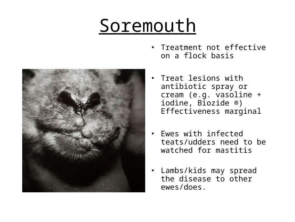
Soremouth• Treatment not effective on a
flock basis
• Treat lesions with antibiotic spray or cream (e.g. vasoline + iodine, Biozide ®) Effectiveness marginal
• Ewes with infected teats/udders need to be watched for mastitis
• Lambs/kids may spread the
disease to other ewes/does.

Prevention of Soremouth
• Maintain a closed flock
• Do not show
• Vaccinate (only if you
have had disease)– In a hairless protected area– Scabs will appear 1 to 3
days after vaccination– 6 weeks before show
season

Orf
• Sore mouth is contagious to humans (painful).
• Need to be careful when working with infected animals and when working with live vaccine.
• Wear gloves

Ringworm Woolrot, Lumpy Wool, Club Lamb FungusClub Lamb Fungus, and Dermatophytosis
• Fungal : Microsporum, Trichophytum
• VERY CONTAGIOUS
• Hairless scaly crusty red circular lesion on face, ears, neck, and sometimes legs.
• Diagnos is based on observation
• Self-limiting in 1-4 months,
• Treat by clipping, exposing areas to air, scrubbing with iodine solution daily for 5-7 days and then weekly if not clearing. Topical antifungal agents
• Isolate affected animals
• Wear gloves, and clean all equipment

Ringworm
• Prevention
• Proper nutrition and care. • Keep sheep and barns as dry as possible. • Avoid nicks and cuts when shearing.• Disinfect equipment (shearing equipment, etc.) with a commercial
fungicide. • Wash and shear show lambs as little as possible. • Use protective gloves when handling infected animals. • Quarantine new arrivals before mixing with flock. • Separate infected from non-infected sheep.• Disinfect pens as needed. • Show equipment such as blankets, towels, and halters should be
cleaned between animals.• Persons handling infected or exposed animals should wear rubber
gloves and clothing with long sleeves. Wash thoroughly after handling animals and wash clothes thoroughly.
• Animals with active lesions should be removed from shows.

Ringworm
• Why is it important to you?
• It moves from flock to flock or from flock to person through contact.
• Sheep can still be contagious without exhibiting external signs of carrying the fungus.
• The fungus is thought to be very hardy and can live for years in barns and equipment hidden from direct sunlight waiting for growing conditions to encourage development.

Ringworm

Enzootic Abortion in Ewes (EAE)(Chlamydia psittici)
• Chlamydia is the most common cause of abortion in ewes • Transmitted from aborting sheep to susceptible individuals • Ewe lambs are usually the most susceptible • Abortion during the last month of pregnancy
• Birth of lambs that die shortly after birth • May cause pneumonia in young lambs• Chlamydia abortions can usually be stopped or reduced
by treating entire flock with tetracycline. High levels may be required in problem flocks
• Vaccine is available

Toxoplamosis
• Protozoan: Toxoplasma gondii
• Important cause abortion, mummification, still births, and birth of weak kids and lambs
• Cats serve as the definitive host; shed oocysts in feces
• If goat ingests oocysts when pregnant, organism can invade placenta and fetus within 2 weeks. Fetuses infected in the first half of pregnancy are more apt to die than fetuses infected later.
• Diagnosis: Doe and fetal serology (fetal fluids) is very specific test, histopathology of placental tissue

Toxoplasmosis• Prevention and Control:
– avoid contamination of feed by cats by storing grain in covered containers. Maintain population of neutered cats, and do not feed raw meat to cats
– Encourage exposure of does/ewes to cats before breeding to develop protective immunity
– Evidence that Rumensin® and Deccox® will partially prevent toxoplasmosis in pregnant ewes.
• Aborted fetuses and placentas should be disposed of properly, wearing gloves when handling these items, proper pasteurization of milk and cooking of meat.
• Pregnant women should be especially careful.

Vibriosis(Vibrio Campylobacteriosis)
• Abortion during last month of pregnancy, stillborn lambs, and birth of weak lambs are typical of vibrio abortions.
• High abortion rates have been recorded (up to 70 percent).• Two strains - Campylobacter jejuni or Campylobacter fetus.• Ewes are infected by oral ingestion.• Incubation period from the time of infection and abortion is only two weeks.• Vaccination can be effective in the face of an outbreak. • Feeding of antibiotics (tetracyclines) has been shown to be effective. Less
effective if C. jejuni strain. • Disease spread can be prevented by isolating aborting ewes, disposal of
fetus and membranes and disinfecting affected area. • Infected ewes usually recover after aborting and are immune to re-infection. • A vaccine is available for Vibrio. • Vibrio vaccines – buy those that contain both intestinalis and jejuni strains.

Summary
Preventative health care and good management are key to successful production and profit; saving time and money in the long run.

AcknowledgementsAcknowledgements• Susan Schoenian – University of Maryland Cooperative Extension Area Sheep Susan Schoenian – University of Maryland Cooperative Extension Area Sheep
and Goat Specialist and Goat Specialist • Bruce Lane – Regional Livestock Specialist, University of Missouri ExtensionBruce Lane – Regional Livestock Specialist, University of Missouri Extension• Dr. Beth Walker – Assistant Professor, Missouri State UniversityDr. Beth Walker – Assistant Professor, Missouri State University• Jim Humphrey - Regional Livestock Specialist, University of Missouri Jim Humphrey - Regional Livestock Specialist, University of Missouri
ExtensionExtension• Randy Saner - Regional Livestock Specialist, University of Nebraska Randy Saner - Regional Livestock Specialist, University of Nebraska
ExtensionExtension• Ken Bolte – Agriculture Business Specialist, University of Missouri ExtensionKen Bolte – Agriculture Business Specialist, University of Missouri Extension• Chris Zumbrunnen – Regional Livestock Specialist, University of Missouri Chris Zumbrunnen – Regional Livestock Specialist, University of Missouri
ExtensionExtension• Dr. Dave Patterson – State Extension Animal Scientist, University of MissouriDr. Dave Patterson – State Extension Animal Scientist, University of Missouri• Dr. Marcia Patterson - State Extension Animal Scientist, University of MissouriDr. Marcia Patterson - State Extension Animal Scientist, University of Missouri