live cell imaging provides novel insights into fungicide ... · live cell imaging provides novel...
TRANSCRIPT

Steinberg G et al., Live Cell Imaging Provides Novel Insights into Fungicide Mode of Action. In: Deising HB; Fraaije B; Mehl A; Oerke EC; Sierotzki H; Stammler G (Eds), "Modern Fungicides and Antifungal Compounds", Vol. VIII, pp. 15-24. © 2017 Deutsche Phytomedizinische Gesellschaft, Braunschweig, ISBN: 978-3-941261-15-0
Live Cell Imaging Provides Novel Insights into Fungicide Mode of Action
Steinberg G; Schuster M; Kilaru S University of Exeter, Biosiences, EX4 4QD, Exeter, United Kingdom Email: [email protected]
ABSTRACT
Over the past two decades, the use of fluorescent proteins has changed our under-standing of living cells. Fused to proteins of interest, green and red proteins enable a quantitative assessment of cellular dynamics. In recent times, fluorescent protein-based live cell imaging was successfully established in various plant pathogenic fungi. Such investigation provided unique insights into the molecular mechanisms of plant invasion. Moreover, when fused to organelle- or cytoskeleton-specific pro-teins, these fluorescent proteins become markers that enable observation of defined cellular compartments and structures. Here, we highlight the potential of fluores-cent protein markers (FPMs) to better understand the mode of action of fungicides.
INTRODUCTION
Over the past twenty years, the use of fluorescent proteins has revolutionized our understan-ding of cellular organization and behaviour. Enhanced green-fluorescent protein (eGFP; Yang et al. 1996) or red-fluorescent proteins, including monomeric red-fluorescent protein (mRFP; Campbell et al. 2002) or monomeric Cherry (mCherry; Shaner et al. 2004) have been widely-used in a broad range of organisms. These include numerous plant pathogenic fungi, such as Ustilago maydis and Zymoseptoria tritici (overview in Kilaru et al. 2015b; Schuster et al. 2015a). When fused to a protein of interest, cells usually target the fluorescent fusion proteins to specific structures or organelles. This enables the study of protein localization or dynamics in living cells (Chalfie et al. 1994). Moreover, when fused to organelle- or structure-specific proteins, such fluorescent fusion proteins become cellular markers (fluorescent protein mar-kers= FPMs) for their respective compartments or cellular structures. A good example is tubu-lin, which is the building subunit of microtubules. Fused to GFP, the modified tubulin incorpo-rates into microtubules, which, when visualized by fluorescent microscopy, show dynamic behaviour that was initially only recognized for purified tubulin in cell-free in vitro assays. In recent years, our group extended this approach to plant pathogenic fungi. Over the past 17 years, the use of FPMs in the corn smut fungus Ustilago maydis (“Maisbeulenbrand”) has pro-vided novel and important insights into the mechanisms underpinning plant infection (e.g. Bielska et al. 2014; Fuchs et al. 2006; Treitschke et al. 2010; Weber et al. 2006; Weber et al.
15
Persistent Identifier: urn:nbn:de:0294-sp-2017-Reinh-02-4

Steinberg et al.
2003) and fundamental principles of fungal cell organisation and growth (e.g. Guimaraes et al. 2015; Lin et al. 2016; Schuster et al. 2016). Moreover, FPM expressing U. maydis cell lines (=strains) have also proven to be powerful tools in understanding the mechanism of fungicide delivery by liposome-like carriers (Steinberg 2012). This overview article highlights the potential use of live cell imaging of FPMs in gaining a better understanding of the mode of action (MoA) of fungicides. This area of research is novel, and thus the results summarised in this article provide only a “snap-shot” of our ongoing research in this arena.
LIVE CELL IMAGING OF FPMS IN FUNGICIDE MODE OF ACTION STUDIES
Example 1: MBC-fungicides Methyl benzimidazole carbamate fungicides (MBC-fungicides) were introduced in the 1960’s – 1970’s and have been widely used as a protective and eradicant anti-fungal to protectcereals, vines, fruit, rice and vegetables (Oliver & Hewitt 2014). The MoA of MBC-fungicides in fungi is well understood. Biochemical and molecular biology studies have shown that these fungicides target the tubulin subunit of the tubulin dimer (overview in Davidse 1986). Binding of the MBC-fungicide to tubulin prevents the addition of the GTP-bound tubulin dimer to the elongating plus-end of the microtubule (Figure 1A). In these non-growing microtubules, the intrinsic hydrolytic activity of tubulin itself cleaves the bound GTP and thus leaves only GDP-tubulin dimers in the microtubule (Figure 1A). The loss of the GTP-cap results into microtubule instability and a switch to “catastrophy”, followed by rapid depolymerisation of the microtubule (Desai & Mitchison 1997). As a consequence, mitotic spindles and interphase microtubules disappear. This results in cell cycle arrest, growth defects and, ultimately, in cell death.
Figure 1 MoA of MBC-fungicides. (A) Schematic drawing of the effect of MBC-fungicides on microtubule dynamics. (B) Fluorescent microtubules in U. maydis cells, treated with the solvent dimethyl-sulfoxide (DMSO), 1 μg/ml benomyl and 1 μg/ml carbendazim for 30-45 minutes. Open arrowheads indicate short microtubule fragments. Scale bar is given in micrometres.
16
Persistent Identifier: urn:nbn:de:0294-sp-2017-Reinh-02-4

Live Cell Imaging Provides Novel Insights into Fungicide Mode of Action
Strikingly, live cell imaging of fluorescent tubulin reveals the MoA of MBC-fungicides in a single experiment. GFP- tubulin expressing U. maydis cells, treated with the solvent dimethyl sulfoxide, contain long microtubules (Figure 1B, DMSO). After 30 minute exposure to 1 μg/ml benomyl or carbendazim, these microtubules disappear, and only short fragments remain in the cytoplasm (arrowhead, Figure 1B). It should be noted that the depolymerisation of microtubules is only one of several microtubule-related phenotypes possible. Depending on the nature of the anti-fungal compound, more stable and longer microtubules, as well as microtubule bundles may be seen. Thus, live cell imaging of fluorescent microtubules provides a rapid way of gaining insight into the MoA of microtubule-affecting fungicides.
Example 2: Dodine
Secondly, we chose to further illustrate the power of live cell imaging of FPMs by investigating the MoA of the fungicide 1-dodecylguanidinium acetate (dodine, FRAC code U12). This protectant fungicide is used to control black spot on apples, pears and roses, and leaf curl in nectarines and peaches. Dodine is a surfactant that likely inserts into membranes. Indeed, early reports in fungi (Brown & Sisler 1960; Somers & Fisher 1967; Somers & Pring 1966), including U. maydis (Solel & Siegel 1984), suggested that dodine perforates the plasma membrane and thus affects the integrity of the cell. On the other hand, an effect on metabolic and other fungal enzymes was suggested (Brown & Sisler 1960; Solel & Siegel 1984), suggesting that dodine enters the fungal cytoplasm. Indeed, work in Neurospora crassa concluded “enzyme inhibition or intracellular reaction appear to be the most probable hypotheses to explain the fungitoxicity of dodine” (citation taken from Somers & Fisher 1967). The FRAC code list 2016 reports the MoA of dodine as “not known” (http://www.frac.info/publications/), which reflects the seemingly contradictory results in these publications. We applied various concentrations of dodine, solved in methanol (MetOH), to FPM-expres-sing strains of U. maydis. In a first set of experiments, we attempted to see an effect of dodine on the plasma membrane in a strain that expresses a fluorescent plasma membrane syntaxin Sso1 (Treitschke et al. 2010). In untreated cells, and in the MetOH solvent controls, this FPM localizes to the periphery of the cell (Figure 2A, 2B). With increasing concentrations of dodine, applied to shaking liquid cultures for 30-45 minutes at 28°C, the plasma membrane marker mis-localizes. This begins with the appearance of a spherical structure (Figure 2B, open arrowhead) that most likely represents the nuclear envelope, but ends in large deposits of the FPM in the cytoplasm at higher concentrations (Figure 2B, closed arrowhead, Figure 2C; both 50 μg/ml dodine). As the plasma membrane marker protein is an integral membrane protein, its concentration in the cytoplasm is likely accompanied by membrane accumulation. This indicates a failure of Sso1 targeting of Sso1-carrying vesicles that may fail to fuse with the plasma membrane. An effect of dodine on the plasma membrane is also indicated by observation of a FBM that labels filamentous actin (F-actin; Schuster et al. 2012). This FPM localizes to actin patches,
17
Persistent Identifier: urn:nbn:de:0294-sp-2017-Reinh-02-4
Steinberg et al.
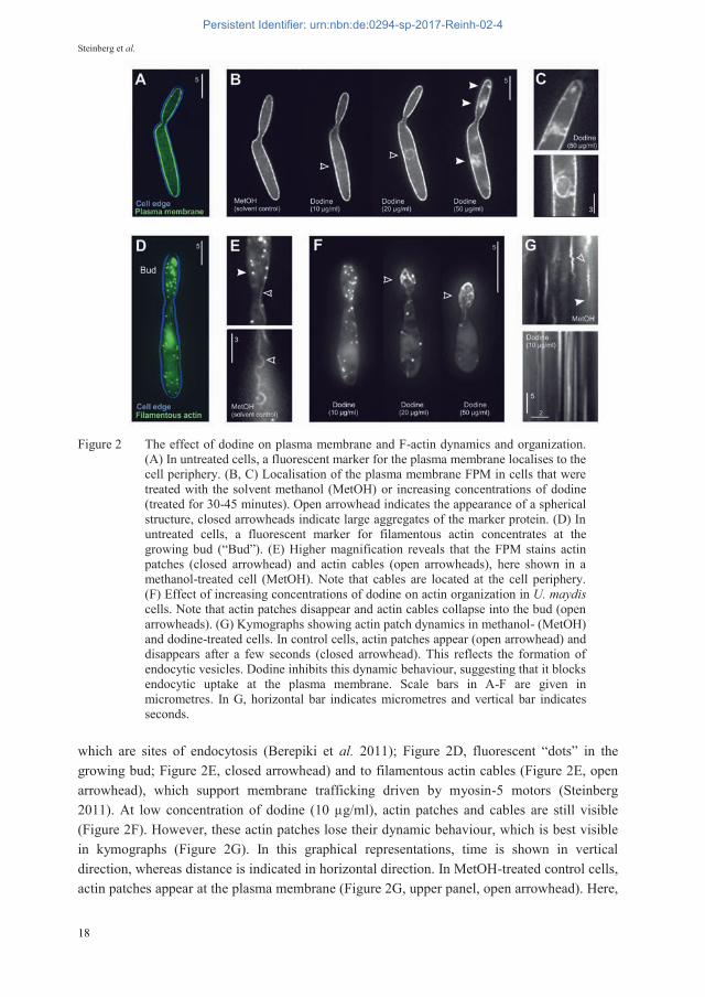
Figure 2 The effect of dodine on plasma membrane and F-actin dynamics and organization. (A) In untreated cells, a fluorescent marker for the plasma membrane localises to the cell periphery. (B, C) Localisation of the plasma membrane FPM in cells that were treated with the solvent methanol (MetOH) or increasing concentrations of dodine (treated for 30-45 minutes). Open arrowhead indicates the appearance of a spherical structure, closed arrowheads indicate large aggregates of the marker protein. (D) In untreated cells, a fluorescent marker for filamentous actin concentrates at the growing bud (“Bud”). (E) Higher magnification reveals that the FPM stains actin patches (closed arrowhead) and actin cables (open arrowheads), here shown in a methanol-treated cell (MetOH). Note that cables are located at the cell periphery. (F) Effect of increasing concentrations of dodine on actin organization in U. maydis cells. Note that actin patches disappear and actin cables collapse into the bud (open arrowheads). (G) Kymographs showing actin patch dynamics in methanol- (MetOH) and dodine-treated cells. In control cells, actin patches appear (open arrowhead) and disappears after a few seconds (closed arrowhead). This reflects the formation of endocytic vesicles. Dodine inhibits this dynamic behaviour, suggesting that it blocks endocytic uptake at the plasma membrane. Scale bars in A-F are given in micrometres. In G, horizontal bar indicates micrometres and vertical bar indicates seconds.
which are sites of endocytosis (Berepiki et al. 2011); Figure 2D, fluorescent “dots” in the growing bud; Figure 2E, closed arrowhead) and to filamentous actin cables (Figure 2E, open arrowhead), which support membrane trafficking driven by myosin-5 motors (Steinberg 2011). At low concentration of dodine (10 μg/ml), actin patches and cables are still visible (Figure 2F). However, these actin patches lose their dynamic behaviour, which is best visible in kymographs (Figure 2G). In this graphical representations, time is shown in vertical direction, whereas distance is indicated in horizontal direction. In MetOH-treated control cells, actin patches appear at the plasma membrane (Figure 2G, upper panel, open arrowhead). Here,
18
Persistent Identifier: urn:nbn:de:0294-sp-2017-Reinh-02-4
Live Cell Imaging Provides Novel Insights into Fungicide Mode of Action
they remain during the formation of an endocytic vesicle, but disappear after this vesicle has “pinched off” the plasma membrane and it moves into the interior of the cell (Figure 2G, closed arrowhead). In the presence of 10 μg/ml dodine, this dynamic behaviour is strongly impaired (Figure 2G, lower panel), indicating that initial endocytosis is blocked by the fungicide. This process occurs at the plasma membrane, suggesting that dodine affects the functionality of membrane-associated processes. Surprisingly, higher concentrations of dodine result in a collapse of actin cables into the growing bud of the FPM-expressing cells (Figure 2F, open arrows). Such reorganization of F-actin could also be due a defect at the plasma membrane, as membrane-associated proteins are known to anchor F-actin to the plasma membrane (Ponuwei 2016). However, further studies are needed to clarify if dodine affects the localization of such actin-binding and anchoring proteins at the plasma membrane.
19
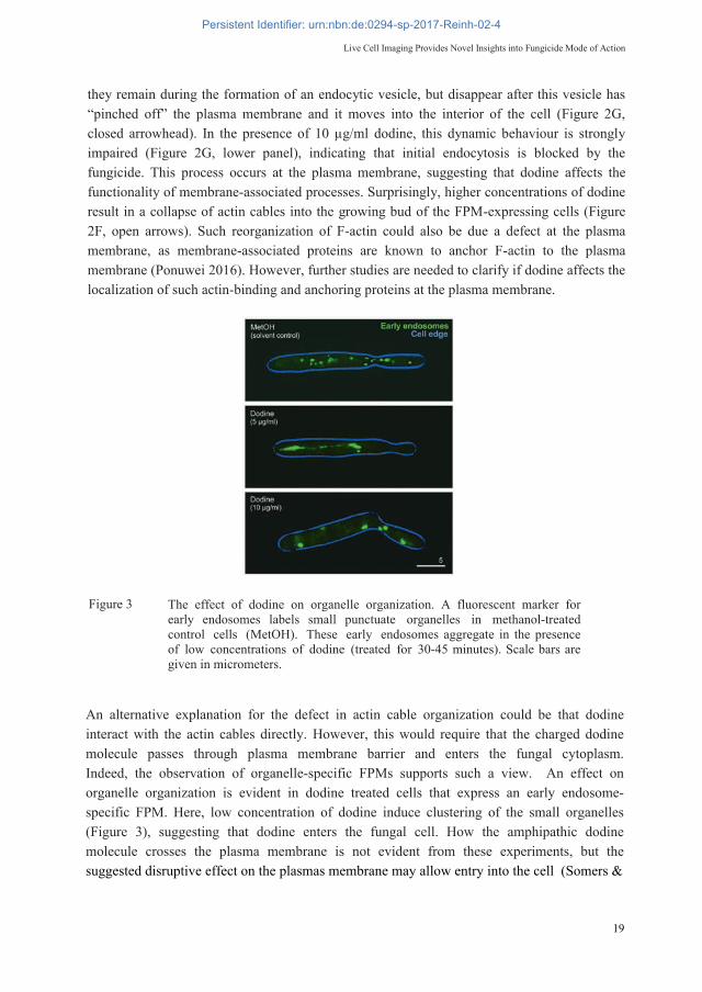
Figure 3 The effect of dodine on organelle organization. A fluorescent marker for early endosomes labels small punctuate organelles in methanol-treated control cells (MetOH). These early endosomes aggregate in the presence of low concentrations of dodine (treated for 30-45 minutes). Scale bars are given in micrometers.
An alternative explanation for the defect in actin cable organization could be that dodine interact with the actin cables directly. However, this would require that the charged dodine molecule passes through plasma membrane barrier and enters the fungal cytoplasm. Indeed, the observation of organelle-specific FPMs supports such a view. An effect on organelle organization is evident in dodine treated cells that express an early endosome-specific FPM. Here, low concentration of dodine induce clustering of the small organelles (Figure 3), suggesting that dodine enters the fungal cell. How the amphipathic dodine molecule crosses the plasma membrane is not evident from these experiments, but the suggested disruptive effect on the plasmas membrane may allow entry into the cell (Somers &
Persistent Identifier: urn:nbn:de:0294-sp-2017-Reinh-02-4

Steinberg et al.
Pring 1966). Clearly, more studies are needed to elucidate the MoA of dodine. However, these results illustrate the power of live cell imaging of FPMs as an important new approach to understand MoA of fungicides.
FPMS IN THE SEPTORIA TRITICI WHEAT BLOTCH FUNGUS ZYMOSEPTORIA TRITICI
Our collection of FPM expressing U. maydis strains derives from 17 years of research (Table 1). Their successful use in better understanding plant infection strategies and fungicide delivery mechanisms (see INTRODUCTION) prompted us to extend this approach to other plant pathogenic fungi. With funding from the British Biotechnology and Biological Sciences Research Council (BBSRC), we have recently established fluorescent proteins, such as mCherry, enhanced GFP and a codon-optimized GFP, as well as numerous FPMs in the wheat blotch fungus Zymoseptoria tritici (=Mycosphaerella graminicola; “Weizen-Blattdürre”); (Guo et al. 2015; Kilaru et al. 2015a; Kilaru et al. 2015b; Schuster et al. 2015a; Schuster et al. 2015b; Table 1). These FPMs allow co-visualization of different Z. tritici strains in axenic culture or inside the infected host plant (Figure 4). Microtubule-specific FPMs, such as the plus end-binding protein ZtPeb1-GFP (Schuster et al. 2015b), the minus end-binding protein ZtGrc1-GFP (Schuster et al. 2015b) and fluorescent -tubulin proteins GFP-ZtTub2 and mCherry-ZtTub2 (Schuster et al. 2015b) provide insight into the organization of the cytoskeleton in yeast-like Z. titrici cells (Figure 5A). Moreover, polarity markers, specifically localizing to the polarisome (GFP-ZtSpa2; Guo et al. 2015), the exocyst (GFP-ZtExo70; Guo et al. 2015) or the Spitzenkörper (GFP-ZtMlc1, GFP-ZtSec4; Guo et al. 2015), reveal details of the organization of the growing hyphal tip in Z. tritici hyphae (Figure 5B). Our current focus is to complete this FPM collection (Table 1), which will allow the use of these FPM-expressing Z. tritici strains in fungicide MoA studies. This opens the unique opportunity to compare fungicide induced phenotypes between the corn smut and the Septoria tritici wheat blotch fungus.
Figure 4: Co-visualisation of two strains of Z. tritici, both in liquid culture and inside infected plants (14 days after infection). Scale bars are given in micrometers.
20
Persistent Identifier: urn:nbn:de:0294-sp-2017-Reinh-02-4

Live Cell Imaging Provides Novel Insights into Fungicide Mode of Action
CONCLUSIONS
An important characteristic of fungicides is their MoA. This describes the biochemical, meta-bolic or anatomical change at the cellular level, caused by the fungicides (e.g. (Opalski et al. 2006), but it is also used to describe the molecular target of an antifungal (e.g. (Fernández-Ortuño et al. 2012). In fact, our knowledge of fungicide MoA is often restricted to either the molecular target, or the way the antifungal affects pathogen physiology. Good examples are SDHIs, which bind to subunits of succinate dehydrogenase, thereby inhibiting cellular respiration (e.g. Rehfus et al. 2016). Whilst this target is well-defined, the “physiological consequences on the level of the cell” (MoA) are not known. By contrast, MoA studies often provide incomplete insight, or even contradictory conclusions with certain fungicides. In this article, we summarised the MoA studies on dodine, where some studies report an effect of the fungicide on the fungal plasma membrane, resulting in increased cell permeability, whilst others report effects on enzyme activity, which are thought to underpin dodine’s fungal toxicity (Brown & Sisler 1960; Solel & Siegel 1984; Somers & Fisher 1967; Somers & Pring 1966). However, these cited MoA studies were published between 1960’s-1980’s and were thus largely restricted by the available techniques of the time. Today, advances in genome sequencing, transciptional profiling, bioinformatics and powerful live cell imaging techniques of fluorescent markers for cellular structures in pathogenic fungi, such as Z. tritici open new avenues for MoA research. This promises unique and novel insights into fungicide MoA in pathogenic fungi, which will enhance our understanding of antifungal activity to inform development of sustainable disease.
Figure 5 FPMs for visualization of microtubule structures in a yeast-like cell structure (A) and the polar growing tip of hyphae (B) in Z. tritici. Sale bars are given in micrometers.
21
Persistent Identifier: urn:nbn:de:0294-sp-2017-Reinh-02-4

Steinberg et al.
REFERENCES
Adamikova L; Straube A; Schulz I; Steinberg G (2004). Calcium signaling is involved in dynein-dependent microtubule organization. Molecular Biology of the Cell 15, 1969-1980.
Berepiki A; Lichius A; Read ND (2011). Actin organization and dynamics in filamentous fungi. Nature Reviews Microbiology 9, 876-887.
Bielska E; Higuchi Y; Schuster M; Steinberg N; Kilaru S; Talbot N J; Steinberg G (2014). Long-distance endosome trafficking drives fungal effector production during plant infection. Nature Communications 5, 5097.
Brown IF; Sisler HD (1960). Mechanisms of fungitoxic action of N-dodecylguanidine acetate. Phytopathology 50, 830-839.
Campbell RE; Tour O; Palmer AE; Steinbach PA; Baird GS; Zacharias DA; Tsien RY (2002). A monomeric red fluorescent protein. Proceedings of the National Academy of Sciences of the United States of America 99, 7877-7882.
22
Persistent Identifier: urn:nbn:de:0294-sp-2017-Reinh-02-4
Live Cell Imaging Provides Novel Insights into Fungicide Mode of Action
Chalfie M; Tu Y; Euskirchen G; Ward WW; Prasher DC (1994). Green fluorescent protein as a marker for gene expression. Science 263, 802-805.
Davidse LC (1986). Benzimidazole fungicides: Mechanism of action and biological impact. Annual Review of Phytopathol 24, 43-65.
Desai A; Mitchison TJ (1997). Microtubule polymerization dynamics. Annual Review of Cell and Developmental Biology 13, 83-117.
Fernández-Ortuño D; Chen F; Schnabel G (2012). Resistance to pyraclostrobin and boscalid in Botrytis cinerea isolates from strawberry fields in the Carolinas. Plant Disease 96, 1198-1203.
Fuchs U; Hause G; Schuchardt I; Steinberg G (2006). Endocytosis is essential for pathogenic development in the corn smut fungus Ustilago maydis. The Plant Cell 18, 2066-2081.
Guimaraes SC; Schuster M; Bielska E; Dagdas G; Kilaru S; Meadows BR; Schrader M; Steinberg G (2015). Peroxisomes, lipid droplets, and endoplasmic reticulum "hitchhike" on motile early endosomes. The Journal of Cell Biology 211, 945-954.
Guo M; Kilaru S; Schuster M; Latz M; Steinberg G (2015). Fluorescent markers for the Spitzenkörper and exocytosis in Zymoseptoria tritici. Fungal Genetics and Biology 79, 158-165.
Higuchi Y; Ashwin P; Roger Y; Steinberg G (2014). Early endosome motility spatially organizes polysome distribution. The Journal of Cell Biology 204, 343-357.
Kilaru S; Schuster M; Latz M; Guo M; Steinberg G (2015a). Fluorescent markers of the endocytic pathway in Zymoseptoria tritici. Fungal Genetics and Biology, 79, 150-157.
Kilaru S; Schuster M; Studholme D; Soanes D; Lin C; Talbot NJ; Steinberg G (2015b). A codon-optimized green fluorescent protein for live cell imaging in Zymoseptoria tritici. Fungal Genetics and Biology 79, 125-131.
Lenz JH; Schuchardt I; Straube A; Steinberg G (2006). A dynein loading zone for retrograde endosome motility at microtubule plus-ends. The EMBO Journal 25, 2275-2286.
Lin C; Schuster M; Guimaraes SC; Ashwin P; Schrader M; Metz J; Hacker C; Gurr SJ; Steinberg G (2016). Active diffusion and microtubule-based transport oppose myosin forces to position organelles in cells. Nature Communications 7, 11814.
Oliver RP; Hewitt G (2014). Fungicides in crop protection, 2nd edn. Oxfordshire, UK, Boston, USA, CABI.
Opalski KS; Tresch S; Kogel KH; Grossmann K; Kohle H; Huckelhoven R (2006). Metrafenone, studies on the mode of action of a novel cereal powdery mildew fungicide. Pest Management Science 62, 393-401.
Ponuwei GA (2016). A glimpse of the ERM proteins. Journal of Biomedical Science 23, 35. Rehfus A; Miessner S; Achenbach J; Strobel D; Bryson R; Stammler G (2016). Emergence of
succinate dehydrogenase inhibitor resistance of Pyrenophora teres in Europe. Pest Management Science 72, 1977-1988.
Schuster M; Kilaru S; Fink G; Collemare J; Roger Y; Steinberg G (2011) Kinesin-3 and dynein cooperate in long-range retrograde endosome motility along a nonuniform microtubule array. Molecular Biology of the Cell 22, 3645-3657.
Schuster M; Kilaru S; Guo M; Sommerauer M; Lin C; Steinberg G (2015a). Red fluorescent proteins for imaging Zymoseptoria tritici during invasion of wheat. Fungal Genetics and Biology 79, 132-140.
23
Persistent Identifier: urn:nbn:de:0294-sp-2017-Reinh-02-4

Steinberg et al.
Schuster M; Kilaru S; Latz M; Steinberg G (2015b). Fluorescent markers of the microtubule cytoskeleton in Zymoseptoria tritici. Fungal Genetics and Biology 79, 141-149.
Schuster M; Martin-Urdiroz M; Higuchi Y; Hacker C; Kilaru S; Gurr S. J; Steinberg G (2016). Co-delivery of cell-wall-forming enzymes in the same vesicle for coordinated fungal cell wall formation. Nature Microbiology 1, 16149.
Schuster M; Treitschke S; Kilaru S; Molloy J; Harmer NJ; Steinberg G (2012). Myosin-5, kinesin-1 and myosin-17 cooperate in secretion of fungal chitin synthase. The EMBO journal 31, 214-227.
Shaner NC; Campbell RE; Steinbach PA; Giepmans BN; Palmer AE; Tsien RY (2004). Improved monomeric red, orange and yellow fluorescent proteins derived from Discosoma sp. red fluorescent protein. Nature Biotechnology 22, 1567-1572.
Solel Z; Siegel MR (1984). Effect of the fungicides guazatine and dodine on growth and metabolism of Ustilago maydis. 91, 273-285.
Somers E; Fisher DJ (1967). Effect of dodine acetate on the electrophoretic mobility of Neurospora crassa conidia. Journal of General Microbiology 48, 147-154.
Somers E; Pring RJ (1966). Uptake and binding of dodine acetate by fungal spores. The Annals of Applied Biology 58, 457-466.
Steinberg G (2011). Motors in fungal morphogenesis, cooperation versus competition. Current Opinion in Microbiology 14, 660-667.
Steinberg G (2012). Cytoplasmic fungal lipases release fungicides from ultra-deformable vesicular drug carriers. PloS One 7, e38181.
Steinberg G; Schuster M (2011). The dynamic fungal cell. Fungal Biology Reviews 25, 14-37. Steinberg G; Wedlich-Söldner R; Brill M; Schulz I (2001). Microtubules in the fungal
pathogen Ustilago maydis are highly dynamic and determine cell polarity. Journal of Cell Science 114, 609-622.
Straube A; Enard W; Berner A; Wedlich-Söldner R; Kahmann R; Steinberg G (2001). A split motor domain in a cytoplasmic dynein. The EMBO Journal 20, 5091-5100.
Theisen U; Straube A; Steinberg G (2008). Dynamic rearrangement of nucleoporins during fungal "open" mitosis. Molecular Biology of the Cell 19, 1230-1240.
Treitschke S; Doehlemann G; Schuster M; Steinberg G (2010). The myosin motor domain of fungal chitin synthase V is dispensable for vesicle motility but required for virulence of the maize pathogen Ustilago maydis. The Plant Cell 22, 2476-2494.
Weber I; Assmann D; Thines E; Steinberg G (2006). Polar localizing class V myosin chitin synthases are essential during early plant infection in the plant pathogenic fungus Ustilago maydis. The Plant Cell 18, 225-242.
Weber I; Gruber C; Steinberg G (2003). A class-V myosin required for mating, hyphal growth, and pathogenicity in the dimorphic plant pathogen Ustilago maydis. The Plant Cell 15, 2826-2842.
Wedlich-Söldner R; Schulz I; Straube A; Steinberg G (2002). Dynein supports motility of endoplasmic reticulum in the fungus Ustilago maydis. Molecular Biology of the Cell 13, 965-977.
Yang TT; Cheng L; Kain SR (1996). Optimized codon usage and chromophore mutations provide enhanced sensitivity with the green fluorescent protein. Nucleic acids research 24, 4592-4593.
24
Persistent Identifier: urn:nbn:de:0294-sp-2017-Reinh-02-4