lm - apps.dtic.mil
TRANSCRIPT

RD-R127 888 RN IN VITRO MODEL FOR THE STUDY OF PIRTELET-VESSEL WALL 1/1NTERACTIONS FOLL (U) ARMY RESEARCH INST OF
ENYIRONMENTAL MEDICINE NATICK MR L R TRUSAL ET AL.UNCLRSSIFIED 16 MAR 83 USARIEM-M-14/83 F/G 6/5 NL
Lm -

02
I..2".1
MICROCOPY RESOLUTION TEST CHART
NAIVtONAL BURE.AU OF STIANDARDS-1963-A

-. . \-.-
"'-'" " . SECURITY CLASSIFICATION OF THIS PAGE (then Data Entered)
READ INSTRUCT14NS" REPORT DOCUMENTATION PAGE BEFORE COMPXETIh FORM
1. REPORT NUMBER 2. GOVT ACCESSION NO. 3. RECIPIENT'S CAT LOG NU BER
4. TITLE (and Subtitle) S. TYPE OF REPO & PAIOD COVERED
An In Vitro Model for the Study of PlateletVessel Wall Interactions Following a Freeze-Thaw
, Injury 6. PERFORMING ORG. REPORT NUMBER
7. AUTHOR(q) 8. CONTRACT OR GRANT NUMBER(e)
Lynn R. Trusal, Ph.D. Albert W. Guzman andODavid L. Moore9. PERFORMING ORGANIZATION NAME AND ADDRESS 10. PROGRAM ELEMENT, PROJECT. TASKCold Research Division AREA & WORK UNIT NUMBERS
US Army Research Institute of EnvironmentalMedicine
Natick- MA 0176011. CONTROLLING OFFICE NAME AND ADDRESS 12. REPORT DATE
US Army Research Institute of Environmental 16 Mar 83
Medicine, Natick, Ma 01760 13. NUMBER OF PAGES27
14. MONITORING AGENCY NAME & AODRESS(II different from Controlling Office) IS. SECURITY CLASS. (of this report)
Same Unclassified
ISa. DECLASSIFICATION/DOWNGRADINGSCHEDULE
16. DISTRIBUTION STATEMENT (of this Report)
Distribution of this document is unlimited.
17. DISTRIBUTION STATEMENT (of the abstract entered in Block 20, it different from Report) D TDistribution of this document is unlimited. ELEI
1. SUPPLEMENTARY NOTES
A
19. KEY WORDS (Continue an reverse @dae It necessary and Identify by block nmrber)
Platelet, endothelial cell, freeze-t tw, subendothelium
AmSTNAcr (cinthae am everm eIf N neceiny and Identify by block nuber)Platelet-endothelial cell interactions are important for maintaining normal
hemodynamics. The intact endothelial cell lining is considered non-thrombogenic" but following disruption of the lining platelets will bind to the subendothelium
' .'..'.. There is also much conjecture concerning the affinity of platelets for danagedendothelial cells. A model is described for the study of platelet-aorta vesselwall interactions following freeze-thaw insult. Using this model, control
;aortas (370C) perfused with platelet rich plasma (PRP) or gel filtered platelets
D ; Un 147 Mn oOF I NOV GIS OBSOLETE Unclassified
SECURITY CLASSIFICATION OF THIS PAGE (When Date Enter )

J* .
-i. SECURITY CLASSIFICATION OF THIS PAGE(W1lan Data Entered)
(GFP) showed no platelet-endothelial cell interactions, though some plateletsdid adhere to areas of exposed subendothelium. Following freeze-thaw insult(-15oC) or -200 C), the endothelial lining was grossly disrupted. Theremaining endothelium was severely damaged, demonstrating holes and pits inthe plasma membranes and separation of adjacent cell borders. Plateletsreadily adhered to the subendothelium, but were rarely noted in sole contactwith the damaged endothelium. Platelet binding did not result in morphological
, changes, degranulation or aggregation. Using transmission electron microcopy,platelets were noted in contact with amorphous material and microfibrils butnot collagen fibers of the subendothelium. It is concluded that this model issuitable for the in vitro study of certain hemodynamic phenomena associatedwith blood vessel freeze-thaw injury. In addition, freeze-thaw damage in thisin vitro model, indicated that platelet-vessel wall interactions were limitedto areas of exposed subendothelium.
SECURITY CLASSIFICATION OF THIS PAGE(V71I Daten Entered)
M ...i-, .-

-. .- - - - . . ...--- - - V . .
S -
-'
I
Title: An In Vitro Model for the Study of Phtelet-Vessel
Wall Interactions Following a Freeze-Thaw Injury
Authors: Lynn R. Trusal, Ph.D., Albert W. Guzman and David L. Moore
Experimental Pathology DivisionU.S. Army Research Institute of Environmental MedicineNatick, MA 01760 ITc7~o o
7:Trs 3 Ion For
°%..
... .......1
o;)" "1

Correspondence Address: Dr. Lynn R. TrusalPathology DivisionU.S. Army Medical Research Institute of Infectious
DiseasesFt. Detrick, Frederick, MD 21701
Running Title: Platelet-Endothelial Interaction
2
- .'-, - .

Lynn R. Trusal-.
ABSTRACT:
-Platelet-endothelial cell interactions are important for maintaining normal
hemodynamics. The intact endothelial cell lining is considered non-
thrombogenic, but following disruption of the lining platelets will bind to the
p osubendothelium. There is also much conjecture concerning the affinity of
platelets for damaged endothelial cells. A model is described for the study of
platelet-aorta vessel wall interactions following freeze-thaw insult. Using this
model, control aortas (37°C) perfused with platelet rich plasma (PRP) or gel
filtered platelets (GFP) showed no platelet-endothelial cell interactions, though
some platelets did adhere to areas of exposed subendothelium. Following freeze-
thaw insult (-15°C or -20 0 C), the endothelial lining was grossly disrupted. The
remaining endothelium was severely damaged, demonstrating holes and pits in
.: the plasma membranes and separation of adjacent cell borders. Platelets readily
adhered to the subendothelium, but were rarely noted in sole contact with the
damaged endothelium. Platelet binding did not result in morphological changes,
degranulation or aggregation. Using transmission electron microscopy, platelets
were noted in contact with amorphous material and microfibrils but not collagen
fibers of the subendothelium. It is concluded that this model is suitable for the
-$ in vitro study of certain hemodynamic phenomena associated with blood vessel
freeze-thaw injury. In addition, freeze-thaw damage in this in vitro model,
indicated that platelet-vessel wall interactions were limited to areas of exposed
subendothelium.
KEY WORDS: Platelet, endothelial cell, freeze-thaw, subendothel urn.
3I.
.°

Lynn R. Trusal
INTRODUCTION:
The intact endothelial cell lining of blood vessels is generally considered
nonthrombogenic (13,21,26,28). Damage or removal of this exposes
subendothelial components and initiates the coagulation mechanisms (7,21).
Activation of platelets leading to their adhesion and aggregation is the first and
perhaps most important step in the coagulation process. Although the
thrombogenicity of subendothelial components such as collagen are well known
(3,4,8,30,27,33). the ability of damaged endothelial cells to initiate platelet
attachment and/or aggregation is subject of debate. Whereas, some
investigators demonstrate platelet interaction with damaged endothelial cells
(2,11,15,19), others are unable to confirm these observations and report that
platelets do not interact with severely damaged endothelial cells 91,9,31,32,34).
In the case of frostbite, endothelial cells are often damaged or removed by
a freeze-thaw injury to the blood vessels of an extremity. This injury initiates
coagulation mechanisms which eventually lead to hemostasis following thaw (23).
The extent of hemostasis is ultimately responsible for determining post-thaw
necrosis and the amount of time necessary for recovery. Although many
variables contribute to post-thaw hemostasis, the role of platelets are
undoubtedly important.
This report describes an in vitro system which can be used to study
platelet-blood vessel wall interactions following a freeze-thaw insult. In
addition, it provides further insight concerning areas of platelet binding
following disruption of the non-thrombogenic endothelial lining.
4
- I.n *. .

Lynn R. Trusal
MATERIALS AND METHODS:
Aorta Source
Thoracic aortas were obtained from freshly slaughtered calves. Each aorta
was rinsed at the slaughterhouse to remove residual blood and transported on ice.
Three inch aorta segments were excised, intercostal arteries ligated and the ends
cannulated with siliconized glass cannulas.
Blood Collection and Plasma Processing
Whole blood was collected from unanestized calves via the jugular vein into
plastic centrifuge bottles, containing 25 ml of anticoagulant (acid citrate
dextrose, NIH formula A). Platelet -rich plasma (PRP) was prepared by
centrifugation of the blood bottles at 1500 rpm for 12-14 min at 22oC, while
platelet poor plasma (PPP) was collected after centrifugation of PRP at 3300
rpm for 30 min. PPP was used as a blank control for PRP aggregometry and as a
dilutent for adjustment of PRP platelet counts.
Gel filtered platelets
Gel filtered platelets (GFP) were used to reduce red .blood cell and plasma
protein contamination. GFP were prepared as follows (18,29). A plastic column
was packed with a slurry of Sepharose 2B gel (Pharmacia Fine Chemicals,
Piscataway, N3), in 0.9% saline containing 0.01% merthiolate. The Sepharose
was previously vacuum washed with acetone (3x) followed by saline rinses 5x) to
remove impurities (18). Prior to filtration, the column was conditioned by the
following elutions: saline (400 ml), Tyrodes with 1% dextrose (200 ml) and
complete Tyrodes with 1% dextrose and 3% albumin (200 ml). Complete Tyrodes
la~i solution consisted of the following: Na Cl (129 mM), Na 3 C H5 O7 (10.9 mM), Na
HCO 3 (8.9 mM),dextrose (1mg/mI), tris base (10 mM), KCI (1.8 mM), KH2 PO4
(0.81 mM), Mg Cl 2 (0.84 mM), Ca Cl2 (2.4mM) and bovine albumin (3 mg/ml), (pH
*G 7.3).
oS
f l '- " ... - ,. - . .~-

Lynn R. Trusal
After conditioning, PRP (40 ml) was carefully layered on the column and
eluted using the complete Tyrodes buffer (40 m~s). GFP were stored in 50 ml
plastic centrifuge tubes at 22 0C until used for aggregometry and subsequent
perfusion.
Platelet Processing
Both suspensions of PRP and GFP were counted by hemacytometer and
3.adjusted to a final count of 250,000/mm using PPP or complete Tryodes,
respectively. These final suspensions were used for both aggregometry and
perfusion.
Aggregometry
Before platelet preparations could be used for perfusion, it was necessary
to establish their viability and aggregation potential. This was tested using a
Payton aggregometer model 600 (Payton Assoc., Buffalo, NY), and a
modification of a previously published procedure (6). Aggregation curves were
recorded on an Omni Scribe chart recorder model B5216-2 (Houston Instru.,
Austin, TX), (P-71). All testing employed a stirring rate of 900 rpm and a
temperature of 37°C. Platelet suspensions were tested for their ability to
aggregate in response to adenosine diphosphate (ADP) ( mg/ml, Sigma Chem.
Co., St. Louis, MO) and collagen (2.6 mg/ml), (Bio/Data Corp., Horsham, PA).
Aggregating agents were added to the cuvettes at a ratio of 1:10 yielding final
concentrations of 0.1 mg/ml ADP and 0.26 mg/ml of collagen. Maximum
aggregation associated with ADP or collagen was measured over a 10 or 30 min
period respectively, and expressed as maximum percent (%) aggregation of the
*i zero baseline value.
Freeze-Thaw Procedure
Both control and experimental aorta segments were filled with complete
* Tyrodes. Control aortas were maintained submerged in a 370C water bath during
66

Lynn R. Trusal
the course of the experiment. The experimental aortas were first placed in
protective plastic bags and submerged in a 95% ethanol refrigerated bath
(Neslab, Portsmouth, NH). Bath temperature was lowered at a rate of
approximately I°C/min. The internal temperature of the aorta lumen was
monitored with a thermocouple which had been inserted through one of the
intercostal arteries before ligaton. Temperature curves were recorded on a
Speedomax chart recorder (Leeds and Northrup, North Wales, PA). When the
internal aorta temperatuare reached -15 or -20°C, the aorta was immediately
thawed in a 37°C water bath at a rate of approximately 8°C/min.
Aorta Perfusion System
A system for perfusing a platelet suspension through the aorta was set up
as illustrated in Figure 1. Both control (37 0 C) and experimental (-15 and -200 C)
aorta segments were perfused under the following conditions: 37°C, 30 min
*'-"duration, 120 mmHg and a flow rate of 5 ml/min. After perfusion each aorta
was flushed with Ringers for 15 min to remove unattached platelets and fixed
with 2.5% glutaraldehyde in 0.1 M cacodylate -0.1 1M sucrose buffer for 45 min.
* Flushing and fixation were performed at the same flow rate, temperature and
physiological pressure mentioned previously. Fixation was continued overnight
(12 hrs) at 220 C and 120 mmHg pressure but without flow.
SEM
Following overnight fixation, small segments, from all regions of the aorta,
were excised and rinsed in fixation buffer (3x, 15 min ea, 22°C, pH 7.3). This
was followed by postfixation in buffered 1% osium tetroxide (Os0 4 ) for 1 hr at
40 C and buffer rinses (3x, 15 min ea, 220 C). A graded series of ethanol (ETOL)
(70%, 95%, 100%, 100%) was then used to dehydrate the specimens for 10 min ea
at 220 C, followed by critical point drying in liquid CO 2 (Samdri PVT-3, Tousimis
Res. Corp., Rockville, MD). Dried aorta segments were mounted on aluminum
7Le

Lynn R. Trusal
stubs, coated with gold/pladium (Technics Hummer 5, Alexandria, VA) and
examined in an AMRay 1000 A SEM (Amer. Metals Res; Bedford, MA).
Platelet suspensions were prepared for SEM by prefixing equal volume
aliquots of PRP or GFP with 0.2% buffered glutaraldehyde for 30 min at 220 C
with aggitation. A 10 ml aliquot was removed for TEM processing while the
remaining 10 ml was added to 10 ml of 6% glutaraldehyde (3% final
'I concentration) for final fixation at 220 C for 24 hrs. Following centrifugtion at
3100 rpm for 10 min, the pellet was washed with deionized water (3x, 10 min ea)
and dehydrated in a graded series of ETOH. Final resuspension was in 1.0 ml of
100% ETOH prior to placing on glass or plastic coverslips for air drying (22).
Finally, platelet preparations were mounted on stubs, sputter coated and viewed
with the SEM.
TEM
" ~. TEM samples were treated identically to SEM ones through the ETOH
dehydration step. After dehydration, small (2 mm) aorta pieces were excised
from the ends and middle of each aorta, and placed in propylene oxide (PO) (2x,
30 min ea, 220 C). This was followed by infiltration with PO and Epon-Araldite
(EA) embedding resin at ratios of (1:1) and (1:3), respectively, for 1 hr ea. The
EA formula used was as previously published (30). Final infiltration occurred in
straight EA for 1 hr followed by polymerization for 3 days at 600C.
Platelet (PRP or GFP) aliquots (10 ml) removed after prefixation for SEM
were centrifuged (3100 rpm, 10 min) and the pellet fixed with 3% buffered
glutaraldehyde for 2 hrs. The pellet was gently removed with an applicator
stick, cut into I mm blocks, and washed (2x, 1i min ea) in the same buffer. This
was followed by postfixation, buffer rinses and dehydration as described for SEM
processing of aorta segments. Platelet blocks were then transferred to PO (2x,
10 min ea) and PO:E 'k mixtures as described. Final infiltration in EA (Q hr)
i .8
Ne

-Lynn R. Trusal
proceeded placement in Beem capsules and polymerization for 3 days at 600 C.
Ultrathin sections were cut using diamond knives, stained with 5% uranyl acetate
(30 min) and 2% lead citrate (6 min), and viewed in a JEOL, TEM, Model IOOB
-" (JEOL, Medford, MA).
9C-
-o'..

L Lynn R. Trusal
RESULTS
Aggregometry and Platelet Morphology
The response of PRP and GFP to collagen and ADP induced aggregation are
illustrated in Table I. Both PRP and GFP responded quickly to ADP induced
aggregation, usually reaching maximal aggregation within 10 min, whereas
maximal aggregation with collagen did not occur for 20 to 30 min.
Figures I A-D illustrates the morphological appearance of both PRP and
GFP as demonstrated by SEM and TEM examination. Platelets from PRP
samples were disc shaped (Fig. IA), with intact dense granules and the presence
of plasma proteins (Figure IB, arrow). .Following gel filtration, the platelet
morphology was largely disc shaped but there was an increase in the number of
sickle shaped platelets (Fig. IC, arrows). TEM examination revealed an internal
morphology identical to the PRP sample with the absence of plasma proteins
from the surrounding media (Fig. ID).
SEM and TEM Examination
The intimal surface of a control (37°C) aorta perfused in the in vitro
system (Fig. 2) is illustrated in Fig 3A. The individual endothelial cells were
clearly visible, forming a contiguous lining over the aorta surface. In isolated
regions where mechanical damage had occurred prior to perfusion, platelets
(GFP) could be seen adhering to exposed regions of the subendothelium. (Fig. 3B,
arrows). Note that no platelets were attached to the undamaged endothelial
cells on the intimal surface.
Following a freeze-thaw insult at either -15 or -20°C, profound changes
occurred on the endothelial surface of the aortas (Fig. 4). In all cases, a
significant proportion of the endothelial cells were totally removed from aorta
surface while in other areas small patches of damaged endothelium remained.
GFP could be seen adhering to the exposed basal lamina (Fig. 4, arrows).
10
4'

Lynn R. Trusal
. Figure 5 (-15°C, GFP) illustrates the numerous platelets which could be seen
attached to large areas of exposed subendothelium. Regions of damaged
endothelium loosely attached to the internal elastic lamina (TEL) were also found
(right side Fig. 5). Note that except for two isolated examples (Fig. 5 insert)
platelets were not seen in contact solely with damaged endothelium, but were
usually located between endothelial gaps, where subendothelium had been
exposed.
A closer examination of several freeze-thaw damaged endothelial cells can
be seen in Figs. 6A and B (Fig. 6A, -15°C and GFP perfusion; Fig. 6B, -20°C and
PRP perfusion). In both examples, the remaining endothelial cells had damaged
plasma membranes with pits and holes over the entire surface (compare to
controls in Fig. 3A). Cell border regions were also characterized by large gaps
between adjacent cells. Again no platelets (PRP or GFP) were noted in sole
contact with damaged endothelium. Platelets adhering to the TEL did not
overlap, remained disc shaped, did not undergo aggregation or differ
morphologically based on platelet source (e.g. PRP versus GFP). A lower
magnification SEM photomicrograph further demonstrated the affinity of GFP
for exposed subendothelium as opposed to the freeze-thaw damaged (-15°C)
endothelium remaining on the aorta surface (Fig. 7).
Similar findings were noted using TEM. In areas devoid of endothelial cells,
after aorta damage (-150 C), attached platelets, still containing their dense
granules, could be seen in contact with amorphous material of the basal lamina
or LEL (Fig. 8A). Note the presence of collagen fibrils (arrows) beneath
successive layers of the TEL but not in contact with attached platelets. A
platelet in contact with microfibrils (arrows) of the TEL can be seen in Fig. 8B.
Also note that platelets had not undergone degranulation and were not in contact
with collagen components of the vessel wall.
:" It

-. 7 . . . ... .. .-
Lynn R. Trusal
An isolated example of a platelet in contact with an endothelial cell is
shown in Fig 9. Note the presence of an amorphous material between the
platelet and the border region of the endothelial cell (Fig. 9, arrow). No collagen
. or microfibrils appear to mediate this attachment.
12
N - .-.

Lynn R. Trusal
DISCUSSION
The present study was primarily undertaken to develop an in vitro system
for the study of freeze-thaw insult to blood vessels and to characterize platelet
interactions in such a system after freeze-thaw injury. The in vitro perfusion
system illustrat d in Fig. 2 proved to be very satisfactory for such a study. It
allowed control of temperature, pressure and flow rate parameters. Perfusion
under physiological pressure was particularly important since it minimized
endothelial artifacts created when blood vessels were not perfused and fixed
under physiological conditions (10,12,14).
In the study of platelet-blood vessel wall interactions the use of viable
platelet suspensions was of extreme importance. We defined viable platelets as
those that demonstrated at least 50% aggregration in response to ADP and
collagen. Platelet suspensions which did not meet these criteria were not used
for perfusion. Typical aggregation responses shown in Table I demonstrated that
both PRP and GFP retained their ability to respond to known aggregating agents
following processing.
The effect of plasma proteins on platelet-endothelial interaction was
examined by gel filteration of PRP which removed non absorbed plasma protein
(29). The SEM and TEM results indicated that the presence (PRP) or absence
(GFP) of plasma proteins played no determining factor in platelet-endothelial
adhesion or platelet-subendothelial interaction. Platelets from both GFP and
PRP suspensions attached to the subendothelium, did not soley interact with
damaged endothelium nor undergo degranulation. It should be noted that while
gel filtration removes plasma proteins, some fibrinogen remains absorbed on the
platelet surface which accounts for their ability to undergo aggregation
responses (Dr. Jack, Lindon, personal communication).
13a',

Lynn R. Trusal
In addition, subjective evaluation of SEM micrographs revealed no real
differences between aorta segments frozen to -15 or -20°C. At both
temperatures severe damage resulted to the intimal surface resulted in with both
removal of endothelium and damage to the remaining cells.
The major finding of this study was the lack of any generalized interaction
of viable platelets with endothelial cells injured by a freeze-thaw insult. In only
rare cases (Figs. 5B, A) were platelets found in sole contact with a damaged
endothelial cell as have been described by some investigators using other forms
of blood vessel insult (2,15,19,25). Therefore, our results are in general
agreement with those who report no generalized platelet-endothelial cell
interactions following ischemia (34), vessel ligation (31,32), enzyme treatment
(9), and heat and hypotonic treatment of blood vessels (35). While platelets
readily adhered to the subendothelium, direct contact with endothelium was
almost exclusively limited to regions between adjacent damaged cells. (Fig. 6A,
arrow). An example of the rare exception was illustrated in Fig. 9. In this
instance, platelet-endothelial cell contact appears mediated by an amorphous
material (Fig. 9 arrows) located between the basal surface of the platelet and
the apical surface of the damaged endothelial cell. The exact nature of this
material is unknown, but does not appear to be microfibril or collagen in nature.
Because platelets did readily adhere to the subendothelium, we examined
the subendothelial components found in contact with attached platelets. In large
musclular arteries, such as the aorta, the subendothelium immediately underlying
the endothelium consists of a thin basal lamina overlying a thick IEL composed
mainly of amorphous elastin and microfibrils (24). The TEM micrographs
demonstrated that platelets attached to the subendothelium were either in
contact with a thin basal lamina (Fig. 8A) or microfibrils of the IEL. These
findings in the bovine are in general agreement with other investigators who
14

..- -...-.. . . .
' "Lynn R. Trusal
have described platelet contact with human glomenular basal lamina (16,17), or
subendothelial microfibrils of rabbit aorta (4,5).
No evidence was found to indicate that collagen was responsible for
mediating platelet adhesion to the subendothelium. Collagen fibers, easily
recognizable in cross or longitudinal section, were found in proximity to the JEL
but not in contact with platelets. (Fig. 8A, 8B). This may account for the
observed phenomenon that while platelets readily adhered to the subendothelium
they did not undergo degranulation 6r aggregation.
In summary, this in vitro model was determined to be suitable for the study
of platelet-vessel wall interactions following freeze-thaw insult. Preliminary
findings from such a model indicated that as reported by others, platelets have
an affinity only for the subendothelium and not endothelial cells that appear
severely damaged by freezing and thawing.
1
.°5
I•

Lynn R. Trusal
ACKNOWLEDGEMENT:
The authors wish to thank Dr. Jack Lindon for construction of the gel
filtration columns and technical assistance, Dr. John Gadarowski for assistance
with aggregation studies, and Ms. Pat Basinger for word processing of the
manuscript.
DISCLAIMER:
The views of the authors do not purport to reflect the positions of the
Department of the Army or the Department of Defense.
16
............

Lynn R. Trusal
REFERENCES
1. Ashford, TP, Freiman, DG: The role of the endothelium in the initialphases of thrombosis: An electron miscroscopic study. Am 3 Pathol 50:257,1967
S-i 2. Ashford, TP, Freiman, DG: Platelet aggregation at sites of minimal*. endothelial injury. Am 3 Pathol 53:599, 1968
3. Barnes, MJ, MacIntyre, DE: Platelet-reactivity of isolated constituents ofthe blood vessel wall. Haemostasis 8:158, 1979
4. Baumgartner, HR., Stemerman, MB, Spaet, TH: Adhesion of bloodplatelets to subendothelial surface: Distinct from adhesion to collagen.Experientia 27:283, 1971
5. Birembaut, P, Legrand, Y3, Bariety, 3, Bretton, R, Fauvel, F, Belair, MF,Pignaud, G, Caen, 3P: Histochemical and ultrastructural characterizationof subendothelial glycoprotein microfibrils interacting with platelets. 3Histochem and Cytochem 30:75, 1982
6. Born, GVR: Aggregation of blood platelets by adenosine diphosphate andits reversal. Nature 194:927, 1962
7. Bunting, S, Modcada, S, Vane 3R: Antithrombotic properties of vascularendothelium. Lancet 2: 1075, 1977
8. Cazenave, 3P, Dejana, E, Kinlough-Rathbone, R, Packham, MA, Mustard,3F: Platelet interactions with the endothelium and the subendothelium:The role of thrombin and prostacyclin. Haemostasis 8; 183, 1979
9. Chen, S, Barnhart, MI: Platelet-vessel wall interaction afterglycohydrolase treatment. In: Scanning Electron Microscopy, Part II,edited by Johari, 0, p. 485, Chicago, IIT Res Inst, 1977
10. Clark, 3M and Glagov, S: Luminal surface of distended arteries by-~ scanning electron microscopy: Eliminating configurational and technical
artefacts. Br 3 Exp Pathol 57:129, 1976
II. Cotran, RS: The delayed and prolonged vascular leakage in inflamation IX.An electron miscroscopy study of the vascular response after thermalinjury. Am 3 Pathol 46:589, 1965
12. Davies, PF, Bowyer, DE: Scanning electron microscopy: Arterialendothelial integrity after fixation at physiological pressure.Atherosclerosis 21:463, 1975
13. French, 3E: The fine structure of experimental thrombi. In Conference onThrombosis, edited by Brinkhaus, S.S., p. 300. Wash., National AcademyScience, 1969
- 14. Gertz, SD, Rennels, ML, Forbes, MS, Nelson, E: Preparation of vascularendothelium for scanning electron microscopy: A comparison of theeffects of perfusion and immersion fixation. 3 Microsc 105:309, 1975
17

Lynn R. Trusal
15. Hovig, T, Mckenzie, FN, Arfors, KE: Measurement of the platelet responseto laser-induced microvascular injury: Ultrastructural studies. ThrombDiathes Harmorrh 32:695, 1974
16. Huang, TW, Lagunoff, D, Benditt, EP: Nonaggregative adherance ofplatelets to basal lamina in vitro. Lab Invest 31:156, 1974
17. Huang, TW, Benditt, EP: Mechanisms of platelet adhesion to the basallamina. Am 3 Pathol 92:99, 1978
18. Lindon, 3N, Rodvien, R, Waugh, DF: Effects of matrix contact during gelfiltration of human platelets in plasma. Thromb Haemost 36:311, 1976
19. Marchesi, VT: Some electron miscropscopic observations on interactionsbetween leukocytes, plateletsi and endothelial cells in acute inflammation.K Ann NY Acad Sci 116:774, 1964.
20. Mason, RG, Mohammad, SF, Chuang, HYK, Richardson, PD: The adhesionof platelets to subendothelium, collagen and artificial surfaces. SeminThromb Hemostas 3:98, 1976
21. Mason, RG, Sharp, D, Chuang, HYK, Mohammad SF: The endothelium:Roles in thrombosis and hemostasis. Arch Pathol Lab Med 101:61, 1977
22. Mattson, 3C., Borgerding, P3, Craft, DL: Fixation of platelets for scanning
and transmission electron microscopy. Stain Technol 52:151, 1977
23. Rabb, 3M, Renaud, ML, Brandt, PA, Witt, CW: Effect of freezing andthawing on the microcirculation and capillary endothelium of the hamstercheek pouch. Cryobiology 11:508, 1974
24. Rhodin, JAG: Histology: A text and atlas, p. 340-344,, New York, OxfordUniv Press, 1974
25. Sohal, RS, Burch, GE, Chu, KG, Leiderman, E, Colcolough, HL:Ultrastructural changes in cardiac virus infected mice. Lab Invest19:399,1968
26. Spaet, TH, Gaynor, E: Vascular endothelial damage and thrombosis. AdvCardiol 4:47, 1970
27. Stemerman, MB, Baumgartner HR, Spaet, TH: The subendothelialmicrofibril and platelets adhesion. Lab Invest 24:179, 1971
28. Stemerman, MB: Vascular intimal components: Precursors of thrombosis.Prog Hemost Thromb 2:1, 1974
29. Tangen, 0, Berman, H3, Marfey, P: Gel filtration: A new technique forseparation of blood platelets from plasma. Thromb et DiathesisHaemorrhagica 25:268, 1971
30. Trusal, LR, Baker C3, Guzman, AW: Transmission and scanning electronmicroscopy of cell monolayes grown on polymethylpentene coverslips.Stain Technol 54:77, 1979
18

Lynn R. Trusal
31. Ts'ao, C, Spaet, TH: Ultramicroscopic changes in the rabbit inferior venacava following partial constriction. Amer 3 Pathol 51:789, 1967
32. Ts'ao, C: Graded endothelial injury of the rabbit aorta: Arch Pathol90:222, 1970
33. Turitto, VT, Baumgartner, HR: Platelet interaction with subendothelium inflowing rabbit blood: Effect of blood shear rate. Microvasc Res 17:38,1979
34. Willms-Kretschmer, K, Majno, G: Ischemia of the skin. Am 3 Pathol54:327, 1969
35. Wechezak, AR, Mansfield, PB, Way, SA: Platelet interaction with culturedendothelial cells following in vitro injury. Artery 1:507, 1975.
19
V...

Lynn R. Trusal
TABLE I
Maximum Platelet Aggregation ()
Aggregatin Agent PRP GFP(final concentration)
ADP (0.1 mg/minI) 64% 71%
Collagen (0.26 mg/mI) 63% 0
*=Data equal means of nine different experiments
20

Lynn R. Trusal
FIGURES
Figure IA SEM micrograph of platelets from PRP. x 5,410.
Figure IB TEM micrograph of platelets from PRP. Note plasma proteins
(arrows). x 12,300.
Figure IC SEM micrograph of gel filtered platelets. x 5,860.
Figure ID - TEM micrograph of gel filtered platelets. Note absence of
plasma proteins. x 12,600.
Figure 2 In vitro perfusion apparatus used to perfuse bovine aorta
segments with platelet suspensions.
Figure 3A Bovine aorta wall maintained at 370C and perfused with GFP.
Note intact endothelial cells and lack of adhering platelets. x
2,500.
Figure 3B Bovine aorta wall perfused with GFP at 37°C. Note region of
mechanical trauma with platelets adhering to the
subendothelium, but not to intact endothelial cells. x 2,080.
Figure 4 Aorta freeze-thawed (-200 C) and perfused with GFP. Note
many platelets attached to subendothelium where endothelial
.K cells have been removed. x 1,040.
21. . . . . . . . . . . . . .
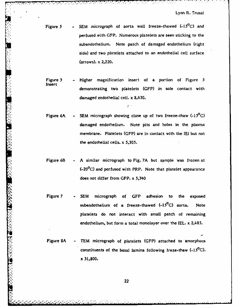
Lynn R. Trusal
t0
Figure 5 SEM micrograph of aorta wall freeze-thawed (-15C) and
perfused with GFP. Numerous platelets are seen sticking to the
subendothelium. Note patch of damaged endothelium (right
side) and two platelets attached to an endothelial cell surface
(arrows). x 2,220.
Figure 5 - Higher magnification insert of a portion of Figure 5Insert
demonstrating two platelets (GFP) in sole contact with
damaged endothelial cell. x 8,430.
Figure 6A - SEM micrograph showing close up of two freeze-thaw (-5 0 C)
damaged endothelium. Note pits and holes in the plasma
membrane. Platelets (GFP) are in contact with the IEI but not
the endothelial cells. x 5,305.
Figure 6B A similar micrograph to Fig. 7A but sample was frozen at
(-200 C) and perfused with PRP. Note that platelet appearance
does not differ from GFP. x 5,340
-.
Figure 7 SEM micrograph of GFP adhesion to the exposed
subendothelium of a freeze-thawed (-I5C) aorta. Note
platelets do not interact with small patch of remaining
endothelium, but form a total monolayer over the IEL. x 2,485.
Figure SA TEM micrograph of platelets (GFP) attached to amorphous
constituents of the basal lamina following freze-thaw (-i50 C).
x 31,800.
22
.. °h - . ° - . -2*\ - . - -. . .. . .

Lynn R. Trusal
Figure 8B - TEM micrograph of platelets (GFP) attached to microfibrils of
the IEL following freze-thaw (-15°C). x 22,500.
Figure 9 TEM micrograph of platelet (PRP) attached to the border
region of a damaged (-15 0 C) endothelial cell. Note presence of
an amorphous material (arrows) mediating cell to cell contact
and absence of collagen (C) involvement. x 22,100.
-2
23
Q . * . .. - . .

I..
k.A IAs- 1 D'

CAG WATRLAT
PERISTARTA
PERFUSIONPUMP
V ~ V
PC. Or, L
ix ~'
U7

r (J'3 '9I Z~r~ Sev

-4
16 h'.

igmt- 'vS
41 wi
V ~ .~* Al
AP-~ .
- R 5.
Fermit t-

The views, opinions, and/or findings contained in this report are those ofthe author(s) and should not be construed as an official Department of theArmy position, policy, or decision, unless so designated by other officialdocumentation.
. ........".
*1
a27

''I
A Z
IIVoU
4 'I t -
j