local inhibition of elastase reduces emilin1 cleavage ... · local inhibition of elastase reduces...
TRANSCRIPT

Clinical Science (2016) 130, 1221–1236 doi: 10.1042/CS20160064
Local inhibition of elastase reduces EMILIN1cleavage reactivating lymphatic vessel function ina mouse lymphoedema modelEliana Pivetta*, Bruna Wassermann*, Lisa Del Bel Belluz*†, Carla Danussi*‡, Teresa Maria Elisa Modica*,Orlando Maiorani*, Giulia Bosisio*, Francesco Boccardo§, Vincenzo Canzonieri‖, Alfonso Colombatti* andPaola Spessotto*
*Experimental Oncology 2, Department of Translational Research, CRO-IRCCS, National Cancer Institute, 33081, Aviano, Italy†Department of Cell and Molecular Biology, Karolinska Institutet, SE-171 77 Stockholm, Sweden‡Human Oncology and Pathogenesis Program, Memorial Sloan Kettering Cancer Center, New York, NY 10065, U.S.A.§Department of Surgery – Unit of Lymphatic Surgery, IRCCS S. Martino University Hospital – IST, National Institute for Cancer Research, University ofGenoa, 16132 Genoa, Italy‖Division of Pathology, Department of Translational Research, CRO-IRCCS, National Cancer Institute, 33081, Aviano, Italy
AbstractLymphatic vasculature critically depends on the connections of lymphatic endothelial cells with the extracellularmatrix (ECM), which are mediated by anchoring filaments (AFs). The ECM protein EMILIN1 is a component of AFsand is involved in the regulation of lymphatic vessel functions: accordingly, Emilin1−/− mice display lymphaticvascular morphological alterations, leading to functional defects such as mild lymphoedema, lymph leakage andcompromised lymph drainage. In the present study, using a mouse post-surgical tail lymphoedema model, we showthat the acute phase of acquired lymphoedema correlates with EMILIN1 degradation due to neutrophil elastase (NE)released by infiltrating neutrophils. As a consequence, the intercellular junctions of lymphatic endothelial cells areweakened and drainage to regional lymph nodes is severely affected. The local administration of sivelestat, aspecific NE inhibitor, prevents EMILIN1 degradation and reduces lymphoedema, restoring a normal lymphaticfunctionality. The finding that, in human secondary lymphoedema samples, we also detected cleaved EMILIN1 withthe typical bands of an NE-dependent pattern of fragmentation establishes a rationale for a powerful strategy thattargets NE inhibition. In conclusion, the attempts to block EMILIN1 degradation locally represent the basis for anovel ‘ECM’ pharmacological approach to assessing new lymphoedema treatments.
Key words: EMILIN1, extracellular matrix, human lymphoedematous tissue, neutrophil elastase, secondary lymphoedema, sivelestat.
INTRODUCTION
The lymphatic vasculature is critical for fluid homoeostasis, im-mune surveillance and fat absorption [1]. It comprises lymphaticcapillaries, adapted for lymph uptake from the tissue interstitium,and collecting vessels, which transport lymph back to the vascu-lar blood system. Lymphatic capillaries are blind-ending vessels,lined by a single thin layer of overlapping lymphatic endothelialcells (LECs) directly connected to the surrounding extracellularmatrix (ECM) by means of elastic anchoring filaments (AFs) [2].Abnormalities of AFs may reduce adsorption from the intersti-tium and propulsion of lymph and cells, and promote patholo-
Abbreviations: AF, anchoring filament; CI, cell index; ECM, extracellular matrix; H&E, haematoxylin and eosin; HMVEC–dLyNeo, human microvascular endothelial cell–dermallymphatic – neonatal; Ig, immunoglobulin; LAEC, lymphangioma-derived endothelial cell; LEC, lymphatic endothelial cell; MMP, matrix metalloproteinase; NE, neutrophil elastase; TGF-β,tumour transforming factor β; VEGF, vascular endothelial growth factor; WT, wild type.
Correspondence: Paola Spessotto (email [email protected]).
gical conditions such as lymphoedema [3]. The secondary lymph-oedema (acquired) frequently arises as a consequence of surgical,malignant, inflammatory or traumatic disruption of the lymphat-ics [4]. Lymphoedema has no cure and current treatments [5] canonly slow its progression. The recent improvements in surgicaland radiotherapy techniques have not substantially reduced theincidence of cancer-related lymphoedema and, although relianceon sentinel node techniques does reduce the risk of lymphoedema,only a small percentage of the at-risk cancer population benefitsfrom this approach. Therefore, the impact of lymphoedema re-mains a serious healthcare problem in patients with cancer, aboveall in those with breast cancer.
1221c© 2016 The Author(s). This is an open access article published by Portland Press Limited on behalf of the Biochemical Society and distributed under theCreative Commons Attribution License 4.0 (CC BY-NC-ND).

E. Pivetta and others
Lymphoedema, wound healing and tumour metastatic pro-cesses, in which lymphangiogenesis is a critical factor to initiateand coordinate the sequence of events, are closely related to themolecular ECM composition of the microenvironment. A betterunderstanding of which and how ECM molecules influence en-dothelial cell functions could provide full insight into how lymph-angiogenesis occurs and contributes to local lymph drainage indisordered tissues. The ECM protein EMILIN1 is expressed inlymphatic vessels, as well as in the connective tissues of a widevariety of organs [6–9]. EMILIN1 is a component of the sys-tem of cell anchorage to elastic lamellae via the α4β1- and/orα9β1-integrin receptors [10,11]. Although mice homozygous fordisruption of emilin1 display no lethal abnormalities [8,12,13],structural and functional defects of the lymphatic vessels arepresent, with a reduction in AFs, enlargement of lymphatic ca-pillaries and alterations of the luminal valves of the collectors,with myofibrillar differentiation and proliferation [11].
There have been few attempts to develop suitable animal mod-els of secondary lymphoedema and to assess new molecular ther-apies: the most recent are the mouse-tail skin wound [14,15] andthe excision of mouse axillary lymph nodes [16]. The identific-ation of several molecular components of the lymphatic vascu-lature has made it feasible to consider treatment with lymphaticgrowth factors. Vascular endothelial growth factor (VEGF)-C andVEGF-D have been tested in animal models, demonstrating thatgrowth factor-induced maturation of lymphatic vessels is possiblein adult mice and that growth factor therapy could become a validapproach for lymphoedema treatment [16–18]. Pharmacologicalapproaches were also attempted; however, although ketoprofenreduced the swelling of the tail in a post-surgical mouse model[19,20], benzo-pyrones applied in human clinical trials were notvery effective, apart from a slight improvement in lymphoedema[21,22].
We have recently demonstrated that neutrophil elastase (NE)was the most effective cleaving enzyme that could fully impairthe regulatory function of EMILIN1 [23]. In the present study wedemonstrate that, in the acute phase of lymphoedema, NE con-tributed to EMILIN1 degradation in the intercellular connectionsof LECs, altering their properties, and that the local administra-tion of sivelestat, a specific NE inhibitor, reactivated lymphaticvessel function in a post-surgical tail lymphoedema model byreducing EMILIN1 cleavage.
MATERIALS AND METHODS
ReagentsSecreted recombinant EMILIN1 protein was obtained by con-stitutive expression in 293E cells, as previously described [24].Briefly, the cells were expanded to mass culture and then main-tained for 2 days in serum-free medium to allow accumulationof EMILIN1 in the cell supernatant. Partial purification wasachieved by dialysis of the conditioned medium at 4 ◦C against0.1 M NaCl, 20 mM Tris/HCI, pH 6.8. A further purificationstep was achieved by chromatography on a DEAE-cellulosecolumn and size exclusion chromatography using Sepharose
CL 4B (1.0×90.0 cm column, Amersham Pharmacia Biotech).Fibronectin was purchased from Sigma-Aldrich.
NE was purchased from Calbiochem (324681, Merk Milli-pore) and MMP-14 from Giotto Biotech S.r.l. Sivelestat sodiumsalt hydrate was purchased from Sigma (S7198) and suspendedin a 20 mg/ml stock solution. GM6001 (Chemicon, Merk Mil-lipore) was used as a broad-spectrum matrix metalloproteinase(MMP) inhibitor.
A complete list of all antibodies used in this study, with theirspecificity and sources/manufacturers, is given in SupplementaryTable S1.
Mouse-tail model of lymphoedemaAll animal procedures and their care were performed according tothe institutional guidelines, in compliance with national laws andauthorization by the Italian Ministry of Health (no. 248/2015).C57Bl/6 mice were purchased from Charles River Laborator-ies. Emilin1−/− mice (C57Bl/6 background) were generated aspreviously described [13] and maintained at the CRO-IRCCSmouse facility. In all experiments 6- to 8-week-old female an-imals and their littermate age-matched controls were used. Micewere anaesthetized with an intraperitoneal injection of ketamine(Imalgene, Merial) (100 mg/kg) and xylazine (Rompun, Bayer)(10 mg/kg). To provide analgesia for the treatment of post-surgical pain, a buprenorphine (Temgesic solution, 0.3 mg/ml,RB Pharmaceutical Ltd) subcutaneous injection was performedbefore surgery. To induce lymphoedema, a circumferential in-cision was made through the dermis close to the tail base (1 cm)to sever the dermal lymphatic vessels. The edges of this incisionwere then pushed apart with a cauterizing iron, thereby disturbingthe deeper lymphatics, preventing superficial bleeding and creat-ing a 2- to 3-mm gap to delay wound closure. Care was taken tomaintain the integrity of the major underlying blood vessels andtendons so that the tail distal to the incision did not become nec-rotic. Gentamicin sulfate 0.1 % ointment (Gentalyn, MSD ItaliaS.r.l.) was applied to prevent infection. To counteract NE, sive-lestat solution (2 mg/kg, final concentration) was intradermicallyinjected with a 29-gauge syringe, immediately after surgery anddaily for the following 4 days. Pilot dose–response experiments(three mice per group) were carried out to find the optimal in-hibitor concentration. Volumetric assessment was performed bytaking a picture of the tail close to a ruler [25]. On acquired im-ages, computer-assisted morphometric analyses were performedusing ImageJ software (accessible at http://imagej.nih.gov/ij/).The tail volume was calculated with the truncated cone formulaand the tail-volume increase for each mouse was calculated onthe basis of presurgical measurements.
Mice were sacrificed at various times up to 14 days from sur-gery. The tails were excised proximally and distally from the siteof the wound. Tail tissues were fixed in 10 % formalin-bufferedsolution, and decalcified with EDTA before OCT medium em-bedding (Kaltek). Cross-cryostat sections of 5 μm were preparedand stored at −80 ◦C until processing.
To test the enzymatic activity of lymph derived from micesubjected to tail surgery, mice were sacrificed at days 4 and7. Oedematous tail tissue was excised and removed close tothe site of wound and immediately centrifuged at 4 ◦C in a
1222 c© 2016 The Author(s). This is an open access article published by Portland Press Limited on behalf of the Biochemical Society and distributed under theCreative Commons Attribution License 4.0 (CC BY-NC-ND).

Control of lymphoedema by local elastase inhibition
microcentrifuge at maximum speed for 30 min. The supernatantwas collected and stored at −80 ◦C until its use for in vitroexperiments.
In vivo administration of anti-Ly6G antibody todeplete neutrophilsMice were injected intraperitoneally with 500 μg of anti-Ly6Gantibody (clone 1A8, BioXCell) dissolved in 200 μl of PBS, theday before tail surgery and daily for the following days. Controlmice received equal amounts of isotope control antibodies (ratimmunoglobulin IgG2a, clone 2A3, BioXcell). Flow cytometrywas performed to check depletion of neutrophils by anti-Ly6Gtreatment as follows: blood samples were collected in tubescontaining EDTA and treated with ammonium chloride eryth-rocyte lysis buffer; cells were stained directly with conjugatedantibodies (see Supplementary Table S1) for 15 min at 4 ◦C in thedark in PBS/1 % BSA. The Gr1 antibody was used here to avoidfalse-negative results because the anti-Ly6G-depleting antibodymay mask the Ly6G epitope. All analyses were performed usinga Beckton Dickinson LSRII flow cytometer using dedicatedDiva software.
Cell culturesHuman microvascular endothelial cell–dermal lymphatic –neonatal (HMVEC–dLyNeo) cells, and the media optimized fortheir growth (EBM-2), were purchased from Lonza (EurocloneSpA). These cells have been characterized as previouslydescribed [8] and reported in Supplementary Figure S1. Mouselymphangioma-derived endothelial cells (LAECs) were isolatedand immortalized following the procedure described previously[26]. Briefly, the mice were injected twice intraperitoneally, witha 15-day interval, with 200 μl of emulsified (1:1 with PBS)incomplete Freund’s adjuvant (Sigma). Hyperplastic vesselswere isolated from the liver and diaphragm at day 30 and treatedwith 0.5 mg/ml of collagenase A (Roche Diagnostics), and theresulting single-cell suspension was cultured. After 7–10 daysof culture, subconfluent cells (LAECs) were recovered withtrypsin/EDTA and immortalized by means of SV40 infec-tion. Immortalized LAECs were characterized for lymphaticendothelial markers as reported previously by us [27].
Human samplesTo analyse EMILIN1 degradation in human lymphoedematoustissues, samples were obtained after signed informed consentfrom patients affected by peripheral secondary lymphoedemawho underwent lymphatic microsurgery at the Centre of Lymph-atic Surgery and Microsurgery of the University of Genoa, Italy.During derivative procedures of multiple lymphatic–venous ana-stomoses or lymphatic pathway reconstruction [28], surgeonssectioned small tissue samples for in vitro analyses. To quantifyinflammatory infiltrate we retrieved biopsy material from two pa-tients with lymphoedema of the vulva, which developed afteradjuvant radiotherapy for cervical cancer and was at a laterstage treated for lymphoedema resolution at CRO-IRCCS, asrecently reported [29]. For histological analysis, formalin-fixed,paraffin-embedded, 5-μm tissue sections were deparaffinized inxylene and rehydrated in a graded series of alcohol. Images of
haematoxylin and eosin (H&E)-stained paraffin sections werecaptured using a camera (ICC50, Leica Microsystems) connec-ted to a Leica DM 750 microscope.
Immunofluorescence and histological analysesFor immunofluorescence staining, cells were washed and fixedwith 4 % paraformaldehyde for 10 min, permeabilized (with 1 %BSA, 0.1 % Triton X-100 and 2 % FBS in PBS) for 5 min andsaturated with the blocking buffer (1 % BSA, 2 % FBS in PBS)for 30 min. Tail-tissue sections were hydrated for 15 min (with1 % Triton X-100, in PBS) and blocked for 30 min (1 % BSA, 2 %FBS or 5 % normal goat serum in PBS). For HMVEC–dLyNeocell staining, rabbit polyclonal anti-human EMILIN1 antibod-ies (As556, produced in our laboratories), mouse monoclonalanti-human CD31 antibodies and rabbit polyclonal anti-humanfibronectin antibodies (a gift of Professor GM Bressan, Univer-sity of Padua, Italy) were used. For tail-tissue cryostat sections ratmonoclonal anti-mouse EMILIN1 antibodies (clone 1007C11A8,produced in our laboratories) and hamster anti-mouse podoplaninantibodies were used. All the incubations with the primary an-tibodies were performed overnight at 4 ◦C (followed by three5-min washes in PBS) and with the appropriate secondary an-tibodies (conjugated with Alexa Fluor 488 or Alexa Fluor 546at 1:200 dilution, Life Technologies) for an additional 1 h atroom temperature. For all samples, negative controls includedthe corresponding isotype or IgG. To-Pro-3 (1:5000, Life Tech-nologies) was used to visualize the nuclei. Images were acquiredwith a true confocal scanner system (TCS SP2, Leica Microsys-tems) equipped with HC PL Fluotar 10×/0.30 NA, HCX PL Apo40×/1.25–0.75 NA and HCX PL Apo 63×/1.40–0.60 NA oilobjectives (Leica), using Leica confocal software.
Cryostat sections were stained with H&E and images wereacquired using an optical microscope (Leica DM750) equippedwith a CCD camera using dedicated software (Las EZ Leica).
Western blot analysisMice were sacrificed at day 4 or 7 after surgery, and lymphsamples were collected from tails and incubated with recombin-ant EMILIN1 for 18 h. Whole lysates of both human samplesand tail tissues from normal and lymphoedematous sites wereprepared using thiourea/urea lysis buffer (7 M urea, 2 M thiourea,2 % CHAPS). Samples were subjected to SDS/4–12%PAGE (us-ing Criterion Precast Gel, BioRad) and blotted on nitrocellu-lose membranes (Amersham Hybond-ECL, Amersham Phar-macia Biotech). Membranes were blocked (5 % non-fat milk,0.1 % Tween-20 in TBS) and incubated with rabbit polyclonalanti-human EMILIN1 (As556) to identify recombinant humanEMILIN1 protein or rabbit anti-mouse EMILIN1 (mC1q, an an-tibody generated to specifically recognize the mouse gC1q do-main) to detect EMILIN1 in mouse tissue lysates. Horseradishperoxidase (HRP)-tagged secondary antibodies (Jackson Immun-oresearch) were used at proper dilutions. Signals were detectedusing ECL reagents (Amersham Western Blotting Detection Sys-tem and HyperFilm ECL, Amersham Pharmacia Biotech).
MicrolymphangiographyMice were anaesthetized as described above and the func-tionality of tail lymphatics was examined by fluorescence
1223c© 2016 The Author(s). This is an open access article published by Portland Press Limited on behalf of the Biochemical Society and distributed under theCreative Commons Attribution License 4.0 (CC BY-NC-ND).

E. Pivetta and others
microlymphangiography; 2 μl of 5 mg/ml FITC–dextran solu-tion (M r ∼2000 kDa, Life Technologies) was injected near thesurgical wound (0.5 cm distant) and diffusion of the dye was im-mediately recorded using a stereomicroscope Leica M205 FA andLeica DFC310 FX digital camera (Leica Microsystems). After5 min, mice were sacrificed and draining iliac lymph nodes wereimaged as above.
Endothelial barrier integrity assaysTo measure endothelial barrier integrity we adopted the techno-logy provided by the Real-Time Cell Analyzer dual plate instru-ment (ACEA Biosciences) and proprietary E-plates 96 [30,31].The principle of this technology relies on the fact that the increasein electrode impedance, expressed as the cell index (CI), is dir-ectly dependent on the number of cells and their spread. Thus,the CI is a reflection of overall cell number, attachment qualityand cell morphology which can change as a function of time.HMVEC-dLyNeo or LAEC cells (5×10³/well) were added toE-plates 96 in EBM-2 medium. After 4 days, different concen-trations of NE were added and the E-plates 96 were monitoredevery 15 min for 12 h. Changes in impedance of confluent en-dothelial cells reflect changes in barrier function. Data analysiswas performed using the Real-Time Cell Analyzer software (ver-sion 1.2) supplied with the instrument. Experiments were per-formed in triplicate and data are expressed as a normalized CI atthe time of NE addition.
Statistical analysisThroughout this study, plotted values are shown asmeans+−S.E.M.s or S.D.s as indicated. The statistical signific-ance of the results was determined using the two-tailed unpairedStudent’s t-test to determine whether the two datasets were signi-ficantly different. A value of P < 0.05 was considered significant.
RESULTS
Lymphatic function is reduced in Emilin1−/− miceUsing a post-surgical tail lymphoedema approach we demonstratethat Emilin1−/− mice show a faster, greater and much longerpersistence of swelling compared with the wild-type (WT) lit-termates (Figures 1A and 1B), suggesting that the absence ofEMILIN1 correlates with the inefficient fluid drainage by thelymphatics. No significant differences in the number of lymphaticvessels were detected, 2 weeks after surgery, in the proximal seg-ment of the wound between WT and Emilin1−/− mice, whereas, inthe distal oedematous tail tissues, very few lymphatic capillarieswere present in Emilin1−/− mice (Figures 1C and 1D). Moreover,new lymphatic vessels of WT mice had a larger lumen comparedwith Emilin1−/− mice (Figures 1C and 1E). To analyse lymph-atic regeneration and function in WT and Emilin1−/− animalsfurther, we performed lymphangiography to visualize draininglymph nodes. After the injection of FITC–dextran in the dermisof distal lymphoedematous tails, the typical hexagonal networkof dermal lymphatics driving the fluorescent tracer was detectablein WT but not Emilin1−/− mice (Figure 2A). Consequently, onlyWT mice were able to drain FITC–dextran to iliac lymph nodes
(Figure 2B). In fact, the uptake of FITC–dextran by lymphaticvessels was easy to detect in both the proximal and the distaltail segments in WT mice, indicating that the regeneration oflymphatics after surgery had occurred successfully (Figure 2C).On the other hand, the fluorescent dye persisted in the dermisof Emilin1−/− mice and no close association of FITC–dextranwith lymphatic vessels was evident (Figure 2C). The lack of awell-defined capillary network in the distal tail and the absenceof fluorescent dye in draining lymph nodes in Emilin1−/− animalssuggested that EMILIN1 deficiency impaired the regeneration offunctional lymphatic vessels.
Neutrophils infiltrate the lymphoedematous tissuesThe surgical approach entails an acute inflammatory responsewith the release of proteolytic enzymes. Neutrophils representedthe prevalent inflammatory cells of the infiltrate 4 days after sur-gery (Figure 3A; see also Figure 7). EMILIN1 was digested to a120-kDa form after in vitro incubation with the lymph extractedfrom post-surgical lymphoedema tissues (Figure 3B). The en-zymatic activity in the lymph persisted up to day 7 after surgeryin both WT and Emilin1−/− mice. This molecular degradationform is reminiscent of that obtained after NE treatment of puri-fied EMILIN1 [23]. This cleavage effect was abolished with theaddition of sivelestat, a well-known NE inhibitor [32–34], but notwith GM6001, a broad-spectrum MMP inhibitor, indicating that,among the enzymes released by neutrophils, NE was the most, ifnot even the only one, effective in EMILIN1 fragmentation (Fig-ure 3C). Consequently, only sivelestat could inhibit the activity ofNE on recombinant EMILIN1 in vitro (Figure 3D). Furthermore,in contrast to normal skin, in lymphoedematous WT tissues theintact form of EMILIN1 was not detectable, whereas a majorEMILIN1 fragment of about 70 kDa was generated (Figure 3E),showing that EMILIN1 digestion occurred after a surgical woundin the tail.
EMILIN1 degradation and neutrophil infiltration inhuman acquired lymphoedemaSamples derived from surgical specimens of peripheral second-ary lymphoedema, obtained during lymphatic microsurgery, wereassayed using Western blots to determine EMILIN1 expressionlevels. All samples expressed different amounts of intact as wellas fragmented EMILIN1 (Figure 4A). Variable amounts of themost characteristic bands detected in the NE-derived fragmenta-tion pattern were present (Figure 4A, black arrows), suggestingthat in human lymphoedematous samples EMILIN1 was alsodigested by NE. The source of NE is probably due to the pres-ence of neutrophils, as detected in human acquired lymphoedemaof the vulva, which developed in two patients affected by cer-vical cancer and treated with adjuvant radiotherapy (Figure 4B).Even if these latter samples have to be considered part of achronic inflammatory process, as demonstrated by the presenceof several macrophages (CD68-positive cells) and a few lympho-cytes (CD3- and CD20-positive cells) (see Supplementary FigureS2), neutrophils were detected nearby or even attached to LECs(Figure 4B).
1224 c© 2016 The Author(s). This is an open access article published by Portland Press Limited on behalf of the Biochemical Society and distributed under theCreative Commons Attribution License 4.0 (CC BY-NC-ND).

Control of lymphoedema by local elastase inhibition
Figure 1 Emilin1−/− mice develop consistent lymphoedema after tail surgery(A, B) Emilin1−/− mice showed a greater and much longer persistence of swelling compared with their WT littermates; sixmice per group were used. NS, not significant. (C) Representative cryostat sections from two WT (left) and two Emilin1−/−(right) mice of the distal part of their tail, demonstrating that the number of podoplanin-positive lymphatic vessels washigher in WT than in Emilin1−/− mice. Isotypical control staining was included. (D) Quantitative analyses of vessel density(podoplanin staining) in proximal and distal parts of the tail. The number of podoplanin-positive lymphatic vessels wascounted in the whole immunofluorescent section acquired by the Leica TCS SP2 confocal system, using Volocity softwareprovided by Perkin Elmer. (E) In distal tails, new lymphatic vessels of WT mice have a larger lumen compared withEmilin1−/− mice. The luminal area was calculated using the measurement area tool of Volocity software. The graphsreport the means+−S.E.M.s obtained from four mice per group, analysing eight fields for each sample. Scale bar = 50 μm.
NE degrades EMILIN1, impairing lymphaticendothelium integrityTo demonstrate that EMILIN1 cleavage directly altered LECproperties, we analysed the integrity of the monolayer by monit-
oring the impedance of confluent endothelial cells after NE treat-ment. The addition of NE resulted in a dose-dependent effect onthe barrier function permeability (Figures 5A and 5B) withoutaffecting cell viability (data not shown), indicating that this
1225c© 2016 The Author(s). This is an open access article published by Portland Press Limited on behalf of the Biochemical Society and distributed under theCreative Commons Attribution License 4.0 (CC BY-NC-ND).

E. Pivetta and others
Figure 2 Reduced lymphatic function in Emilin1−/− mice after tail surgeryFITC–dextran was subcutaneously injected into the tail of WT and Emilin1−/− mice at day 14 after wounding. (A) Rep-resentative fluorescence images of subcutaneous lymphatic vessels in the tail are shown: note the typical hexagonalnetwork in WT tails. White arrows indicate the FITC–dextran injection site. (B) At 5 min from injection, images were takento visualize draining iliac lymph nodes. (C) Tails were harvested and stained with anti-podoplanin antibodies. Note theuptake of FITC–dextran by lymphatic vessels (yellow asterisks) in WT mice and the persistence of the dye in the dermisin Emilin1−/− mice. White arrows indicate podoplanin-positive lymphatic vessels. Isotypical control staining was included.Scale bars = 2 mm (A, B) and 50 μm (C).
1226 c© 2016 The Author(s). This is an open access article published by Portland Press Limited on behalf of the Biochemical Society and distributed under theCreative Commons Attribution License 4.0 (CC BY-NC-ND).

Control of lymphoedema by local elastase inhibition
Figure 3 Neutrophil infiltration after tail surgery is associated with EMILIN1 cleavage(A) Representative H&E staining of cryostat tail section (left) and its boxed area magnification (right) at day 4 after surgery,showing an abundant neutrophil infiltrate. (B) Western blot analysis of recombinant EMILIN1 (E1) fragmentation in vitroafter an 18-h incubation with the lymph extracted from WT and Emilin1−/− mice at day 4 (d4) or 7 (d7) after wounding.(C) Sivelestat, but not GM6001, blocked the enzymatic activity of the lymph on recombinant EMILIN1. (D) CoomassieBlue-stained gels show that recombinant EMILIN1 cleavage using NE was specifically blocked by sivelestat. Fibronectinwas used as positive control for MMP-14 and the MMP inhibitor GM6001. (E) Western blot analysis of EMILIN1 degradationin normal (N) and lymphoedematous (L) tail tissue extracts. Scale bar = 20 μm.
enzyme treatment could loosen cell junctions as demonstratedby CD31 distribution (Figures 5C–5F). After NE treatment,cells appeared smaller with looser intercellular junctions, andEMILIN1-positive fibrils were not evident in the gaps betweencells, suggesting that its cleavage correlated with the markedly re-duced cellular contacts (Figure 5D). Fibronectin staining seemedto be unmodified intercellularly after NE treatment (Figures 5Eand 5F), probably because the elected substrate for NE in thiscontext was EMILIN1 and the integrity of EMILIN1 determ-
ined the stability of the LEC junctions. CD31 staining wasclearly visible and intense in cellular protrusions after NE treat-ment, indicating that the NE-dependent formation of intercel-lular gaps was probably attributed to ECM degradation ratherthan to proteolysis of membrane receptors (Figure 5D). Ac-cordingly, LECs obtained from Emilin1−/− mice were not af-fected by NE treatment (Figure 5B), suggesting that NE’s ef-fects on LEC integrity were exerted by affecting primarilyEMILIN1.
1227c© 2016 The Author(s). This is an open access article published by Portland Press Limited on behalf of the Biochemical Society and distributed under theCreative Commons Attribution License 4.0 (CC BY-NC-ND).

E. Pivetta and others
Figure 4 EMILIN1 cleavage and neutrophil infiltration in human acquired lymphoedema(A) Western blotting analysis of EMILIN1 expression and cleavage in human acquired lymphoedema samples. Blackarrows and red arrow indicate the most representative bands of cleaved EMILIN1 and undigested EMILIN1, respectively.(B) Paraffin-embedded sections of vulvar lymphoedema samples obtained from two patients (a and b, patient no. 1; c,patient no. 2) stained with H&E. Arrows indicate neutrophils (a′, b′, c′) and asterisks enlarged lymphatic vessels (a, b, c);a′, b′ and c′ correspond to the boxed areas in a, b and c, respectively. Scale bars = 20 μm (a′, b′, c′); 50 μm (a, b, c).
1228 c© 2016 The Author(s). This is an open access article published by Portland Press Limited on behalf of the Biochemical Society and distributed under theCreative Commons Attribution License 4.0 (CC BY-NC-ND).

Control of lymphoedema by local elastase inhibition
Figure 5 EMILIN1 degradation by NE impairs lymphatic endothelium integrity(A, B) Dynamic monitoring of lymphatic endothelium integrity in response to NE measured with XCelligence instrumentand expressed as normalized CI. Cells were grown to confluence and then (black arrows in the graphs) different dosesof NE were added. HMVEC (A) and mouse LECs from WT (LAEC WT) and Emilin1−/− (LAEC KO) mice (B) were used. Themeans+−S.D.s of three independent experiments are reported in the graphs. (C–F) Confluent HMVECs were incubated for5 h with NE and then fixed and stained with anti-CD31 and anti-EMILIN1 (C, D) or anti-fibronectin (E, F) antibodies. Dottedlines highlight loss of EMILIN1 (D) or the well-detected and distributed fibres of fibronectin (F) within the area separatingadjacent cells after NE treatment. (G) Isotypical control staining of confluent HMVECs. Scale bar = 20 μm.
1229c© 2016 The Author(s). This is an open access article published by Portland Press Limited on behalf of the Biochemical Society and distributed under theCreative Commons Attribution License 4.0 (CC BY-NC-ND).

E. Pivetta and others
Figure 6 Sivelestat specifically reduces tissue swelling in a post-surgically induced lymphoedema model(A) Dose–response effect of sivelestat on tail-volume increase after induction of surgical lymphoedema. Three mice pergroup were subjected to local treatment. PBS (indicated as ‘0 mg/kg’) was used as control ‘treatment’. (B–D) Sivelestat,but not GM6001, already significantly reduced the extent of swelling in WT mice from the first days after injury. The vehicleDMSO was used as a control for GM6001. (E) Sivelestat had no effect when locally injected in Emilin1−/− mice. Thenumbers of mice used in (D) and (E) are reported in parentheses. Data are expressed as means+−S.E.M.s.
Treatment with NE inhibitor reduces lymphoedemaWe hypothesized that local treatment with NE inhibitors, bypreventing EMILIN1 degradation, could reduce the extent oflymphoedema in WT mice. Immediately before wound surgeryand daily for the next 4 days, WT animals were treated withsivelestat, which displays powerful treatment effects in variouspreclinical models [35]. We observed that, right from the earlydays after surgery, sivelestat-treated mice developed a signific-antly (P < 0.001) less intense swelling of the distal tail comparedwith the control (PBS-treated) mice (Figures 6A–6C, 7A and 7B).On the contrary, the local administration of GM6001 (5 mg/kg,following the same schedule used for sivelestat treatment) wasunsuccessful in reducing lymphoedema (Figures 6B and 6D), in-dicating that MMPs were not responsible for the pathogenesisof lymphoedema. The possibility that EMILIN1 was the crucialtarget of NE in the acute phase of lymphoedema was stronglysuggested by the results in Emilin1−/− mice, in which sivelestathad no effect in reducing tail swelling (Figure 6E).
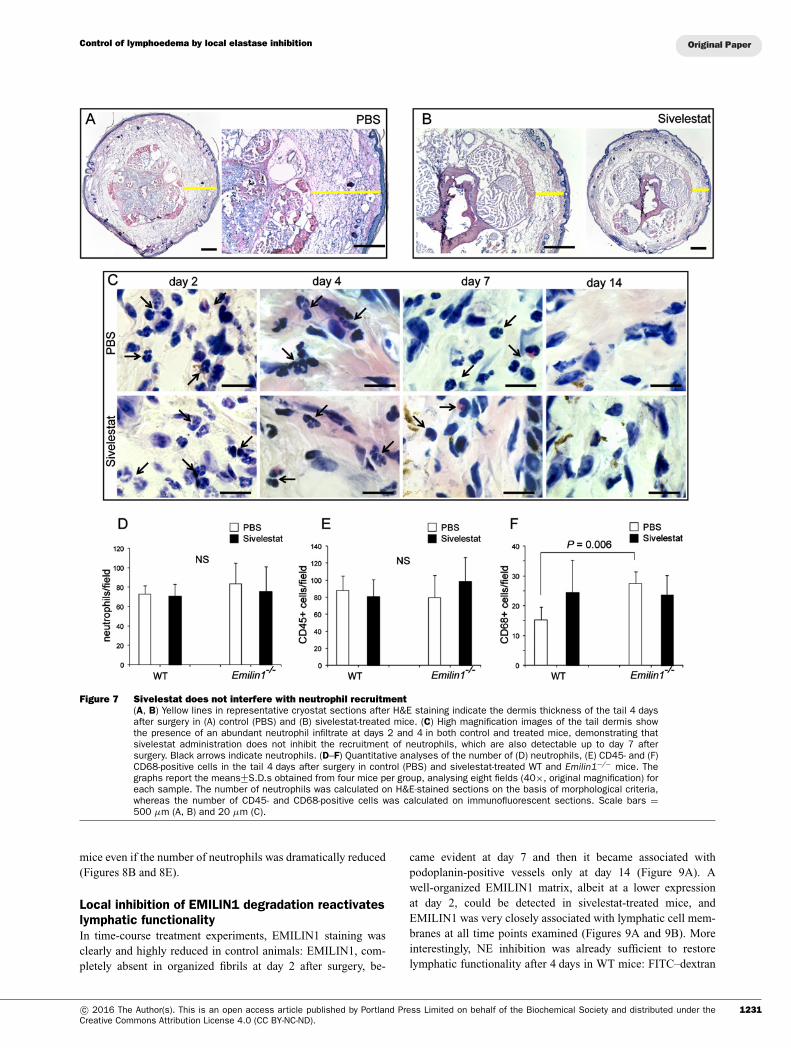
The role of neutrophils in the tail lymphoedemamodelSivelestat did not inhibit the recruitment of neutrophils, whichcould also be detected up to day 7 after surgery in the
tail dermis, as well as in PBS-treated mice (Figures 7C and7D). The numbers of CD45- and CD68-positive cells didnot significantly change after sivelestat administration (Fig-ures 7E and 7F), indicating that this synthetic inhibitor spe-cifically acted primarily on the enzymatic NE activity, andwas ineffective on inflammatory cell recruitment. Sivelestat wasalso not influential in Emilin1−/− mice, which, however, hadhigher basal numbers of macrophages compared with WT mice(Figure 7E).
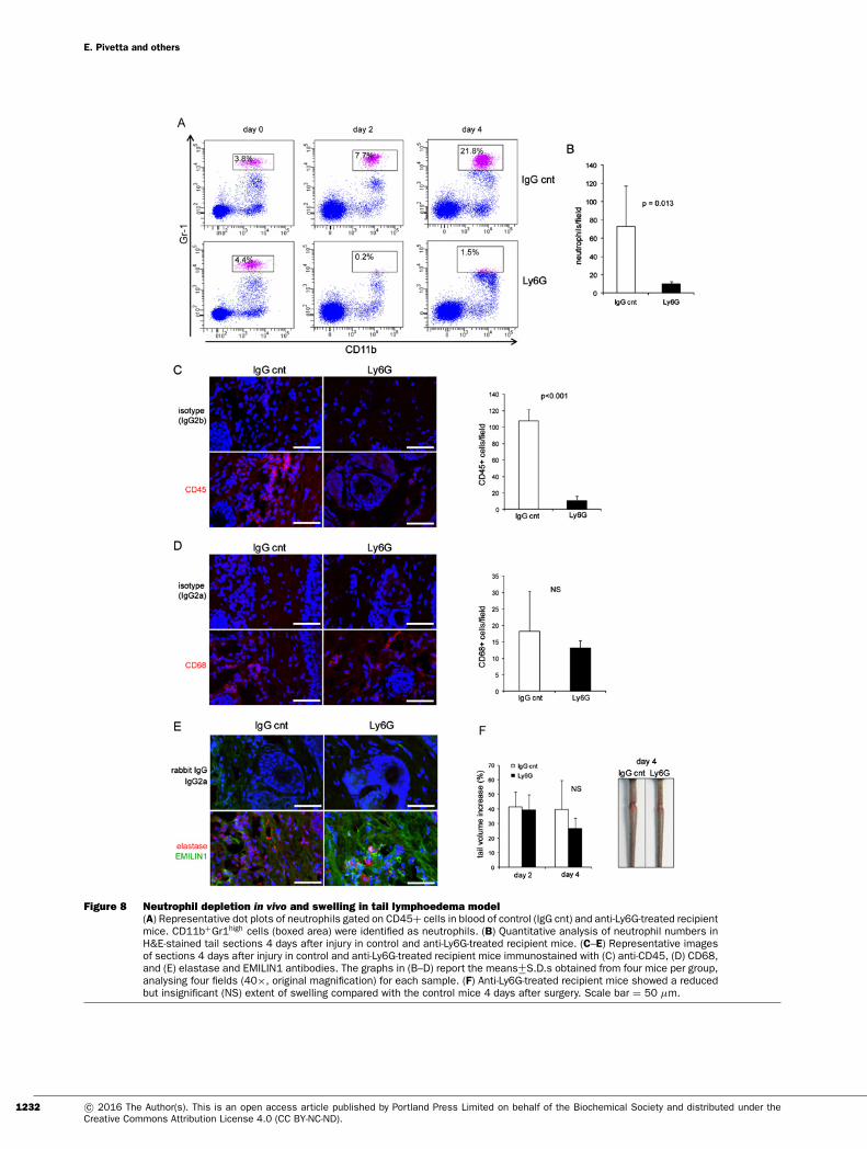
As neutrophils are the principal source of NE, we reasoned thatneutrophil depletion could provide the same effect as obtained bysivelestat. We successfully depleted neutrophils by daily in vivoadministration of anti-Ly6G monoclonal antibody (Figures 8A–8C). Accordingly, the number of CD45+ cells was dramaticallyreduced (Figure 8C). No significant change in the number ofCD68+ cells was detected (Figure 8D). Two consequences of thecell depletion were the reduction in the extent of tail swelling4 days after surgery (Figure 8F) and a higher EMILIN1 stain-ing compared with control mice (Figure 8E). The effects ofLy6G treatment suggest that EMILIN1 fragmentation was re-duced, but not sufficiently to prevent the loss of its integrity andobtain a significant reduction in swelling, as occurred after sive-lestat treatment. In fact, NE was still detectable in Ly6G-treated
1230 c© 2016 The Author(s). This is an open access article published by Portland Press Limited on behalf of the Biochemical Society and distributed under theCreative Commons Attribution License 4.0 (CC BY-NC-ND).
Control of lymphoedema by local elastase inhibition
Figure 7 Sivelestat does not interfere with neutrophil recruitment(A, B) Yellow lines in representative cryostat sections after H&E staining indicate the dermis thickness of the tail 4 daysafter surgery in (A) control (PBS) and (B) sivelestat-treated mice. (C) High magnification images of the tail dermis showthe presence of an abundant neutrophil infiltrate at days 2 and 4 in both control and treated mice, demonstrating thatsivelestat administration does not inhibit the recruitment of neutrophils, which are also detectable up to day 7 aftersurgery. Black arrows indicate neutrophils. (D–F) Quantitative analyses of the number of (D) neutrophils, (E) CD45- and (F)CD68-positive cells in the tail 4 days after surgery in control (PBS) and sivelestat-treated WT and Emilin1−/− mice. Thegraphs report the means+−S.D.s obtained from four mice per group, analysing eight fields (40×, original magnification) foreach sample. The number of neutrophils was calculated on H&E-stained sections on the basis of morphological criteria,whereas the number of CD45- and CD68-positive cells was calculated on immunofluorescent sections. Scale bars =500 μm (A, B) and 20 μm (C).
mice even if the number of neutrophils was dramatically reduced(Figures 8B and 8E).
Local inhibition of EMILIN1 degradation reactivateslymphatic functionalityIn time-course treatment experiments, EMILIN1 staining wasclearly and highly reduced in control animals: EMILIN1, com-pletely absent in organized fibrils at day 2 after surgery, be-
came evident at day 7 and then it became associated withpodoplanin-positive vessels only at day 14 (Figure 9A). Awell-organized EMILIN1 matrix, albeit at a lower expressionat day 2, could be detected in sivelestat-treated mice, andEMILIN1 was very closely associated with lymphatic cell mem-branes at all time points examined (Figures 9A and 9B). Moreinterestingly, NE inhibition was already sufficient to restorelymphatic functionality after 4 days in WT mice: FITC–dextran
1231c© 2016 The Author(s). This is an open access article published by Portland Press Limited on behalf of the Biochemical Society and distributed under theCreative Commons Attribution License 4.0 (CC BY-NC-ND).
E. Pivetta and others
Figure 8 Neutrophil depletion in vivo and swelling in tail lymphoedema model(A) Representative dot plots of neutrophils gated on CD45+ cells in blood of control (IgG cnt) and anti-Ly6G-treated recipientmice. CD11b+Gr1high cells (boxed area) were identified as neutrophils. (B) Quantitative analysis of neutrophil numbers inH&E-stained tail sections 4 days after injury in control and anti-Ly6G-treated recipient mice. (C–E) Representative imagesof sections 4 days after injury in control and anti-Ly6G-treated recipient mice immunostained with (C) anti-CD45, (D) CD68,and (E) elastase and EMILIN1 antibodies. The graphs in (B–D) report the means+−S.D.s obtained from four mice per group,analysing four fields (40×, original magnification) for each sample. (F) Anti-Ly6G-treated recipient mice showed a reducedbut insignificant (NS) extent of swelling compared with the control mice 4 days after surgery. Scale bar = 50 μm.
1232 c© 2016 The Author(s). This is an open access article published by Portland Press Limited on behalf of the Biochemical Society and distributed under theCreative Commons Attribution License 4.0 (CC BY-NC-ND).

Control of lymphoedema by local elastase inhibition
Figure 9 Local treatment with sivelestat restores lymphatic functionality(A) EMILIN1 staining was highly reduced in PBS-treated mice even at day 7 after wounding, very probably becauseof degradation by NE, whereas in silvelestat-treated mice EMILIN1 fibres appeared to be well deposited and in closecontact with lymphatic vessels (arrows). (B) The z-section of the sivelestat-treated tail at day 14 as reported in (A); thecorresponding x and y projections clearly provide evidence for the close (yellow) association of EMILIN1 with lymphaticvessel cell membranes. (C) FITC–dextran was already drained by lymph nodes in sivelestat-treated but not control (PBS)mice at day 4 after surgery. Scale bars = 20 μm (A), 2 mm (C).
drained to lymph nodes in sivelestat-treated but not control mice(Figure 9C).
DISCUSSION
To the best of our knowledge, this is the first report to focus onthe role of an ECM component in the pathogenesis of secondarylymphoedema. Most of the recent research has focused on themolecular basis of lymphatic development and generated severalgenetic animal models that are useful to identify key regulatorsof embryonic lymphatic development and provide insight into themechanisms of primary lymphatic insufficiency [36]. The tissueresponses to the interplay of local inflammation, matrix remod-elling and lymphangiogenesis in secondary lymphatic insuffi-ciency are less well understood. Inflammation triggers lymphan-giogenesis [37,38], and various lymphangiogenic factors, such as
VEGF-C, VEGF-D and hepatocyte growth factor, were identifiedas necessary to regulate lymphatic regeneration [39]. However,the crucial regulators in the coordination of the repair processesafter lymphatic injury still need to be elucidated. Optimism overthe promise of growth factor therapy for the treatment of sec-ondary lymphoedema has been dampened [40,41]. Contrastingobservations emerged to show that exogenous growth factor ad-ministration to sites of lymphatic injury augmented early LECproliferation, but without the development of a functionally com-petent vasculature [42]. This was the case for delivery of exogen-ous VEGF-C [43], suggesting that VEGF-C/VEGF receptor-3signalling may not be sufficient for the organizational evolutionof lymphangiogenesis. Furthermore, it seems that the ability ofthe lymphatic vasculature to regenerate after injury is governedby a controlled balance between pro- and anti-lymphangiogeniccytokines [44]. In this context, the role of ECM has been poorlyconsidered. The Emilin1−/− mouse model has represented thefirst abnormal lymphatic phenotype associated with deficiency
1233c© 2016 The Author(s). This is an open access article published by Portland Press Limited on behalf of the Biochemical Society and distributed under theCreative Commons Attribution License 4.0 (CC BY-NC-ND).
E. Pivetta and others
of an ECM protein, thus providing a unique and useful tool todemonstrate the role of a key structural element in the mainten-ance of lymphatic vessels’ integrity and in regulation of lymph-angiogenesis. Our previous studies, in fact, demonstrated thatEMILIN1 genetic deficiency induces a significant reduction ofAFs in the lymphatic capillaries [8], and unveiled EMILIN1 asa novel ligand for LEC α9-integrin, suggesting that EMILIN1played a direct role in growth and maintenance of lymphaticvessels [11].
In the present study, we propose EMILIN1 as a ‘structural’regulator for a competent vasculature. Through use of apost-surgical tail lymphoedema model, we show that EMILIN1degradation by NE is an early event in the pathogenesis oflymphoedema.
We also demonstrate that, in the first phases of the inflam-matory process after tail surgery, the cellular infiltrate is mainlyrepresented by neutrophils. It is well known that neutrophilssynthesize a number of different proteases, including NE andMMP-8 and MMP-9 [45]. Among these enzymes, NE is the onlyone that can cleave EMILIN1 because sivelestat fully blockedthe effect of lymph extracted from lymphoedematous tissues,whereas GM6001, a broad-spectrum MMP inhibitor, was totallyineffective. In a recent study, Rigby et al. [46] found that,during neutrophil transit of the inflamed lymphatic endothelium,MMP-8, MMP-9 and NE promote the endothelial retractionthat increases junctional permeability, enabling neutrophiltransmigration. They did not identify the substrates of the neut-rophil enzymes, but hypothesized that ECM components wereinvolved. The present study provides evidence that EMILIN1is the elective ECM substrate, as demonstrated in vitro byexperiments on the increased permeability of LECs after NEtreatment and EMILIN1 degradation.
The crucial role of neutrophils in lymphoedema pathogen-esis remains to be fully and well characterized. Our neutrophildepletion approach clearly demonstrated that it is necessary tofully inhibit NE to restore lymphatic endothelial functionality:the presence of few neutrophils after Ly6G treatment, but posit-ivity for elastase staining, did not allow a strong and significanteffect in reducing tail swelling.
In fact, only the treatment with sivelestat significantly inhib-ited the development of lymphoedema in the acute phase; in sodoing it could also delay or even avoid the onset of the chronicphase. It is known that transforming growth factor (TGF)-β in-creases the tissue fibrosis that characterizes the chronic phaseof lymphoedema and impairs lymphatic regeneration [47]. Asthe genetic inactivation of EMILIN1 causes increased TGF-βlevels [12], it is reasonable to hypothesize that, in the absenceof EMILIN1, as in Emilin1− /− mice (see Figure 1), or after itscleavage as in secondary lymphoedema, the lymphangiogenic re-sponse could also be altered via an indirect TGF-β-dependentmechanism.
The rationale for using sivelestat as a novel ‘ECM’ pharmaco-logical approach to assess new lymphoedema treatments is highlysupported by ex vivo analyses of human lymphoedema samples,in which few neutrophils were still present, even if among sev-eral cells typical of the chronic inflammatory process. DegradedEMILIN1 was, however, detected.
In summary, EMILIN1 is a structural element that assuresa proper functionality of the LEC intercellular junctions. Thus,prevention of EMILIN1 cleavage in the acute phase of inflamma-tion, by locally inhibiting the activity of NE, could be a powerfulstrategy to avoid lymphatic vessel impairment and the consequentsevere development of secondary lymphoedema.
CLINICAL PERSPECTIVE
• As a therapeutically relevant treatment of secondary lymph-oedema has yet to be achieved, the impact of lymphoedemaremains a serious healthcare problem, above all in cancer pa-tients. The role of ECM in lymphoedema still needs to beelucidated.
• We show that the degradation of the ECM protein EMILIN1by NE is an early event in the acute phase of lymphoedema.
• The local administration of a specific elastase inhibitor pre-vents EMILIN1 degradation, reduces lymphoedema and re-stores lymphatic functionality, thus providing the rationale fora powerful strategy to avoid the consequent development ofsevere secondary lymphoedema.
AUTHOR CONTRIBUTION
Eliana Pivetta and Paola Spessotto conceived and designed thestudy; Eliana Pivetta, Bruna Wassermann, Carla Danussi, LisaDel Bel Belluz, Teresa Maria Elisa Modica, Giulia Bosisio andOrlando Maiorani developed methodology and performed in vitro
and in vivo experiments; Francesco Boccardo and Vincenzo Can-zonieri provided and performed analyses on human lymphoedemasamples; Alfonso Colombatti and Paola Spessotto reviewed and in-terpreted data; Eliana Pivetta and Paola Spessotto analysed dataand supervised the study; Paola Spessotto wrote the manuscript.All authors read and approved the final manuscript.
ACKNOWLEDGEMENTS
We thank Gustavo Baldassarre for critical comments on the manu-script.
FUNDING
This work was supported by Associazione Italiana per la Ricercasul Cancro (AIRC) [IG 14192) and the Ministry of Health [RF-2010–2309719].
REFERENCES
1 Alitalo, K., Tammela, T. and Petrova, T.V. (2005)Lymphangiogenesis in development and human disease. Nature438, 946–953 CrossRef PubMed
2 Gerli, R., Ibba, L. and Fruschelli, C. (1991) Ultrastructuralcytochemistry of anchoring filaments of human lymphaticcapillaries and their relation to elastic fibers. Lymphology 24,105–112 PubMed
3 De Cock, H.E., Affolter, V.K., Farver, T.B., Van, B.L., Scheuch, B.and Ferraro, G.L. (2006) Measurement of skin desmosine as anindicator of altered cutaneous elastin in draft horses with chronicprogressive lymphedema. Lymphat. Res. Biol. 4, 67–72CrossRef PubMed
1234 c© 2016 The Author(s). This is an open access article published by Portland Press Limited on behalf of the Biochemical Society and distributed under theCreative Commons Attribution License 4.0 (CC BY-NC-ND).

Control of lymphoedema by local elastase inhibition
4 Rockson, S.G. (2012) Update on the biology and treatment oflymphedema. Curr. Treat. Options Cardiovasc. Med. 14,184–192 CrossRef PubMed
5 Witte, M.H., Bernas, M.J., Martin, C.P. and Witte, C.L. (2001)Lymphangiogenesis and lymphangiodysplasia: from molecular toclinical lymphology. Microsc. Res. Tech. 55, 122–145CrossRef PubMed
6 Colombatti, A., Bressan, G.M., Castellani, I. and Volpin, D.(1985) Glycoprotein 115, a glycoprotein isolated from chickblood vessels, is widely distributed in connective tissue. J. CellBiol. 100, 18–26 CrossRef PubMed
7 Colombatti, A., Poletti, A., Bressan, G.M., Carbone, A. andVolpin, D. (1987) Widespread codistribution of glycoprotein gp115 and elastin in chick eye and other tissues. Coll. Relat. Res.7, 259–275 CrossRef PubMed
8 Danussi, C., Spessotto, P., Petrucco, A., Wassermann, B.,Sabatelli, P., Montesi, M., Doliana, R., Bressan, G.M. andColombatti, A. (2008) Emilin1 deficiency causes structural andfunctional defects of lymphatic vasculature. Mol. Cell Biol. 28,4026–4039 CrossRef PubMed
9 Danussi, C., Petrucco, A., Wassermann, B., Pivetta, E., Modica,T.M., Belluz, L.B., Colombatti, A. and Spessotto, P. (2011)EMILIN1-α4/α9 integrin interaction inhibits dermal fibroblast andkeratinocyte proliferation. J. Cell Biol. 195, 131–145CrossRef PubMed
10 Spessotto, P., Cervi, M., Mucignat, M.T., Mungiguerra, G.,Sartoretto, I., Doliana, R. and Colombatti, A. (2003)β1-Integrin-dependent cell adhesion to EMILIN-1 is mediated bythe gC1q domain. J. Biol. Chem. 278, 6160–6167CrossRef PubMed
11 Danussi, C., Del Bel, B.L., Pivetta, E., Modica, T.M., Muro, A.,Wassermann, B., Doliana, R., Sabatelli, P., Colombatti, A. andSpessotto, P. (2013) EMILIN1/alpha9beta1 integrin interaction iscrucial in lymphatic valve formation and maintenance. Mol. CellBiol. 33, 4381–4394 CrossRef PubMed
12 Zacchigna, L., Vecchione, C., Notte, A., Cordenonsi, M., Dupont,S., Maretto, S., Cifelli, G., Ferrari, A., Maffei, A., Fabbro, C. et al.(2006) Emilin1 links TGF-beta maturation to blood pressurehomeostasis. Cell 124, 929–942CrossRef PubMed
13 Zanetti, M., Braghetta, P., Sabatelli, P., Mura, I., Doliana, R.,Colombatti, A., Volpin, D., Bonaldo, P. and Bressan, G.M. (2004)EMILIN-1 deficiency induces elastogenesis and vascular celldefects. Mol. Cell Biol. 24, 638–650CrossRef PubMed
14 Swartz, M.A., Kaipainen, A., Netti, P.A., Brekken, C., Boucher, Y.,Grodzinsky, A.J. and Jain, R.K. (1999) Mechanics ofinterstitial-lymphatic fluid transport: theoretical foundation andexperimental validation. J. Biomech. 32, 1297–1307CrossRef PubMed
15 Jin, da P., An, A., Liu, J., Nakamura, K. and Rockson, S.G. (2009)Therapeutic responses to exogenous VEGF-C administration inexperimental lymphedema: immunohistochemical and molecularcharacterization. Lymphat. Res. Biol. 7, 47–57CrossRef PubMed
16 Tammela, T., Saaristo, A., Holopainen, T., Lyytikka, J., Kotronen,A., Pitkonen, M., Abo-Ramadan, U., Yla-Herttuala, S., Petrova,T.V. and Alitalo, K. (2007) Therapeutic differentiation andmaturation of lymphatic vessels after lymph node dissection andtransplantation. Nat. Med. 13, 1458–1466 CrossRef PubMed
17 Saaristo, A., Karkkainen, M.J. and Alitalo, K. (2002) Insights intothe molecular pathogenesis and targeted treatment oflymphedema. Ann. N. Y. Acad. Sci. 979, 94–110CrossRef PubMed
18 Tervala, T.V., Hartiala, P., Tammela, T., Visuri, M.T., Yla-Herttuala,S., Alitalo, K. and Saarikko, A.M. (2015) Growth factor therapyand lymph node graft for lymphedema. J. Surg. Res. 196,200–207 CrossRef PubMed
19 Nakamura, K., Radhakrishnan, K., Wong, Y.M. and Rockson, S.G.(2009) Anti-inflammatory pharmacotherapy with ketoprofenameliorates experimental lymphatic vascular insufficiency inmice. PLoS One 4, e8380 CrossRef PubMed
20 Tabibiazar, R., Cheung, L., Han, J., Swanson, J., Beilhack, A., An,A., Dadras, S.S., Rockson, N., Joshi, S., Wagner, R. et al. (2006)Inflammatory manifestations of experimental lymphaticinsufficiency. PLoS Med 3, e254 CrossRef PubMed
21 Casley-Smith, J.R. (1999) Benzo-pyrones in the treatment oflymphoedema. Int. Angiol. 18, 31–41 PubMed
22 Badger, C., Preston, N., Seers, K. and Mortimer, P. (2004)Benzo-pyrones for reducing and controlling lymphoedema of thelimbs. Cochrane Database Syst. Rev. CD003140 PubMed
23 Pivetta, E., Danussi, C., Wassermann, B., Modica, T.M., Del Bel,B.L., Canzonieri, V., Colombatti, A. and Spessotto, P. (2014)Neutrophil elastase-dependent cleavage compromises the tumorsuppressor role of EMILIN1. Matrix Biol 34, 22–32CrossRef PubMed
24 Mongiat, M., Mungiguerra, G., Bot, S., Mucignat, M.T.,Giacomello, E., Doliana, R. and Colombatti, A. (2000)Self-assembly and supramolecular organization of EMILIN. J.Biol. Chem. 275, 25471–25480 CrossRef PubMed
25 Sitzia, J. (1995) Volume measurement in lymphoedematreatment: examination of formulae. Eur. J. Cancer Care (Engl.)4, 11–16 CrossRef PubMed
26 Mancardi, S., Stanta, G., Dusetti, N., Bestagno, M., Jussila, L.,Zweyer, M., Lunazzi, G., Dumont, D., Alitalo, K. and Burrone, O.R.(1999) Lymphatic endothelial tumors induced by intraperitonealinjection of incomplete Freund’s adjuvant. Exp. Cell Res. 246,368–375 CrossRef PubMed
27 Danussi, C., Petrucco, A., Wassermann, B., Modica, T.M.,Pivetta, E., Del Bel, B.L., Colombatti, A. and Spessotto, P. (2012)An EMILIN1-negative microenvironment promotes tumor cellproliferation and lymph node invasion. Cancer Prev. Res. (Phila)5, 1131–1143 CrossRef PubMed
28 Boccardo, F., Fulcheri, E., Villa, G., Molinari, L., Campisi, C.,Dessalvi, S., Murdaca, G., Campisi, C., Santi, P.L., Parodi, A.et al. (2013) Lymphatic microsurgery to treat lymphedema:techniques and indications for better results. Ann. Plast. Surg.71, 191–195 CrossRef PubMed
29 Sopracordevole, F., Mancioli, F., Canzonieri, V., Buttignol, M.,Giorda, G. and Ciavattini, A. (2015) Laser CO treatment for vulvarlymphedema secondary to gynecological cancer therapy: a reportof two cases and review of the literature. Oncol. Lett. 9,1889–1892 PubMed
30 Sun, M., Fu, H., Cheng, H., Cao, Q., Zhao, Y., Mou, X., Zhang, X.,Liu, X. and Ke, Y. (2012) A dynamic real-time method formonitoring epithelial barrier function in vitro. Anal. Biochem.425, 96–103 CrossRef PubMed
31 Xing, J.Z., Zhu, L., Jackson, J.A., Gabos, S., Sun, X.J., Wang, X.B.and Xu, X. (2005) Dynamic monitoring of cytotoxicity onmicroelectronic sensors. Chem. Res. Toxicol. 18, 154–161CrossRef PubMed
32 Feng, L., Zhu, W., Huang, C. and Li, Y. (2012) Direct interactionof ONO-5046 with human neutrophil elastase through 1H NMRand molecular docking. Int. J. Biol. Macromol. 51, 196–200CrossRef PubMed
33 Wang, Z.Q., Chen, L.Q., Yuan, Y., Wang, W.P., Niu, Z.X., Yang, Y.S.and Cai, J. (2015) Effects of neutrophil elastase inhibitor inpatients undergoing esophagectomy: a systematic review andmeta-analysis. World J. Gastroenterol. 21, 3720–3730CrossRef PubMed
34 Tagami, T., Tosa, R., Omura, M., Fukushima, H., Kaneko, T.,Endo, T., Rinka, H., Murai, A., Yamaguchi, J., Yoshikawa, K. et al.(2014) Effect of a selective neutrophil elastase inhibitor onmortality and ventilator-free days in patients with increasedextravascular lung water: a post hoc analysis of the PiCCOPulmonary Edema Study. J. Intensive Care 2, 67CrossRef PubMed
1235c© 2016 The Author(s). This is an open access article published by Portland Press Limited on behalf of the Biochemical Society and distributed under theCreative Commons Attribution License 4.0 (CC BY-NC-ND).

E. Pivetta and others
35 Henriksen, P.A. (2014) The potential of neutrophil elastaseinhibitors as anti-inflammatory therapies. Curr. Opin. Hematol.21, 23–28 CrossRef PubMed
36 Eklund, L., Bry, M. and Alitalo, K. (2013) Mouse models forstudying angiogenesis and lymphangiogenesis in cancer. Mol.Oncol. 7, 259–282 CrossRef PubMed
37 Kim, H., Kataru, R.P. and Koh, G.Y. (2014)Inflammation-associated lymphangiogenesis: a double-edgedsword? J. Clin. Invest 124, 936–942 CrossRef
38 Liao, S. and von der Weid, P.Y. (2014) Inflammation-inducedlymphangiogenesis and lymphatic dysfunction. Angiogenesis 17,325–334 CrossRef PubMed
39 Shin, W.S. and Rockson, S.G. (2008) Animal models for themolecular and mechanistic study of lymphatic biology anddisease. Ann. N. Y. Acad. Sci. 1131, 50–74CrossRef PubMed
40 Goldman, J., Rutkowski, J.M., Shields, J.D., Pasquier, M.C., Cui,Y., Schmokel, H.G., Willey, S., Hicklin, D.J., Pytowski, B. andSwartz, M.A. (2007) Cooperative and redundant roles of VEGFR-2and VEGFR-3 signaling in adult lymphangiogenesis. FASEB J. 21,1003–1012 CrossRef PubMed
41 Rutkowski, J.M., Boardman, K.C. and Swartz, M.A. (2006)Characterization of lymphangiogenesis in a model of adult skinregeneration. Am. J. Physiol Heart Circ. Physiol. 291,H1402–H1410 CrossRef PubMed
42 Rutkowski, J.M., Moya, M., Johannes, J., Goldman, J. andSwartz, M.A. (2006) Secondary lymphedema in the mouse tail:Lymphatic hyperplasia, VEGF-C upregulation, and the protectiverole of MMP-9. Microvasc. Res. 72, 161–171 CrossRef PubMed
43 Goldman, J., Le, T.X., Skobe, M. and Swartz, M.A. (2005)Overexpression of VEGF-C causes transient lymphatichyperplasia but not increased lymphangiogenesis in regeneratingskin. Circ. Res. 96, 1193–1199 CrossRef PubMed
44 Zampell, J.C., Avraham, T., Yoder, N., Fort, N., Yan, A., Weitman,E.S. and Mehrara, B.J. (2012) Lymphatic function is regulated bya coordinated expression of lymphangiogenic andanti-lymphangiogenic cytokines. Am. J. Physiol Cell Physiol. 302,C392–C404 CrossRef PubMed
45 Nathan, C. (2006) Neutrophils and immunity: challenges andopportunities. Nat. Rev. Immunol. 6, 173–182 CrossRef PubMed
46 Rigby, D.A., Ferguson, D.J., Johnson, L.A. and Jackson, D.G.(2015) Neutrophils rapidly transit inflamed lymphatic vesselendothelium via integrin-dependent proteolysis andlipoxin-induced junctional retraction. J. Leukoc. Biol. 98,897–912 CrossRef PubMed
47 Avraham, T., Daluvoy, S., Zampell, J., Yan, A., Haviv, Y.S.,Rockson, S.G. and Mehrara, B.J. (2010) Blockade oftransforming growth factor-beta1 accelerates lymphaticregeneration during wound repair. Am. J. Pathol. 177,3202–3214 CrossRef PubMed
Received 22 September 2015; accepted 26 February 2016
Accepted Manuscript online 26 February 2016, doi: 10.1042/CS20160064
1236 c© 2016 The Author(s). This is an open access article published by Portland Press Limited on behalf of the Biochemical Society and distributed under theCreative Commons Attribution License 4.0 (CC BY-NC-ND).