location of activation of tarsal joints on spect ct … · location of activation of tarsal joints...
TRANSCRIPT

Location of Activation of Tarsal Joints on
SPECT CT Scan Predicts Preoperative
Functional and Pain Scores on
Supramalleolar Osteotomy Patients
Christopher E. Gross, MD1; William Barfield, PhD1; Christine
Schweizer2; Helmut Rasch, MD3; Michael T. Hirschmann, MD4;
Beat Hintermann, MD2; Markus Knupp, MD4
1Department of Orthopedics, Medical University of South Carolina, Charleston, SC, USA2Department of Orthopedic Surgery and Traumatology, Kantonsspital Baselland Standort Bruderholz, Switzerland
3Department of Radiology, Kantonsspital Baselland Standort Bruderholz, Switzerland4Department of Orthopedic Surgery and Traumatology, Kantonsspital Baselland, Liestal, Switzerland

NO CONFLICTS TO DISCLOSE
The utility of the ankle
SPECT/CT scan to predict
functional and clinical outcomes in
supramalleolar osteotomy patients
We have no potential conflicts with this presentation.
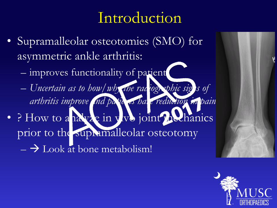
Introduction
• Supramalleolar osteotomies (SMO) for
asymmetric ankle arthritis:
– improves functionality of patients
– Uncertain as to how/why the radiographic signs of
arthritis improve and patients have reduction in pain.
• ? How to analyze in vivo joint mechanics
prior to the supramalleolar osteotomy
– Look at bone metabolism!
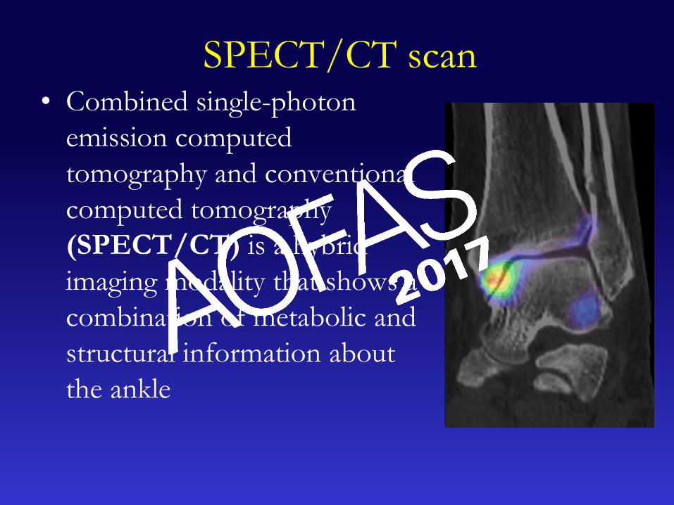
SPECT/CT scan• Combined single-photon
emission computed
tomography and conventional
computed tomography
(SPECT/CT) is a hybrid
imaging modality that shows a
combination of metabolic and
structural information about
the ankle

In the Literature• Degenerative joint
in the ankle and
midfoot
• Painful total ankle
replacements
• Malalignment
• Osteochondral
lesions
• Diabetic foot
infection
• Accessory bones
• Stress fractures
• Tarsal coalitions
• Impingement and
soft tissue
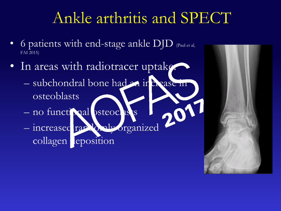
Ankle arthritis and SPECT
• 6 patients with end-stage ankle DJD (Paul et al,
FAI 2015)
• In areas with radiotracer uptake:
– subchondral bone had an increase in
osteoblasts
– no functional osteoclasts
– increased randomly organized
collagen deposition
PurposeInvestigate if the location of bone scan activation is
related to pre and post-operative functional and pain
scores.
Hypothesis• Uptake in specific locations within the ankle joint
can be associated with worse pre- and post-
operative functional and pain scores.
• SPECT/CT scan can help predict which patients
will have a successful SMO

Methods
• 80 supramalleolar osteotomies performed
(4/2006-1/2015)
• All pts were evaluated pre and post-op
clinically and radiographically
• Pre and post-operative functional and pain
scores were recorded for each patient at an
average of 3.7±2 years post-operatively
• Failure rates recorded
Methods
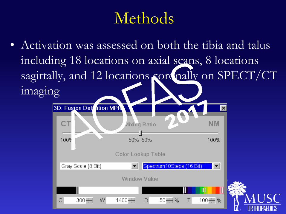
• Activation was assessed on both the tibia and talus
including 18 locations on axial scans, 8 locations
sagittally, and 12 locations coronally on SPECT/CT
imaging
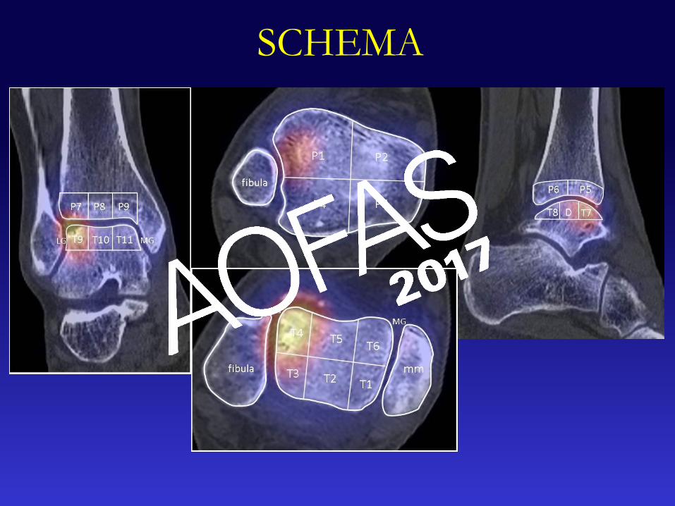
SCHEMA

Statistical analysis
• χ2 test: association between 2 categorical variables
• Two-sample, independent sample t-test: compare
mean difference between 2 groups.
• Significance level for all tests was p<0.05.
• SAS version 9.4 (Cary, NC; United States).
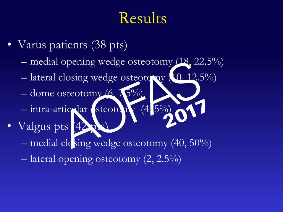
Results
• Varus patients (38 pts)
– medial opening wedge osteotomy (18, 22.5%)
– lateral closing wedge osteotomy (10, 12.5%)
– dome osteotomy (6, 7.5%)
– intra-articular osteotomy (4, 5%)
• Valgus pts (42 pts)
– medial closing wedge osteotomy (40, 50%)
– lateral opening osteotomy (2, 2.5%)

Pre-operatively
• Talonavicular (6) activation:
– worse malalignment (AOFAS Hindfoot-A subscore)
– worse a functional status (AOFAS-F)
• Subtalar joint activation (10):
– significantly worse (p<.05) pre-operative VAS pain
scores.
– worse AOFAS-F, AOFAS Hindfoot, and FAOS-S
scores
• Calcaneocuboid activation (1) did not have any
correlation to pre-operative pain or functional
scores.

Post-operatively
• Activation in these areas
were not associated with
any post-operative
functional or pain scores.
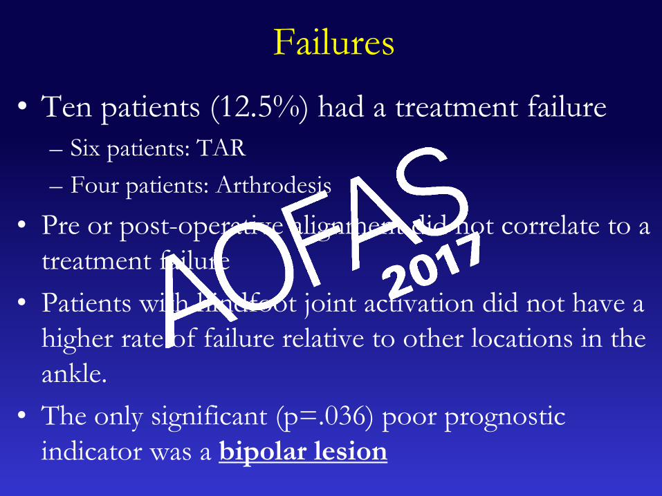
Failures
• Ten patients (12.5%) had a treatment failure
– Six patients: TAR
– Four patients: Arthrodesis
• Pre or post-operative alignment did not correlate to a
treatment failure
• Patients with hindfoot joint activation did not have a
higher rate of failure relative to other locations in the
ankle.
• The only significant (p=.036) poor prognostic
indicator was a bipolar lesion
Limitations
• Cannot quantify amount of radiotracer uptake
– Needed to rely on a 10 point SPECTRUM scale
• We are able to correlate certain pre- and post-
operative functional outcomes to areas of
radiotracer uptake
– Unclear if there are any clinical implications for this data
Conclusion
• Pre-operative SPECT/CT can be used to
clinically correlate patient-specific factors in
the pre and post-operative period
• We caution against performing a SMO in
patients with bipolar activation on a pre-
operative SPECT-CT scan

THANK YOU

• 1. Knupp M, Hintermann B. Treatment of asymmetric arthritis of the ankle joint with supramalleolar osteotomies. Foot
Ankle Int. 2012 Mar;33(3):250-2.
• 2. Takakura Y, Takaoka T, Tanaka Y, Yajima H, Tamai S. Results of opening-wedge osteotomy for the treatment of a
post-traumatic varus deformity of the ankle. J Bone Joint Surg Am. 1998 Feb;80(2):213-8.
• 3. Lee WC, Moon JS, Lee K, Byun WJ, Lee SH. Indications for supramalleolar osteotomy in patients with ankle
osteoarthritis and varus deformity. J Bone Joint Surg Am. 2011 Jul 6;93(13):1243-8.
• 4. Knupp M, Stufkens SA, van Bergen CJ, Blankevoort L, Bolliger L, van Dijk CN, et al. Effect of supramalleolar varus
and valgus deformities on the tibiotalar joint: a cadaveric study. Foot Ankle Int. 2011 Jun;32(6):609-15.
• 5. Knupp M, Pagenstert GI, Barg A, Bolliger L, Easley ME, Hintermann B. SPECT-CT compared with conventional
imaging modalities for the assessment of the varus and valgus malaligned hindfoot. J Orthop Res. 2009 Nov;27(11):1461-6.
• 6. Williams T, Cullen N, Goldberg A, Singh D. SPECT-CT imaging of obscure foot and ankle pain. Foot Ankle Surg.
2012 Mar;18(1):30-3.
• 7. Chicklore S, Gnanasegaran G, Vijayanathan S, Fogelman I. Potential role of multislice SPECT/CT in impingement
syndrome and soft-tissue pathology of the ankle and foot. Nucl Med Commun. 2013 Feb;34(2):130-9.
• 8. Singh VK, Javed S, Parthipun A, Sott AH. The diagnostic value of single photon-emission computed tomography
bone scans combined with CT (SPECT-CT) in diseases of the foot and ankle. Foot Ankle Surg. 2013 Jun;19(2):80-3.
• 9. Ha S, Hong SH, Paeng JC, Lee DY, Cheon GJ, Arya A, et al. Comparison of SPECT/CT and MRI in diagnosing
symptomatic lesions in ankle and foot pain patients: diagnostic performance and relation to lesion type. PLoS One.
2015;10(2):e0117583.
• 10. Claassen L, Uden T, Ettinger M, Daniilidis K, Stukenborg-Colsman C, Plaass C. Influence on therapeutic decision
making of SPECT-CT for different regions of the foot and ankle. Biomed Res Int. 2014;2014:927576.
• 11. Fornaciari P, Gilgen A, Zwicky L, Horn Lang T, Hintermann B. Isolated talonavicular fusion with tension band for
Muller-Weiss syndrome. Foot Ankle Int. 2014 Dec;35(12):1316-22.
• 12. Parthipun A, Moser J, Mok W, Paramithas A, Hamilton P, Sott AH. 99mTc-HDP SPECT-CT Aids Localization of
Joint Injections in Degenerative Joint Disease of the Foot and Ankle. Foot Ankle Int. 2015 Aug;36(8):928-35.
• 13. Paul J, Barg A, Kretzschmar M, Pagenstert G, Studler U, Hugle T, et al. Increased Osseous (99m)Tc-DPD Uptake
in End-Stage Ankle Osteoarthritis: Correlation Between SPECT-CT Imaging and Histologic Findings. Foot Ankle Int. 2015
Dec;36(12):1438-47.
• 14. Mason LW, Wyatt J, Butcher C, Wieshmann H, Molloy AP. Single-photon-emission computed tomography in painful
total ankle replacements. Foot Ankle Int. 2015 Jun;36(6):635-40.
• 15. Meftah M, Katchis SD, Scharf SC, Mintz DN, Klein DA, Weiner LS. SPECT/CT in the management of osteochondral
lesions of the talus. Foot Ankle Int. 2011 Mar;32(3):233-8.
• 16. Heiba SI, Kolker D, Mocherla B, Kapoor K, Jiang M, Son H, et al. The optimized evaluation of diabetic foot infection
by dual isotope SPECT/CT imaging protocol. J Foot Ankle Surg. 2010 Nov-Dec;49(6):529-36.