longitudinal and integrative biomodeling of effector and ... filemalka tindel,‖ martine louet,#...
TRANSCRIPT

of September 6, 2018.This information is current as
VaccinationCompartments after Inactivated InfluenzaEffector and Memory Immune Longitudinal and Integrative Biomodeling of
Werf, Stephane Jauréguiberry and Behazine CombadiereBricaire, Assia Samri, Dominique Rousset, Sylvie van der Loulergue, Gwenaelle Badre, Christine Katlama, FrançoisTindel, Martine Louet, Florent Desert, Odile Launay, Pierre Jordan Dimitrov, Srini V. Kaveri, Angélique Curjol, MalkaBenhabiles, Fabrice Carrat, Anne Krivine, Flore Rozenberg, Olivia Bonduelle, Nora Yahia, Sophie Siberil, Nora
ol.1203483http://www.jimmunol.org/content/early/2013/06/17/jimmun
published online 17 June 2013J Immunol
MaterialSupplementary
3.DC1http://www.jimmunol.org/content/suppl/2013/06/17/jimmunol.120348
average*
4 weeks from acceptance to publicationFast Publication! •
Every submission reviewed by practicing scientistsNo Triage! •
from submission to initial decisionRapid Reviews! 30 days* •
Submit online. ?The JIWhy
Subscriptionhttp://jimmunol.org/subscription
is online at: The Journal of ImmunologyInformation about subscribing to
Permissionshttp://www.aai.org/About/Publications/JI/copyright.htmlSubmit copyright permission requests at:
Email Alertshttp://jimmunol.org/alertsReceive free email-alerts when new articles cite this article. Sign up at:
Print ISSN: 0022-1767 Online ISSN: 1550-6606. Immunologists, Inc. All rights reserved.Copyright © 2013 by The American Association of1451 Rockville Pike, Suite 650, Rockville, MD 20852The American Association of Immunologists, Inc.,
is published twice each month byThe Journal of Immunology
by guest on September 6, 2018
http://ww
w.jim
munol.org/
Dow
nloaded from
by guest on September 6, 2018
http://ww
w.jim
munol.org/
Dow
nloaded from

The Journal of Immunology
Longitudinal and Integrative Biomodeling of Effector andMemory Immune Compartments after Inactivated InfluenzaVaccination
Olivia Bonduelle,*,1 Nora Yahia,*,1 Sophie Siberil,*,1 Nora Benhabiles,† Fabrice Carrat,‡
Anne Krivine,x Flore Rozenberg,x Jordan Dimitrov,{ Srini V. Kaveri,{ Angelique Curjol,‖
Malka Tindel,‖ Martine Louet,# Florent Desert,# Odile Launay,** Pierre Loulergue,**
Gwenaelle Badre,** Christine Katlama,‖ Francois Bricaire,‖ Assia Samri,*
Dominique Rousset,†† Sylvie van der Werf,†† Stephane Jaureguiberry,‖ and
Behazine Combadiere*,‡‡
Most vaccines, including those against influenza, were developed by focusing solely on humoral response for protection. However, vac-
cination activates different adaptive compartments that might play a role in protection. We took advantage of the pandemic 2009 A
(H1N1) influenza vaccination to conduct a longitudinal integrative multiparametric analysis of seven immune parameters in vaccinated
subjects. A global analysis underlined the predominance of induction of humoral and CD4 T cell responses, whereas pandemic 2009 A
(H1N1)–specific CD8 responses did not improve after vaccination. A principal component analysis and hierarchical clustering of
individuals showed a differential upregulation of influenza vaccine–specific immunity including hemagglutination inhibition titers,
IgA+ and IgG+ Ab-secreting cells, effector CD4 or CD8 T cell frequencies at day 21 among individuals, suggesting a fine-tuning of the
immune parameters after vaccination. This is related to individual factors including the magnitude and quality of influenza-specific
immune responses before vaccination. We propose a graphical delineation of immune determinants that would be essential for a better
understanding of vaccine-induced immunity in vaccination strategies. The Journal of Immunology, 2013, 191: 000–000.
The principal reference criterion for evaluating the efficacyof influenza vaccination in clinical trials is the magnitudeof the Ag-specific Ab titer. Ever since Hobson et al. (1)
determined the threshold of hemagglutination inhibition (HI) Abtiters for protection after influenza infection in 1972, these titershave been regarded as the cornerstone of an anti-influenza im-mune response. However, both the quality and the quantity of hu-
moral responses such as avidity, mucosal responses, or frequenciesof Ab-secreting B cells (ASCs) (2, 3) are important immunolog-ical parameters of protection (4–6). Khurana et al. (7) observedqualitatively superior humoral responses in elderly individuals withpre-existing immunity after pandemic 2009 A(H1N1) (A[H1N1]pdm09) vaccination. Although humoral responses have been de-fined as the sole indicators of the correlates of protection in sea-sonal influenza vaccination (1, 8), renewed attention is being paid
to T cell responses because of their role in decreasing diseaseseverity and their capacity for long-term maintenance after im-
munization (9–11). In the context of pandemics, where no or few
pre-existing Abs can rapidly control the infection, T cells might
mediate protection or limit the severity of the influenza-associated
illness in humans (12–14) and murine models (15, 16). T cells re-
main highly cross-reactive among influenza strains and also rec-
ognize constant epitopes of influenza proteins (17, 18).Assessing the potency and immunogenicity of vaccines would be
essential in designing novel vaccination strategies. Global de-
scription of immunogenicity of vaccines is a laborious and cost-
effective task that is partly due to the multiplicity of phenotypes
and functions that can be studied for each immune compartment.
We took advantage of the influenza A(H1N1)pdm09 vaccination
to perform a multiparametric analysis of effector and memory
*INSERM Unite Mixte de Recherche S-945, Laboratory of Immunity and Infection,University of Pierre and Marie Curie (Paris 06), 75013 Paris, France; †DepartmentSensors Signal and Information, Centre d’Energie Atomique Saclay, CEA, InstitutCarnot Laboratoire d’Integration des Systemes et des Technologies, 91191 Gif-sur-Yvette, France; ‡INSERM UMRS-707, University of Pierre and Marie Curie, Unitede Sante Publique, Hopital Saint-Antoine, Assistance Publique/Hopitaux de Paris,75012 Paris, France; xVirology Laboratory, Hopital Cochin-Saint Vincent de Paul,Assistance Publique/Hopitaux de Paris, 75014 Paris, France; {INSERM U872, Uni-versite Paris Descartes (Paris 05), Centre de Recherche des Cordeliers, 75006 Paris,France; ‖Infectious and Tropical Diseases Unit, Hopital Pitie-Salpetriere, Assis-tance Publique/Hopitaux de Paris, 75013 Paris, France; #Occupationnal MedecineUnit, Hopital Pitie-Salpetriere, Assistance Publique/Hopitaux de Paris, 75013 Paris,France; **INSERM Centre d’Investigation Clinique-BT505, Universite Paris Des-cartes (Paris 05), Hopital Cochin, Assistance Publique/Hopitaux de Paris, 75014Paris, France; ††Institut Pasteur, Unite de Genetique Moleculaire des Virus a ARN,Departement de Virologie and Centre National de Recherche Scientifique Unite deRecherche Associee 3015, Universite Paris Diderot, Sorbonne Paris Cite, 75015Paris, France; and ‡‡Hopital Pitie-Salpetriere, Assistance Publique/Hopitaux de Paris,75013 Paris, France
1O.B., N.Y., and S.S. contributed equally to this work.
Received for publication December 20, 2012. Accepted for publication May 13,2013.
This work was supported by a grant from the Programme de Recherche, A(H1N1)coordinated by the Institut de Microbiologie et Maladies Infectieuses (France). B.C.has been awarded an INSERM-Assistance Publique/Hopitaux de Paris Interface Pro-gram contract and is supported by the Fondation pour la Recherche Medicale.
Address correspondence and reprint requests to Dr. Behazine Combadiere, INSERMU945, 91 Boulevard de l’Hopital, 75013 Paris, France. E-mail address: [email protected]
The online version of this article contains supplemental material.
Abbreviations used in this article: A(H1N1)pdm09, pandemic 2009 A(H1N1); ASC,Ab-secreting B cell; D0, day 0; GMT, geometric mean titer; HA, hemagglutinin; HI,hemagglutination inhibition; M4, month 4; MN, microneutralization; PCA, principalcomponent analysis; SP, single-positive.
Copyright� 2013 by TheAmerican Association of Immunologists, Inc. 0022-1767/13/$16.00
www.jimmunol.org/cgi/doi/10.4049/jimmunol.1203483
Published June 17, 2013, doi:10.4049/jimmunol.1203483 by guest on Septem
ber 6, 2018http://w
ww
.jimm
unol.org/D
ownloaded from
immune responses to vaccine, and to define immune behaviorand longitudinal equilibrium in a target population. In this study,we explored the plexus of immune parameters that could be usedas hallmarks of vaccine efficacy and the relation between pre-existing immunity and immune response to influenza upon vac-cination. We propose a novel model of evaluation of vaccineimmunogenicity that takes into account heterogeneity of individualimmune responses to influenza vaccination.
Materials and MethodsStudy design
One dose of an adjuvanted A(H1N1)pdm09 influenza vaccine (Pandemrix;GlaxoSmithKline, Marly-le-Roi, France) was given i.m. to 147 hospitalhealth care staff members enrolled in a Phase IV clinical trial from October21, 2009, through December 16, 2009, in two university hospitals locatedin Paris (France); 10 subjects discontinued their participation in the study.Eligibility criteria were the following: age $ 18 y, clinical examinationand interview for medical history, documented history of influenza vac-cinations, and written informed consent. Further exclusion criteria wereany acute or chronic illness, local or systemic immunosuppressive treat-ments, and pregnancy that might interfere with the study protocol. Sup-plemental Table I summarizes the subjects’ demographic characteristics.Information on the total number of previous influenza vaccinations wasrecorded for 127 individuals with an average of 2.59 6 3.00 seasonalinfluenza vaccinations during their life (A[H1N1]pdm09 vaccination(Supplemental Table II). Blood samples at day 0 (D0), day 21 (D21), andmonth 4 (M4) after A(H1N1)pdm09 vaccination were collected for im-munological analyses. PBLs were isolated on Ficoll gradients (Eurobio,Courtaboeuf, France). Sera and PBL were frozen, and samples (D0, D21,and M4) were treated simultaneously for each subject.
Ethics committee approval and health authorities
The trial was conducted in accordance with the latest version of theDeclaration of Helsinki, Good Clinical Practice, and International Con-ference on Harmonisation regulatory guidelines. The study protocol andpatient information forms were approved by the Ethics Committee of Pitie-Salpetriere Hospital. Written informed consent was obtained from eachvolunteer before study entry. This trial is registered with ClinicalTrials.gov: NCT01063608.
Hemagglutination inhibition assay
Serum Abs against nonadjuvanted A(H1N1)pdm09 influenza vaccine(Panenza; Sanofi Pasteur) were measured by a microtiter HI assay modifiedfrom Kendal et al. (19). In brief, after treatment by receptor-destroyingenzyme, serial 2-fold dilutions of serum (from 1:10) were tested against 4hemagglutinin (HA) units of Ag, on human O Rh-RBCs. The HI titerswere defined as the reciprocal of the highest serum dilution that completelyinhibited hemagglutination.
Microneutralization assay
Neutralizing Abs titers were measured with standard techniques by mi-croneutralization (MN) assays. Sera were first heat-inactivated at 56˚Cfor 30 min. Serial 2-fold dilutions of serum (from 1:10) were added with103 TCID50 A/California/07/2009 (H1N1) influenza virus and incubated at37˚C for 2 h before being transferred onto 96-well microtiter plates con-taining confluent MDCK cells. The neutralization titer is expressed as thereciprocal of the highest serum dilution at which virus infection is blockedafter 3 d of culture.
Serum avidity assay
Serum avidity of anti-HA Abs was evaluated by ELISA (20). Sera fromdonors were incubated with recombinant HA from A/California/07/2009(H1N1) influenza virus (Protein Sciences) coated on ELISA plates ata dilution equivalent to 2 mg/ml HA, in the presence of serial dilutions ofsodium thiocyanate (Sigma). HRP-conjugated mouse anti-human IgG(Southern Biotech) was incubated before revelation with o-phenylenedi-amine peroxidase substrate buffer. Reaction was stopped with 2N HCl, andOD was red with a GENios lector using the Xfluor4 software at 492 nm.Serum avidity was defined as the concentration of sodium thiocyanaterequired to induce a 50% inhibition of Ab binding.
ASC detection
Differentiation of memory B cells into ASCs was induced after 6 d of PBLculture in complete medium (RPMI 1640 supplemented with 10% heat-
inactivated FCS [PAA], L-glutamine, and antibiotics [Life TechnologiesBRL, Life Technology]) supplemented with 55 mM 2-ME (Sigma-Aldrich)and a mix of PWM, protein A from Staphylococcus aureus (Sigma-Aldrich), and CpG oligodesoxyribonucleotides (InvivoGen). This methodwas previously described by Crotty et al. (21). ELISPOT plates werecoated with Pandemrix vaccine (without adjuvant) at a dilution equivalentto 2 mg/ml HA or PBS (background). IgA+ or IgG+ ASCs were detectedwith alkaline phosphatase–conjugated goat anti-human IgA or IgG Abs(Sigma-Aldrich), were revealed with 5-bromo-4-chloro-3-indolyl phos-phate/nitro blue tetrazolium substrate (Sigma-Aldrich), and were countedwith an automated microscope (Zeiss, Le Pecq, France). ELISPOT read-outs were expressed as the number of A(H1N1)pdm09-specific IgA or IgGASCs/106 PBLs.
Intracellular cytokine assay
Frozen PBLs were available for further analysis of A(H1N1)pdm09-specific T cells. Cells were stimulated for 16 h at 37˚C with or without(background) Pandemrix vaccine (without adjuvant) at a dilution equiva-lent to 60 ng/ml HA. Brefeldin A and monensin (Sigma-Aldrich) in thepresence of CD107a-PE-Cy5 mAbs (BD Biosciences) were added duringthe last 14 h. Cells were washed and stained in PBS-2% FCS at 4˚C withCD3-AmCyan, CD4-Pacific Blue, CD8-allophycocyanin-H7, CD27-PE(BD), and CD45RA-ECD (Beckman Coulter) mAbs. Cytofix/Cytopermkit (BD) was used to permeabilize cells, according to the manufacturer’sinstructions, before staining with IL-2–FITC, IFN-g–Alexa Fluor 700 andTNF-a–PE–Cy7 (BD) mAbs. Flow cytometry was performed with anLSRII flow cytometer (BD). At least 1,000,000 live events were accu-mulated and analyzed for Boolean combination gating with the FlowJosoftware (Tree Star).
Statistical analyses
In univariate analyses, we used Wilcoxon matched-pairs tests for kineticimmune responses and Mann–Whitney tests for continuous variables. ABonferroni correction was applied to compare groups. Statistical signifi-cance was set at p , 0.05. All statistical analyses were performed withSPSS statistical software 17.0 (SPSS, Chicago, IL) and SAS 9.2 (SASInstitute, Cary, NC), and Prism 5.0 or Microsoft Excel for Mac OS X fordata handling and graphic representation.
Radar chart, principal component analysis
The radar charts were designed with R, a free software environment forstatistical computing and graphics (http://www.r-project.org/). The analysisby principal component analysis (PCA) and hierarchical clustering is basedon the fold increase of immune responses between D0 and D21. Data on all7 parameters were available for 79 subjects. Log10 fold increases werenormalized using MeV 4.7.4 software. To stratify the population and vi-sualize the clusters, we used a part of the TM4 software suite, the MultiExperiment Viewer, MeV (22). The best partition for the initial populationwas obtained for five clusters (inflection point of the Figure of Merit) (23).To cluster the data, for example, to stratify the initial population, we rana K-Mean Clustering for five clusters and performed Hierarchical Clus-tering on the elements in each cluster created (24). The hierarchicalclustering was done using complete linkage and Pearson correlation. Eachcluster was then colored separately. A PCA was run and used to attributethe overall variability in the data to a reduced set of variables, for example,the principal components. We used the three first principal components tomap each element into a three-dimensional viewer.
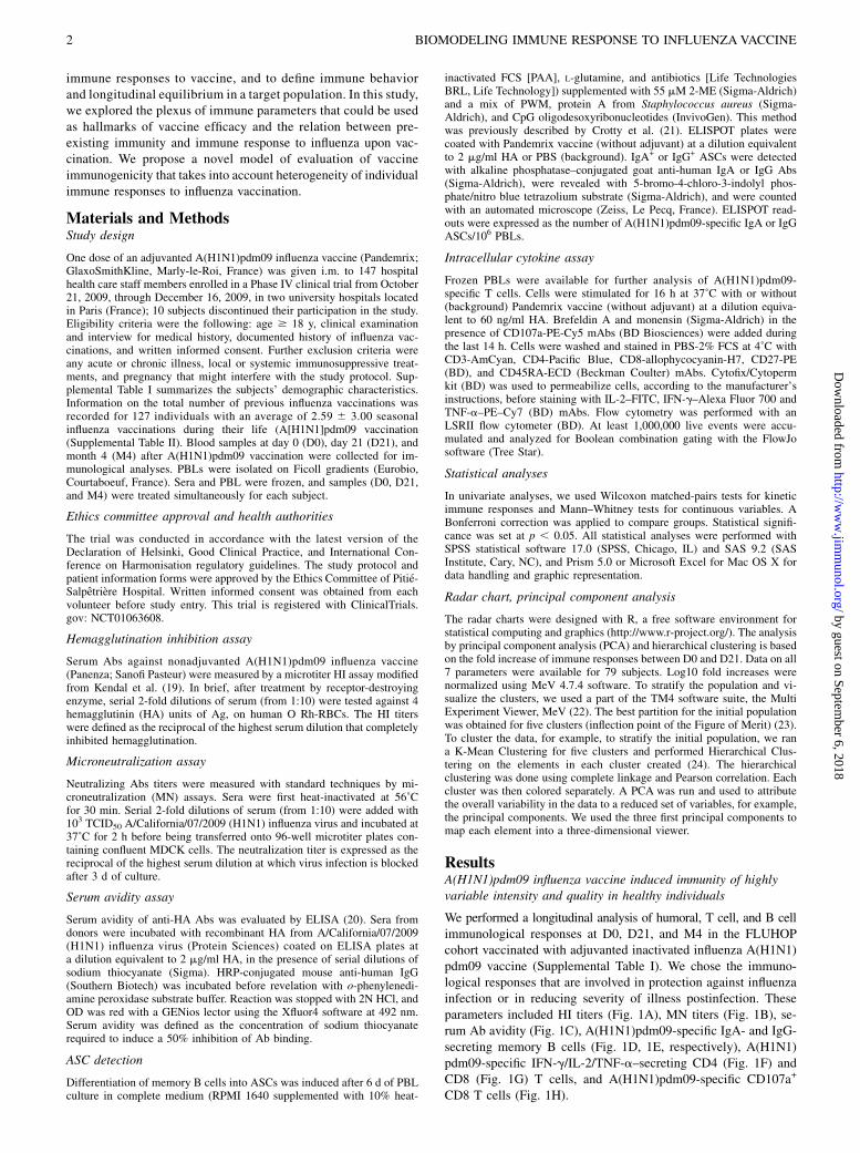
ResultsA(H1N1)pdm09 influenza vaccine induced immunity of highlyvariable intensity and quality in healthy individuals
We performed a longitudinal analysis of humoral, T cell, and B cellimmunological responses at D0, D21, and M4 in the FLUHOPcohort vaccinated with adjuvanted inactivated influenza A(H1N1)pdm09 vaccine (Supplemental Table I). We chose the immuno-logical responses that are involved in protection against influenzainfection or in reducing severity of illness postinfection. Theseparameters included HI titers (Fig. 1A), MN titers (Fig. 1B), se-rum Ab avidity (Fig. 1C), A(H1N1)pdm09-specific IgA- and IgG-secreting memory B cells (Fig. 1D, 1E, respectively), A(H1N1)pdm09-specific IFN-g/IL-2/TNF-a–secreting CD4 (Fig. 1F) andCD8 (Fig. 1G) T cells, and A(H1N1)pdm09-specific CD107a+
CD8 T cells (Fig. 1H).
2 BIOMODELING IMMUNE RESPONSE TO INFLUENZA VACCINE
by guest on September 6, 2018
http://ww
w.jim
munol.org/
Dow
nloaded from
As expected, HI titers increased significantly between D0(geometric mean titer [GMT] = 11.45, 17.52% HI titers $ 40)and D21 (GMT = 133.5, 94.81% HI titers $ 40) and then de-creased at M4 (GMT = 72.44, 81.75% HI titers $ 40; p , 0.0001compared with D21; Fig. 1A, Supplemental Table II). Trends inGMTs were similar when seroprotection was defined as an HItiter $80 postvaccination (Supplemental Table II). In addition,Fig. 1B and 1C show that the neutralization activity and serumavidity of A(H1N1)pdm09-specific Abs also increased at D21compared with D0 (p , 0.0001) and decreased at M4 (comparedwith D21, p , 0.0001). These results, which are consistent withthe literature (25–28), present the conventional way to measurevaccination immunogenicity.
In view of the prime role of B cells in the generation of Abs,we measured A(H1N1)pdm09-specific IgA–Ab-secreting memoryB cells (Fig. 1D) and IgG-ASC (Fig. 1E). We found that bothtypes of ASCs were significantly amplified at D21 (IgA-ASC andIgG-ASC: p , 0.0001) and decreased at M4 (compared withD21, IgA-ASC: p = 0.0046 and IgG-ASC: p = 0.0018; Fig. 1D, 1E).One hallmark of the efficacy of T cell responses against viral
infection is the production of multiple cytokines by CD4 andCD8 cells, most specifically IFN-g. Accordingly, we analyzedthe longitudinal frequency of single-positive (SP; IFN-g+IL-22
TNF-a2, or IFNg2IL-2+TNF-a2 or IFN-g2IL-22TNF-a+),double-positive (IFN-g+IL-2+TNF-a2, or IFNg2IL-2+TNF-a+ orIFN-g+IL-22TNF-a+), and triple-positive (IFN-g+IL-2+TNF-a+)
FIGURE 1. A(H1N1)pdm09 influenza vaccine elicited heterogeneous humoral and cellular immune responses. Immune responses were evaluated in
blood before (D0) and at D21 and M4 after Pandemrix vaccination. Box and whisker plots with 10th and 90th percentiles are depicted for each parameter
with log10 scale of intensity of immune responses. (A–C) Humoral response was assessed by HI assay (n = 137) (A), MN assay (n = 51) (B), and serum
avidity assay (n = 46) (C). (D and E) A(H1N1)pdm09-specific memory B cell responses were measured by ELISPOT assays: A(H1N1)pdm09-specific IgA
ASCs (D) and IgG ASCs (E) (n = 79). (F–H) A(H1N1)pdm09-specific T cell responses were measured by intracellular cytokine staining (Boolean gating of
IFN-g, IL-2, and/or TNF-a) in CD4 T cells (F), CD8 T cells (G), or by degranulation marker expression (CD107a) (H) (n = 100). (I) Radar chart presents the
minimum and maximum values for each assay as indicated in log10 scale. The mean influenza-specific immune responses are presented at D0 (black), D21
(red), and M4 (blue). Statistical analyses were performed with the Wilcoxon matched pairs test. Statistical significance is indicated as: *p , 0.05, **p ,0.01, ***p , 0.001, ****p , 0.0001. ns, not significant.
The Journal of Immunology 3
by guest on September 6, 2018
http://ww
w.jim
munol.org/
Dow
nloaded from
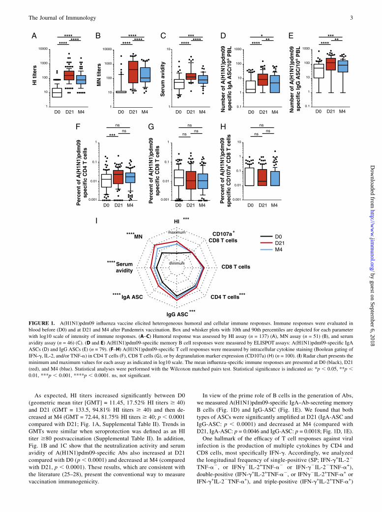
cytokine-secreting T cells. In addition, the expression of CD107a,a molecule defining degranulation capacity, was evaluated forCD8 T cell response. Fig. 1F and 1G showed the percentages of A(H1N1)pdm09-specific total cytokine (IFN-g, IL-2, and/or TNF-a)-secreting CD4 and CD8 T cells, respectively (after subtractionof background nonstimulated cells). A(H1N1)pdm09-specificCD4 T cells increase significantly between D0 and D21 (p 50.0005) and do not change between D21 and M4 (Fig. 1F). In-terestingly, the A(H1N1)pdm09-specific CD8 responses (eithercytokine+ or CD107a+ cells) did not change significantly (Fig. 1G,1H).The polyfunctionality of T cells was represented in pie-chart
analyses (Fig. 2). A significant difference was observed for theamplification of IFN-g+IL-22TNF-a2–producing CD4 T cellsbetween D0 and D21 (p = 0.0004) and its subsequent decreaseat M4 (compared with D21, p = 0.0001; Fig. 2A). The CD8 cy-tokine profile, however, did not change significantly over time(Fig. 2B). Furthermore, the increase in A(H1N1)pdm09-specificCD4 T cells observed from D0 to D21 was correlated with in-creased HI titers (p = 0.0048), attesting of potential helper func-tion of CD4 T cells.A radar chart summarizing the view of overall adaptive im-
munity showed that influenza vaccination shaped immune re-sponses toward major humoral responses, including amplificationof effector CD4 T cells (Fig. 1I). However, we noted the absenceof A(H1N1)pdm09-specific CD8 T cell amplification in the pop-ulation and the extreme heterogeneity of humoral and cellularimmune responses both at baseline and postvaccination.
Heterogeneity of magnitude and quality of influenza-specificimmune compartments
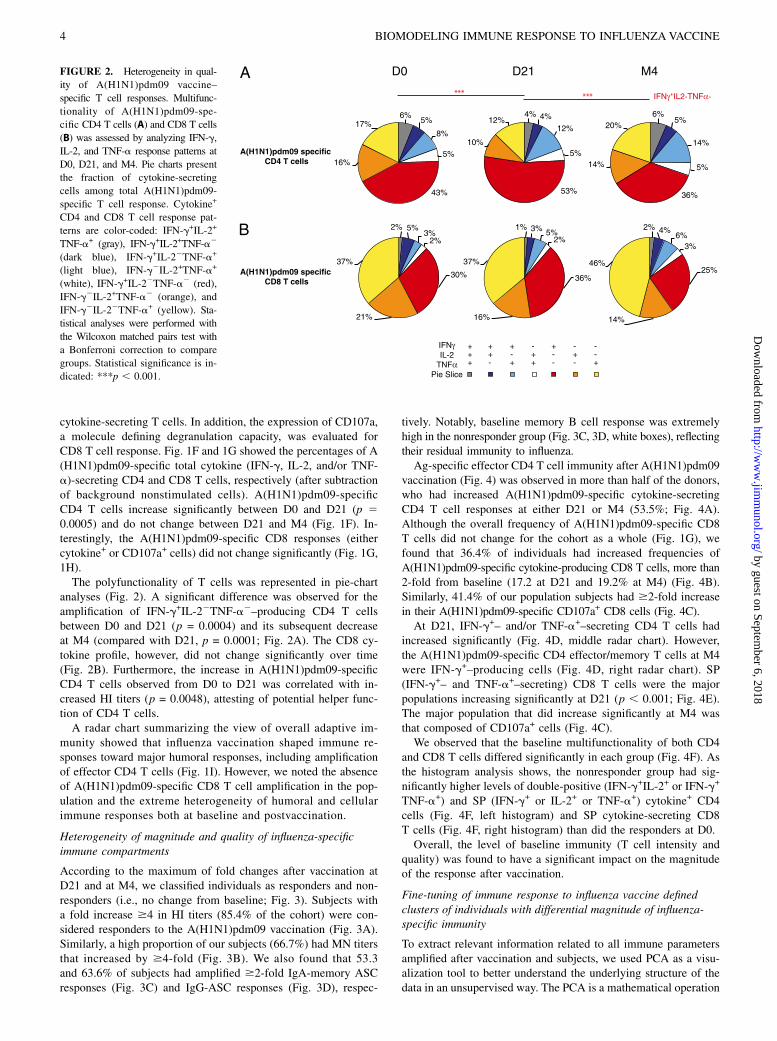
According to the maximum of fold changes after vaccination atD21 and at M4, we classified individuals as responders and non-responders (i.e., no change from baseline; Fig. 3). Subjects witha fold increase $4 in HI titers (85.4% of the cohort) were con-sidered responders to the A(H1N1)pdm09 vaccination (Fig. 3A).Similarly, a high proportion of our subjects (66.7%) had MN titersthat increased by $4-fold (Fig. 3B). We also found that 53.3and 63.6% of subjects had amplified $2-fold IgA-memory ASCresponses (Fig. 3C) and IgG-ASC responses (Fig. 3D), respec-
tively. Notably, baseline memory B cell response was extremelyhigh in the nonresponder group (Fig. 3C, 3D, white boxes), reflectingtheir residual immunity to influenza.Ag-specific effector CD4 T cell immunity after A(H1N1)pdm09
vaccination (Fig. 4) was observed in more than half of the donors,who had increased A(H1N1)pdm09-specific cytokine-secretingCD4 T cell responses at either D21 or M4 (53.5%; Fig. 4A).Although the overall frequency of A(H1N1)pdm09-specific CD8T cells did not change for the cohort as a whole (Fig. 1G), wefound that 36.4% of individuals had increased frequencies ofA(H1N1)pdm09-specific cytokine-producing CD8 T cells, more than2-fold from baseline (17.2 at D21 and 19.2% at M4) (Fig. 4B).Similarly, 41.4% of our population subjects had $2-fold increasein their A(H1N1)pdm09-specific CD107a+ CD8 cells (Fig. 4C).At D21, IFN-g+– and/or TNF-a+–secreting CD4 T cells had
increased significantly (Fig. 4D, middle radar chart). However,the A(H1N1)pdm09-specific CD4 effector/memory T cells at M4were IFN-g+–producing cells (Fig. 4D, right radar chart). SP(IFN-g+– and TNF-a+–secreting) CD8 T cells were the majorpopulations increasing significantly at D21 (p , 0.001; Fig. 4E).The major population that did increase significantly at M4 wasthat composed of CD107a+ cells (Fig. 4C).We observed that the baseline multifunctionality of both CD4
and CD8 T cells differed significantly in each group (Fig. 4F). Asthe histogram analysis shows, the nonresponder group had sig-nificantly higher levels of double-positive (IFN-g+IL-2+ or IFN-g+
TNF-a+) and SP (IFN-g+ or IL-2+ or TNF-a+) cytokine+ CD4cells (Fig. 4F, left histogram) and SP cytokine-secreting CD8T cells (Fig. 4F, right histogram) than did the responders at D0.Overall, the level of baseline immunity (T cell intensity and
quality) was found to have a significant impact on the magnitudeof the response after vaccination.
Fine-tuning of immune response to influenza vaccine definedclusters of individuals with differential magnitude of influenza-specific immunity
To extract relevant information related to all immune parametersamplified after vaccination and subjects, we used PCA as a visu-alization tool to better understand the underlying structure of thedata in an unsupervised way. The PCA is a mathematical operation
FIGURE 2. Heterogeneity in qual-
ity of A(H1N1)pdm09 vaccine–
specific T cell responses. Multifunc-
tionality of A(H1N1)pdm09-spe-
cific CD4 T cells (A) and CD8 T cells
(B) was assessed by analyzing IFN-g,
IL-2, and TNF-a response patterns at
D0, D21, and M4. Pie charts present
the fraction of cytokine-secreting
cells among total A(H1N1)pdm09-
specific T cell response. Cytokine+
CD4 and CD8 T cell response pat-
terns are color-coded: IFN-g+IL-2+
TNF-a+ (gray), IFN-g+IL-2+TNF-a2
(dark blue), IFN-g+IL-22TNF-a+
(light blue), IFN-g2IL-2+TNF-a+
(white), IFN-g+IL-22TNF-a2 (red),
IFN-g2IL-2+TNF-a2 (orange), and
IFN-g2IL-22TNF-a+ (yellow). Sta-
tistical analyses were performed with
the Wilcoxon matched pairs test with
a Bonferroni correction to compare
groups. Statistical significance is in-
dicated: ***p , 0.001.
4 BIOMODELING IMMUNE RESPONSE TO INFLUENZA VACCINE
by guest on September 6, 2018
http://ww
w.jim
munol.org/
Dow
nloaded from
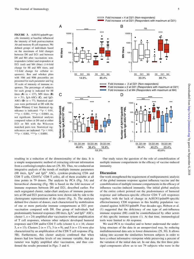
resulting in a reduction of the dimensionality of the data. It isa simple nonparametric method of extracting relevant informationfrom a confusing/complex data set (29, 30). Thus, we conducted anintegrative analysis of the trends of multiple immune parameters(HI titers, IgA+ and IgG+ ASCs, cytokine-producing CD4 andCD8 T cells, CD107a+ CD8 T cells), all of them available at alltime points in 79 donors. The analysis by PCA (Fig. 5A) andhierarchical clustering (Fig. 5B) is based on the fold increase ofimmune responses between D0 and D21, described earlier. Foreach segregated cluster, radar-chart analyses of immune parame-ters at D0 and D21 postvaccination were shown side by side to theclustergrams representation of the cluster (Fig. 5). The analysesdefined five clusters of donors, each characterized by mobilizationof one or more particular immune compartments at D21 post-vaccination compared with D0. One group of individuals hadpredominantly humoral responses (HI titers, IgA+ and IgG+ ASCs;cluster 1, n = 24) amplified after vaccination without amplificationof T cell responses, whereas other subjects developed humoralresponses and CD8 and/or CD4 T cells (cluster 4, n = 16; cluster5, n = 13). Clusters 2 (n = 17), 3 (n = 9), and 5 (n = 13) were alsocharacterized by an amplification of the CD8 T cell response (Fig.5B). Furthermore, this cluster analysis confirmed that whendonors had low baseline levels of one immune variable, that pa-rameter was highly amplified after vaccination, and thus con-firmed the results presented in Figs. 3 and 4.
Our study raises the question of the role of comobilization ofmultiple immune compartments in the efficacy of vaccine-inducedimmunity.
DiscussionOur work strengthened the requirement of multiparametric analysisof the global immune response against influenza vaccine and thecomobilization of multiple immune compartments in the efficacy ofinfluenza vaccine–induced immunity. Our initial global analysisof the entire cohort pointed out the predominance of humoralresponse and influenza-specific effector CD4 T cell responsestogether, with the lack of change in A(H1N1)pdm09-specificeffector/memory CD8 responses in this healthy population vac-cinated against A(H1N1)pdm09. Four decades ago, Hobson et al.(1) suggested that the deficiency of one type of anti-influenzaimmune response (HI) could be counterbalanced by other actorsof this specific immune system (1). At that time, immunologicaltools were limited to Ab response.We used PCA to visualize and to better understand the under-
lying structure of the data in an unsupervised way, by reducingmultidimensional data sets to lower dimensions (29, 30). It allowstaking into account the similarities between subjects in order tohave a robust informative viewpoint while preserving a percent ofthe variation of the initial data set. In our data, the first three prin-cipal components allow us to see 79 subjects who were in the
FIGURE 3. A(H1N1)pdm09-spe-
cific immunity at baseline influenced
the intensity of both postvaccination
Ab and memory B cell responses. We
defined groups of individuals based
on the maximum of fold changes
between D0 and D21 and between
D0 and M4 after vaccination: non-
responders (white) and responders at
D21 (red) and M4 (blue) ($4-fold
change for HI and MN titers, and
$2-fold change for cellular re-
sponses). Box and whisker plots
with 10th and 90th percentiles are
presented for each parameter and log
10 scale of intensity of immune re-
sponses. The percentage of subjects
for each group is indicated for HI
titers (A) (n = 137), MN titers (B)
(n = 51), IgA-ASCs (C), and IgG-
ASCs (D) (n = 77). Statistical anal-
yses were performed at D0 with the
Mann–Whitney U test. Statistical sig-
nificance is indicated: **p , 0.01,
***p , 0.001, ****p , 0.0001. ns,
not significant. Statistical analyses
compared values at D0 and at either
D21 or M4 with the Wilcoxon
matched pairs test. Statistical sig-
nificances are indicated: **p , 0.01,
***p , 0.001, ****p , 0.0001.
The Journal of Immunology 5
by guest on September 6, 2018
http://ww
w.jim
munol.org/
Dow
nloaded from
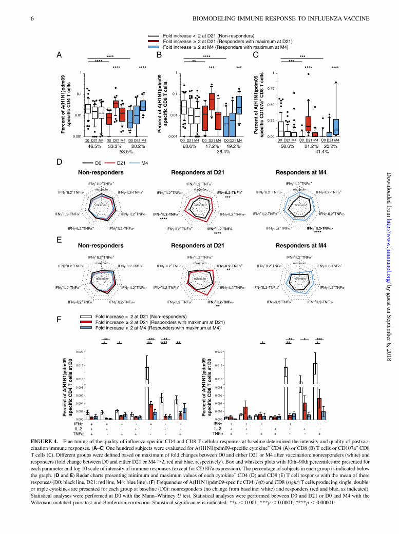
FIGURE 4. Fine-tuning of the quality of influenza-specific CD4 and CD8 T cellular responses at baseline determined the intensity and quality of postvac-
cination immune responses. (A–C) One hundred subjects were evaluated for A(H1N1)pdm09-specific cytokine+ CD4 (A) or CD8 (B) T cells or CD107a+ CD8
T cells (C). Different groups were defined based on maximum of fold changes between D0 and either D21 or M4 after vaccination: nonresponders (white) and
responders (fold change between D0 and either D21 or M4$2, red and blue, respectively). Box and whiskers plots with 10th–90th percentiles are presented for
each parameter and log 10 scale of intensity of immune responses (except for CD107a expression). The percentage of subjects in each group is indicated below
the graph. (D and E) Radar charts presenting minimum and maximum values of each cytokine+ CD4 (D) and CD8 (E) T cell response with the mean of these
responses (D0: black line, D21: red line, M4: blue line). (F) Frequencies of A(H1N1)pdm09-specific CD4 (left) and CD8 (right) T cells producing single, double,
or triple cytokines are presented for each group at baseline (D0): nonresponders (no change from baseline; white) and responders (red and blue, as indicated).
Statistical analyses were performed at D0 with the Mann–Whitney U test. Statistical analyses were performed between D0 and D21 or D0 and M4 with the
Wilcoxon matched pairs test and Bonferroni correction. Statistical significance is indicated: **p , 0.001, ***p , 0.0001, ****p , 0.00001.
6 BIOMODELING IMMUNE RESPONSE TO INFLUENZA VACCINE
by guest on September 6, 2018
http://ww
w.jim
munol.org/
Dow
nloaded from

original space in 6 dimensions (6-fold increased variables). Thisreduction takes into account 80.5% of the variation of the initialinformation, thus allowing for conserving most information. Thismathematical procedure can be seen as a simple visual way toreveal the internal “hidden” structure of the data.Using PCA, we demonstrated that anti-influenza immunity is the
result of a balance between the different immune compartments foreach cluster of individuals. Our longitudinal integrative study ofmultiple immune parameters before and after A(H1N1)pdm09vaccination (HI titers, IgA+ and IgG+ ASCs, cytokine+ CD4 andCD8 T cells, CD107a+ CD8 T cells) has allowed us to definedifferent profiles of immune responses represented by five clustersof subjects. Each cluster was characterized by an important foldchange for one or more particular immune components.Predominance of humoral responses early after influenza A
(H1N1)pdm09 vaccination is consistent with previous data, in-cluding validation of vaccine efficacy in healthy individuals (25,
26, 28, 31). This predominance of HI titers is observed in all fiveclusters; this is not surprising because the influenza A(H1N1)pdm09 vaccine is adjuvanted and has been designed to inducehigh HI titers. The presence of adjuvant might shape the immunitytoward the humoral responses (31). In addition, the route of ad-ministration will also impact the immunological outcomes (32,33). In accordance, we also observed increased serum aviditydirected against A(H1N1)pdm09 vaccine and increased influenza-specific neutralizing Abs. A(H1N1)pdm09-specific memory Bcells producing IgG or IgA were positively correlated with thefold increase (D21/D0) in HI titers (p , 0.01).We demonstrated a significant amplification of the effector CD4
T cell response, predominantly IFN-g+–producing CD4 T cells, inthe first weeks after A(H1N1)pdm09 vaccination, and it positivelycorrelated with the increased HI titers at D21 (p = 0.0048). Pre-vious studies suggest that CD4 T cells might exert antiviral ac-tivities via effector functions mediated by the production of IFN-g
FIGURE 5. Differential mobiliza-
tion of immune response after A
(H1N1)pdm09 influenza vaccination.
(A) The analysis by PCA and hier-
archical clustering is based on the
fold increase of immune responses
between D0 and D21. PCA of im-
mune response revealed that 79 sub-
jects were segregated on the basis of
log 10 fold increases (D21/D0) into
5 clusters: cluster 1 (n = 21; green),
cluster 2 (n = 17; blue), cluster 3 (n =
9; red), cluster 4 (n = 16; yellow),
and cluster 5 (n = 13; pink). (B) For
each segregated cluster, radar-chart
(right) analyses of immune parame-
ters were shown side by side to the
clustergrams representation of the
cluster (left) representing a hierar-
chical clustering of subjects of
immune responses. The radar chart
(right) presents the minimum and
maximum values of each assay as
indicated in log10 scale. The mean
of influenza-specific immune respon-
ses is presented at D0 (black) and
D21 (red). The dominant immune
compartments mobilized in each
cluster are indicated in bold.
The Journal of Immunology 7
by guest on September 6, 2018
http://ww
w.jim
munol.org/
Dow
nloaded from
and perforin, and the activation of innate responses in infectedtissue (34–36). Two independent studies have shown that one doseof adjuvanted subunit vaccine containing proteins from eitherH5N1 or A(H1N1)pdm09 virus is sufficient to induce amplifica-tion of specific circulating CD4 T cells in the first weeks post-vaccination (31, 37). The study of donors vaccinated with H5N1vaccine showed that the expansion of specific activated CD4T cells predicted the subsequent increase of neutralizing Abs afterbooster immunization and their persistence at 6 months (37).Clustering analyses showed that different combinations of HItiters, IgA+- and IgG+-ASCs, and/or CD107a+ CD8 T cells in-creased highly in these individuals by D21 even if CD4 cell fre-quencies did not change (clusters 1, 2, 3 and 5). One cluster ofindividuals (cluster 4) showed an increased frequency of vaccine-specific CD4 T cells together with vaccine-specific CD8 T cells.Although we observed no significant change in either the
magnitude or the quality of the Ag-specific CD8 T cell response inthe cohort as a whole, we did distinguish in this study ∼37% ofsubjects who had .2-fold increase in A(H1N1)pdm09-specificcytokine+ CD8 T cells at D21 and at M4. At the three studypoints, A(H1N1)pdm09-specific CD8 T cells were predominantlyTNF-a+, IFN-g+, or both. In addition, we found a sharp increase inCD107a+ T cells, which reached a very high frequency at M4,suggesting a continuous differentiation into a memory CD8+ Tcell pool. These results are particularly important and call intoquestion the effect of late induction of cytotoxic CD8 cells uponvaccination of elderly individuals, and its potential impact in theseverity of influenza illness for individuals vaccinated later duringthe vaccination season. One could also hypothesize that vacci-nated individuals were re-exposed to circulating influenza AH1N1pdm 09 virus that could boost their immune system. How-ever, we found that individuals with high CD107a+ CD8 T cellsdid not present higher HI titers at M4 that would witness a po-tential Ag re-exposure.In accordance with the literature, we also found that fold change
in HI titers was inversely correlated to the age of the subject(p = 0.0293; r = 20.1877); however, we did not find any corre-lation between the age of the subject and fold change (D21/D0) ofother immune parameters.Vaccination campaign with A(H1N1)pdm09-adjuvanted vaccine
in our clinical trial began in week 43 of 2009 (October 21, 2009)and lasted until week 51 of 2009 (December 16, 2009). In France,influenza-illness incidence, predominantly infections with the A(H1N1)pdm09 strain, has been shown to achieve the epidemicthreshold between weeks 42 and 53 (38). However, the number ofcases reported revealed to be much lower than the initial estima-tion. For our study, weekly telephonic surveys have shown thatonly 10 donors of the cohort have influenza-illness symptoms overthe period of our study. However, all were negative for A(H1N1)pdm09 virus–specific PCR excluding an impact of potential recentinfection with influenza virus on the immune status of the donorsbefore and after vaccination. In addition, the level of HI titers didnot increase at M4. However, we cannot exclude potential re-encountering of influenza viruses during the season.Our data also suggest that the anti-influenza immune status
before vaccination influences humoral and cellular outcomes, andthat might explain, in part, the intraindividual heterogeneity ofimmune responses. Most adults have memory immunity againstinfluenza Ags, typically established after Ag encounter at seasonalvaccination or during infection. The emergence of the new virus A(H1N1)pdm09 strain, genetically and antigenically distinguishablefrom previously circulating seasonal viruses, has provided anopportunity to assess the cross-reactivity that might help to limitdisease severity and to mount effective immune responses (39, 40).
Broadly cross-reactive Abs, directed against the stem region ofHA and derived from memory B cells, are protective against A(H1N1)pdm09 and other heterosubtypic influenza viruses (3).A recent study showed the presence of pre-existing serum anti-influenza Abs that cross-reacted with, but did not protect against,A(H1N1)pdm09 virus in middle-aged adults with severe influenzadisease (5). The avidity of these nonprotective Abs for A(H1N1)pdm09 influenza Ags was low; indeed, the Abs were associatedwith the formation of low-avidity deleterious pulmonary immunecomplexes.The heterogeneity of specific immune responses and individual
capacity to mobilize/recall one or more particular immune com-ponents in response to vaccine also results from host factors such asage, global immune status, and genetic characteristics. The basalvariations in the healthy human immune system and the complexityof its evaluation also probably contribute to the difficulty in pre-dicting specific immune responses to vaccines (41). Some recentstudies using high-throughput technologies and systems biologyhave led to progress in identifying genes, molecules, and networksof molecules involved in the immune response to vaccines andearly predictive molecular signatures (42–47). Our longitudinalintegrative analysis of the intensity and quality of multiple im-mune parameters induced shortly or several months after vacci-nation is a complementary and relevant tool, not only for assessinga given vaccine’s immunogenicity, but also for understanding theunderlying mechanisms of the immunity induced by vaccine.Our work proposes an overall evaluation of immunity to in-
fluenza vaccine that could be further extended to other immunecompartments (mucosal immunity) for future vaccine design andevaluation of protective capacity. Challenging vaccination-inducedefficacy to identify correlates of long-term protection is a key issuein vaccinology and often also a complicated step in the develop-ment of vaccination strategies against specific diseases.
AcknowledgmentsWe thank the FLUHOP study group for helpful discussion and staff mem-
bers of Hopital Pitie-Salpetriere “Service des Maladies Infectieuses et
Tropicales” and “Service de la Sante au Travail” for valuable services
to the FLUHOP cohort during the A(H1N1)pdm09 season. We also thank
Etienne Thevenot (CEA-Laboratoire d’Integration des Systemes et des
Technologies, Laboratory of Data Analysis Tools) for computational
programming of the radar chart.
DisclosuresF.C. has received consulting fees fromRoche, Aventis, and Chiron-Novartis,
and has attended sponsor-funded meetings. B.C. has received consulting
fees from Sanofi-Pasteur MSD, Pfizer, and Merck, and has attended sponsor-
funded meetings. S.v.d.W. has received support from GlaxoSmithKline for
meeting attendance, attended sponsor-funded meetings, and through her
research unit received consulting fees from Danone. O.L. has participated
as an investigator in vaccine studies sponsored by Sanofi-Pasteur MSD,
and GlaxoSmithKline, and has attended sponsor-funded meetings. P.L. has
participated as an investigator in vaccines studies sponsored by Sanofi-
Pasteur MSD and GlaxoSmithKline, and has attended sponsor-funded
meetings. The other authors have no financial conflicts of interest.
References1. Hobson, D., R. L. Curry, A. S. Beare, and A. Ward-Gardner. 1972. The role of
serum haemagglutination-inhibiting antibody in protection against challengeinfection with influenza A2 and B viruses. J. Hyg. (Lond.) 70: 767–777.
2. Sasaki, S., M. C. Jaimes, T. H. Holmes, C. L. Dekker, K. Mahmood,G. W. Kemble, A. M. Arvin, and H. B. Greenberg. 2007. Comparison of theinfluenza virus-specific effector and memory B-cell responses to immunizationof children and adults with live attenuated or inactivated influenza virus vac-cines. J. Virol. 81: 215–228.
3. Wrammert, J., K. Smith, J. Miller, W. A. Langley, K. Kokko, C. Larsen,N. Y. Zheng, I. Mays, L. Garman, C. Helms, et al. 2008. Rapid cloning of high-
8 BIOMODELING IMMUNE RESPONSE TO INFLUENZA VACCINE
by guest on September 6, 2018
http://ww
w.jim
munol.org/
Dow
nloaded from

affinity human monoclonal antibodies against influenza virus. Nature 453: 667–671.
4. Arulanandam, B. P., R. H. Raeder, J. G. Nedrud, D. J. Bucher, J. Le, andD. W. Metzger. 2001. IgA immunodeficiency leads to inadequate Th cell primingand increased susceptibility to influenza virus infection. J. Immunol. 166: 226–231.
5. Monsalvo, A. C., J. P. Batalle, M. F. Lopez, J. C. Krause, J. Klemenc,J. Z. Hernandez, B. Maskin, J. Bugna, C. Rubinstein, L. Aguilar, et al. 2011.Severe pandemic 2009 H1N1 influenza disease due to pathogenic immunecomplexes. Nat. Med. 17: 195–199.
6. Renegar, K. B., P. A. Small, Jr., L. G. Boykins, and P. F. Wright. 2004. Role ofIgA versus IgG in the control of influenza viral infection in the murine respi-ratory tract. J. Immunol. 173: 1978–1986.
7. Khurana, S., N. Verma, K. R. Talaat, R. A. Karron, and H. Golding. 2012. Im-mune response following H1N1pdm09 vaccination: differences in antibodyrepertoire and avidity in young adults and elderly populations stratified by ageand gender. J. Infect. Dis. 205: 610–620.
8. Larson, H. E., D. A. Tyrrell, C. H. Bowker, C. W. Potter, and G. C. Schild. 1978.Immunity to challenge in volunteers vaccinated with an inactivated current orearlier strain of influenza A(H3N2). J. Hyg. (Lond.) 80: 243–248.
9. McMichael, A. J., F. M. Gotch, D. W. Dongworth, A. Clark, and C. W. Potter.1983. Declining T-cell immunity to influenza, 1977-82. Lancet 2: 762–764.
10. Seder, R. A., P. A. Darrah, and M. Roederer. 2008. T-cell quality in memory andprotection: implications for vaccine design. Nat. Rev. Immunol. 8: 247–258.
11. Wagar, L. E., L. Rosella, N. Crowcroft, B. Lowcock, P. C. Drohomyrecky,J. Foisy, J. Gubbay, A. Rebbapragada, A. L. Winter, C. Achonu, et al. 2011.Humoral and cell-mediated immunity to pandemic H1N1 influenza in a Cana-dian cohort one year post-pandemic: implications for vaccination. PLoS ONE 6:e28063.
12. McMichael, A. J., and B. A. Askonas. 1978. Influenza virus-specific cytotoxicT cells in man; induction and properties of the cytotoxic cell. Eur. J. Immunol. 8:705–711.
13. McMichael, A. J., F. M. Gotch, G. R. Noble, and P. A. Beare. 1983. CytotoxicT-cell immunity to influenza. N. Engl. J. Med. 309: 13–17.
14. Plotkin, S. A. 2008. Vaccines: correlates of vaccine-induced immunity. Clin.Infect. Dis. 47: 401–409.
15. Belz, G. T., D. Wodarz, G. Diaz, M. A. Nowak, and P. C. Doherty. 2002.Compromised influenza virus-specific CD8(+)-T-cell memory in CD4(+)-T-cell-deficient mice. J. Virol. 76: 12388–12393.
16. Eichelberger, M., W. Allan, M. Zijlstra, R. Jaenisch, and P. C. Doherty. 1991.Clearance of influenza virus respiratory infection in mice lacking class I majorhistocompatibility complex-restricted CD8+ T cells. J. Exp. Med. 174: 875–880.
17. Biddison, W. E., S. Shaw, and D. L. Nelson. 1979. Virus specificity of humaninfluenza virus-immune cytotoxic T cells. J. Immunol. 122: 660–664.
18. Greenbaum, J. A., M. F. Kotturi, Y. Kim, C. Oseroff, K. Vaughan, N. Salimi,R. Vita, J. Ponomarenko, R. H. Scheuermann, A. Sette, and B. Peters. 2009. Pre-existing immunity against swine-origin H1N1 influenza viruses in the generalhuman population. Proc. Natl. Acad. Sci. USA 106: 20365–20370.
19. Kendal, A. P., and T. R. Cate. 1983. Increased sensitivity and reduced specificityof hemagglutination inhibition tests with ether-treated influenza B/Singapore/222/79. J. Clin. Microbiol. 18: 930–934.
20. Dimitrov, J. D., S. Lacroix-Desmazes, and S. V. Kaveri. 2011. Importantparameters for evaluation of antibody avidity by immunosorbent assay. Anal.Biochem. 418: 149–151.
21. Crotty, S., R. D. Aubert, J. Glidewell, and R. Ahmed. 2004. Tracking humanantigen-specific memory B cells: a sensitive and generalized ELISPOT system.J. Immunol. Methods 286: 111–122.
22. Saeed, A. I., V. Sharov, J. White, J. Li, W. Liang, N. Bhagabati, J. Braisted,M. Klapa, T. Currier, M. Thiagarajan, et al. 2003. TM4: a free, open-sourcesystem for microarray data management and analysis. Biotechniques 34: 374–378.
23. Yeung, K. Y., D. R. Haynor, and W. L. Ruzzo. 2001. Validating clustering forgene expression data. Bioinformatics 17: 309–318.
24. Soukas, A., P. Cohen, N. D. Socci, and J. M. Friedman. 2000. Leptin-specificpatterns of gene expression in white adipose tissue. Genes Dev. 14: 963–980.
25. Greenberg, M. E., M. H. Lai, G. F. Hartel, C. H. Wichems, C. Gittleson,J. Bennet, G. Dawson, W. Hu, C. Leggio, D. Washington, and R. L. Basser. 2009.Response to a monovalent 2009 influenza A (H1N1) vaccine. N. Engl. J. Med.361: 2405–2413.
26. Hancock, K., V. Veguilla, X. Lu, W. Zhong, E. N. Butler, H. Sun, F. Liu,L. Dong, J. R. DeVos, P. M. Gargiullo, et al. 2009. Cross-reactive antibodyresponses to the 2009 pandemic H1N1 influenza virus. N. Engl. J. Med. 361:1945–1952.
27. Madhun, A. S., P. E. Akselsen, H. Sjursen, G. Pedersen, S. Svindland,J. K. Nøstbakken, M. Nilsen, K. Mohn, A. Jul-Larsen, I. Smith, et al. 2010. Anadjuvanted pandemic influenza H1N1 vaccine provides early and long termprotection in health care workers. Vaccine 29: 266–273.
28. Roman, F., T. Vaman, B. Gerlach, A. Markendorf, P. Gillard, and J. M. Devaster.2010. Immunogenicity and safety in adults of one dose of influenza A H1N1v2009 vaccine formulated with and without AS03A-adjuvant: preliminary reportof an observer-blind, randomised trial. Vaccine 28: 1740–1745.
29. Daffertshofer, A., C. J. Lamoth, O. G. Meijer, and P. J. Beek. 2004. PCA instudying coordination and variability: a tutorial. Clin. Biomech. (Bristol, Avon)19: 415–428.
30. Roden, J. C., B. W. King, D. Trout, A. Mortazavi, B. J. Wold, and C. E. Hart.2006. Mining gene expression data by interpreting principal components. BMCBioinformatics 7: 194.
31. Roman, F., F. Clement, W. Dewe, K. Walravens, C. Maes, J. Willekens, F. DeBoever, E. Hanon, and G. Leroux-Roels. 2011. Effect on cellular and humoralimmune responses of the AS03 adjuvant system in an A/H1N1/2009 influenzavirus vaccine administered to adults during two randomized controlled trials.Clin. Vaccine Immunol. 18: 835–843.
32. Combadiere, B., and C. Liard. 2011. Transcutaneous and intradermal vaccina-tion. Hum. Vaccin. 7: 811–827.
33. Durando, P., R. Iudici, C. Alicino, M. Alberti, D. de Florentis, F. Ansaldi, andG. Icardi. 2011. Adjuvants and alternative routes of administration towards thedevelopment of the ideal influenza vaccine. Hum. Vaccin. 7(Suppl.): 29–40.
34. Brown, D. M., A. M. Dilzer, D. L. Meents, and S. L. Swain. 2006. CD4 T cell-mediated protection from lethal influenza: perforin and antibody-mediatedmechanisms give a one-two punch. J. Immunol. 177: 2888–2898.
35. Doherty, P. C., S. J. Turner, R. G. Webby, and P. G. Thomas. 2006. Influenza andthe challenge for immunology. Nat. Immunol. 7: 449–455.
36. Strutt, T. M., K. K. McKinstry, J. P. Dibble, C. Winchell, Y. Kuang, J. D. Curtis,G. Huston, R. W. Dutton, and S. L. Swain. 2010. Memory CD4+ T cells induceinnate responses independently of pathogen. Nat. Med. 16: 558–564, 1p fol-lowing 564.
37. Galli, G., D. Medini, E. Borgogni, L. Zedda, M. Bardelli, C. Malzone, S. Nuti,S. Tavarini, C. Sammicheli, A. K. Hilbert, et al. 2009. Adjuvanted H5N1 vaccineinduces early CD4+ T cell response that predicts long-term persistence of pro-tective antibody levels. Proc. Natl. Acad. Sci. USA 106: 3877–3882.
38. Pelat, C., A. Falchi, F. Carrat, A. Mosnier, I. Bonmarin, C. Turbelin, S. Vaux,S. van der Werf, J. M. Cohen, B. Lina, et al. 2011. Field effectiveness of pan-demic and 2009-2010 seasonal vaccines against 2009-2010 A(H1N1) influenza:estimations from surveillance data in France. PLoS ONE 6: e19621.
39. Monto, A. S., and A. P. Kendal. 1973. Effect of neuraminidase antibody on HongKong influenza. Lancet 1: 623–625.
40. Wilkinson, T. M., C. K. Li, C. S. Chui, A. K. Huang, M. Perkins, J. C. Liebner,R. Lambkin-Williams, A. Gilbert, J. Oxford, B. Nicholas, et al. 2012. Preexistinginfluenza-specific CD4+ T cells correlate with disease protection against influ-enza challenge in humans. Nat. Med. 18: 274–280.
41. Maecker, H. T., J. P. McCoy, and R. Nussenblatt. 2012. Standardizing immu-nophenotyping for the Human Immunology Project. Nat. Rev. Immunol. 12: 191–200.
42. Monaco, A., F. M. Marincola, M. Sabatino, Z. Pos, M. L. Tornesello,D. F. Stroncek, E. Wang, G. K. Lewis, F. M. Buonaguro, and L. Buonaguro.2009. Molecular immune signatures of HIV-1 vaccines in human PBMCs. FEBSLett. 583: 3004–3008.
43. Nakaya, H. I., J. Wrammert, E. K. Lee, L. Racioppi, S. Marie-Kunze,W. N. Haining, A. R. Means, S. P. Kasturi, N. Khan, G. M. Li, et al. 2011.Systems biology of vaccination for seasonal influenza in humans. Nat. Immunol.12: 786–795.
44. Querec, T. D., R. S. Akondy, E. K. Lee, W. Cao, H. I. Nakaya, D. Teuwen,A. Pirani, K. Gernert, J. Deng, B. Marzolf, et al. 2009. Systems biology approachpredicts immunogenicity of the yellow fever vaccine in humans. Nat. Immunol.10: 116–125.
45. Arico, E., E. Wang, M. L. Tornesello, M. Tagliamonte, G. K. Lewis,F. M. Marincola, F. M. Buonaguro, and L. Buonaguro. 2005. Immature mono-cyte derived dendritic cells gene expression profile in response to Virus-LikeParticles stimulation. J. Transl. Med. 3: 45.
46. Buonaguro, L., A. Monaco, E. Arico, E. Wang, M. L. Tornesello, G. K. Lewis,F. M. Marincola, and F. M. Buonaguro. 2008. Gene expression profile of pe-ripheral blood mononuclear cells in response to HIV-VLPs stimulation. BMCBioinformatics 9(Suppl. 2): S5.
47. Gaucher, D., R. Therrien, N. Kettaf, B. R. Angermann, G. Boucher, A. Filali-Mouhim, J. M. Moser, R. S. Mehta, D. R. Drake, III, E. Castro, et al. 2008.Yellow fever vaccine induces integrated multilineage and polyfunctional im-mune responses. J. Exp. Med. 205: 3119–3131.
The Journal of Immunology 9
by guest on September 6, 2018
http://ww
w.jim
munol.org/
Dow
nloaded from