low-frequency normal modes that describe allosteric ... · allosteric transitions in biological...
TRANSCRIPT
Low-frequency normal modes that describeallosteric transitions in biological nanomachinesare robust to sequence variationsWenjun Zheng*†, Bernard R. Brooks*†, and D. Thirumalai†‡
*Laboratory of Computational Biology, National Heart, Lung, and Blood Institute, National Institutes of Health, Bethesda, MD 20892; and ‡BiophysicsProgram, Institute for Physical Science and Technology, University of Maryland, College Park, MD 20742
Edited by George H. Lorimer, University of Maryland, College Park, MD, and approved March 1, 2006 (received for review December 4, 2005)
By representing the high-resolution crystal structures of a numberof enzymes using the elastic network model, it has been shownthat only a few low-frequency normal modes are needed todescribe the large-scale domain movements that are triggered byligand binding. Here we explore a link between the nearly invari-ant nature of the modes that describe functional dynamics at themesoscopic level and the large evolutionary sequence variations atthe residue level. By using a structural perturbation method (SPM),which probes the residue-specific response to perturbations (ormutations), we identify a sparse network of strongly conservedresidues that transmit allosteric signals in three structurally unre-lated biological nanomachines, namely, DNA polymerase, myosinmotor, and the Escherichia coli chaperonin. Based on the responseof every mode to perturbations, which are generated by inter-changing specific sequence pairs in a multiple sequence alignment,we show that the functionally relevant low-frequency modes aremost robust to sequence variations. Our work shows that robust-ness of dynamical modes at the mesoscopic level is encoded in thestructure through a sparse network of residues that transmitallosteric signals.
DNA polymerase � myosin � GroEL � elastic network model � robustness
A common theme in the function of many biological nanoma-chines is that they undergo large-scale domain movements in
response to binding of ligands or other biomolecules. DNA poly-merases are well studied examples in which such large conforma-tional changes have been described using crystal structures andbiophysical studies (1, 2). The global structure of polymerases isdescribed by using the hand metaphor (3). The first step in thefunction involves the binding of the duplex DNA to the unligandedpolymerases, which triggers the closing of the thumb domainaround the DNA. Subsequent binding of dNTP to the binarycomplex results in the rotation of the fingers from the openconformation to the closed state. Similarly, large-scale conforma-tional changes, induced by ATP binding and hydrolysis, are involvedin the directed movements of myosins on actin filaments (4). Inanother class of nanomachines, binding of ATP to the equatorialdomain of the Escherichia coli chaperonin GroEL results in adownward movement of the intermediate domain, which results inthe locking of the ATP-binding sites (5). Upon binding of GroES,the apical domain swings upward and simultaneously twists, thusdoubling the volume of the cavity as compared with the unligandedstate. Such large-scale conformational changes are linked to thefunction of GroEL (6).
To obtain insights into these universally prevalent motions,normal modes analysis (NMA) of the elastic network model (ENM)representations of large protein complexes have been used todescribe ligand-induced conformational changes. A number ofstudies on vastly different enzymes have shown that the domainmovements are dominated by one or a few normal modes (7–17).To understand how the allosteric transitions are executed with highfidelity, it is important to explore the relationship between the
global dynamics at the macromolecular level and the amino acidvariations at the microscopic level.
In the context of ENM, we introduce a previously undescribedmethod to assess the robustness of all of the normal modes to thevariations in model parameters (force constants). Such variationsnaturally arise from differences in sequences in homologous pro-teins. We propose that evolution should preferentially conservestrongly those contacts that are critical to the functional dynamics,while leaving those functionally unimportant contacts susceptible tounconstrained mutations. Therefore, we hypothesize that the func-tionally relevant normal modes computed from the NMA of theENM are robust to the sequence variations.
Recently, we showed, using the structural perturbation method(SPM), that in a number of polymerases a sparse network ofphysically connected residues that transmit allosteric signalsthrough the functionally relevant modes are also strongly conserved(18). In this work, we establish, by probing the variations in theresponse of all of the normal modes to changes in the interactionsthat are expressed in forms of perturbations involving contacts, thatthe low-frequency modes are robust to large sequence variationsacross a given family. Applications to three biological nanomach-ines, namely, DNA polymerase, myosin II, and the E. coli chap-eronin to sequence variations. Moreover, other subdominantmodes show that robustness of the dynamically relevant modes isnot only an indicator of the functional relevance but also may beencoded in the structures of biological nanomachines.
ResultsTo test our hypothesis, we consider the conformational changes inDNA polymerase, myosin II, and the E. coli chaperonin GroEL.The open�closed transitions in polymerases are well described byjust one normal mode (8). Similarly, only one or two modes accountfor the large conformational changes in myosin II upon ATPbinding (13). The ligand-induced changes in the structure of GroELanalyzed by ENM (19) also showed the dominance of only thelow-frequency modes. We first evaluated the relevance of eachnormal mode to the observed functional conformational changes.Then, we quantitatively assessed the robustness of each mode tosequence variations using two complementary methods (contact-based and ��-based; see Methods). The final step involved cross-examining the above plots of relevance-vs.-mode and robustness-vs.-mode to assess possible correlations between them.
Thermus aquaticus (Taq) DNA Polymerase I. The transition from theopen form [Protein Data Bank (PDB) ID code 2KTQ) to the closed
Conflict of interest statement: No conflicts declared.
This paper was submitted directly (Track II) to the PNAS office.
Abbreviations: ENM, elastic network model; MSA, multiple sequence alignment; NMA,normal modes analysis; PDB, Protein Data Bank; SPM, structural perturbation method; Taq,Thermus aquaticus.
†To whom correspondence may be addressed. E-mail: [email protected],[email protected], or [email protected].
© 2006 by The National Academy of Sciences of the USA
7664–7669 � PNAS � May 16, 2006 � vol. 103 � no. 20 www.pnas.org�cgi�doi�10.1073�pnas.0510426103

form (PDB ID code 3KTQ) results in the closing of the finger-palmcrevice (20). The large conformational change can be deconvolutedinto two rotations successively affecting different parts of thefingers domain. First, a 6° rigid-body rotation of helices N, O, O1,and O2 results in a partial closing of the crevice (see figure 4 of ref.20 for definition of secondary structures). This motion is amplifiedby a second rotation of 40°, affecting only the N and O helices.Higher modes are involved in the open�closed transition. The NMA forthe open-form structure 2KTQ yields a number of low-frequencynormal modes that describe the observed conformational changes.The open�closed transition is dominated by mode 4 (overlap �0.50; see ref. 8) that involves the fingers domain bending toward theactive site. Several subdominant modes (modes 5, 7, and 10; see Fig.1 Upper) also supplement mode 4 in describing the fine details ofthe observed structural changes (see below). As the mode numberincreases, the overlap value decreases rapidly (Fig. 1 Upper).Dominant modes are most robust to sequence variations. We usedcontact-based SPM (see Methods) to calculate f�E for the 100 lowest
modes. The relevance of a mode to the open�closed transitionincreases as f�E decreases. The values of f�E for the various modesshow that the global minimum occurs for mode 4. There are othersubdominant modes (modes 7 and 10) that also have relatively lowf�E values (Fig. 1). The minimum in f�E together with negative Zscore (Z � �3.98) for mode 4 establishes it to be the most dominantin the open�closed transition as well as the most robust to sequencevariations. There is a weak trend of increasing f�E as mode numberincreases that also is accompanied by decaying statistical signifi-cance (Z score less negative). This finding is consistent with theobservation that the lowest modes are more functionally relevantthan higher modes (see Table 1).
To ensure that our results do not depend on the exact imple-mentation of the SPM, we also used a variation of the methodintroduced in ref. 18. The dependence of f��E on the mode numberalso exhibits a minimum for mode 4 (see Fig. 7 and Supporting Text,which are published as supporting information on the PNAS website). The associated Z score is large and negative (Z � �5.21). Justas in Fig. 1 Upper, we also find modes 7 and 10 to be robust, whichmake subdominant but significant contribution to the open�closedtransition.
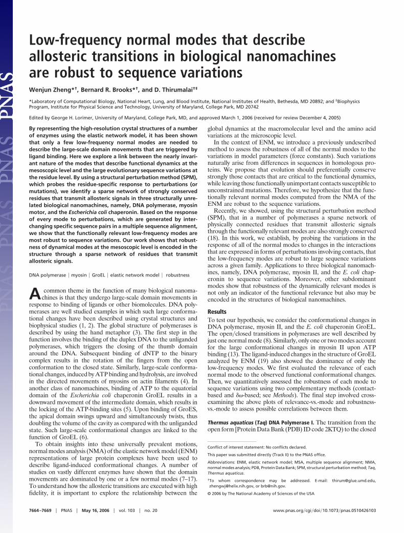
The two independent methods reveal a significant correlationbetween mode relevance and its robustness. Both pinpoint mode 4as the most robust mode to sequence variations. The contact-basedSPM confirms that specific contacts are ‘‘conserved,’’ which impliesthat for functional reasons only those mutations that preserve thecontacts are tolerated. This finding is in accord with a number ofexperiments that have probed the fidelity of DNA polymerases tospecific mutations (see below and Supporting Text). The strongconservation and robustness of the dominant and a few subdomi-nant modes may be a requirement for the high-fidelity replicationfunction of DNA polymerase I.Dynamical domain partition on each mode reveals the specific structuresinvolved in the open�closed transition. For mode 4 (Fig. 2a), the fingersdomain is comprised of three smaller dynamical domains. The topone (T, in yellow) includes the O1�O2 helices, the middle one (M,in cyan) sits in between, and the bottom one (B, in purple) is at theinterface with the palm domain (in blue). The O, N helices are inthe hinge�bending region (green). Therefore, the movement of thefingers domain consists of three coupled hinge motions. The firstcorresponds to twisting of T relative to the M, the second representstwisting of B relative to T, and the third describes the bending of Brelative to the palm domain (see three arrows in Fig. 2a). Thehinge�bending residues (green) are distributed across the fingersdomain, which is in accordance with the finding of an extensivenetwork of eight clusters of dynamically important residues (18).Besides the fingers motions that open�close the cleft, mode 4 alsodescribes a hinge motion of the thumb domain (red), which may,however, be locked by DNA binding.
Fig. 1. ENM and sequence analysis for Taq DNA polymerase’s open�closedtransition (2KTQ 3 3KTQ). (Upper) Overlap between each mode and theobserved conformational changes for the lowest 100 modes. (Lower) Themode-dependent robustness ( f��) computed by the contact-based SPM forthe lowest 100 modes, where the red curve (with circles) is for the original MSAand the blue curve (with error bars) represents the average (and standarddeviation) of f�� distribution computed for 100 randomly permuted MSA.
Table 1. Dominant modes in domain movements and their robustness to sequence variations
Proteins PDB pairs* Mode† Overlap‡
��-based analysis§
Contact-basedanalysis¶
f��E� z-score** f�E
� Z score**
Taq DNA pol I 2KTQ3 3KTQ 4 0.50 0.0811 �5.210 0.0338 �3.979Dictyostelium myosin II 1VON3 1MMA 1 0.56 0.0414 �3.460 0.0161 �2.597
2 0.39 0.0453 �2.954 0.0125 �2.667E. coli GroEL 1AON�A3 1GRL 1 0.81 0.0485 �3.315 0.0198 �2.159
*The two structures represent the beginning and end states in the allosteric transitions.†Dominant mode(s) in the specific transition.‡Overlap describes the extent to which the mode describes the domain movements. A value close to unity impliesthat a single mode suffices to represent a given allosteric transition. A value �0.5 is highly significant.
§��-based SPM to probe robustness of modes to perturbations (see Supporting Text).¶SPM in which contact energy is varied to probe robustness of modes to perturbations (see Methods).�A dimensionless score ( f�E or f��E) to assess the robustness of a mode (see Eq. 9).**Statistical significance of f�E or f��E (see Eq. 11).
Zheng et al. PNAS � May 16, 2006 � vol. 103 � no. 20 � 7665
BIO
PHYS
ICS
For mode 7 (Fig. 2b), the top half of the fingers (including theN�O�O1�O2 helices; T, colored in yellow) forms a dynamicaldomain, whereas the bottom half is fused with the palm (blue) asone dynamical domain. Although mode 7 describes a bendingmotion of T, the bending region is smaller and more localized thanin mode 4, which is consistent with its accessory role in theopen�closed transition. In mode 10 (Fig. 2c), a localized twisting ofO1 and O2 helices (red) is seen. This mode has the second-highestoverlap with the measured conformational changes. This findingsupports the importance of internal flexibility of fingers in theopen�closed transition.
Therefore, a detailed examination of the above robust modessuggests that they involve different hinge motions of the fingersdomain, although they share some bending regions. Although thecontributions from the subdominant modes to the open�closedtransition are smaller than mode 4, they are still functionallyimportant. Several modes are needed to describe it with accuracy.The importance of the subdominant modes is further supported bythe observed strong robustness to evolutionary sequence variations.
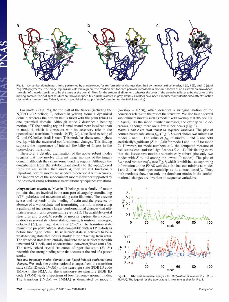
Dictyostelium Myosin II. Myosin II belongs to a family of motorproteins that are involved in the transport of cargo by coordinatingATP hydrolysis and movement along actin filaments. The enzymesenses and responds to the binding of actin and the presence orabsence of a �-phosphate and transmitting this information alonga pathway of increasingly larger conformational changes that ulti-mately results in a force-generating event (21). The available crystalstructures and cryo-EM results of myosins capture their confor-mations in several structural states, namely, transition, near-rigor,detached (22), and rigor-like states (23–25). The transition statemimics the prepower-stroke state compatible with ATP hydrolysisbefore binding to actin. The near-rigor state is believed to be aweak-binding state that occurs shortly after detaching from actin.The detached state is structurally similar to the near-rigor state withunwound SH1 helix and unconstrained converter�lever arm (22).The newly solved crystal structures of rigor-like state (23, 24)resemble the strong-binding state that occurs at the end of a powerstroke.Two low-frequency modes dominate the ligand-induced conformationalchanges. We study the conformational changes from the transitionstate (PDB ID code 1VOM) to the near-rigor state (PDB ID code1MMA). The NMA for the transition-state structure (PDB IDcode 1VOM) yields a spectrum of low-frequency normal modes.The transition (1VOM 3 1MMA) is dominated by mode 1
(overlap � 0.558), which describes a swinging motion of theconverter relative to the rest of the structure. We also found severalsubdominant modes (such as mode 2 with overlap � 0.388; see Fig.3 Upper). As the mode number increases, the overlap value de-creases, although there are a few minor peaks (Fig. 3).Modes 1 and 2 are most robust to sequence variations. The plot ofcontact-based robustness f�E (Fig. 3 Lower) shows two minima atmodes 2 and 1. The value of f�E of modes 1 and 2 are bothstatistically significant (Z � �2.60 for mode 1 and �2.67 for mode2). However, for mode numbers � 3, the computed measure ofrobustness loses statistical significance (Z � �1). This finding showsthat the lowest two modes are statistically robust (the only twomodes with Z � �2 among the lowest 10 modes). The plot of��-based robustness f��E (see Fig. 8, which is published as supportinginformation on the PNAS web site) also has two minima at modes1 and 2. It has similar peaks and dips as the contact-based f�E. Thus,both methods show that only the dominant modes in the confor-mational changes are invariant to sequence variations.
Fig. 2. Dynamical domain partitions, performed by using DYNDOM, for conformational changes described by the most robust modes, 4 (a), 7 (b), and 10 (c), ofTaq DNA polymerase. The hinge regions are colored in green. The rotation axis for each pairwise interdomain motion is shown as an axis with an arrowhead;the color of the axis stem is set to be the same as the domain fixed for the structural alignment, whereas the color of the arrowhead is set to be the color of themoving domain. The hot-spot residues are shown in space-filled circles colored in gray. Residues in black have been experimentally identified to affect function(for residue numbers, see Table 2, which is published as supporting information on the PNAS web site).
Fig. 3. ENM and sequence analysis for Dictyostelium myosin (1VOM 31MMA). The legend for the two graphs is the same as that for Fig. 1.
7666 � www.pnas.org�cgi�doi�10.1073�pnas.0510426103 Zheng et al.
The domain partition for modes 1 and 2 (Fig. 4) reveals twodynamical domains. One is the converter (blue), and the other is therest of the motor domain (red). Mode 1 describes a closure motionof the converter, whereas mode 2 describes a twisting motion. Thehot-spot residues of modes 1 and 2 (Fig. 4) are mostly distributedover the relay helix, SH1 helix, and the converter. They partiallyoverlap with the bending region (green) identified by DYNDOM (26).These hinge residues are critical in mediating signals that ultimatelylead to force generation. Preservation of the function requires thatthese residues be highly conserved.
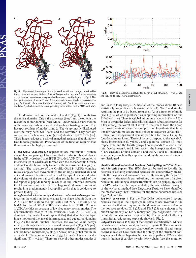
E. coli GroEL Chaperonin. Chaperonins are multisubunit proteinassemblies comprising of two rings that are stacked back-to-back.In the ATP-hydrolyzed state [PDB ID code 1AON (5)], asymmetricintermediates of GroEL are formed with the cochaperonin GroESand nucleotides bound only to one of the seven-subunit rings (thecis ring). The structure of the GroEL–GroES–(ADP)7 complexreveals large en bloc movements of the cis ring’s intermediate andapical domains. Elevation and twist of the apical domains doublethe volume of the central cavity that results in the burial of thehydrophobic peptide-binding residues at the interface betweenGroEL subunits and GroES. The large-scale domain movementresults in a predominantly hydrophilic cavity that is conducive toprotein folding (6).A single mode dominates the ligand-induced conformational changes. Wefocus on the conformational change for a single subunit from theADP�GROES state to the apo state (1AON�A 3 1GRL). TheNMA for the ADP�GROES state structure (PDB ID code1AON�A) yields a spectrum of low-frequency normal modes. Theconformational change in the transition 1AON�A 3 1GRL isdominated by mode 1 (overlap � 0.806) that describes multiplehinge motions of the apical, intermediate, and equatorial domains(19). As the mode number increases, the overlap value decaysrapidly, although there are occasional minor peaks (Fig. 5 Upper).Low-frequency modes are robust to sequence variations. The measure ofcontact-based robustness f�E (Fig. 5 Lower) has a global minimumat mode 1. The minimum value of f�E for mode 1 is statisticallysignificant (Z � �2.16). There are several other modes (modes 2
and 3) with fairly low f�E. Almost all of the modes above 10 havestatistically insignificant robustness (Z � �1). We found similarresults in the plot of ��-based robustness f��E as a function of mode(see Fig. 9, which is published as supporting information on thePNAS web site). There is a global minimum at mode 1 (Z � �3.32).Most of the modes lack statistically significant robustness except fora few among the lowest 10. Therefore, the results from the abovetwo assessments of robustness support our hypothesis that func-tionally relevant modes are most robust to sequence variations.
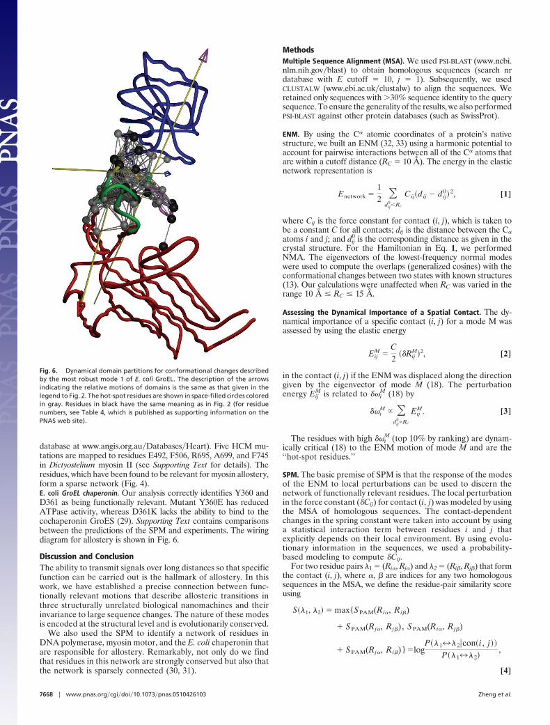
Based on the dynamical domain partition for mode 1 (Fig. 6),four domains are found. Three of them correspond to the apical (A,blue), intermediate (I, yellow), and equatorial domain (E, red),respectively, and the fourth (purple) corresponds to a loop at theinterface between A and I. For mode 1, the hot-spot residues (Fig.6) are clustered around domain I and the A–I and E–I interfaceswhere many functionally important and highly conserved residuesare distributed.
Identification of Network of Residues (‘‘Wiring Diagram’’) That Trans-mit Allosteric Signals. The SPM also can be used to identify thenetwork of distantly connected residues that cooperatively orches-trate the large-scale domain movements. By assessing the degree ofresponse to site-specific perturbations, the importance of a givenresidue in facilitating allosteric transitions can be gauged. By usingthe SPM, which can be implemented by the contact-based analysisor the ��-based method (see Supporting Text), we have identifiedthe mechanically ‘‘hot’’ residues for the three nanomachines (seeFigs. 2, 4, and 6).Taq DNA polymerase I. For the Taq DNA polymerase I severalresidues that span the fingers�palm domains are involved in thethree modes that are required in the domain movements. Amongthe hot-spot residues, I614 (27) and F667 (28) were shown to becritical for fidelity of DNA replication (see Supporting Text fordetailed comparisons with experiments). The network of allosterytransmitting residues are explicitly shown in Fig. 2.Dictyostelium myosin II. Many of the residues identified by SPM havebeen shown to be functionally relevant in myosin II. The amino acidsequence similarity between Dictyostelium myosin II and human�-cardiac myosin have facilitated the study of the structural con-sequences of those hypertrophic cardiomyopathy (HCM) muta-tions in human �-cardiac myosin heavy chain (see the mutation
Fig. 4. Dynamical domain partitions for conformational changes described bythe most robust modes, 1 (a) and 2 (b), of Dictyostelium myosin. For the meaningof the relative domain motions given by the arrows, see the legend to Fig. 1. Thehot-spot residues of modes 1 and 2 are shown in space-filled circles colored ingray. Residues in black have the same meaning as in Fig. 2 (for residue numbers,see Table 3, which is published as supporting information on the PNAS web site).
Fig. 5. ENM and sequence analysis for E. coli GroEL (1AON�A3 1GRL). Seethe legend to Fig. 1 for a description.
Zheng et al. PNAS � May 16, 2006 � vol. 103 � no. 20 � 7667
BIO
PHYS
ICS
database at www.angis.org.au�Databases�Heart). Five HCM mu-tations are mapped to residues E492, F506, R695, A699, and F745in Dictyostelium myosin II (see Supporting Text for details). Theresidues, which have been found to be relevant for myosin allostery,form a sparse network (Fig. 4).E. coli GroEL chaperonin. Our analysis correctly identifies Y360 andD361 as being functionally relevant. Mutant Y360E has reducedATPase activity, whereas D361K lacks the ability to bind to thecochaperonin GroES (29). Supporting Text contains comparisonsbetween the predictions of the SPM and experiments. The wiringdiagram for allostery is shown in Fig. 6.
Discussion and ConclusionThe ability to transmit signals over long distances so that specificfunction can be carried out is the hallmark of allostery. In thiswork, we have established a precise connection between func-tionally relevant motions that describe allosteric transitions inthree structurally unrelated biological nanomachines and theirinvariance to large sequence changes. The nature of these modesis encoded at the structural level and is evolutionarily conserved.
We also used the SPM to identify a network of residues inDNA polymerase, myosin motor, and the E. coli chaperonin thatare responsible for allostery. Remarkably, not only do we findthat residues in this network are strongly conserved but also thatthe network is sparsely connected (30, 31).
MethodsMultiple Sequence Alignment (MSA). We used PSI-BLAST (www.ncbi.nlm.nih.gov�blast) to obtain homologous sequences (search nrdatabase with E cutoff � 10, j � 1). Subsequently, we usedCLUSTALW (www.ebi.ac.uk�clustalw) to align the sequences. Weretained only sequences with �30% sequence identity to the querysequence. To ensure the generality of the results, we also performedPSI-BLAST against other protein databases (such as SwissProt).
ENM. By using the C� atomic coordinates of a protein’s nativestructure, we built an ENM (32, 33) using a harmonic potential toaccount for pairwise interactions between all of the C� atoms thatare within a cutoff distance (RC � 10 Å). The energy in the elasticnetwork representation is
Enetwork �12 �
dij0�Rc
Cij�dij � dij02, [1]
where Cij is the force constant for contact (i, j), which is taken tobe a constant C for all contacts; dij is the distance between the C�
atoms i and j; and dij0 is the corresponding distance as given in the
crystal structure. For the Hamiltonian in Eq. 1, we performedNMA. The eigenvectors of the lowest-frequency normal modeswere used to compute the overlaps (generalized cosines) with theconformational changes between two states with known structures(13). Our calculations were unaffected when RC was varied in therange 10 Å RC 15 Å.
Assessing the Dynamical Importance of a Spatial Contact. The dy-namical importance of a specific contact (i, j) for a mode M wasassessed by using the elastic energy
EijM �
C2
��RijM2, [2]
in the contact (i, j) if the ENM was displaced along the directiongiven by the eigenvector of mode M (18). The perturbationenergy Eij
M is related to ��iM (18) by
��iM �
dij0Rc
EijM. [3]
The residues with high ��iM (top 10% by ranking) are dynam-
ically critical (18) to the ENM motion of mode M and are the‘‘hot-spot residues.’’
SPM. The basic premise of SPM is that the response of the modesof the ENM to local perturbations can be used to discern thenetwork of functionally relevant residues. The local perturbationin the force constant (�Cij) for contact (i, j) was modeled by usingthe MSA of homologous sequences. The contact-dependentchanges in the spring constant were taken into account by usinga statistical interaction term between residues i and j thatexplicitly depends on their local environment. By using evolu-tionary information in the sequences, we used a probability-based modeling to compute �Cij.
For two residue pairs 1 � (Ri�, Rj�) and 2 � (Ri�, Rj�) that formthe contact (i, j), where �, � are indices for any two homologoussequences in the MSA, we define the residue-pair similarity scoreusing
S�1, 2 � max{SPAM(Ri�, Ri�)
� SPAM(Rj�, Rj�), SPAM(Ri�, Rj�)
� SPAM(Rj�, Ri�)}�logP�172�con� i , j
P�172,
[4]
Fig. 6. Dynamical domain partitions for conformational changes describedby the most robust mode 1 of E. coli GroEL. The description of the arrowsindicating the relative motions of domains is the same as that given in thelegend to Fig. 2. The hot-spot residues are shown in space-filled circles coloredin gray. Residues in black have the same meaning as in Fig. 2 (for residuenumbers, see Table 4, which is published as supporting information on thePNAS web site).
7668 � www.pnas.org�cgi�doi�10.1073�pnas.0510426103 Zheng et al.
where P(1 7 2�con(i, j)) is the conditional probability of occur-rence of residues pair substitution 17 2 provided contact (i, j) isconserved. By ‘‘conserved’’ we mean that the context-dependentstatistical interaction is maintained upon 17 2 substitution. Herewe allowed the swapping of residues between (i, j) (see the secondterm in Eq. 4), because that should not alter the physical pairwiseinteraction between them. We use the PAM250 substitution matrixto evaluate residue similarity. We assume that the commonlyobserved residue substitutions in homologous proteins preserveresidue–residue contact interactions within the protein nativestructure.
The variation of the statistical interaction for contact (i, j) due tothe residue pair substitution 17 2 was taken to be proportionalto the probability of not conserving the contact (i, j) if thesubstitution 1 7 2 was allowed
�E�1, 2 � �1 � P�con(i , j �172] ��
� �1 � P�con(i , j �es�1,2] �� , [5]
where � is a constant that yields the maximal variation ofinteraction.
For two randomly generated pairs (1, 2) (for each pair 1 or 2,both residues are randomly chosen from all 20 types of aminoacids), we computed the average Srand � S(1, 2)�rand and set
Prand���(1�P�con(i , j) �eSrand)��. [6]
By using Eq. 5 and Bayes theorem, we get
�E�1, 2 � �1 � �1 � Prand)�eS�1,2�Srand]��. [7]
Here we set Prand � 0.5. The variation of the force constant forcontact (i, j) is
�Cij � �E�1, 2�MSA�Rc2, [8]
where the average is over all (1, 2) for the given pair (i, j) fromthe MSA at column i and j.
The robustness of a mode to sequence variations was assessedby using the fractional variation of the distortional energy E
f�E ��EE
. [9]
If f�E is small, then the specific mode is robust to sequencevariations. For the contact-based analysis
�E � ��i, j
�Cij
C�Eij
M, E � ��i, j
EijM, [10]
where �Cij is computed in Eq. 8 and EijM is defined in Eq. 2.
Statistical Significance of the Mode Robustness. To evaluate thestatistical significance of the mode robustness, we calculated thedistribution of f�E for randomly generated sequence–structurealignments. We performed 100 random permutations of the one-to-one mapping between the positions (columns), the MSA, and thepositions (nodes) in the ENM. This procedure eliminates allcorrelations encoded in the original mapping of the protein struc-ture. For each mode, we compute f�E for every random permutationand obtain a distribution of f�E, from which we can compute bothits average f�E�rand and standard deviation �rand. The Z score forf�E is
Z �f�E � f�E�rand
�rand, [11]
which is used to quantitatively assess the statistical significance off�E. The more negative Z is, the more statistically significant is f�E.A mode is robust if f�E is small and if Z is as negative as possible.
Dynamical Domains Partition. To visualize the collective conforma-tional changes described by each low-frequency normal mode (seeFigs. 2, 4, and 6), we used the dynamical domains partition analysisbased on analyzing the interdomain conformational changes de-scribed by the eigenvector of each mode. This analysis was per-formed by a computational tool called DYNDOM (26).
This work was supported in part by National Institutes of Health (NIH)Grant 1R01GM067851-01 and by the Intramural Research Program ofthe National Heart, Lung, and Blood Institute of the NIH.
1. Patel, P. H. & Loeb, L. A. (2001) Nat. Struct. Biol. 8, 656–659.2. Steitz, T. (1999) J. Biol. Chem. 274, 17395–17398.3. Ollis, D. L., Brick, P., Hamlin, R., Xuong, N. G. & Steitz, T. A. (1985) Nature
313, 762.4. Houdusse, A. & Sweeney, H. (2001) Curr. Opin. Struct. Biol. 11, 182–194.5. Xu, Z., Horwich, A. L. & Sigler, P. B. (1997) Nature 388, 741–750.6. Thirumalai, D. & Lorimer, G. H. (2001) Annu. Rev. Biophys. Biomol. Struct. 30,
245–269.7. Atilgan, A. R., Durell, S. R., Jernigan, R. L., Demirel, M. C., Keskin, O. &
Bahar, I. (2001) Biophys. J. 80, 505–515.8. Delarue, M. & Sanejouand, Y. H. (2002) J. Mol. Biol. 320, 1011–1024.9. Tama, F. & Sanejouand, Y. H. (2001) Protein Eng. 14, 1–6.
10. Bahar, I. & Jernigan, R. L. (1999) Biochemistry 38, 3478–3490.11. Rader, A. J., Vlad, D. H., Bahar, I. (2005) Structure (London) 13, 413–421.12. Bahar, I. & Rader, A. J. (2005) Curr. Opin. Struct. Biol. 15, 586–592.13. Zheng, W. & Doniach, S. (2003) Proc. Natl. Acad. Sci. USA 100, 13253–
13258.14. Bahar, I., Atilgan, A. R. & Erman, B. (1997) Fold Des. 2, 173–181.15. Haliloglu, T., Bahar, I. & Erman, B. (1997) Phys. Rev. Lett. 79, 3090–3093.16. Kim, M. K., Jernigan, R. L. & Chirikjian G. S. (2002) Biophys. J. 83, 1620–1630.17. Demirel, M. C., Atilgan, A. R., Jernigan, R. L., Erman, B. & Bahar, I. (1998)
Protein Sci. 7, 2522–2532.18. Zheng, W., Brooks, B. R., Doniach, S. & Thirumalai, D. (2005) Structure (London)
13, 565–577.
19. Keskin, O., Bahar, I., Flatow, D., Covell, D. G. & Jernigan, R. L. (2002)Biochemistry 41, 491–501.
20. Li, Y., Korolev, S. & Waksman, G. (1998) EMBO J. 17, 7514–7525.21. Geeves, M. A. & Holmes, K. C. (1999) Annu. Rev. Biochem. 68, 687–728.22. Houdusse, A., Szent-Gyorgyi, A. G. & Cohen, C. (2000) Proc. Natl. Acad. Sci.
USA 97, 11238–11243.23. Coureux, P. D., Wells, A. L., Menetrey, J., Yengo, C. M., Morris, C. A.,
Sweeney, H. L. & Houdusse, A. (2003) Nature 425, 419–423.24. Reubold, T. F., Eschenburg, S., Becker, A., Kull, F. J. & Manstein, D. J. (2003)
Nat. Struct. Biol. 10, 826–830.25. Holmes, K. C., Angert, I., Kull, F. J., Jahn, W. & Schroder, R. R. (2003) Nature
425, 423–427.26. Patel, P. H., Kawate, H., Adman, E., Ashbach, M. & Loeb, L. A. (2000) J. Biol.
Chem. 276, 5044–5051.27. Suzuki, M., Yoshida, S., Adman, E. T., Blank, A. & Loeb, L. A. (2000) J. Biol.
Chem. 275, 32728–32735.28. Fenton, W. A., Kashi, Y., Furtak, K. & Horwich, A. L. (1994) Nature 371,
614–619.29. Lockless, S. W. & Ranganathan, R. (1999) Science. 286, 295–299.30. Suel, G. M., Lockless, S. W., Wall, M. A. & Ranganathan R. (2003) Nat. Struct.
Biol. 10, 59–69.31. Doruker, P., Atilgan, A. R. & Bahar, I. (2000) Proteins 40, 512–524.32. Tirion, M. M. (1996) Phys. Rev. Lett. 77, 1905–1908.33. Hayward, S. & Berendsen, H. J. (1998) Proteins 30, 144–154.
Zheng et al. PNAS � May 16, 2006 � vol. 103 � no. 20 � 7669
BIO
PHYS
ICS