lung cancer (non small cell)
TRANSCRIPT
Lung Cancer (Non-Small Cell)
What is non-small cell lung cancer? Lung cancer starts when cells of the lung become abnormal and begin to grow out of control. As more cancer cells develop, they can form into a tumor and spread to other areas of the body. (To learn more about how cancers start and spread, see What Is Cancer?)
Types of non-small cell lung cancer
There are 2 main types of lung cancer:
• About 80% to 85% of lung cancers are non-small cell lung cancer (NSCLC)
• About 10% to 15% are small cell lung cancer (SCLC)
These types of lung cancer are treated very differently. This information covers non-small cell lung cancer. For information on small cell lung cancer, see Lung Cancer (Small Cell).
There are subtypes of NSCLC, which start from different types of lung cells. But they are grouped together as NSCLC because the approach to treatment and prognosis (outlook) are often similar.
Adenocarcinoma: About 40% of lung cancers are adenocarcinomas. These cancers start in early versions of the cells that would normally secrete substances such as mucus.
This type of lung cancer occurs mainly in current or former smokers, but it is also the most common type of lung cancer seen in non-smokers. It is more common in women than in men, and it is more likely to occur in younger people than other types of lung cancer.
Adenocarcinoma is usually found in outer parts of the lung. Though it tends to grow slower than other types of lung cancer and is more likely to be found before it has spread, this varies from patient to patient.
People with a type of adenocarcinoma called adenocarcinoma in situ (previously called bronchioloalveolar carcinoma) tend to have a better outlook than those with other types of lung cancer.
Squamous cell (epidermoid) carcinoma: About 25% to 30% of all lung cancers are squamous cell carcinomas. These cancers start in early versions of squamous cells, which are flat cells that line the inside of the airways in the lungs. They are often linked to a history of smoking and tend to be found in the central part of the lungs, near a main airway (bronchus).
Large cell (undifferentiated) carcinoma: This type accounts for about 10% to 15% of lung cancers. It can appear in any part of the lung. It tends to grow and spread quickly, which can make it harder to treat. A subtype of large cell carcinoma, known as large cell neuroendocrine carcinoma, is a fast-growing cancer that is very similar to small cell lung cancer.
Other subtypes: A few other subtypes of NSCLC, such as adenosquamous carcinoma and sarcomatoid carcinoma, are much less common.
Other types of lung tumors
Along with the 2 main types of lung cancer, other tumors can occur in the lungs.
Lung carcinoid tumors: Carcinoid tumors of the lung account for fewer than 5% of lung tumors. Most of these grow slowly. For more information about these tumors, see Lung Carcinoid Tumor.
Other lung tumors: Other types of lung cancer such as adenoid cystic carcinomas, lymphomas, and sarcomas, as well as benign lung tumors such as hamartomas are rare. These are treated differently from the more common lung cancers and are not discussed here.
Cancers that spread to the lungs: Cancers that start in other organs (such as the breast, pancreas, kidney, or skin) can sometimes spread (metastasize) to the lungs, but these are not lung cancers. For example, cancer that starts in the breast and spreads to the lungs is still breast cancer, not lung cancer. Treatment for metastatic cancer to the lungs is based on where it started (the primary cancer site).
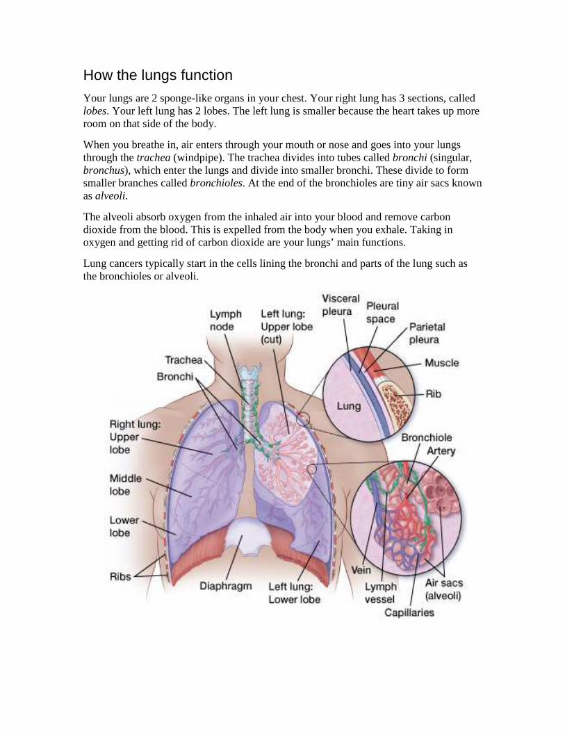
How the lungs function
Your lungs are 2 sponge-like organs in your chest. Your right lung has 3 sections, called lobes. Your left lung has 2 lobes. The left lung is smaller because the heart takes up more room on that side of the body.
When you breathe in, air enters through your mouth or nose and goes into your lungs through the trachea (windpipe). The trachea divides into tubes called bronchi (singular, bronchus), which enter the lungs and divide into smaller bronchi. These divide to form smaller branches called bronchioles. At the end of the bronchioles are tiny air sacs known as alveoli.
The alveoli absorb oxygen from the inhaled air into your blood and remove carbon dioxide from the blood. This is expelled from the body when you exhale. Taking in oxygen and getting rid of carbon dioxide are your lungs’ main functions.
Lung cancers typically start in the cells lining the bronchi and parts of the lung such as the bronchioles or alveoli.

A thin lining layer called the pleura surrounds the lungs. The pleura protects your lungs and helps them slide back and forth against the chest wall as they expand and contract during breathing.
Below the lungs, a thin, dome-shaped muscle called the diaphragm separates the chest from the abdomen. When you breathe, the diaphragm moves up and down, forcing air in and out of the lungs.
Key statistics for lung cancer Most lung cancer statistics include both small cell and non-small cell lung cancers.
How common is lung cancer?
Lung cancer (both small cell and non-small cell) is the second most common cancer in both men and women (not counting skin cancer). In men, prostate cancer is more common, while in women breast cancer is more common. About 14% of all new cancers are lung cancers.
The American Cancer Society’s estimates for lung cancer in the United States for 2016 are:
• About 224,390 new cases of lung cancer (117,920 in men and 106,470 in women)
• About158,080 deaths from lung cancer (85,920 in men and 72,160 in women)
Lung cancer is by far the leading cause of cancer death among both men and women; about 1 out of 4 cancer deaths are from lung cancer. Each year, more people die of lung cancer than of colon, breast, and prostate cancers combined.
Lung cancer mainly occurs in older people. About 2 out of 3 people diagnosed with lung cancer are 65 or older, while less than 2% are younger than 45. The average age at the time of diagnosis is about 70.
Lifetime chance of getting lung cancer
Overall, the chance that a man will develop lung cancer in his lifetime is about 1 in 14; for a woman, the risk is about 1 in 17. These numbers include both smokers and non-smokers. For smokers the risk is much higher, while for non-smokers the risk is lower.
Black men are about 20% more likely to develop lung cancer than white men. The rate is about 10% lower in black women than in white women. Both black and white women

have lower rates than men, but the gap is closing. The lung cancer rate has been dropping among men over the past few decades, but only for about the last decade in women.
Statistics on survival in people with lung cancer vary depending on the stage (extent) of the cancer when it is diagnosed. For survival statistics based on the stage of the cancer, see “Non-small cell lung cancer survival rates by stage.”
Despite the very serious prognosis (outlook) of lung cancer, some people with earlier stage cancers are cured. More than 430,000 people alive today have been diagnosed with lung cancer at some point.
Visit the American Cancer Society’s Cancer Statistics Center for more key statistics.
Non-small cell lung cancer risk factors A risk factor is anything that affects a person’s chance of getting a disease such as cancer. Different cancers have different risk factors. Some risk factors, like smoking, can be changed. Others, like a person’s age or family history, can’t be changed.
But having a risk factor, or even several, does not mean that you will get the disease. And some people who get the disease may have few or no known risk factors.
Several risk factors can make you more likely to develop lung cancer.
Risk factors you can change
Tobacco smoke
Smoking is by far the leading risk factor for lung cancer. About 80% of lung cancer deaths are thought to result from smoking. The risk for lung cancer among smokers is many times higher than among non-smokers. The longer you smoke and the more packs a day you smoke, the greater your risk.
Cigar smoking and pipe smoking are almost as likely to cause lung cancer as cigarette smoking. Smoking low-tar or “light” cigarettes increases lung cancer risk as much as regular cigarettes. Smoking menthol cigarettes might increase the risk even more since the menthol allows smokers to inhale more deeply.
Secondhand smoke: If you don’t smoke, breathing in the smoke of others (called secondhand smoke or environmental tobacco smoke) can increase your risk of developing lung cancer. Secondhand smoke is thought to cause more than 7,000 deaths from lung cancer each year.
If you or someone you care about needs help quitting, see our Guide to Quitting Smoking or call the American Cancer Society at 1-800-227-2345
Exposure to radon
Radon is a naturally occurring radioactive gas that results from the breakdown of uranium in soil and rocks. You can’t see, taste, or smell it. According to the US Environmental Protection Agency (EPA), radon is the second leading cause of lung cancer in this country, and is the leading cause among non-smokers.
Outdoors, there is so little radon that it is not likely to be dangerous. But indoors, radon can be more concentrated. Breathing it in exposes your lungs to small amounts of radiation. This may increase a person’s risk of lung cancer.
Homes and other buildings in nearly any part of the United States can have high indoor radon levels (especially in basements).
For more information, see Radon and Cancer.
Exposure to asbestos
People who work with asbestos (such as in mines, mills, textile plants, places where insulation is used, and shipyards) are several times more likely to die of lung cancer. Lung cancer risk is much greater in workers exposed to asbestos who also smoke. It’s not clear how much low-level or short-term exposure to asbestos might raise lung cancer risk.
People exposed to large amounts of asbestos also have a greater risk of developing mesothelioma, a type of cancer that starts in the pleura (the lining surrounding the lungs). For more on this type of cancer, see Malignant Mesothelioma.
In recent years, government regulations have greatly reduced the use of asbestos in commercial and industrial products. It’s still present in many homes and other older buildings, but it’s not usually considered harmful as long as it’s not released into the air by deterioration, demolition, or renovation. For more information, see Asbestos and Cancer Risk.
Exposure to other cancer-causing agents in the workplace
Other carcinogens (cancer-causing agents) found in some workplaces that can increase lung cancer risk include:
• Radioactive ores such as uranium
• Inhaled chemicals such as arsenic, beryllium, cadmium, silica, vinyl chloride, nickel compounds, chromium compounds, coal products, mustard gas, and chloromethyl ethers
• Diesel exhaust

The government and industry have taken steps in recent years to help protect workers from many of these exposures. But the dangers are still there, so if you work around these agents, be careful to limit your exposure whenever possible.
Arsenic in drinking water
Studies of people in parts of Southeast Asia and South America with high levels of arsenic in their drinking water have found a higher risk of lung cancer. In most of these studies, the levels of arsenic in the water were many times higher than those typically seen in the United States, even areas where arsenic levels are above normal. For most Americans who are on public water systems, drinking water is not a major source of arsenic.
Certain dietary supplements
Studies looking at the possible role of vitamin supplements in reducing lung cancer risk have had disappointing results. In fact, 2 large studies found that smokers who took beta carotene supplements actually had an increased risk of lung cancer. The results of these studies suggest that smokers should avoid taking beta carotene supplements.
Risk factors you cannot change
Previous radiation therapy to the lungs
People who have had radiation therapy to the chest for other cancers are at higher risk for lung cancer, particularly if they smoke; for example, people who have been treated for Hodgkin disease or women who get radiation after a mastectomy for breast cancer. Women who have radiation therapy to the breast after a lumpectomy do not appear to have a higher than expected risk of lung cancer.
Air pollution
In cities, air pollution (especially near heavily trafficked roads) appears to raise the risk of lung cancer slightly. This risk is far less than the risk caused by smoking, but some researchers estimate that worldwide about 5% of all deaths from lung cancer may be due to outdoor air pollution.
Personal or family history of lung cancer
If you have had lung cancer, you have a higher risk of developing another lung cancer.

Brothers, sisters, and children of people who have had lung cancer may have a slightly higher risk of lung cancer themselves, especially if the relative was diagnosed at a younger age. It’s not clear how much of this risk might be due to shared genes among family members and how much might be from shared household exposures (such as tobacco smoke or radon).
Researchers have found that genetics seems to play a role in some families with a strong history of lung cancer. (See “Do we know what causes non-small cell lung cancer?”).
Factors with uncertain or unproven effects on lung cancer risk
Smoking marijuana
There are some reasons to think that smoking marijuana might increase lung cancer risk. Marijuana smoke contains tar and many of same cancer-causing substances that are in tobacco smoke. (Tar is the sticky, solid material that remains after burning, and is thought to contain most of the harmful substances in smoke.)
Marijuana cigarettes (joints) are typically smoked all the way to the end, where tar content is the highest. Marijuana is also inhaled very deeply and the smoke is held in the lungs for a long time, which gives any cancer-causing substances more opportunity to deposit in the lungs. And because marijuana is still illegal in many places, it may not be possible to control what other substances it might contain.
But those who use marijuana tend to smoke fewer marijuana cigarettes in a day or week than the amount of tobacco consumed by cigarette smokers. The lesser amount smoked would make it harder to see an impact on lung cancer risk.
It’s been hard to study whether there is a link between marijuana and lung cancer because marijuana has been illegal in many places for so long, and it’s not easy to gather information about the use of illegal drugs. Also, in studies that have looked at past marijuana use in people who had lung cancer, most of the marijuana smokers also smoked cigarettes. This can make it hard to know how much any increased risk is from tobacco and how much might be from marijuana. More research is needed to know the cancer risks from smoking marijuana.
Talc and talcum powder
Talc is a mineral that in its natural form may contain asbestos. Some studies have suggested that talc miners and millers might have a higher risk of lung cancer and other respiratory diseases because of their exposure to industrial grade talc. But other studies have not found an increase in lung cancer rate.

Talcum powder is made from talc. By law since the 1970s, all home-use talcum products (baby, body, and facial powders) in the United States have been asbestos-free. The use of cosmetic talcum powder has not been found to increase lung cancer risk.
What causes non-small cell lung cancer? We don’t know what causes each case of lung cancer. But we do know many of the risk factors for these cancers (see “Non-small cell lung cancer risk factors”) and how some of them cause cells to become cancerous.
Smoking
Tobacco smoking is by far the leading cause of lung cancer. About 80% of lung cancer deaths are caused by smoking, and many others are caused by exposure to secondhand smoke.
Smoking is clearly the strongest risk factor for lung cancer, but it often interacts with other factors. Smokers exposed to other known risk factors such as radon and asbestos are at even higher risk. Not everyone who smokes gets lung cancer, so other factors like genetics likely play a role as well (see below).
Lung cancer in non-smokers
Not all people who get lung cancer are smokers. Many people with lung cancer are former smokers, but many others never smoked at all.
Lung cancer in non-smokers can be caused by exposure to radon, secondhand smoke, air pollution, or other factors. Workplace exposures to asbestos, diesel exhaust, or certain other chemicals can also cause lung cancers in some people who don’t smoke.
A small portion of lung cancers occur in people with no known risk factors for the disease. Some of these might just be random events that don’t have an outside cause, but others might be due to factors that we don’t yet know about.
Lung cancers in non-smokers are often different in some ways from those that occur in smokers. They tend to occur at younger ages. Lung cancers in non-smokers often have certain gene changes that are different from those in tumors from smokers. In some cases, these changes can be used to guide treatment.
Gene changes that may lead to lung cancer
Scientists know how some of the risk factors for lung cancer can cause certain changes in the DNA of lung cells. These changes can lead to abnormal cell growth and, sometimes, cancer. DNA is the chemical in our cells that makes up our genes, which control how our

cells function. We usually look like our parents because they are the source of our DNA. But DNA also can influence our risk for developing certain diseases, including some kinds of cancer.
Some genes help control when cells grow, divide to make new cells, and die:
• Genes that help cells grow, divide, or stay alive are called oncogenes.
• Genes that help keep cell division under control or cause cells to die at the right time are called tumor suppressor genes.
Cancers can be caused by DNA changes that turn on oncogenes or turn off tumor suppressor genes.
Inherited gene changes
Some people inherit DNA mutations (changes) from their parents that greatly increase their risk for developing certain cancers. But inherited mutations alone are not thought to cause very many lung cancers.
Still, genes do seem to play a role in some families with a history of lung cancer. For example, people who inherit certain DNA changes in a particular chromosome (chromosome 6) are more likely to develop lung cancer, even if they don’t smoke or only smoke a little.
Some people seem to inherit a reduced ability to break down or get rid of certain types of cancer-causing chemicals in the body, such as those found in tobacco smoke. This could put them at higher risk for lung cancer.
Other people inherit faulty DNA repair mechanisms that make it more likely they will end up with DNA changes. People with DNA repair enzymes that don’t work normally might be especially vulnerable to cancer-causing chemicals and radiation.
Researchers are developing tests that may help identify such people, but these tests are not yet used routinely. For now, doctors recommend that all people avoid tobacco smoke and other exposures that might increase their cancer risk.
Acquired gene changes
Gene changes related to lung cancer are usually acquired during life rather than inherited. Acquired mutations in lung cells often result from exposure to factors in the environment, such as cancer-causing chemicals in tobacco smoke. But some gene changes may just be random events that sometimes happen inside a cell, without having an outside cause.
Acquired changes in certain genes, such as the TP53 or p16 tumor suppressor genes and the K-RAS or ALK oncogenes, are thought to be important in the development of non-small cell lung cancer. Changes in these and other genes may also make some lung
cancers more likely to grow and spread than others. Not all lung cancers share the same gene changes, so there are undoubtedly changes in other genes that have not yet been found.
Can non-small cell lung cancer be prevented? Not all lung cancers can be prevented. But there are things you can do that might lower your risk, such as changing the risk factors that you can control.
Stay away from tobacco
The best way to reduce your risk of lung cancer is not to smoke and to avoid breathing in other people’s smoke.
If you stop smoking before a cancer develops, your damaged lung tissue gradually starts to repair itself. No matter what your age or how long you’ve smoked, quitting may lower your risk of lung cancer and help you live longer. If you would like help quitting smoking, see our Guide to Quitting Smoking or call the American Cancer Society at 1-800-227-2345.
Avoid radon
Radon is an important cause of lung cancer. You can reduce your exposure to radon by having your home tested and treated, if needed. For more information, see Radon and Cancer.
Avoid or limit exposure to cancer-causing chemicals
Avoiding exposure to known cancer-causing chemicals, in the workplace and elsewhere, may also be helpful (see What are the risk factors for non-small cell lung cancer?). When people work where these exposures are common, they should be kept to a minimum.
Eat a healthy diet
A healthy diet with lots of fruits and vegetables may also help reduce your risk of lung cancer. Some evidence suggests that a diet high in fruits and vegetables may help protect against lung cancer in both smokers and non-smokers. But any positive effect of fruits and vegetables on lung cancer risk would be much less than the increased risk from smoking.

Attempts to reduce the risk of lung cancer in current or former smokers by giving them high doses of vitamins or vitamin-like drugs have not been successful so far. In fact, some studies have found that supplements of beta-carotene, a nutrient related to vitamin A, appear to increase the rate of lung cancer in these people.
Some people who get lung cancer do not have any clear risk factors. Although we know how to prevent most lung cancers, at this time we don’t know how to prevent all of them.
Can non-small cell lung cancer be found early? Usually symptoms of lung cancer do not appear until the disease is already at an advanced stage. Even when lung cancer does cause symptoms, many people may mistake them for other problems, such as an infection or long-term effects from smoking. This may delay the diagnosis.
Some lung cancers are found early by accident as a result of tests for other medical conditions. For example, lung cancer may be found by tests done for other reasons in people with heart disease, pneumonia, or other lung conditions. A small portion of these people do very well and may be cured of lung cancer.
Screening is the use of tests or exams to find a disease in people who don’t have symptoms. Doctors have looked for many years for a good screening test for lung cancer, but only in recent years has a study shown that a test known as a low-dose CT (LDCT) scan can help lower the risk of dying from this disease.
The National Lung Screening Trial
The National Lung Screening Trial (NLST) was a large clinical trial that looked at using LDCT of the chest to screen for lung cancer. CT scans of the chest provide more detailed pictures than chest x-rays and are better at finding small abnormal areas in the lungs. Low-dose CT of the chest uses lower amounts of radiation than a standard chest CT and does not require the use of intravenous (IV) contrast dye.
The NLST compared LDCT of the chest to chest x-rays in people at high risk of lung cancer to see if these scans could help lower the risk of dying from lung cancer. The study included more than 50,000 people ages 55 to 74 who were current or former smokers and were in fairly good health. To be in the study, they had to have at least a 30 pack-year history of smoking.
A pack-year is the number of cigarette packs smoked each day multiplied by the number of years a person has smoked. Someone who smoked a pack of cigarettes per day for 30 years has a 30 pack-year smoking history, as does someone who smoked 2 packs a day for 15 years.

Former smokers could enter the study if they had quit within the past 15 years. The study did not include people if they had a history of lung cancer or lung cancer symptoms, if they had part of a lung removed, if they needed to be on oxygen at home to help them breathe, or if they had other serious medical problems.
People in the study got either 3 LDCT scans or 3 chest x-rays, each a year apart, to look for abnormal areas in the lungs that might be cancer. After several years, the study found that people who got LDCT had a 16% lower chance of dying from lung cancer than those who got chest x-rays. They were also 7% less likely to die overall (from any cause) than those who got chest x-rays.
Screening with LDCT was also shown to have some downsides that need to be considered. One drawback of this test is that it also finds a lot of abnormalities that have to be checked out with more tests, but that turn out not to be cancer. (About 1 out of 4 people in the NLST had such a finding.) This may lead to additional tests such as other CT scans or more invasive tests such as needle biopsies or even surgery to remove a portion of lung in some people. These tests can sometimes lead to complications (like a collapsed lung) or rarely, death, even in people who do not have cancer (or who have very early stage cancer).
LDCTs also expose people to a small amount of radiation with each test. It is less than the dose from a standard CT, but it is more than the dose from a chest x-ray. Some people who are screened may end up needing further CT scans, which means more radiation exposure. When done in tens of thousands of people, this radiation may cause a few people to develop breast, lung, or thyroid cancers later on.
The NLST was a large study, but it left some questions that still need to be answered. For example, it’s not clear if screening with LDCT scans would have the same effect if different people were allowed in the study, such as those who smoke less (or not at all), or people younger than age 55 or older than 74. Also, in the NLST, patients got 3 scans over 2 years. It’s not yet clear what the effect would be if people were screened for longer than 2 years.
These factors, and others, need to be taken into account by people and their doctors who are considering whether or not screening with LDCT scans is right for them.
American Cancer Society’s guidelines for lung cancer screening
The American Cancer Society has thoroughly reviewed the subject of lung cancer screening and issued guidelines that are aimed at doctors and other health care providers:
Patients should be asked about their smoking history. Patients who meet ALL of the following criteria may be candidates for lung cancer screening:
• 55 to 74 years old
• In fairly good health (discussed further down)
• Have at least a 30 pack-year smoking history (discussed above)
• Are either still smoking or have quit smoking within the last 15 years
These criteria were based on what was used in the NLST.
Doctors should talk to these patients about the benefits, limitations, and potential harms of lung cancer screening. Screening should only be done at facilities that have the right type of CT scanner and that have a lot of experience using low-dose CT (LDCT) scans for lung cancer screening. The facility should also have a team of specialists that can provide the appropriate care and follow-up of patients with abnormal results on the scans.
For patients
If you fit all of the criteria listed above for lung cancer screening, you and your doctor (or other health care provider) should talk about screening, including possible benefits and harms, as well as the limitations of screening.
The main benefit is a lower chance of dying of lung cancer, which accounts for many deaths in current and former smokers. Still, it’s important to be aware that, like with any type of screening, not everyone who gets screened will benefit. Screening with LDCT will not find all lung cancers, and not all of the cancers that are found will be found early.
Even if a cancer is found by screening, you may still die from lung cancer. Also, LDCT often finds things that turn out not to be cancer, but have to be checked out with more tests to know what they are. You might need more CT scans, or even invasive tests such as a lung biopsy, in which a piece of lung tissue is removed with a needle or during surgery. These tests have risks of their own (see above).
Screening should only be done at facilities that have the right type of CT scanner and that have experience in LDCT scans for lung cancer screening. The facility should also have a team of specialists that can give patients the appropriate care and follow-up if there are abnormal results on the scans. You might not have the right kind of facility nearby, so you may need to travel some distance to be screened.
If you and your doctor decide that you should be screened, you should get a LDCT every year until you reach the age of 74, as long as you are still in good health.
If you smoke, you should get counseling about stopping. You should be told about your risk of lung cancer and referred to a smoking cessation program. Screening is not a good alternative to stopping smoking. For help quitting, see our Guide to Quitting Smoking or call the American Cancer Society at 1-800-227-2345.

What does “in fairly good health” mean?
Screening is meant to find cancer in people who do not have symptoms of the disease. People who already have symptoms that might be caused by lung cancer may need tests such as CT scans to find the underlying cause, which in some cases may be cancer. But this kind of testing is for diagnosis and is not the same as screening. Some of the possible symptoms of lung cancer that kept people out of the NLST were coughing up blood and weight loss without trying.
To get the most benefit from screening, patients need to be in good health. For example, they need to be able to have surgery and other treatments to try to cure lung cancer if it is found. Patients who need home oxygen therapy probably couldn’t withstand having part of a lung removed, and so are not candidates for screening. Patients with other serious medical problems that would shorten their lives or keep them from having surgery might not benefit enough from screening for it to be worth the risks, and so should also not be screened.
Metal implants in the chest (like pacemakers) or back (like rods in the spine) can interfere with x-rays and lead to poor quality CT images of the lungs. People with these types of implants were also kept out of the NLST, and so should not be screened with CT scans for lung cancer according to the ACS guidelines.
Non-small cell lung cancer signs and symptoms Most lung cancers do not cause any symptoms until they have spread, but some people with early lung cancer do have symptoms. If you go to your doctor when you first notice symptoms, your cancer might be diagnosed at an earlier stage, when treatment is more likely to be effective. The most common symptoms of lung cancer are:
• A cough that does not go away or gets worse
• Coughing up blood or rust-colored sputum (spit or phlegm)
• Chest pain that is often worse with deep breathing, coughing, or laughing
• Hoarseness
• Weight loss and loss of appetite
• Shortness of breath
• Feeling tired or weak
• Infections such as bronchitis and pneumonia that don’t go away or keep coming back
• New onset of wheezing
When lung cancer spreads to distant organs, it may cause:
• Bone pain (like pain in the back or hips)
• Nervous system changes (such as headache, weakness or numbness of an arm or leg, dizziness, balance problems, or seizures), from cancer spread to the brain or spinal cord
• Yellowing of the skin and eyes (jaundice), from cancer spread to the liver
• Lumps near the surface of the body, due to cancer spreading to the skin or to lymph nodes (collections of immune system cells), such as those in the neck or above the collarbone
Most of these symptoms are more likely to be caused by something other than lung cancer. Still, if you have any of these problems, it’s important to see your doctor right away so the cause can be found and treated, if needed.
Some lung cancers can cause syndromes, which are groups of very specific symptoms.
Horner syndrome
Cancers of the top part of the lungs (sometimes called Pancoast tumors) sometimes can affect certain nerves to the eye and part of the face, causing a group of symptoms called Horner syndrome:
• Drooping or weakness of one eyelid
• A smaller pupil (dark part in the center of the eye) in the same eye
• Reduced or absent sweating on the same side of the face
Pancoast tumors can also sometimes cause severe shoulder pain.
Superior vena cava syndrome
The superior vena cava (SVC) is a large vein that carries blood from the head and arms back to the heart. It passes next to the upper part of the right lung and the lymph nodes inside the chest. Tumors in this area can press on the SVC, which can cause the blood to back up in the veins. This can lead to swelling in the face, neck, arms, and upper chest (sometimes with a bluish-red skin color). It can also cause headaches, dizziness, and a change in consciousness if it affects the brain. While SVC syndrome can develop gradually over time, in some cases it can become life-threatening, and needs to be treated right away.

Paraneoplastic syndromes
Some lung cancers can make hormone-like substances that enter the bloodstream and cause problems with distant tissues and organs, even though the cancer has not spread to those tissues or organs. These problems are called paraneoplastic syndromes. Sometimes these syndromes can be the first symptoms of lung cancer. Because the symptoms affect organs besides the lungs, patients and their doctors may suspect at first that a disease other than lung cancer is causing them.
Some of the more common paraneoplastic syndromes that can be caused by non-small cell lung cancer include:
• High blood calcium levels (hypercalcemia), which can cause frequent urination, thirst, constipation, nausea, vomiting, belly pain, weakness, fatigue, dizziness, confusion, and other nervous system problems
• Excess growth/thickening of certain bones, especially those in the finger tips, which is often painful
• Blood clots
• Excess breast growth in men (gynecomastia)
Again, many of these symptoms are more likely to be caused by something other than lung cancer. Still, if you have any of these problems, it’s important to see your doctor right away so the cause can be found and treated, if needed.
Tests for non-small cell lung cancer Some lung cancers can be found by screening, but most lung cancers are found because they are causing problems. If you have possible signs or symptoms of lung cancer, see your doctor, who will examine you and may order some tests. The actual diagnosis of lung cancer is made by looking at a sample of lung cells under a microscope.
Medical history and physical exam
Your doctor will ask about your medical history to learn about your symptoms and possible risk factors. Your doctor will also examine you to look for signs of lung cancer or other health problems.
If the results of your history and physical exam suggest you might have lung cancer, more tests will be done. These could include imaging tests and/or getting biopsies of lung tissue.

Imaging tests
Imaging tests use x-rays, magnetic fields, sound waves, or radioactive substances to create pictures of the inside of your body. Imaging tests may be done for a number of reasons both before and after a diagnosis of lung cancer, including:
• To look at suspicious areas that might be cancer
• To learn how far cancer may have spread
• To help determine if treatment is working
• To look for possible signs of cancer coming back after treatment
Chest x-ray
This is often the first test your doctor will do to look for any abnormal areas in the lungs. Plain x-rays of your chest can be done at imaging centers, hospitals, and even in some doctors’ offices. If something suspicious is seen, your doctor may order more tests.
Computed tomography (CT) scan
A CT scan uses x-rays to make detailed cross-sectional images of your body. Instead of taking one picture, like a regular x-ray, a CT scanner takes many pictures as it rotates around you while you lie on a table. A computer then combines these pictures into images of slices of the part of your body being studied.
A CT scan is more likely to show lung tumors than routine chest x-rays. It can also show the size, shape, and position of any lung tumors and can help find enlarged lymph nodes that might contain cancer that has spread from the lung. This test can also be used to look for masses in the adrenal glands, liver, brain, and other internal organs that might be due to the spread of lung cancer.
CT-guided needle biopsy: If a suspected area of cancer is deep within your body, a CT scan can be used to guide a biopsy needle into the suspected area.
Magnetic resonance imaging (MRI) scan
Like CT scans, MRI scans provide detailed images of soft tissues. But MRI scans use radio waves and strong magnets instead of x-rays. A contrast material called gadolinium is often injected into a vein before the scan to better see details.
MRI scans are most often used to look for possible spread of lung cancer to the brain or spinal cord. Rarely, MRI of the chest may be done to see if the cancer has grown into central structures in the chest.
Positron emission tomography (PET) scan
For this test, a form of radioactive sugar (known as FDG) is injected into the blood. Because cancer cells in the body are growing quickly, they absorb more of the radioactive sugar. This radioactivity can be seen with a special camera.
PET/CT scan: Often a PET scan is combined with a CT scan using a special machine that can do both at the same time. This lets the doctor compare areas of higher radioactivity on the PET scan with the more detailed appearance of that area on the CT scan. This is the type of PET scan most often used in patients with lung cancer.
If you appear to have early stage lung cancer, your doctor can use this test to help see if the cancer has spread to nearby lymph nodes or other areas, which can help determine if surgery may be an option for you. This test can also be helpful in getting a better idea if an abnormal area on another imaging test might be cancer.
PET/CT scans can also be useful if your doctor thinks the cancer might have spread but doesn’t know where. They can show spread of cancer to the liver, bones, adrenal glands, or some other organs. They are not as useful for looking at the brain, since all brain cells use a lot of glucose.
PET/CT scans are often helpful in diagnosing lung cancer, but their role in checking whether treatment is working is unproven. Most doctors do not recommend PET/CT scans for routine follow up of patients with lung cancer after treatment.
Bone scan
For this test, a small amount of low-level radioactive material is injected into the blood. The substance settles in areas of bone changes throughout the entire skeleton. This radioactivity can be seen with a special camera.
A bone scan can help show if a cancer has spread to the bones. But this test isn’t needed very often because PET scans, which are often done in patients with non-small cell lung cancer, can usually show if cancer has spread to the bones. Bone scans are done mainly when there is reason to think the cancer may have spread to the bones (because of symptoms such as bone pain) and other test results aren’t clear.
Tests for diagnosing lung cancer
Symptoms and the results of certain tests may strongly suggest that a person has lung cancer, but the actual diagnosis is made by looking at lung cells with a microscope.
The cells can be taken from lung secretions (sputum or phlegm), fluid removed from the area around the lung (thoracentesis), or from a suspicious area using a needle or surgery (known as a biopsy). The choice of which test(s) to use depends on the situation.
Sputum cytology
A sample of mucus you cough up from the lungs (sputum) is looked at under a microscope to see if it has cancer cells. The best way to do this is to get early morning samples from you 3 days in a row. This test is more likely to help find cancers that start in the major airways of the lung, such as squamous cell lung cancers. It may not be as helpful for finding other types of non-small cell lung cancer. If your doctor suspects lung cancer, further testing will be done even if no cancer cells are found in the sputum.
Thoracentesis
If there is a buildup of fluid around the lungs (called a pleural effusion), doctors can perform thoracentesis to find out if it is caused by cancer spreading to the lining of the lungs (pleura). The buildup might also be caused by other conditions, such as heart failure or an infection.
For this procedure, the skin is numbed and a hollow needle is inserted between the ribs to drain the fluid. (In a similar test called pericardiocentesis, fluid is removed from within the sac around the heart.) The fluid is checked under a microscope for cancer cells. Chemical tests of the fluid are also sometimes useful in telling a malignant (cancerous) pleural effusion from one that is not.
If a malignant pleural effusion has been diagnosed, thoracentesis may be repeated to remove more fluid. Fluid buildup can keep the lungs from filling with air, so thoracentesis can help a person breathe better.
Needle biopsy
Doctors can often use a hollow needle to get a small sample from a suspicious area (mass).
• In a fine needle aspiration (FNA) biopsy, the doctor uses a syringe with a very thin, hollow needle to withdraw (aspirate) cells and small fragments of tissue.
• In a core biopsy, a larger needle is used to remove one or more small cores of tissue. Samples from core biopsies are larger than FNA biopsies, so they are often preferred.
An advantage of needle biopsies is that they don’t require a surgical incision. The drawback is that they remove only a small amount of tissue. In some cases (particularly with FNA biopsies), the amount removed might not be enough to both make a diagnosis and to classify DNA changes in the cancer cells that can help doctors choose anticancer drugs.
Transthoracic needle biopsy: If the suspected tumor is in the outer part of the lungs, the biopsy needle can be inserted through the skin on the chest wall. The area where the needle is to be inserted may be numbed with local anesthesia first. The doctor then guides
the needle into the area while looking at the lungs with either fluoroscopy (which is like an x-ray, but creates a moving image on a screen rather than a single picture on film) or CT scans.
If CT is used, the needle is inserted toward the mass (tumor), a CT image is taken, and the direction of the needle is guided based on the image. This is repeated a few times until the needle is within the mass.
A possible complication of this procedure is that air may leak out of the lung at the biopsy site and into the space between the lung and the chest wall. This is called a pneumothorax. It can cause part of the lung to collapse and possibly trouble breathing. If the air leak is small, it often gets better without any treatment. Large air leaks are treated by putting a small tube into the chest space and sucking out the air over a day or two, after which it usually heals on its own.
Other approaches to needle biopsies: An FNA biopsy may also be done to check for cancer in the lymph nodes between the lungs:
• Transtracheal FNA or transbronchial FNA is done by passing the needle through the wall of the trachea (windpipe) or bronchi (the large airways leading into the lungs) during bronchoscopy or endobronchial ultrasound (described below).
• In some patients an FNA biopsy is done during endoscopic esophageal ultrasound (described below) by passing the needle through the wall of the esophagus.
Bronchoscopy
Bronchoscopy can help the doctor find some tumors or blockages in the larger airways of the lungs, which can often be biopsied during the procedure.
For this exam, a lighted, flexible fiber-optic tube (called a bronchoscope) is passed through the mouth or nose and down into the windpipe and bronchi. The mouth and throat are sprayed first with a numbing medicine. You may also be given medicine through an intravenous (IV) line to make you feel relaxed.
Small instruments can be passed down the bronchoscope to take biopsy samples. The doctor can also sample cells from the lining of the airways with a small brush (bronchial brushing) or by rinsing the airways with sterile saltwater (bronchial washing). These tissue and cell samples are then looked at under a microscope.
Tests to find lung cancer spread in the chest
If lung cancer has been found, it’s often important to know if it has spread to the lymph nodes in the space between the lungs (mediastinum) or other nearby areas. This can affect a person’s treatment options. Several types of tests can be used to look for this cancer spread.
Endobronchial ultrasound
Ultrasound is a type of imaging test that uses sound waves to create pictures of the inside of your body. For this test, a small, microphone-like instrument called a transducer gives off sound waves and picks up the echoes as they bounce off body tissues. The echoes are converted by a computer into an image on a computer screen.
For endobronchial ultrasound, a bronchoscope is fitted with an ultrasound transducer at its tip and is passed down into the windpipe. This is done with numbing medicine (local anesthesia) and light sedation.
The transducer can be pointed in different directions to look at lymph nodes and other structures in the mediastinum (the area between the lungs). If suspicious areas such as enlarged lymph nodes are seen on the ultrasound, a hollow needle can be passed through the bronchoscope and guided into these areas to obtain a biopsy. The samples are then sent to a lab to be looked at under a microscope.
Endoscopic esophageal ultrasound
This test is like endobronchial ultrasound, except the doctor passes an endoscope (a lighted, flexible scope) down the throat and into the esophagus (the tube connecting the throat to the stomach). This is done with numbing medicine (local anesthesia) and light sedation.
The esophagus is just behind the windpipe and is close to some lymph nodes inside the chest to which lung cancer may spread. As with endobronchial ultrasound, the transducer can be pointed in different directions to look at lymph nodes and other structures inside the chest that might contain lung cancer. If enlarged lymph nodes are seen on the ultrasound, a hollow needle can be passed through the endoscope to get biopsy samples of them. The samples are then sent to a lab to be looked at under a microscope.
Mediastinoscopy and mediastinotomy
These procedures may be done to look more directly at and get samples from the structures in the mediastinum (the area between the lungs). They are done in an operating room by a surgeon while you are under general anesthesia (in a deep sleep). The main difference between the two is in the location and size of the incision.
Mediastinoscopy: A small cut is made in the front of the neck and a thin, hollow, lighted tube is inserted behind the sternum (breast bone) and in front of the windpipe to look at the area. Instruments can be passed through this tube to take tissue samples from the lymph nodes along the windpipe and the major bronchial tube areas. Looking at the samples under a microscope can show if they have cancer cells.
Mediastinotomy: The surgeon makes a slightly larger incision (usually about 2 inches long) between the left second and third ribs next to the breast bone. This lets the surgeon reach some lymph nodes that can’t be reached by mediastinoscopy.
Thoracoscopy
Thoracoscopy can be done to find out if cancer has spread to the spaces between the lungs and the chest wall, or to the linings of these spaces. It can also be used to sample tumors on the outer parts of the lungs as well as nearby lymph nodes and fluid, and to assess whether a tumor is growing into nearby tissues or organs. This procedure is not often done just to diagnose lung cancer, unless other tests such as needle biopsies are unable to get enough samples for the diagnosis.
Thoracoscopy is done in the operating room while you are under general anesthesia (in a deep sleep). A small cut (incision) is made in the side of the chest wall. (Sometimes more than one cut is made.) The doctor then puts a thin, lighted tube with a small video camera on the end through the incision to view the space between the lungs and the chest wall. Using this, the doctor can see possible cancer deposits on the lining of the lung or chest wall and remove small pieces of tissue for examination. (When certain areas can’t be reached with thoracoscopy, the surgeon may need to make a larger incision in the chest wall, known as a thoracotomy.)
Thoracoscopy can also be used as part of the treatment to remove part of a lung in some early-stage lung cancers. This type of operation, known as video-assisted thoracic surgery (VATS), is described in more detail in Surgery for non-small cell lung cancer.
Lab tests of biopsy and other samples
Samples that have been collected during biopsies or other tests are sent to a pathology lab. A pathologist, a doctor who uses lab tests to diagnose diseases such as cancer, will look at the samples with a microscope and may do other special tests to help better classify the cancer. (Cancers from other organs can spread to the lungs. It’s very important to find out where the cancer started, because treatment is different depending on the type of cancer.)
The results of these tests are described in a pathology report, which is usually available within about a week. If you have any questions about your pathology results or any diagnostic tests, talk to your doctor. If needed, you can get a second opinion of your pathology report by having your tissue samples sent to a pathologist at another lab.
For more information on understanding your pathology report, see the Lung Pathology section of our website.

Immunohistochemical tests
For this test, very thin slices of the samples are attached to glass microscope slides. The samples are then treated with special proteins (antibodies) that attach only to a specific substance found in certain cancer cells. If the cancer cells have that substance, the antibody will attach to the cells. Chemicals are then added so that antibodies change color. The doctor who looks at the sample under a microscope can see this color change.
Molecular tests
In some cases, doctors may look for specific gene changes in the cancer cells that could mean certain targeted drugs might help treat the cancer. For example:
• The epidermal growth factor receptor (EGFR) is a protein that sometimes appears in high amounts on the surface of cancer cells and helps them grow. Some drugs that target EGFR seem to work best against lung cancers with certain changes in the EGFR gene, which are more common in certain groups, such as non-smokers, women, and Asians. But these drugs don’t seem to be as helpful in patients whose cancer cells have changes in the KRAS gene. Many doctors now test for changes in genes such as EGFR and KRAS to determine if these newer treatments are likely to be helpful.
• About 5% of non-small cell lung cancers (NSCLCs) have a change in a gene called ALK. This change is most often seen in non-smokers (or light smokers) who have the adenocarcinoma subtype of NSCLC. Doctors may test cancers for changes in the ALK gene to see if drugs that target this change may help them.
• About 1% to 2% of NSCLCs have a rearrangement in the ROS1 gene, which might make the tumor respond to certain targeted drugs. A similar percentage have a rearrangement in the RET gene. Certain drugs that target cells with RET gene changes might be options for treating these tumors.
Newer lab tests for certain other genes or proteins may also help guide the choice of treatment. Some of these are described in What’s new in non-small cell lung cancer research and treatment?
Blood tests
Blood tests are not used to diagnose lung cancer, but they can help to get a sense of a person’s overall health. For example, they can be used to help determine if a person is healthy enough to have surgery.
A complete blood count (CBC) looks at whether your blood has normal numbers of different types of blood cells. For example, it can show if you are anemic (have a low number of red blood cells), if you could have trouble with bleeding (due to a low number

of blood platelets), or if you are at increased risk for infections (because of a low number of white blood cells). This test will be repeated regularly if you are treated with chemotherapy, because these drugs can affect blood-forming cells of the bone marrow.
Blood chemistry tests can help spot abnormalities in some of your organs, such as the liver or kidneys. For example, if cancer has spread to the liver and bones, it may cause abnormal levels of certain chemicals in the blood, such as a high level of lactate dehydrogenase (LDH).
Pulmonary function tests
Pulmonary function tests (PFTs) are often done after lung cancer is diagnosed to see how well your lungs are working (for example, how much emphysema or chronic bronchitis is present). This is especially important if surgery might be an option in treating the cancer. Surgery to remove lung cancer may mean removing part or all of a lung, so it’s important to know how well the lungs are working beforehand. Some people with poor lung function (like those with lung damage from smoking) don’t have enough lung reserve to withstand removing even part of a lung. These tests can give the surgeon an idea of whether surgery is a good option, and if so, how much lung can safely be removed.
There are different types of PFTs, but they all basically have you breathe in and out through a tube that is connected to a machine that measures airflow.
Sometimes PFTs are coupled with a test called an arterial blood gas. In this test, blood is removed from an artery (instead of from a vein, like most other blood tests) to measure the amount of oxygen and carbon dioxide that it contains.
Non-small cell lung cancer stages
What is the stage of a cancer?
The stage of a cancer describes how far it has spread. Your treatment and prognosis (outlook) depend, to a large extent, on the cancer’s stage.
There are actually 2 types of staging descriptions for non-small cell lung cancer (NSCLC).
• The clinical stage is based on the results of physical exams, biopsies, imaging tests (CT scan, chest x-ray, PET scan, etc.), and other tests, which are described in How is non-small cell lung cancer diagnosed?
• If you have surgery, your doctor can also determine the pathologic stage, which is based on the same factors as the clinical stage, plus what is found as a result of the surgery.
The clinical and pathologic stages might be different in some cases. For example, during surgery the doctor may find cancer in an area that did not show up on imaging tests, which might give the cancer a more advanced pathologic stage.
Because many people with NSCLC do not have surgery, the clinical stage is often used when describing the extent of this cancer. But when it is available, the pathologic stage is likely to be more accurate than the clinical stage, as it uses the additional information obtained at surgery.
Understanding the stage of your NSCLC
The system used most often to stage NSCLC is the American Joint Committee on Cancer (AJCC) TNM system, which is based on:
• The size of the main (primary) tumor (T) and whether it has grown into nearby areas.
• Whether the cancer has spread to nearby (regional) lymph nodes (N). Lymph nodes are small bean-shaped collections of immune system cells to which cancers often spread before going to other parts of the body.
• Whether the cancer has spread (metastasized; M) to other organs of the body. The most common sites are the brain, bones, adrenal glands, liver, kidneys, and the other lung.
Numbers or letters appear after T, N, and M to provide more details about each of these factors. Higher numbers mean the cancer is more advanced. Once the T, N, and M categories have been determined, this information is combined in a process called stage grouping, and an overall stage is assigned.
Details of the TNM staging system
The TNM staging system is complex and can be hard for patients (and even some doctors) to understand. If you have any questions about the stage of your cancer, ask your doctor to explain it to you.
T categories for lung cancer
TX: The main (primary) tumor can’t be assessed, or cancer cells were seen on sputum cytology or bronchial washing but no tumor can be found.
T0: There is no evidence of a primary tumor.
Tis: The cancer is found only in the top layers of cells lining the air passages. It has not invaded into deeper lung tissues. This is also known as carcinoma in situ.

T1: The tumor is no larger than 3 centimeters (cm)—slightly less than 1¼ inches—across, has not reached the membranes that surround the lungs (visceral pleura), and does not affect the main branches of the bronchi.
If the tumor is 2 cm (about 4/5 of an inch) or less across, it is called T1a. If the tumor is larger than 2 cm but not larger than 3 cm across, it is called T1b.
T2: The tumor has 1 or more of the following features:
• It is larger than 3 cm across but not larger than 7 cm.
• It involves a main bronchus, but is not closer than 2 cm (about ¾ inch) to the carina (the point where the windpipe splits into the left and right main bronchi).
• It has grown into the membranes that surround the lungs (visceral pleura).
• The tumor partially clogs the airways, but this has not caused the entire lung to collapse or develop pneumonia.
If the tumor is 5 cm or less across, it is called T2a. If the tumor is larger than 5 cm across (but not larger than 7 cm), it is called T2b.
T3: The tumor has 1 or more of the following features:
• It is larger than 7 cm across.
• It has grown into the chest wall, the breathing muscle that separates the chest from the abdomen (diaphragm), the membranes surrounding the space between the two lungs (mediastinal pleura), or membranes of the sac surrounding the heart (parietal pericardium).
• It has grown into a main bronchus and is closer than 2 cm (about ¾ inch) to the carina, but it does not involve the carina itself.
• It has grown into the airways enough to cause an entire lung to collapse or to cause pneumonia in the entire lung.
• Two or more separate tumor nodules are present in the same lobe of a lung.
T4: The cancer has 1 or more of the following features:
• A tumor of any size has grown into the space between the lungs (mediastinum), the heart, the large blood vessels near the heart (such as the aorta), the windpipe (trachea), the tube connecting the throat to the stomach (esophagus), the backbone, or the carina.
• Two or more separate tumor nodules are present in different lobes of the same lung.
N categories for lung cancer
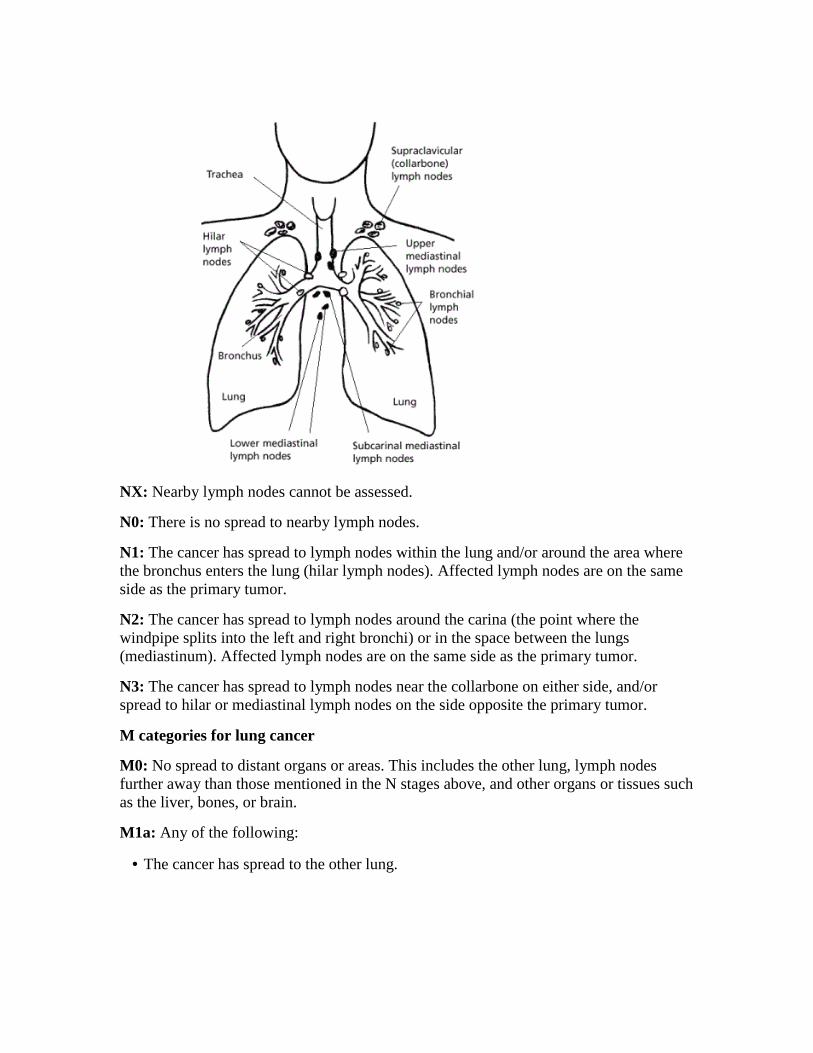
NX: Nearby lymph nodes cannot be assessed.
N0: There is no spread to nearby lymph nodes.
N1: The cancer has spread to lymph nodes within the lung and/or around the area where the bronchus enters the lung (hilar lymph nodes). Affected lymph nodes are on the same side as the primary tumor.
N2: The cancer has spread to lymph nodes around the carina (the point where the windpipe splits into the left and right bronchi) or in the space between the lungs (mediastinum). Affected lymph nodes are on the same side as the primary tumor.
N3: The cancer has spread to lymph nodes near the collarbone on either side, and/or spread to hilar or mediastinal lymph nodes on the side opposite the primary tumor.
M categories for lung cancer
M0: No spread to distant organs or areas. This includes the other lung, lymph nodes further away than those mentioned in the N stages above, and other organs or tissues such as the liver, bones, or brain.
M1a: Any of the following:
• The cancer has spread to the other lung.
• Cancer cells are found in the fluid around the lung (called a malignant pleural effusion).
• Cancer cells are found in the fluid around the heart (called a malignant pericardial effusion).
M1b: The cancer has spread to distant lymph nodes or to other organs such as the liver, bones, or brain.
Stage grouping for lung cancer
Once the T, N, and M categories have been assigned, this information is combined to assign an overall stage of 0, I, II, III, or IV. This process is called stage grouping. Some stages are subdivided into A and B. The stages identify cancers that have a similar outlook (prognosis) and thus are treated in a similar way. Patients with lower stage numbers tend to have a better outlook.
Occult (hidden) cancer
TX, N0, M0: Cancer cells are seen in a sample of sputum or other lung fluids, but the cancer isn’t found with other tests, so its location can’t be determined.
Stage 0
Tis, N0, M0: The cancer is found only in the top layers of cells lining the air passages. It has not invaded deeper into other lung tissues and has not spread to lymph nodes or distant sites.
Stage IA
T1a/T1b, N0, M0: The cancer is no larger than 3 cm across, has not reached the membranes that surround the lungs, and does not affect the main branches of the bronchi. It has not spread to lymph nodes or distant sites.
Stage IB
T2a, N0, M0: The cancer has 1 or more of the following features:
• The main tumor is larger than 3 cm across but not larger than 5 cm.
• The tumor has grown into a main bronchus, but is not within 2 cm of the carina (and it is not larger than 5 cm).
• The tumor has grown into the visceral pleura (the membranes surrounding the lungs) and is not larger than 5 cm.
• The tumor is partially clogging the airways (and is not larger than 5 cm).
The cancer has not spread to lymph nodes or distant sites.

Stage IIA
Three main combinations of categories make up this stage.
T1a/T1b, N1, M0: The cancer is no larger than 3 cm across, has not grown into the membranes that surround the lungs, and does not affect the main branches of the bronchi. It has spread to lymph nodes within the lung and/or around the area where the bronchus enters the lung (hilar lymph nodes). These lymph nodes are on the same side as the cancer. It has not spread to distant sites.
OR
T2a, N1, M0: The cancer has 1 or more of the following features:
• The main tumor is larger than 3 cm across but not larger than 5 cm.
• The tumor has grown into a main bronchus, but is not within 2 cm of the carina (and it is not larger than 5 cm).
• The tumor has grown into the visceral pleura (the membranes surrounding the lungs) and is not larger than 5 cm.
• The tumor is partially clogging the airways (and is not larger than 5 cm).
The cancer has also spread to lymph nodes within the lung and/or around the area where the bronchus enters the lung (hilar lymph nodes). These lymph nodes are on the same side as the cancer. It has not spread to distant sites.
OR
T2b, N0, M0: The cancer has 1 or more of the following features:
• The main tumor is larger than 5 cm across but not larger than 7 cm.
• The tumor has grown into a main bronchus, but is not within 2 cm of the carina (and it is between 5 and 7 cm across).
• The tumor has grown into the visceral pleura (the membranes surrounding the lungs) and is between 5 and 7 cm across.
• The tumor is partially clogging the airways (and is between 5 and 7 cm across).
The cancer has not spread to lymph nodes or distant sites.
Stage IIB
Two combinations of categories make up this stage.
T2b, N1, M0: The cancer has 1 or more of the following features:
• The main tumor is larger than 5 cm across but not larger than 7 cm.
• The tumor has grown into a main bronchus, but is not within 2 cm of the carina (and it is between 5 and 7 cm across).
• The tumor has grown into the visceral pleura (the membranes surrounding the lungs) and is between 5 and 7 cm across.
• The cancer is partially clogging the airways (and is between 5 and 7 cm across).
It has also spread to lymph nodes within the lung and/or around the area where the bronchus enters the lung (hilar lymph nodes). These lymph nodes are on the same side as the cancer. It has not spread to distant sites.
OR
T3, N0, M0: The main tumor has 1 or more of the following features:
• It is larger than 7 cm across.
• It has grown into the chest wall, the breathing muscle that separates the chest from the abdomen (diaphragm), the membranes surrounding the space between the lungs (mediastinal pleura), or membranes of the sac surrounding the heart (parietal pericardium).
• It invades a main bronchus and is closer than 2 cm (about ¾ inch) to the carina, but it does not involve the carina itself.
• It has grown into the airways enough to cause an entire lung to collapse or to cause pneumonia in the entire lung.
• Two or more separate tumor nodules are present in the same lobe of a lung.
The cancer has not spread to lymph nodes or distant sites.
Stage IIIA
Three main combinations of categories make up this stage.
T1 to T3, N2, M0: The main tumor can be any size. It has not grown into the space between the lungs (mediastinum), the heart, the large blood vessels near the heart (such as the aorta), the windpipe (trachea), the tube connecting the throat to the stomach (esophagus), the backbone, or the carina. It has not spread to different lobes of the same lung.
The cancer has spread to lymph nodes around the carina (the point where the windpipe splits into the left and right bronchi) or in the space between the lungs (mediastinum). These lymph nodes are on the same side as the main lung tumor. The cancer has not spread to distant sites.
OR
T3, N1, M0: The cancer has 1 or more of the following features:
• It is larger than 7 cm across.
• It has grown into the chest wall, the breathing muscle that separates the chest from the abdomen (diaphragm), the membranes surrounding the space between the lungs (mediastinal pleura), or membranes of the sac surrounding the heart (parietal pericardium).
• It invades a main bronchus and is closer than 2 cm to the carina, but it does not involve the carina itself.
• Two or more separate tumor nodules are present in the same lobe of a lung.
• It has grown into the airways enough to cause an entire lung to collapse or to cause pneumonia in the entire lung.
It has also spread to lymph nodes within the lung and/or around the area where the bronchus enters the lung (hilar lymph nodes). These lymph nodes are on the same side as the cancer. It has not spread to distant sites.
OR
T4, N0 or N1, M0: The cancer has 1 or more of the following features:
• A tumor of any size has grown into the space between the lungs (mediastinum), the heart, the large blood vessels near the heart (such as the aorta), the windpipe (trachea), the tube connecting the throat to the stomach (esophagus), the backbone, or the carina.
• Two or more separate tumor nodules are present in different lobes of the same lung.
It may or may not have spread to lymph nodes within the lung and/or around the area where the bronchus enters the lung (hilar lymph nodes). Any affected lymph nodes are on the same side as the cancer. It has not spread to distant sites.
Stage IIIB
Two combinations of categories make up this stage.
Any T, N3, M0: The cancer can be of any size. It may or may not have grown into nearby structures or caused pneumonia or lung collapse. It has spread to lymph nodes near the collarbone on either side, and/or has spread to hilar or mediastinal lymph nodes on the side opposite the primary tumor. The cancer has not spread to distant sites.
OR

T4, N2, M0: The cancer has 1 or more of the following features:
• A tumor of any size has grown into the space between the lungs (mediastinum), the heart, the large blood vessels near the heart (such as the aorta), the windpipe (trachea), the tube connecting the throat to the stomach (esophagus), the backbone, or the carina.
• Two or more separate tumor nodules are present in different lobes of the same lung.
The cancer has also spread to lymph nodes around the carina (the point where the windpipe splits into the left and right bronchi) or in the space between the lungs (mediastinum). Affected lymph nodes are on the same side as the main lung tumor. It has not spread to distant sites.
Stage IV
Two combinations of categories make up this stage.
Any T, any N, M1a: The cancer can be any size and may or may not have grown into nearby structures or reached nearby lymph nodes. In addition, any of the following is true:
• The cancer has spread to the other lung.
• Cancer cells are found in the fluid around the lung (called a malignant pleural effusion).
• Cancer cells are found in the fluid around the heart (called a malignant pericardial effusion).
OR
Any T, any N, M1b: The cancer can be any size and may or may not have grown into nearby structures or reached nearby lymph nodes. It has spread to distant lymph nodes or to other organs such as the liver, bones, or brain.
Non-small cell lung cancer survival rates, by stage Survival rates tell you what portion of people with the same type and stage of cancer are still alive a certain amount of time (usually 5 years) after they were diagnosed. These numbers can’t tell you how long you will live, but they may help give you a better understanding about how likely it is that your treatment will be successful. Some people will want to know the survival rates for their cancer type and stage, and some people won’t. If you don’t want to know, you don’t have to.

What is a 5-year survival rate?
Statistics on the outlook for a certain type and stage of cancer are often given as 5-year survival rates, but many people live longer – often much longer – than 5 years. The 5-year survival rate is the percentage of people who live at least 5 years after being diagnosed with cancer. For example, a 5-year survival rate of 80% means that an estimated 80 out of 100 people who have that cancer are still alive 5 years after being diagnosed. Keep in mind, however, that many of these people live much longer than 5 years after diagnosis.
But remember, the 5-year survival rates are estimates – your outlook can vary based on a number of factors specific to you.
Survival rates don’t tell the whole story
Survival rates are often based on previous outcomes of large numbers of people who had the disease, but they can’t predict what will happen in any particular person’s case. There are a number of limitations to keep in mind:
• The numbers below are among the most current available. But to get 5-year survival rates, doctors have to look at people who were treated at least 5 years ago. As treatments are improving over time, people who are now being diagnosed with non-small cell lung cancer (NSCLC) may have a better outlook than these statistics show.
• These statistics are based on the stage of the cancer when it was first diagnosed. They do not apply to cancers that later come back or spread, for example.
• The outlook for people with NSCLC varies by the stage (extent) of the cancer – in general, the survival rates are higher for people with earlier stage cancers. But many other factors can affect a person’s outlook, such as the subtype of NSCLC, gene changes in the cancer cells, the person’s age and overall health, and how well the cancer responds to treatment. The outlook for each person is specific to his or her circumstances.
Your doctor can tell you how these numbers may apply to you, as he or she is familiar with your particular situation.
Survival rates for non-small cell lung cancer, by stage
The numbers below are calculated from the National Cancer Institute’s SEER database, based on people who were diagnosed with NSCLC between 1998 and 2000. Although they are based on people diagnosed several years ago, they are the most recent rates published for the current AJCC staging system.
These survival rates include people who die from causes other than cancer.
• The 5-year survival rate for people with stage IA NSCLC is about 49%. For people with stage IB NSCLC, the 5-year survival rate is about 45%.
• For stage IIA cancer, the 5-year survival rate is about 30%. For stage IIB cancer, the survival rate is about 31%.
• The 5-year survival rate for stage IIIA NSCLC is about 14%. For stage IIIB cancers the survival rate is about 5%.
• NSCLC that has spread to other parts of the body is often hard to treat. Metastatic, or stage IV NSCLC, has a 5-year survival rate of about 1%. Still, there are often many treatment options available for people with this stage of cancer.
Remember, these survival rates are only estimates – they can’t predict what will happen to any individual person. We understand that these statistics can be confusing and may lead you to have more questions. Talk to your doctor to better understand your specific situation.
Non-small cell lung cancer treatment If you’ve been diagnosed with non-small cell lung cancer (NSCLC), your cancer care team will discuss your treatment options with you. It’s important that you think carefully about your choices. You will want to weigh the benefits of each treatment option against the possible risks and side effects.
Which treatments are used for NSCLC?
Depending on the stage of the cancer and other factors, treatment options for people with NSCLC can include:
• Surgery
• Radiofrequency ablation (RFA)
• Radiation therapy
• Chemotherapy
• Targeted therapies
• Immunotherapy
Palliative treatments can also be used to help with symptoms.
In many cases, more than one of type of treatment is used. To learn about the most common approaches to treating these cancers, see Treatment choices for non-small cell lung cancer, by stage.
Which doctors treat NSCLC?
You may have different types of doctors on your treatment team, depending on the stage of your cancer and your treatment options. These doctors could include:
• A thoracic surgeon: a doctor who treats diseases of the lungs and chest with surgery
• A radiation oncologist: a doctor who treats cancer with radiation therapy
• A medical oncologist: a doctor who treats cancer with medicines such as chemotherapy, targeted therapy, and immunotherapy
• A pulmonologist: a doctor who specializes in medical treatment of diseases of the lungs
You might have many other specialists on your treatment team as well, including physician assistants (PAs), nurse practitioners (NPs), nurses, respiratory therapists, nutrition specialists, social workers, and other health professionals. See Health Professionals Associated With Cancer Care for more on this.
Making treatment decisions
It’s important to discuss all of your treatment options, including their goals and possible side effects, with your doctors to help make the decision that best fits your needs. It’s also very important to ask questions if there is anything you’re not sure about. See What should you ask your doctor about non-small cell lung cancer? for ideas.
Getting a second opinion
You may also want to get a second opinion. This can give you more information and help you feel more certain about the treatment plan you choose. If you aren’t sure where to go for a second opinion, ask your doctor for help.
Thinking about taking part in a clinical trial
Clinical trials are carefully controlled research studies that are done to get a closer look at promising new treatments or procedures. Clinical trials are one way to get state-of-the art cancer treatment. In some cases they may be the only way to get access to newer treatments. They are also the best way for doctors to learn better methods to treat cancer. Still, they are not right for everyone.
If you would like to learn more about clinical trials that might be right for you, start by asking your doctor if your clinic or hospital conducts clinical trials. You can also call our clinical trials matching service at 1-800-303-5691 for a list of studies that meet your medical needs, or see Clinical Trials to learn more.
Considering complementary and alternative methods
You may hear about complementary or alternative methods that your doctor hasn’t mentioned to treat your cancer or relieve symptoms. These methods can include vitamins, herbs, and special diets, or other methods such as acupuncture or massage, to name a few.
Complementary methods refer to treatments that are used along with your regular medical care. Alternative treatments are used instead of a doctor’s medical treatment. Although some of these methods might be helpful in relieving symptoms or helping you feel better, many have not been proven to work. Some might even be dangerous.
As you consider your options, look for “red flags” that might suggest fraud. Does the method promise to cure all or most cancers? Are you told not to have regular medical treatments? Is the treatment a “secret” that requires you to visit certain providers or travel to another country?
Be sure to talk to your cancer care team about any method you are thinking about using. They can help you learn what is known (or not known) about the method, which can help you make an informed decision. See Complementary and Alternative Medicine to learn more.
Choosing to stop treatment or choosing no treatment at all
For some people, when treatments have been tried and are no longer controlling the cancer, it could be time to weigh the benefits and risks of continuing to try new treatments. Whether or not you continue treatment, there are still things you can do to help maintain or improve your quality of life. Learn more in If Cancer Treatments Are No Longer Working.
Some people, especially if the cancer is advanced, might not want to be treated at all. There are many reasons you might decide not to get cancer treatment, but it’s important to talk this through with your doctors before you make this decision. Remember that even if you choose not to treat the cancer, you can still get supportive care to help with pain or other symptoms.
Help getting through treatment
Your cancer care team will be your first source of information and support, but there are other resources for help when you need it. Hospital- or clinic-based support services are

an important part of your care. These might include nursing or social work services, financial aid, nutritional advice, rehab, or spiritual help.
The American Cancer Society also has programs and services – including rides to treatment, lodging, support groups, and more – to help you get through treatment. Call our National Cancer Information Center at 1-800-227-2345 and speak with one of our trained specialists on call 24 hours a day, every day.
The treatment information in this document is not official policy of the American Cancer Society and is not intended as medical advice to replace the expertise and judgment of your cancer care team. It is intended to help you and your family make informed decisions, together with your doctor. Your doctor may have reasons for suggesting a treatment plan different from these general treatment options. Don't hesitate to ask him or her questions about your treatment options..
Surgery for non-small cell lung cancer Surgery to remove the cancer (often along with other treatments) may be an option for early stage non-small cell lung cancer (NSCLC). If surgery can be done, it provides the best chance to cure NSCLC. Lung cancer surgery is a complex operation that can have serious consequences, so it should be done by a surgeon who has a lot of experience operating on lung cancers.
If your doctor thinks the lung cancer can be treated with surgery, pulmonary function tests will be done beforehand to see if you would still have enough healthy lung tissue left after surgery. Other tests will check the function of your heart and other organs to be sure you’re healthy enough for surgery.
Because surgery doesn’t help more advanced stage lung cancers, your doctor will also want to check if the cancer has already spread to the lymph nodes between the lungs. This is often done just before surgery with mediastinoscopy or with some of the other techniques described in How is non-small cell lung cancer diagnosed?
Types of lung surgery
Different operations can be used to treat (and possibly cure) NSCLC:
• Pneumonectomy: This surgery removes an entire lung. This might be needed if the tumor is close to the center of the chest.
• Lobectomy: The lungs are made up of 5 lobes (3 on the right and 2 on the left). In this surgery, the entire lobe containing the tumor(s) is removed. This is often the preferred type of operation for NSCLC if it can be done.
• Segmentectomy or wedge resection: In these surgeries, only part of a lobe is removed. This approach might be used, for example, if a person doesn’t have enough lung function to withstand removing the whole lobe.
• Sleeve resection: This operation may be used to treat some cancers in large airways in the lungs. If you think of the large airway with a tumor as similar to the sleeve of a shirt with a stain a couple of inches above the wrist, the sleeve resection would be like cutting across the sleeve above and below the stain and then sewing the cuff back onto the shortened sleeve. A surgeon may be able to do this operation instead of a pneumonectomy to preserve more lung function.
With any of these operations, nearby lymph nodes are also removed to look for possible spread of the cancer. These operations require general anesthesia (where you are in a deep sleep) and are usually done through a surgical incision between the ribs in the side of the chest (called a thoracotomy).
The type of operation your doctor recommends depends on the size and location of the tumor and on how well your lungs are functioning. Doctors often prefer to do a more extensive operation (for example, a lobectomy instead of a segmentectomy) if a person’s lungs are healthy enough, as it may provide a better chance to cure the cancer.
When you wake up from surgery, you will have a tube (or tubes) coming out of your chest and attached to a special canister to allow excess fluid and air to drain out. The tube(s) will be removed once the fluid drainage and air leak subside. Generally, you will need to spend 5 to 7 days in the hospital after the surgery.
Video-assisted thoracic surgery (VATS)
Increasingly, doctors now treat early-stage lung cancers in the outer parts of the lung with a procedure called video-assisted thoracic surgery (VATS), which requires smaller incisions than a thoracotomy.
During this operation, a thin, rigid tube with a tiny video camera on the end is placed through a small cut in the side of the chest to help the surgeon see inside the chest on a TV monitor. One or two other small cuts are created in the skin, and long instruments are passed through these cuts to do the same operation that would be done using an open approach (thoracotomy). One of the incisions is enlarged if a lobectomy or pneumonectomy is done to allow the specimen to be removed. Because only small incisions are needed, there is usually less pain after the surgery and a shorter hospital stay – typically 4 to 5 days.
Most experts recommend that only early-stage tumors near the outside of the lung be treated this way. The cure rate after this surgery seems to be the same as with surgery done with a larger incision. But it’s important that the surgeon doing this procedure is experienced, because it requires a great deal of technical skill.
Possible risks and side effects of lung surgery
Surgery for lung cancer is a major operation and can have serious side effects, which is why surgery isn’t a good idea for everyone. While all surgeries carry some risks, these depend to some degree on the extent of the surgery and the person’s health beforehand.
Possible complications during and soon after surgery can include reactions to anesthesia, excess bleeding, blood clots in the legs or lungs, wound infections, and pneumonia. While it is rare, some people may not survive the surgery.
Recovering from lung cancer surgery typically takes weeks to months. If the surgery is done through a thoracotomy (a long incision in the chest), the surgeon must spread ribs to get to the lung, so the area near the incision will hurt for some time after surgery. Your activity might be limited for at least a month or two. People who have VATS instead of thoracotomy tend to have less pain after surgery and to recover more quickly.
If your lungs are in good condition (other than the presence of the cancer) you can usually return to normal activities after some time if a lobe or even an entire lung has been removed. If you also have another lung disease such as emphysema or chronic bronchitis (which are common among long-time smokers), you might become short of breath with certain levels of activity after surgery.
Surgery for lung cancers with limited spread to other organs
If the lung cancer has spread to your brain or to an adrenal gland and there is only one tumor, you may benefit from having the tumor removed. This surgery should be considered only if the tumor in the lung can also be removed completely. Even then, not all lung cancer experts agree with this approach, especially if the tumor is in the adrenal gland.
For tumors in the brain, this is done by surgery through a hole in the skull (called a craniotomy). It should only be done if the tumor can be removed without damaging vital areas of the brain.
For more general information about surgery, see A Guide to Cancer Surgery.
Radiofrequency ablation (RFA) for non-small cell lung cancer This treatment might be an option for some people some small lung tumors that are near the outer edge of the lungs, especially if they can’t tolerate surgery.
RFA uses high-energy radio waves to heat the tumor. A thin, needle-like probe is put through the skin and moved in until the tip is in the tumor. Placement of the probe is

guided by CT scans. Once the tip is in place, an electric current is passed through the probe, which heats the tumor and destroys the cancer cells.
RFA is usually done as an outpatient procedure, using local anesthesia (numbing medicine) where the probe is inserted. You may be given medicine to help you relax as well.
You might have some pain where the needle was inserted for a few days after the procedure. Major complications are uncommon, but they can include the partial collapse of a lung (which often goes away on its own) or bleeding into the lung.
Radiation therapy for non-small cell lung cancer Radiation therapy uses high-energy rays (such as x-rays) or particles to kill cancer cells.
When might radiation therapy be used?
Depending on the stage of the non-small cell lung cancer (NSCLC) and other factors, radiation therapy might be used:
• As the main treatment (sometimes along with chemotherapy), especially if the lung tumor can’t be removed because of its size or location, if a person isn’t healthy enough for surgery, or if a person doesn’t want surgery.
• After surgery (alone or along with chemotherapy) to try to kill any small areas of cancer that surgery might have missed.
• Before surgery (usually along with chemotherapy) to try to shrink a lung tumor to make it easier to operate on.
• To treat a single area of cancer spread, such as a tumor in the brain or an adrenal gland. (This might be done along with surgery to treat the main lung tumor.)
• To relieve (palliate) symptoms of advanced NSCLC such as pain, bleeding, trouble swallowing, cough, or problems caused by spread to other organs such as the brain. For example, brachytherapy is most often used to help relieve blockage of large airways by cancer.
Types of radiation therapy
There are 2 main types of radiation therapy:
• External beam radiation therapy
• Brachytherapy (internal radiation therapy)
External beam radiation therapy
External beam radiation therapy (EBRT) focuses radiation from outside the body on the cancer. This is the type of radiation therapy most often used to treat NSCLC or its spread to other organs.
Before your treatments start, the radiation team will take careful measurements to determine the correct angles for aiming the radiation beams and the proper dose of radiation. This planning session, called simulation, usually includes getting imaging tests such as CT scans.
Treatment is much like getting an x-ray, but the radiation dose is stronger. The procedure itself is painless. Each treatment lasts only a few minutes, although the setup time – getting you into place for treatment – usually takes longer. Most often, radiation treatments to the lungs are given 5 days a week for 5 to 7 weeks, but this can vary based on the type of EBRT and the reason it’s being given.
In recent years, newer EBRT techniques have been shown to help doctors treat lung cancers more accurately while lowering the radiation exposure to nearby healthy tissues. These include:
Three-dimensional conformal radiation therapy (3D-CRT): 3D-CRT uses special computers to precisely map the tumor’s location. Radiation beams are then shaped and aimed at the tumor(s) from several directions, which makes it less likely to damage normal tissues.
Intensity modulated radiation therapy (IMRT): IMRT is an advanced form of 3D therapy. It uses a computer-driven machine that moves around you as it delivers radiation. Along with shaping the beams and aiming them at the tumor from several angles, the intensity (strength) of the beams can be adjusted to limit the dose reaching nearby normal tissues. This technique is used most often if tumors are near important structures such as the spinal cord. Many cancer centers now use IMRT.
A variation of IMRT is called volumetric modulated arc therapy (VMAT). It uses a machine that delivers radiation quickly as it rotates once around the body. This allows each treatment to be given over just a few minutes.
Stereotactic body radiation therapy (SBRT): SBRT, also known as stereotactic ablative radiotherapy (SABR), is sometimes used to treat very early-stage lung cancers when surgery isn’t an option due to a person’s health or in people who don’t want surgery.
Instead of giving a small dose of radiation each day for several weeks, SBRT uses very focused beams of high-dose radiation given in fewer (usually 1 to 5) treatments. Several beams are aimed at the tumor from different angles. To target the radiation precisely, you
are put in a specially designed body frame for each treatment. This reduces the movement of the lung tumor during breathing. Like other forms of external radiation, the treatment itself is painless.
Early results with SBRT for smaller lung tumors have been very promising, and it seems to have a low risk of complications. It is also being studied for tumors that have spread to other parts of the body, such as the bones or liver.
Stereotactic radiosurgery (SRS): SRS is a type of stereotactic radiation therapy that is given in only one session. It can sometimes be used instead of or along with surgery for single tumors that have spread to the brain. In one version of this treatment, a machine called a Gamma Knife® focuses about 200 beams of radiation on the tumor from different angles over a few minutes to hours. Your head is kept in the same position with a rigid frame. In another version, a linear accelerator (a machine that creates radiation) that is controlled by a computer moves around your head to deliver radiation to the tumor from many different angles. These treatments can be repeated if needed.
Brachytherapy (internal radiation therapy)
In people with NSCLC, brachytherapy is sometimes used to shrink tumors in the airway to relieve symptoms.
For this type of treatment, the doctor places a small source of radioactive material (often in the form of small pellets) directly into the cancer or into the airway next to the cancer. This is usually done through a bronchoscope, but it may also be done during surgery. The radiation travels only a short distance from the source, limiting the effects on surrounding healthy tissues. The radiation source is usually removed after a short time. Less often, small radioactive “seeds” are left in place permanently, and the radiation gets weaker over several weeks.Possible side effects of radiation therapy
If you are going to get radiation therapy, it’s important to ask your doctor beforehand about the possible side effects so you know what to expect. Common side effects depend on where the radiation is aimed and can include:
• Fatigue
• Nausea and vomiting
• Loss of appetite and weight loss
• Skin changes in the area being treated, which can range from mild redness to blistering and peeling
• Hair loss where the radiation enters the body
Often these go away after treatment. When radiation is given with chemotherapy, the side effects are often worse.
Radiation therapy to the chest may damage your lungs and cause a cough, problems breathing, and shortness of breath. These usually improve after treatment is over, although sometimes they may not go away completely.
Your esophagus, which is in the middle of your chest, may be exposed to radiation, which could cause a sore throat and trouble swallowing during treatment. This might make it hard to eat anything other than soft foods or liquids for a while. This also improves after completion of treatment.
Radiation therapy to large areas of the brain can sometimes cause memory loss, headaches, trouble thinking, or reduced sexual desire. Usually these symptoms are minor compared with those caused by a brain tumor, but they can affect your quality of life.
For more information, see the Radiation Therapy section of our website.
Chemotherapy for non-small cell lung cancer Chemotherapy (chemo) is treatment with anti-cancer drugs injected into a vein or taken by mouth. These drugs enter the bloodstream and go throughout the body, making this treatment useful for cancer anywhere in the body.
When might chemotherapy be used?
Depending on the stage of non-small cell lung cancer (NSCLC) and other factors, chemo may be used in different situations:
• Before surgery (sometimes along with radiation therapy) to try to shrink a tumor. This is known as neoadjuvant therapy.
• After surgery (sometimes along with radiation therapy) to try to kill any cancer cells that might have been left behind. This is known as adjuvant therapy.
• Along with radiation therapy (concurrent therapy) for some cancers that can’t be removed by surgery because the cancer has grown into nearby important structures
• As the main treatment (sometimes along with radiation therapy) for more advanced cancers or for some people who aren’t healthy enough for surgery.

Chemo is often not recommended for patients in poor health, but advanced age by itself is not a barrier to getting chemo.
Drugs used to treat NSCLC
The chemo drugs most often used for NSCLC include:
• Cisplatin
• Carboplatin
• Paclitaxel (Taxol)
• Albumin-bound paclitaxel (nab-paclitaxel, Abraxane)
• Docetaxel (Taxotere)
• Gemcitabine (Gemzar)
• Vinorelbine (Navelbine)
• Irinotecan (Camptosar)
• Etoposide (VP-16)
• Vinblastine
• Pemetrexed (Alimta)
Most often, treatment for NSCLC uses a combination of 2 chemo drugs. Studies have shown that adding a third chemo drug doesn’t add much benefit and is likely to cause more side effects. Single-drug chemo is sometimes used for people who might not tolerate combination chemotherapy well, such as those in poor overall health or who are elderly.
If a combination is used, it often includes cisplatin or carboplatin plus one other drug. Sometimes combinations that do not include these drugs, such as gemcitabine with vinorelbine or paclitaxel, may be used.
For people with advanced lung cancers who meet certain criteria, a targeted therapy drug such as bevacizumab (Avastin), ramucirumab (Cyramza), or necitumumab (Portrazza) may be added to treatment as well.
Doctors give chemo in cycles, with a period of treatment (usually 1 to 3 days) followed by a rest period to allow the body time to recover. Some chemo drugs, though, are given every day. Chemo cycles generally last about 3 to 4 weeks.
For advanced cancers, the initial chemo combination is often given for 4 to 6 cycles. Some doctors now recommend giving treatment beyond this with a single chemo or targeted drug, even in people who have had a good response to their initial chemotherapy. Some studies have found that this continuing treatment, known as maintenance therapy, might help keep the cancer in check and help some people live longer. For more information, see What’s new in non-small cell lung cancer research?
If the initial chemo treatment for advanced lung cancer is no longer working, the doctor may recommend second-line treatment with a single chemo drug such as docetaxel or pemetrexed, or with a targeted therapy or immunotherapy drug. Again, advanced age is no barrier to receiving these drugs as long as the person is in good general health.
Possible side effects
Chemo drugs attack cells that are dividing quickly, which is why they work against cancer cells. But other cells in the body, such as those in the bone marrow (where new blood cells are made), the lining of the mouth and intestines, and the hair follicles, also divide quickly. These cells are also likely to be affected by chemo, which can lead to certain side effects.
The side effects of chemo depend on the type and dose of drugs given and how long they are taken. Some common side effects include:
• Hair loss
• Mouth sores
• Loss of appetite
• Nausea and vomiting
• Diarrhea or constipation
• Increased chance of infections (from having too few white blood cells)
• Easy bruising or bleeding (from having too few blood platelets)
• Fatigue (from having too few red blood cells)
These side effects usually go away after treatment is finished. There are often ways to lessen these side effects. For example, drugs can be given to help prevent or reduce nausea and vomiting.
Some drugs can have specific side effects. For example, drugs such as cisplatin, vinorelbine, docetaxel, or paclitaxel can cause nerve damage (peripheral neuropathy). This can sometimes lead to symptoms (mainly in the hands and feet) such as pain, burning or tingling sensations, sensitivity to cold or heat, or weakness. In most people

this goes away or gets better once treatment is stopped, but it may last a long time in some people. For more information, see Peripheral Neuropathy Caused by Chemotherapy.
Be sure to report any side effects you notice while getting chemo to your medical team so that they can be treated promptly. In some cases, the doses of the chemo drugs may need to be reduced or treatment may need to be delayed or stopped to prevent the effects from getting worse.
To learn more about chemo, see the Chemotherapy section of our website.
Targeted therapy drugs for non-small cell lung cancer As researchers have learned more about the changes in non-small cell lung cancer (NSCLC) cells that help them grow, they have developed newer drugs to specifically target these changes. Targeted drugs work differently from standard chemotherapy (chemo) drugs. They sometimes work when chemo drugs don’t, and they often have different (and less severe) side effects. At this time, they are most often used for advanced lung cancers, either along with chemo or by themselves.
Drugs that target tumor blood vessel growth (angiogenesis)
For tumors to grow, they need to form new blood vessels to keep them nourished. This process is called angiogenesis. Some targeted drugs, called angiogenesis inhibitors, block this new blood vessel growth:
• Bevacizumab (Avastin) is used to treat advanced NSCLC. It is a monoclonal antibody (a man-made version of a specific immune system protein) that targets vascular endothelial growth factor (VEGF), a protein that helps new blood vessels to form. This drug is often used with chemo for a time. Then if the cancer responds, the chemo may be stopped and the bevacizumab given by itself until the cancer starts growing again.
• Ramucirumab (Cyramza) can also be used to treat advanced NSCLC. VEGF has to bind to cell proteins called receptors to act. This drug is a monoclonal antibody that targets a VEGF receptor. This helps stop the formation of new blood vessels. This drug is most often given after another treatment stops working. It is often combined with chemo.
Side effects
Common side effects of these drugs include:

• High blood pressure
• Tiredness
• Bleeding
• Low white blood cell counts (with increased risk of infections)
• Headaches
• Mouth sores
• Loss of appetite
• Diarrhea
Rare but possibly serious side effects can include blood clots, severe bleeding, holes (called perforations) forming in the intestine, heart problems, and slow wound healing. If a hole forms in the intestine it can lead to severe infection and may require surgery to correct.
Because of the risks of bleeding, these drugs typically aren’t used in people who are coughing up blood or who are taking drugs called blood thinners. The risk of serious bleeding in the lungs is higher in patients with the squamous cell type of NSCLC, which is why most current guidelines do not recommend using bevacizumab in people with this type of lung cancer.
Drugs that target cells with EGFR changes
Epidermal growth factor receptor (EGFR) is a protein on the surface of cells. It normally helps the cells grow and divide. Some NSCLC cells have too much EGFR, which makes them grow faster. Drugs called EGFR inhibitors can block the signal from EGFR that tells the cells to grow. Some of these drugs can be used to treat NSCLC.
EGFR inhibitors used in NSCLC with EGFR gene mutations
• Erlotinib (Tarceva)
• Afatinib (Gilotrif)
• Gefitinib (Iressa)
These drugs can be used alone (without chemo) as the first treatment for advanced NSCLCs that have certain mutations in the EGFR gene. These are more common in women and people who haven’t smoked. Erlotinib can also be used for advanced NSCLC without these mutations if chemo isn’t working. All of these medicines are taken as pills.

EGFR inhibitors that also target cells with the T790M mutation
EGFR inhibitors can often shrink tumors for several months or more. But eventually these drugs stop working for most people, usually because the cancer cells develop another mutation in the EGFR gene. One such mutation is known as T790M. But some newer EGFR inhibitors also work against cells with the T790M mutation, including osimertinib (Tagrisso).
Doctors now commonly get another tumor biopsy when EGFR inhibitors have stopped working to see if the patient has developed the T790M mutation.
EGFR inhibitors used for squamous cell NSCLC
Necitumumab (Portrazza) is a monoclonal antibody (a man-made version of an immune system protein) that targets EGFR. It can be used along with chemotherapy as the first treatment in people with advanced squamous cell NSCLC. This drug is given as an infusion into a vein (IV).
Side effects
Common side effects of all EGFR inhibitors include:
• Skin problems
• Diarrhea
• Mouth sores
• Loss of appetite
Skin problems can include an acne-like rash on the face and chest, which in some cases can lead to skin infections. For more detailed information on the skin problems that can result from anti-EGFR drugs, see Targeted Therapy.
These drugs can also cause more serious, but less common, side effects. For example, necitumumab can lower the levels of certain minerals in the blood, which can affect the heart rhythm and in some cases might be life-threatening.
Drugs that target cells with ALK gene changes
About 5% of NSCLCs have a rearrangement in a gene called ALK. This change is most often seen in non-smokers (or light smokers) who have the adenocarcinoma subtype of NSCLC. The ALK gene rearrangement produces an abnormal ALK protein that causes the cells to grow and spread. Drugs that target the abnormal ALK protein include:
• Crizotinib (Xalkori)

• Ceritinib (Zykadia)
• Alectinib (Alecensa)
These drugs can often shrink tumors in people whose lung cancers have the ALK gene change. Although they can help after chemo has stopped working, they are often used instead of chemo in people whose cancers have the ALK gene rearrangement.
These drugs also seem to be useful in treating people whose cancers have changes in the ROS1 gene.
These drugs are taken as pills.
Side effects
Common side effects of ALK inhibitors include:
• Nausea and vomiting
• Diarrhea
• Constipation
• Fatigue
• Changes in vision
Other side effects are also possible. Some side effects can be severe, such as low white blood cell counts, lung inflammation, liver damage, and heart rhythm problems.
Immunotherapy for non-small cell lung cancer Immunotherapy is the use of medicines to stimulate a person’s own immune system to recognize and destroy cancer cells more effectively. Immunotherapy can be used to treat some forms of non-small cell lung cancer (NSCLC).
Immune checkpoint inhibitors
An important part of the immune system is its ability to keep itself from attacking normal cells in the body. To do this, it uses “checkpoints” – molecules on immune cells that need to be turned on (or off) to start an immune response. Cancer cells sometimes use these checkpoints to avoid being attacked by the immune system. But newer drugs that target these checkpoints hold a lot of promise as cancer treatments.

• Nivolumab (Opdivo) and pembrolizumab (Keytruda) target PD-1, a protein on immune system cells called T cells that normally helps keep these cells from attacking other cells in the body. By blocking PD-1, these drugs boost the immune response against cancer cells. This can shrink some tumors or slow their growth.
• Atezolizumab (Tecentriq) targets PD-L1, a protein related to PD-1 that is found on some tumor cells and immune cells. Blocking this protein can also help boost the immune response against cancer cells.
These drugs can be used in people with certain types of NSCLC whose cancer starts growing again after chemotherapy or other drug treatments. Pembrolizumab can also be used as the first treatment (instead of chemo or other drugs) in some people.
These drugs are given as an intravenous (IV) infusion every 2 or 3 weeks.
Possible side effects
Side effects of these drugs can include fatigue, cough, nausea, itching, skin rash, loss of appetite, constipation, joint pain, and diarrhea.
Other, more serious side effects occur less often. These drugs work by basically removing the brakes on the body’s immune system. Sometimes the immune system starts attacking other parts of the body, which can cause serious or even life-threatening problems in the lungs, intestines, liver, hormone-making glands, kidneys, or other organs.
It’s very important to report any new side effects to your health care team promptly. If serious side effects do occur, treatment may need to be stopped and you may get high doses of corticosteroids to suppress your immune system.
Palliative procedures for non-small cell lung cancer Palliative, or supportive care, is aimed at relieving symptoms and improving a person’s quality of life.
People with lung cancer often benefit from procedures to help with problems caused by the cancer. For example, people with advanced lung cancer can have shortness of breath. This can be caused by a number of things, including fluid around the lung or an airway that is blocked by a tumor. Although treating the cancer with chemotherapy or other drugs may help with this, other treatments may be needed as well.

Treating fluid buildup in the area around the lungs
Sometimes fluid can build up in the chest outside of the lungs. This is called a pleural effusion. It can press on the lungs and cause trouble breathing.
Thoracentesis
This is done to drain the fluid. For this procedure, the doctor will numb an area in the chest, and then place a hollow needle into the space between the lungs and the ribs to drain the fluid. This is often done using ultrasound to guide the needle into the fluid.
Pleurodesis
This procedure might be done to remove the fluid and keep it from coming back.
One way to do this is to make a small cut in the skin of the chest wall, and place a hollow tube (called a chest tube) into the chest to remove the fluid. Then a substance is instilled into the chest through the tube that causes the linings of the lung (visceral pleura) and chest wall (parietal pleura) to stick together, sealing the space and limiting further fluid buildup. A number of substances can be used for this, such as talc, the antibiotic doxycycline, or a chemotherapy drug like bleomycin. The tube is often left in for a couple of days to drain any new fluid that might collect.
Another way to do this is to blow talc into the space around the lungs during an operation. This is done through a small incision using thoracoscopy.
Catheter placement
This is another way to control the buildup of fluid. One end of the catheter (a thin, flexible tube) is placed in the chest through a small cut in the skin, and the other end is left outside the body. This is done in a doctor’s office or hospital. Once in place, the catheter can be attached to a special bottle or other device to allow the fluid to drain out on a regular basis.
Treating fluid buildup around the heart
Lung cancer can sometimes spread to the area around the heart. This can lead to fluid buildup inside the sac around the heart (called a pericardial effusion). The fluid can press on the heart, affecting how well it works.

Pericardiocentesis
In this procedure, the fluid is drained with a needle placed into the space around the heart. This is usually done using an ultrasound of the heart (echocardiogram) to guide the needle.
Creating a pericardial window
This procedure can be done to keep the fluid from building up again. During surgery, a piece of the sac around the heart (the pericardium) is removed to allow the fluid to drain into the chest or belly.
Treating an airway blocked by a tumor
If the cancer is growing into an airway in the lung, it can block the airway and cause problems like pneumonia or shortness of breath. Treatments can often relieve the blockage in the airway.
Photodynamic therapy (PDT)
This type of treatment can be used to treat very early-stage lung cancers that are only in the outer layers of the lung airways, when other treatments aren’t appropriate. It can also be used to help open up airways blocked by tumors to help people breathe better.
For this technique, a light-activated drug called porfimer sodium (Photofrin) is injected into a vein. This drug collects more in cancer cells than in normal cells. After a couple of days (to give the drug time to build up in the cancer cells), a bronchoscope is passed down the throat and into the lung. This may be done with either local anesthesia (where the throat is numbed) and sedation, or with general anesthesia (where you are in a deep sleep). A special laser light on the end of the bronchoscope is aimed at the tumor, which activates the drug and causes the cells to die. The dead cells are then removed a few days later during a bronchoscopy. This process can be repeated if needed.
PDT can cause swelling in the airway for a few days, which may lead to some shortness of breath, as well as coughing up blood or thick mucus. Some of this drug also collects in normal cells in the body, such as skin and eye cells. This can make you very sensitive to sunlight or strong indoor lights. Too much exposure can cause serious skin reactions (like a severe sunburn), so doctors recommend staying out of any strong light for several weeks after the injection.
For more information on PDT, see Photodynamic Therapy.
Laser therapy
Lasers can sometimes be used to treat very small tumors in the linings of airways. They can also be used to help open up airways blocked by larger tumors to help people breathe better.
You are usually asleep (under general anesthesia) for this type of treatment. The laser is on the end of a bronchoscope, which is passed down the throat and next to the tumor. The doctor then aims the laser beam at the tumor to burn it away. This treatment can usually be repeated, if needed.
Stent placement
If a lung tumor has grown into an airway and is causing problems, sometimes a hard silicone or metal tube called a stent is placed in the airway to help keep it open using a bronchoscope. This is often done after other treatments such as PDT or laser therapy.
Treatment choices for non-small cell lung cancer, by stage The treatment options for non-small cell lung cancer (NSCLC) are based mainly on the stage (extent) of the cancer, but other factors, such as a person’s overall health and lung function, as well as certain traits of the cancer itself, are also important.
If you smoke, one of the most important things you can do to be ready for treatment is to try to quit. Studies have shown that patients who stop smoking after a diagnosis of lung cancer tend to have better outcomes than those who don’t.
Treating occult cancer
For these cancers, malignant cells are seen on sputum cytology but no obvious tumor can be found with bronchoscopy or imaging tests. They are usually early-stage cancers. Bronchoscopy and possibly other tests are usually repeated every few months to look for a tumor. If a tumor is found, treatment will depend on the stage.
Treating stage 0 NSCLC
Because stage 0 NSCLC is limited to the lining layer of airways and has not invaded deeper into the lung tissue or other areas, it is usually curable by surgery alone. No chemotherapy or radiation therapy is needed.
If you are healthy enough for surgery, you can usually be treated by segmentectomy or wedge resection (removal of part of the lobe of the lung). Cancers in some locations

(such as where the windpipe divides into the left and right main bronchi) may be treated with a sleeve resection, but in some cases they may be hard to remove completely without removing a lobe (lobectomy) or even an entire lung (pneumonectomy).
In some cases, treatments such as photodynamic therapy (PDT), laser therapy, or brachytherapy (internal radiation) may be alternatives to surgery for stage 0 cancers. If your cancer is truly stage 0, these treatments should cure you.
Treating stage I NSCLC
If you have stage I NSCLC, surgery may be the only treatment you need. This may be done either by taking out the lobe of the lung containing the tumor (lobectomy) or by taking out a smaller piece of the lung (sleeve resection, segmentectomy, or wedge resection). At least some lymph nodes within the lung and in the space between the lungs will also be removed and checked for cancer cells.
Segmentectomy or wedge resection is generally an option only for very small stage I cancers and for patients with other health problems that make removing the entire lobe dangerous. Still, most surgeons believe it is better to do a lobectomy if the patient can tolerate it, as it offers the best chance for cure.
For people with stage I NSCLC that has a higher risk of coming back (based on size, location, or other factors), adjuvant chemotherapy after surgery may lower the risk that cancer will return. But doctors aren’t always sure how to determine which people are likely to be helped by chemo. New lab tests that look at the patterns of certain genes in the cancer cells may help with this. Studies are now being done to see if these tests are accurate.
After surgery, the removed tissue is checked to see if there are cancer cells at the edges of the surgery specimen (called positive margins). This could mean that some cancer has been left behind, so a second surgery might be done to try to ensure that all the cancer has been removed. (This might be followed by chemotherapy as well.) Another option might be to use radiation therapy after surgery.
If you have serious health problems that prevent you from having surgery, you may get stereotactic body radiation therapy (SBRT) or another type of radiation therapy as your main treatment. Radiofrequency ablation (RFA) may be another option if the tumor is small and in the outer part of the lung.
Treating stage II NSCLC
People who have stage II NSCLC and are healthy enough for surgery usually have the cancer removed by lobectomy or sleeve resection. Sometimes removing the whole lung (pneumonectomy) is needed.
Any lymph nodes likely to have cancer in them are also removed. The extent of lymph node involvement and whether or not cancer cells are found at the edges of the removed tissues are important factors when planning the next step of treatment.
In some cases, chemotherapy (often along with radiation) may be recommend before surgery to try to shrink the tumor to make the operation easier.
After surgery, the removed tissue is checked to see if there are cancer cells at the edges of the surgery specimen (called positive margins). This might mean that some cancer has been left behind, so a second surgery might be done to try to remove any remaining cancer. This may be followed by chemotherapy (chemo). Another option is to treat with radiation, sometimes along with chemo.
Even if positive margins are not found, chemo is usually recommended after surgery to try to destroy any cancer cells that might have been left behind. As with stage I cancers, newer lab tests now being studied may help doctors find out which patients need this adjuvant treatment and which are less likely to benefit from it.
If you have serious medical problems that would keep you from having surgery, you may get only radiation therapy as your main treatment.
Treating stage IIIA NSCLC
Treatment for stage IIIA NSCLC may include some combination of radiation therapy, chemotherapy (chemo), and/or surgery. For this reason, planning treatment for stage IIIA NSCLC often requires input from a medical oncologist, radiation oncologist, and a thoracic surgeon. Your treatment options depend on the size of the tumor, where it is in your lung, which lymph nodes it has spread to, your overall health, and how well you are tolerating treatment.
For patients who can tolerate it, treatment usually starts with chemo, often combined with radiation therapy. Surgery may be an option after this if the doctor thinks any remaining cancer can be removed and the patient is healthy enough. (In some cases, surgery may be an option as the first treatment.) This is often followed by chemo, and possibly radiation therapy if it hasn’t been given before.
For people who are not healthy enough for surgery, radiation therapy, which may be combined with chemo, is often used.
Treating stage IIIB NSCLC
Stage IIIB NSCLC has spread to lymph nodes that are near the other lung or in the neck, and may also have grown into important structures in the chest. These cancers can’t be removed completely by surgery. As with other stages of lung cancer, treatment depends on the patient’s overall health. If you are in fairly good health you may be helped by
chemotherapy (chemo) combined with radiation therapy. Some people can even be cured with this treatment. Patients who are not healthy enough for this combination are often treated with radiation therapy alone, or, less often, chemo alone.
These cancers can be hard to treat, so taking part in a clinical trial of newer treatments may be a good option for some people.
Treating stage IV NSCLC
Stage IV NSCLC is widespread when it is diagnosed. Because these cancers have spread to distant sites, they are very hard to cure. Treatment options depend on where the cancer has spread, the number of tumors, and your overall health.
If you are in otherwise good health, treatments such as surgery, chemotherapy (chemo), targeted therapy, immunotherapy, and radiation therapy may help you live longer and make you feel better by relieving symptoms, even though they aren’t likely to cure you.
Other treatments, such as photodynamic therapy (PDT) or laser therapy, may also be used to help relieve symptoms. In any case, if you are going to be treated for advanced NSCLC, be sure you understand the goals of treatment before you start.
Cancer that has spread to only one other site
Cancer that is limited in the lungs and has only spread to one other site (such as the brain) is not common, but it can sometimes be treated (and even potentially cured) with surgery and/or radiation therapy to treat the area of cancer spread, followed by treatment of the cancer in the lung. For example, a single tumor in the brain may be treated with surgery or stereotactic radiation, followed by radiation to the whole brain. Treatment for the lung tumor is then based on its T and N stages, and may include surgery, chemo, radiation, or some of these in combination.
Cancer that has spread widely
For cancers that have spread widely throughout the body, before any treatments start, your tumor will be tested for common gene mutations (such as in the EGFR, ALK, or ROS1 genes). If one of these genes is mutated in your cancer cells, your first treatment will likely be a targeted therapy drug:
• For tumors that have the ALK gene change, crizotinib (Xalkori) is often the first treatment. Ceritinib (Zykadia) or alectinib (Alecensa) can be used if crizotinib stops working or is not well tolerated.
• For people whose cancers have certain changes in the EGFR gene, the anti-EGFR drugs erlotinib (Tarceva), gefitinib (Iressa), or afatinib (Gilotrif) may be used as the first treatment.

• For people whose cancers have changes in the ROS1 gene, an ALK inhibitor such as crizotinib might be used.
Your tumor cells might also be tested for the PD-L1 protein. Tumors with higher levels of PD-L1 are more likely to respond to certain immunotherapy drugs, so treatment with pembrolizumab (Keytruda) might be an option as the first treatment.
For most other cancers that have spread, chemo is usually the main treatment, as long as the person is healthy enough for it. For people who are not at high risk for bleeding (that is, they do not have squamous cell NSCLC and have not coughed up blood), the targeted drug bevacizumab (Avastin) might be given with chemo. Some people with squamous cell cancer might still be given bevacizumab, as long as the tumor is not near large blood vessels in the center of the chest. If bevacizumab is used, it is often continued even after chemo is finished. Another option for people with squamous cell NSCLC is to get chemo along with the targeted drug necitumumab (Portrazza).
If the cancer has caused fluid buildup in the space around the lungs (a malignant pleural effusion), the fluid may be drained. If it keeps coming back, options include pleurodesis or placement of a catheter into the chest through the skin to let the fluid drain out. (Details of these are discussed in “Palliative procedures for non-small cell lung cancer.”)
As with other stages, treatment for stage IV lung cancer depends on a person’s overall health. For example, some people not in good health might get only 1 chemo drug instead of 2. For people who can’t have chemo, radiation therapy is usually the treatment of choice. Local treatments such as laser therapy, PDT, or stent placement may also be used to help relieve symptoms caused by lung tumors.
Because treatment is unlikely to cure these cancers, taking part in a clinical trial of newer treatments may be a good option.
You can also find more information about living with stage IV cancer in Advanced Cancer.
Cancer that progresses or recurs after treatment
If cancer continues to grow during treatment (progresses) or comes back (recurs), further treatment will depend on the location and extent of the cancer, what treatments have been used, and on the person’s health and desire for more treatment. It’s important to understand the goal of any further treatment – if it is to try to cure the cancer, to slow its growth, or to help relieve symptoms – as well as the likelihood of benefits and risks.
If cancer continues to grow during initial treatment such as radiation therapy, chemotherapy (chemo) may be tried. If a cancer continues to grow during chemo as the first treatment, second line treatment most often consists of a single chemo drug such as docetaxel or pemetrexed, the targeted therapy erlotinib (Tarceva), or chemo plus a targeted drug like ramucirumab (Cyramza). If a targeted drug was the first treatment and

is no longer working, another targeted drug or combination chemo might be tried. For some people with certain types of NSCLC, treatment with an immunotherapy drug such as nivolumab (Opdivo), pembrolizumab (Keytruda), or atezolizumab (Tecentriq) might be an option.
Smaller cancers that recur locally in the lungs can sometimes be retreated with surgery or radiation therapy (if it hasn’t been used before). Cancers that recur in the lymph nodes between the lungs are usually treated with chemo, possibly along with radiation if it hasn’t been used before. For cancers that return at distant sites, chemo, targeted therapies, and/or immunotherapy are often the treatments of choice.
For more on dealing with a recurrence, see When Your Cancer Comes Back: Cancer Recurrence.
In some people, the cancer may never go away completely. These people may get regular treatments with chemo, radiation therapy, or other therapies to try to help keep the cancer in check. Learning to live with cancer that does not go away can be difficult and very stressful. It has its own type of uncertainty. Our document When Cancer Doesn’t Go Away talks more about this.
The treatment information here is not official policy of the American Cancer Society and is not intended as medical advice to replace the expertise and judgment of your cancer care team. It is intended to help you and your family make informed decisions, together with your doctor. Your doctor may have reasons for suggesting a treatment plan different from these general treatment options. Don't hesitate to ask him or her questions about your treatment options.
What should you ask your health care team about non-small cell lung cancer? It’s important to have honest, open discussions with your cancer care team. You should ask any question, no matter how small it might seem. Here are some questions you might want to ask:
When you’re told you have lung cancer
• What kind of lung cancer do I have?
• Where exactly is the cancer? Has it spread beyond where it started?
• What is the stage of my cancer, and what does that mean in my case?
• Will I need any other tests before we can decide on treatment?
• Have the cancer cells been checked for gene changes that could affect my treatment options?

• Do I need to see any other doctors or health professionals?
• If I’m concerned about the costs and insurance coverage for my diagnosis and treatment, who can help me?
When deciding on a treatment plan
• How much experience do you have treating this type of cancer?
• What are my treatment choices?
• What do you recommend and why?
• What is the goal of my treatment?
• Should I get a second opinion? How do I do that? Can you recommend someone?
• What are the chances my cancer can be cured with these options?
• How quickly do we need to decide on treatment?
• What should I do to be ready for treatment?
• How long will my treatment last?
• What will treatment be like?
• Where will my treatment be done?
• What are the risks and side effects with the treatments you suggest?
• Will treatment affect my daily activities?
During treatment
Once treatment begins, you’ll need to know what to expect and what to look for. Not all of these questions may apply to you, but asking the ones that do may be helpful.
• How will we know if the treatment is working?
• Is there anything I can do to help manage side effects?
• What symptoms or side effects should I tell you about right away?
• How can I reach you on nights, holidays, or weekends?
• Do I need to change what I eat during treatment?
• Are there any limits on what I can do?

• What kind of exercise should I do, and how often?
• Can you suggest a mental health professional I can see if I start to feel overwhelmed, depressed, or distressed?
After treatment
• Are there any limits on what I can do?
• What symptoms should I watch for?
• What kind of exercise should I do now?
• What type of follow-up will I need after treatment?
• How often will I need to have follow-up exams and imaging tests?
• Will I need any blood tests?
• How will we know if the cancer has come back? What should I watch for?
• What will my options be if the cancer comes back?
Along with these sample questions, be sure to write down some of your own. For instance, you might want more information about recovery times. Or you may want to ask about getting a second opinion or about clinical trials for which you may qualify.
Keep in mind that doctors aren’t the only ones who can give you information. Other health care professionals, such as nurses and social workers, can answer some of your questions. To find out more about speaking with your health care team, see Talking With Your Doctor.
Living as a non-small cell lung cancer survivor For some people, treatment may remove or destroy the lung cancer. The end of treatment can be both stressful and exciting. You’ll be relieved to finish treatment, yet it’s hard not to worry about cancer coming back. This is very common if you’ve had cancer.
For other people, the lung cancer may never go away completely. Some people may get regular treatments with chemotherapy, radiation therapy, or other therapies to try to keep the cancer in check for as long as possible. Learning to live with cancer that does not go away can be difficult and very stressful.
Living with cancer is different from living after cancer. Life after lung cancer means returning to some familiar things and also making some new choices.

Ask your doctor for a survivorship care plan
Talk with your doctor about developing a survivorship care plan for you. This plan might include:
• A suggested schedule for follow-up exams and tests
• A schedule for other tests you might need in the future, such as early detection (screening) tests for other types of cancer, or tests to look for long-term health effects from your cancer or its treatment
• A list of potential late or long-term side effects from your treatment, including what to watch for and when you should contact your doctor
• Suggestions for things you can do that might improve your health, including possibly lowering your chances of the cancer coming back
Typical follow-up schedule
If you have completed treatment, your doctors will still want to watch you closely. It’s very important to go to all of your follow-up appointments. During these visits, your doctors will ask if you are having any problems and may do exams and lab tests or imaging tests to look for signs of cancer or treatment side effects.
Almost any cancer treatment can have side effects. Some might only last for a few days or weeks, but others might last a long time. Some side effects might not even show up until years after you have finished treatment. Your doctor visits are a good time to ask questions and talk about any changes or problems you notice or concerns you have.
For all lung cancer survivors, it’s important to let your doctor know about any new symptoms or problems, because they could be caused by the cancer coming back or by a new disease or second cancer.
Doctor visits and tests
In people with no signs of cancer remaining, many doctors recommend follow-up visits and CT scans about every 6 to 12 months for the first 2 years after treatment, and yearly visits and CT scans after this, although doctor visits might be more frequent at first.
Keeping health insurance and copies of your medical records
Even after treatment, it’s very important to keep health insurance. Tests and doctor visits cost a lot, and even though no one wants to think of their cancer coming back, this could happen.
At some point after your cancer treatment, you might find yourself seeing a new doctor who doesn’t know about your medical history. It’s important to keep copies of your medical records to give your new doctor the details of your diagnosis and treatment. Learn more in Keeping Copies of Important Medical Records.
Can I lower the risk of my cancer progressing or coming back?
Staying as healthy as possible is more important than ever after lung cancer treatment. Quitting smoking and eating right may help you lower your risk of your lung cancer coming back, and may help protect you from other health problems.
Quitting smoking
If you smoke, quitting is important. Quitting has been shown to help people with lung cancer live longer, even if the cancer has spread. It also lowers the chance of getting another lung cancer, which is especially important for people with early-stage lung cancer.
Of course, quitting smoking can have other health benefits as well, including lowering your risk of some other cancers. If you need help quitting, talk to your doctor or call the American Cancer Society at 1-800-227-2345.
Diet, nutrition, and dietary supplements
The possible link between diet and lung cancer growing or coming back is much less clear. Some studies have suggested that diets high in fruits and vegetables might help prevent lung cancer from developing in the first place, but this hasn’t been studied in people who already have lung cancer.
Some early studies have suggested that people with early-stage lung cancer who have higher vitamin D levels might have better outcomes, but so far no study has shown that taking extra vitamin D (as a supplement) helps. On the other hand, studies have found that beta carotene supplements may actually increase the risk of lung cancer in smokers.
Dietary supplements are not regulated like medicines in the United States – they do not have to be proven effective (or even safe) before being sold, although there are limits on what they’re allowed to claim they can do. If you’re thinking about taking any type of nutritional supplement, talk to your health care team. They can help you decide which ones you can use safely while avoiding those that could be harmful.

If the cancer comes back
If cancer does recur at some point, your treatment options will depend on where the cancer is, what treatments you’ve had before, and your health. Surgery, radiation therapy, chemotherapy, targeted therapy, immunotherapy, or some combination of these might be options. Other types of treatment might also be used to help relieve any symptoms from the cancer. For more on how recurrent cancer is treated, see Treatment choices by stage for non-small cell lung cancer. For more general information on dealing with a recurrence, you may also want to read When Your Cancer Comes Back: Cancer Recurrence.
Could I get a second cancer after lung cancer treatment?
People who’ve had lung cancer can still get other cancers, although most don’t get cancer again. Lung cancer survivors are at higher risk for getting another lung cancer, as well as some other types of cancer. Learn more in Second cancers after non-small cell lung cancer.
Moving on after lung cancer
Emotional support
Some amount of feeling depressed, anxious, or worried is normal when lung cancer is a part of your life. Some people are affected more than others. But everyone can benefit from help and support from other people, whether friends and family, religious groups, support groups, professional counselors, or others. Learn more in Emotions After Cancer Treatment
Second cancers after non-small cell lung cancer Non-small cell lung cancer survivors can be affected by a number of health problems, but often a major concern is facing cancer again. Cancer that comes back after treatment is called a recurrence. But some cancer survivors develop a new, unrelated cancer later. This is called a second cancer.
Unfortunately, being treated for lung cancer doesn’t mean you can’t get another cancer. People who have had lung cancer can still get the same types of cancers that other people get. In fact, they might be at higher risk for certain types of cancer.
Survivors of non-small cell lung cancer can get any type of second cancer, but they have an increased risk of:
• A second lung cancer (This is different from the first cancer coming back.)
• Cancer of the larynx (voice box)
• Cancer of the mouth and throat
• Esophagus cancer
• Stomach cancer
• Small intestine cancer
• Colon cancer
• Rectal cancer
• Pancreas cancer
• Bladder cancer
• Cancer of the kidney and renal pelvis
• Thyroid cancer
• Acute myeloid leukemia (AML)
Lung cancer is the most common second cancer in someone with a previous lung cancer. Smoking is a risk factor for many of these cancers, and the risks of a second cancer are especially high among lung cancer survivors who continue to smoke.
Follow-up after lung cancer treatment
After completing treatment, you should still see your doctor regularly to look for any new symptoms or problems, because they could be caused by the cancer coming back, or by a new disease or second cancer. See “Living as a non-small cell lung cancer survivor” for more on the types of tests you might need after treatment.
Lung cancer survivors should also follow the American Cancer Society guidelines for the early detection of cancer, such as those for colorectal, breast, cervical, and prostate cancer. Screening tests can find some cancers early, when they are likely to be easier to treat. For people who have had lung cancer, most experts don’t recommend any additional testing to look for second cancers unless you have symptoms.
Can I lower my risk of getting a second cancer?
There are steps you can take to lower your risk and stay as healthy as possible. For example, people who have had lung cancer should do their best to stay away from

tobacco products. Smoking increases the risk of dying from lung cancer, as well as of developing many of the second cancers seen after lung cancer.
To help maintain good health, lung cancer survivors should also:
• Get to and stay at a healthy weight
• Keep physically active
• Eat a healthy diet, with an emphasis on plant foods
• Limit alcohol to no more than 1 drink per day for women or 2 per day for men
These steps may also lower the risk of some other health problems.
See Second Cancers in Adults for more information about causes of second cancers.
What’s new in non-small cell lung cancer research? Research into the prevention, early detection, and treatment of lung cancer is being done in many medical centers throughout the world.
Prevention
Tobacco
Prevention offers the greatest opportunity to fight lung cancer. Although decades have passed since the link between smoking and lung cancers became clear, smoking is still responsible for most lung cancer deaths. Research is continuing on:
• Ways to help people quit smoking and stay tobacco-free through counseling, nicotine replacement, and other medicines
• Ways to convince young people to never start smoking
• Inherited differences in genes that may make some people much more likely to get lung cancer if they smoke or are exposed to someone else’s smoke
Environmental causes
Researchers also continue to look into some of the other causes of lung cancer, such as exposure to radon and diesel exhaust. Finding new ways to limit these exposures could potentially save many more lives.

Diet, nutrition, and medicines
Researchers are looking for ways to use vitamins or medicines to prevent lung cancer in people at high risk, but so far none have been shown to clearly reduce risk.
Some studies have suggested that a diet high in fruits and vegetables may offer some protection, but more research is needed to confirm this. While any protective effect of fruits and vegetables on lung cancer risk is likely to be much smaller than the increased risk from smoking, following the American Cancer Society dietary recommendations (such as staying at a healthy weight and eating a diet high in fruits, vegetables, and whole grains) may still be helpful.
Early detection
As mentioned in Can non-small cell lung cancer be found early?, screening with spiral CT scans in people at high risk of lung cancer (due to smoking history) lowers the risk of death from lung cancer, when compared to chest x-rays.
Another approach now being studied uses newer, sensitive tests to look for cancer cells in sputum samples. Researchers have found several changes often seen in the DNA of lung cancer cells. Studies are looking at tests that can spot these DNA changes to see if they can find lung cancers at an earlier stage.
Diagnosis
Fluorescence bronchoscopy
Also known as autofluorescence bronchoscopy, this technique might help doctors find some lung cancers earlier, when they are likely to be easier to treat. For this test, the doctor inserts a bronchoscope through the mouth or nose and into the lungs. The end of the bronchoscope has a special fluorescent light on it, instead of a normal (white) light.
The fluorescent light causes abnormal areas in the airways to show up in a different color than healthy parts of the airway. Some of these areas might not be visible under white light, so the color difference can help doctors find these areas sooner. Some cancer centers now use this technique to look for early lung cancers, especially if there are no obvious tumors seen with normal bronchoscopy.
Virtual bronchoscopy
This imaging test uses a chest CT scan to create a detailed 3-dimensional picture of the airways in the lungs. The images can be viewed as if the doctor were actually using a bronchoscope.

Virtual bronchoscopy has some possible advantages over standard bronchoscopy. First, it is non-invasive and doesn’t require anesthesia. It also helps doctors view some airways that they might not able to get to with standard bronchoscopy, such as those being blocked by a tumor. But this test has some drawbacks as well. For example, it doesn’t show color changes in the airways that might indicate a problem. It also doesn’t let a doctor take samples of suspicious areas like bronchoscopy does. Still, it can be a useful tool in some situations, such as in people who might be too sick to get a standard bronchoscopy.
This test will probably become more available as the technology improves.
Electromagnetic navigation bronchoscopy
Lung tumors near the center of the chest can be biopsied during bronchoscopy, but bronchoscopes have trouble reaching the outer parts of the lungs, so tumors in these areas often need to be biopsied using a needle passed through the skin.
This newer approach can help a doctor use a bronchoscope to biopsy a tumor in the outer part of the lung. First, CT scans are used to create a virtual bronchoscopy. The abnormal area is identified, and a computer helps guide a bronchoscope to the area so that it can be biopsied. The bronchoscope used has some special attachments that allow it to reach further than a regular bronchoscope.
This takes special equipment and training, and it is not widely available at this time.
Treatment
Surgery
Doctors now use video-assisted thoracic surgery (VATS) to treat some small lung tumors. This procedure lets doctors remove parts of the lung through smaller incisions, which can result in shorter hospital stays and less pain for patients. Doctors are now studying if VATS can be used for larger lung tumors.
In a newer approach to this type of operation, the surgeon sits at a specially designed control panel inside the operating room to maneuver long surgical instruments using robotic arms. This approach, known as robotic-assisted surgery, is now being used in some larger cancer centers.
Real-time tumor imaging
Researchers are looking to use new imaging techniques, such as four-dimensional computed tomography (4DCT), to help improve treatment. In this technique, the CT machine scans the chest continuously for about 30 seconds. It shows where the tumor is
in relation to other structures as a person breathes, as opposed to just giving a ‘snapshot’ of a point in time, like a standard CT does.
4DCT can be used to determine exactly where the tumor is during each part of the breathing cycle, which can help doctors deliver radiation to a tumor more precisely. This technique might also be used to help show if a tumor is attached to or invading important structures in the chest, which could help doctors determine if a patient might be eligible for surgery.
Radiation therapy
Several newer methods of giving radiation therapy have become available in recent years, For example, some newer radiation therapy machines have imaging scanners built into them. This advance, known as image guided radiation therapy (IGRT), lets the doctor take pictures of the lung and make minor adjustments in aiming just before giving the radiation. This may help deliver the radiation more precisely, which might result in fewer side effects.
Chemotherapy
New combinations: Many clinical trials are looking at newer combinations of chemotherapy drugs to determine which are the safest and most effective. This is especially important in patients who are older and have other health problems. Doctors are also studying better ways to combine chemotherapy with radiation therapy and other treatments.
Lab tests to help predict if chemo will be helpful: Doctors know that adjuvant chemotherapy after surgery may be more helpful for some people with early (stage I or II) cancers than for others, but figuring out which patients to give it to is not easy. In early studies, newer lab tests that look at patterns of certain genes in the cancer cells have shown promise in telling which people might benefit most. Larger studies of these tests are now trying to confirm their usefulness.
Other lab tests may help predict whether a lung cancer will respond to particular chemo drugs. For example, studies have found that tumors with high levels of the ERCC1 protein are less likely to respond to chemo that includes cisplatin or carboplatin, while tumors with high levels of the RRM1 protein seem less likely to respond to chemo with gemcitabine. Doctors are now looking to see if tests for these markers can help guide the choice of treatment, so these are not a part of standard treatment.
Targeted therapy drugs
Researchers are learning more about the inner workings of lung cancer cells that control their growth and spread. This is being used to develop new targeted therapy drugs. Many of these are already being used to treat NSCLC. Others are now being tested in clinical

trials to see if they can help people with advanced lung cancer live longer or relieve their symptoms. Newer targeted drugs being studied include ganetespib, nintedanib, selumetinib, dacomitinib, and custirsen.
Researchers are also working on lab tests to help predict which patients might be helped by which drugs. Studies have found that some patients do not benefit from certain targeted therapies, whereas others are more likely to have their tumors shrink. For example, a test can find changes in the EGFR gene that make it much more likely that a person’s lung cancer will respond to treatment with a targeted drug called an EGFR inhibitor. Similar gene tests for other targeted treatments are now being studied. Predicting who might benefit could save some people from trying treatments that are unlikely to work for them and would probably cause unneeded side effects.
Maintenance therapy
For people with advanced lung cancers who get chemotherapy, combinations of 2 chemo drugs (sometimes along with a targeted drug) are typically given for about 4 to 6 cycles. Some studies have found that with cancers that have not progressed, continuing treatment beyond the 4 to 6 cycles with a single chemo drug such as pemetrexed or with a targeted drug may help some people live longer. This is known as maintenance therapy. A possible downside to this continued treatment is that people may not get a break from treatment side effects. Some doctors now recommend maintenance therapy, while others await further research on this topic.
Immune treatments
Researchers are developing immunotherapy drugs that can help the body’s immune system fight the cancer.
Immune checkpoint inhibitors: Cancer cells can sometimes avoid being attacked by the body’s immune system by using certain “checkpoints” that normally keep the immune system in check. For example, cancer cells often have a lot of a protein called PD-L1 on their surface that helps them evade the immune system. New drugs that block the PD-L1 protein, or the corresponding PD-1 protein on immune cells called T cells, can help the immune system recognize the cancer cells and attack them.
Nivolumab (Opdivo) and pembrolizumab (Keytruda) are anti-PD-1 drugs that have been shown to shrink or slow the growth of some tumors. Atezolizumab (Tecentriq) is an anti-PD-L1 drug that has also been shown to shrink some tumors. These drugs are now approved for use in advanced NSCLC. They are typically used after certain other treatments have been tried, although pembrolizumab can also be used as the first treatment in some cases.
Other, similar drugs such as MEDI4736 might also shrink some lung cancer tumors. Large studies of these new drugs are now being done.
Vaccines: Several types of vaccines for boosting the body’s immune response against lung cancer cells are being tested in clinical trials. Unlike vaccines against infections like measles or mumps, these vaccines are designed to help treat, not prevent, lung cancer. These types of treatments seem to have very limited side effects, so they might be useful in people who can’t tolerate other treatments.
Some vaccines are made up of parts of proteins commonly found on lung cancer cells. For example, the MUC1 protein is found on some lung cancer cells. A vaccine called TG4010 causes the immune system to react against the MUC1 protein. Early research has suggested this vaccine might be helpful when given along with chemo. More studies are planned to see if the vaccine will help patients live longer.
At this time, lung cancer vaccines are only available in clinical trials.
Additional resources for non-small cell lung cancer
More information from your American Cancer Society
We have a lot more information that you might find helpful. Explore www.cancer.org or call our National Cancer Information Center toll-free number, 1-800-227-2345. We’re here to help you any time, day or night.
Other national organizations and websites*
Along with the American Cancer Society, other sources of information and support include:
American Lung Association Toll-free number: 1-800-586-4872 (1-800-LUNGUSA) Website: www.lungusa.org
Offers information on lung cancer and coping with breathing problems, side effects, and physical activity
Lungcancer.org Toll-free number: 1-800-813-4673 (1-800-813-HOPE) Website: www.lungcancer.org
Provides information, support, and other assistance to people with lung cancer. Also offers phone counseling and phone support groups for people with lung cancer, with online support for caregivers
Lung Cancer Alliance Toll-free number: 1-800-298-2436 Website: www.lungcanceralliance.org
Offers lung cancer treatment information, including a lung cancer information line, as well as a phone buddy program, referrals to support groups, and more
*Inclusion on this list does not imply endorsement by the American Cancer Society.
References: Non-small cell lung cancer detailed guide Alberg AJ, Brock MV, Stuart JM. Epidemiology of lung cancer: Looking to the future. J Clin Oncol. 2005;23:3175–3185.
The Alpha-Tocopherol, Beta Carotene Cancer Prevention Study Group. The effect of vitamin E and beta carotene on the incidence of lung cancer and other cancers in male smokers. N Engl J Med. 1994;330:1029-1035.
American Cancer Society. Cancer Facts & Figures 2016. Atlanta, Ga: American Cancer Society; 2016.
American Cancer Society. Cancer Prevention & Early Detection Facts & Figures 2015-2016. Atlanta, Ga: American Cancer Society; 2015.
American Cancer Society. Cancer Treatment & Survivorship Facts & Figures 2014-2015. Atlanta, Ga: American Cancer Society; 2015.
American Cancer Society. Cancer Facts & Figures for African Americans 2013-2014. Atlanta, Ga: American Cancer Society; 2013.
American Joint Committee on Cancer. Lung. AJCC Cancer Staging Manual. 7th ed. New York: Springer. 2010:253–266.
Amos CI, Pinney SM, Li Y, et al. A susceptibility locus on chromosome 6q greatly increases lung cancer risk among light and never smokers. Cancer Res. 2010;70:2359–2367.
Berthiller J, Straif K, Boniol M, et al. Cannabis smoking and risk of lung cancer in men: A pooled analysis of three studies in Maghreb. J Thorac Oncol. 2008;3:1398-1403.
Caporaso N, Dodd KW, Tucker MA. New Malignancies Following Cancer of the Respiratory Tract. In: Curtis RE, Freedman DM, Ron E, Ries LAG, Hacker DG, Edwards BK, Tucker MA, Fraumeni JF Jr. (eds). New Malignancies Among Cancer Survivors: SEER Cancer Registries, 1973-2000. National Cancer Institute. NIH Publ. No. 05-5302. Bethesda, MD, 2006. Accessed on 4/18/2014 at http://seer.cancer.gov/archive/publications/mpmono/MPMonograph_complete.pdf.
Ciuleanu T, Brodowicz T, Zielinski C, et al. Maintenance pemetrexed plus best supportive care versus placebo plus best supportive care for non-small-cell lung cancer: A randomised, double-blind, phase 3 study. Lancet. 2009;374:1432−1440.
Cohen AJ, Ross Anderson H, Ostro B, et al. The global burden of disease due to outdoor air pollution. J Toxicol Environ Health A. 2005;68:1301–1307.
Detterbeck FC, Decker RH, Tanoue L, Lilenbaum RC. Chapter 41: Non-small cell lung cancer. In: DeVita VT, Lawrence TS, Rosenberg SA, eds. DeVita, Hellman, and Rosenberg’s Cancer: Principles and Practice of Oncology. 10th ed. Philadelphia, Pa: Lippincott Williams & Wilkins; 2015.
Groome PA, Bolejack V, Crowley JJ, et al. The IASLC Lung Cancer Staging Project: Validation of the proposals for revision of the T, N, and M descriptors and consequent stage groupings in the forthcoming (seventh) edition of the TNM classification of malignant tumours. J Thorac Oncol. 2007;2:694–705.
Hashibe M, Morgenstern H, Cui Y, et al. Marijuana use and the risk of lung and upper aerodigestive tract cancers: Results of a population-based case-control study. Cancer Epidemiol Biomarkers Prev. 2006;15:1829-1834.
Horn L, Eisenberg R, Gius D, et al. Cancer of the lung: Non-small cell lung cancer and small cell lung cancer. In: Niederhuber JE, Armitage JO, Doroshow JH, Kastan MB, Tepper JE, eds. Abeloff’s Clinical Oncology. 5th ed. Philadelphia, Pa: Elsevier; 2014:1143–1192.
Howlader N, Noone AM, Krapcho M, et al (eds). SEER Cancer Statistics Review, 1975-2012, National Cancer Institute. Bethesda, MD, http://seer.cancer.gov/csr/1975_2012/, based on November 2014 SEER data submission, posted to the SEER web site, April 2015.
Kaufman EL, Jacobson JS, Hershman DL, et al. Effect of breast cancer radiotherapy and cigarette smoking on risk of second primary lung cancer. J Clin Oncol. 2008;26:392–398.
Kwak EL, Bang Y, Camidge DR, et al. Anaplastic lymphoma kinase inhibition in non-small cell lung cancer. New Engl J Med. 2010;363:1693–1703.
Kushi LH, Doyle C, McCullough M, et al. American Cancer Society Guidelines on nutrition and physical activity for cancer prevention: Reducing the risk of cancer with healthy food choices and physical activity. CA Cancer J Clin. 2012;62:30-67.
Leong S, Ju H, Marshall H, et al. Electromagnetic navigation bronchoscopy: A descriptive analysis. J Thorac Dis. 2012;4:173-185.
Moir D, Rickert WS, Levasseur G, et al. A comparison of mainstream and sidestream marijuana and tobacco cigarette smoke produced under two machine smoking conditions. Chem Res Toxicol. 2008; 21 : 494-502.
National Cancer Institute. Physician Data Query (PDQ). Non-Small Cell Lung Cancer Treatment. 2015. Accessed at www.cancer.gov/types/lung/hp/non-small-cell-lung-treatment-pdq on November 25, 2015.
National Comprehensive Cancer Network. NCCN Clinical Practice Guidelines in Oncology: Non-Small Cell Lung Cancer. V.2.2016. Accessed at www.nccn.org/professionals/physician_gls/PDF/nscl.pdf on November 25, 2015.
National Lung Screening Trial Research Team, Aberle DR, Adams AM, Berg CD, et al. Reduced lung-cancer mortality with low-dose computed tomographic screening. N Engl J Med. 2011;365:395–409.
Obedian E, Fischer DB, Haffty BG. Second malignancies after treatment of early-stage breast cancer: Lumpectomy and radiation therapy versus mastectomy. J Clin Oncol. 2000;18:2406–2412.
Omenn GS, Goodman GE, Thornquist et al. Risk factors for lung cancer and for intervention effects in CARET, the Beta-Carotene and Retinol Efficacy Trial. J Natl Cancer Inst. 1996;88:1550-1559.
Parsons A, Daley A, Begh R, Aveyard P. Influence of smoking cessation after diagnosis of early stage lung cancer on prognosis: Systematic review of observational studies with meta-analysis. BMJ. 2010;340:b5569.
Pinsky PF, Church TR, Izmirlian G, Kramer BS. The National Lung Screening Trial: Results stratified by demographics, smoking history, and lung cancer histology. Cancer. 2013;119:3976-3983.
Posther KE, Harpole DH. The surgical management of lung cancer. Cancer Investigation. 2006;24:56–67.
Quoix E, Ramlau R, Westeel V, et al. Therapeutic vaccination with TG4010 and first-line chemotherapy in advanced non-small-cell lung cancer: A controlled phase 2B trial. Lancet Oncol. 2011;12:1125–1133.
Rock CL, Doyle C, Demark-Wahnefried W, et al. Nutrition and physical activity guidelines for cancer survivors. CA Cancer J Clin. 2012;62: 242–274.
Schottenfeld D. The etiology and epidemiology of lung cancer. In: Pass HI, Carbone DP, Johnson DH, Minna JD, Scagliotti GV, Turrisi AT, eds. Principles and Practice of Lung Cancer. 4th ed. Philadelphia, Pa: Lippincott Williams & Wilkins. 2010:3–22.
Sequist LV, Yang JC, Yamamoto N, et al. Phase III study of afatinib or cisplatin plus pemetrexed in patients with metastatic lung adenocarcinoma with EGFR mutations. J Clin Oncol. 2013;31:3327-3334.
Topalian SL, Hodi FS, Brahmer JR, et al. Safety, activity, and immune correlates of anti-PD-1 antibody in cancer. N Engl J Med. 2012;366:2443–2454.

Travis WD, Brambilla E, Noguchi M, et al. International Association for the Study of Lung Cancer/American Thoracic Society/European Respiratory Society international multidisciplinary classification of lung adenocarcinoma. J Thorac Oncol. 2011;6:244–285.
US Department of Health and Human Services. The Health Consequences of Smoking – 50 Years of Progress. A Report of the Surgeon General. 2014. Accessed at www.surgeongeneral.gov/library/reports/50-years-of-progress/full-report.pdf on July 9, 2014.
Wender R, Fontham E, Barrera E, et al. American Cancer Society lung cancer screening guidelines. CA Cancer J Clin. 2013;63:106–117.
Wozniak AJ, Gadgeel SM. Clinical presentation of non-small cell carcinoma of the lung. In: Pass HI, Carbone DP, Johnson DH, Minna JD, Scagliotti GV, Turrisi AT, eds. Principles and Practice of Lung Cancer. 4th ed. Philadelphia, Pa: Lippincott Williams & Wilkins. 2010:327–340.
Zhou W, Heist RS, Liu G, et al. Circulating 25-hydroxyvitamin D levels predict survival in early-stage non-small-cell lung cancer patients. J Clin Oncol. 2007;25:479–485.
Last Medical Review: 2/8/2016 Last Revised: 10/25/2016 2016 Copyright American Cancer Society