magnetic resonance imaging (mri) equipment, … resonance imaging (mri) equipment, operations and...
TRANSCRIPT

April 2017
Magnetic resonance imaging (MRI) equipment, operations and planning in the NHS Report from the Clinical Imaging Board
www.ipem.ac.uk
www.sor.org
Contents
Foreword 3
1. Executive summary 4
2. Introduction and background 4
3. Objectives 5
4. Methodology 5
5. Data returns 6
6. Discussion of findings 16
7. Summary findings 18
8. Summary points and recommendations 19
Appendix 1. Clinical Imaging Board Terms of Reference for Report working party 22
Appendix 2. MRI equipment survey questionnaire 24
3Magnetic resonance imaging (MRI) equipment, operations and planning in the NHS Report from the Clinical Imaging Board
www.rcr.ac.uk
Foreword This report was commissioned by the Clinical Imaging Board (CIB), an intercollegiate board representing The Royal College of Radiologists (RCR), the College of Radiographers (CoR) and the Institute of Physics and Engineering in Medicine (IPEM).
This report has been compiled by a multidisciplinary working group comprising of Dr Martin Graves, Consultant Clinical Scientist and working party lead, Dr Paul Malcolm, Consultant Radiologist, Ms Alex Lipton, CoR Professional Officer, Ms Erica Scurr and Ms Debbie Horne, Magnetic Resonance Imaging (MRI) Radiographers, with help from Jemimah Eve and Alexandra Hall from the Workforce Intelligence unit of the IPEM.
The CIB has accepted the recommendations of the working group in this report.
Dr Nicola Strickland President, The Royal College of Radiologists
Mr Steve Herring President, the Society and College of Radiographers
Professor David Brettle President, the Institute of Physics and Engineering in Medicine
4Magnetic resonance imaging (MRI) equipment, operations and planning in the NHS Report from the Clinical Imaging Board
www.rcr.ac.uk
1. Executive summary
This report was commissioned by the CIB to assess the current state of the UK NHS MRI equipment base together with its usage and organisations’ plans for replacement. An electronic survey was conducted across the NHS. The results represented approximately 42% of the UK installed base of MRI systems.
The results demonstrate a wide variation in equipment ages with 29% of clinical systems ten or more years old and only 44% aged five years or under. This compares unfavourably with other European countries and is unlikely to improve given the current economic challenges being experienced by the NHS. Only half of the responding organisations stated that they had a replacement plan in place. There were no replacement plans for nearly 40% of systems that are already seven or more years old.
Comparing with data from the Organisation of Economic Cooperation and Development (OECD) the UK has one of the lowest number of MRI systems per million population (6.1) yet performs 56.3 scans per 1,000 population, just slightly under the OECD average, clearly demonstrating a very high level of utilisation.8 Furthermore, while respondents predicted a 13% growth in future activity, many reported being unable to undertake state-of-the-art MRI investigations due either to equipment and/or capacity limitations. These issues are further exacerbated by the national shortage of radiographers and radiologists.
It is imperative that the NHS invest in additional, and replacement, MRI equipment together with the required staff to deliver world-class diagnostic services.
The main findings and recommendations of the report are as follows:
§ The CIB believes that valuable overview information about MRI equipment in the NHS has been gathered through this exercise
§ The CIB believes there is a need to raise awareness of the low numbers of MRI systems per million population compared to many other similar countries
§ While many NHS radiology services have future MRI replacement plans in place, it is of great concern that a significant number do not. This requires action by NHS radiology services and all stakeholders
§ All NHS radiology departments need a future equipment plan, looking forward at least five years. Professional bodies working together should produce a template or framework to assist development for equipment plans
§ The CIB notes that 29% of MRI equipment is ten or more years old and does not have state-of-the-art imaging capability, limiting access to high-quality imaging for some patients
§ Expected growth in MRI activity in the coming years will require additional equipment as well as increasing operational hours. These issues are further exacerbated by the national shortage of radiographers and radiologists.
2. Introduction and background
The quantity and complexity of imaging investigations continues to increase within the NHS. This is the result of the increasing volume of demand and technological developments resulting in new applications for imaging technologies. MRI has been at the forefront of these changes as demand increases. For example, guidance from the National Institute of Health and Care Excellence (NICE) now recommends multi-parametric prostate MRI in prostate cancer and whole-body MRI in myeloma.1,2 There is an increasing demand
5Magnetic resonance imaging (MRI) equipment, operations and planning in the NHS Report from the Clinical Imaging Board
www.rcr.ac.uk
for urgent MRI, and standards requiring timely provision of the appropriate imaging, for example in metastatic spinal cord compression.3
MRI is expensive to provide and, in comparison to CT for example, examinations are lengthy. As demand increases, there have been increases in working hours. However, there is also a need for a capital replacement programme as MRI applications evolve and scanners age. The capital cost of MRI is high. From 2000–07 the NHS Cancer Plan provided almost three quarters of CT and MRI scanners bought by the NHS in England.4 This programme no longer exists.
With this background in mind, the CIB, representing the RCR, CoR and IPEM, asked a working group to examine the MRI equipment base in the NHS in the UK. The purpose was to determine the specification and age of the installed equipment to see whether it was of appropriate standard to meet the current demand. The study also asked whether there was an appropriate replacement programme and whether planned capacity would be likely to meet future demand (see Appendix 1. Terms of reference).
3. Objectives
The aims of this report are listed below.
§ To produce a report detailing installed equipment and operations of MRI services in the NHS across the whole UK.
§ To establish an overview of service forward planning intentions, to determine if sufficient future MRI capacity will be available and to help quantify any shortfall.
4. Methodology
An Excel-based questionnaire was developed by the group and made available to download distributed electronically via IPEM and CoR, asking the organisation to respond to a series of questions and provide data about each installed diagnostic MRI scanner at their NHS institution.
The survey was promoted through the CoR and the British Association of MR Radiographers (BAMRR). The survey was promoted to all CoR members through their membership database (Managers and MRI Leads), CoR website, e-zine publication (TopTalk), their MR advisory group (MRAG) and regional manager network. BAMRR engaged their members through a mailshot and on their website. The UK MRI physics community was engaged via the MRI-PHYSICS list server. Completed spreadsheets were returned to the Workforce Intelligence Manager at IPEM.
After initial distribution of the survey in March 2016, 25 responses were received. Anecdotal reports suggested that that this was due to survey fatigue, the level of detail required and inconvenience of the Excel spreadsheet format.
The questionnaire was therefore reformatted as an online ‘SmartSurvey’.5 This work was kindly undertaken by Jemimah Eve at IPEM. A revised survey was distributed at the end of August 2016 (Appendix 2).
Although no explicit deadline was given, data was collected up until the end of November 2016. A small number of partial responders were then contacted directly and asked to complete their responses.
6Magnetic resonance imaging (MRI) equipment, operations and planning in the NHS Report from the Clinical Imaging Board
www.rcr.ac.uk
There were 73 complete responses, representing 171 MRI systems, obtained primarily from NHS organisations across the UK.
Independent sector providers were not surveyed; however, several NHS organisations reported that their systems were provided by a third party.
5. Data returns
The structure of the questionnaire provided data for each MRI scanner within a radiology service enabling analysis of the profile of each scanner, rather than the provision by trust or locality. The results were collated by Jemimah Eve and Alexandra Hall of IPEM. Analysis of the data was performed by the report authors.
System demographics73 organisations from across the UK responded to the survey detailing information about 171 MRI systems. The information provided by these respondents forms the basis of the content of the remainder of this report.
From the English respondents, this represents an organisational response rate of 32.6% based on 175 NHS organisations listed in the 2016–17 NHS England Diagnostic Dataset.6
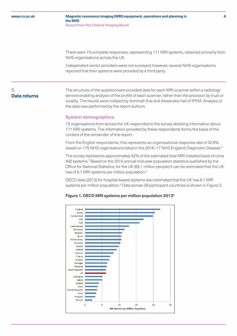
The survey represents approximately 42% of the estimated total MRI installed base of circa 400 systems.7 Based on the 2015 annual mid-year population statistics published by the Office for National Statistics, for the UK (65.1 million people) it can be estimated that the UK has of 6.1 MRI systems per million population.8
OECD data (2013) for hospital-based systems also estimated that the UK has 6.1 MRI systems per million population.9 Data across 28 participant countries is shown in Figure 3.
Figure 1. OECD MRI systems per million population 20139
7Magnetic resonance imaging (MRI) equipment, operations and planning in the NHS Report from the Clinical Imaging Board
www.rcr.ac.uk
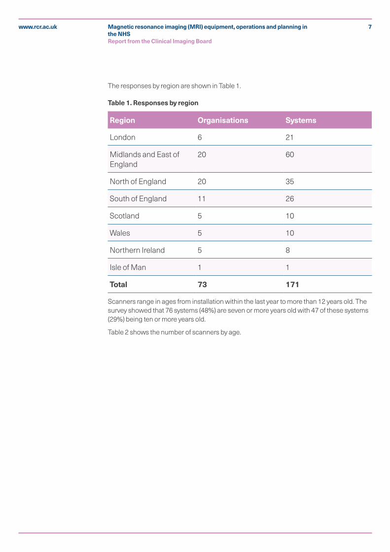
The responses by region are shown in Table 1.
Table 1. Responses by region
Region Organisations Systems
London 6 21
Midlands and East of England
20 60
North of England 20 35
South of England 11 26
Scotland 5 10
Wales 5 10
Northern Ireland 5 8
Isle of Man 1 1
Total 73 171
Scanners range in ages from installation within the last year to more than 12 years old. The survey showed that 76 systems (48%) are seven or more years old with 47 of these systems (29%) being ten or more years old.
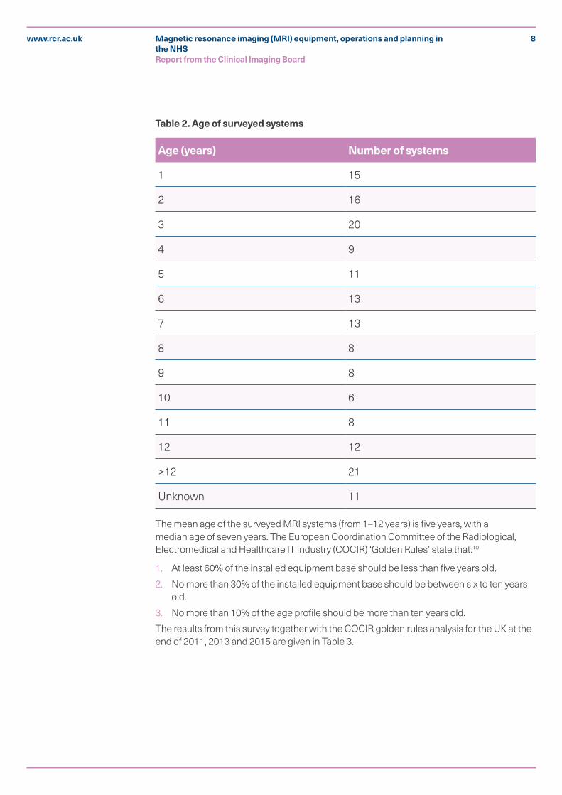
Table 2 shows the number of scanners by age.
8Magnetic resonance imaging (MRI) equipment, operations and planning in the NHS Report from the Clinical Imaging Board
www.rcr.ac.uk
Table 2. Age of surveyed systems
Age (years) Number of systems
1 15
2 16
3 20
4 9
5 11
6 13
7 13
8 8
9 8
10 6
11 8
12 12
>12 21
Unknown 11
The mean age of the surveyed MRI systems (from 1–12 years) is five years, with a median age of seven years. The European Coordination Committee of the Radiological, Electromedical and Healthcare IT industry (COCIR) ‘Golden Rules’ state that:10
1. At least 60% of the installed equipment base should be less than five years old.
2. No more than 30% of the installed equipment base should be between six to ten years old.
3. No more than 10% of the age profile should be more than ten years old.
The results from this survey together with the COCIR golden rules analysis for the UK at the end of 2011, 2013 and 2015 are given in Table 3.
9Magnetic resonance imaging (MRI) equipment, operations and planning in the NHS Report from the Clinical Imaging Board
www.rcr.ac.uk
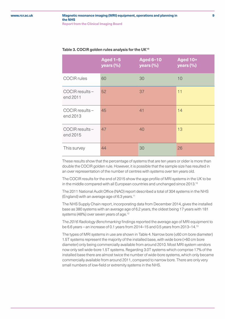
Table 3. COCIR golden rules analysis for the UK10
Aged 1–5 years (%)
Aged 6–10 years (%)
Aged 10+ years (%)
COCIR rules 60 30 10
COCIR results – end 2011
52 37 11
COCIR results – end 2013
45 41 14
COCIR results – end 2015
47 40 13
This survey 44 30 26
These results show that the percentage of systems that are ten years or older is more than double the COCIR golden rule. However, it is possible that the sample size has resulted in an over representation of the number of centres with systems over ten years old.
The COCIR results for the end of 2015 show the age profile of MRI systems in the UK to be in the middle compared with all European countries and unchanged since 2013.10
The 2011 National Audit Office (NAO) report described a total of 304 systems in the NHS (England) with an average age of 6.3 years.11
The NHS Supply Chain report, incorporating data from December 2014, gives the installed base as 380 systems with an average age of 6.2 years, the oldest being 17 years with 181 systems (48%) over seven years of age.12
The 2016 Radiology Benchmarking findings reported the average age of MRI equipment to be 6.6 years – an increase of 0.1 years from 2014–15 and 0.5 years from 2013–14.13
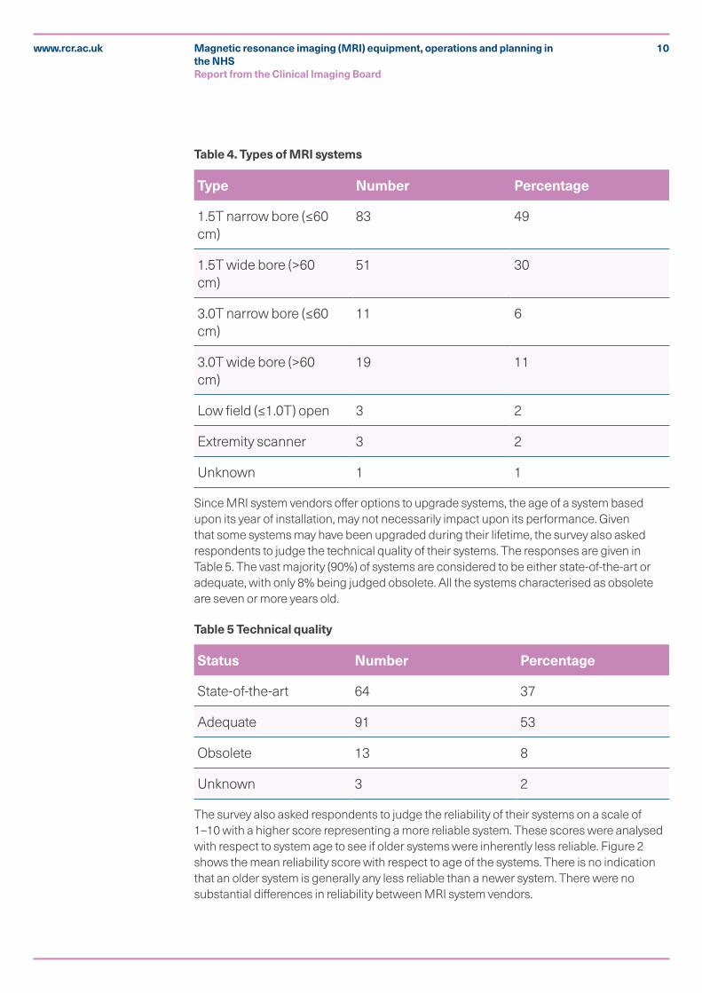
The types of MRI systems in use are shown in Table 4. Narrow bore (≤60 cm bore diameter) 1.5T systems represent the majority of the installed base, with wide bore (>60 cm bore diameter) only being commercially available from around 2010. Most MRI system vendors now only sell wide-bore 1.5T systems. Regarding 3.0T systems which comprise 17% of the installed base there are almost twice the number of wide-bore systems, which only became commercially available from around 2011, compared to narrow bore. There are only very small numbers of low-field or extremity systems in the NHS.
10Magnetic resonance imaging (MRI) equipment, operations and planning in the NHS Report from the Clinical Imaging Board
www.rcr.ac.uk
Table 4. Types of MRI systems
Type Number Percentage
1.5T narrow bore (≤60 cm)
83 49
1.5T wide bore (>60 cm)
51 30
3.0T narrow bore (≤60 cm)
11 6
3.0T wide bore (>60 cm)
19 11
Low field (≤1.0T) open 3 2
Extremity scanner 3 2
Unknown 1 1
Since MRI system vendors offer options to upgrade systems, the age of a system based upon its year of installation, may not necessarily impact upon its performance. Given that some systems may have been upgraded during their lifetime, the survey also asked respondents to judge the technical quality of their systems. The responses are given in Table 5. The vast majority (90%) of systems are considered to be either state-of-the-art or adequate, with only 8% being judged obsolete. All the systems characterised as obsolete are seven or more years old.
Table 5 Technical quality
Status Number Percentage
State-of-the-art 64 37
Adequate 91 53
Obsolete 13 8
Unknown 3 2
The survey also asked respondents to judge the reliability of their systems on a scale of 1–10 with a higher score representing a more reliable system. These scores were analysed with respect to system age to see if older systems were inherently less reliable. Figure 2 shows the mean reliability score with respect to age of the systems. There is no indication that an older system is generally any less reliable than a newer system. There were no substantial differences in reliability between MRI system vendors.

11Magnetic resonance imaging (MRI) equipment, operations and planning in the NHS Report from the Clinical Imaging Board
www.rcr.ac.uk
Figure 2. Mean system reliability versus age
MRI equipment purchase methods Table 5 shows the purchase methods for the systems. Capital funding is the main method of purchase with managed equipment services (MES) constituting 20% of purchases.
Table 6. MRI equipment funding
Funding method Number Percentage
Owned by your hospital/trust/Health Board
103 60
Part of a managed equipment service
35 20
Leased by your hospital/trust/Health Board
12 7
Don't know 10 6
Provided by a third party
8 5
Grant funded 3 2
12Magnetic resonance imaging (MRI) equipment, operations and planning in the NHS Report from the Clinical Imaging Board
www.rcr.ac.uk
Replacement planningThe survey indicates that, of the 76 systems that are seven years old or more, 27 (37.5%) have no replacement plans in place. The majority of organisations with plans in place (45 systems) are due to replace them in the period 2016–18. Only two systems considered to be obsolete do not currently have replacement plans.
Of the 35 systems that are part of a MES, 11 currently have no replacement plans. However, the majority are only 1–2 years old.
Meeting demandData from the NHS England Diagnostic Imaging Dataset (DID) demonstrate annual growth in MRI activity of 11.3%, 10.5% and 6.7% over 2013–14, 2014–15 and 2015–16 respectively.2
The DID data from 2015–16 shows that MRI activity increased by 6.7% from 2014–15 to 3,084,815 examinations.6 Unusually, this growth outstripped the increase in CT (6.2%) during this period. Based on annual mid-year population statistics published by the Office for National Statistics, this equates to 56.3 MRI studies per 1,000 population.8
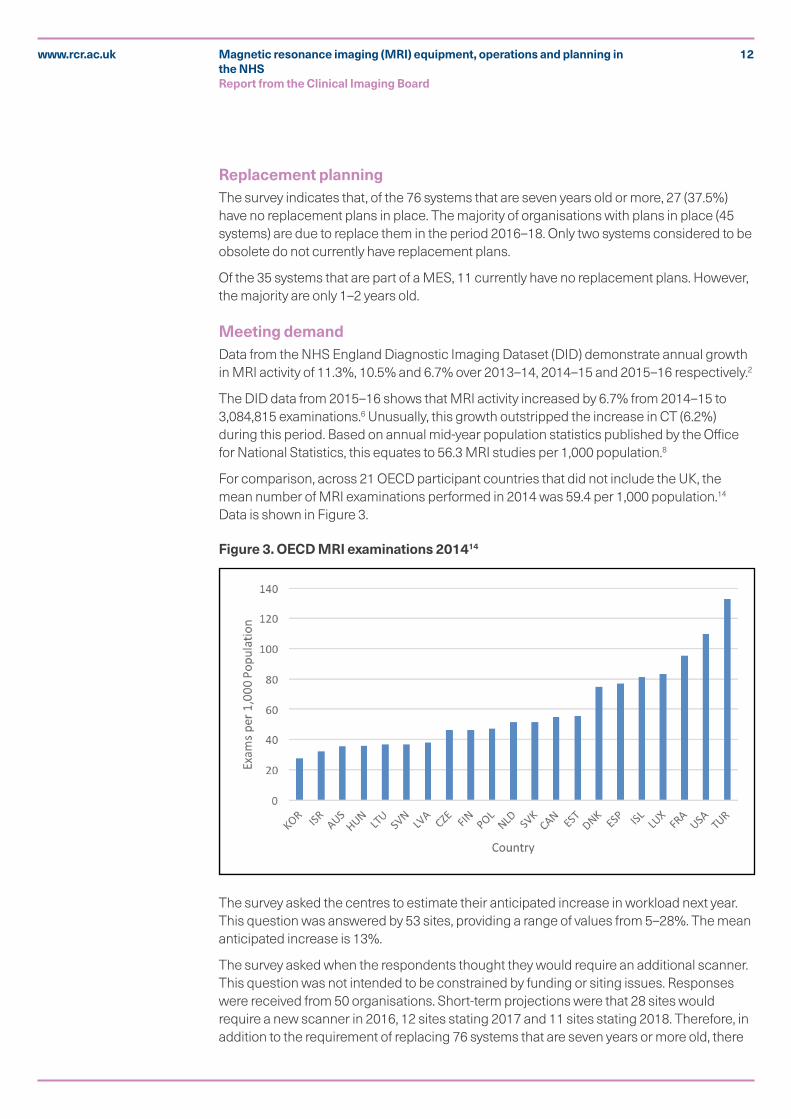
For comparison, across 21 OECD participant countries that did not include the UK, the mean number of MRI examinations performed in 2014 was 59.4 per 1,000 population.14 Data is shown in Figure 3.
Figure 3. OECD MRI examinations 201414
The survey asked the centres to estimate their anticipated increase in workload next year. This question was answered by 53 sites, providing a range of values from 5–28%. The mean anticipated increase is 13%.
The survey asked when the respondents thought they would require an additional scanner. This question was not intended to be constrained by funding or siting issues. Responses were received from 50 organisations. Short-term projections were that 28 sites would require a new scanner in 2016, 12 sites stating 2017 and 11 sites stating 2018. Therefore, in addition to the requirement of replacing 76 systems that are seven years or more old, there
13Magnetic resonance imaging (MRI) equipment, operations and planning in the NHS Report from the Clinical Imaging Board
www.rcr.ac.uk
is an expected requirement of an additional 49 systems to address the increasing demand. Note, however that, these figures only represent 42% of the estimated installed base.
Assuming the existing installed base is working at capacity then the anticipated growth of 13% in studies could potentially require a 13% increase in the number of MRI systems which, assuming an installed base of 400 systems, would equate to an additional 52 systems.
There are several ways of increasing MRI capacity, from a growth in the absolute numbers of pieces of equipment, to different staffing skills-mix and workload models, through to extending hours of operation both during a five-day week, and extending to seven-day working. The latter is already increasingly commonplace in the NHS, due to clinical demands.
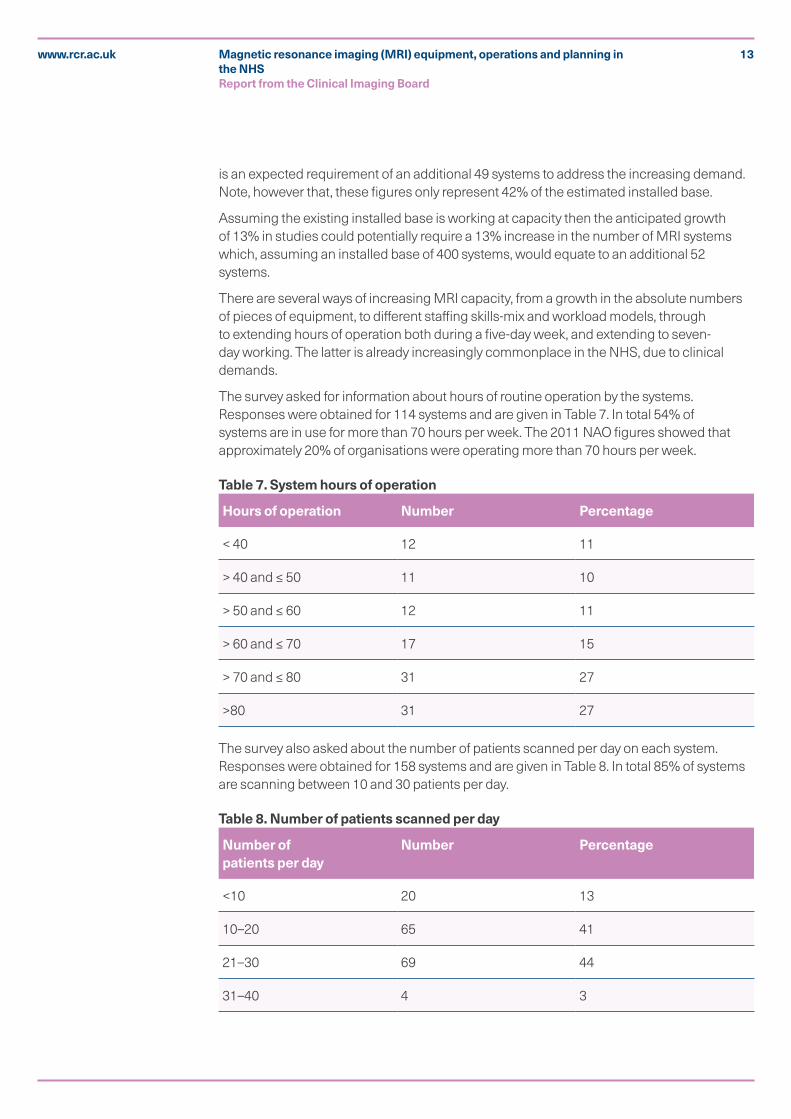
The survey asked for information about hours of routine operation by the systems. Responses were obtained for 114 systems and are given in Table 7. In total 54% of systems are in use for more than 70 hours per week. The 2011 NAO figures showed that approximately 20% of organisations were operating more than 70 hours per week.
Table 7. System hours of operation
Hours of operation Number Percentage
< 40 12 11
> 40 and ≤ 50 11 10
> 50 and ≤ 60 12 11
> 60 and ≤ 70 17 15
> 70 and ≤ 80 31 27
>80 31 27
The survey also asked about the number of patients scanned per day on each system. Responses were obtained for 158 systems and are given in Table 8. In total 85% of systems are scanning between 10 and 30 patients per day.
Table 8. Number of patients scanned per day
Number of patients per day
Number Percentage
<10 20 13
10–20 65 41
21–30 69 44
31–40 4 3
14Magnetic resonance imaging (MRI) equipment, operations and planning in the NHS Report from the Clinical Imaging Board
www.rcr.ac.uk
The survey asked how many patients per week were being scanned external to the organisation. 32 organisations provided a numerical response totalling 2,125 patients. Ten organisations are externally scanning 100 patients or more per week, with one organisation scanning 200 per week.
The survey did not specifically ask about emergency activity or out-of-hours on-call availability.
Technical issues
MRI technology
MRI system technology advances can be categorised as follows:
§ Static magnetic field. The two main field strengths in clinical use in the NHS are 1.5T and 3.0T. 3.0T systems potentially offer an improved signal-to-noise ratio (SNR) compared to 1.5T that can be traded for improved image quality, spatial or temporal resolution or reduced acquisition times. However, 3.0T systems may also display a higher level of image artefacts in some situations. The increased power deposition, known as the specific absorption rate (SAR), associated with 3.0T may also limit some of the achievable performance.
§ Gradient system. The gradient system encodes the MRI signal. Gradient performance is characterised by the maximum amplitude and the time it takes the gradients to switch to the required amplitude, known as the rise-time. The achievable performance is determined by the gradient amplifiers and the gradient coils. The increased diameter of wide-bore systems is generally achieved through a reduction in the physical dimensions of the gradient coils and consequently the gradient system will have a reduced performance compared to an equivalent narrow-bore system. Some MRI systems can be upgraded with improved amplifiers and/or coils
§ Radiofrequency (RF) system. The RF system is responsible for exciting the nuclei and detecting the nuclear magnetic resonance (NMR) signal. While the excitation is generally performed using an in-built body coil, there is a wide range of signal-detection coils that generally comprise of multiple individual elements each connected to separate receiver channels. The use of dedicated RF coils for specific body areas can improve the SNR, which can be traded-off as above, and can also be used to reduce acquisition times through the application of parallel imaging techniques. MRI systems can be upgraded with new RF coils and possibly with an increased number of receiver channels.
§ Software. The most common type of upgrade to an MRI system is via software. This may comprise new pulse sequences and/or user interface changes that can provide new imaging capabilities, such as those referred to below. Occasionally software upgrades may require additional hardware such as host or reconstruction computers. It may be that certain software options are also not supported on the particular MRI system in use, requiring a complete system replacement. Sometimes vendors make it possible to retain just the magnet whilst the rest of the system is replaced.
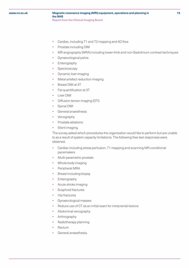
The survey asked which procedures the organisation would like to perform but are unable to as a result of system hardware/technical limitations. The following free-text responses were obtained.
§ Whole body imaging (including diffusion-weighted imaging [DWI])
15Magnetic resonance imaging (MRI) equipment, operations and planning in the NHS Report from the Clinical Imaging Board
www.rcr.ac.uk
§ Cardiac, including T1 and T2 mapping and 4D flow
§ Prostate including DWI
§ MR angiography (MRA) including lower-limb and non-Gadolinium contrast techniques
§ Gynaecological pelvis
§ Enterography
§ Spectroscopy
§ Dynamic liver imaging
§ Metal artefact reduction imaging
§ Breast DWI at 3T
§ Fat quantification at 3T
§ Liver DWI
§ Diffusion tensor imaging (DTI)
§ Spinal DWI
§ General anaesthesia
§ Venography
§ Prostate ablations
§ Silent imaging.
The survey asked which procedures the organisation would like to perform but are unable to as a result of system capacity limitations. The following free-text responses were obtained.
§ Cardiac including stress perfusion, T1 mapping and scanning MR-conditional pacemakers
§ Multi-parametric prostate
§ Whole body imaging
§ Peripheral MRA
§ Breast including biopsy
§ Enterography
§ Acute stroke imaging
§ Scaphoid fractures
§ Hip fractures
§ Gynaecological masses
§ Reduce use of CT as an initial exam for intracranial lesions
§ Abdominal venography
§ Arthrography
§ Radiotherapy planning
§ Rectum
§ General anaesthesia.
16Magnetic resonance imaging (MRI) equipment, operations and planning in the NHS Report from the Clinical Imaging Board
www.rcr.ac.uk
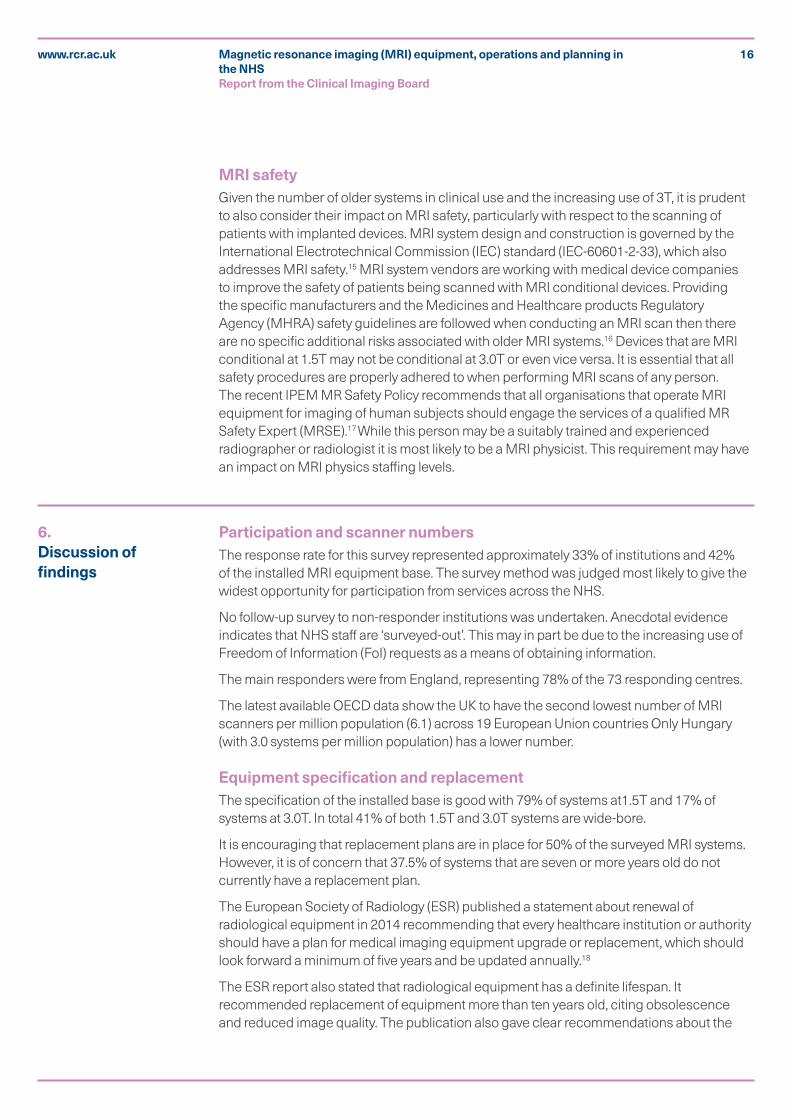
MRI safetyGiven the number of older systems in clinical use and the increasing use of 3T, it is prudent to also consider their impact on MRI safety, particularly with respect to the scanning of patients with implanted devices. MRI system design and construction is governed by the International Electrotechnical Commission (IEC) standard (IEC-60601-2-33), which also addresses MRI safety.15 MRI system vendors are working with medical device companies to improve the safety of patients being scanned with MRI conditional devices. Providing the specific manufacturers and the Medicines and Healthcare products Regulatory Agency (MHRA) safety guidelines are followed when conducting an MRI scan then there are no specific additional risks associated with older MRI systems.16 Devices that are MRI conditional at 1.5T may not be conditional at 3.0T or even vice versa. It is essential that all safety procedures are properly adhered to when performing MRI scans of any person. The recent IPEM MR Safety Policy recommends that all organisations that operate MRI equipment for imaging of human subjects should engage the services of a qualified MR Safety Expert (MRSE).17 While this person may be a suitably trained and experienced radiographer or radiologist it is most likely to be a MRI physicist. This requirement may have an impact on MRI physics staffing levels.
6. Discussion of findings
Participation and scanner numbersThe response rate for this survey represented approximately 33% of institutions and 42% of the installed MRI equipment base. The survey method was judged most likely to give the widest opportunity for participation from services across the NHS.
No follow-up survey to non-responder institutions was undertaken. Anecdotal evidence indicates that NHS staff are ‘surveyed-out’. This may in part be due to the increasing use of Freedom of Information (FoI) requests as a means of obtaining information.
The main responders were from England, representing 78% of the 73 responding centres.
The latest available OECD data show the UK to have the second lowest number of MRI scanners per million population (6.1) across 19 European Union countries Only Hungary (with 3.0 systems per million population) has a lower number.
Equipment specification and replacementThe specification of the installed base is good with 79% of systems at1.5T and 17% of systems at 3.0T. In total 41% of both 1.5T and 3.0T systems are wide-bore.
It is encouraging that replacement plans are in place for 50% of the surveyed MRI systems. However, it is of concern that 37.5% of systems that are seven or more years old do not currently have a replacement plan.
The European Society of Radiology (ESR) published a statement about renewal of radiological equipment in 2014 recommending that every healthcare institution or authority should have a plan for medical imaging equipment upgrade or replacement, which should look forward a minimum of five years and be updated annually.18
The ESR report also stated that radiological equipment has a definite lifespan. It recommended replacement of equipment more than ten years old, citing obsolescence and reduced image quality. The publication also gave clear recommendations about the
17Magnetic resonance imaging (MRI) equipment, operations and planning in the NHS Report from the Clinical Imaging Board
www.rcr.ac.uk
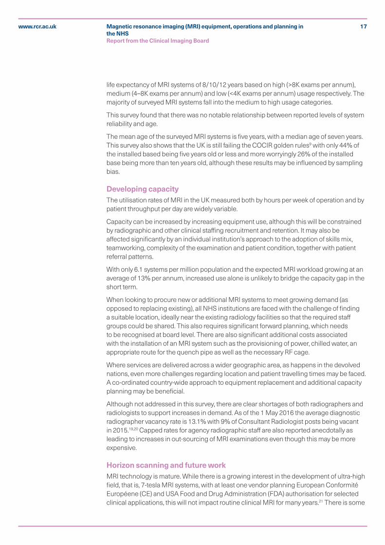
life expectancy of MRI systems of 8/10/12 years based on high (>8K exams per annum), medium (4–8K exams per annum) and low (<4K exams per annum) usage respectively. The majority of surveyed MRI systems fall into the medium to high usage categories.
This survey found that there was no notable relationship between reported levels of system reliability and age.
The mean age of the surveyed MRI systems is five years, with a median age of seven years. This survey also shows that the UK is still failing the COCIR golden rules9 with only 44% of the installed based being five years old or less and more worryingly 26% of the installed base being more than ten years old, although these results may be influenced by sampling bias.
Developing capacityThe utilisation rates of MRI in the UK measured both by hours per week of operation and by patient throughput per day are widely variable.
Capacity can be increased by increasing equipment use, although this will be constrained by radiographic and other clinical staffing recruitment and retention. It may also be affected significantly by an individual institution’s approach to the adoption of skills mix, teamworking, complexity of the examination and patient condition, together with patient referral patterns.
With only 6.1 systems per million population and the expected MRI workload growing at an average of 13% per annum, increased use alone is unlikely to bridge the capacity gap in the short term.
When looking to procure new or additional MRI systems to meet growing demand (as opposed to replacing existing), all NHS institutions are faced with the challenge of finding a suitable location, ideally near the existing radiology facilities so that the required staff groups could be shared. This also requires significant forward planning, which needs to be recognised at board level. There are also significant additional costs associated with the installation of an MRI system such as the provisioning of power, chilled water, an appropriate route for the quench pipe as well as the necessary RF cage.
Where services are delivered across a wider geographic area, as happens in the devolved nations, even more challenges regarding location and patient travelling times may be faced. A co-ordinated country-wide approach to equipment replacement and additional capacity planning may be beneficial.
Although not addressed in this survey, there are clear shortages of both radiographers and radiologists to support increases in demand. As of the 1 May 2016 the average diagnostic radiographer vacancy rate is 13.1% with 9% of Consultant Radiologist posts being vacant in 2015.19,20 Capped rates for agency radiographic staff are also reported anecdotally as leading to increases in out-sourcing of MRI examinations even though this may be more expensive.
Horizon scanning and future workMRI technology is mature. While there is a growing interest in the development of ultra-high field, that is, 7-tesla MRI systems, with at least one vendor planning European Conformité Européene (CE) and USA Food and Drug Administration (FDA) authorisation for selected clinical applications, this will not impact routine clinical MRI for many years.21 There is some
18Magnetic resonance imaging (MRI) equipment, operations and planning in the NHS Report from the Clinical Imaging Board
www.rcr.ac.uk
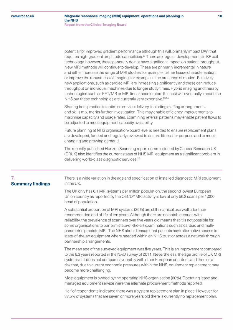
potential for improved gradient performance although this will, primarily impact DWI that requires high-gradient amplitude capabilities.22 There are regular developments in RF coil technology, however, these generally do not have significant impact on patient throughput. New MRI methods will continue to develop. These are primarily incremental in nature and either increase the range of MRI studies, for example further tissue characterisation, or improve the robustness of imaging, for example in the presence of motion. Relatively new applications, such as cardiac MRI are increasing significantly and these can reduce throughput on individual machines due to longer study times. Hybrid imaging and therapy technologies such as PET/MR or MRI linear accelerators (Linacs) will eventually impact the NHS but these technologies are currently very expensive.23,24
Sharing best practice to optimise service delivery, including staffing arrangements and skills mix, merits further investigation. This may enable efficiency improvements to maximise capacity and usage rates. Examining referral patterns may enable patient flows to be adjusted to meet equipment capacity availability.
Future planning at NHS organisation/board level is needed to ensure replacement plans are developed, funded and regularly reviewed to ensure fitness for purpose and to meet changing and growing demand.
The recently published Horizon Scanning report commissioned by Cancer Research UK (CRUK) also identifies the current status of NHS MRI equipment as a significant problem in delivering world-class diagnostic services.25
7. Summary findings
There is a wide variation in the age and specification of installed diagnostic MRI equipment in the UK.
The UK only has 6.1 MRI systems per million population, the second lowest European Union country as reported by the OECD.9 MRI activity is low at only 56.3 scans per 1,000 head of population.
A substantial proportion of MRI systems (26%) are still in clinical use well after their recommended end of life of ten years. Although there are no notable issues with reliability, the prevalence of scanners over five years old means that it is not possible for some organisations to perform state-of-the-art examinations such as cardiac and multi-parametric prostate MRI. The NHS should ensure that patients have alternative access to state-of-the-art equipment where needed within an NHS trust or across a network through partnership arrangements.
The mean age of the surveyed equipment was five years. This is an improvement compared to the 6.3 years reported in the NAO survey of 2011. Nevertheless, the age profile of UK MRI systems still does not compare favourably with other European countries and there is a risk that, due to current economic pressures within the NHS, equipment replacement may become more challenging.
Most equipment is owned by the operating NHS organisation (60%). Operating lease and managed equipment service were the alternate procurement methods reported.
Half of respondents indicated there was a system replacement plan in place. However, for 37.5% of systems that are seven or more years old there is currently no replacement plan.
19Magnetic resonance imaging (MRI) equipment, operations and planning in the NHS Report from the Clinical Imaging Board
www.rcr.ac.uk
Respondents predicted a growth in activity of 13% per annum for 2017, whereas the DID data reported an actual growth of 6.7% from 2014–15 to 2015–16.5 Weekly hours of operation and patient throughput vary widely between systems and organisations. Increasing demand will require additional equipment or working longer clinical hours. More staff will be required to support this both to undertake the imaging and to provide the requisite reporting.
Readers should note that this report has only considered access to MRI equipment and has not considered the workforce issues related to driving acquisition and delivering the required reporting of studies, nor the impact of increasing workload for referrers receiving radiological reports.
8. Summary points and recommendations
The CIB believes that valuable overview information about MRI equipment in the NHS has been gathered through this exercise.
The CIB believes there is a need to raise awareness of the low numbers of MRI systems per million population compared to many other similar countries.
While many NHS radiology services have future MRI replacement plans in place, it is of great concern that a significant number do not. This requires action by NHS radiology services and all stakeholders.
All NHS radiology departments need a future equipment plan, looking forward at least five years. Professional bodies working together should produce a template or framework to assist in their development.
The CIB notes that more than 26% of MRI equipment is ten or more years old and does not have state-of-the-art imaging capability, limiting access to high-quality imaging for some patients.
Expected growth in MRI activity in the coming years will require additional equipment as well as increasing operational hours. These issues are further exacerbated by the national shortage of radiographers and radiologists.
Approved by the Faculty of Clinical Radiology of The Royal College of Radiologists, the Institute of Physics and Engineering in Medicine and the Society and College of Radiolgraphers: February 2017.
20Magnetic resonance imaging (MRI) equipment, operations and planning in the NHS Report from the Clinical Imaging Board
www.rcr.ac.uk
References
1. National Institute for Heath and Care Excellence (NICE). Prostate cancer: diagnosis and management. Clinical guideline [CG175]. London: NICE, 2014.
2. National Institute for Heath and Care Excellence (NICE). Myeloma: diagnosis and management. Clinical guideline [NG35]. London: NICE, 2016.
3. National Institute for Heath and Care Excellence (NICE). Metastatic spinal cord compression in adults: risk assessment, diagnosis and management. Clinical guideline [CG75]. London: NICE, 2008.
4. Department of Health. The NHS Cancer Plan. A plan for investment. A plan for reform. London: DoH, 2000.
5. www.smartsurvey.co.uk/s/CIBMREquip/ (last accessed 21/4/17)
6. NHS England. Diagnostic Imaging Dataset Annual Statistical Release 2016/16. Leeds: NHS England, 2016.
7. www.statista.com/statistics/473302/number-of-magnetic-resonance-imaging-units-united-kingdom-uk/ (last accessed 21/4/17)
8. www.ons.gov.uk/peoplepopulationandcommunity/populationandmigration/populationestimates/bulletins/annualmidyearpopulationestimates/mid2015 (last accessed 21/4/17)
9. https://data.oecd.org/healtheqt/magnetic-resonance-imaging-mri-units.htm (last accessed 21/4/17)
10. European Coordination Committee of the Radiological, Electromedical and Healthcare IT Industry (COCIR). Medical imaging equipment age profile and density. Brussels: COCIR, 2016.
11. National Audit Office, Department of Health. Managing high value capital equipment in the NHS in England. A report by the comptroller and auditor general. London: The Stationery Office, 2011.
12. NHS Supply Chain. Why it is important that the NHS has access to the latest medical equipment. Maidstone: NHS Supply Chain, 2014.
13. NHS Benchmarking Network. 2016 Radiology Benchmarking findings. London: NHS Benchmarking Network, 2016.
14. https://data.oecd.org/healthcare/magnetic-resonance-imaging-mri-exams.htm (last accessed 21/4/17)
15. International Electrotechnical Commission (IEC). Medical electrical equipment - Part 2-33: Particular requirements for the basic safety and essential performance of magnetic resonance equipment for medical diagnosis. Geneva: IEC, 2015.
16. Medicines and Healthcare products Regulatory Agency (MHRA). Safety guidelines for magnetic resonance imaging equipment in clinical use. London: MHRA, 2015.
17. Institute of Physics and Engineering in Medicine (IPEM). IPEM policy statement: scientific safety advice to magnetic resonance imaging units that undertake human imaging. York: IPEM, 2013.
18. European Society of Radiology (ESR). Renewal of radiological equipment. Insights Imaging 2014; 5: 543–546.
19. www.sor.org/sites/default/files/document-versions/20161107_diagnostic_radiography_uk_workforce_report_cd_v2.pdf (last accessed 21/4/17)
20. The Royal College of Radiologists. Clinical Radiology UK workforce census 2015 report. London: RCR, 2016.
21. Balchandani, P and Naidich, TP. Ultra-high-field MR neuroimaging. Am J Neuroradiol 2015; 36: 1204–1215.
22. McNab J, Edlow BL, Witzel T et al. The Human Connectome Project and beyond: initial applications of 300 mT/m gradients. Neuroimage 2013; 80: 234–245
23. Vandenberghe S and Marsden PK. PET-MRI: a review of challenges and solutions in the development of integrated multimodality imaging. Phys Med Biol 2015; 60: R115–R154.
24. Lagendijk JJ, Raaymakers BW, Raajmakers AJ et al. MRI/linac integration. Radiother Oncol 2008; 86: 25–29.

21Magnetic resonance imaging (MRI) equipment, operations and planning in the NHS Report from the Clinical Imaging Board
www.rcr.ac.uk
25. 2020 Delivery. Horizon scanning. An evaluation of imaging capacity across the NHS in England. London: Cancer Research UK, 2015.
22Magnetic resonance imaging (MRI) equipment, operations and planning in the NHS Report from the Clinical Imaging Board
www.rcr.ac.uk
Appendix 1. Clinical Imaging Board Terms of Reference for Report working party
Purpose/Remit To produce a report for the Clinical Imaging Board on the current state of UK NHS MRI equipment. This will endeavour to establish whether current equipment is fit for purpose, its utilisation and plans for replacement. It will also attempt to predict whether these plans will be adequate for anticipated future demand.
Several approaches are needed to measure these endpoints. It can be difficult to define a useful lifetime for MRI systems since it is possible to perform major hardware upgrades whilst maintaining some of the original components, eg magnet. However, it may not be cost-effective to continually upgrade the system. This survey will evaluate the type and age of the existing MRI systems in use within the UK NHS. However, it will also ask questions about the perceived technical status of the systems to identify older scanners that have been upgraded, whilst also asking about the perceived reliability of the system. Furthermore, given the developing technical capabilities of MRI the questionnaire will also seek to identify whether there are certain procedures that the system is technically incapable of performing or that capacity is limiting the introduction of these new capabilities.
The questionnaire will also seek to determine the operating hours and throughput of the systems and identify the weekly usage for the main clinical indications.
The questionnaire will ask about plans to replace existing systems as well as asking about the anticipated increase in workload over the next year and when the organisation would expect that additional capacity would be required. We will also ask if the organisation is outsourcing work to external organisations.
The questions will include:
§ Location, ie trust/Health Board
§ Manufacturer, model and type
§ Date of installation
§ Planned year of replacement
§ Subjective technical status, eg obsolete, adequate, state-of-the-art
§ Subjective reliability (score 1–10)
§ Elective hours worked
§ Average daily throughput
§ How is the system funded?
§ Number of patients that are externally scanned
§ Desired procedures that it is not technically possible to perform on this system(s)
§ Anticipated increase in workload over the next year
§ When would you envisage requiring an additional system(s).The results will provide a snapshot of the existing MRI equipment base and utilisation within the NHS and will identify areas where procedures are limited due to either equipment or capacity issues.
This work will complement the work of the National Audit Office in England in 2011 (www.nao.org.uk/report/managing-high-value-capital-equipment-in-the-nhs-in-england/) with a particular perspective of the professions involved in MRI scanning.
23Magnetic resonance imaging (MRI) equipment, operations and planning in the NHS Report from the Clinical Imaging Board
www.rcr.ac.uk
The inclusion of the Independent Sector (IS) was considered. Our current opinion is that we should initially survey the NHS and then based on the findings of the report this will inform a decision as to whether a second survey should be undertaken with the IS. It is likely that a slightly different set of questions will need to be formulated for this purpose.
ObjectivesThis small, expert, tri-partite group will:
§ Develop and agree with the Clinical Imaging Board the detailed scope for the above work
§ Set out the necessary methodology
§ Carry out the work
§ Produce a final report and guidance for publication by CIB.
Specific tasksThe Working Party is to convene an initial meeting via teleconference. Subsequent work will be via teleconference or email. A final meeting may be required. Administrative support will be provided by IPEM.
It may be necessary during the development of this publication for the Chair of the working group to co-opt/appoint additional members as the group’s work evolves. These may include representatives of other professional groups with whom the Clinical Radiology specialty works in close association – for example, radiographers, medical physicists, clinical radiologists and oncologists, clinical nurse specialists. Nominations from the relevant professional body will need to be sought at this stage.
MembershipThe Working Party membership will include:
Dr Martin Graves, Consultant Clinical Scientist (Chair)
Dr Paul Malcolm, Consultant Radiologist
Ms Erica Scurr, MRI Radiographer
Ms Debbie Horne, MRI Radiographer
Ms Alex Lipton, CoR
Pam Black, SCOR Past-President (advisor)
ProductThe report will be produced by the CIB, and will be the property of, the three professional bodies that make up the “CIB”. The report will be available in electronic format and available on the RCR, IPEM and SCOR websites.
CostsAny meeting expenses and travel costs will be met by the RCR (The Royal College of Radiologists, Faculty of Clinical Radiology), IPEM (Institute of Physics and Engineering in Medicine) and the SCOR (Society and College of Radiographers) respectively for each member.
24Magnetic resonance imaging (MRI) equipment, operations and planning in the NHS Report from the Clinical Imaging Board
www.rcr.ac.uk
All direct costs (for example, editorial and/or design costs) related to the production of the guidance will be met by the RCR, IPEM and SCOR equally.
Proposed circulationCommissioners of diagnostic services, radiology service managers, trust board/health board chairs etc. The guidance will be published on the RCR, IPEM and SCOR website(s). Members and Fellows will be notified when the publication becomes available.
Copyright The report will be published under the copyright of the RCR, IPEM and CoR in all media now known or in future existing.
TimescaleThe work is expected to be completed by January 2017.
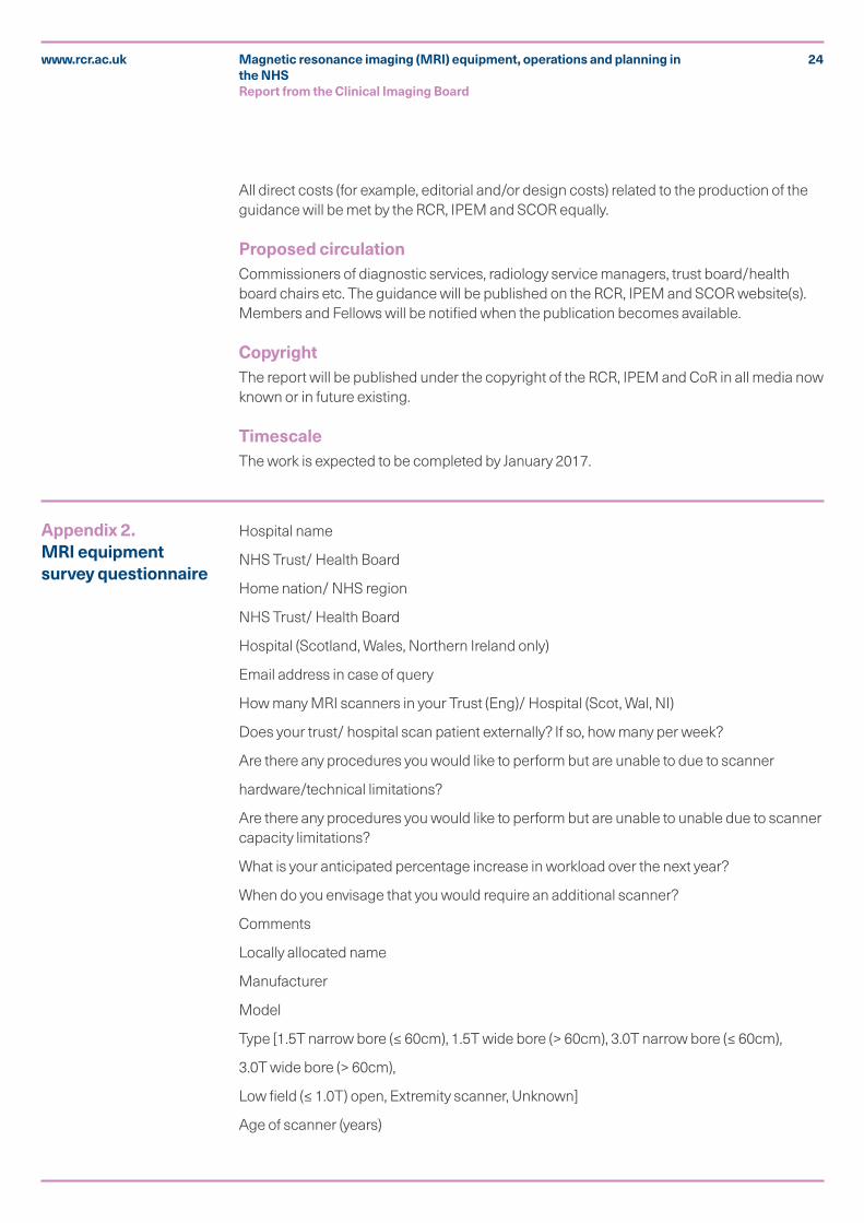
Appendix 2. MRI equipment survey questionnaire
Hospital name
NHS Trust/ Health Board
Home nation/ NHS region
NHS Trust/ Health Board
Hospital (Scotland, Wales, Northern Ireland only)
Email address in case of query
How many MRI scanners in your Trust (Eng)/ Hospital (Scot, Wal, NI)
Does your trust/ hospital scan patient externally? If so, how many per week?
Are there any procedures you would like to perform but are unable to due to scanner
hardware/technical limitations?
Are there any procedures you would like to perform but are unable to unable due to scanner capacity limitations?
What is your anticipated percentage increase in workload over the next year?
When do you envisage that you would require an additional scanner?
Comments
Locally allocated name
Manufacturer
Model
Type [1.5T narrow bore (≤ 60cm), 1.5T wide bore (> 60cm), 3.0T narrow bore (≤ 60cm),
3.0T wide bore (> 60cm),
Low field (≤ 1.0T) open, Extremity scanner, Unknown]
Age of scanner (years)

25Magnetic resonance imaging (MRI) equipment, operations and planning in the NHS Report from the Clinical Imaging Board
www.rcr.ac.uk
Which year do you plan to replace this scanner?
Do you consider this scanner to be technically [State-of-the-art, Adequate, Obsolete, Unknown]
On a scale of 1 to 10 how to you rate the reliability of this system [1,…,10]
How many hours does this machine operate in a typical week? [<40, >40 and ≤50, >50 and ≤60, >60 and ≤70, >70 and ≤80, >80]
Average daily patient throughput on this machine [<10, >10 and ≤20, >20 and ≤30, >30 and ≤40]
How is this scanner owned? [Owned by your Hospital/Trust/Health Board, Part of a managed equipment service, Leased by your Hospital/Trust/Health Board, Grant funded, Provided by a 3rd party, Don’t know]
The Royal College of Radiologists 63 Lincoln’s Inn Fields London WC2A 3JW
+44 (0)20 7405 1282 [email protected] www.rcr.ac.uk
@RCRadiologists
The Royal College of Radiologists. Magnetic resonance imaging (MRI) equipment, operations and planning in the NHS. London: The Royal College of Radiologists, 2017.
© The Royal College of Radiologists, May 2017.
For permission to reproduce any of the content contained herein, please email: [email protected]
This material has been produced by The Royal College of Radiologists (RCR) for use internally within the specialties of clinical oncology and clinical radiology in the United Kingdom. It is provided for use by appropriately qualified professionals, and the making of any decision regarding the applicability and suitability of the material in any particular circumstance is subject to the user’s professional judgement.
While every reasonable care has been taken to ensure the accuracy of the material, RCR cannot accept any responsibility for any action taken, or not taken, on the basis of it. As publisher, RCR shall not be liable to any person for any loss or damage, which may arise from the use of any of the material. The RCR does not exclude or limit liability for death or personal injury to the extent only that the same arises as a result of the negligence of RCR, its employees, Officers, members and Fellows, or any other person contributing to the formulation of the material.