management of children with spina bifida in the age...
TRANSCRIPT

FRANCOIS I. LUKS
PETRA KLINGE
Management of children with spina bifida in the age of fetal intervention
www.revolutionhealth.com

Spina Bifida and Neural Tube Defects
Epidemiology
One of the most common birth defects: 1-2 cases/1,000 births
Certain populations have a greater risk:
Highest incidence in Ireland and Wales
More common in girls
U.S.: 0.7/1,000 live births
Higher on the East Coast than on the West Coast
Higher in whites (1/1,000 births)
Lower in African-Americans (0.1-0.4/1,000 births)

Spina Bifida and Neural Tube Defects
Epidemiology
Risk factors:
Race and ethnicity
Family history of neural tube defects
Folate deficiency
Medication/teratogenic effect: valproic acid
Maternal age
Diabetes
Obesity
Increased body temperature
Hol FA et al, Clinical Genetics, 2008

Embryology of spina bifida
Weeks 3-4 of gestation
3 phases:
Neurulation
Canalization
Retrogressive differentiation
Management of children with spina bifida in the age of fetal intervention

Spina Bifida and Neural Tube Defects
Definitions and Classification
Open spina bifida (Aperta)
Meningocele in 5%
Myelomeningocele (cord and cauda equina exposed) in 95%
Closed spina bifida (Occulta)
50% have cutaneous stigmata
Lipomyelomeningocele
Filum terminale lipoma
“Fatty” filum terminale
Dermoid sinus and dermoid tumor

Current management of spina bifida
Primary treatment
Perinatal care (protection of the neural tube, infections)
Closure of the defect
Management of hydrocephalus
Chiari II hindbrain herniation
Formal evaluation of spina bifida (overlaps with treatment)
Physical examination: deformities, neuro exam; continence/tone
Ultrasound
MRI – brain
MRI – spine
Other: genetic testing, specialized imaging
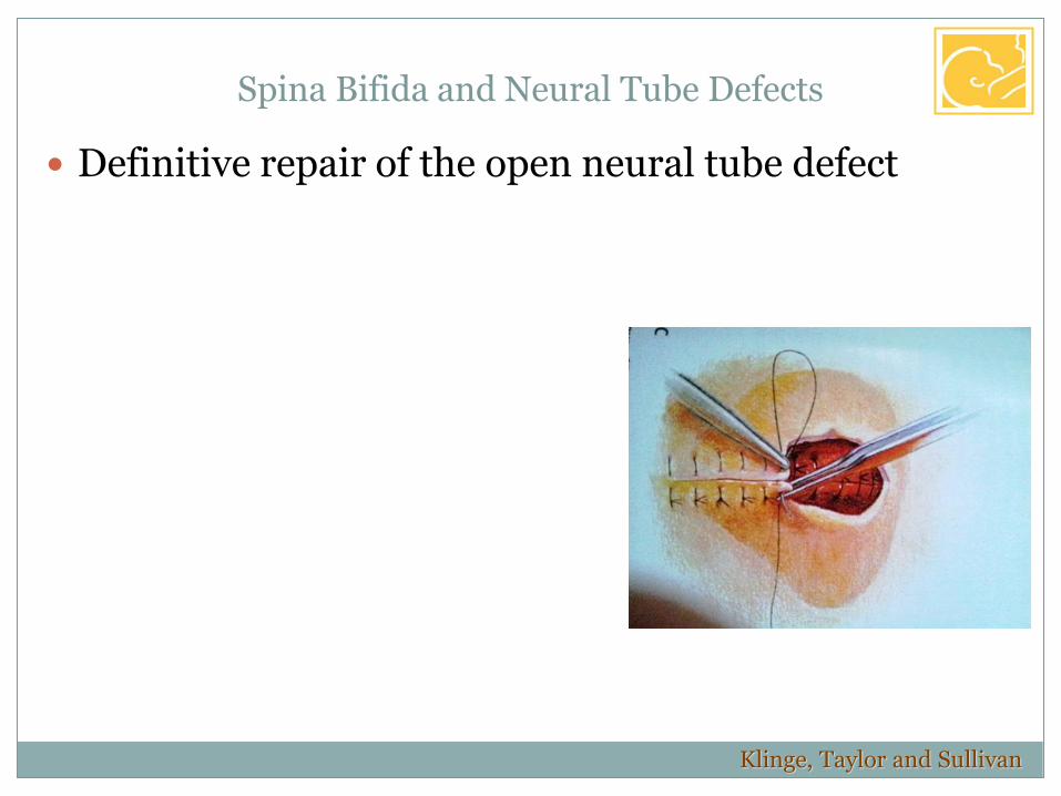
Spina Bifida and Neural Tube Defects

Spina Bifida and Neural Tube Defects
Definitive repair of the open neural tube defect
Posterior vertebral defect
Thecal sac
Cord extruded into the sac (placode)
Plate of embryonic epithelial cells: spinal cord

Spina Bifida and Neural Tube Defects
Definitive repair of the open neural tube defect
Closure within 24 hours
No evidence that immediate/urgent closure improves function
But: early closure reduces risk of infection
Wound colonization after 36 hours
Surgical technique: (neurosurgeon + plastic surgeon team)
Placode dissected off arachnoid
Allowed to drop into spinal canal
Dura dissected off skin and lumbodorsal fascia
Dura closed
Muscular fascia closed
Skin closed
CSF Placode
Meninges
SKIN
FASCIA

Spina Bifida and Neural Tube Defects
Definitive repair of the open neural tube defect
Surgical technique: Sharp microdissection of the placode

Spina Bifida and Neural Tube Defects
Definitive repair of the open neural tube defect
Continued dissection toward the placode
Detethering
Klinge, Taylor and Sullivan

Spina Bifida and Neural Tube Defects
Definitive repair of the open neural tube defect
Detethering of aberrant nerve roots
Klinge, Taylor and Sullivan

Spina Bifida and Neural Tube Defects
Definitive repair of the open neural tube defect
Paraspinal muscle closure
Klinge, Taylor and Sullivan
Spina Bifida and Neural Tube Defects
Definitive repair of the open neural tube defect
Klinge, Taylor and Sullivan

Spina Bifida and Neural Tube Defects
Pathophysiology and associated disorders
Hydrocephalus
80-95% incidence in myelomeningocele
100% of 35 thoracic lesions
88% of 114 lumbar lesions
68% of 40 sacral lesions
Significant in 20% at birth
Rintoul et al, Pediatrics 2002

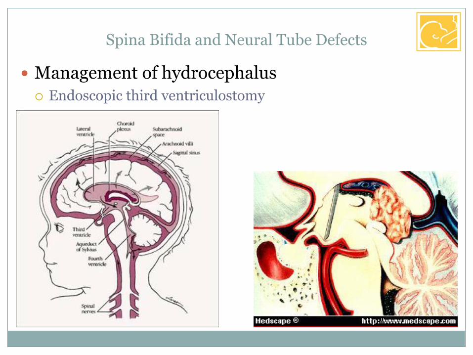
Spina Bifida and Neural Tube Defects
Management of hydrocephalus
Imaging: ventriculomegaly (Ventricular index >0.33)
Pediatric characteristics:
Selective thinning of the occipiatl cranial vault and cortex:
Rigid nuclear masses (basal ganglia) in the frontal lobe
Monitor head circumference!
Ventricular index > 0.33
47.65 mm
137.96 mm

Spina Bifida and Neural Tube Defects
Management of hydrocephalus
Serial head ultrasounds in the newborn:

Spina Bifida and Neural Tube Defects
Management of hydrocephalus
Temporary drainage:
Lumbar puncture
External ventricular drainage, reservoir
Shunt
Weight >2.5 kg
No active infection
Medically stable
Endoscopic third ventriculostomy

Spina Bifida and Neural Tube Defects
Management of hydrocephalus
Types of shunts:
Adjustable valves
Spina Bifida and Neural Tube Defects
Management of hydrocephalus
Endoscopic third ventriculostomy

Spina Bifida and Neural Tube Defects
Pathophysiology and associated disorders
Chiari II malformation
99% of myelomeningocele have radiographic Chiari II
Only symptomatic ones require treatment (30% at 5 years)
Responsible for 15-20% of deaths in children with MMC
Respiratory failure/arrest
Syringomyelia

Spina Bifida and Neural Tube Defects
Treatment of Chiari II malformation

Current management of spina bifida
Secondary management
Relatively recent: now that these children survive long-term
The most difficult – chronic vigilance
CNS monitoring:
VP shunt management
Management of tethered cord (10%)
Physical therapy evaluation/motor function of lower extremities
Preventive medicine – insensate lower body
Psychological support
Spina Bifida and Neural Tube Defects

Current management of spina bifida
Secondary management
Management of tethered cord
Second detethering surgery for decline in function and/or before correction of scoliosis
Spina Bifida and Neural Tube Defects
Tethering at the MMC closure site after surgery

Which organ systems does it affect?
Neuro-motor
Neurodevelopmental, hydrocephalus, CNS development
Spina Bifida and Neural Tube Defects

Which organ systems does it affect?
Neuro-motor
Neurodevelopmental, hydrocephalus, CNS development
Urogenital
Gastrointestinal
Gastroesophageal reflux disease (GERD)
Constipation
More commonly: incontinence
Other
Variability in severity for all systems (GI specifically)
Spina Bifida and Neural Tube Defects

Spina Bifida and Neural Tube Defects Management of children with spina bifida
in the age of fetal intervention
Peripheral effects of open neural tube defect
Exposed spinal cord during gestation
(Progressive?) damage to the exposed neural tube
Variable paresis, urine & stool incontinence
CSF leak into amniotic cavity
Basis for prenatal testing: leakage of alpha-fetoprotein (AFP)
Increased concentration in the amniotic fluid (amniocentesis)
Maternal Serum AFP (MSAFP) elevated as well
False-positives: any other cause of AFP leakage: gastroschisis

Spina Bifida and Neural Tube Defects Management of children with spina bifida
in the age of fetal intervention
Peripheral effects of open neural tube defect
Exposed spinal cord during gestation
(Progressive?) damage to the exposed neural tube
Could spina bifida be cured – or even prevented ?

Embryology of spina bifida – can it be prevented?
Progressive development theory
Is only one theory – and the most simplistic one
Prolonged in utero exposure of the neural tube leads to
Chronic leakage of CSF
Gradual siphoning and hindbrain herniation
Increased risk of hydrocephalus
Progressive damage to the neural placode
Progressive peripheral nerve damage
• Lower extremity function
• Sphincter function
Management of children with spina bifida in the age of fetal intervention

Spina bifida – can it be diagnosed in utero?
Ultrasound
Spinal defect
“Lemon” sign: abnormally shaped skull (head circumference)
“Banana” sign: abnormally shaped cerebellum
Hydrocephalus
Management of children with spina bifida in the age of fetal intervention

Spina bifida – can it be diagnosed in utero?
Magnetic Resonance Imaging
Management of children with spina bifida in the age of fetal intervention

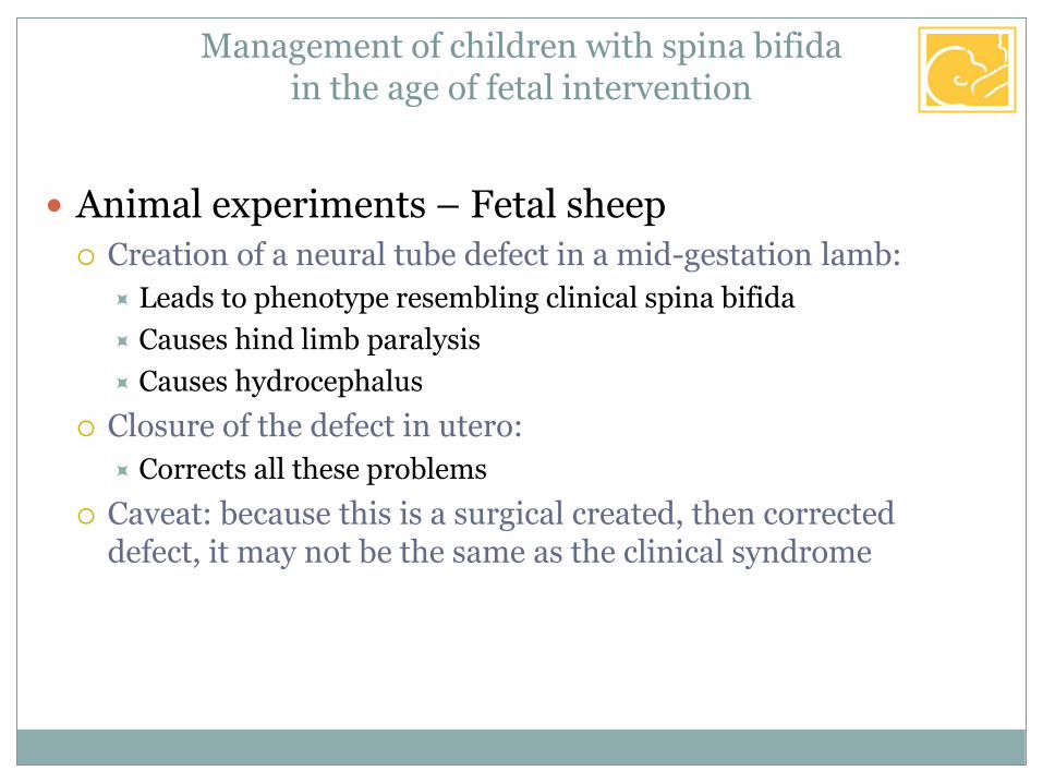
Animal experiments – Fetal sheep
Creation of a neural tube defect in a mid-gestation lamb:
Leads to phenotype resembling clinical spina bifida
Causes hind limb paralysis
Causes hydrocephalus
Management of children with spina bifida in the age of fetal intervention
Normal Spina bifida Repaired Spina bifida
Meuli M et al, Nature Medicine 1995

Animal experiments – Fetal sheep
Creation of a neural tube defect in a mid-gestation lamb:
Leads to phenotype resembling clinical spina bifida
Causes hind limb paralysis
Causes hydrocephalus
Closure of the defect in utero:
Corrects all these problems
Management of children with spina bifida in the age of fetal intervention
Meuli M et al, Nature Medicine 1995
Animal experiments – Fetal sheep
Creation of a neural tube defect in a mid-gestation lamb:
Leads to phenotype resembling clinical spina bifida
Causes hind limb paralysis
Causes hydrocephalus
Closure of the defect in utero:
Corrects all these problems
Caveat: because this is a surgical created, then corrected defect, it may not be the same as the clinical syndrome
Management of children with spina bifida in the age of fetal intervention

Animal experiments – better models?
Mouse models: loss of grainyhead-like (Grhl) gene function:
Grhl-3 mutation: ct (curly-tail) mouse
Grhl-2 mutation: Axd (axial defects) mouse
Management of children with spina bifida in the age of fetal intervention
Brouns MR et al, Human Molec Genet 2011 Brouns MR et al, Drug Disc Today 2005

Fetal surgery for spina bifida: from sheep to man
Proof of concept in animal model
Management of children with spina bifida in the age of fetal intervention

Fetal surgery for spina bifida: from sheep to man
Proof of concept in animal model
Progress in fetal surgery for other indications
Management of children with spina bifida in the age of fetal intervention
Luks FI et al, J Pediatr Surg 1993

Fetal surgery for spina bifida: from sheep to man
Proof of concept in animal model
Progress in fetal surgery for other indications
Endoscopic fetal surgery for Twin-to-twin Transfusion Syndrome
Management of children with spina bifida in the age of fetal intervention

Fetal surgery for spina bifida: from sheep to man
Proof of concept in animal model
Progress in fetal surgery for other indications
Endoscopic fetal surgery for Twin-to-twin Transfusion Syndrome
1998: Vanderbilt reports on endoscopic repair of MMC
2/4 survivors – technique abandoned
Management of children with spina bifida in the age of fetal intervention
Bruner JP et al, Am J Obstet Gynecol 1998

Fetal surgery for spina bifida: from sheep to man
Proof of concept in animal model
Progress in fetal surgery for other indications
Endoscopic fetal surgery for Twin-to-twin Transfusion Syndrome
1998: Vanderbilt reports on endoscopic repair of MMC
2/4 survivors – technique abandoned
Early 2000: anecdotal, then non-randomized series
Vanderbilt, CHOP, UCSF
In utero repair is feasible
Management of children with spina bifida in the age of fetal intervention
Fetal surgery for spina bifida: from sheep to man
Proof of concept in animal model
Progress in fetal surgery for other indications
Endoscopic fetal surgery for Twin-to-twin Transfusion Syndrome
1998: Vanderbilt reports on endoscopic repair of MMC
2/4 survivors – technique abandoned
Early 2000: anecdotal, then non-randomized series
Vanderbilt, CHOP, UCSF
In utero repair is feasible
Possible improvement over postnatal repair? Less hydrocephalus?
Final conclusion: it does NOT improve motor function
Management of children with spina bifida in the age of fetal intervention

Started in 2003
Randomized to 3 prenatal centers or postnatal R/
Goal: 100 patients/arm
Prenatal closure at 19-25 weeks
All deliveries in a MOMS center
Vanderbilt, Nashville
University of California San Francisco
Children’s Hospital of Philadelphia
Hypothesis:
Fetal repair delays hydrocephalus, prevents Chiari II
Not: Better chance of walking!
Management Of Myelomeningocele Study: The MOMS trial

Started in 2003
Was supposed to take only 3 years
By 2010: Still only 140 patients recruited (of 200 needed)
Late 2011: Study suddenly stopped at 85% recruitment
Why? Because of better-than-expected results!
Management Of Myelomeningocele Study: The MOMS trial
New York Times 2011
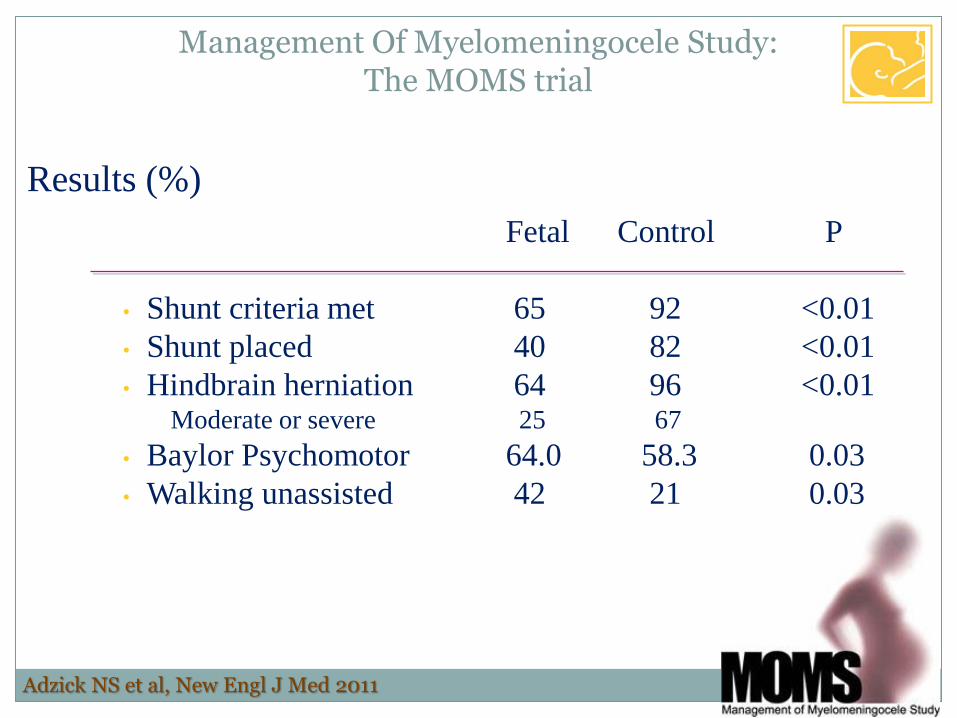
Results (%)
Fetal Control P
• Shunt criteria met 65 92 <0.01
• Shunt placed 40 82 <0.01
• Hindbrain herniation 64 96 <0.01 Moderate or severe 25 67
• Baylor Psychomotor 64.0 58.3 0.03
• Walking unassisted 42 21 0.03
Management Of Myelomeningocele Study: The MOMS trial
Adzick NS et al, New Engl J Med 2011

Complications (%)
Maternal complications Fetal Control P
• Pulmonary edema 6 0 0.03
• Placental abruption 6 0 0.03
• Chorioamnionitis 3 0 0.24
• Preecclampsia 4 0 0.12
• Blood transfusion 9 1 0.03
Management Of Myelomeningocele Study: The MOMS trial
Adzick NS et al, New Engl J Med 2011
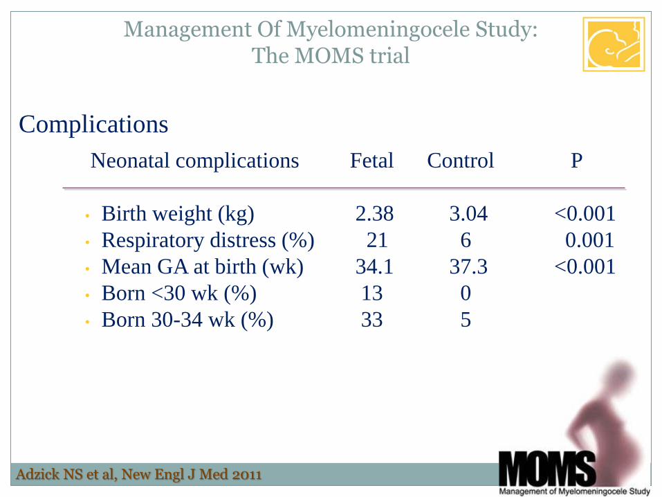
Complications
Neonatal complications Fetal Control P
• Birth weight (kg) 2.38 3.04 <0.001
• Respiratory distress (%) 21 6 0.001
• Mean GA at birth (wk) 34.1 37.3 <0.001
• Born <30 wk (%) 13 0
• Born 30-34 wk (%) 33 5
Management Of Myelomeningocele Study: The MOMS trial
Adzick NS et al, New Engl J Med 2011
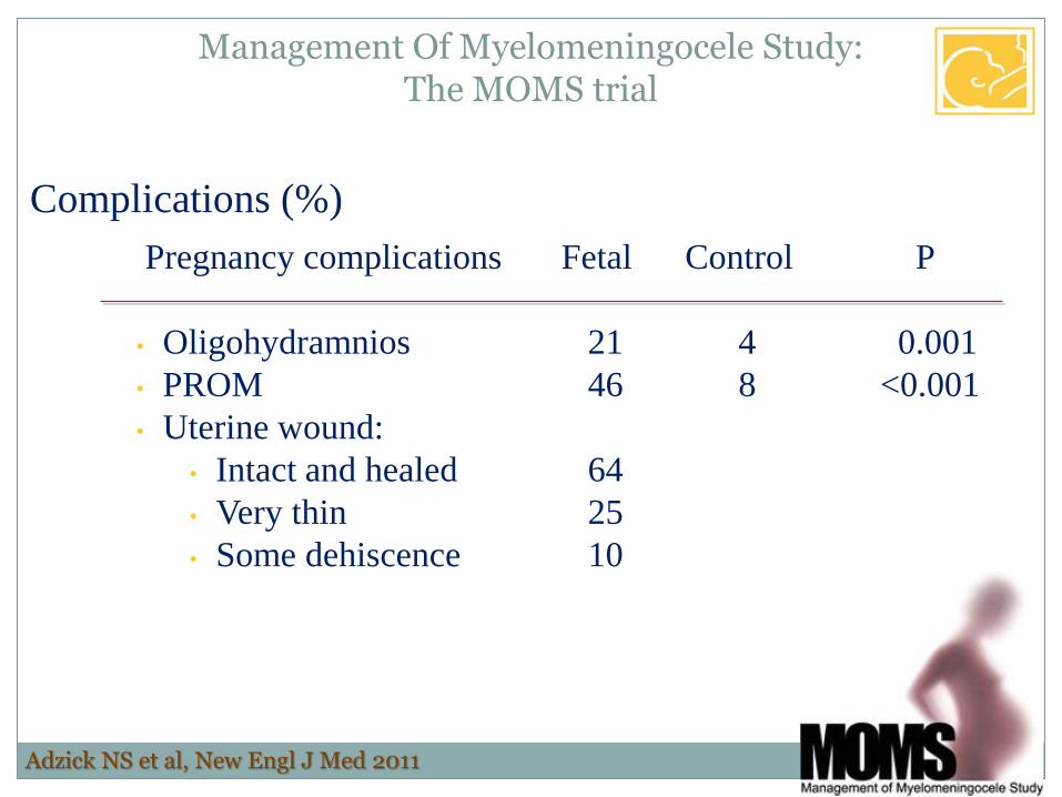
Complications (%)
Pregnancy complications Fetal Control P
• Oligohydramnios 21 4 0.001
• PROM 46 8 <0.001
• Uterine wound:
• Intact and healed 64
• Very thin 25
• Some dehiscence 10
Management Of Myelomeningocele Study: The MOMS trial
Adzick NS et al, New Engl J Med 2011

In utero repair of spina bifida: how is it done?
Maternal and fetal anesthesia
General anesthesia
Uterine relaxation
Inhalation anesthesia
Preserved placental circulation
Arterial line
Epidural for analgesia
MgSO4 for CP prevention
Steroids (prematurity)
Management of children with spina bifida in the age of fetal intervention

In utero repair of spina bifida: how is it done?
Multidisciplinary team approach
Maternal Anesthesia
Maternal-Fetal Medicine
Pediatric Surgery
Pediatric Neurosurgery
Pediatric Plastic Surgery
Neonatalogy
Management of children with spina bifida in the age of fetal intervention

In utero repair of spina bifida: how is it done?
Wide maternal laparotomy
Full exposure of the uterus
Management of children with spina bifida in the age of fetal intervention

In utero repair of spina bifida: how is it done?
Partial exteriorization of the uterus
Ultrasound-guided mapping of the placenta, fetus
Stapled hysterotomy (preservation of membranes)
Management of children with spina bifida in the age of fetal intervention

In utero repair of spina bifida: how is it done?
Exposure of the neural tube defect
Management of children with spina bifida in the age of fetal intervention

In utero repair of spina bifida: how is it done?
Exposure of the neural tube defect
Meticulous, but rapid closure
Management of children with spina bifida in the age of fetal intervention
3 -4 hours!
0.5 hours!
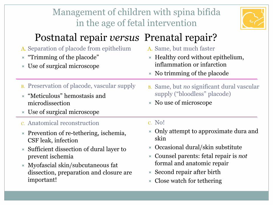
Postnatal repair versus Prenatal repair?
Management of children with spina bifida in the age of fetal intervention
A. Separation of placode from epithelium
“Trimming of the placode”
Use of surgical microscope
B. Preservation of placode, vascular supply
“Meticulous” hemostasis and microdissection
Use of surgical microscope
C. Anatomical reconstruction
Prevention of re-tethering, ischemia, CSF leak, infection
Sufficient dissection of dural layer to prevent ischemia
Myofascial skin/subcutaneous fat dissection, preparation and closure are important!
A. Same, but much faster
Healthy cord without epithelium, inflammation or infarction
No trimming of the placode
B. Same, but no significant dural vascular supply (“bloodless” placode)
No use of microscope
C. No!
Only attempt to approximate dura and skin
Occasional dural/skin substitute
Counsel parents: fetal repair is not formal and anatomic repair
Second repair after birth
Close watch for tethering
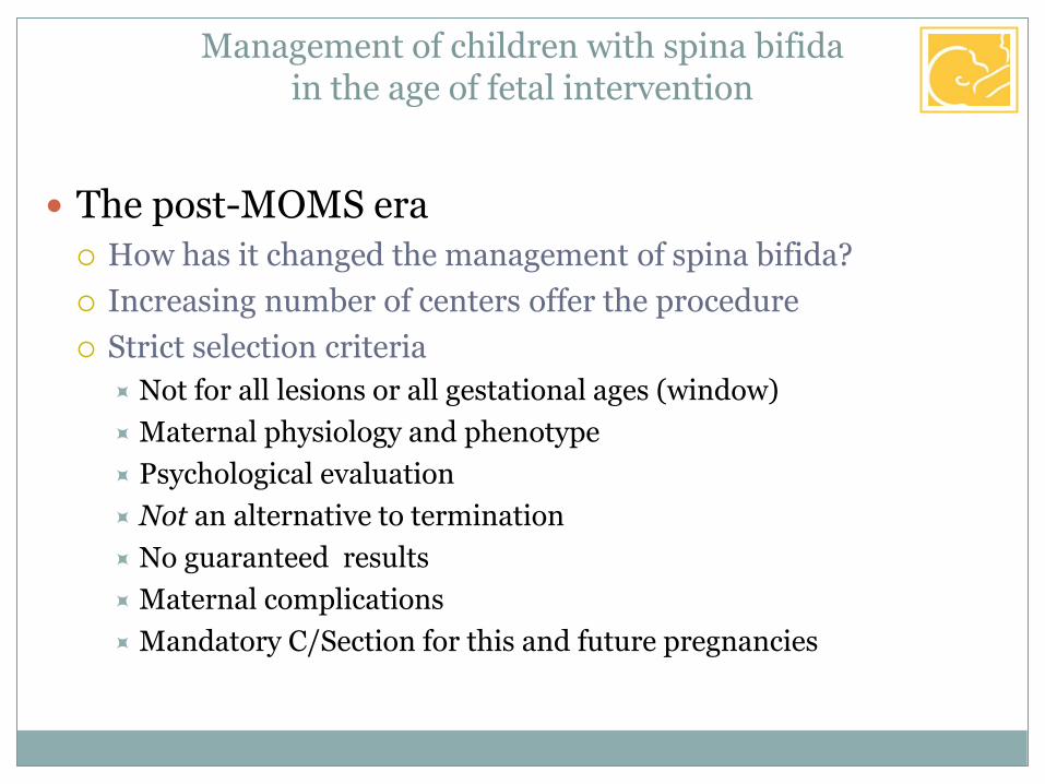
The post-MOMS era
How has it changed the management of spina bifida?
Increasing number of centers offer the procedure
Strict selection criteria
Not for all lesions or all gestational ages (window)
Maternal physiology and phenotype
Psychological evaluation
Not an alternative to termination
No guaranteed results
Maternal complications
Mandatory C/Section for this and future pregnancies
Management of children with spina bifida in the age of fetal intervention

MOMS II
Further analysis of the results in the initial cohort
It improves motor function
Does it improve GERD?
No real evidence (25% if shunted, v. 8% if not shunted)
Does it improve continence?
No word yet – but the answer appears to be “no”
Does it improve cognitive outcome?
Unclear – but encouraging results at 30 months…
Does it prevent/ Improve Tethering?
No word yet – but appears to be the opposite
Management of children with spina bifida in the age of fetal intervention
Danzer E et al, Neuropediatrics 2008
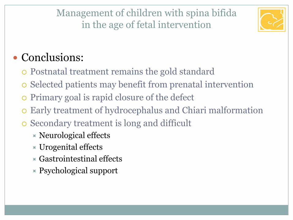
Conclusions:
Postnatal treatment remains the gold standard
Selected patients may benefit from prenatal intervention
Primary goal is rapid closure of the defect
Early treatment of hydrocephalus and Chiari malformation
Secondary treatment is long and difficult
Neurological effects
Urogenital effects
Gastrointestinal effects
Psychological support
Management of children with spina bifida in the age of fetal intervention