management of hand burns - asht · pdf filemanagement of hand burns ... • identify burn...
TRANSCRIPT

MANAGEMENT OF HAND BURNS
Including Tips and Tricks for the Hand Therapist Treating Wounds,
Grafts and Flaps
Nora Barrett, MS, OTR/L, CHTHand Specialist
Burn Rehabilitation TherapistBend, OR

3
Why Hands?
3

5
Objectives• Identify burn wound characteristics, surgical
options, and therapy priorities to promote healing and recovery
• Identify major components of hand rehabilitation throughout phases of burn recovery
• Recognize the purpose and use of orthotics throughout phases of burn healing and scar formation
• Identify biomedical and topical wound coverings that may be useful in treating non-burn wounds in the outpatient setting

Burn Depth• 1st Degree —> Epidermis (.05mm-1mm in adult)
– Non-vascular, stratified epithelial cells– Capable of rapid regeneration
• 2nd Degree—> Dermis (10 times thicker than epidermis)– Vascular layer containing collagen strands with nerve
endings, hair follicles, oil & sweat glands, lymph spaces– Epidermal cells line deep structures in dermis
• 3rd Degree—> Subcutaneous tissue– Adipose tissue & fibrous connective tissue
• 4th Degree—> Muscle or bone

15
Depths of Burn Injury
15

BURN DEPTHSUPERFICIAL THICKNESS• 1st degree burn• Epidermis only• Pink or red• Erythema due to vasodilation• Painful• Characterized by cell damage
without cell death• Complete scarless healing
within 7 days via re-epithelialization
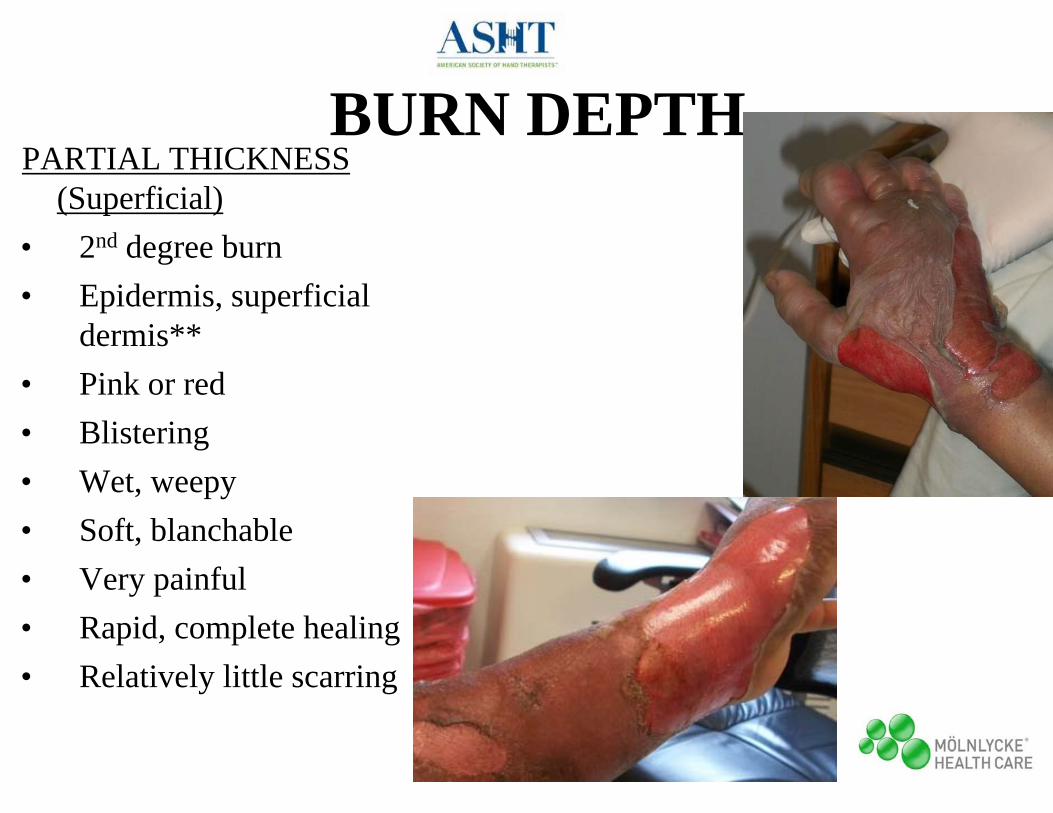
BURN DEPTHPARTIAL THICKNESS
(Superficial)• 2nd degree burn• Epidermis, superficial
dermis**• Pink or red• Blistering• Wet, weepy• Soft, blanchable• Very painful• Rapid, complete healing• Relatively little scarring

13
BURN DEPTHPARTIAL THICKNESS (Deep)• 2nd degree, potential conversion to
3rd degree• Most of dermis• Red with overlying eschar• Relatively insensate, potential for
pressure• Delayed healing potential (poor
quality)• Copious scarring
13

14
BURN DEPTHFULL THICKNESS• 3rd or 4th degree burn• White, brown, tan, black or red• Dry and leathery• Firm, non-blanchable• Insensate• No potential for healing• Profuse scarring if closes without
excision, grafting (3rd)• Elaborate debridement/
reconstruction/amputation (4th)

BIOLOGICAL DRESSINGS & WOUND COVERAGE
Commonly Used with Hand Burns
• Aquaphor• Xeroform• Mepitel• Mepilex• Acticoat• Silvadene• VAC
1st Superficial 2nd
Deep 2nd
3rd/4th

AQUAPHORPROS:
• Easy, comfortable
• Inexpensive, OTC
• Under glove
CONS:
• Acne, ? reaction
• Thick, greasy

21
XEROFORMPROS:• Relatively easy• Comfortable• Allows relative mobility
CONS:
• Can be difficult under compression
• Cannot be used if wound bed already moist

MEPITELPROS:
• Preserves injured epithelium
• Reduced pain and trauma at removal
• Perforated- allows fluid drainage
CONS:
• Does not lower risk of infection

Mepitel in Hand Clinic
14

MEPILEX-AGPROS:
• Used on any size area
• Can be left in place 4-7 days
• Donor sites
• Easy application
CONS:
• Indicated for low to moderate exuding wounds
• May need to be changed frequently with high exudate wounds

ACTICOAT
Pros:• Effective vs.
MRSA, VRE• 7-Day
Dressing• Used on grafts
and synthetics
Cons:• Difficult to use on
large wounds• Must be kept
moist• Not transparent • Silver chloride
stain

SILVER SULFADIAZINE“Silvadene”
PROS:
• Broad spectrum
• Not painful
• Lower cost
• 24 hour microbial coverage
CONS:
• Limited diffusion into eschar

Electrical

Chemical

20
EMERGENT PHASE(Initial 72 hours post-burn)
Major Hand Considerations
• Edema• Escharotomy• Positioning• Orthosis Intervention• Motion• Patient/family
education

21
Post-Burn Edema
9

22
Escharotomy/Fasciotomy
11

23
Positioning
13

24
ORTHOSIS INDICATIONS• Purpose: immobilize, support, position hand• Characteristics: nonconforming, nonconstrictive• Not standardized across burn units• Many parameters for initiating orthotic use• General indicators:
– Sedated patient unable to participate– Significant edema resulting in clawing– Unable to actively achieve intrinsic plus position – Circumferential hand burn
25
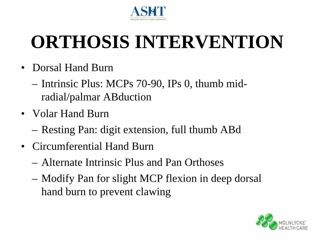
ORTHOSIS INTERVENTION• Dorsal Hand Burn
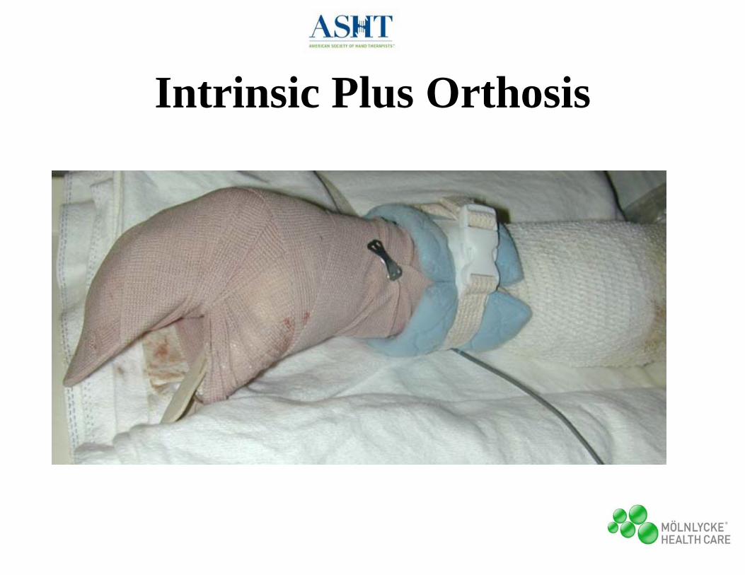
– Intrinsic Plus: MCPs 70-90, IPs 0, thumb mid-radial/palmar ABduction
• Volar Hand Burn– Resting Pan: digit extension, full thumb ABd
• Circumferential Hand Burn– Alternate Intrinsic Plus and Pan Orthoses– Modify Pan for slight MCP flexion in deep dorsal
hand burn to prevent clawing
26
Intrinsic Plus Orthosis
16

27
Motion• Preserve motion, prevent deformity• Promote tendon gliding, active muscle function• AROM as soon as awake, participating• Full available motion in superficial dorsal injuries• Protected ROM in deep dorsal hand burns• Digital ABd/ADd considered safe for all depths
• Activates intrinsics, mobilizes fluid• ROM permitted after escharotomy/fasciotomy• PROM in sedated patients

Cutaneous Functional Units (CFUs)• Fields of skin associated with normal movement• Skin recruited serially as joint ROM increases• Most skin motion occurs at skin crease of joint but skin
recruited beyond joint itself• Contracture risk regardless if skin crease involved• Isolated MCP vs composite flexion
– No difference in recruitment of uninjured dorsal hand skin (excludes digits)
28

Elbow extension
29

Composite Fisting
30

31
ACUTE PHASE(Emergent Phase through Wound Closure)
Major Hand Considerations• Motion• Tendon Integrity• Persistent Edema• Orthosis Intervention• Functional use/ADL participation and
modifications
19

32
Motion• Minimize scar contraction, promote function• Daily monitoring for loss of motion or limitations,
initial deformity, maladaptive positioning • Challenges in acute phase: pain, fibrous edema,
increasing tautness, inelastic eschar• What is limiting AROM? functional use?• Disruption of the coordinated interplay of intrinsic
and extrinsic muscles, tendons and joint is the underlying cause of most post-burn functional disturbances
34
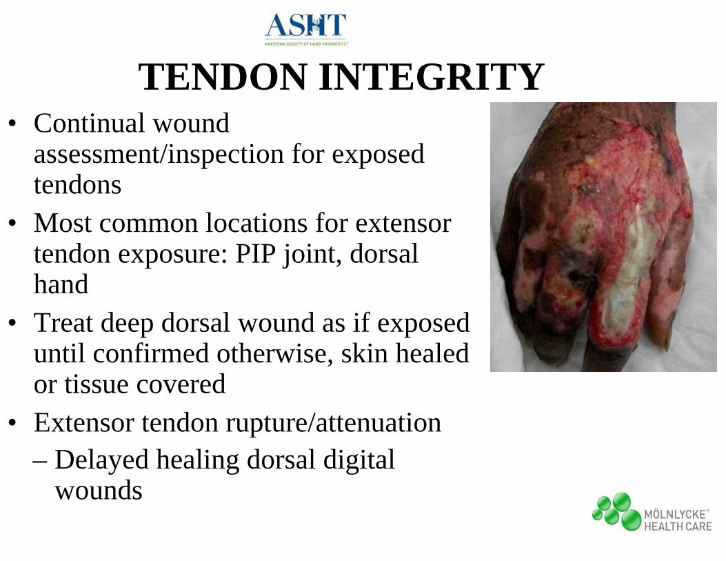
TENDON INTEGRITY• Continual wound
assessment/inspection for exposed tendons
• Most common locations for extensor tendon exposure: PIP joint, dorsal hand
• Treat deep dorsal wound as if exposed until confirmed otherwise, skin healed or tissue covered
• Extensor tendon rupture/attenuation– Delayed healing dorsal digital
wounds

35
PERSISTENT EDEMA• Restricts motion, causes stiffness• Can lead to tissue ischemia, fibrosis,
progressive scar formation, deformity• Compromised blood flow to hand, digits
– Contributes to intrinsic tightness
• Fibrosis + thickened eschar can lead to delayed tissue death, “crushing effect” on extensor mechanism
• Treatment options/combinations– Fluff wrap, Coban, gloves– AROM, functional use 33

36
PERSISTENT EDEMA
34

37
STATIC ORTHOSES• Purpose: prevent contracture• Adjust for edema changes, decreasing
dressing bulk• Indications/Schedule:
– Continue at night for optimal position– Limited use daytime if awake, participating– Intermittent daytime use with prolonged sedation or
decreased functional use, maladaptive positioning– Uninterrupted use with tendon exposure
• Position ET on slack to prevent rupture but prevent excessive shortening

38
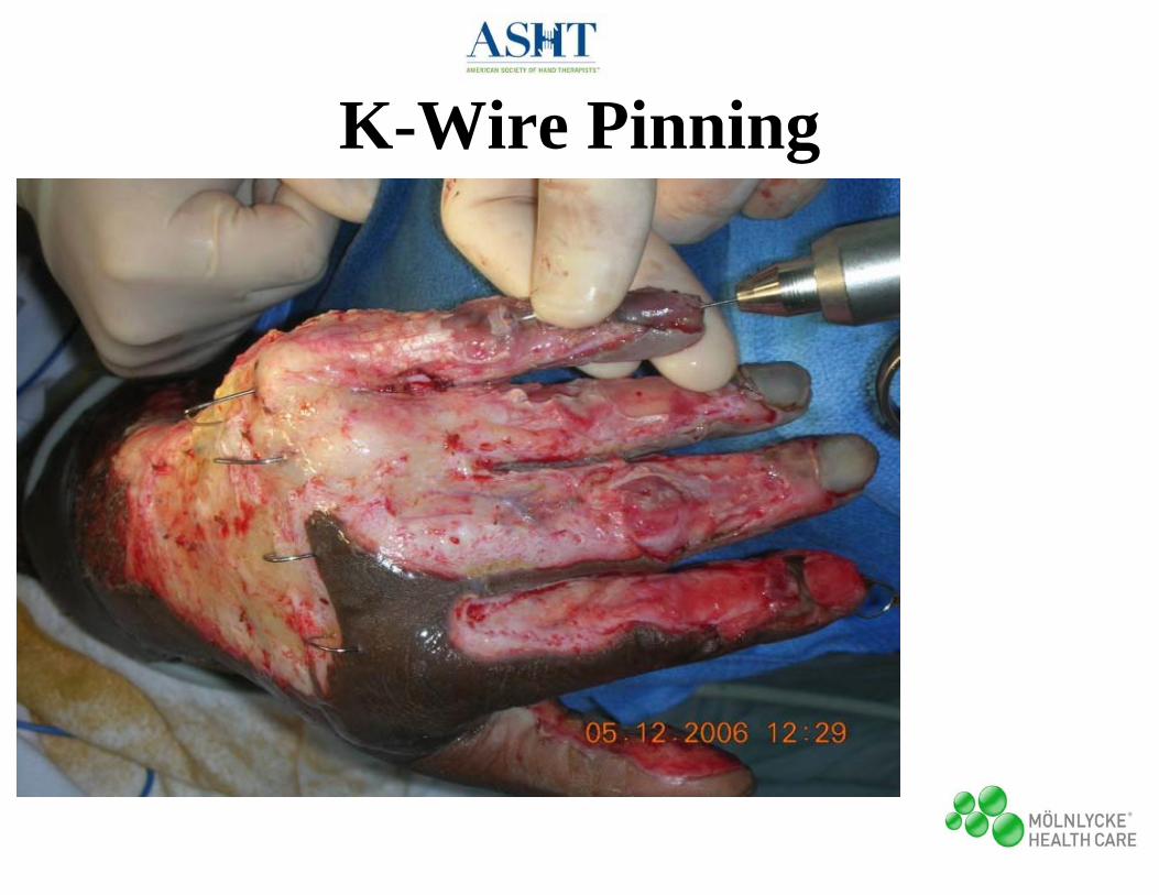
PIN FIXATION• Deep, non-healing wounds unresponsive to
orthosis• Likely tendon/joint exposure with loss of extensor
mechanism• K-wire pins driven through MCPs in maximal
flexion, IPs in 0 degrees extension• Pins kept in place up to 6 weeks for temporary
positioning, >6 weeks for permanent positioning until pseudo-arthrodesis via scarring
39
K-Wire Pinning
37

40
Delayed Pin Removal
38

MOBILIZATION ORTHOSES• Adjunct to active exercise, manual stretching• Force application amount determined by
tissue response• Dynamic traction used for early stiff hand
– “Subtle suggestiveness”– Ideal when PROM responds to stretch,
inflammation subsiding• Cautious use of composite mobilization
orthoses until dorsal wounds closed

CONTRACTURE IN BURN INJURY(at time of hospital discharge)
• Small joints– 23% at least 1 wrist or hand joint contracture
• Statistically significant predictors of contracture development– Concomitant medical problems, TBSA grafted, presence of hand burn
and hand grafting• Statistically significant predictors of contracture #
– Length of stay, concomitant medical problems, burn size, presence of hand burn and hand grafting
42
Contractures in Burn Injury Part II: Investigating Joints of the HandJ Burn Care & Research, 2008
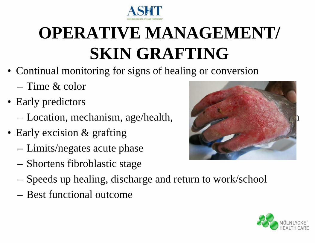
• Continual monitoring for signs of healing or conversion – Time & color
• Early predictors– Location, mechanism, age/health, occupation
• Early excision & grafting– Limits/negates acute phase– Shortens fibroblastic stage– Speeds up healing, discharge and return to work/school– Best functional outcome
OPERATIVE MANAGEMENT/ SKIN GRAFTING

38
WOUND COVERAGE (SURGICAL)Commonly Used with Hand Burns
• Xenograft• Homograft• Autograft• VAC• Integra• Flap
Superficial 2nd Deep 2nd
3rd
4th

XENOGRAFT Pigskinaka EZ Derm
• Temporary wound coverage
• Minimizes fluid loss• Controls pain via nerve
ending coverage• Stimulates re-
epithelialization

HOMOGRAFT/ALLOGRAFTCadaver
• Extended temporary wound coverage
• Tests recipient bed for viability
• Decreases pain• Protective
covering/seals wound– Reduces heat loss– Prevents infection– Minimizes fluid loss

41
AUTOGRAFTPatient’s Own Skin
• Permanent coverage• No risk of rejection• Skin depth and color
matching• Sheet for optimal
cosmesis, durability• Meshed for enlarged
coverage area• Donor site can be
reharvested

48
SPLIT-THICKNESS SKIN GRAFT (STSG)
• Most commonly used autograft• Includes epidermis and part of dermis• Vascular ingrowth within 24-28 hours• Typically harvested from ipsilateral
anterior thigh• Donor site requires wound care, dressing• Initial sensibility 4-6 weeks

49
FULL-THICKNESS SKIN GRAFT (FTSG)
• Less common but standard of choice for palmar wounds
• Includes epidermis and dermis• Advantages
– Increased depth-->higher quality coverage
– Less contraction within wound bed
• Disadvantages– Harvest site requires skin graft

50
AUTOGRAFTPOST-OPERATIVE CARE
• Bulky post-op dressing to immobilize hand, digits and prevent shearing
• Dressing removed POD#3 for STSG, POD#5 for FTSG
• Assessed for “take” (%)– Adherence to wound bed– Viability– Presence of fluid pockets or hematoma

51
AUTOGRAFT (STSG)POST-OPERATIVE CARE
• Therapist role– POD#3-5
• Trim excess autograft • Dressing to control edema, allow mobility
– Xeroform, gauze, Coban• Resume gentle AROM** (fibrin glue)
– POD#5 and beyond• Progress to limited dressing and edema glove• Intermediate pressure glove once little to no
dressing• Advance to aggressive AROM, PROM and
orthosis use as necessary, unrestricted ADL including shower

52
Autograft Trimming

WOUND VACAdvantages:
• Enhances granulation tissue
• Less dressing changes
• OR or bedside
Disadvantages:
• Unable to visualize wound
• Difficult application to smaller hands

WOUND VAC APPLICATION

WOUND VAC

INTEGRA• Bilayer matrix wound dressing
• Inner porous matrix allows rebuilding of blood supply, replaces dermis
• Outer silicone layer acts as epidermis, removed after dermal ingrowth for thin epidermal skin graft
• Closely monitored for infection
– Serum collection removed daily to prevent failure, loss

57
FLAP• Used for traumatic
defects involving soft tissue loss
• Provides wound coverage/closure
• Local skin flap uses nearby skin and subcutaneous tissue– Rotational– V-Y Advancement– Cross-Finger

58
FLAP• Used for traumatic defects
involving extensive soft tissue loss, exposed bone/tendon, inefficient blood supply
• Axial flap for reconstruction of distal UE injuries– Groin flap
• Secure at 5 days, PROM initiated
59
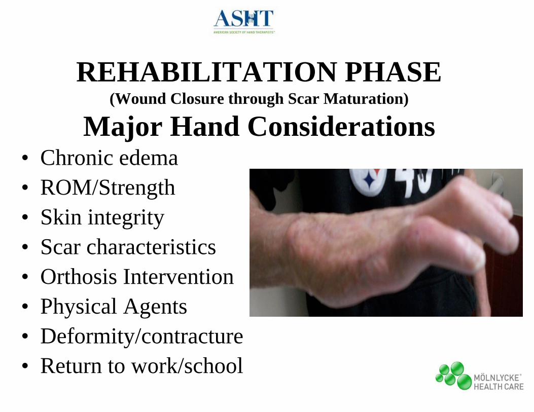
REHABILITATION PHASE(Wound Closure through Scar Maturation)
Major Hand Considerations• Chronic edema• ROM/Strength• Skin integrity• Scar characteristics• Orthosis Intervention• Physical Agents• Deformity/contracture• Return to work/school

60
CHRONIC EDEMA• Source of progressive scar formation &
restriction of motion• Compounded by lymphatic/vessel damage• Contributes to intrinsic, extrinsic tightness
– Due to ischemia, fibrosis, ROM limitations
• Treatment options– Compression gloves, sleeves– Jobst compression pump (home)

61
ROM/STRENGTH• Manual examination to
determine which structures limit motion– Multiple positions, target
tissue on slack & tension• Must consider soft tissue
structures beyond skin/scar– Intrinsic/extrinsic tightness,
joint stiffness• Resistive exercise in burn
recovery

62
SKIN INTEGRITY
• Recurrent exposed tendons/joints
• Assess readiness for pressure
• Protect bony prominences from shearing, blistering in garments and with return to activity
63
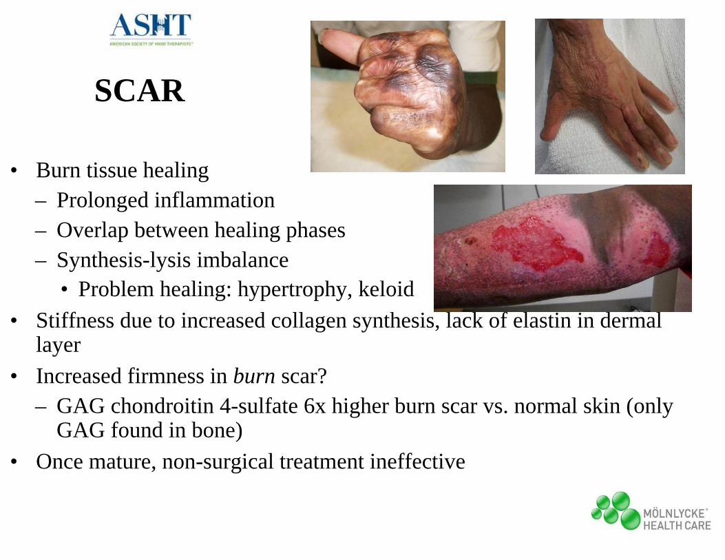
SCAR
• Burn tissue healing– Prolonged inflammation– Overlap between healing phases – Synthesis-lysis imbalance
• Problem healing: hypertrophy, keloid• Stiffness due to increased collagen synthesis, lack of elastin in dermal
layer• Increased firmness in burn scar?
– GAG chondroitin 4-sulfate 6x higher burn scar vs. normal skin (only GAG found in bone)
• Once mature, non-surgical treatment ineffective

64
SCAR ASSESSMENT• Vancouver Scar Scale (VSS)
– Most commonly used• Patient and Observer Scar
Assessment Scale (POSAS)– Developed in the Netherlands– Patient scale: color, pliability,
thickness, relief, itching, pain– Observer scale: vascularization,
pigmentation, pliability, thickness, relief
– Concurrent validity with VSS– Suitable for rating burn scars
Consistency ReliabilityVSS .49 .69POSAS .76 (P) .69 (O) .73 (O)
VSS

65
SCAR MANAGEMENTPressure
• Alters disposition of collagen fibers in dermal hypertrophic healing
• Custom fitted pressure garments– Measured when remaining wounds
no larger than quarter– “Intermediate” gloves/garments used
in interim to control scar/edema, prepare skin
– 2 sets for laundering– Worn 23 hours/day– Modifications for enhanced grip
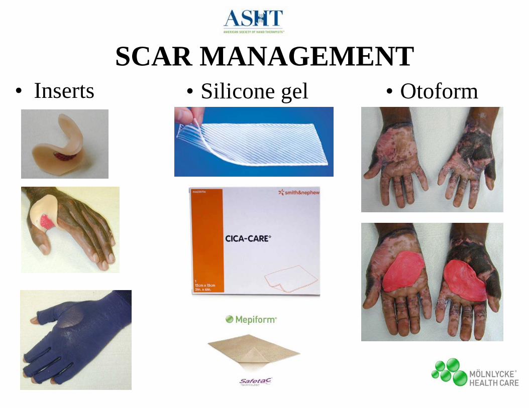
66
SCAR MANAGEMENT• Inserts • Silicone gel • Otoform

67
ORTHOSIS INDICATIONS• Purpose:
– Reduce non-surgical contracture– Prevent/reduce deformity– Maintain/promote natural body contours– Complement pressure treatment
• Goal:– Maintain sustained stretch to scar tissue– Maintain range achieved with stretch/ROM– Immobilize joint at end-range– Avoid pressure, excessive stretch

68
ORTHOSIS INTERVENTION• Static
– Thumb webspacer: 1st webspace tightness– PIP gutter: Boutonniere– DIP gutter: Mallet

69
ORTHOSIS INTERVENTION• Static progressive
– Significant resistance at end of passive stretch– Tension applied with joint at maximum range,
adjusted when tissue response allows repositioning to new length• MCP extension contractures

70
ORTHOSIS INTERVENTION• Serial static (casting)
– Resistive joint, firm to hard end-feel– Joint immobilized in stationary position, cast
remolded at new maximum length after tissue accommodation• Fixed contracture

71
PHYSICAL AGENTS• Paraffin with sustained stretch
– Most commonly used PAM with burns (cooler temp)– Softens skin, promotes increased tissue motion prior to exercise
• Iontophoresis (slow delivery)– Saline or iodine for scar softening
• Fluidotherapy• Ultrasound: limited success treating burn scar• Laser: multiple types for delayed scar treatment,
prophylactic prevention hypertrophic scar w/o good evidence
72
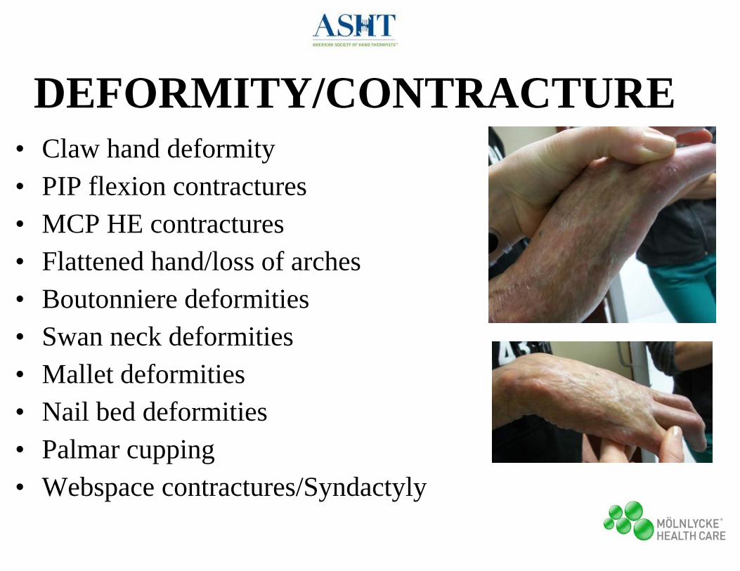
DEFORMITY/CONTRACTURE • Claw hand deformity• PIP flexion contractures• MCP HE contractures• Flattened hand/loss of arches• Boutonniere deformities• Swan neck deformities• Mallet deformities• Nail bed deformities• Palmar cupping• Webspace contractures/Syndactyly

Boutonniere deformities• More likely with deep dorsal hand, digits, thumb burns• Mechanism of injury
– Immediate: direct thermal injury to central slip– Delayed: tendon ischemia – Chronic: Scar banding &/or ORL tightness
• Arthrodesis is primary surgical correction option– Unsatisfactory tenoplasty options– No soft tissue coverage needed
73

Swan neck deformities
• MF incidence most prominent• Causes for PIP hyperextension
– EDC adherence– Intrinsic ischemic contracture– Joint stiffness/improper immobilization– Burn scar contracture
74

Mallet deformities• Mechanism of injury
– Immediate: direct thermal injury to terminal slip– Delayed: tendon ischemia (crushing of tendon between
dorsal surface eschar and P3 base)• Increased during PIP flexion
75

Nail bed deformities• Mechanism of injury
– Dorsal scarring over DIP with distortion of eponychial fold, eponychium retraction, proximal nail exposure
• Consequences of injury– Limits finger stability with pinching, fine motor
dexterity– Cosmetically disabling
• Surgical treatment– Tightness w/o retraction= skin release, graft– Tightness with retraction= proximally based
lateral skin flaps
76

Palmar cupping• Mechanism of injury
– Deep palmar burn (peds, contact)• Consequences of injury
– Thumb MCP HE contractures– Sensory deficits– Loss of stable grasping surface
• Surgical treatment– Multiple reconstruction procedures
and extensive therapy

Webspace contractures• Mechanism of injury
– Adjacent digits burned (fingers fuse together)– Digital skin granulation or contractures allow
distal web migration• Consequences of injury
– Limits digital ABduction and thumb opening– Cannot place thumb away from palmar plane
• Surgical treatment– Z-plasty variations
(lowest recurrence rate)– FTSG if not sufficient skin
78
79
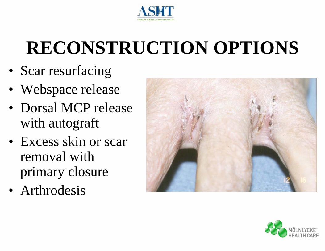
RECONSTRUCTION OPTIONS• Scar resurfacing• Webspace release• Dorsal MCP release
with autograft• Excess skin or scar
removal with primary closure
• Arthrodesis

80
RETURN TO WORK/SCHOOL• Collaborative effort• Referral to work-hardening program• Strongest indicators
• RTW time: % TBSA, grafting requirements, B hand involvement
• Successful school re-entry: tutors during hospitalization, school environment/ personnel & peer preparedness
• MHQ: hand function deterioration 68%• Most affected: ADL 76%, work 59%
23

81
RESOURCES & PROGRAMSFor Burn Survivors
• Phoenix Society www.phoenix-society.org– Survivors Offering Assistance & Recovery– Image enhancement– Local support groups
• American Burn Association www.ameriburn.org• International Association of Firefighters
– Regional Burn Camps • Adaptive Sports Center, Crested Butte CO
– Burn specific adaptive sports, outdoor programs

82
RESOURCESFor Therapists
• American Burn Association Rehabilitation Committee, Special Interest Group www.ameriburn.org
• BurnTherapist.com• Textbooks
– Burn Care and Rehabilitation: Principles and Practice (Richard, RL)
– Total Burn Care (2nd ed. Herndon)– Rehabilitation of the Hand and Upper Extremity (6th ed. Skirven
et al)

Support was provided by:
Mölnlycke Health Care is a world-leading provider of single-use surgical and wound care products for customers, healthcare professionals and patients. Our products provide value to our customers and are supported by clinical and health economic evidence. And we strive to find new ways to minimize community and hospital-acquired conditions.
http://www.molnlycke.us/
Contact email for therapists: [email protected]

References– Daugherty MB, Carr-Collins JA. Splinting Techniques for the Burn Patient in Burn Care and Rehabilitation: Principles and Practice.
Richard RL, Staley MJ. Philadelphia, PA: FA Davis, 1994.
– Dewey WS et al. A review of compression glove modifications to enhance functional grip: a case series. J Burn Care & Research Nov/Dec 2007; 28(6):888-891.
– Draaijers LJ et al. The patient and observer scar assessment scale: a reliable and feasible tool for scar evaluation. Plast & Recon Surg June 2004; 113(7):1960-1965.
– Esselman PC, Thombs BD, Magyar-Russell G, Fauerbach JA. Burn Rehabilitation: State of the science. Am J Phys Med Rehab 2006; 85(4): 383-413.
– Evans EB. Musculoskeletal Changes Secondary to Thermal Burns in Total Burn Care, 2nd ed. Herndon. London: Saunders, 2002.
– Germann G, Weigel G. The Burned Hand in Green’s Operative Hand Surgery, 6th ed. Green & Wolfe. Philadelphia, PA: Elsevier (Churchill Livingstone), 2011.
– Greenhalgh DG. Wound Healing in Total Burn Care, 2nd ed. Herndon. London: Saunders, 2002.
– Howell JW. Management of the Burned Hand in Burn Care and Rehabilitation: Principles and Practice. Richard RL, Staley MJ. Philadelphia, PA: FA Davis, 1994.
– Levi B, Peterson JR, De La Rosa S, Lisiecki JL, Rinkinen J, Su GL, Buchman SR, Cederna PS, Krebsbach PH, Wang SC. “Effect of Burn Injury on Mesenchymal Stem Cell Niches: The Mechanism and Possible Treatment for Heterotopic Ossification” presented at theAmerican Burn Association Annual Meeting, Palm Springs, CA, 2013.

References– Liew SH et al. Prophylactic treatment of deep dermal burn scar to prevent hypertrophic scarring using the pulsed dye laser: a preliminary
study. Annals Plast Surg Nov 2002; 49(5):472-475.
– Linares HA. Pathophysiology of the Burn Scar in Total Burn Care, 2nd ed. Herndon. London: Saunders, 2002.
– Moore ML, Dewey WS & Richard RL. Rehabilitation of the burned hand. Hand Clinics Nov 2009; 25(4):529-541.
– Muller MJ, Herndon DN. Operative Wound Management in Total Burn Care, 2nd ed. Herndon. London: Saunders, 2002.
– Mustoe, TA. Evolution of silicone therapy and mechanism of action in scar management. Aesth Plast Surg 2008; 32:82-92.
– Nelson ER, Wong VW, Krebsbach PH, Wang SC, Levi B. Heterotopic ossification following burn injury: The role of stem cells. J Burn Care & Research 2012; 33(4):463-470.
– Richard RL. Burns in Splinting the Hand and Upper Extremity: Principles and Process. Jacobs ML, Austin NM. Philadelphia, PA: Lippincott Williams and Wilkins, 2003.
– Richard RL et al. Identification of cutaneous functional units related to burn scar contracture development. J Burn Care & Research July/Aug 2009; 30(4):625-631.
– Serghiou, MA, Evans EB, Ott S, Calhoun JH, Morgan D, Hannon L. Comprehensive Rehabilitation of the Burn Patient in Total Burn Care, 2nd ed. Herndon. London: Saunders, 2002.
– Schneider JC, Holavanahalli R, Helm P, Goldstein R, Kowalske K. Contractures in Burn Injury: Defining the Problem. J Burn Care & Research 2006; 27(4): 508-514.
– Schneider JC, Holavanahalli R, Helm P, O’Neil C, Goldstein R, Kowalske K. Contractures in Burn Injury Part II: Investigating Joints of the Hand. J Burn Care & Research 2008; 29(4): 606-613.

References– Scott PG, Ghahary A, Tredget EE. Molecular and Cellular Basis of Hypertrophic Scarring in Total Burn Care, 2nd ed. Herndon. London:
Saunders, 2002.
– Simpson RL. Management of Burns of the Upper Extremity in Rehabilitation of the Hand and Upper Extremity, 6th ed. Skirven, Osterman, Fedorczyk, Amadio. Philadelphia, PA: Elsevier (Mosby), 2011.
– Suman OE, Spies RJ, Celis MM, Mlcak RP, Herndon DN. Effects of a 12-wk resistance exercise program on skeletal muscle strength in children with burn injuries. J Appl Physiol 2001; 91:1168-1175.
– Tufaro PA, Bondoc SL. Therapist’s Management of the Burned Hand in Rehabilitation of the Hand and Upper Extremity, 6th ed. Skirven, Osterman, Fedorczyk, Amadio. Philadelphia, PA: Elsevier (Mosby), 2011.
– Whitehead C, Serghiou M. A 12 year comparison of common interventions in the burn unit. J Burn Care & Research March/April 2009; 30(2):281-287.
– Williams WG. Pathophysiology of the Burn Wound in Total Burn Care, 2nd ed. Herndon. London: Saunders, 2002.