marginal bone resorption at dental implant – rtg … of dental problems and solutions issn:...
TRANSCRIPT
Journal of Dental Problems and Solutions
ISSN: 2394-8418 DOI CC By
001
Citation: Pejeva E, Papakoca K, Ambarkova V, Todorovska G (2018) Marginal bone resorption at dental implant – RTG analysis. J Dent Probl Solut 5(1): 001-011. DOI: http://doi.org/10.17352/2394-8418.000055
Clinical Group
AbstractDental implants are modern solutions, they compensate the lost tooth or teeth in the maxilla or
mandible. Their target is to improve the mastication, esthetics and phonation. They are made of metal, metal alloys or non-metallic materials. An important factor for successful implantation is biocompactibility. Bone resorption is destruction of bone tissue, and the loss of bone tissue is conducted by osteoclasts and mononuclear cells. In this fi nal paper are described the peri-implant diseases exactly the peri-implant mucositis and peri-implantitis. This diseases are complication of ossteointegrated dental implants, and they make changes of the soft and bone tissue around the implant. They bring marginal bone resorption, around the dental implant, and they do destruction of the bone tissue. RTG analysis were made, with intention to see how much the marginal bone resorption is around the dental implants.
Research Article
Marginal bone resorption at dental implant – RTG analysis
Pejeva E1, Papakoca K2, Ambarkova V3* and Todorovska G4
1Dentist Interim at the University Dental Clinic Center Ss.Pantelejmon, Skopje, Republic of Macedonia2University Goce Delcev, School of Medical studies, Shtip, Republic of Macedonia3University Ss.Cyril & Methodius, School of Dental Medicine, Skopje, Republic of Macedonia4Health Center- Skopje
Dates: Received: 29 November, 2017; Accepted: 30 December, 2017; Published: 02 January, 2018
*Corresponding author: Vesna Ambarkova PhD, MSc, DDS, University St. Cyril and Methodius, Faculty of Dental Medicine, Department of Paediatric and Preventive Dentistry, Vodnjanska 17 University Dental Clinic Center Sv.Pantelejmon Skopje 1000 , Republic of Macedonia, Tel: ++38970686333; E-mail:
Keywords: Dental implants; Implantation; Bone; Patient; Resorption; X-ray analysis; Peri-implant mucositis; Peri-implantitis
https://www.peertechz.com
Introduction
Dental implantology is an area of dentistry that involves the insertion of dental implants in order to compensate for the lost tooth and improve the function of mastication, phonation and aesthetics [1]. This paper presents 12 patients who have been implanted 23 dental implants. Osteointegrated dental implants, after a period of 4 to 12 months, are characterized by changes in the alveolar bone. Certain factors lead to resorption of the alveolar bone, especially in the marginal part of the alveolar bone. One of the factors is the infectious agents i.e. the presence of pathogens on the site where the implant will be placed, their presence and around the abutment itself. Furthermore, the impact of traumatic injuries, occlusal trauma in the patient, excessive strain on the dental implant are also one of the factors that lead to marginal bone resorption [2].
Bone resorption is the destruction of bone tissue, whereby bone tissue loss occurs and is carried out by osteoclasts and mononuclear cells. Osteoclasts are responsible for the demineralization of the bone, while mononuclear cells play a major role in the process of degradation of the organic matrix. Bone resorption is the most critical factor in the loss of attachment process. The height and density of the alveolar bone is maintained by means of an equilibrium between the apposition and bone resorption, which is regulated by systemic and local infl uences. When we have bone resorption, which exceeds the formation, bone height and density are reduced.
The resorption of the alveolar bone can be of a vertical or horizontal type. The vertical type of bone resorption, localized marginally, around the osteointegrated dental implant is char-acterized by the presence of defects that are placed under a slant angle in relation to the longitudinal axis of the implant. While at horizonal loss of the alveolar bone, we have a bone that is reduced in height, but the bone edges are perpendicu-lar to the tooth surface. The tissue healing process around the dental implant is similar to the physiological recovery of bone tissue. Studies on titanium implants in terms of their recovery are divided into three phases: osteoconductive, osteoarthritic and osteophilic [3].
The success of surgery, aesthetically and functionally de-pends on the amount of bone tissue and gingival tissue. The amount of alveolar bone loss in the fi rst year affects sulcus. In-creasing the depth of sulcus affects the longevity of the dental implant [4]. Radiographic analysis has shown that the shape of the micronut is very important in minimizing the loss of marginal alveolar bone during the stress-free phase and func-tional load. Such a surface on the microwave is recommended, around the neck of the implant acts preventively for the loss of the marginal alveolar bone and helps the early biomechanical adaptation of the load [5,6].
RTG - analyzes in front of the dental implants
Before the lining of dental implants, we had a thorough planning phase, in which we determined how many dental implants would be placed, their position and length. In planning, rhg analyzes enabled us to fi nd out more data on the anatomical structures of the maxilla and mandible. When

02
Citation: Pejeva E, Papakoca K, Ambarkova V, Todorovska G (2018) Marginal bone resorption at dental implant – RTG analysis. J Dent Probl Solut 5(1): 001-011. DOI: http://doi.org/10.17352/2394-8418.000055
planning any surgery, it is necessary to determine the location of the mandibular canal, when placing implants in the lower jaw.
The maxillary posterior quadrant is characterized by certain features, which are very important in terms of placement of the dental implant and the establishment of the chewing function. The volume, height of the alveolar bone is lost as a result of periodontal disease and bone resorption after the loss of teeth. The loss of maxillary side teeth leads to a decrease in the width of the bone and the labial bone lamella, and as a result, pneumatization of the maxillary sinus occurs. The width of the lateral parts of the upper jaw decreases much faster than other distal regions. Bone quality is in direct relation with bone density. Bone density is 5 to 10 times smaller than the symphony region in the lower jaw. The maxillary sinus is the largest of the four paranasal sinuses. It passes through several phases of seepage, ie air fi lling, expansion, spread to the lateral walls.
X-ray methods are used are the orthopantomograma of the jaws, lateral cephalometry, periapical radiography, occlusal X-ray, conventional and computed tomography [7]. In computed tomography a three-dimensional image is obtained - shows the confi guration of the bone not only in the sagittal but also in the transverse it provides innovative radiological technology, which undoubtedly replaces the conventional fi lm-based technology in all forms used in dentistry [8]. We made a digital volume tomography in a patient in the area of the maxillary lateral quadrant, which indicates that we have a lowered sinus. Using this RTG analysis, we make the treatment plan (Figure 1).
The plan of therapy involved raising the maxillary sinus, the goal is to increase the vertical dimension of the alveolar bone and to obtain space for the dental implant. The vertical dimension of the bone between the sinus fl oor and the alveolar ridge top is 8-12 mm. It is necessary to raise the maxillary sinus and to get a space of 2-4 mm more, to accommodate the future implant. At fi rst, we made osteotomy, then we raised the sinus and placed a bone graft. Various materials such as allogeneic bone, autogenous bone, alopeciae and combinations are used for sinus grafting [9].
The localization of canalis mandibularis is also very important when we want to place a dental implant in the mandible. If we determine with the rtg footage, that the localization of canalis mandibularis is appropriate for implant placement, then we make a plan of therapy.
The purpose of this paper is to demonstrate marginal bone resorption in dental implants that occurs as a result of peri-implant disease. With the help of RTG recordings, analyzes were made of how much resorption of bone tissue around the dental implants occurred. After a period of 4 months to 12 months, additional RTG recordings are made, where it is analyzed:
- Did we have marginal bone resorption and how much it is
- Is there a vertical or horizontal type of marginal bone resorption?
- Is the resorption of the alveolar ridge more expressed in maxilla or mandible?
- From which side of the dental implant we have more resorption, whether from a mesial or distal one
- To assess what type of implant we used, what is its location, diameter, length and age and gender of the patient.
During the surgical intervention itself, when placing dental implants in their alveolar bone, traumatic injury to the tissue itself occurs. The impact of traumatic injuries, the presence of pathogens on the site where the implant is placed, around the abutment itself cause bone resorption in the implant. The occlusal trauma in the patient, the excessive load on the dental implant, also contributes to marginal bone resorption. Inadequate maintenance of oral hygiene, poor plaque control, irregular dental examinations are also one of the factors that lead to the occurrence of peri-implant mucositis and peri-implantitis, and thus to marginal bone resorption.
Technique of implanting dental implants
Before placing the dental implants, it is necessary to take an exhaustive history of the patient, to make the diagnosis and the plan of therapy. The procedure for implanting a dental implant involves fi rst giving a local anesthetic a scalpel of size 15 and taking a sulcus incision, the buccal length of the implant site, and a vertical relaxation incision to protect the adjacent papilla. Before the initial osteotomy, we approach which measurement of the diameter of the alveolus and we estimate its architecture using special depth lines. Initial osteotomy is performed with the help of special borers. When doing osteotomy, we should pay attention to the depth due to the restorative platform that should not be deeper than 2-3mm apical from the cement enamel boundary of the future fi nal restoration. Placement of the implant is carried out with a torsion force of 30Ncm. The fi rst suture is set for the correct positioning of the cortical margin of the lamp using a thread for sutures of 5-0 or lower. After 7-10 days the strings are removed. After the completed modeling and soft tissue formation around the implant, the defi nitive restoration is made. Once the implant is placed in the bone, after a certain period the phase of their association, i.e. osteointegration and fi brous integration, is followed. After the osteointegration of the implants, the opening of the buried implants, mounting of the abutment and preparation of a prosthetic superstructure is followed. After the selection of the suprastructure, we approach the fi ngerprint for the working Figure 1: Lowered maxillar sinus.

03
Citation: Pejeva E, Papakoca K, Ambarkova V, Todorovska G (2018) Marginal bone resorption at dental implant – RTG analysis. J Dent Probl Solut 5(1): 001-011. DOI: http://doi.org/10.17352/2394-8418.000055
model, the prosthetic compensation is made and the defi nitive setting of the suprastructure and the prostitution fee follows [1].
Peri-implant diseases: Peri-implant diseases are infl ammatory conditions that affect the soft and hard tissues around the dental implants. As with the natural tooth, the dental plaque supra and subgangially accumulates in the dental implant as well.
Dental plaque bacteria irritate the gingiva around the dental implant and cause infl ammation, damage to the soft tissue, and if it is not diagnosed and treated on time, alveolar bone tissue around the dental implant will be lost.
Peri-implant diseases are classifi ed into two categories: - Peri-implant mucositis, which would be described as a bacterial-induced, infl ammatory process that involves soft tissue. Clinically, redness of the gingiva is noticeable, it is oedematous and bleeding occurs during the probing.
- Peri-implantitis characterized by progressive, infl ammatory destruction of the alveolar bone around the implant. Deepening of the gingival sulcus occurs, the purulent and bloody contents are drained during the projection [10,11] (Figures 2,3).
In the previous section, I tried to describe periodic implant diseases only briefl y. According to my research, there are certain risk factors for these peri-implant diseases to progress.
Risk factors for the progression of peri-implant di-seases
Early periodontal disease: Peri-implantitis as a serious illness is more noticeably observed in patients who have previously had periodontitis in their dental history.
Weak plaque control: The implant’s prototype implant is designed to allow easy mechanical cleaning of the dental implant. With the use of a toothbrush, a dental tie and an interdental brush, effective dental plaque control is ensured. All patients in terms of maintaining oral hygiene vary. According to studies conducted by poorly maintained oral hygiene patients, poorly used dental plaque controls, such as the oral cavity, lead to peri-implant mucositis and gradually to peri-implantitis [12].
Smoking cigarettes: According to the research, I noted that in 50% of patients we had dental implants, they had the peri-implantitis diagnosis. While in non-smokers, about 40% reported peri-implantitis [12,13].
Genetic factors: The association between the IL-1 gene polymorphism and peri-implantitis is still very long-established. The IL-1RN gene-incorporated polymorphism has long been considered a risk factor for the progression of peri-implantitis [12-14].
Diabetes: A high amount of blood glucose affects the progression of peri-implantitis. It also causes interruption of collagen homeostasis in the extracellular matrix and is
associated with neutrophil dysfunction and a decline in the immune system [14,15].
In peri-implant mucositis, the marginal gingiva in lightly bleeding bleeds. Gingiva is hyperemic and oedematous. The patient feels discomfort, mastication of food also leads to bleeding (Figure 4) [16].
With the progression of the peri-implant mucositis, the soft tissue condition around the dental implant is aggravated. Bone tissue destruction begins inside the alveolar bone around the implant itself, causing bone resorption. This process is irreversible in relation to peri-implant mucositis that is a reversible process (Figures 5-7) [17].
Classifi cation of peri-implantitis
According to the research I have done, I can classify Peri-implant peri-implantitis in an early stage, a moderate stage and an advanced stage.
An early stage of peri-implantitis is when the clinical examination in the patient, in the course of the probing, the depth of the gingival sulcus is. In the case of probing, there is a cranny and sometimes drainage of purulent contents. Marginal bone resorption is <25% of the length of the dental implant.
Figure 2: Peri-implant mucositis.
Figure 3: Peri-implantitis.
Figure 4: Peri-implant mucositis, bleeding on probing.

04
Citation: Pejeva E, Papakoca K, Ambarkova V, Todorovska G (2018) Marginal bone resorption at dental implant – RTG analysis. J Dent Probl Solut 5(1): 001-011. DOI: http://doi.org/10.17352/2394-8418.000055
A moderate stage of peri-implantitis exists when, when probe around the dental implant, the probe in the gingival sulcus collapses and drainage of bloody or purulent content occurs. Bone tissue resorption is from 25% to 50% of the length of the dental implant.
Late stage of peri-implantitis, we have when when probe around the dental implant, the probe falls into the sulcus in depth. Bleeding and purulent content drainage is being drained. Bone resorption is> 50% of the length of the dental implant (Figures 8-10) [18].
In this scientifi c research, 12 patients were included, aged 30-60. 23 titanium implants (Ankylos DENTSPLY Implants) were placed. The method of work is the analysis of the dental implants on the RTG images, which we made in patients from their placement in the alveolar ridge and after a period of up to 12 months passed. In this past period, we made an insight into the changes that occurred on the alveolar bone in dental implants. Patients were diagnosed with peri-implant diseases. Furthermore, RTG recordings were performed that concluded
that there was bone resorption around the dental implants, and discussed the type of marginal bone resorption.
The material from which we received the necessary data for this research paper were RTG recordings. The use of X-rays is an inevitable procedure in high-quality dental diagnostics. DVT or digital volume tomography representing innovative radiological technology. The research process was conducted in a private health institution - a dental offi ce Dent-Estet in Stip.
Results
Clinical case no 1
The fi rst patient was at the age of 48 with male gender and generally with good health. There are no contraindications to make oral surgery. The patient is not allergic to any medication. A dental implant was placed 12 months ago (Figures 11,12), the dental implant’s position was 46. The implant was 4.1 mm in diameter and 12 mm in length. The patient has peri-implantitis, a RTG intraoral radiography image was recorded and marginal bone resorption was observed. The depth of the gingival sulcus was 10mm. Dehiscencia was seen in the patient
Figure 5: Peri – implant mucositis.
Figure 6: Xray reveals of peri-implantitis with bone resorption.
Figure 7: Surgery exposure of the place where there is bone loss around the dental implant.
Figure 8: Early stadium of peri-implantitis.
Figure 9: Moderate sadium of peri-implantitis.
Figure 10: Advanced stadium of peri-implantitis.

05
Citation: Pejeva E, Papakoca K, Ambarkova V, Todorovska G (2018) Marginal bone resorption at dental implant – RTG analysis. J Dent Probl Solut 5(1): 001-011. DOI: http://doi.org/10.17352/2394-8418.000055
from the buccal side of the implant (Figure 13). The bone defect was closed with a bone graft (Figures 14-16).
Clinical case no 2
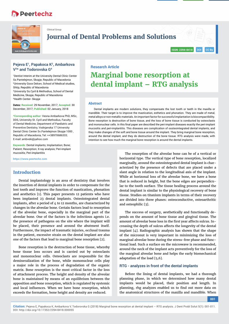
Woman at aged 37 year age, in good health condition and without any systemic diseases, allergic to penicillin came for prosthetic rehabilitation. She was without second premolar and fi rst molar in the lower right quadrant. The patient was provided with dental implants at 45 and 46 (Figures 17-19). After a month on a 45-implant, retrograde peri-implantitis appeared. In chronological order, X-ray images are shown on fi gures from 17 to 20.
Different treatment strategies were performed in order to remove peri-implantitis. Provided treatment include: debridement of bone tissue, use of bone grafts from known biocompatible grafting materials, i.e. BioOS, disinfection of the surface of the dental implant. After 12 months, another
Figure 11: On the probing, the mesial depth is 11mm and the distal depth is 12 mm.
Figure 12: Preoperative intraoral radiography, where bone resorption was seen.
Figure 13: Careful removal of granulation tissue, circular loss of bone tissue.
Figure 14: The bone defect was closed with a bone graft.
Figure 15: After the bone graft placement, a collagen membrane was applied.
Figure 16: Postoperative intraoral imaging. A bone defect with bone graft, which is in contact with the surface of the implant, is seen to be complete.
Figure 17: Retroalveolar radiography was taken after 2 weeks of the dental implants placement. At 45 implant, a small periapical radioluscence was noted.
Figure 18: Rtg taken after 1 month of the dental implants placement.
06
Citation: Pejeva E, Papakoca K, Ambarkova V, Todorovska G (2018) Marginal bone resorption at dental implant – RTG analysis. J Dent Probl Solut 5(1): 001-011. DOI: http://doi.org/10.17352/2394-8418.000055
radiography record was made, where ceramic crowns over the implants were seen and there was no longer peri-implantitis (Figure 20) [19,20].
Clinical case no 3
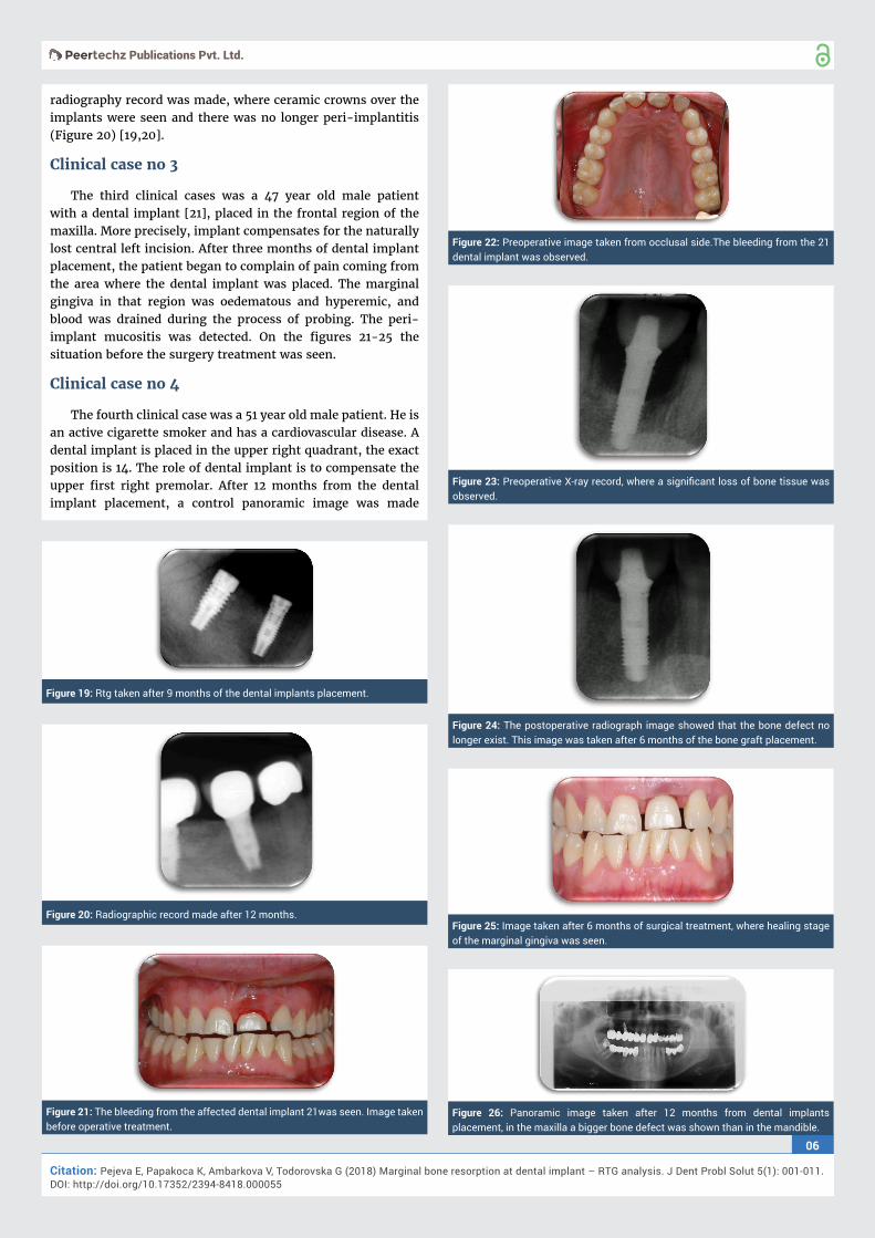
The third clinical cases was a 47 year old male patient with a dental implant [21], placed in the frontal region of the maxilla. More precisely, implant compensates for the naturally lost central left incision. After three months of dental implant placement, the patient began to complain of pain coming from the area where the dental implant was placed. The marginal gingiva in that region was oedematous and hyperemic, and blood was drained during the process of probing. The peri-implant mucositis was detected. On the fi gures 21-25 the situation before the surgery treatment was seen.
Clinical case no 4
The fourth clinical case was a 51 year old male patient. He is an active cigarette smoker and has a cardiovascular disease. A dental implant is placed in the upper right quadrant, the exact position is 14. The role of dental implant is to compensate the upper fi rst right premolar. After 12 months from the dental implant placement, a control panoramic image was made
Figure 19: Rtg taken after 9 months of the dental implants placement.
Figure 20: Radiographic record made after 12 months.
Figure 21: The bleeding from the affected dental implant 21was seen. Image taken before operative treatment.
Figure 22: Preoperative image taken from occlusal side.The bleeding from the 21 dental implant was observed.
Figure 23: Preoperative X-ray record, where a signifi cant loss of bone tissue was observed.
Figure 24: The postoperative radiograph image showed that the bone defect no longer exist. This image was taken after 6 months of the bone graft placement.
Figure 25: Image taken after 6 months of surgical treatment, where healing stage of the marginal gingiva was seen.
Figure 26: Panoramic image taken after 12 months from dental implants placement, in the maxilla a bigger bone defect was shown than in the mandible.

07
Citation: Pejeva E, Papakoca K, Ambarkova V, Todorovska G (2018) Marginal bone resorption at dental implant – RTG analysis. J Dent Probl Solut 5(1): 001-011. DOI: http://doi.org/10.17352/2394-8418.000055
in order to see the condition of the dental implant. In the orthopantomographic image below (Figure 26), the extensive bone resorption was noted in both jaws, which is particularly expressed in the maxilla.
Clinical case no 5
The fi fth clinical case is female patient at the age of 48, without any systemic disease but an active cigarette smoker. Four dental implants were placed, three of which ware in the maxilla, and one in mandible. In the upper right quadrant, two dental implants were placed and they compensate the lost fi rst molar. Two crowns with premolar size were placed on two dental implants, in order to avoid excessive overload due to the masticatory pressure that mostly affects that area.
In the upper left quadrant on posterior region, one more titanium dental implant was placed, which compensates the lost fi rst molar. In the lower left quadrant on the posterior region, another dental implant was placed instеad of the lower fi rst natural molar. After 9 months a control panoramic X-ray was made in order to follow the condition of the bone tissue in the both jaws. From the fi gure 27 it’s evident that bone resorption is more expressed in maxilla than in mandible. The larger bone defect was noted in the upper posterior right quadrant, where two dental implants are placed next to each other.
Clinical case no 6
The sixth clinical case was a 29 year old female patient with excellent health condition. Three dental implants were placed in the lower right quadrant. Dental implants compensate previously lost fi rst and second permanent molars. On the panoramic image below (Figure 28), could can be seen that bone resorption was more pronounced in the mandible, especially in the lower right posterior quadrant, than in the maxilla. The panoramic record was done after 11 months of dental implants placement (Figure 28).
Clinical case no 7
The seventh clinical case was male 33-year-old patient with good health condition and without any illness. He received dental implants in maxilla both in the right and the left posterior quadrant, 10 months ago. On the panoramic X-ray image marginal bone resorption changes were present in both jaws (maxilla and mandible). Bone resorption was observed on mesial and distal side from dental implants, also horizontal type of resorption is present (Figure 29).
Clinical case no 8
The eighth clinical case is a female 42 year old patient, which is in good health and with good oral hygiene habits. Before a 9-months, she received two titanium dental implants. The position of the placed dental implants were in the mandible, in the posterior right quadrant. The dental implants were placed due to the missing of the second premolar and the fi rst mandibular molar. A control X-ray image was made to the patient because of the suspicion that she has a peri-implant
disease. In the clinical examination, it was found that the gingiva around the dental implants was swollen and hyperemic. The impressions and minimal bleeding were observed on the gingival palpation. The bleeding from the gingiva occur during the process of probing, and pocket depth of 3.5 mm was detected. After the performed x-ray examination, a very small marginal bone resorption was detected аnd no surgical intervention was required (Figures 30-32).
Clinical case no 9
The ninth clinical case was a thirty-year-old male patient. He received titanium dental implant 12 months ago, due to the missing of the second premolar in the month. The position of the dental implant is 15, i.e. it was on a position of the maxillary right second premolar. The patient was with diabetic disease, with no allergic reactions to drugs and with good oral
Figure 27: Panoramic image taken after 9 months from the implant placement.
Figure 28: Panoramic X-ray image taken after 11 months of dental implants placement.
Figure 29: After ten months from dental implants placement, a control panoramic X-ray was taken.
Figure 30: Dental implants placed at positions 45 and 46.

08
Citation: Pejeva E, Papakoca K, Ambarkova V, Todorovska G (2018) Marginal bone resorption at dental implant – RTG analysis. J Dent Probl Solut 5(1): 001-011. DOI: http://doi.org/10.17352/2394-8418.000055
hygiene habits. Due to the patient’s symptomatology, we made an X-ray in order to control the situation and the state of the tissues around the dental implant. In the clinical examination of the patient during the probing, drainage of blood with pus impurities was noticed. The patient had a painful reaction on palpation, the gums were swollen and with red color. The pocket depth was 6 mm. On the x-ray marginal bone resorption was detected with loss of tissue attachment and defect in bone structure (Figure 33).
Clinical case no 10
The tenth clinical case was a 30 year old female patient. Before a period of 7 months she received dental implant due to a missing of the frontal right central incision 11. The patient was in excellent health and without allergy to any medication. An X-ray was made, showing changes in bone tissue around the dental implant. Around dental implant (from the mesial and distal side) a bone defect was detected, which occurred after a period of seven months. During the probing around the dental implant, the probe went down 4 mm in pocket from the mesial side and 3 mm from the distal side. The greater loss of bone tissue was detected from the mesial side of the dental implant than from the distal side. The patient was identifi ed to have peri-implant disease (Figure 34).
Clinical case no 11
The eleventh clinical case was a 53 year old male patient with cardiovascular disease, but well treated by cardiologist He received 5 titanium dental implants. He received three titanium dental implants in maxilla in the left posterior quadrant and two titanium dental implants in mandible, in the right posterior quadrant half-year ago. Due to patient’s inadequate maintenance of the oral hygiene, during consecutive 6 months, infl ammatory changes and bleeding from the gums occurred during the act of mastication. After the RTG examination, resorption of the bone tissue in both jaws was detected. Bone destruction is more pronounced in the maxilla than in the mandible (Figure 35).
Clinical case no 12
Two dental titanium implants were placed in a 37 years old, male patient. These two dental titanium implants were placed one year ago and their position is 36, they compensate the lower left fi rst molar. The patient come to dental offi ce on control because he felt pain in the area of dental implants. The pain, except during mastication, was also manifested during the brushing of the teeth, and was accompanied by bleeding from the gums. Gingiva was hyperemic and oedematous (Figure 36), and X-ray control images were also made, which can be seen below (Figure 37-40). During the process of probing, the dental probe drowned in bone pocket around the dental implants at a depth of 10 mm.
Discussion
Early detection of peri-implant as a peri-implant disease allows timely treatment, and for that purpose, a larger bone mass is preserved. According to studies, the
Figure 32: After 10 months from the implants placement, good condition of the gums were observed, because the patient followed the instructions for improvement of oral hygiene. Gingiva was no longer swollen and hyperemic.
Figure 33: Control x-ray record taken after 12 months from the titanium dental implant placement.
Figure 34: Marginal bone resorption around a titanium dental implant, seven months after a dental implant placement.
Figure 35: Control X-ray panorama was made after 6 months from the dental implants placement. Bone resorption was seen in both maxilla and mandible.
Figure 31: Control X-ray taken after a period of 9 months from the implants placement. Minimal marginal bone resorption was observed.

09
Citation: Pejeva E, Papakoca K, Ambarkova V, Todorovska G (2018) Marginal bone resorption at dental implant – RTG analysis. J Dent Probl Solut 5(1): 001-011. DOI: http://doi.org/10.17352/2394-8418.000055
Figure 36: Probing around the dental implants, where the depth of the pocket that is formed was 10 mm.
Figure 37: Control retroalveolar X-ray record, where the presence of a bone defect around the titanium dental implants was observed.
Figure 38: Removal of granulation tissue around the dental implants and debridement of bone tissue around the implants.
Figure 39: The placement of bone graft in order to close the bone defect.
Figure 40: Post-surgical x-ray control. Helthy bone tissue around the dental implants.
level of marginal gingiva can vary in patients with systemic diseases and the presence of periodontal disease [19]. It is very important to make a good diagnosis when it comes to peri-implant mucositis. Because there are cases when peri-implant mucositis can be ascertained as a periodontal disease. According to a certain group of authors led by Schwartz, it has been established that if the initial marginal bone resorption is less than 2mm, then no surgical procedures are undertaken in order to prevent further progression of the disease. While the amount of marginal bone resorption is greater than 2 mm, there is greater destruction of the bone mass, then surgical treatment is expected. Surgical treatment includes the treatment of mucoperiodic lobe, which is localized apical. Raising mucoperiodistomal lampo, bone mass around the dental implant. Furthermore, placement of bone grafts, in order to complete the defect, and in some cases a resorptive collagen membrane is placed [20].
Patients who were given dental implants were also given instructions. That is, to increase the level of maintenance of oral hygiene, to constantly control dental plaque, to come to dental examination. After placing the dental implants, patients were also advised to use a soft-fi ber brush for at least the fi rst month after placing the dental implants.
In patients initially when peri-implant disease is peri-implant mucositis, I would add if the patients would control dental plaque by treating the plaque removal from the surface of the implant, avoiding the irritation of the gingiva. Thus, infl ammation of the marginal gingival tissue will not occur, no edema, bleeding disorders, and most of all loss of bone tissue and progression in peri-implantitis [21,22].
In the above-described clinical case in a patient who had retrograde peri-implantitis at 45, research was carried out and one of the factors for the occurrence of retrograde peri-implantitis is the over-heating of the bone. But by applying appropriate treatment over a period of 12 months, the retrograde peri-implantitis disappeared [23].
The surgical protocol which was applied, enabled the establishment of the basic amount of bone tissue that existed previously. Flap techniques, followed by muco-periostеal fl aps often were primarily used when we have a horizontal type of resorption and moderate bone defects.
Conclusion
Bone resorption is the most critical factor in the loss of attachment process. The height and density of the alveolar bone is maintained by means of an equilibrium between the apposition and bone resorption, which is regulated by systemic and local infl uences.- There are certain factors that infl uence the success of dental implantation; it is of particular importance that the successful teamwork between the surgeon and the prosthetist.
- The material from which the dental implant is made, as well as its size and shape, is very important. The technique for embedding the dental implant is also of great importance,

010
Citation: Pejeva E, Papakoca K, Ambarkova V, Todorovska G (2018) Marginal bone resorption at dental implant – RTG analysis. J Dent Probl Solut 5(1): 001-011. DOI: http://doi.org/10.17352/2394-8418.000055
as well as the development of a functional and aesthetic suprastructure.
- Of course, just like the implant, the suprastructure is equally important. High quality metal - ceramic products are the minimal thing to do with dental implants. We had the opportunity to see manufactured zirconium ceramic or complete zirconium supraventures over dental implants that after a few years have a perfect aesthetic and functional role and are perfectly fi tted into the dental - facial system.
The failure of dental implantation is also infl uenced by fac-tors.
- The use of traumatic work, that is, the use of excessive force during the surgical procedure itself leads to failure in the im-plantation. Healing of bone tissue, at the very act of making a new alveoloma, leads most often to the occurrence of peri-implantitis.
- Insuffi cient professional preparation and inexperience of the team that performs the procedure are more causes of early implant failure - prosthetic restoration.
Implanting or placement of dental implants, as a surgical procedure, and then opening the implants for setting the su-prastructure - in inadequate or more precisely stated non-ster-ile conditions, are also one of the more frequent causes of early implant loss. Most often, patients are those who cause fail-ure to the dental implant itself.
- Patients poorly maintain oral hygiene, and this leads to continuous supra-gingival and sub-gingival accumulation of the dental plaque.
- The accumulated dental plaque on the surface of the dental implant acts on the marginal gingiva irritatively and causes infl ammation. Gingiva is further followed by hyperae-mia, oedematousness, bleeding at the time of prophylaxis, and sometimes purulent impurities are present. This indicates the existence of a peri-implant disease, that is, peri-implant mu-cositis. Further non-treatment leads to peri-implantitis. This disease, which is characterized by irreversible changes, is seri-ous and leads to a large amount of bone loss.
- For occurrence of peri-implant diseases, there are certain risk factors that we need to pay attention to. Such as patients with chronic systemic disease, diabetic who do not control the disease, previous periodontal diseases, smoking cigarettes, poor plaque control, genetic factors.
Today there are modern methods for diagnosing peri-im-plant diseases.
- X-ray diagnostics is an inevitable procedure in high-quality dental therapy. The use of digital tomography, which is an innovative X-ray technology, can enable effi cient diagnosis of peri-implantology.
To have a good prognosis for already osteointegrated im-plants, timely diagnosis of peri-implant mucositis and peri-implantitis is necessary.
References
1. Papakoca K, Dimova C, Zlatanovska K, Zarkova J Dental implantology - script, University Goce Delcev, School of medical studies, Shtip, Republic of Mace-donia.
2. Misch CE (2005) Dental implant prosthetich; Library of Congres Catalogingin-Publicatioin Data. St Louis: Mosby.
3. Krhen J, Canjuga I, Jerolimov V, Krhen T (2009) Implant stability measure-ment six weeks after implantation. Acta Stomatol Croat 43: 45-51. Link: https://goo.gl/WtJZLg
4. Živko-Babić J, Jakovac M, Carek A, Lovrić Ž (2009) Implantoprosthetic therapy of a missing front tooth. Acta Stomatol Croat 43: 234-241. Link: https://goo.gl/YL1HPv
5. Nickenig HJ, Wichmann M, Schlegel KA, Nkenke E, Eitner S (2009) Radio-graphic evaluation of marginal bone levels adjacent to parallel- screw cyl-inder machined-neck implants and rough-surfaced microthreaded implants using digitized panoramic radiographs. Clin Oral Implants Res 20: 550-554. Link: https://goo.gl/KEMzb2
6. Piao CM, Lee JE, Koak JY, Kim SK, Rhyu IC, et al. (2010) Marginal bone loss around three different implant systems: radiographic evaluation after 1 year. J Oral Rehabil 37: 538-544. Link: https://goo.gl/EwmpQQ
7. Evelina Markova, Milen Markov, Javor Simeonov Klinika Plovdiv (2012) Den-tal implants. Link: https://goo.gl/5kc1wa
8. Alan L Rosenfeld, Richard A Mecall (1998) Using computerized tomography to develop realistic treatment objectives for the implant team.
9. Kao DW, Kubota A Nevins M, Fiorellini JP (2012) The negative effect of combining rhBMP-2 and Bio-Oss on bone formation for maxillary si-nus augmentation. Int J Periodontics Restorative Dent 32: 61-67. Link: https://goo.gl/QENrky
10. (2016) American Academy of Periodontology. Link: https://goo.gl/kr4ju4
11. Khammissa RAG, Feller L, Meyerov R, Lemmer J (2012) Peri-implant muco-sitis and peri-implantitis: clinical and histopathological characteristics and treatment. SADJ 67: 124-126. Link: https://goo.gl/6YzvEy
12. Rocchietta I, Nisand D (2012) A review assessing the quality of reporting of risk factor research in implant dentistry using smoking, diabetes and periodontitis and implant loss as an outcome: Critical aspects in design and outcome assessment. J Clin Periodontol 39 (Suppl.12): 114-121. Link: https://goo.gl/dMkFA2
13. Klokkevold PR, Han TJ (2007) How do smoking, diabetes, and periodontitis affect outcomes of implant treatment? Int J Oral Maxillofac Implants 22(Sup-pl.): 173-202 Link: https://goo.gl/oyG8eJ
14. Hinode D, Tanabe S, Yokoyama M, Fujisawa K, Yamauchi E, et al. (2006) Infl u-ence of smoking on osseointegrated implant failure: A meta-analysis. Clin Oral Implants Res 17: 473-478. Link: https://goo.gl/8Fnj21
15. Bornstein MM, Cionca N, Mombelli A (2009) Systemic conditions and treat-ments as risks for implant therapy. Int J Oral Maxillofac Implants 24(Suppl.): 12-27. Link: https://goo.gl/32DsQF
16. Khammissa RA, Feller L, Meyerov R, Lemmer J (2012) Peri-implant muco-sitis and peri-implantitis: bacterial infection. SADJ 67: 70, 72-74. Link: https://goo.gl/VSvHiG
17. Paul Rosen, Donald Clem, David Cochran, Stuart Froum, Bradley McAllister, et al. (2013) Academy Report: Peri-Implant Mucositis and Peri-Implantitis: A Current Understanding of Their Diagnoses and Clinical Implications. J Peri-odontol 84: 436-443. Link: https://goo.gl/8t8Se4
18. Sugiura T, Yamamoto K, Horita S, Murakami K, Tsutsumi S, Kirita T (2016) The effects of bone density and crestal cortical bone thickness on micromotion

011
Citation: Pejeva E, Papakoca K, Ambarkova V, Todorovska G (2018) Marginal bone resorption at dental implant – RTG analysis. J Dent Probl Solut 5(1): 001-011. DOI: http://doi.org/10.17352/2394-8418.000055
Copyright: © 2018 Pejeva E, et al. This is an open-access article distributed under the terms of the Creative Commons Attribution License, which permits unrestricted use, distribution, and reproduction in any medium, provided the original author and source are credited.
and peri-implant bone strain distribution in an immediately loaded implant: A nonlinear fi nite element analysis. J Periodontal Implant Sci 46: 136–151. Link: https://goo.gl/GudMDE
19. Renvert S, Aghazadeh A, Hallström H, Persson GR (2014) Factors related to peri-implantitis - a retrospective study. Clin Oral Implants Res 25: 522–529. Link: https://goo.gl/VnbFm6
20. Lagervall M, Jansson LE (2013) Treatment outcome in patients with peri-implantitis in a periodontal clinic: a retrospective study. J Periodontol 84: 1365–1373. Link: https://goo.gl/imNS6Z
21. Chen S, Darby I (2003) Dental implants: Maintenance, care and treatment of peri-implant infection. Aust Dent J 48: 212–220. Link: https://goo.gl/rdHhuo
22. Romeo E, Ghisolfi M, Murgolo N, Chiapasco M, Lops D, et al. (2005) Therapy of peri-implantitis with resective surgery. A 3-year clinical trial on rough screw-shaped oral implants. Part I: Clinical outcome. Clin Oral Implants Res16: 9–18. Link: https://goo.gl/sdBqJF
23. Chang LC, Hsu CS, Lee YL (2011) Successful medical treatment of an im-plant periapical lesion: a casereport. Chang Gung Med J 34: 109-114. Link: https://goo.gl/ynb487