mast cell cultures: bench to bedside
TRANSCRIPT

REVIEW
Mast cell cultures: bench to bedside
G. L. ROSSI, V. DI COMITE and D. OLIVIERI
Department of Respiratory Disease, University of Parma, Italy
Introduction
The mast cell plays a prominent role in allergic reactionsand is implicated in the genesis of other diseases such aspulmonary fibrosis and graft-vs-host disease. Moreover,mast cells participate in important physiological responsessuch as the defence against bacteria and parasites and mayalso contribute to the growth of solid tumours or to immunedefence against them [1,2]. As regards bronchial asthma, theperception of the mast cell’s role has been changed overtime, and the present view states that mast cells, along withT lymphocytes and eosinophils, substantially contribute tothe establishment of bronchial inflammation [3].
In 1989, several independent investigators reported thatcultivated murine mast cells were able to produce an arrayof multiple proinflammatory cytokinesin vitro [4–6]. Fiveyears later, Braddinget al., using immunohistochemicalanalysis of endobronchial biopsies, showed that mast cellswere the major source of IL-4, IL-5, IL-6 and TNFa (both innormal individuals and in subjects with mild atopic asthma[7]. This work showed that studiesin vitro can anticipateimportant findings in humans that are relevant to thepathogenesis of asthma.
The aim of the present article is to briefly outline thepotential role of mast cell cultures as a source of informa-tion, useful for clinical investigators in allergy, respiratorymedicine and other fields of research.
In vitro and in vivo studies on mast cells are influenced bymast cell heterogeneity
The mast cell, unlike the basophil or the eosinophil, does notleave the bone marrow as a mature cell but in the form of animmature progenitor that, although committed to the mastcell lineage, lacks the phenotype and the functions of mastcells. The progenitors released from the bone marrow enterthe bloodstream and reach peripheral tissues and organswhere they differentiate under the influence of local envir-onmental factors and assume the morphological and func-tional characteristics of mast cells (reviewed in [8]).
Much research has been addressed to mature,differentiated mast cells present in tissues. Overall, immu-nocytochemical studies strongly point out the presence of alarge heterogeneity in the phenotype of mast cell popula-tions both in rodents [9,10] and humans (reviewed in [11]).A variability of mast cell phenotype — for example, thedifferences in the content of cellular proteases — is presentin different anatomical sites of the same individual andreflects the action of the local microenvironment.
Also, a pronounced functional heterogeneity exists inmast cells, as demonstrated byin vitro studies performedwith mast cells recovered from various tissues and organsboth in rodents (lung, gut, peritoneal cavity, etc.) andhumans (lung, skin, tonsil, intestine). More specifically,the responses to different activating stimuli and the phar-macological inhibition of mast cell activation vary amongcell populations derived from different tissues of the sameindividual or from individuals of different species.
Therefore, the study of the ‘end product’ (i.e. the differ-entiated mast cells present in tissues) does not easily rendera comprehensive view of the role of the mast cell in a moregeneral context.
Mast cell cultures: from the mouse to the man
In 1981, five independent groups reported that normalmouse bone marrow, cultivated in a medium containingparticular growth factors, can generate a uniform populationof cells with the feature of immature mast cells. The mostimportant of such growth factors (in this murine system)was later identified as IL-3. In a recent article, Galli hasexhaustively reviewed the ‘historical’ steps that led to theestablishment of these normal (non-transformed) murinemast cell cultures and how they contributed to elucidatefundamental biological questions related to mast cell originand development [12]. Therefore we refer the reader to thisarticle for more details.
We would like to underscore that studies with mousebone marrow-derived mast cells (BMMC) helped to clarifythree important points: mast cell precursors are locatedin the bone marrow; IL-3 is the crucial factor for mastcell differentiation in the mouse; and mast cell phenotypecan vary according to environmental stimuli. The latter
1182 q 1998 Blackwell Science Ltd
Clinical and Experimental Allergy,1998, Volume 28, pages 1182–1190
Correspondence: G. L. Rossi, Department of Respiratory Disease,University of Parma, Rasori Hospital, Via Rasori 10, 43100 Parma, Italy.

phenomenon was elegantly demonstrated with the injectionof BMMC (whose phenotype in liquid cultures is similar tomucosal mast cells of the airways or gastric mucosa) ingenetically mast cell-deficient mice (W/Wv), which arevirtually devoid of mast cells in tissues. The interestingresults were that mast cells of donor origin assumed thecharacteristics of the connective tissue-type mast cell whenthey homed in the skin or in the peritoneal cavity [13].
Parallel studies identified another soluble factor strictlyinvolved in the biology of the mast cell, i.e. thec-kit ligandor stem cell factor (SCF) — mainly produced by fibroblasts,but also from endothelial cells and keratinocytes — whichinteracts with thec-kit receptor present on mast cells andmast cell precursors. The numerous activities of stem cellfactor have been previously reviewed [14]. This factorpermits the mast cell to survive and develop in tissues andalso regulates many functions of mature mast cells. Mastcell precursors are characterized by the presence ofc-kitreceptor, while in W/Wv mice thec-kit receptor is absent. InBMMC, SCF prevents the apoptosis induced by IL-3deprivation [15] and promotes the release of IL-6, TNFa
and other mediators [16].The studies on stem cell factor in the murine system
opened the way to human primary mast cell cultures. In fact,in recent years SCF was shown to be a major growth anddifferentiation factor for human mast cell progenitors incord blood mononuclear cells [17], bone marrow [18,19]and fetal liver [20]. However, in order to fully obtaindifferentiated human mast cells, co-culture with murineskin fibroblasts [21] or additional growth factors (IL-6,prostaglandin E2) [22] may be needed.
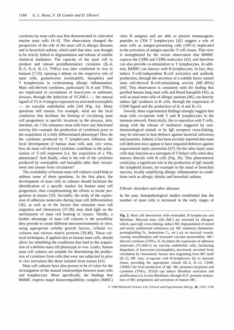
IL-3 does not bind to human lung mast cells [23],although it may support SCF-induced human mast celldifferentiation [18,20], acting probably on SCF-responsiveprogenitors [24]. Recently it has been reported that IL-3prolongs the survival of cultured human mast cells thatappear to bear receptors for this cytokine [25]. Interestingly,IL-3 alone — which promotes the growth of murine mastcells — is a specific growth factor for human basophils[26–28]. Thus, in humans, IL-3 and SCF originate basophilsand mast cells, respectively, acting on haemopoietic undif-ferentiated precursors (CD 34þ) (Fig. 1). Mature mast cellsstill retain the receptor for SCF and basophils circulating inparallel display a high affinity receptor for IL-3 [27],indicating that these specific growth factors not only actduring differentiation but also regulate the functions ofmature cells.
Mast cell cultures: are they useful to fill the gap betweenbasic science and clinical research?
Non-transformed mast cell cultures provide a large numberof normal mast cells and so they facilitate basic research.
For example, the mechanisms of mast cell activation andsignal transduction have been extensively investigated, andvery recent reports indicate that BMMC can still provideimportant information on specific features, such as the co-stimulatory receptors involved in cell activation [29], signaltransduction in genetically manipulated cells [30] or theinhibition of cell activation [31]. Moreover, mast cellcultures enable us to study the response to various activatingstimuli during differentiation, even in very early stageswhen cells have not yet acquired degranulation capacity[32]. This longitudinal approach can also be applied toexamine the morphological characteristics of developingmast cells [21].
It is far beyond the aim of this paper to discuss in detailthe contribution of mast cell cultures to the advancement ofthe knowledge of mast cell biology that has been accom-plished in recent years. We would only like to expand a fewpoints that are relevant to the pathogenesis of humandiseases and that should also be of interest to the clinicalinvestigator.
Allergic inflammation–bronchial asthma–immune defences
As mentioned above, the production of a variety of
Mast cell cultures 1183
q 1998 Blackwell Science Ltd,Clinical and Experimental Allergy, 28, 1182–1190
Fig. 1. Differentiation of human mast cells and basophils. Baso-phils differentiate in the bone marrow from multilineage hemo-poietic progenitors (CD34þ) under the influence of interleukin-3(IL-3). Committed mast cell progenitors migrate to peripheralconnective tissues where, under the influence of stem cell factor(SCF) — produced by fibroblasts — differentiate into mature mastcells.
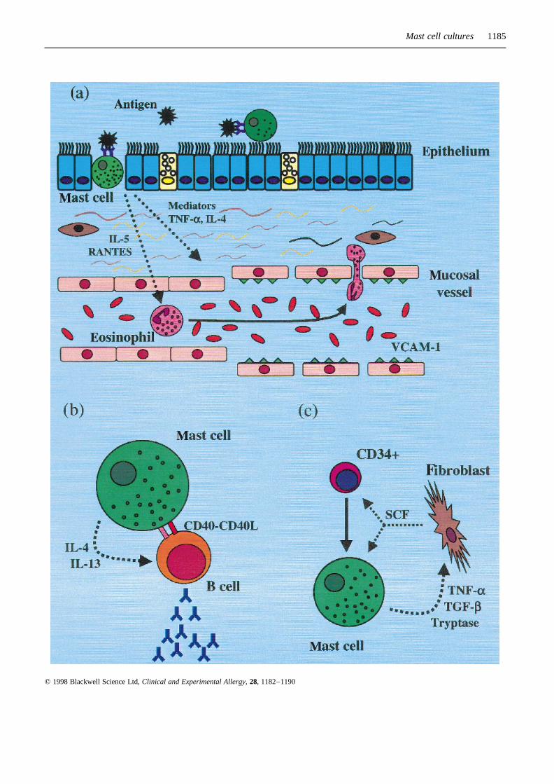
cytokines by mast cells was first demonstrated in cultivatedmurine mast cells [4–6]. This observation changed theperspective of the role of the mast cell in allergic diseasesand in bronchial asthma, which until that time, was thoughtto be strictly linked to degranulation and release of solublechemical mediators. The capacity of the mast cell toproduce and release proinflammatory cytokines (IL-4,IL-5, IL-6, IL-13, TNFa) was later confirmedin vivo inhumans [7,33], opening a debate on the respective role ofmast cells, granulocytes (eosinophils, basophils) andT lymphocytes in orchestrating allergic inflammation.Mast cell-derived cytokines, particularly IL-4 and TNFa,are implicated in recruitment of leucocytes in asthmaticairways, through the induction of VCAM-1 — the naturalligand of VLA-4 integrin expressed on activated eosinophils— on vascular endothelial cells [34] (Fig. 2a). Manyquestions still remain. For example, what are the localconditions that facilitate the homing of circulating mastcell progenitors in specific locations in the airways, skin,intestine, etc.? Do immature mast cells have any functionalactivity (for example the production of cytokines) prior tothe acquisition of a fully differentiated phenotype? How dothe cytokines produced by T lymphocytes influence thelocal development of human mast cells and, vice versa,how do mast cell-derived cytokines contribute to the polar-ization of T-cell responses (i.e. the acquisition of a TH2
phenotype)? And finally, what is the role of the cytokinesproduced by eosinophils and basophils after their recruit-ment into tissues from circulation?
The availability of human mast cell cultures could help toaddress some of these questions. In the first place, thedevelopment of mast cells in cultures should facilitate theidentification of a specific marker for human mast cellprogenitors, thus complementing the efforts to locate pro-genitors in tissues [35]. Secondly, the study of the expres-sion of adhesion molecules during mast cell differentiation[36], as well as of the factors that stimulate mast cellmigration and chemotaxis [37,38], may shed light on themechanisms of mast cell homing in tissues. Thirdly, afurther advantage of mast cell cultures is the possibilitythey provide to create different microenvironmentsin vitro,using appropriate soluble growth factors, cellular co-cultures and various matrix proteins [39,40]. These cul-tural techniques, if applied also to human mast cells, shouldallow for rebuilding the conditions that lead to the acquisi-tion of a definite mast cell phenotypein vivo. Lastly, humanmast cell cultures are suitable for determining the produc-tion of cytokines from cells that were not subjected to priorin vivo activation like those isolated from tissues [41].
Mast cell cultures have also proven to be of value for theinvestigation of the mutual relationships between mast cellsand lymphocytes. More specifically, the findings thatBMMC express major histocompatibility complex (MHC)
class II antigens and are able to present immunogenicpeptides to CD4 T lymphocytes [42] suggest a role ofmast cells as antigen-presenting cells (APCs) implicatedin the activation of antigen-specific T-cell clones. This viewis strengthened by the recent observation that BMMCexpress the CD80 and CD86 molecules [43], and thereforecan also provide co-stimulation to T lymphocytes. In addi-tion, BMMC can interact with B lymphocytes. In fact, theyinduce T-cell-independent B-cell activation and antibodyproduction, through the secretion of a soluble factor namedmast cell-derived B-cell-stimulating activity (MC-BSA)[44]. This observation is consistent with the finding thatpurified human lung mast cells and blood basophils [45], aswell as nasal mast cells of allergic patients [46], can directlyinduce IgE synthesis in B cells, through the expression ofCD40 ligand and the production of IL-4 and IL-13.
Overall, these experimental findings strongly suggest thatmast cells co-operate with T and B lymphocytes in theimmune network. Particularly, the co-operation with T cells,along with the release of mediators triggered by non-immunological stimuli or by IgE receptors cross-linking,may be relevant to host defence against bacterial infectionsand parasites. Indeed, it has been recently reported that mastcell-deficient mice appear to have impaired defences againstexperimental septic peritonitis [47]. On the other hand, mastcells may function as a surrogate of T helper cells when theyinteract directly with B cells (Fig. 2b). This phenomenoncould play a significant role in the production of IgE outsidethe lymphoid tissues, for example in the nasal or bronchialmucosa, locally amplifying allergic inflammation in condi-tions such as allergic rhinitis and bronchial asthma.
Fibrotic disorders and other diseases
In the past, histopathological studies established that thenumber of mast cells is increased in the early stages of
1184 G. L. Rossi, V. Di Comite and D. Olivieri
q 1998 Blackwell Science Ltd,Clinical and Experimental Allergy, 28, 1182–1190
Fig. 2. Mast cell interactions with eosinophil, B lymphocyte andfibroblast. Mucosal mast cells (MC) are activated by allergenswhich, upon IgE cross-linking, induce MC degranulation of storedand newly synthesized substances (a). MC mediators (histamine,prostaglanding D2. leukotriene C4, etc.) act on mucosal vessels,causing vasodilatation and increased vascular permeability. MC-derived cytokines (TNFa, IL-4) induce the expression of adhesionmolecules (VCAM-1) on vascular endothelial cells, facilitatingdiapedesis of leucocytes (eosinophils), previously recruited fromcirculation by chemotactic factors also originating from MC itself(IL-5). MC may co-operate with B-lymphocyte (b) in mucosaltissue, providing the appropriate stimuli (IL-4, IL-13, CD40,CD40L) for local production of IgE. MC proteases (tryptase) andcytokines (TNFa, TGFb) can induce fibroblast activation andproliferation (c); in turn fibroblasts, through SCF, promote matura-tion of MC progenitors and activation of mature MC.
Mast cell cultures 1185
q 1998 Blackwell Science Ltd,Clinical and Experimental Allergy, 28, 1182–1190

fibrosis and in areas of tissue repair and remodelling,particularly around blood vessels. This observation suggestsa role for the mast cell in the pathogenesis of conditionssuch as fibrotic lung disorders, graft vs host disease, sclero-derma and wound healing [48]. Clearly, the pattern of mastcell activation in fibrosis — a chronic process — isdifferent from that of immediate allergic reactions. Accord-ingly, a peculiar type of mast cell degranulation (piecemealdegranulation) has been described [49], characterized by theretention of partially filled and empty granules, distinct fromclassic anaphylactic degranulation characterized by granuleextrusion. In this regard, very recently Dvorak and Morganhave documented the secretion of histaminein vivo duringpiecemeal degranulation of human mast cells [50], suggest-ing that the same mediator can be secreted by the mast cellwith different mechanisms. Although the nature of mast cellactivators that take part in fibrotic diseases has not beendetermined, it has been speculated that chronic mast cellactivation is caused by the constant presence of endogenousstimuli in contrast with immediate allergic activation causedby exposure to exogenous allergens [48]. Interestingly, thenumber of eosinophils can also be augmented in fibroticdisorders and severalin vitro studies have tried to unravelthe complex interactions between mast cells, eosinophilsand fibroblasts [48]. A recent study reported that tryptasestimulates both the synthesis of procollagen mRNA andchemotaxis in fibroblasts [51] and that BMMC promotefibroblast proliferation through the release of TNFa andTGFb [52] (Fig 2c). These findings indicate that our under-standing of the cellular and molecular mechanisms offibrotic diseases is progressing and that mast cell culturescan substantially contribute to this interesting field ofresearch.
Similarly, the availability of a large number of non-transformed mast cells (especially of human origin) mayfacilitate the comprehension of the role of the mast cell in agrowing list of diseases in which this multifunctionaleffector cell has been implicated. While the efforts ofmany basic and clinical investigators still continue toelucidate the pathogenesis of classical, widespread immu-nological disorders such as chronic urticaria and atopicdermatitis, others are evaluating the contribution of mastcells to inflammatory bowel diseases (Crohn’s disease andulcerative colitis) [53] and rheumatoid arthritis [54]. More-over, the interactions between mast cells and nerves havebeen actively investigated [55,56], as has also been the roleof human heart mast cells in atherosclerosis and anaphylaxis[57]. As regards cardiac mast cells, an original procedure fortheir isolation and partial purification from human hearts hasbeen recently developed, thus facilitating the study of thefunctions of these cellsin vitro [57].
A recent study reports that human mast cells stimulatevascular tube formation by secreting tryptase, which acts as
a potent and previously unrecognized angiogenic factor[58]. This observation is relevant to previous studies indi-cating the accumulation of mast cells and basophils aroundtumours [59], as well as a possible role of mast cells intumour-associated angiogenesis — an essential requirementfor the growth of solid tumours [60] — and tumourproliferation [61]. Hopefully, future studies will ascertainwhether the increased density of mast cells in tumoursindicates host defence or growth promotion.
Lastly, study of the action of the various growth factorsand cytokines that affect mast cell differentiation andfunction (that is clearly facilitated by the developing ofmast cells in culture) is essential for the comprehension ofthe pathogenetic mechanisms that underlie the differentforms of systemic mastocytosis, varying from mildhyperplasia to fatal mast-cell leukaemia [62].
Mast cell cultures and pharmacological research
The usefulness of mast cell cultures in pharmacologicalstudies derives from their ease of producing large numbersof cells, thus this technique has been widely exploited. Theactivity of many anti-allergic drugs has been tested inexperimental systems based on the IgE-mediated and non-IgE-mediated activation of BMMC.
Since the purpose of this paper does not include anextensive review of the large body of literature concerningpharmacological investigations with cultured mast cells, wewould like to focus on selected studies performed withcorticosteroids and cromones and to briefly outline potentialfuture applications of mast cell cultures in pharmacologicaland toxicological research.
Corticosteroids and mast cells
Although glucocorticoids (GC) may inhibit mediatorsrelease from murine mast cells [63,64], they do not appearto have a direct inhibitory effect on human lung mast cells[65]. However, it has recently been shown that GC exertother effects on mast cells that may be relevant to theirtherapeutic action in bronchial asthma and allergic diseases.More specifically, dexamethasone inhibits the production ofTNFa by BMMC in vitro [66,67], as well as the TNFa-dependent inflammation in micein vivo [66]. Moreover, inBMMC, glucocorticoids inhibit the SCF- and IL-3-inducedproliferation [67], the IL-10-mediated expression of chy-mases [67] and the IgE aggregation-dependent expression oflymphotactin (a lymphocyte-specific chemokine) mRNA[68].
Taken together, these findings suggest that GC regulatethe activity of mast cells at different levels, independentlyfrom the classical IgE-mediated degranulation and media-tors release. In addition, the recent report by Finottoet al.
1186 G. L. Rossi, V. Di Comite and D. Olivieri
q 1998 Blackwell Science Ltd,Clinical and Experimental Allergy, 28, 1182–1190
that GC down-regulate SCF mRNA expression and SCFprotein production in fibroblasts helps to clarify, at themolecular level, the mechanism by which corticosteroidsreduce tissue mast cells number [69], a phenomenon that isstrictly relevant to the effectiveness of long-term treatmentwith inhaled steroids in asthma.
In the near future, human mast cell cultures will probablyfacilitate the understanding of the action of glucocorticoidson human mast cells. Accordingly, preliminary studiessuggest that dexamethasone inhibits the production ofcytokines, but not the release of histamine from culturedhuman mast cells [70]. Further studies will clarify whetherthe inhibition of cytokine production in mast cells hasa therapeutic relevance in comparison to the well-knownGC-dependent inhibition of cytokine production inlymphocytes and eosinophils.
Cromones and mast cells
Despite the large use of the so called ‘mast cell stabilizers’in the treatment of respiratory and gastro-intestinal allergy,their mechanism of action has not yet been completelyunderstood. Previous studies indicated that sodium cromo-glycate inhibits allergen-induced histamine release from ratperitoneal mast cells [71] but not from intestinal mucosalmast cells [72]. In BMMC a global inhibition of mediatorsrelease appears only after chronic exposure (at least 5 days)to sodium cromoglycate [73]. Further studies demonstratedthat high concentrations of sodium cromoglycate and nedo-cromil sodium are required to achieve even a modestinhibition of mediator releasein vitro from human mastcells isolated from different tissues, and also that skin, lung,tonsillar, adenoidal and intestinal mast cells largely varytheir sensitivity to cromones [74,75].
Overall, the data available suggest that the inhibition ofmediators release from mast cells may not be the mainpharmacological action of cromones. Other mechanisms,for example the inhibition of sensory nerves, may be morerelevant to their therapeutic effects or, alternatively, theiraction on mast cells may involve other cellular functions,such as the release of cytokines [76]. Additional studies areneeded to better clarify the action of cromones on thevarious aspects of mast cell activation.
Potential future applications
Mast cell cultures provide an ideal opportunity to confirm theputative anti-allergic properties that have been claimed forseveral drugs, for example the new antihistamines [77]. More-over, human mast cell cultures may facilitate the testing ofmany drugs and substances that display mast cell degranulat-ing effects, such as muscle relaxants, general anaesthetics,opioids, benzodiazepines and radiocontrast media [78].
This same method could be also applied to the screeningof the numerous new chemicals and potentially allergenicsubstances that are continually introduced in the environ-ment. Additionally,in vitro screening might be easier, morereliable and also more ethical when compared to traditionalin vivo animal models.
Finally, mast cell cultures may contribute to ascertainwhether air pollutants exert any direct effect on mast cells.This topic is relevant to the epidemiological evidence ofincreased prevalence and incidence of allergic diseases inwestern countries. Although the assessment of the effects ofair pollutants on cultured mast cells entails considerabletechnical difficulties, by adapting the experimental modelsdescribed in published experiments regarding the effects ofair pollutants on several types of cultured cells, includinghuman fibroblasts, B-lymphocytes, alveolar macrophagesand epithelial cells [79] similar investigations in mast cellsmight be facilitated.
Conclusions
The methods for obtaining pure populations of murine mastcells have been established for many years. More recently,human mast cells have also been grown in culture. Thedevelopment of mast cells in cultures is particularly import-ant when considering the peculiar differentiation of the mastcell in tissues when compared to other inflammatory cellssuch as the neutrophil, the eosinophil and the basophil thatleave the bone marrow as fully differentiated cells. Indeed,mast cells mature in peripheral tissues, where their progeni-tors are subjected to the influence of a variety of micro-environmental factors that, at least in part, can bereproducedin vitro. Furthermore, mast cell cultures yielda large number of cells, while the isolation and purificationof mast cells from tissues is sometimes a difficult procedure.
Until now, mast cell cultures have been mainly used bybasic investigators and, consequently, they have been offundamental importance in clarifying many aspects of mastcell biology. We think that the time has also come forclinical investigators who are motivated in pursuing highquality research to take advantage from thisin vitro model.In fact, mast cell cultures are suitable for the evaluation ofdrugs, both mast cell stabilizers and mast cell activators,provided that researchers devote the necessary time andeffort to acquire familiarity with the techniques of cellculture and mediator release assays. Moreover, culturesmay help complement observations that originate fromstudiesin vivo (for example histopathological findings ofbronchial biopsies) focusing on functional aspects. For thelarge majority of clinical investigators in allergy andrespiratory medicine, the collaboration with basicresearchers will hopefully furnish cultural stimuli thatfacilitate a better comprehension of disease mechanisms,
Mast cell cultures 1187
q 1998 Blackwell Science Ltd,Clinical and Experimental Allergy, 28, 1182–1190
particularly but not exclusively allergic disorders, bronchialasthma and fibrosis. In this regard, mast cell cultures havealready proven to be instrumental.
Acknowledgements
The authors would like to thank Ms Elizabeth De Young, ofthe Language Centre of the University of Parma, for thecareful revision of this manuscript.
References
1 Church MK, Levi-Schaffer F. The human mast cell. J AllergyClin Immunol 1997; 99:155–60.
2 Warner JA, Kroegel C. Pulmonary immune cells in health anddisease: mast cells and basophils. Eur Respir J 1994; 7:1326–41.
3 Rossi GL, Olivieri D. Does the mast cell still have a key role inasthma? Chest 1997; 112:523–9.
4 Plaut M, Pierce JH, Watson CJ et al. Mast cell lines producelymphokines in response to cross-linkage of FceRI or calciumionophore. Nature 1989; 339:64–7.
5 Wodnar-Filipowicz A, Heusser CH, Moroni C. Production ofthe haemopoietic growth factors GM-CSF and interleukin-3 bymast cells in response to IgE receptor-mediated activation.Nature 1989; 339:150–2.
6 Burd PR, Rogers HW, Gordon JR et al. Interleukin 3-dependent and -independent mast cells stimulated with IgEand antigen express multiple cytokines. J Exp Med 1989;179:245–57.
7 Bradding P, Roberts JA, Britten KM et al. Interleukin-4, -5, -6and tumor necrosis factor-a in normal and asthmatic airways:evidence for the human mast cells as a source of thesecytokines. Am J Respir Cell Mol Biol 1994; 10:471–80.
8 Enerback L. The differentiation and maturation of inflamma-tory cells involved in the allergic response: mast cells andbasophils. Allergy 1997; 52:4–10.
9 Enerback L. Mast cell heterogeneity: the evolution of theconcept of a specific mucosal mast cell. In: Befus AD, Bienen-stock J, Denburg JA eds. Mast cell differentiation and Hetero-geneity. New York: Raven Press 1986:1–26.
10 Bienenstock J, Befus AD, Denburg JA. Mast cell heterogene-ity: Basic questions and clinical implications. In: Befus AD,Bienenstock J, Denburg JA eds. Mast cell differentiation andHeterogeneity. New York: Raven Press 1986:391–402.
11 Church MK, Okayama Y, Bradding P. Functional mast cellheterogeneity. In: Busse WW, Holgate ST eds. Asthma andrhinitis. Boston: Blackwell Scientific 1995:209–20.
12 Galli SJ. The mast cell: a versatile effector cell for a challen-ging world. Int Arch Allergy Immunol 1997; 113:14–22.
13 Nakano T, Sonoda T, Hayashi C et al. Fate of bone marrow-derived cultured mast cells after intracutaneous, intraperito-neal, and intravenous transfer into genetically deficient W/Wvmice. Evidence that cultured mast cells can give rise to bothconnective tissue type and mucosal mast cell. J Exp Med 1985;162:1025–43.
14 Galli SJ, Tsai M, Wershil BK, Tam SY, Costa JJ. Regulation ofmouse and human mast cell development, survival and func-tion by stem cell factor, the ligand for c-kit receptor. Int ArchAllergy Immunol 1995; 107:51–3.
15 Mekori YA, Metcalfe DD. Transforming growth factor-b
prevents stem cell factor-mediated rescue of mast cells fromapoptosis after IL-3 deprivation. J Immunol 1994; 153:2194–203.
16 Gagari E, Tsai M, Lantz CS, Fox LG, Galli SJ. Differentialrelease of mast cell interleukin-6 via c-kit. Blood 1997;89:2654–63.
17 Durand B, Migliaccio G, Yee NS et al. Long term generation ofhuman mast cells in serum-free cultures of CD34þ cord bloodcells stimulated with stem cell factor and interleukin-3. Blood1994; 84:3667–74.
18 Valent P, Spanblochl E, Sperr WR et al. Induction of differ-entiation of human mast cells from bone marrow and peripheralblood mononuclear cells by recombinant human stem cellfactor/kit-ligand in a long term culture. Blood 1992; 80:2237.
19 Kirshenbaum AS, Kessler SW, Goff JP et al. Effect of IL-3 andstem cell factor on the appearance of human basophils and mastcells from CD34þ pluripotent progenitor cells. J Immunol1992; 148:772.
20 Irani AM, Nilsson G, Miettinen U et al. Recombinant humanstem cell factor stimulates differentiation of mast cells fromdispersed human fetal liver cells. Blood 1992; 80:3009.
21 Dvorak AM, Furitsu T, Kissell-Rainville S., Ishizaka T. Ultra-structural identification of human mast cells resembling skinmast cells stimulated to develop in long term human cord bloodmononuclear cells cultured with 3T3 murine skin fibroblasts. JLeukoc Biol 1992; 51:557–69.
22 Saito H, Ebisawa M, Tachimoto H et al. Selective growth ofhuman mast cells induced by steel factor, IL-6 and prostaglan-din E2 from cord blood mononuclear cells. J Immunol 1996;157:343–50.
23 Valent P, Besemer J, Sillaber CH et al. Failure to detect IL-3binding sites on human mast cells. J Immunol 1990; 145:3432–7.
24 Metcalfe DD. Hematopoietic regulators: redundancy orsubtlety? Blood 1993; 82:3515–23.
25 Yanagida M, Fukamaki H, Ohgami K et al. Effects of T-helper2-type cytokines, interleukin-3 (IL-3), IL-4, IL-5, and IL-6 onthe survival of cultured human mast cells. Blood 1995; 86(10):3705–14.
26 Valent P, Schmidt G, Besemer J et al. Interleukin-3 is adifferentiation factor for human basophils. Blood 1989;73:1763–9.
27 Valent P, Besemer J, Muhm M et al. Interleukin-3 activatehuman basophils via high affinity binding site. Proc Natl AcadSci USA 1989; 86:5542–6.
28 Kirshenbaum AS, Goff JP, Dreskin SC et al. IL-3 dependentgrowth of basophil-like and mast-like cells from human bonemarrow. J Immunol 1989; 142:2424–9.
29 Tashiro M, Kawakami Y, Abe R et al. Increased secretions ofTNF-a by costimulation of mast cells via CD28 and FceRI. JImmunol 1997; 158:2382–9.
30 Nishizumi H, Yamamoto T. Impaired tyrosine phosphorylationand Ca2þ mobilization, but not degranulation, in lyn-deficient
1188 G. L. Rossi, V. Di Comite and D. Olivieri
q 1998 Blackwell Science Ltd,Clinical and Experimental Allergy, 28, 1182–1190

bone marrow-derived mast cells. J Immunol 1997; 158:2350–5.
31 Katz HR, Austen KF. A newly recognized pathway for thenegative regulation of mast cell-dependent hypersensivity andinflammation mediated by an endogenous cell surface receptorof the gp49 family. J Immunol 1997; 158:5065–70.
32 Rossi GL, Yen A, Barrett KE. IgE and adenosine 50 tripho-sphate receptors on immature murine mast cells are function-ally linked to signal transduction mechanisms. J Allergy ClinImmunol 1992; 90 (5):765–71.
33 Bradding P. Human mast cells cytokines. Clin Exp Allergy1996; 26:13–9.
34 Bochner BS, Schleimer RP. The role of adhesion molecules inhuman eosinophils and basophil recruitment. J Allergy ClinImmunol 1994; 94:427–38.
35 Kawabori S, Kanai N, Tosho T, Adachi T. Existence of c-kitreceptor-positive, tryptase negative, IgE-negative cells inhuman allergic nasal mucosa: a candidate for mast cell pro-genitor. Int Arch Allergy Immunol 1997; 112:36–43.
36 Shimizu Y, Irani AA, Brown EJ, Ashman LK, Schwartz LB.Human mast cells derived from fetal liver cells cultured withstem cell factor express a functional CD51/CD61 (avb3)integrin. Blood 1995; 86:930–9.
37 Nilsson G, Butterfield JH, Nilsson K, Siegbahn A. Stem cellfactor is a chemiotactic factor for human mast cells. J Immunol1994; 153:3717.
38 Nilsson G, Johnell M, Hammer CH et al. C3a and C5a arechemotaxins for human mast cells and act through distinctreceptors via a pertussis toxin-sensitive signal transductionpathway. J Immunol 1996; 157:1693–8.
39 Dastych J, Hardison MC, Metcalfe DD. Aggregation of lowaffinity IgG receptors induces mast cell adherence to fibronec-tin. J Immunol 1997; 158:1803–09.
40 Ogasawara T, Murakami M, Suzuki-Nishimura T, Uchida MK,Kudo I. Mouse bone marrow-derived mast cells undergoexocytosis, prostanoid generation, and citokine expression inresponse to G-protein activating polybasic compounds aftercocoltures with fibroblasts in the presence of c-kit ligand. JImmunol 1997; 158:393–404.
41 Bressler RB, Lesko J, Jones ML et al. Production of IL-5 andgranulocyte-macrophage colony-stimulating factor by naivehuman mast cells activated by high affinity IgE receptorligation. J Allergy Clin Immunol 1997; 99:508–14.
42 Frandji P, Oskeritzian C, Cacaraci F et al. Antigen-dependentstimulation by bone marrow-derived mast cells of MHC classII-restricted T cells hybridoma. J Immunol 1993; 151:6318–28.
43 Frandji P, Tkaczyk C, Oskeritzian C et al. Exogenous andendogenous antigens are differentially presented by mast cellsto CD4þ T lymphocytes. Eur J Immunol 1996; 26:2517–28.
44 Tkaczyk C, Frandji P, Botros HG et al. Mouse bone marrow-derived mast cells and mast cell lines constitutively produce Bcell growth and differentiation activities. J Immunol 1996;157:1720–8.
45 Gauchat JF, Henchoz S, Mazzei G et al. Induction of humanIgE synthesis in B cells by mast cells and basophils. Nature1993; 365 (6444):340–3.
46 Pawankar R, Okuda M, Yssel H, Okomura K, Ra C. Nasal mastcells in perennial allergic rhinitis exhibit increased expressionof the FceRI, CD40L, IL-4, and IL-13, and can induce IgEsynthesis in B cells. J Clin Invest 1997; 99 (7):1492–9.
47 Echtenacher B, Mannel DN, Hultner L. Critical protective roleof mast cells in a model of acute septic peritonitis. Nature 1996;381:75–7.
48 Levi-Schaffer F, Weg VB. Mast cells, eosinophils and fibrosis.Clin Exp Allergy 1997; 27:64–70.
49 Dvorak AM, Kissell S. Granule changing of human skin mastcells characteristics of piecemeal degranulation and associatedwith recovery during wound healing in situ. J Leukoc Biol1991; 49:147–210.
50 Dvorak AM, Morgan ES. Diamine oxidase-gold enzyme affi-nity ultrastructural demonstration that human gut mucosal mastcells secrete histamine by piecemeal degranulation in vivo. JAllergy Clin Immunol 1997; 99:812–20.
51 Gruber BL, Kew RR, Jelaska A et al. Human mast cellsactivate fibroblasts. J Immunol 1997; 158:2310–7.
52 Kendall JC, Li XH, Galli SJ, Gordon JR. Promotion of mousefibroblast proliferation by IgE-dependent activation of mousemast cells: role for mast cell tumor necrosis factor-alpha andtransforming growth factor-beta 1. J Allergy Clin Immunol1997; 99:113–23.
53 Wershil BK. Role of mast cells and basophils in gastrointest-inal inflammation. In: Marone G, ed. Human basophil and mastcells: clinical aspects. Chem Immunol Basel, Karger 1995;62:187–203.
54 Arnason JA, Malone DG. Role of mast cells in arthritis. InMarone G, ed. Human basophil and mast cells: clinical aspects.Chem Immunol Basel, Karger 1995; 62:204–38.
55 Bienenstock J, Blennerhasset M, Kakuta Y et al. Evidence forcentral and peripheral nervous system interactions with mastcells. In: Galli SJ, Austen KF eds. Mast cells and basophilsdifferentiation and function in health and disease. New York:Raven Press 1989:275–284.
56 Marshall JS, Waserman S. Mast cells and the nerves potentialinteractions in the context of chronic disease. Clin Exp Allergy1995; 25:102–10.
57 Patella V, Genovese A, Marone G. What are human heart mastcells for? In: Marone G ed. Human basophil and mast cells:clinical aspects. Chem Immunol Basel, Karger 1995; 62:108–31.
58 Blair RJ, Meng H, Marchese MJ et al. Human mast cellstimulate vascular tube formation. J Clin Invest 1997; 99(11):2691–700.
59 Harveit F. Mast cells and methacromasia in human breastcancer: their occurrence significance and consequence. Apreliminary report. J Pathol 1981; 134:7–11.
60 Crowle PK, Starkley JR. Mast cells and tumour associatedangiogenesis. In: Galli SJ, Austen KF, eds. Mast cells andbasophils differentiation and function in health and disease.New York: Raven Press 1989:307–17.
61 Roche WR. Mast cells and tumours: the specific enhancementof tumour proliferation in vitro. Am J Pathol 1985; 119:57–64.
62 Austen KF. Systemic mastocytosis. N Engl J Med 1992; 326(9):639–40.
Mast cell cultures 1189
q 1998 Blackwell Science Ltd,Clinical and Experimental Allergy, 28, 1182–1190
63 Daeron M, Sterk AR, Hirata F, Ishizaka T. Biochemicalanalysis of glucocorticoid-induced inhibition of IgE-mediatedhistamine release from mouse mast cells. J Immunol 1982;129:1212–8.
64 Marquardt DL. Modulation of rat serosal mast cell biochem-istry by in vivo dexamethasone administration. J Immunol1983; 131 (2):934–9.
65 Schleimer RP. Effects of glucocorticoids on inflammatory cellsrelevant to their therapeutic application in asthma. Am RevRespir Dis 1990; 141:S59–69.
66 Wershil BK, Furota GT, Lavigne JA et al. Dexamethasone orcyclosporin A suppress mast cell-leukocyte cytokine cascades.J Immunol 1995; 154:1391–8.
67 Eklund KK, Humphries DE, Xia Z et al. Glucocorticoidsinhibit the cytokine-induced proliferation of mast cells, thehigh affinity IgE receptor-mediated expression of TNF-a, andthe IL-10 induced expression of chimases. J Immunol 1997;158:4373–80.
68 Rumsaeng V, Vliagofits H, Oh CK, Metcalf DD. Lymphotactingene expression in mast cells following Fce receptor I aggrega-tion. J Immunol 1997; 158:1353–60.
69 Finotto S, Mekori YA, Metcalfe DD. Glucocorticoids decreasetissue mast cell number by reducing the production of the c-kitligand, stem cell factor, by resident cells. J Clin Invest 1997;99:1721–28.
70 Tachimoto H, Iikura Y, Saito H. Corticosteroids inhibit cyto-kine production but not histamine release from cultured humanmast cells. J Allergy Clin Immunol 1997; 99:S123.
71 Pearce FL. Inhibition by disodium cromoglycate of anaphy-lactic histamine secretion from rat peritoneal mast cells in thepresence of phosphatidil serine. Agents Action 1983; 13:212–5.
72 Pearce FL, Befus AD, Gauldie J, Bienenstock J. Mucosal mastcells II. Effects of anti-allergic compounds on histaminesecretion by isolated intestinal mast cells. J Immunol 1982;128:2481–6.
73 Marquardt DL, Walker LL, Wassermann SI. Cromolyn inhibi-tion of mediator release in mast cells derived from mouse bonemarrow. Am Rev Respir Dis 1986; 133:1105–9.
74 Di Comite V, Rossi GL, Bertorelli G, Olivieri D. Nedocromilsodium does not affectb-hexosaminidase release in bonemarrow derived mast cells after acute or prolonged exposure.Abst. accepted for presentation at World Asthma Meeting,Barcelona, 10–13 December 1998.
75 Okayama Y, Benyon RC, Rees PH, et al. Inhibition profile ofsodium cromoglycate and nedocromil sodium on mediatorrelease from mast cells of human skin, lungs, tonsil, adenoidand intestine. Clin Exp Allergy 1992; 22:401–9.
76 Bissonnette EY, Enciso JA, Befus AD. Inhibition of tumournecrosis factor alpha (TNF-a) release from mast cells by theanti-inflammatory drugs, sodium cromoglycate and nedocromilsodium. Clin Exp Immunol 1995; 102:78–84.
77 Genovese A, Patella V, De Crescenzo G, et al. Loratidine anddesethoxylcarbonyl-loratidine inhibits the immunologicalrelease of mediators from human FceRIþ cells. Clin ExpAllergy 1997; 27:559–67.
78 Stellato C, Marone G. Mast cells and basophils in adversereactions to drugs used during general anaesthesia. In: MaroneG, ed. Human basophil and mast cells: clinical aspects. ChemImmunol Basel, Karger 1995; 62:108–31.
79 Devalia JL, Bayram H, Rusznak C et al. Mechanisms ofpollution-induced airway disease: in vitro studies in theupper and lower airways. Allergy 1997; 52 (38):45–51.
1190 G. L. Rossi, V. Di Comite and D. Olivieri
q 1998 Blackwell Science Ltd,Clinical and Experimental Allergy, 28, 1182–1190