mastitis in dairy cattle causes and treatment
TRANSCRIPT

*Dr Hamed Attia
*Dr Mohamed Tharwat
*Professor of Veterinary Internal
Medicine
Zagazig University-Egypt
Mastitis in Dairy Cattle
Mastitis
An inflammation of the milk secreting tissues of the udder, caused by microbial infections in one or more quarters.
Affects 25 to 30 percent of all quarters
The most costly disease of dairy cattle
$200 /cow/year
What’s mastitis ?
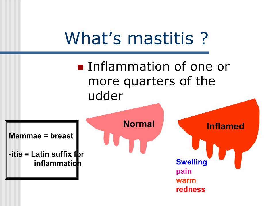
Inflammation of one or more quarters of the udder
Normal Inflamed
Swelling
pain
warm
redness
Mammae = breast
-itis = Latin suffix for
inflammation

Determinants of Mastitis
Host
Infectious AgentEnvironment
Pathogenicity, virulence, number
Resistance, nutrition, stress
Housing, equipment, hygiene, weather, bedding

How does mastitis develop ?
Cow Predisposing conditions
• Existing trauma (milking machine, heat or cold, injury)
• Teat end injury
• Lowered immunity (following calving, surgery)
• Nutrition
Organisms
EnvironmentEnvironment
Organism
Cow
Mastitis Infection
Almost always caused by bacteria that generally enter through the teat canal.
The environment inside the udder is warm and moist with plenty of available nutrients, so bacteria multiply rapidly.
What causes mastitis ?
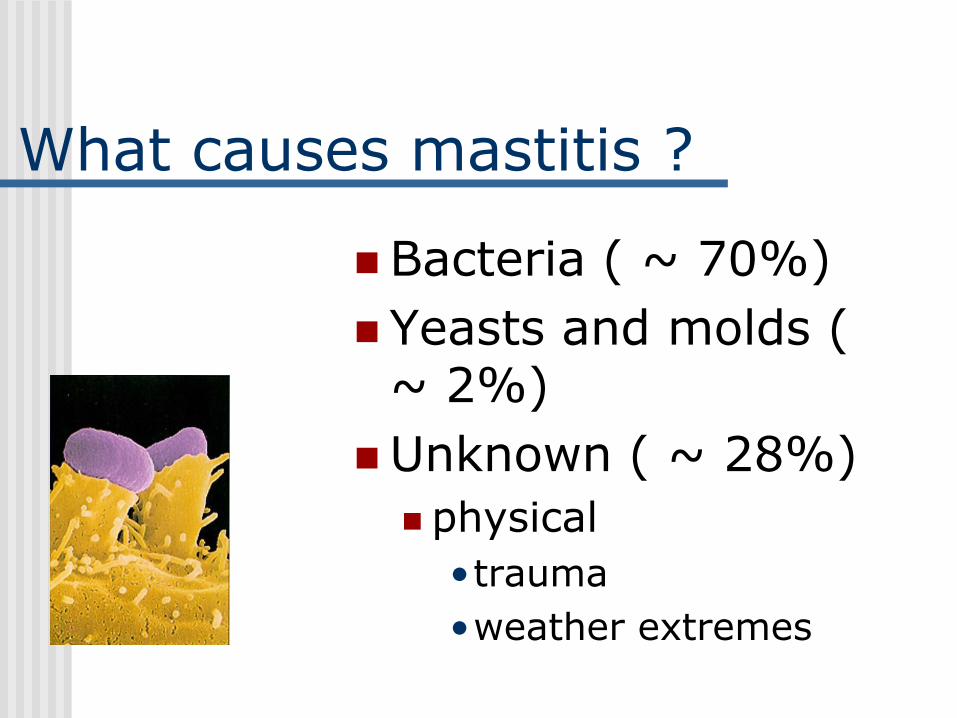
Bacteria ( ~ 70%)
Yeasts and molds ( ~ 2%)
Unknown ( ~ 28%)
physical
•trauma
•weather extremes
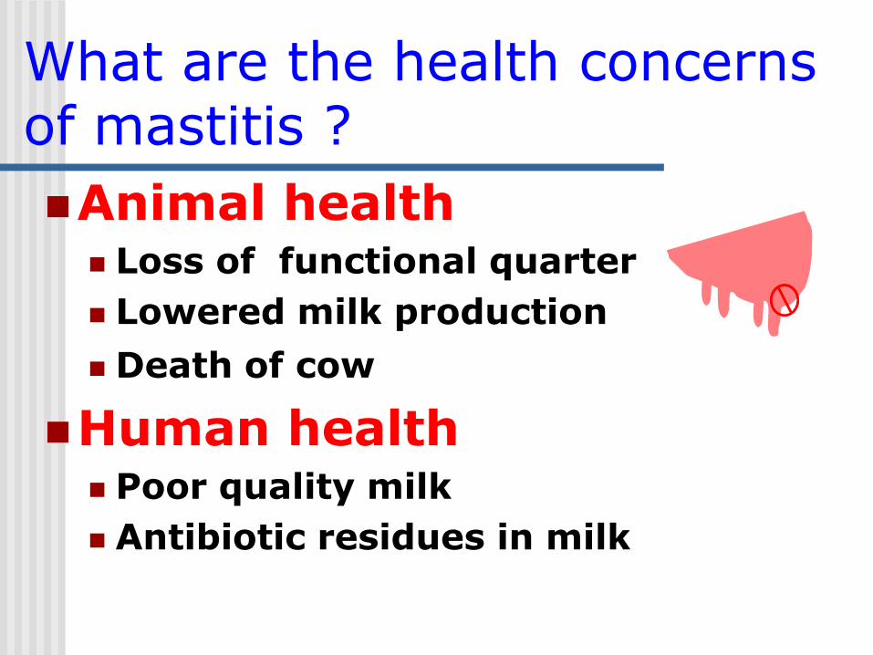
What are the health concerns of mastitis ?
Animal health Loss of functional quarter
Lowered milk production
Death of cow
Human health Poor quality milk
Antibiotic residues in milk

Types of Mastitis
Contagious
Environmental


Contagious Mastitis
Primary habitat bacteria live on/in the udder and teat lesions
Poor survival of bacteria in the environment Is spread from cow to cow, primarily during
milking by milk-contaminated fomites at milking, sponge, milker's hands, milking machine
Staphylococcus aureus, Streptococcus agalactia, Mycoplasma bovis and sometimes streptococcus uberis are contagious mastitis causing organisms.
Usually chronic, subclinical mastitis
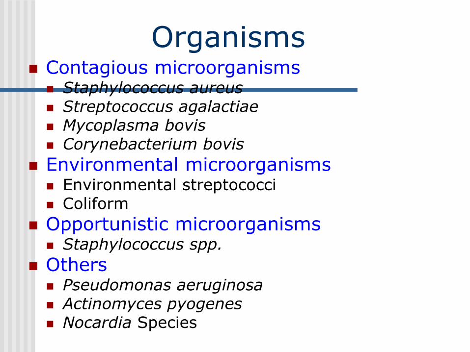
Organisms Contagious microorganisms Staphylococcus aureus Streptococcus agalactiae Mycoplasma bovis Corynebacterium bovis
Environmental microorganisms Environmental streptococci Coliform
Opportunistic microorganisms Staphylococcus spp.
Others Pseudomonas aeruginosa Actinomyces pyogenes Nocardia Species


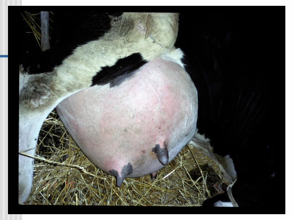
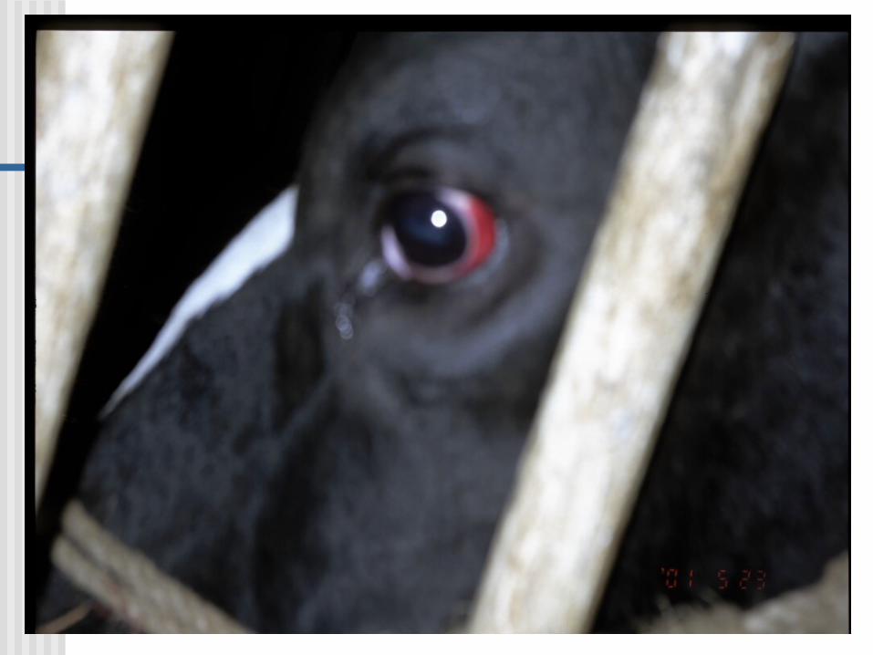
Coliform Mastitis
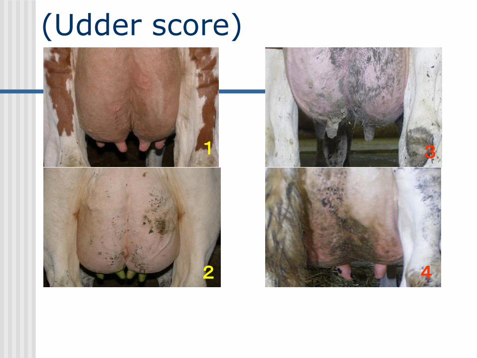
(Udder score)
1
2
3
4


Mastitis Clinical Syndromes
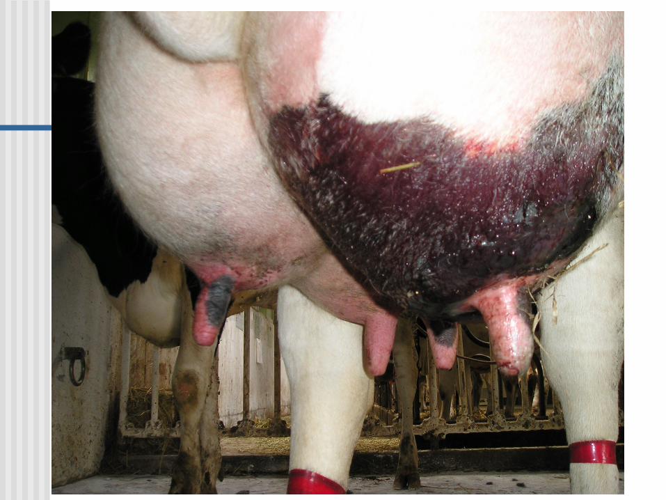
Peracute Mastitis: sudden onset, severe inflammation of the udder, and serous milk-Systemic illness often precedes the symptoms manifested in the milk and mammary gland.
Acute Mastitis: sudden onset, moderate to severe inflammation of the udder, decreased production, and occurrence of serous milk/fibrin clots, Systemic signs are similar but less severe than for the peracute form.

Mastitis Clinical Syndromes
Subacute Mastitis: mild inflammation, no visible
changes in udder, but there generally are small
flakes or clots in the milk, and the milk may have
an off-color. There are no systemic signs of illness.
Chronic Mastitis: chronic mastitis may persist in a
subclinical form for months or years with
occasional clinical flare-ups. Treatment usually
involves treating the clinical flare-ups, or culling
the cow from the herd.
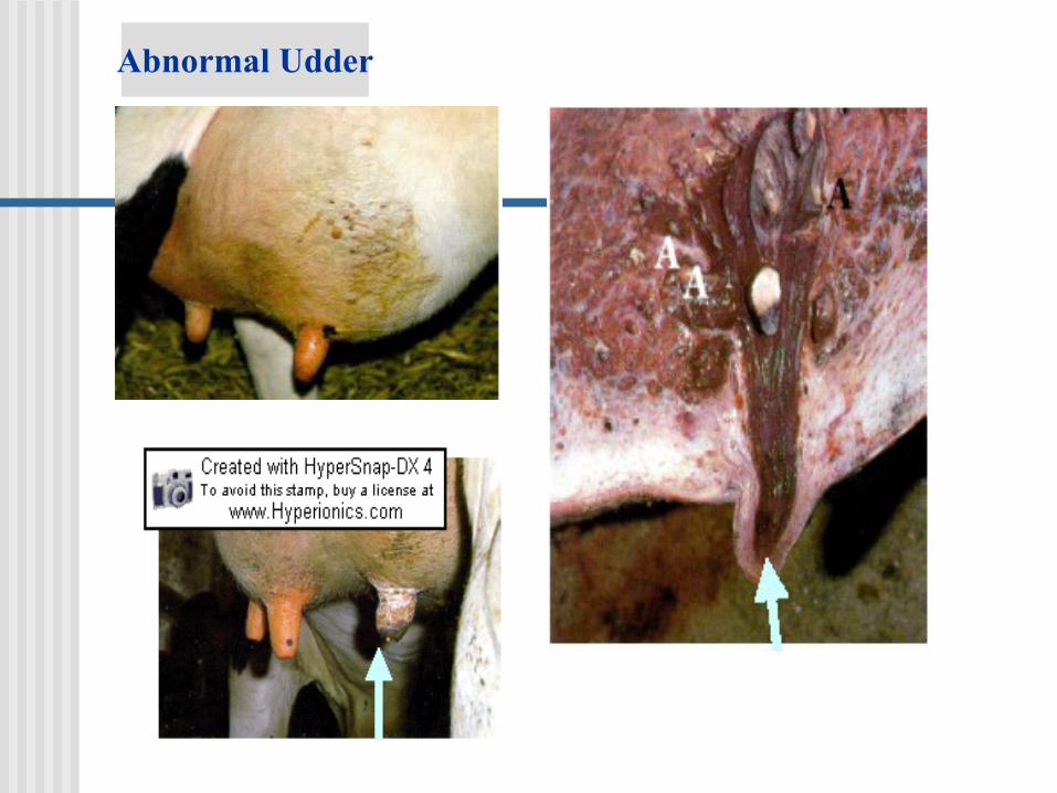
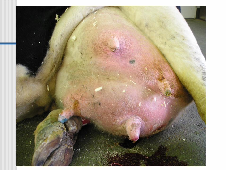
Abnormal Udder
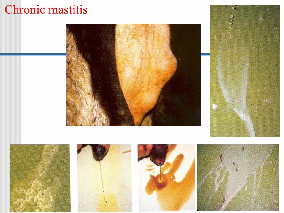
Chronic mastitis
Subclinical Mastitis: the most common
form of mastitis, more common than
clinical mastitis.
No gross inflammation of the udder and
no gross changes in the milk.
Decreased production and decreased
milk quality.
Elevated SCC.
Mastitis Clinical Syndromes
Detecting Subclinical Mastitis
Increased Somatic Cells
Bulk Tank SCC
Individual Cow SCC
CMT
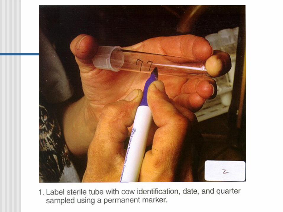
Sterile Milk Culture
Find mastitis causing organisms
Antibody ELISA


How is mastitis diagnosed ?
Physical examination Signs of inflammation
Empty udder
Differences in firmness
Unbalanced quarters
Culture
Cowside tests California Mastitis
test

Visualization and palpation of the udderVisualization and palpation of the udder
Detection of Somatic CellsDetection of Somatic Cells
California Mastitis TestCalifornia Mastitis Test
Detection of MastitisDetection of Mastitis
NN--acetylacetyl--ßß--DD--glucosaminidase (NAGase) glucosaminidase (NAGase)
-- a lysosomal enzyme which increases in milk a lysosomal enzyme which increases in milk
when mastitis is present when mastitis is present

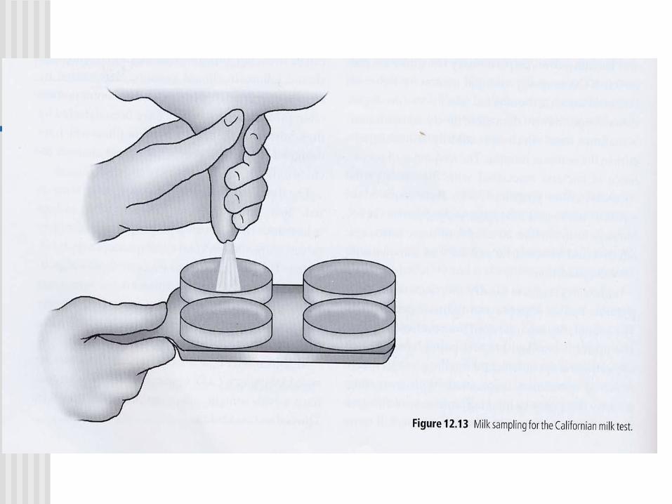
California Mastitis Test (CMT)
The CMT reagent reacts with somatic cells present in
milk to form a gel.
A plastic paddle having four shallow cups marked A, B,
C and D for easy identification of the individual quarter.
Approximately 1/2 teaspoon (2 cc) of milk is poured. An
equal amount of the CMT reagent is added to the milk.
A circular rotating to thoroughly mix the contents. Score
in approximately ten seconds while still rotating.
Read the test quickly as the reaction tends to disintegrate
after about 20 seconds.
Rinse the paddle thoroughly with water and it is ready
for the next test.
Rupture of the
suspensory ligament


Line of
Treatment of
Masitis
Anti-
Inflamm.
AntibioticFluid
therapy
Vitamin
A&COxytocine
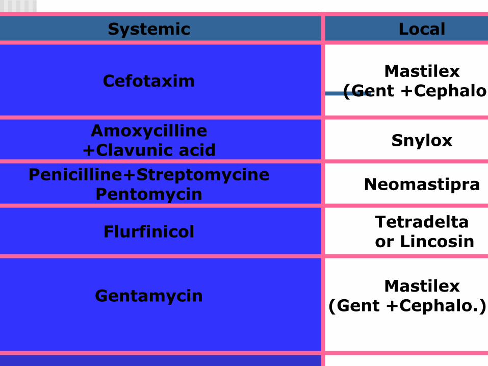
LocalSystemic
Mastilex(Gent +Cephalo.)
Cefotaxim
Snylox Amoxycilline
+Clavunic acid
Neomastipra Penicilline+Streptomycine
Pentomycin
Tetradeltaor Lincosin
Flurfinicol
Mastilex(Gent +Cephalo.)
Gentamycin

LocalSystemic
NoMarbocil
OxyOxyte-teracycline
(oxy 5%)
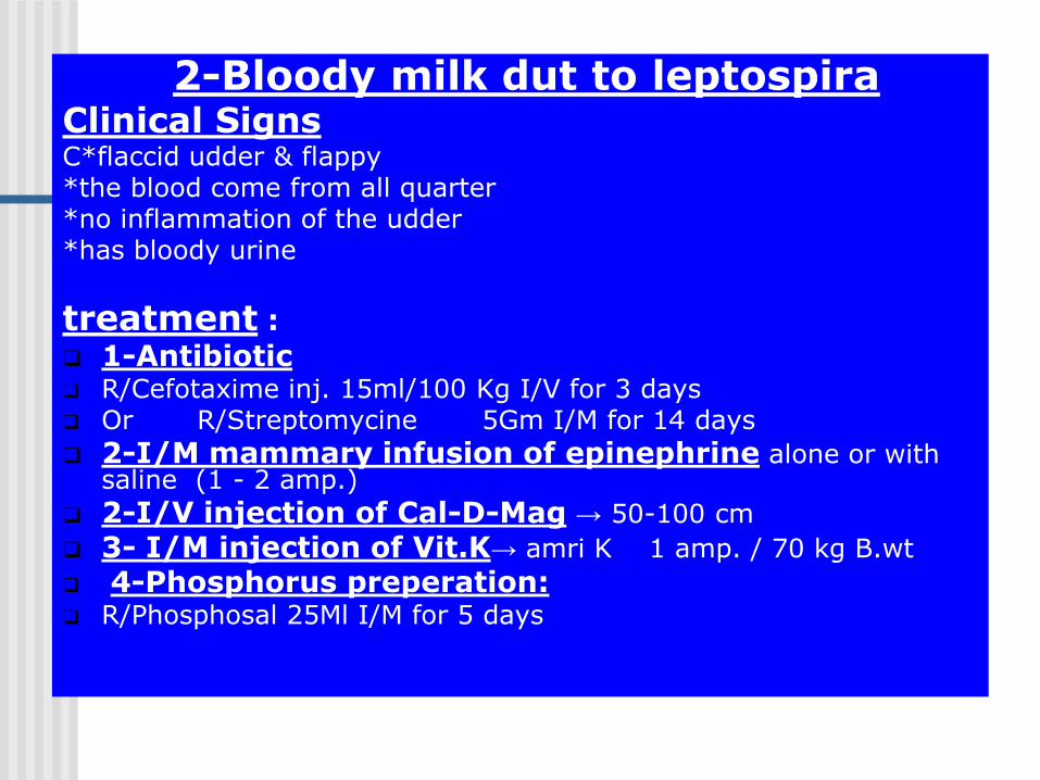
2-Bloody milk dut to leptospiraClinical SignsC*flaccid udder & flappy*the blood come from all quarter *no inflammation of the udder*has bloody urine
treatment :
1-Antibiotic R/Cefotaxime inj. 15ml/100 Kg I/V for 3 days Or R/Streptomycine 5Gm I/M for 14 days
2-I/M mammary infusion of epinephrine alone or with saline (1 - 2 amp.)
2-I/V injection of Cal-D-Mag → 50-100 cm
3- I/M injection of Vit.K→ amri K 1 amp. / 70 kg B.wt
4-Phosphorus preperation: R/Phosphosal 25Ml I/M for 5 days

3-bloody milk due to truma:
* blood only without milk
*treatment of trumatic bloody milk:
1-I/ mammary infusion of epinephrine alone or with saline (1 - 2 amp.)
2-I/V injection of Cal-D-Mag → 50-100 cm
3- I/M injection of Vit.K→ amri K 1 amp. / 70 kg B.wt
4- cold application

4- supportive treatment :
*I/V injection of glucose 25% → 1-2 liter
*I/V injection of Avil → 1 amp./70 kg B.wt
*local application of cold fomentation (ice bag) on the udder(in acute cases)

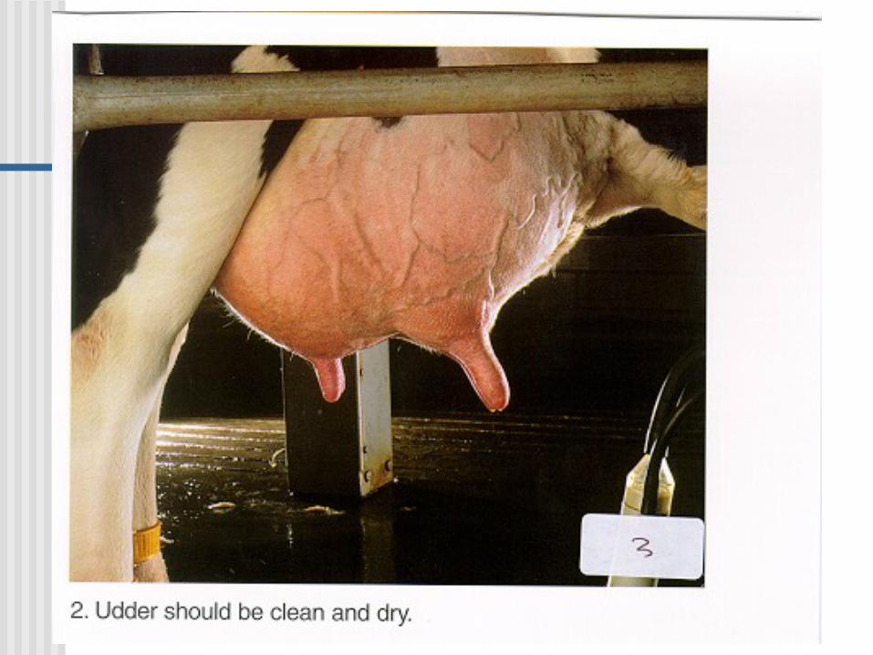
Mastitis Prevention Proper Milking Techniques
Procedures, training, monitoring
Keep cows clean! Proper Bedding
• Sand is the best bedding
• Organic bedding (sawdust, etc.) must be dry
• Stall sized to fit cows
• Tail docking
Nutrition Vitamins and minerals
Milk contagious cows last
Maintain milking equipment
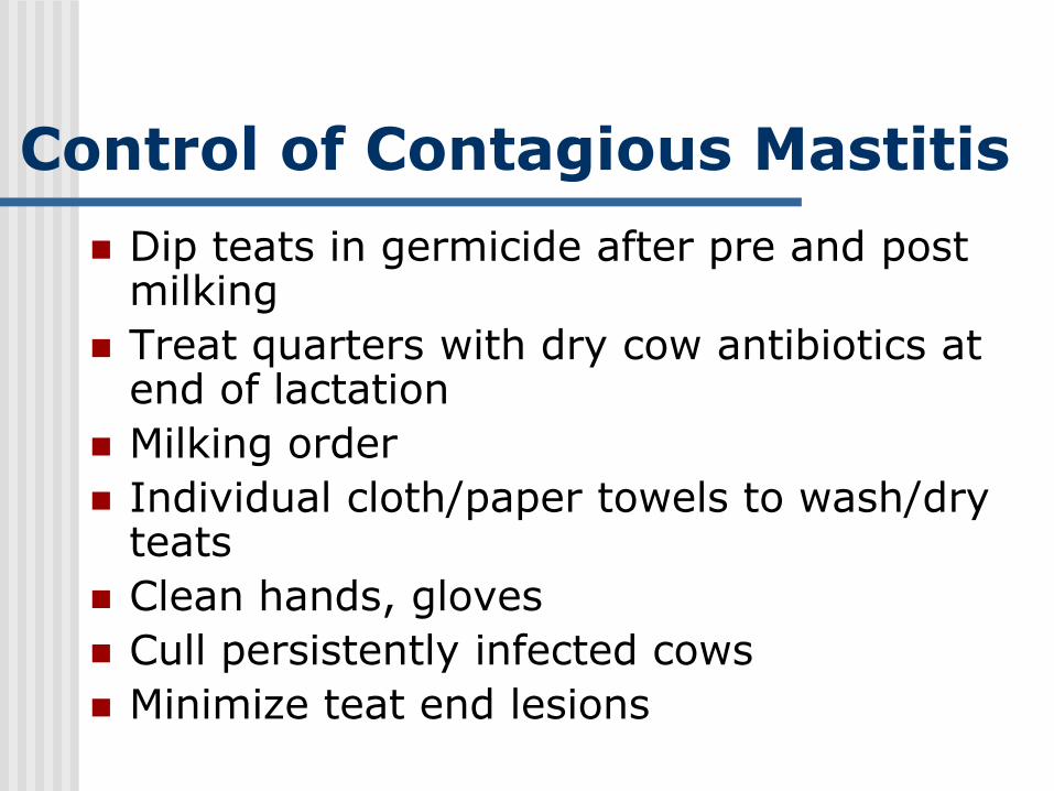
Control of Contagious Mastitis
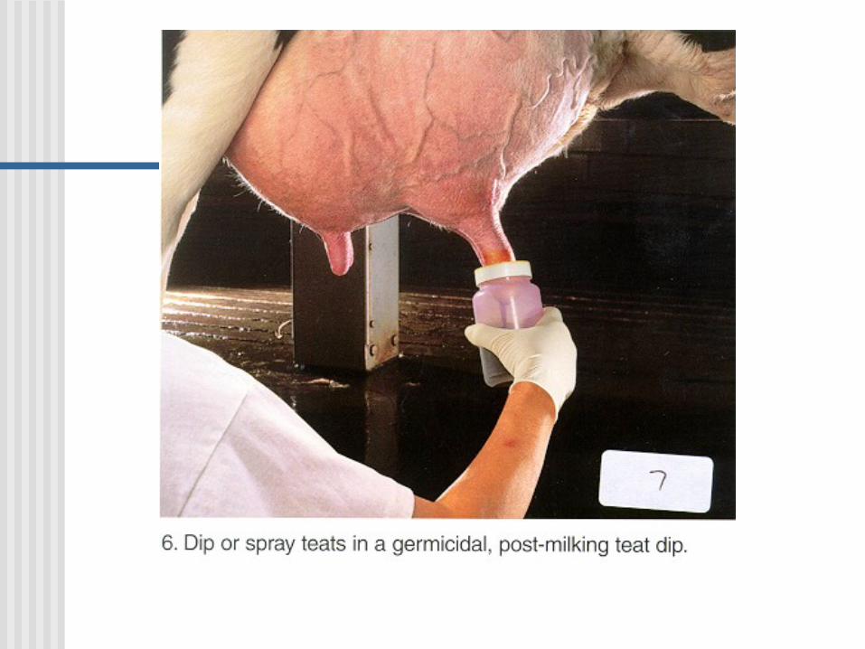
Dip teats in germicide after pre and post milking
Treat quarters with dry cow antibiotics at end of lactation
Milking order
Individual cloth/paper towels to wash/dry teats
Clean hands, gloves
Cull persistently infected cows
Minimize teat end lesions
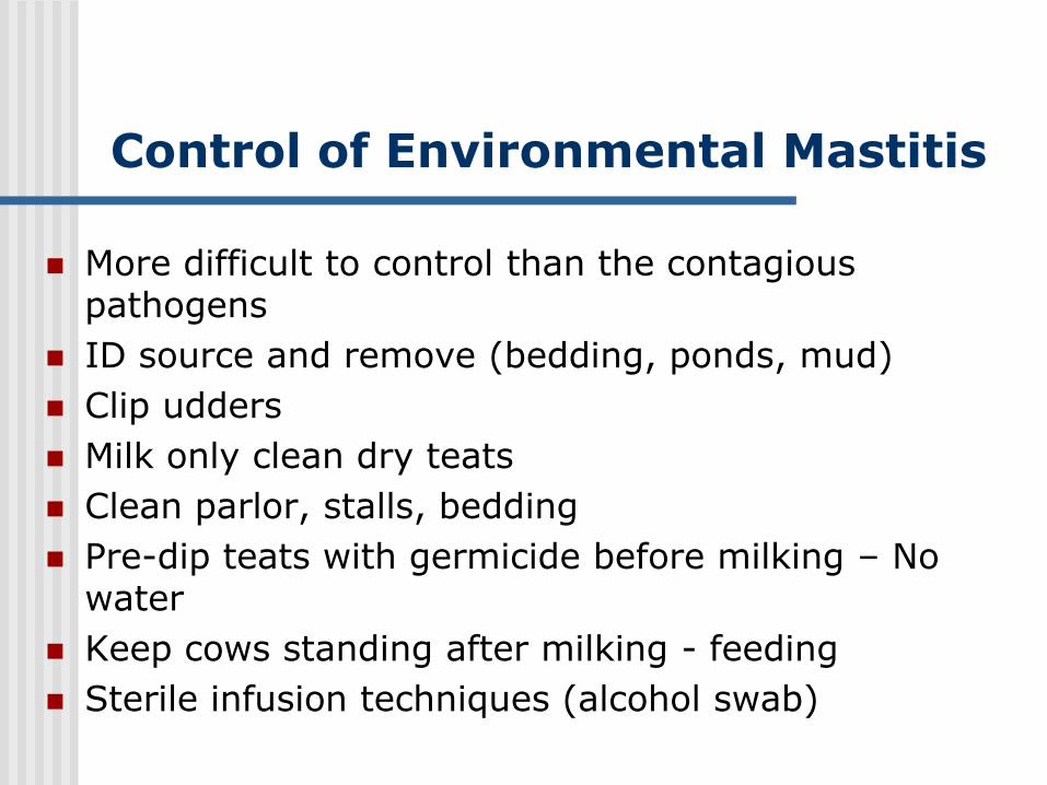
Control of Environmental Mastitis
More difficult to control than the contagious pathogens
ID source and remove (bedding, ponds, mud)
Clip udders
Milk only clean dry teats
Clean parlor, stalls, bedding
Pre-dip teats with germicide before milking – No water
Keep cows standing after milking - feeding
Sterile infusion techniques (alcohol swab)