material properties of the posterior human sclerabioeng.nus.edu.sg/oeil/publications/material...
TRANSCRIPT

Available online at www.sciencedirect.com
www.elsevier.com/locate/jmbbm
j o u r n a l o f t h e m e c h a n i c a l b e h a v i o r o f b i o m e d i c a l m a t e r i a l s 2 9 ( 2 0 1 4 ) 6 0 2 – 6 1 7
1751-6161/$ - see frohttp://dx.doi.org/10.
☆Supported in paHealth, Bethesda, MPrevent Blindness P
nCorresponding autE-mail addresses:
Research Paper
Material properties of the posterior human sclera$
Rafael Grytza, Massimo A. Fazioa, Michael J.A. Girardb, Vincent Libertiauxa,Luigi Brunoc, Stuart Gardinerd, Christopher A. Girkina, J. Crawford Downsa,n
aOphthalmology, University of Alabama at Birmingham, Birmingham, AL, USAbBioengineering, National University of Singapore, SingaporecMechanical Engineering, University of Calabria, Cosenza, ItalydDevers Eye Institute, Portland, OR, USA
a r t i c l e i n f o
Article history:
Received 18 January 2013
Accepted 26 March 2013
Available online 20 April 2013
Keywords:
Sclera
Inverse analysis
Homeostasis
Collagen fibril strain
nt matter & 2013 Elsevie1016/j.jmbbm.2013.03.027
rt by U.S. Public Health Garyland; Legacy Good Shysician-Scientist Awardhor. Tel.: +1 205 996 [email protected] (R. Grytz
a b s t r a c t
To characterize the material properties of posterior and peripapillary sclera from human
donors, and to investigate the macro- and micro-scale strains as potential control mechanisms
governing mechanical homeostasis. Posterior scleral shells from 9 human donors aged 57–90
years were subjected to IOP elevations from 5 to 45mmHg and the resulting full-field
displacements were recorded using laser speckle interferometry. Eye-specific finite element
models were generated based on experimentally measured scleral shell surface geometry and
thickness. Inverse numerical analyses were performed to identify material parameters for each
eye by matching experimental deformation measurements to model predictions using
a microstructure-based constitutive formulation that incorporates the crimp response and
anisotropic architecture of scleral collagen fibrils. The material property fitting produced models
that fit both the overall and local deformation responses of posterior scleral shells very well. The
nonlinear stiffening of the sclera with increasing IOP was well reproduced by the uncrimping of
scleral collagen fibrils, and a circumferentially aligned ring of collagen fibrils around the scleral
canal was predicted in all eyes. Macroscopic in-plane strains were significantly higher in
peripapillary region then in the mid-periphery. In contrast, the meso- andmicro-scale strains at
the collagen network and collagen fibril level were not significantly different between regions.
The elastic response of the posterior human sclera can be characterized by the anisotropic
architecture and crimp response of scleral collagen fibrils. The similar collagen fibril strains in
the peripapillary and mid-peripheral regions support the notion that the scleral collagen
architecture including the circumpapillary ring of collagen fibrils evolved to establish optimal
load bearing conditions at the collagen fibril level.
& 2013 Elsevier Ltd. All rights reserved.
1. Introduction
Biomechanics is likely to be important in the developmentand progression of glaucoma, as it provides a direct link
r Ltd. All rights reserved.
rants R01-EY18926 and Ramaritan Foundation, Po.
), [email protected] (J. Cra
between intraocular pressure (IOP) and the microenviron-ment of the optic nerve head (ONH) where glaucomatousdamage to the retinal ganglion cell axons is thought to occur(Burgoyne et al., 2005; Downs et al., 2008). The sclera is an
01-EY19333 from the National Eye Institute, National Institutes ofrtland, OR; Eye Sight Foundation of Alabama; and Research to
wford Downs).
j o u r n a l o f t h e m e c h a n i c a l b e h a v i o r o f b i o m e d i c a l m a t e r i a l s 2 9 ( 2 0 1 4 ) 6 0 2 – 6 1 7 603
important driver of ONH biomechanics, as it imposes theprincipal mechanical boundary condition on the containedlamina cribrosa and neural canal tissues and the intrascleralbranches of the short posterior ciliary arteries provide theprimary blood supply for the lamina cribrosa. Several com-putational studies have shown the sclera to be among themost important determinants of ONH stress and strain (Sigalet al., 2005, 2011a, 2011b).
Computational modeling studies are necessary to studyONH biomechanics, as no experimental methods are avail-able to measure or estimate stress and strain in the ONHin vivo. However, without accurate material properties, thesemodels yield inaccurate stress and strain predictions in thesclera, and hence ONH. No previous studies have reportedmaterial property estimates for the posterior and peripapil-lary human sclera that incorporate the inhomogeneous,hyperelastic, anisotropic nature of its material response.Histologic studies have shown that there is a circumferentialring of highly aligned collagen fibrils surrounding the ONH,and computational simulations have suggested that this ringserves to shield the relatively compliant ONH from excessivestrains (Girard et al., 2009b; Grytz et al., 2011a; Coudrillieret al., in press). Experimental studies have shown thatthe sclera's mechanical response changes in response toage (Girard et al., 2009c; Coudrillier et al., 2012) and exposureto chronically elevated IOP (Girard et al., 2011b), although nowork has been done to elucidate the mechanical factorsdriving these changes.
In this study, we estimated eye-specific scleral materialproperties by matching the inflation response of an eye-specific computational model to the experimentally mea-sured displacements of the same posterior scleral shellsubjected to an inflation test. The material properties wereiteratively fit to a mechanistic constitutive model formulatedsuch that its parameters capture physiologically relevantmechanical behavior at the macro- and micro-scale. Thisconstitutive model represents the collagen fibril, network,and non-fibrillar extracellular matrix (ECM) as separate com-ponents that combine to determine the overall mechanicalresponse of the tissue. As such, the material properties fitwith this mechanistic model can help elucidate the mechan-isms underlying changes in scleral biomechanics with age,race, and IOP-driven remodeling associated with aging ordisease. This is not the case with many existing phenomen-ological constitutive models (Coudrillier et al., 2012; Downset al., 2005; Elsheikh et al., 2010; Woo et al., 1972), which areaccurate mathematical descriptions of the mechanical beha-vior but lack parameters that describe the underlying beha-vior of the connective tissue constituents, e.g., collagen fibrilsand non-fibrillar extracellular matrix (ECM).
The sclera is a living soft tissue and its material propertiesevolve and change over time, e.g., through growth remodel-ing of its collagen structure. The underlying stimuli remainunclear, although different mechanical stimuli have beenproposed to drive growth and remodeling in collagenoussoft tissues. Most existing computational formulations usemacroscopic stress or strain variables at the tissue level tomotivate growth and remodeling (Taber and Humphrey, 2001;Gleason and Humphrey, 2004; Hariton et al., 2007; Rickenet al., 2007; Driessen et al., 2004; Kuhl et al., 2005; Himpel
et al., 2008; Kuhl and Holzapfel, 2007; Driessen et al., 2008;Hariton et al., 2007; Grytz and Meschke, 2010; Grytz et al.,2011a). An increasing number of studies of anisotropicgrowth and remodeling theories assume the existence of ahomeostatic tissue strain or stress value in the direction ofthe collagen fibril (Watton et al., 2009; Nagel and Kelly, inpress; Zeinali-Davarani et al., 2011a,b; Martufi and Gasser,2012). Recently, we proposed a homeostatic strain controlmechanism at the collagen fibril level to motivate thethickening of the lamina cribrosa in early stages of experi-mental glaucoma (Grytz et al., 2011b). Recent experimentalevidence also points toward the existence of a homeostaticcontrol mechanism at the collagen fibril level in collagenoussoft tissues (Camp et al., 2011; Flynn et al., 2010; Bhole et al.,2009; Foolen and van Donkelaar, 2010). To investigate thepotential homeostatic strain control mechanism in the pos-terior sclera, we calculated the relative differences in differ-ent strain variables across the scleral shell. We computedstrain variables at the different length scales of our constitu-tive model to determine if these variables were uniformacross the scleral shell and therefore could be considered ascandidate variables driving homeostatic strain control.
The outline of this manuscript is as follows. In Section 2the experimental and computational methods are presentedthat were used to estimate the material properties of theposterior human sclera. The results of the inverse analysisand the investigation of the different strain measures arepresented in Section 3. We discuss the obtained results andthe limitations of this study in Section 4. The theoreticalbackground of our microstructure based constitutive formulationand the calculation of the cost function, which was used for theinverse analysis, are summarized in the Appendices A and B,respectively.
2. Materials and methods
2.1. Inflation testing and laser speckle interferometry
2.1.1. Human donor specimensNine pairs of eyes from normal human donors aged 20–90years old (average age of 56.7, 12 males, 8 females) wereobtained from the Lions Eye Bank of Oregon in Portland,Oregon and the Alabama Eye Bank in Birmingham, Alabama.Donor eyes were deemed normal by next-of-kin question-naire; donors with a history of glaucoma, severe myopia,diabetes, or gross anatomic abnormalities on inspection wereexcluded. All specimens were stored in isotonic saline at 4 1Cimmediately after enucleation and tested within 48 h postmortem as follows.
The surface strain data for these donor eyes were pub-lished in a previous report focused on regional and sectorialmechanical strain variations calculated directly from experi-mental inflation tests (Fazio et al., 2012). In the current study,we fit our mechanistic constitutive model formulation to theinflation test displacement data using a new inverse finiteelement technique in order to estimate material propertyparameters for these same eyes.
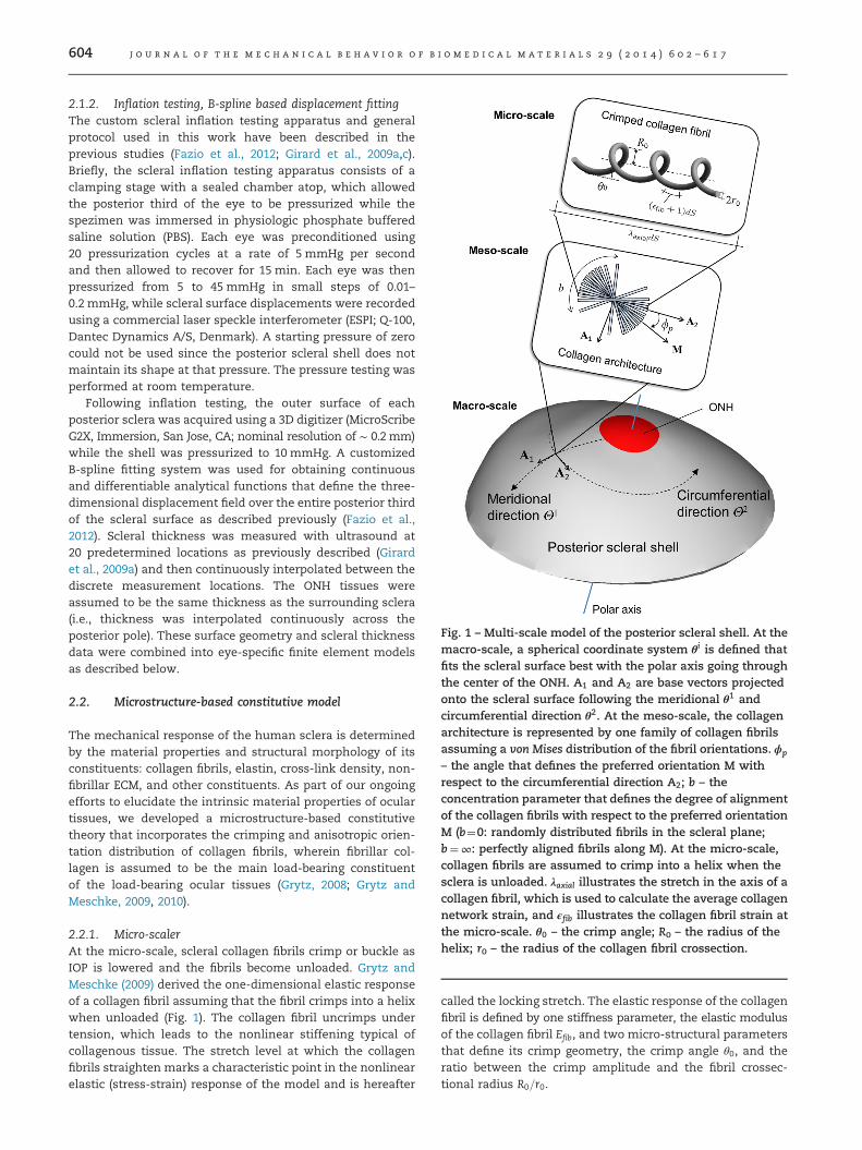
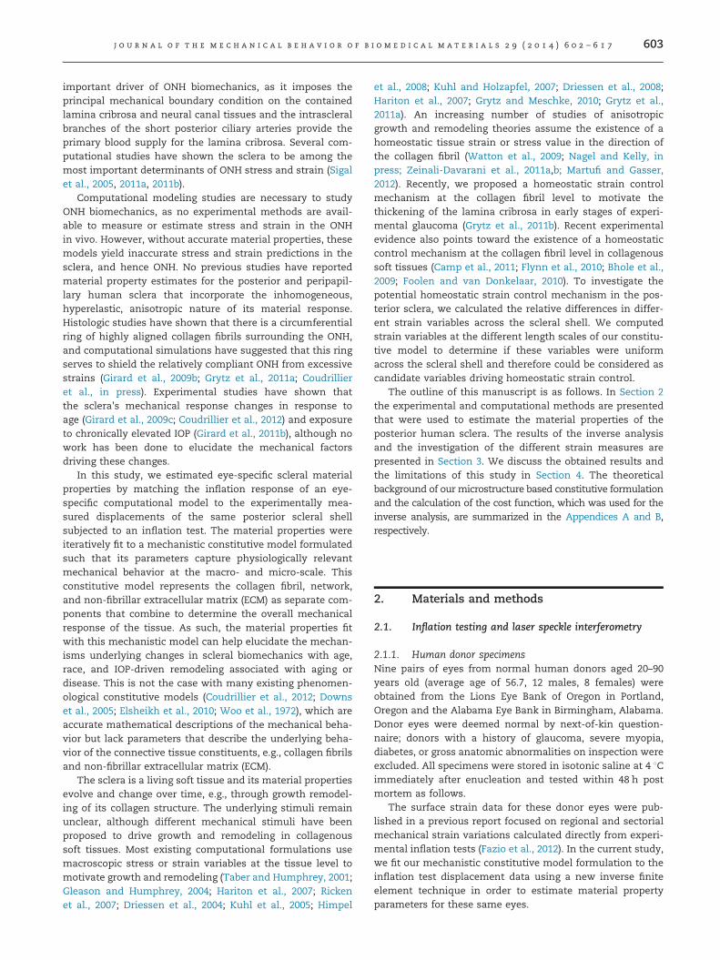
Fig. 1 – Multi-scale model of the posterior scleral shell. At themacro-scale, a spherical coordinate system θi is defined thatfits the scleral surface best with the polar axis going throughthe center of the ONH. A1 and A2 are base vectors projectedonto the scleral surface following the meridional θ1 andcircumferential direction θ2. At the meso-scale, the collagenarchitecture is represented by one family of collagen fibrilsassuming a von Mises distribution of the fibril orientations. ϕp
– the angle that defines the preferred orientation M withrespect to the circumferential direction A2; b – theconcentration parameter that defines the degree of alignmentof the collagen fibrils with respect to the preferred orientationM (b¼0: randomly distributed fibrils in the scleral plane;b¼∞: perfectly aligned fibrils along M). At the micro-scale,collagen fibrils are assumed to crimp into a helix when thesclera is unloaded. λaxial illustrates the stretch in the axis of acollagen fibril, which is used to calculate the average collagennetwork strain, and ϵfib illustrates the collagen fibril strain atthe micro-scale. θ0 – the crimp angle; R0 – the radius of thehelix; r0 – the radius of the collagen fibril crossection.
j o u r n a l o f t h e m e c h a n i c a l b e h a v i o r o f b i o m e d i c a l m a t e r i a l s 2 9 ( 2 0 1 4 ) 6 0 2 – 6 1 7604
2.1.2. Inflation testing, B-spline based displacement fittingThe custom scleral inflation testing apparatus and generalprotocol used in this work have been described in theprevious studies (Fazio et al., 2012; Girard et al., 2009a,c).Briefly, the scleral inflation testing apparatus consists of aclamping stage with a sealed chamber atop, which allowedthe posterior third of the eye to be pressurized while thespezimen was immersed in physiologic phosphate bufferedsaline solution (PBS). Each eye was preconditioned using20 pressurization cycles at a rate of 5 mmHg per secondand then allowed to recover for 15 min. Each eye was thenpressurized from 5 to 45 mmHg in small steps of 0.01–0.2 mmHg, while scleral surface displacements were recordedusing a commercial laser speckle interferometer (ESPI; Q-100,Dantec Dynamics A/S, Denmark). A starting pressure of zerocould not be used since the posterior scleral shell does notmaintain its shape at that pressure. The pressure testing wasperformed at room temperature.
Following inflation testing, the outer surface of eachposterior sclera was acquired using a 3D digitizer (MicroScribeG2X, Immersion, San Jose, CA; nominal resolution of ∼ 0.2 mm)while the shell was pressurized to 10 mmHg. A customizedB-spline fitting system was used for obtaining continuousand differentiable analytical functions that define the three-dimensional displacement field over the entire posterior thirdof the scleral surface as described previously (Fazio et al.,2012). Scleral thickness was measured with ultrasound at20 predetermined locations as previously described (Girardet al., 2009a) and then continuously interpolated between thediscrete measurement locations. The ONH tissues wereassumed to be the same thickness as the surrounding sclera(i.e., thickness was interpolated continuously across theposterior pole). These surface geometry and scleral thicknessdata were combined into eye-specific finite element modelsas described below.
2.2. Microstructure-based constitutive model
The mechanical response of the human sclera is determinedby the material properties and structural morphology of itsconstituents: collagen fibrils, elastin, cross-link density, non-fibrillar ECM, and other constituents. As part of our ongoingefforts to elucidate the intrinsic material properties of oculartissues, we developed a microstructure-based constitutivetheory that incorporates the crimping and anisotropic orien-tation distribution of collagen fibrils, wherein fibrillar col-lagen is assumed to be the main load-bearing constituentof the load-bearing ocular tissues (Grytz, 2008; Grytz andMeschke, 2009, 2010).
2.2.1. Micro-scalerAt the micro-scale, scleral collagen fibrils crimp or buckle asIOP is lowered and the fibrils become unloaded. Grytz andMeschke (2009) derived the one-dimensional elastic responseof a collagen fibril assuming that the fibril crimps into a helixwhen unloaded (Fig. 1). The collagen fibril uncrimps undertension, which leads to the nonlinear stiffening typical ofcollagenous tissue. The stretch level at which the collagenfibrils straighten marks a characteristic point in the nonlinearelastic (stress-strain) response of the model and is hereafter
called the locking stretch. The elastic response of the collagenfibril is defined by one stiffness parameter, the elastic modulusof the collagen fibril Efib, and two micro-structural parametersthat define its crimp geometry, the crimp angle θ0, and theratio between the crimp amplitude and the fibril crossec-tional radius R0=r0.

j o u r n a l o f t h e m e c h a n i c a l b e h a v i o r o f b i o m e d i c a l m a t e r i a l s 2 9 ( 2 0 1 4 ) 6 0 2 – 6 1 7 605
2.2.2. Meso-scaleAt the scleral meso-scale, collagen fibrils aggregate and formcomplex architectures with directional (anisotropic) stiffness.We assume that collagen fibrils are tangent to the scleralsurface as observed in the previous histologic studies(Watson and Young, 2004). We use a semicircular von Misesdistribution function to describe the anisotropic collagenfibril orientation distribution in which ϕp is an angle thatdefines the preferred orientation and b is the concentrationparameter of the collagen fibril distribution (Fig. 1) (Grytz,2008; Girard et al., 2009b). Collagen fibrils are randomlyoriented in the scleral plane for b¼0 and become increasinglyaligned along the preferred orientation for increasing valuesof b. A detailed discussion on the anisotropic materialresponse for varying meso-structural parameters is providedin our previous publication (Girard et al., 2009b).
2.2.3. Macro-scaleWe assume that the anisotropic collagen network is embeddedin a nearly incompressible non-fibrillar tissue matrix withisotropic (orientation independent) material properties. Thisisotropic tissue matrix represents all non-collagenous tissuecomponents (e.g., elastin, glycosaminoglycans, proteogly-cans, cells, and fluid), as well as the isotropic component ofthe collagen fibril network. The isotropic elastic response ofthe tissue matrix is described by the shear modulus μ.
In total, the constitutive model contains two micro-structuralparameters (θ0, R0=r0), two meso-structural parameters (ϕp, b),and two stiffness parameters (μ, Efib). In contrast to phenom-enological constitutive models that describe elastic behavior
Fig. 2 – (a) The eye-specific FE mesh of one eye showing the postethe original shape and thickness of each eye. (b) Section throughincluding the simple support at the outer surface and the spring(red lines) used to model local variations in the collagen fibril archb) between the control points (red and green circles). A standard bin the spherical coordinate space for each element of the extendscleral boundaries, where control points 13–16 are located at the vdistances. The extended mesh can be rotated by an offset angle ofthe red control points and fitted at the green control points. (For inreader is referred to the web version of this article.)
but whose formulation has no physical relevance (Coudrillieret al., 2012; Downs et al., 2005; Elsheikh et al., 2010; Woo et al.,1972), the structural parameters of our mechanistic modelhave clear physical interpretations and can be directlyobtained from experimental observations. The strain energydensity function and main equations of the constitutivetheory are presented in Appendix A.
2.3. Eye-specific finite element (FE) modeling
2.3.1. FE meshThe surface geometry and thickness of each posterior scleralshell were obtained experimentally as described above andcombined to generate eye-specific FE models. The FE meshwas comprised of 256 quadratic hexahedral elements forthe posterior sclera and 80 for the ONH using one elementthrough the shell thickness (Fig. 2a). We used a fitted, eye-specific model to test the appropriateness of this meshdensity in terms of numerical accuracy. This mesh conver-gence test showed that doubling the mesh density in both in-plane directions and in thickness direction changed thedisplacement predictions of the model by less than 5%. Basedon this test the mesh density was considered sufficient toensure numerical accuracy of the results.
2.3.2. Boundary conditionsThe posterior sclera for each eye was clamped to a custom-builtpressurization apparatus to determine inflation-induced dis-placements. Scleral clamping was achieved by positioning theposterior scleral shell onto a radiused plastic ring and then
rior scleral shell and the lamina cribrosa. The mesh capturesthe FE mesh showing the boundary conditions at the clamp,support through the scleral thickness. (c) The extended meshitecture by interpolating the meso-structural parameters (ϕp,i-linear interpolation of the control point values is performeded mesh. The extended mesh extends beyond the posteriorirtual equator of the eye. d1, d2 represent variable meridionalfset. The meso-structural parameters (ϕp, b) are pre-defined atterpretation of the references to color in this figure caption, the

j o u r n a l o f t h e m e c h a n i c a l b e h a v i o r o f b i o m e d i c a l m a t e r i a l s 2 9 ( 2 0 1 4 ) 6 0 2 – 6 1 7606
elevating a vertical stage to squeeze the sclera between the ringand the machined conical surface of a fixed, stainless steelclamping plate. Modeling the mechanical boundary conditionsat the scleral clamping site presents several challenges, as theclamping process squeezes and prestresses the tissue and thedegree to which the clamping process constraints the deforma-tion of the scleral shell is likely eye-specific. Previously, weassumed that scleral deformations were perfectly constrainedthroughout the scleral thickness between the plastic ring andthe clamping plate (a fixed boundary condition) (Girard et al.,2009a). Preliminary studies showed that this approach isreasonable for nonhuman primate scleral shells but overconstrains human scleral shells, which is likely due to thesignificantly greater thickness of human sclera. As a result, weused eye-specific boundary conditions that allow us to adjustthe stiffness of the clamping constraint such that the computa-tional model best fit the experimental displacements. At theclamp, we assume a simple support at the outer surface of thesclera and spring support through the scleral thickness (Fig. 2b).Our experimental clamping condition does not result in aperfectly rigid support, and therefore this spring support repre-sents the experimental condition more accurately than a fixedboundary condition. The eye-specific boundary conditionsrequire the fitting of a spring stiffness k for each eye, whichwas fitted as an unknown parameter in the inverse identifica-tion algorithm presented in the subsequent section. The fittedeye-specific boundary conditions were found to significantlyimprove the quality of the material property fits compared to afixed boundary condition.
2.3.3. Extended FE meshTo model local differences in tissue anisotropy (Zeinali-Davarani et al., 2011a), we used an extended mesh on theouter scleral surface that allowed us to interpolate the meso-structural parameters (ϕp, b) across the entire posterior scleralshell beyond the clamp boundary (Fig. 2c). The extended
Table 1 – Fitted parameters describing the scleral material respparameter search space. The limit values in brackets representdifferential evolution method (Section 2.4.2) if different from th
Parameter
Micro-scale (collagen fibril):θ0 crimp angleR0=r0 ratio between crimp amplitude and cross-section radiusEfib elastic modulus of the collagen fibrils
Meso-scale (collagen architecture):ϕp;5–ϕp;8 preferred collagen fibril orientations at each control point 5
ϕp;9−12 one preferred collagen fibril orientation for control points 9
b5–b12 collagen fibril concentration parameters at each control poi12
Macro-scale (posterior scleral shell):μ shear modulus of the tissuek surface spring stiffness for the clamping boundary conditio
offset circumferential offset of the extended meshd1 meridional distance between control points 1–4and 5–8d2 meridional distance between control points 5–8 and 9–12
Total
mesh contains 16 control points at which the parameters ϕp,b must be defined. The extended mesh is defined with respectto a spherical coordinate system, which was defined suchthat the polar axis is going through the center of the ONH andthe origin of a sphere that best fits the posterior scleral shell(see Fig. 1). A standard bi-linear interpolation along thespherical coordinates is performed for each element of theextended mesh. The interpolated values are used to solve theconstitutive equations at each Gauss point of the regular(non-extended) FE mesh (Appendix A).
2.4. Inverse parameter identification
2.4.1. Model assumptionsWe assume that the stiffness parameters (μ, Efib), as well asthe micro-structural parameters (θ0, R0=r0) are uniform acrossthe entire scleral shell for each eye. The meso-structuralparameters that define local anisotropy (ϕp, b) are definedat the 16 control points of the extended mesh. To reducethe number of unknowns, further assumptions were maderegarding the meso-structure. In accordance with the pre-vious experimental (Goldbaum et al., 1989; Winkler et al.,2010) and numerical studies (Grytz et al., 2011a), the align-ment of collagen fibrils around the scleral canal was assumedto be circumferential by setting ϕp ¼ 0 and b¼10 at controlpoints 1–4 (Fig. 2). At the equator, collagen fibrils wereassumed to be planar isotropic by setting ϕp ¼ 0 and b¼0at control points 13–16. To further reduce the number ofunknowns, the preferred orientation angles at the peripheryof the scleral shell (ϕp;9 ¼ ϕp;10 ¼ ϕp;11 ¼ ϕp;12) were assumed tobe identical. In total, the inverse FE problem for each eyeconsisted of 21 unknown parameters as summarized inTable 1. The lower and the upper limits assumed for eachparameter (Table 1) were enforced by using the bounce backmethod (Price et al., 2005).
onse, including the assumed lower and upper limits of thethe bounds of the first random population of the globale parameter search space.
Number ofunknowns
Lowerlimits
Upper limits
1 21 ∞ (81)1 1 101 10.0 MPa ∞ (100 MPa)
–8 4 −901 901
–12 1 −901 901
nt 5– 8 0 ∞ (5)
1 0.01 MPa ∞ (1 MPa)n 1 0 ∞ (100 N/
mm3)1 −451 4511 51 1511 151 451
21

j o u r n a l o f t h e m e c h a n i c a l b e h a v i o r o f b i o m e d i c a l m a t e r i a l s 2 9 ( 2 0 1 4 ) 6 0 2 – 6 1 7 607
For the sake of simplicity, the ONH tissues (including thelamina cribrosa) were modeled by interpolating the scleralthickness measurements across the scleral canal. To com-pensate for the ONHs lower structural stiffness, which resultsfrom the significantly lower thickness and collagen fibrildensity of the lamina cribrosa compared with the surround-ing sclera, the stiffness parameters of the ONH were reducedby a factor of 15 compared to the scleral values (Efib;ONH¼Efib;Sclera=15). The micro-structural parameters (θ0, R0=r0)were set to the scleral values. The ONH was also assumedto have planar isotropic oriented collagen fibrils (bONH ¼ 0).
2.4.2. Cost functionA crucial part of any inverse optimization scheme is thedefinition of an objective cost function that defines thequality of the fit. Let cost be the cost function that must beminimized by the optimization algorithm. We propose a costfunction that is based on the sum of squared residuals (error)when comparing the FE model and the experimental displa-cements, integrated over the outer surface of the sclera. Toaccurately capture the nonlinear stiffening of the sclera, wesum the cost over nine IOP elevation ranges (from 5mmHg to7, 10, 15, 20, 25, 30, 35, 40, and 45 mmHg). The cost function isboth normalized and weighted such that each IOP level andeach displacement component (X, Y, and Z) have a similaroverall impact on the cost function. Detailed derivations ofthe cost function are presented in Appendix B. To neglectunphysical solutions from the search space of the fittingalgorithm, a penalty function, penalty, was used. The penaltyfunction was defined based on the assumption that at least80% of scleral collagen fibrils uncrimp for a pressure elevationfrom 5 to 45 mmHg (see Appendix B). The penalty functionwas found to effectively remove unphysical solutions and toimprove the convergence rate of the inverse problem.
2.4.3. Global optimization approachThe inverse finite element problem of each posterior scleralshell presents a large-scale optimization problem character-ized by multiple local minima in the cost function. Thedifferential evolution (DE) approach initially proposed byStorn and Price (1997) has proven to be a robust algorithmin many applications that converges faster and with morecertainty than many other global optimization schemes.Recent advances in the DE approach were summarized byNeri and Tirronen (2010). We use the DE algorithm recentlyproposed by Brest and Maučec (2010). This algorithm wasdesigned to solve large-scale optimization problems withmultiple local minima similar to the inverse problem pre-sented here. The initial control parameters of the algorithmwere chosen as proposed by Brest and Maučec (2010) exceptfor the maximum number of iterations, which was set to2000. We tested the convergence properties and the repeat-ability of the method by solving five independent trials fortwo inverse problems.
2.4.4. High performance computing (HPC)Global optimization schemes such as the algorithm used inthis study require a large number of cost function computa-tions (number of iteration times the size of the population). Inthe present case, each cost calculation represents the solution
of a nonlinear eye-specific FE model, which takes about1–1.5 min solution time on a standard PC. Approximately47,000 cost calculations are necessary to perform one eye-specific scleral material property fit, which would require33–51 days on a standard PC. To handle this high demandon computational calculations in a reasonable amount of time,a parallelized solution strategy was developed. DE algorithmsare parallel in nature and well suited for parallel computingstrategies. We developed a HPC strategy that largely follows amaster-slave scheme running on approximately 160 idle CPUcores distributed among the Apple Mac Pro workstations inour department. This HPC setup led to an average solutiontime of 13 h for each eye-specific global optimization problem.
2.4.5. PrestressIn general, the posterior human sclera maintains its sphericalshape at zero IOP but it may buckle locally. To exclude thepotential for unbuckling of the scleral shell during the inflationtests, we preloaded the shells with 5 mmHg IOP prior todisplacement recording (Fazio et al., 2012). This preloadinduces a prestress state that needs to be taken into accountin the inverse parameter identification process if one seeks toidentify the intrinsic material properties of the sclera. The FE-mesh was constructed using scleral surface geometric infor-mation collected at an IOP of 10mmHg (Fazio et al., 2012)while the preload of the tissues for the inflation test was5 mmHg. The scleral shell deforms from 0 to 10mmHg, butthese deformations are small (Fazio et al., 2012), and below the250 μ m resolution of the 3D digitizer we used to collect thescleral surface geometry data. We recently proposed a newnumerical method (Grytz and Downs, in press) to accuratelycalculate this pre-existing stress state. However, we have alsoshown that an indirect approach (Grytz and Meschke, 2010;Grytz and Downs, in press) using relative displacements wassuitable for the inverse parameter identification of scleralelasticity from inflation tests. The indirect approach requiressignificantly less computational time than the accuratemethod and was found to perform better (Grytz and Downs,in press) compared to an alternative approach based onrelative IOP values (Girard et al., 2009a,c). Consideringthe large number of calculations and the associated computa-tional cost, we applied the indirect method using relativedisplacements (Grytz and Downs, in press) in the presentstudy to estimate the prestressed state of each eye.
2.5. Strain variables as candidates for homeostatic control
To determine if tissue and fibril level strains were uniformacross the posterior sclera and therefore could be consideredas candidate variables for homeostatic strain control, weinvestigated the relative differences in strain across thescleral shell at the macro-scale and collagen fibril level. Toaccomplish this, the results of the inverse FE models wereanalyzed with respect to strain predictions obtained at thedifferent length scales of the multi-scale model. We used fourstrain-based measures: (i) the in-plane strain, which representsthe average tissue strain in the scleral shell plane; (ii) thecollagen network strain, which represents the average axialstrain within the dispersed collagen fibril orientations;(iii) the collagen fibril strain; and (iv) the percentile of locked
j o u r n a l o f t h e m e c h a n i c a l b e h a v i o r o f b i o m e d i c a l m a t e r i a l s 2 9 ( 2 0 1 4 ) 6 0 2 – 6 1 7608
collagen fibrils, which represents the percentile of scleralcollagen fibrils that are straightened (uncrimped). The math-ematical definition of the different strain-based values can befound in Appendix A and a graphical illustration of thecollagen fibril strain is shown in Fig. 1. Average strain-basedmeasures were computed for the peripapillary and mid-peripheral regions. The peripapillary region was defined asa 10-degree-wide band adjacent to the ONH (approximately2.2 mm wide) and the mid-peripheral region was defined foreach eye such that it had the same surface area as theperipapillary region. We calculated the volume average ofthe strain responses in these two regions within each eye. Weused linear mixed effects statistical models, which accountfor the difference between intra- and inter-donor variability,to determine if strains were significantly different betweenthe two regions.
The overall IOP-dependent elastic response of the inversemodel is primarily dictated by the two stiffness parameters(μ, Efib) and the two micro-structural parameters (θ0, R0=r0). Toinvestigate the variation and the biomechanical implications ofthese model parameters on IOP-dependent scleral elasticity, aparametric study was performed. The eye-specific model thatexhibited the set of fitted model parameters closest to the meanvalues for the entire group was used for the parametric study. Toinvestigate the sensitivity and the physical interpretations of thematerial property parameters, the four strain-based measuresintroduced above were computed for the peripapillary and mid-peripheral region by setting one of the four constitutive para-meters to theminimum andmaximum values of the fitted rangewhile keeping the other parameters at their mean values.
3. Results
3.1. Convergence and repeatability
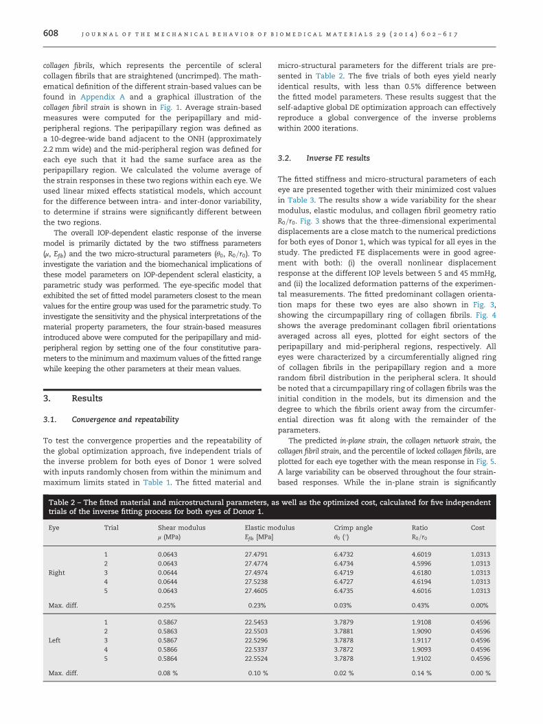
To test the convergence properties and the repeatability ofthe global optimization approach, five independent trials ofthe inverse problem for both eyes of Donor 1 were solvedwith inputs randomly chosen from within the minimum andmaximum limits stated in Table 1. The fitted material and
Table 2 – The fitted material and microstructural parameters, atrials of the inverse fitting process for both eyes of Donor 1.
Eye Trial Shear modulus Elastic moμ (MPa) Efib [MPa]
1 0.0643 27.47912 0.0643 27.4774
Right 3 0.0644 27.49744 0.0644 27.52385 0.0643 27.4605
Max. diff. 0.25% 0.23%
1 0.5867 22.54532 0.5863 22.5503
Left 3 0.5867 22.52964 0.5866 22.53375 0.5864 22.5524
Max. diff. 0.08 % 0.10 %
micro-structural parameters for the different trials are pre-sented in Table 2. The five trials of both eyes yield nearlyidentical results, with less than 0.5% difference betweenthe fitted model parameters. These results suggest that theself-adaptive global DE optimization approach can effectivelyreproduce a global convergence of the inverse problemswithin 2000 iterations.
3.2. Inverse FE results
The fitted stiffness and micro-structural parameters of eacheye are presented together with their minimized cost valuesin Table 3. The results show a wide variability for the shearmodulus, elastic modulus, and collagen fibril geometry ratioR0=r0. Fig. 3 shows that the three-dimensional experimentaldisplacements are a close match to the numerical predictionsfor both eyes of Donor 1, which was typical for all eyes in thestudy. The predicted FE displacements were in good agree-ment with both: (i) the overall nonlinear displacementresponse at the different IOP levels between 5 and 45 mmHg,and (ii) the localized deformation patterns of the experimen-tal measurements. The fitted predominant collagen orienta-tion maps for these two eyes are also shown in Fig. 3,showing the circumpapillary ring of collagen fibrils. Fig. 4shows the average predominant collagen fibril orientationsaveraged across all eyes, plotted for eight sectors of theperipapillary and mid-peripheral regions, respectively. Alleyes were characterized by a circumferentially aligned ringof collagen fibrils in the peripapillary region and a morerandom fibril distribution in the peripheral sclera. It shouldbe noted that a circumpapillary ring of collagen fibrils was theinitial condition in the models, but its dimension and thedegree to which the fibrils orient away from the circumfer-ential direction was fit along with the remainder of theparameters.
The predicted in-plane strain, the collagen network strain, thecollagen fibril strain, and the percentile of locked collagen fibrils, areplotted for each eye together with the mean response in Fig. 5.A large variability can be observed throughout the four strain-based responses. While the in-plane strain is significantly
s well as the optimized cost, calculated for five independent
dulus Crimp angle Ratio Costθ0 (1) R0=r0
6.4732 4.6019 1.03136.4734 4.5996 1.03136.4719 4.6180 1.03136.4727 4.6194 1.03136.4735 4.6016 1.0313
0.03% 0.43% 0.00%
3.7879 1.9108 0.45963.7881 1.9090 0.45963.7878 1.9117 0.45963.7872 1.9093 0.45963.7878 1.9102 0.4596
0.02 % 0.14 % 0.00 %

Table 3 – The fitted stiffness and microstructural parameters including the optimized cost of the inverse models of the 18donor eyes. M – male; F – female; R – right; L – left; STDEV – standard deviation.
Donor Sex Age Eye Shear modulus Elastic modulus Crimp angle Ratio Costμ (MPa) Efib (MPa) θ0 (1) R0=r0
1 M 81 R 0.13 27.48 6.47 4.60 1.03L 0.59 22.53 3.79 1.91 0.46
2 M 80 R 0.45 45.48 4.49 3.29 0.41L 0.29 24.79 6.08 2.51 0.69
3 F 66 R 0.25 42.30 6.31 4.19 0.72L 0.11 32.67 6.99 10.00 1.41
4 F 77 R 0.82 71.51 3.87 5.03 0.84L 0.09 23.83 6.92 4.00 0.61
5 F 82 R 0.18 25.48 6.10 5.59 1.29L 0.26 25.49 5.16 3.50 1.23
6 M 90 R 0.24 59.83 4.49 2.04 0.32L 0.42 35.50 5.26 4.40 0.83
7 M 78 R 0.37 40.69 5.58 4.73 0.81L 0.21 19.31 7.25 2.37 0.68
8 M 88 R 0.40 16.32 3.97 10.00 0.84L 0.69 67.23 2.84 1.45 0.66
9 F 57 R 0.23 70.97 6.21 8.98 0.92L 0.26 101.53 4.54 3.10 1.31
Mean 77.67 0.33 41.83 5.35 4.54 0.84STDEV 10.05 0.20 23.37 1.28 2.63 0.31
j o u r n a l o f t h e m e c h a n i c a l b e h a v i o r o f b i o m e d i c a l m a t e r i a l s 2 9 ( 2 0 1 4 ) 6 0 2 – 6 1 7 609
higher in the peripapillary region (po0:001), there are nosignificant regional differences in the collagen network andfibril strains (p¼0.11 and 0.34, respectively). For IOP rangesbetween 0 and ∼ 20mmHg, the in-plane and the collagennetwork strain curves show a nonlinear stiffening of the tissueand the collagen network, respectively. These strain responsesbecome almost linear for IOPs larger than 20mmHg, becausethe majority of collagen fibrils have straightened (locked) at thatIOP. At the collagen fibril level, the strains follow an almostlinear pattern through the entire IOP range. The collagen fibrilstrains at the micro-scale are much smaller than the strainmeasures at the higher scales. At an IOP of 15 mmHg, the meanvalues of the collagen fibril strain over both regions were0:04670:025%, collagen network strain was 0:4470:2%, andthe in-plane strain was 1:0970:48%. In both regions, thepercentile of fibrils that are stretched beyond their locking pointincreases with IOP, and the majority of collagen fibrils arerecruited to participate in bearing the IOP load between 7 and20mmHg.
3.3. Parameter study
The right posterior scleral shell of donor 7 was used for aparametric study as its fitted model parameters were closestto the mean values of all the eyes in this study. Theindependent effects of the two macro-structural (μ, Efib) andtwo micro-structural parameters (θ0, R0=r0) on the in-planestrain, the collagen network strain, the collagen fibril strain, andthe percentile of locked collagen fibrils are shown in Fig. 6. Foreach parameter, the response curves for the maximum,minimum, and mean parameter values (Table 3) were com-pared, while the other model parameters were kept at theirmean values.
At the macro-scale, the in-plane strain was higher in theperipapillary region compared to the mid-periphery for each
of the parameter variations. An increasing shear modulus (μ)led to an overall stiffening of the tissue and vice-versa. Anincrease in the elastic modulus of collagen fibrils (Efib)increased the tissue stiffness at IOP levels 10 mmHg andhigher and vice-versa. An increasing collagen fibril crimpangle (θ0) increases the in-plane strain by a constant shift ofthe response curve and vice-versa. Variation in the collagenfibril geometry ratio R0=r0 only had a very small impact on thetissue stiffness around the transition region between the softand stiff tissue response (at an IOP of ∼10mmHg).
At the meso-scale, the aforementioned parameter varia-tions elicited similar effects on the collagen network straincurves compared to the in-plane strain except for the followingobservations: the IOP-dependent collagen network strains arealmost identical for the peripapillary and mid-peripheralregions; the shear modulus has an significant lower impacton the collagen network strain compared to the in-plane strain;and variations in the collagen crimp angle have the greatestimpact on the variation of collagen network strain.
At the micro-scale, the collagen fibril strain and the percentileof locked collagen fibrils were similar between the peripapillaryand the mid-peripheral regions for the different parametervariations. The elastic modulus of the collagen fibrils had themost significant effect on the collagen fibril strains.
4. Discussion
An inverse numerical fitting strategy was proposed to identifymaterial properties of posterior human scleral shells usingthree-dimensional, full-field displacement measurementsfrom experimental inflation tests. The model is based on amechanistic constitutive formulation (Grytz and Meschke, 2009)that derives the inhomogeneous, hyperelastic, and anisotropicnature of the scleral material response from the microstructure

Fig. 3 – Top: columns show the comparison of experimentally measured and FE model-predicted displacements for both eyes(both shown in right eye configuration) of one donor (81 years old) for three IOP elevations from 5 to 15, 30, and 45 mmHg. Therows show the comparison between the experimentally measured and computationally predicted meridional, circumferential,and radial surface displacements. Column and row-wise comparisons show that the predicted displacements are in goodagreement with both: (i) the overall nonlinear displacement response and (ii) the localized deformation patterns of theexperimental measurements. Bottom: the predicted collagen fibril architecture for both eyes showing the concentration ofcollagen fibrils (contour plot) along their preferred orientations (white lines). A ring of circumferentially aligned collagen fibrils isseen in the peripapillary scleral region around the scleral canal. Exp. – experimental.
j o u r n a l o f t h e m e c h a n i c a l b e h a v i o r o f b i o m e d i c a l m a t e r i a l s 2 9 ( 2 0 1 4 ) 6 0 2 – 6 1 7610
collagen fibrils. The proposed inverse model was found toreproduce both the overall and local deformation response ofthe posterior scleral shells from individual eyes. The fittedmaterial parameters of our mechanistic model have a clearphysical meaning and can be compared to direct experimen-tal observations. As such, the material properties fit with thismodel can help elucidate the mechanisms underlying micro-and macroscopic changes in scleral biomechanics with age,race, and IOP-driven remodeling in glaucoma.
The proposed constitutive model is based on the sum ofisotropic and anisotropic strain energies, where the latter isassumed to represent the elastic contribution of the
anisotropic collagen fibril architecture. Based on the 9 pairsof human eyes investigated here (57–90 years old), the inverseanalysis showed a large variation in the estimated modelparameters. The estimated elastic modulus (per tissue volume)of collagen fibrils, Efib, was 41:83723:37MPa (mean, standarddeviation) and the crimp angle of collagen fibrils, θ0, was5:35171:281. These values are very close to our previousestimates (Efib ¼ 37:42MPa, θ0¼5.091) (Grytz, 2008; Grytz andMeschke, 2010), which were based on fitting parameters toexperimental inflation measurements for a single meridionalsection as reported by Woo et al. (1972). The estimated shearmodulus of the human sclera (μ¼ 0:3370:2MPa) and the ratio

Fig. 4 – Average collagen fibril density distributions in 8sectors of the peripapillary and mid-peripheral region of thesclera averaged over all eyes, as represented by individualpolar plots of the average collagen fibril distribution at eachsector location. A high concentration of collagen fibrils in thecircumferential direction can be seen across all sectors of theperipapillary region. More randomly organized collagen fibrilorientations are observed in the mid-peripheral region.
j o u r n a l o f t h e m e c h a n i c a l b e h a v i o r o f b i o m e d i c a l m a t e r i a l s 2 9 ( 2 0 1 4 ) 6 0 2 – 6 1 7 611
between the crimp amplitude and the cross-section radius ofcollagen fibrils (R0=r0 ¼ 4:5472:6) were significantly higherthan our previous estimates of 0.01 MPa and 1.09, respectively(Grytz, 2008; Grytz and Meschke, 2010). This difference maybe the result of the much simpler modeling approach used inour previous work (Grytz, 2008; Grytz and Meschke, 2010) and/or the one-dimensional displacement measurements by Wooet al. (1972). The three-dimensional, full-field displacementmeasurements, coupled with the much more sophisticatedmodeling and fitting approach used here, provide the bestestimates reported to date for human scleral material proper-ties. The rather high shear stiffness we report may relate to ahigh concentration of collagen crosslinks or the existence of alarge isotropic component of the collagen fibril network, thepresence of which has been suggested by Girard et al. (2011a)based on recent work in the rat sclera.
We also estimated local differences in the anisotropiccollagen fibril architecture for each eye. While the modelparameters associated with scleral collagen fibril anisotropywere constrained within certain limits, recurring structureswere observed in the fitted results. A circumpapillary ring ofcollagen fibrils surrounding the scleral canal was a character-istic structure in all eyes, which is in agreement with theexperimental observations (Goldbaum et al., 1989; Winkleret al., 2010; Girard et al., 2011a; Pijanka et al., 2012) andprevious numerical predictions (Girard et al., 2009a; Grytzet al., 2011a; Girard et al., 2011b). At the periphery ofthe posterior sclera, collagen fibrils were estimated to bemore randomly oriented, which has also been observedexperimentally (Boote et al., 2010).
The use of a microstructurally motivated constitutivemodel allowed for the numerical investigation of IOP-dependent scleral strains at different length scales that havedifferent interpretations and ramifications. At the macro-scale, the in-plane strains were found to be significantly
higher in peripapillary region than in the mid-periphery,which matches our recent direct calculations of macro-scalescleral strain in these same eyes (Fazio et al., 2012). Incontrast, the collagen network and the collagen fibril strainsthat occur at the meso- and micro-scale were very similarin both regions. This finding has implications for both theresident cell populations and in determining the biomecha-nical signals and mechanisms that drive IOP-related scleralremodeling processes. These findings suggest that the fibril-level strains are a candidate for driving the mechanisms thatmaintain scleral mechanical homeostasis, while the tissuelevel strains are not good candidates.
We have previously shown that the sclera stiffens in themonkey eye when exposed to chronic IOP elevations (Girardet al., 2011b). We hypothesized that this stiffening is aremodeling mechanism that strives to maintain homeostaticloading conditions at the collagen fibril level (Grytz et al.,2012). Recent experimental evidence supports the hypothesisthat evolution, growth, and remodeling mechanisms in softcollagenous tissues are driven by the loading conditions ofthe collagen fibrils themselves (Camp et al., 2011; Flynn et al.,2010; Foolen and van Donkelaar, 2010). Our finding that thecollagen network strain and collagen fibril strain are similar inthe peripapillary and mid-peripheral regions even though themacro-scale in-plane strains are different support this hypoth-esis. This finding also supports that both strain measures, thecollagen network strain and the collagen fibril strain, are reason-able candidate variables to drive homeostatic strain controlin the sclera. In addition, previous modeling studies haveshown that IOP-related stress concentrates around the scleralcanal due to the presence of the more compliant ONH. Ourresults further suggest that the ring of collagen fibrils aroundthe scleral canal is necessary to establish optimal loadbearing conditions at the collagen fibril level, while alsoprotecting the contained ONH from excessive strain inducedby scleral canal expansion. Previously, we estimated ahomeostatic collagen fibril strain of 0.1% in our study onthe remodeling of the lamina cribrosa in early glaucoma(Grytz et al., 2011b). In the present study, an average collagenfibril strain of 0:046%70:025% was identified for the peripa-pillary and mid-peripheral region at 15 mmHg IOP. Bothestimates are in the same order of magnitude, which sup-ports the notion that collagen fibril strain of this magnituderepresents a homeostatic zone for scleral collagen fibrils.
Grytz and Meschke (2009) showed that the crimp angleparameter and the collagen fibril strain predictions of thecrimped collagen fibril model compare well to experimentalmeasurements in rat tail tendons (Diamant et al., 1972) andbovine Achilles tendons (Sasaki and Odajima, 1996), respec-tively. The collagen architecture of the sclera appears morecomplex in that it incorporates interwoven and more ran-domly oriented lamellae. Consequently, inter-lamellar inter-actions are more likely to play an important role in the elasticresponse of the scleral collagen network. Neither fibril norlamellar interaction have been considered in the constitutivemodel used here. Accordingly, it remains unclear to whatextent the collagen fibril strain and the crimp angle com-puted in this study compare to the scleral microstructure andits deformation in the living tissue, and they should thereforeonly be considered as estimates.

Fig. 5 – From top to bottom, IOP versus the average in-plane strain, average collagen network strain, the average collagen fibrilstrain, and the percentile of locked collagen fibrils plots showing the model responses of all scleral shells (grey lines) and themean response (black lines) for the peripapillary scleral (left plots) and mid-peripheral region (right plots). The in-plane strainand collagen network strain curves show the nonlinear and IOP-dependent stiffening of the sclera, while the collagen fibrilstrain increases nearly linear with increasing IOP. The average in-plane strain is higher in the peripapillary region than in themid-periphery, while the average collagen network and fibril strains are almost identical for both regions. Collagen fibrils startto lock at about 7 mmHg and more fibrils get recruited to bear the load for increasing IOP. All plots show a significant variabilityin the data set.
j o u r n a l o f t h e m e c h a n i c a l b e h a v i o r o f b i o m e d i c a l m a t e r i a l s 2 9 ( 2 0 1 4 ) 6 0 2 – 6 1 7612
Our constitutive formulation does not directly model theelastic response of cells, proteoglycans, elastin, glycosamino-glycans, and other extracellular tissue constituents because
the overall elastic response of all these constituents iscombined into one parameter, the shear modulus. Conse-quently, the contribution of each constituent to the material

Fig. 6 – Parameter study using the posterior scleral shell from the right eye of Donor 7 and the range of model parametersobtained from the fitting analysis using the full range of fitted parameters found in all the eyes. The influence of the twostiffness parameters (shear modulus of the tissue, elastic modulus of collagen fibrils) and two micro-structural parameters(collagen fibril crimp angle and ratio R0=r0) on the IOP-dependent responses is shown for the peripapillary scleral (red) and mid-peripheral region (blue): from left to right, the average in-plane strain, average collagen network strain, the average collagenfibril strain, and the percentile of locked collagen fibrils. For each parameter, the response for the maximum (dashed line withtriangles up), minimum (dashed line with triangles down) and mean values (solid lines) are compared, while keeping the othermodel parameters at their mean values. The fitted meso-structural parameters (b, ϕp) were not varied. (For interpretation of thereferences to color in this figure caption, the reader is referred to the web version of this article.)
j o u r n a l o f t h e m e c h a n i c a l b e h a v i o r o f b i o m e d i c a l m a t e r i a l s 2 9 ( 2 0 1 4 ) 6 0 2 – 6 1 7 613
properties or the interaction of these constituents with thecollagen fibril network cannot be directly obtained from ourresults. While it would be ideal to describe these separateconstituents with separate parameters in our constitutiveformulation, it would also increase the number of unknownparameters that must be estimated. As our current modelalready incorporates an accurate mathematical descriptionof the hyperelastic and anisotropic material response of thesclera, the elastic contribution of any additional constituentwould overlap with the elastic contribution of existing para-meters and diminish the repeatability and uniqueness of theinverse fitting procedure. If elastin contributes significantly tothe anisotropic material response of the sclera, that behaviorwill be captured in the model as part of the collagen network.To incorporate additional constituents and mechanisms intoour model and to further elucidate the intrinsic materialproperties of ocular tissues, additional experimental data willbe required to eliminate additional unknowns. In this con-text, mechanistic constitutive models have a clear advantage
over phenomenological models, as structural parameterssuch as the crimp angle of collagen fibrils (Diamant et al.,1972; Hill et al., 2012) and the distribution of collagen fibrilorientations (Girard et al., 2011a; Pijanka et al., 2012; Meekand Quantock, 2001) can be directly obtained from experi-mental observations.
In other constitutive formulations, the nonlinear stiffen-ing of soft collagenous tissues is derived from a load depen-dent recruitment of collagen fibrils based on a distribution ofcollagen fibril undulation (Cacho et al., 2007; Lokshin andLanir, 2009; Martufi and Gasser, 2011; Raz and Lanir, 2009). Wedid not consider this, but assumed a constant collagen fibrilcrimp angle throughout the scleral shell. However, the modelstill predicted a nonlinear recruitment of collagen fibrils,as demonstrated by the increase in the percentile of lockedfibrils with increasing IOP. This effect is mainly driven by themechanism that in-plane strains are in general higher at theexterior than at the interior surface of scleral shells subjectedto IOP. This in turn leads to an earlier recruitment of collagen

j o u r n a l o f t h e m e c h a n i c a l b e h a v i o r o f b i o m e d i c a l m a t e r i a l s 2 9 ( 2 0 1 4 ) 6 0 2 – 6 1 7614
fibrils at the exterior surface of the scleral model, which is aconsequence of our model assumptions and might be differ-ent in the living tissue. If the homeostatic theory presentedabove holds and collagen fibrils are remodeled to reside athomeostatic strain in the living tissue, collagen fibrils shouldhave a wavier structure (higher crimp angle) at the exteriorsurface compared to the interior surface when the sclera isunloaded. However, this mechanism remains to be experi-mentally investigated.
The presented inverse material property identificationmethod presents a novel strategy to elucidate intrinsic elasticproperties of the posterior sclera. The method presents apromising tool to investigate biomechanical mechanismsthat may alter the elastic properties during aging or glau-coma. The parameter study quantified the effects of each ofthe constitutive model parameters on the overall mechanicalresponse of the sclera, and thereby elucidates the physiolo-gical relevance of each parameter. The reported materialproperty parameter estimates provide the most comprehen-sive characterization of the human scleral mechanicalresponse, and can serve as inputs to future computationalmodels of the posterior scleral shell and ONH.
Appendix A. Microstructure based constitutivemodel
Let the function describing the strain energy density W of thescleral tissue be composed of three parts: the energy densityrelated to the isotropic tissue response Wiso, the anisotropiccollagen network Wcol, and a pure hydrostatic part U
W¼Wiso þWcol þ U ðA:1Þ
The energy contribution of the ground substance is modeledusing an isochoric Neo-Hookean material model (Simo et al.,1985)
Wiso ¼ 12μðJ−2=3tr C−3Þ ðA:2Þ
with J¼ det F and C¼ FTF, where F is the deformation gradi-ent. The model contains one material parameter, the shearmodulus μ, which represents the isotopic stiffness of thetissue. The pure hydrostaic part U controls the compressi-bility of the of the material and is defined as
U¼ 12κðln JÞ2 ðA:3Þ
where κ is the bulk modulus. The bulk modulus was set toκ¼ 1000 μ to model the near incompressibility of the sclera.
Let θi be a curvilinear spherical coordinate system thatwas fit to the posterior scleral shell with the polar axis goingthrough the center of the lamina cribrosa (see Fig. 1). Therelated (orthonormal) base vectors Gi ¼ ∂X=∂θi can be obtainedby standard derivations (Başar and Weichert, 2000) at anypoint X. To define the collagen fibril architecture, the ortho-normal basis Ai is introduced here. A2 is obtained fromprojection of the circumferential direction G2 onto the scleralsurface, A3 is the normal vector of the experimentallyobtained scleral surface and A1 ¼A2 �A3. Let e0 be a unitvector defined in the A1–A2 plane by means of an Eulerianangle ϕ∈½−π=2; π=2�e0ðϕÞ ¼ sinðϕp þ ϕÞG1 þ cosðϕp þ ϕÞG2 ðA:4Þ
where ϕp is the angle that defines the preferred orientation ofthe collagen fibril network (see meso-scale in Fig. 1). The axialstretch λaxial of one collagen fibril pointing in direction ϕ canbe computed as
λaxialðϕÞ ¼ ðe0ðϕÞCe0ðϕÞÞ1=2 ðA:5Þ
This axial stretch can be used to compute the energycontribution of collagen fibrils WfibðλaxialðϕÞÞ pointing in thedirection ϕ based on the microstructure-oriented model,which was developed by Grytz and Meschke (2009) formodeling crimped collagen fibrils. See the original paper fordetail derivations of Wfib. The collagen network is assumed tobe composed of collagen fibrils with distributed orientationse0. We use a normalized von Mises distribution function ρ todefine the in-plane distributed collagen fibrils (Grytz, 2008;Girard et al., 2009b)
ρðϕÞ ¼ expðb cosð2ϕÞÞI0ðbÞπ
ðA:6Þ
where b is the concentration parameter and I0 the modifiedBessel function of the first kind and order zero. The strainenergy function of the collagen network is represented bythe integral over the strain energy contributions of the collagenfibrils weighted by the distribution function ρ
Wcol ¼Z π=2
−π=2ρðϕÞWfibðλaxialðϕÞÞ dϕ ðA:7Þ
We use a standard Gauss-Legendre quadrature of 15th order tonumerically compute the above integral. For further derivationincluding the computation of stress and elasticity tensors seeour previous work (Grytz, 2008; Girard et al., 2009b).
To investigate the changing IOP-dependent elastic proper-ties of the sclera at the different length scales of the purposedconstitutive model, we introduce the in-plane strain ϵIP
ϵIP ¼ JðA3C−1A3Þ1=2−1 ðA:8Þ
the collagen network strain ϵcol
ϵcol ¼Z π=2
−π=2ρðϕÞðλaxialðϕÞ−1Þ dϕ ðA:9Þ
and collagen fibril strain ϵfib. The fibril strain is derived fromthe micro-scale model for crimped collagen fibrils. See Fig. 1for a graphical interpretation and the original paper for adetailed derivation of ϵfib (Grytz and Meschke, 2009). Further-more, the percentile of locked collagen fibrils is introduced
ϑlock ¼ 100π
Z π=2
−π=2HðλaxialðϕÞÞ dϕ
with HðλaxialÞ ¼(0; λaxialoλlock1; λaxial≥λlock
ðA:10Þ
where λlock represents the locking stretch at which collagenfibrils straighten (uncrimp).
Appendix B. Cost function
We propose a following cost function as an objective functionto estimate the quality of the inverse parameter fitting
cost¼ ∑9
p ¼ 1
RΩðuFE
p −uexpp ÞWpðuFE
p −uexpp Þ dAR
Ωuexpp
Wpuexpp dA ðB:1Þ

j o u r n a l o f t h e m e c h a n i c a l b e h a v i o r o f b i o m e d i c a l m a t e r i a l s 2 9 ( 2 0 1 4 ) 6 0 2 – 6 1 7 615
Herein,RΩ dA represents the integration over the outer sur-
face of the posterior scleral shell. The numerator in the aboveterm represents the weighted integral of squared residualsbetween the finite element displacements uFE and the experi-mental displacements uexp integrated over the outer surfaceof the sclera. We use the denominator to normalize theresiduals with the squares of experimental displacementssuch that the cost related to each IOP p has a similar impacton the total cost, where p represents the nine IOP elevationsfrom 5 mmHg to 7, 10, 15, 20, 25, 30, 35, 40, 45 mmHg. InEq. (B.1), Wp is a weighting tensor which is used to accountfor large differences in the three displacement componentswith respect to the spherical coordinate system
Wp ¼w11 0 0
0 w22 0
0 0 w33
264
375p
Gi⊗Gj ðB:2Þ
The components of the weighting tensor are defined in thefollowing:
ðwiiÞp ¼ 1−RΩju
expp � Gij dAR
Ωðjuexpp � G1j þ juexp
p � G2j þ juexpp � G3jÞ dA
for i¼ 1; 2; 3 ðB:3ÞHerein, Gi are the base vectors of the spherical coordinatesystem introduced in Appendix A. To penalize unphysicalsolution based on the tissue volume containing straightenscleral collagen fibrils the following dynamic penalty termwas used:
penalty¼ ð0:5 � iterÞ2ðϑlock;totð45Þ−ϑlock;totð5Þ−80Þ2 ðB:4Þ
ϑlock;totðpÞ represents percentile of locked collagen fibrils of theentire posterior scleral shell at the IOP level p (mmHg) and iteris the current iteration of the DE algorithm. The pressure onunphysical solutions is increasing with increasing iterationsteps due to the first term in (B.4). The penalty term (B.4) wasadded to the cost function (B.1) if less than 80% of scleralcollagen fibrils were straighten for an IOP elevation from 5 to45 mmHg. The objective function f used to evaluate the DEpopulation was defined as
f ¼ costþ penalty if ϑlock;totð45Þ−ϑlock;totð5Þo80
cost otherwise
�ðB:5Þ
r e f e r e n c e s
Başar, Y., Weichert, D., 2000. Nonlinear Continuum Mechanics ofSolids. Springer Verlag, Berlin.
Bhole, A.P., Flynn, B.P., Liles, M., Saeidi, N., Dimarzio, C.A.,Ruberti, J.W., 2009. Mechanical strain enhances survivabilityof collagen micronetworks in the presence of collagenase:implications for load-bearing matrix growth and stability.Philosophical Transactions of the Royal Society A 367(September (1902)), 3339–3362.
Boote, C., Sorensen, T., Coudrillier, B., Myers, K., Meek, K., Quigley,H., Nguyen, T., 2010. Posterior scleral collagen architecture innormal and glaucoma human eyes, as determined usingwide-angle x-ray scattering. ARVO Abstract 51, 4900.
Brest, J., Maučec, M., 2010. Self-adaptive differential evolutionalgorithm using population size reduction and three
strategies. Soft Computing—A Fusion of Foundations,Methodologies and Applications 15 (11), 2157–2174.
Burgoyne, C.F., Downs, J.C., Bellezza, A.J., Suh, J.-K.F., Hart, R.T.,2005. The optic nerve head as a biomechanical structure: anew paradigm for understanding the role of IOP-related stressand strain in the pathophysiology of glaucomatous opticnerve head damage. Progress in Retinal and Eye Research 24(January (1)), 39–73.
Cacho, F., Elbischger, P.J., Rodríguez, J.F., Dolbaré, M., Holzapfel, G.A., 2007. A constitutive model for fibrous tissues consideringcollagen fiber crimp. International Journal of Non-LinearMechanics 42, 391–402.
Camp, R.J., Liles, M., Beale, J., Saeidi, N., Flynn, B.P., Moore, E.,Murthy, S.K., Ruberti, J.W., 2011. Molecularmechanochemistry: low force switch slows enzymaticcleavage of human type I collagen monomer. Journal of theAmerican Chemical Society 133 (March (11)), 4073–4078.
Coudrillier, B., Boote, C., Quigley, H.A., Nguyen, T.D., in press.Scleral anisotropy and its effects on the mechanical responseof the optic nerve head. Biomechanics and Modeling inMechanobiology, http://dx.doi.org/10.1007/s10237-012-0455-y.
Coudrillier, B., Tian, J., Alexander, S., Myers, K.M., Quigley, H.A.,Nguyen, T.D., 2012. Biomechanics of the human posteriorsclera: age-and glaucoma-related changes measured usinginflation testing. Investigative Ophthalmology and VisualScience 53 (4), 1714–1728.
Diamant, J., Keller, A., Baer, E., Litt, M., Arridge, R., 1972. Collagen:Ultrastructure and its relation to mechanical properties as afunction of aging. Proceedings of Royal Society of London,Series B 180, 293–315.
Downs, J.C., Roberts, M.D., Burgoyne, C.F., 2008. Mechanicalenvironment of the optic nerve head in glaucoma. Optometryand Vision Science 85 (June (6)), 425–435.
Downs, J.C., Suh, J.K., Thomas, K.A., Bellezza, A.J., Hart, R.T.,Burgoyne, C.F., 2005. Viscoelastic material properties of theperipapillary sclera in normal and early-glaucoma monkey eyes.Investigative Ophthalmology and Visual Science 46, 540–546.
Driessen, N.J.B., Cox, M.A.J., Bouten, C.V.C., Baaijens, P.T.B., 2008.Remodelling of the angular collagen fiber distribution incardiovascular tissues. Biomechanics and Modeling inMechanobiology 7, 93–103.
Driessen, N.J.B., Wilson, W., Bouten, C.V.C., Baaijens, F.P.T., 2004.A computational model for collagen fibre remodelling in thearterial wall. Journal of Theoretical Biology 226, 53–64.
Elsheikh, A., Geraghty, B., Alhasso, D., Knappett, J., Campanelli,M., Rama, P., 2010. Regional variation in the biomechanicalproperties of the human sclera. Experimental Eye Research 90(May (5)), 624–633.
Fazio, M.A., Grytz, R., Bruno, L., Girard, M.J.A., Gardiner, S., Girkin,C.A., Downs, J.C., 2012. Regional variations in mechanicalstrain in the posterior human sclera. InvestigativeOphthalmology and Visual Science 53 (September (9)),5326–5333.
Flynn, B.P., Bhole, A.P., Saeidi, N., Liles, M., Dimarzio, C.A.,Ruberti, J.W., 2010. Mechanical strain stabilizes reconstitutedcollagen fibrils against enzymatic degradation by mammaliancollagenase matrix metalloproteinase 8 (MMP-8). PLoS ONE 5(8), e12337.
Foolen, J., van Donkelaar, C.C., Soekhradj-Soechit, S., Ito, K., 2010.European Society of Biomechanics SM Perren Award 2010: anadaptation mechanism for fibrous tissue to sustainedshortening. Journal of Biomechanics 43 (September (16)),3168–3176.
Girard, M.J.A., Dahlmann-Noor, A., Rayapureddi, S., Bechara, J.A.,Bertin, B.M.E., Jones, H., Albon, J., Khaw, P.T., Ethier, C.R.,2011a. Quantitative mapping of scleral fiber orientation innormal rat eyes. Investigative Ophthalmology and VisualScience 52 (13), 9684–9693.

j o u r n a l o f t h e m e c h a n i c a l b e h a v i o r o f b i o m e d i c a l m a t e r i a l s 2 9 ( 2 0 1 4 ) 6 0 2 – 6 1 7616
Girard, M.J.A., Downs, J.C., Bottlang, M., Burgoyne, C.F., Suh, J.F.,2009a. Peripapillary and posterior scleral mechanics. Part II.Experimental and inverse finite element characterization.Journal of Biomechanical Engineering 131 (5), 051012.
Girard, M.J.A., Downs, J.C., Burgoyne, C.F., Suh, J.F., 2009b.Peripapillary and posterior scleral mechanics. Part I.Development of an anisotropic hyperelastic constitutivemodel. Journal of Biomechanical Engineering 131 (5), 051011.
Girard, M.J.A., Suh, J.-K.F., Bottlang, M., Burgoyne, C.F., Downs, J.C.,2009c. Scleral biomechanics in the aging monkey eye.Investigative Ophthalmology and Visual Science 50 (November(11)), 5226–5237.
Girard, M.J.A., Suh, J.-K.F., Bottlang, M., Burgoyne, C.F., Downs, J.C.,2011b. Biomechanical changes in the sclera of monkey eyesexposed to chronic IOP elevations. Investigative Ophthalmologyand Visual Science 52 (August (8)), 5656–5669.
Gleason, R.L., Humphrey, J.D., 2004. A mixture model of arterialgrowth and remodeling in hypertension: altered muscle toneand tissue turnover. Journal of Vascular Research 41 (4),352–363.
Goldbaum, M.H., Jeng, S.Y., Logemann, R., Weinreb, R.N., 1989.The extracellular matrix of the human optic nerve. Archives ofOphthalmology 107 (August (8)), 1225–1231.
Grytz, R., 2008. Computational modeling and remodeling ofhuman eye tissues as biomechanical structures at multiplescales. Ph.D. Thesis, Ruhr-University Bochum, Germany.
Grytz, R., Downs, J.C., in press. A forward incrementalprestressing method with application to inverse parameterestimations and eye-specific simulations of posterior scleralshells. Computer Methods in Biomechanics and BiomedicalEngineering, http://dx.doi.org/10.1080/10255842.2011.641119.
Grytz, R., Girkin, C., Libertiaux, V., Downs, J., 2012. Perspectives onbiomechanical growth and remodeling mechanisms inglaucoma. Mechanics Research Communications 42, 92–106.
Grytz, R., Meschke, G., 2009. Constitutive modeling of crimpedcollagen fibrils in soft tissues. Journal of the MechanicalBehavior of Biomedical Materials 2 (October (5)), 522–533.
Grytz, R., Meschke, G., 2010. A computational remodelingapproach to predict the physiological architecture of thecollagen fibril network in corneo-scleral shells. Biomechanicsand Modeling in Mechanobiology 9 (April (2)), 225–235.
Grytz, R., Meschke, G., Jonas, J.B., 2011a. The collagen fibrilarchitecture in the lamina cribrosa and peripapillary sclerapredicted by a computational remodeling approach.Biomechanics and Modeling in Mechanobiology 10 (3),371–382.
Grytz, R., Sigal, I.A., Ruberti, J.W., Meschke, G., Downs, J.C., 2011b.Lamina cribrosa thickening in early glaucoma predicted by amicrostructure motivated growth and remodeling approach.Mechanics of Materials 44, 99–109.
Hariton, I., deBotton, G., Gasser, T.C., Holzapfel, G.A., 2007. Stress-driven collagen fiber remodeling in arterial walls.Biomechanics and Modelling in Mechanobiology 6 (April (3)),163–175.
Hill, M.R., Duan, X., Gibson, G.A., Watkins, S., Robertson, A.M.,2012. A theoretical and non-destructive experimentalapproach for direct inclusion of measured collagenorientation and recruitment into mechanical models of theartery wall. Journal of Biomechanics 45 (February), 762–771.
Himpel, G., Menzel, A.A.K., Steinmann, P., 2008. Time-dependentfiber reorientation of transversely isotropic continua-finiteelement formulation and consistent linearization.International Journal for Numerical Methods in Engineering73, 1413–1433.
Kuhl, E., Garikipati, K., Arruda, E., Grosh, K., 2005. Remodeling ofbiological tissues: mechanically induced reorientation of atransversely isotropic chain network. Journal of theMechanics and Physics of Solids 53, 1552–1573.
Kuhl, E., Holzapfel, G., 2007. A continuum model for remodelingin living structures. Journal of Materials Science 42, 8811–8823.
Lokshin, O., Lanir, Y., 2009. Micro and macro rheology of planartissues. Biomaterials 30 (June (17)), 3118–3127.
Martufi, G., Gasser, T.C., 2011. A constitutivemodel for vascular tissuethat integrates fibril, fiber and continuum levels with applicationto the isotropic and passive properties of the infrarenal aorta.Journal of Biomechanics 44 (September (14)), 2544–2550.
Martufi, G., Gasser, T.C., 2012. Turnover of fibrillar collagen in softbiological tissue with application to the expansion ofabdominal aortic aneurysms. Journal of the Royal SocietyInterface 9 (August), 3366–3377.
Meek, K.M., Quantock, A.J., 2001. The use of x-ray scatteringtechniques to determine corneal ultrastructure. Progress inRetinal and Eye Research 20 (January (1)), 95–137.
Nagel, T., Kelly, D.J., 2012. Remodelling of collagen fibre transitionstretch and angular distribution in soft biological tissues andcell-seeded hydrogels. Biomechanics and Modelling inMechanobiology 11, 325–339.
Neri, F., Tirronen, V., 2010. Recent advances in differentialevolution: a survey and experimental analysis. ArtificialIntelligence Review 33 (1), 61–106.
Pijanka, J.K., Coudrillier, B., Ziegler, K., Sorensen, T., Meek, K.M.,Nguyen, T.D., Quigley, H.A., Boote, C., 2012. Quantitativemapping of collagen fiber orientation in non-glaucoma andglaucoma posterior human scleras. InvestigativeOphthalmology and Visual Science 53 (July (9)), 5258–5270.
Price, K., Storn, R., Lampinen, J., 2005. Differential Evolution: APractical Approach to Global Optimization. Springer-Verlag,New York Inc..
Raz, E., Lanir, Y., 2009. Recruitment viscoelasticity of the tendon.Journal of Biomechanical Engineering 131 (November (11)),111008.
Ricken, T., Schwarza, A., Bluhm, J., 2007. A triphasic model oftransversely isotropic biological tissue with applications tostress and biologically induced growth. ComputationalMaterials Science 39, 124–136.
Sasaki, N., Odajima, S., 1996. Elongation mechanism of collagenfibrils and force-strain relations of tendon at each level ofstructural hierarchy. Journal of Biomechanics 29, 1131–1136.
Sigal, I.A., Flanagan, J.G., Ethier, C.R., 2005. Factors influencingoptic nerve head biomechanics. Investigative Ophthalmologyand Visual Science 46 (November (11)), 4189–4199.
Sigal, I.A., Yang, H., Roberts, M.D., Burgoyne, C.F., Downs, J.C.,2011a. IOP-induced lamina cribrosa displacement and scleralcanal expansion: an analysis of factor interactions usingparameterized eye-specific models. InvestigativeOphthalmology and Visual Science 52 (March (3)), 1896–1907.
Sigal, I.A., Yang, H., Roberts, M.D., Grimm, J.L., Burgoyne, C.F.,Demirel, S., Downs, J.C., 2011b. IOP-induced lamina cribrosadeformation and scleral canal expansion: independent orrelated?. Investigative Ophthalmology and Visual Science 52(12), 9023–9032.
Simo, J., Taylor, R., Pister, K., 1985. Variational and projectionmethods for the volume constraint in finite deformationelasto-plasticity. Computational Methods in AppliedMechanics and Engineering 51, 177–208.
Storn, R., Price, K., 1997. Differential evolution—a simple andefficient heuristic for global optimization over continuousspaces. Journal of Global Optimization 11 (4), 341–359.
Taber, L.A., Humphrey, J.D., 2001. Stress-modulated growth,residual stress, and vascular heterogeneity. Journal ofBiomechanical Engineering 123, 528–535.
Watson, P.G., Young, R.D., 2004. Scleral structure, organisation anddisease: a review. Experimental Eye Research 78 (March (3)),609–623.

j o u r n a l o f t h e m e c h a n i c a l b e h a v i o r o f b i o m e d i c a l m a t e r i a l s 2 9 ( 2 0 1 4 ) 6 0 2 – 6 1 7 617
Watton, P.N., Ventikos, Y., Holzapfe, G.A., 2009. Modelling thegrowth and stabilization of cerebral aneurysms. MathematicalMedicine and Biology 26 (June (2)), 133–164.
Winkler, M., Jester, B., Nien-Shy, C., Massei, S., Minckler, D.S.,Jester, J.V., Brown, D.J., 2010. High resolution three-dimensional reconstruction of the collagenous matrix of thehuman optic nerve head. Brain Research Bulletin 81 (3),339–348.
Woo, S.L., Kobayashi, A.S., Schlegel, W.A., Lawrence, C., 1972.Nonlinear material properties of intact cornea and sclera.Experimental Eye Research 14, 29–39.
Zeinali-Davarani, S., Raguin, L.G., Vorp, D.A., Baek, S., 2011a.
Identification of in vivo material and geometric parameters of
a human aorta: toward patient-specific modeling of
abdominal aortic aneurysm. Biomechanics and Modeling in
Mechanobiology 10 (October (5)), 689–699.
Zeinali-Davarani, S., Sheidaei, A., Baek, S., 2011b. A finite element
model of stress-mediated vascular adaptation: application to
abdominal aortic aneurysms. Computer Methods in
Biomechanics and Biomedical Engineering 14 (April), 803–817.