materials science & engineering c · the ranking of release following sintering was 57 > 56...
TRANSCRIPT
Contents lists available at ScienceDirect
Materials Science & Engineering C
journal homepage: www.elsevier.com/locate/msec
Sintered electrospun polycaprolactone for controlled model drug delivery
Francisco J. Chaparroa, Kayla F. Presleya, Marco A. Coutinho da Silvab, John J. Lannuttia,⁎
a Department of Materials Science and Engineering, The Ohio State University, 2041 College Road, Columbus, OH 43210, USAbDepartment of Veterinary Clinical Sciences, The Ohio State University, 601 Vernon Tharp St., Columbus, OH 43210, USA
A R T I C L E I N F O
Keywords:PharmaceuticalElutionContraceptiveEquineSustained releaseMethadone
A B S T R A C T
Electrospinning has been used widely for drug delivery applications due to its versatility and ease of mod-ification of spun fiber properties. Net drug loading and release is typically limited by the inherent surface-area ofthe sample. In a relatively novel approach, sintering of electrospun fiber was used to create a capsule favoringlong-term delivery. We showed that electrospun polycaprolactone (PCL) retained its initial morphology out to1042 days of in vitro exposure, illustrating its potential for extended performance. Sintering decreased theelectrospun pore size by 10- and 28-fold following 56 and 57 °C exposures, respectively. At 58 and 59 °C, the PCLcapsules lost all apparent surface porosity, but entrapped pores were observed in the 58 °C cross-section. The useof Rhodamine B (RhB, 479.02 gmol−1), Rose Bengal (RB, 1017.64 gmol−1) and albumin-fluorescein iso-thiocyanate conjugate from bovine serum (BSA-FITC, ~66,000 gmol−1) as model compounds demonstrated thatrelease (RhB > RB≫ BSA-FITC) is controlled both by molecular weight and available porosity. Interestingly,the ranking of release following sintering was 57 > 56 > 59 > 58 °C; COMSOL simulations explored the ef-fects of capsule wall thickness and porosity on release rate. It was hypothesized that model drug adsorption onthe available fiber surface-area (57 versus 56 °C) and entrapped porosity (59 versus 58 °C) could have alsoattributed to the observed ranking of release rates. While the 56 and 57 °C exposures allowed the bulk of therelease to occur in< 1 day, the capsules sintered at 58 and 59 °C exhibited release that continued after 12 days ofexposure.
1. Introduction
Electrospinning is a versatile technique used in diverse fields such asenergy generation [1], water treatment [2], air filtration [3], sensors[4] and biomedical engineering [5]. Electrospinning can produce bio-compatible, biodegradable, porous and interconnected scaffolds thatsupport cell proliferation and migration, making this technique an idealchoice for a variety of scaffolding materials [6,7]. Electrospun fibershave been used in many biomedical applications due to their similarityto the extracellular matrix (ECM) [5,8–10]. The ability to createblended polymer compositions (both natural and synthetic) in-corporating either inorganic or organic components and the ability tomodify the resulting surface chemistry and physical properties makethis technique particularly useful in biomedicine. Via precise controlover diameter and surface hydrophobicity and the incorporation ofdifferent compounds by means of pre- and/or post-fiber modification,electrospinning has become valuable for applications involving drug
delivery. Drug delivery vehicles have been an area of intense interestdue to the potential for surface-area based control over release [11–15].
Researchers have reported delivery utilizing electrospun fibers frombioabsorbable polymers such as polycaprolactone (PCL) [11,16,17],poly(lactic-co-glycolic acid) (PLGA) [17,18], poly(lactic acid) (PLA)[17,19], natural polymers such as chitosan and gelatin, and blends ofbioabsorbable [15,16] and natural polymers (PCL:PLGA [18] andPCL:gelatin [11,20]). In particular, PCL has been extensively studied fordrug delivery due to its biodegradability, biocompatibility, low cost andits approval for use in vivo by the FDA [11,21–24]. PCL is widely re-garded as an initially hydrophobic compound (due to the presence of afive‑carbon aliphatic chain) that becomes less so as hydrolysis is in-itiated.
In this context, Geiger et al. studied the interaction of two modeldrugs, BODIPY and Rhodamine B (RhB), with PCL and PCL:gelatinblended electrospun fibers [11]. It was demonstrated that hydrophobicBODIPY has a greater affinity for hydrophobic PCL and RhB has a
https://doi.org/10.1016/j.msec.2019.01.095Received 6 September 2018; Received in revised form 21 December 2018; Accepted 18 January 2019
Abbreviations: ECM, extracellular matrix; FDA, food and drug administration; HFP, 1,1,1,3,3,3-hexafluoro-2-propanol; MW, molecular weight; PBS, phosphatebuffered saline; PCL, polycaprolactone; PLA, poly (lactic acid); PLGA, poly (lactic-co-glycolic acid); RhB, Rhodamine B; RB, Rose Bengal; SEM, scanning electronmicroscope; THC, tetracycline hydrochloride; TPSA, topological polar surface-area
⁎ Corresponding author at: 2041 College Road, Columbus, OH 43210, USA.E-mail address: [email protected] (J.J. Lannutti).
Materials Science & Engineering C 99 (2019) 112–120
Available online 25 January 20190928-4931/ © 2019 Elsevier B.V. All rights reserved.
T
higher affinity for blended PCL:gelatin fibers due to hydrophilic inter-actions with gelatin. This follows the expected trends dominated byhydrogen bond formation and/or hydrophobic interactions betweenfunctional groups of both entities [25]. Tailored long-term drug de-livery can only be engineered by carefully selecting the appropriatepolymer-drug combination.
In recent years, various strategies have been reported to achievetunable release from electrospun fibers. Coaxial and triaxial electro-spinning have commonly been used to achieve specific fiber structureswith tunable release [26–30]. For instance, Singh et al. reported thataltering the flow rate of the core-shell solution can alter the core dia-meter which was shown to affect the release of a model protein. [27]Similarly, Ball et al. showed that variable core and shell flow rates canbe used to alter the shell thickness of polyvinylpyrrolidone-ethyl cel-lulose core-shell fibers to achieve tunable release of maraviroc [28].Additionally, Liu et al. recently reported a method to produce carefullycontrolled fibers via triaxial electrospinning. In this case, a celluloseacetate layer was precisely varied in order to control the rate of drugrelease [30]. Control over polymer blend composition is anothermethod to tailor the drug release rate [26,31,32]. For instance, delValle et al. altered the ratio of PLA:PCL in electrospun fibers and foundthat increased [PLA] led to faster release of triclosan [32].
Biphasic release from as-spun polymeric samples is often observed[11,15–21] where initial burst release occurs invariably followed bylinear release. If the initial burst release is not well controlled, toxiclevels could be achieved in the surrounding tissues. Some studies haveemployed core-shell fibers to suppress and control burst release duringthe first few hours [11,16,17]. By engineering the hydrophobic char-acter of the fiber core and shell, burst release can be modified. Malekiet al. demonstrated that when normal electrospinning was used, thecontact angle on pure PLGA was 130° [16]. When a PLGA- tetracyclinehydrochloride (THC) blend was used, the contact angle decreased to70°, showing that the drug imparted a hydrophilic surface to the scaf-folds. In contrast, when coaxial electrospun fibers made up of a corecarrying THC and a shell of PLGA were created, the hydrophobicproperties of the shell polymer were maintained. Importantly, eventhough burst release was also observed from these core-shell fibers, asuppression of burst release from 35 to 20% of THC was observed.[16]Despite the advantages provided by core-shell fibers as drug releasevehicles, the amount of drug that can be loaded is limited especially iflong-term delivery is desired. A technique that can improve this is su-percritical CO2 infusion [11,33,34] which requires a polymer that isheavily plasticized in the presence of high-pressure CO2. Drugs infusedinto polymers using CO2 do not exhibit an ideal zero order release[11,33,34]. Therefore, different encapsulation approaches are desired.
Tubular shaped electrospun scaffolds have been previously used tomimic blood vessels and other body tissues [5,35–37]. Chaparro et al.[5], Drilling et al. [35] and Lee et al. [36] created PCL tubular hollowscaffolds able to meet and/or surpass native blood vessel tissue prop-erties. These examples provide guidance toward the use of electrospunhollow cylinders to encapsulate a larger drug payload than currentlyavailable to existing electrospinning strategies. During a study of theuse of tubular PCL scaffolds as tracheal tissue engineering scaffolds[37], Nelson et al. sintered electrospun PCL at temperatures rangingfrom 55 to 58 °C for 24 h. Both pore size and porosity were tightlycontrolled by annealing at temperatures close to the melting point(~60 °C). [37]
Due to the low melting point of PCL, enclosing the open end ofelectrospun PCL scaffolds can be readily achieved by employing asimple heat sealer. In this context, tubular samples could provide anoption to deliver drugs inside a capsule composed of sintered electro-spun fibers. Using this novel approach, we explored release from elec-trospun tubular shaped PCL capsules using sintering to control poresize. We loaded different model drug compounds inside these sinteredelectrospun capsules to study their release behavior. Model compoundswere: Rhodamine B (RhB), Rose Bengal (RB) and albumin-fluorescein
isothiocyanate conjugate from bovine serum (BSA-FITC). These com-pounds are readily used as model drugs [11,21,38–40] due to theirwater solubility, their fluorescent nature and the broad range of mo-lecular weights (RhB: 479.02mol−1, RB: 1017.64 gmol−1, BSA-FITC:~66,000 gmol−1). In the case of RhB and RB, these two hydrophiliccompounds were selected to explore the effect of molecular weight andhydrodynamic radius on release of lower molecular weight drugs. BSA-FITC was selected as a model protein to examine release of a muchlarger compound. Modeling was conducted to better understand thelinkage between sintered microstructure and diffusion mechanismscontrolling model drug release.
2. Materials and methodology
2.1. Scaffold preparation
To create the desired drug delivery capsule (Schematic shown asFig. S1a in Supplementary Material), electrospun tubes were first pre-pared as follows. 5 wt% PCL (Aldrich, St. Louis, MO, Mn=80,000) wasdissolved in 1,1,1,3,3,3-hexafluoro-2-propanol (HFP) (OakwoodChemical, West Columbia, SC) via continuous stirring for 24 h at roomtemperature producing a homogeneous solution. Once the solution wascompletely dissolved and appeared to be well mixed, it was transferredto a 20 cm3 plastic syringe (BD Luer Lok, Franklin Lakes, NJ) tippedwith a 20-gauge blunt needle (EFD, East Providence, RI). The followingconditions were then used to produce electrospun fibers: a voltagepotential of 24 kV, a flow rate of 5mL h−1 and a source to grounddistance of 20 cm. Samples were spun at 500 rpm onto rotating 316stainless steel rods having lengths of 6 cm and a diameter of 3.00mm.Any trace of residual solvents was removed using exposure to vacuum[41] (−30 in Hg) for 18 h at room temperature. Using a Keyence lasermicrometer (Keyence, Model LS-7030) [35] the initial electrospun wallthickness was measured and determined to be 603 ± 110 μm.
2.2. Capsule preparation
To modify the as-spun microstructure, the tubes were sintered at 56,57, 58 and 59 °C. The as-spun samples adhered to the metal mandrel(Fig. S1b in Supplementary Material) were inserted into zip-lock plasticbags placed into a water bath (VWR, Model WB05, temperaturestability± 0.1 °C) for 3 h at the desired temperature. A partial immer-sion thermometer was used inside the chamber of the water bath tomonitor the temperature. Following these sintering exposures, thick-ness was then established using a laser micrometer. Diametric capsuleshrinkage was calculated as follows: % shrinkage= 100 ∗ (t0-t1)/t0,where t0 is the original thickness of the capsule and t1 is the thicknessafter sintering.
Specimens 2.50 cm in length were cut and removed from the metalrod to create tubes enabling compound isolation followed by release. Tocreate the desired reservoir, both ends were sealed following the in-troduction of the payload of interest using a TTS-8 table top sealer (UHeat Seal Solutions, Corona, CA). Each payload was a 40 μL aliquot of a5.0 mgmL−1 solution of a model compound (see Section 2.3) in dis-tilled water. Note that by sintering the capsules on identical 3.00mmdiameter stainless steel rods and by cutting the capsules to the samelength (2.5 cm), the final capsule geometry was maintained regardlessof sintering condition; only fiber morphology, porosity and capsulethickness were affected. Additionally, since the model compounds wereadded to the capsules post-sintering, they were not exposed to theseelevated (56–59 °C) temperatures.
2.3. Model compound release
Three different model compounds were used: RhB (99+%, ACROS,Morris, NJ), RB (95%, Aldrich, St. Louis, MO) and BSA-FITC (Catalognumber: A9771, Sigma, St. Louis, MO). A stock solution of 5.0mgmL−1
F.J. Chaparro et al. Materials Science & Engineering C 99 (2019) 112–120
113
for each compound was prepared using distilled water. For each sin-tered condition, an aliquot of 40 μL of the solution containing ap-proximately 200 μg of each compound was placed inside each of thecapsules which were then sealed as described previously (Section 2.2).An example of the final capsule structure with one of the encapsulatedmodel drugs is shown in Fig. S1c in Supplementary Material.
Each sealed capsule was then placed into capped 15mL glass vialscontaining 10mL 1× phosphate buffered saline (PBS) (FisherBioReagents, Hampton, NH) and 0.02 wt% sodium azide (Aldrich, St.Louis, MO). The capsule-vial combinations were then maintained at37 °C in an incubator (Thermo Electron Corporation, Model 320) for thedesired period of study. Aliquots (100 μL) were extracted from the vialand stored in a 96-well plate (Costar®, Salt Lake City, UT). After eachsampling point, the vial media was completely exchanged with freshPBS. The amount of each compound released over time was establishedusing a plate reader (SpectraMax®, M Series, Molecular Devices) bymeasuring the fluorescence resulting from the excitation and emissionof each dye. The excitation wavelengths of RhB, RB and BSA-FITC were535, 540 and 485 nm, respectively; the emission wavelengths were 612,575, 535 nm, respectively. A calibration curve for each compound wasconstructed using concentrations ranging from 1×10⁻⁵ to0.1 μgmL−1. If the concentration was above 0.1 μgmL−1, the solutionwas diluted to fall inside the calibration curve and was then measuredand multiplied by the dilution factor. All experiments were performedin triplicate.
2.4. Scanning Electron Microscope (SEM) analysis
Samples were placed on conductive carbon tape (SPI Supplies, WestChester, PA) adhered to aluminum SEM sample mounts (TED Pella,Redding, CA) and sputter coated (EMS, model 150TS) with ~14 nm ofgold palladium (60:40) at an emission current of 30mA.Microstructural changes were then observed within an SEM (FEI Nova400) at an accelerating voltage of 5 kV. Fiber diameter (n=50) andpore size area (n=50) were estimated via ImageJ (v. 1.6.0) by refer-encing the scale bars at a magnification of 30,000 and 10,000 X, re-spectively. To quantify pore area, image brightness and contrast wereadjusted and background fibers manually eliminated prior to measuringthe area between the fibers in a given plane using the “polygon selec-tion” tool in ImageJ.
2.5. Statistical analysis
The presence or absence of statistical difference between capsuleshrinkage and calculated pore area for the various sintering conditionswas assessed. This analysis was performed by means of a one-wayANOVA using JMP Pro software (v. 14.0.0). A post-hoc Tukey-KramerHSD test was used to compare statistical differences between differentsintering conditions. For pore area calculations, the pore area of sin-tered capsules was compared to that of the control (the as-spun cap-sule). For calculated diametric shrinkage, results were compared be-tween the different sintering conditions. Significance was attained at avalue of p < 0.05. All data is presented as mean ± standard deviation.
2.6. COMSOL modeling
Supplemental modeling simulations [42] were performed usingCOMSOL Multiphysics software (v. 5.3a) to better understand factorsgoverning RhB release through the PCL capsules sintered at 56, 57, 58and 59 °C. For all simulations, the geometry was simplified to a 2Dcapsule cross-section. The inner radius of the capsule was assumed to be1.50mm. The outer radius was established using the measured thick-nesses of the electrospun capsules. The outer radius of the COMSOLgeometry (representing the inner diameter of the glass vial) was heldconstant at 8.50mm.
The inner capsule was assumed to be filled with PBS, and the initial
dye concentration within the capsule was set to 2.36mol m−3 derivedfrom the capsule geometry. The concentration elsewhere (either withinthe PCL wall or in the surrounding PBS solution) was initially assumedto be 0mol m−3. Effects from adsorption and convection were ignored,and only dye diffusion was considered. The diffusivity of RhB in PBS (inthe capsule interior and the surrounding solution) was set to be2.77×10−10 m2 s−1 [43]. The effective diffusivity of RhB in PCL wasestablished as a variable parameter. All simulations monitored the totalconcentration of RhB outside the capsule in the surrounding PBS so-lution versus time. These measurements of concentration were thenconverted into the net percentage of RhB release. To better match theresults of the experiment, simulations monitored release for 14 days.
3. Results
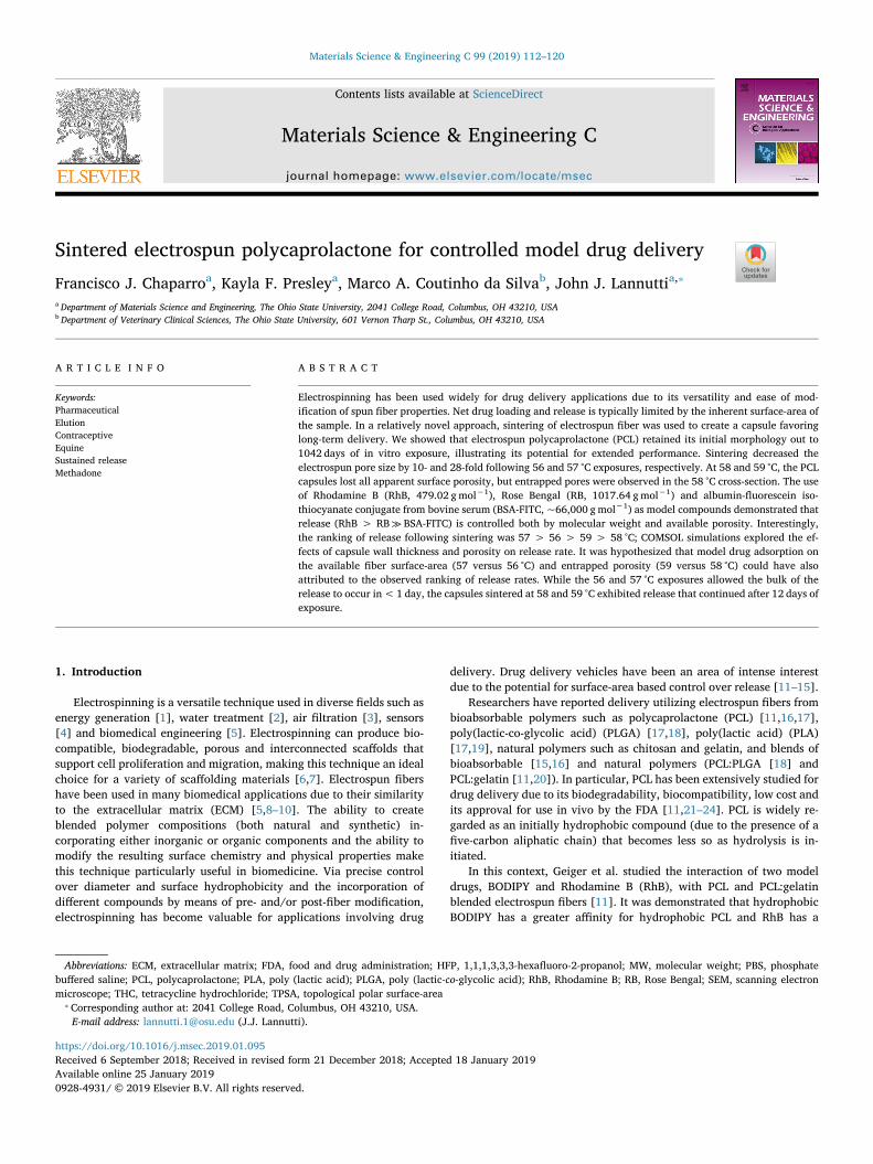
Fig. 1 shows the molecular structure of the fluorescent compoundsused as models for drug release in this study. The variable molecularweights affect release behavior through PCL. The molecular weights forRhB (Fig. 1a), RB (Fig. 1b) and BSA-FITC (Fig. 1c) are 479.02, 1017.64and ~66,000 gmol−1, respectively. For size reference, the structure ofthe BSA protein consists of 607 amino acids.
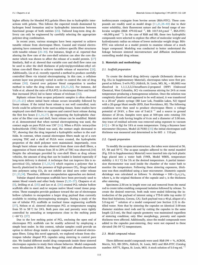
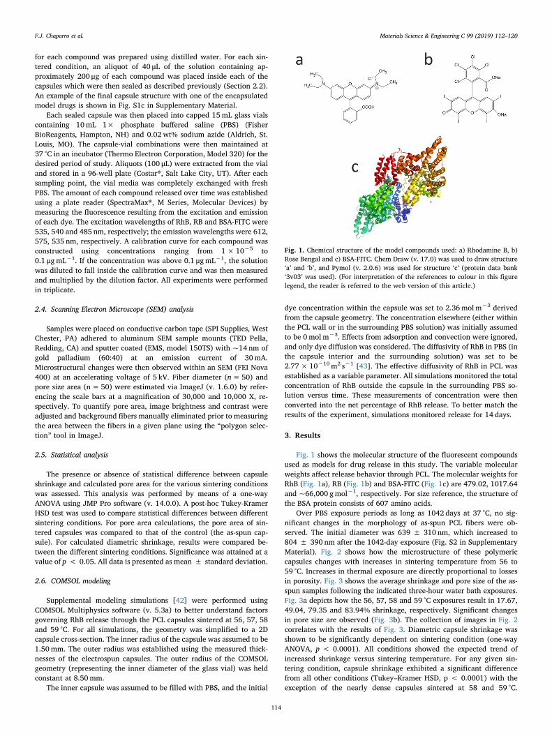
Over PBS exposure periods as long as 1042 days at 37 °C, no sig-nificant changes in the morphology of as-spun PCL fibers were ob-served. The initial diameter was 639 ± 310 nm, which increased to804 ± 390 nm after the 1042-day exposure (Fig. S2 in SupplementaryMaterial). Fig. 2 shows how the microstructure of these polymericcapsules changes with increases in sintering temperature from 56 to59 °C. Increases in thermal exposure are directly proportional to lossesin porosity. Fig. 3 shows the average shrinkage and pore size of the as-spun samples following the indicated three-hour water bath exposures.Fig. 3a depicts how the 56, 57, 58 and 59 °C exposures result in 17.67,49.04, 79.35 and 83.94% shrinkage, respectively. Significant changesin pore size are observed (Fig. 3b). The collection of images in Fig. 2correlates with the results of Fig. 3. Diametric capsule shrinkage wasshown to be significantly dependent on sintering condition (one-wayANOVA, p < 0.0001). All conditions showed the expected trend ofincreased shrinkage versus sintering temperature. For any given sin-tering condition, capsule shrinkage exhibited a significant differencefrom all other conditions (Tukey–Kramer HSD, p < 0.0001) with theexception of the nearly dense capsules sintered at 58 and 59 °C.
Fig. 1. Chemical structure of the model compounds used: a) Rhodamine B, b)Rose Bengal and c) BSA-FITC. Chem Draw (v. 17.0) was used to draw structure‘a’ and ‘b’, and Pymol (v. 2.0.6) was used for structure ‘c’ (protein data bank‘3v03’ was used). (For interpretation of the references to colour in this figurelegend, the reader is referred to the web version of this article.)
F.J. Chaparro et al. Materials Science & Engineering C 99 (2019) 112–120
114
Sintering temperature also had a significant effect on pore area (one-way ANOVA, p < 0.0001). Pore areas measured for 56 °C– and 57 °C–sintered capsules were significantly lower than the as-spun control(Tukey-Kramer HSD, p < 0.0001). At 56 °C (Figs. 2a-b) the averagepore area of the fibers decreased 10-fold versus the as-spun counter-parts (Fig. 3b). By increasing the temperature to 57 °C (Fig. 2c–d) theaverage pore area decreased 28-fold versus the as-spun capsule. Inter-estingly, once 58 °C is reached, the PCL capsule completely loses itsinitial apparent surface porosity (Fig. 2e–f) although some fiber-likestructures can still be distinguished. A similar elimination of surfaceporosity is seen following the 59 °C exposure but no remaining fiber-likestructures were observed (Fig. 2g–h).
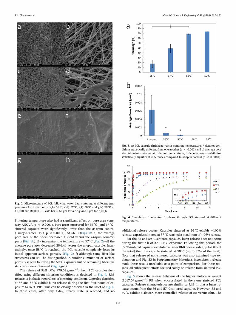
The release of RhB (MW 479.02 gmol−1) from PCL capsules den-sified using different sintering conditions is depicted in Fig. 4. RhBrelease is biphasic regardless of sintering condition. Capsules densifiedat 56 and 57 °C exhibit burst release during the first four hours of ex-posure to 37 °C PBS. This can be clearly observed in the inset of Fig. 4.In those cases, after only 1 day, steady state is reached, and no
additional release occurs. Capsules sintered at 56 °C exhibit ~100%release; capsules sintered at 57 °C reached a maximum of ~96% release.
For the 58 and 59 °C-sintered capsules, burst release does not occurduring the first 4 h of 37 °C PBS exposure. Following this period, the59 °C-sintered capsules exhibited a faster RhB release rate (up to 88% ofthe total) than the capsule sintered at 58 °C (up to 83% of the total).Note that release of non-sintered capsules was also examined (see ex-planation and Fig. S3 in Supplementary Material). Inconsistent releasemade these results unreliable as a point of comparison. For these rea-sons, all subsequent efforts focused solely on release from sintered PCLcapsules.
Fig. 5 shows the release behavior of the higher molecular weight(1017.64 gmol−1) RB when encapsulated in the same sintered PCLcapsules. Release characteristics are similar to RhB in that a burst re-lease occurs from the 56 and 57 °C-sintered capsules. However, 58 and59 °C exhibit a slower, more controlled release of RB versus RhB. The
Fig. 2. Microstructure of PCL following water bath sintering at different tem-peratures for three hours: a,b) 56 °C, c,d) 57 °C, e,f) 58 °C and g,h) 59 °C at10,000 and 30,000×. Scale bar= 50 μm for a,c,e,g and 4 μm for b,d,f,h.
Fig. 3. a) PCL capsule shrinkage versus sintering temperature; * denotes con-ditions statistically different from one another (p < 0.001) and b) average poresize following sintering at different temperatures; * denotes results exhibitingstatistically significant differences compared to as-spun control (p < 0.0001).
Fig. 4. Cumulative Rhodamine B release through PCL sintered at differenttemperatures.
F.J. Chaparro et al. Materials Science & Engineering C 99 (2019) 112–120
115
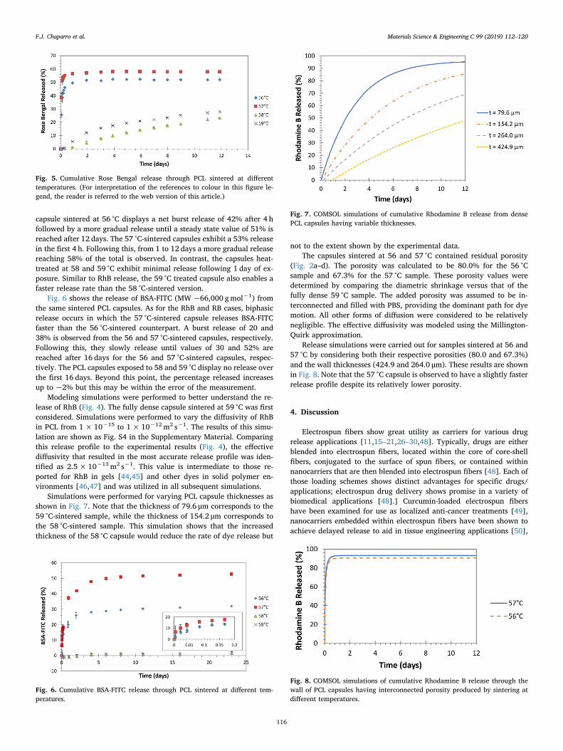
capsule sintered at 56 °C displays a net burst release of 42% after 4 hfollowed by a more gradual release until a steady state value of 51% isreached after 12 days. The 57 °C-sintered capsules exhibit a 53% releasein the first 4 h. Following this, from 1 to 12 days a more gradual releasereaching 58% of the total is observed. In contrast, the capsules heat-treated at 58 and 59 °C exhibit minimal release following 1 day of ex-posure. Similar to RhB release, the 59 °C treated capsule also enables afaster release rate than the 58 °C-sintered version.
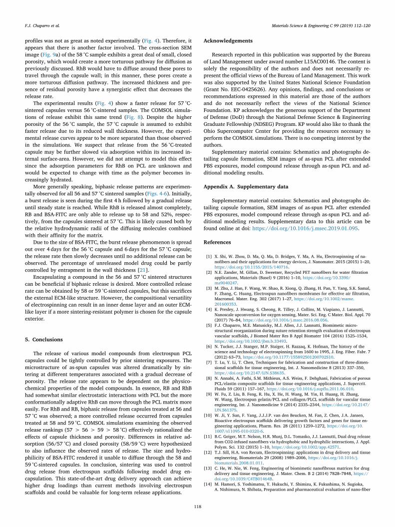
Fig. 6 shows the release of BSA-FITC (MW ~66,000 gmol−1) fromthe same sintered PCL capsules. As for the RhB and RB cases, biphasicrelease occurs in which the 57 °C-sintered capsule releases BSA-FITCfaster than the 56 °C-sintered counterpart. A burst release of 20 and38% is observed from the 56 and 57 °C-sintered capsules, respectively.Following this, they slowly release until values of 30 and 52% arereached after 16 days for the 56 and 57 °C-sintered capsules, respec-tively. The PCL capsules exposed to 58 and 59 °C display no release overthe first 16 days. Beyond this point, the percentage released increasesup to ~2% but this may be within the error of the measurement.
Modeling simulations were performed to better understand the re-lease of RhB (Fig. 4). The fully dense capsule sintered at 59 °C was firstconsidered. Simulations were performed to vary the diffusivity of RhBin PCL from 1×10⁻¹⁵ to 1× 10⁻¹² m2 s−1. The results of this simu-lation are shown as Fig. S4 in the Supplementary Material. Comparingthis release profile to the experimental results (Fig. 4), the effectivediffusivity that resulted in the most accurate release profile was iden-tified as 2.5× 10⁻¹³ m2 s−1. This value is intermediate to those re-ported for RhB in gels [44,45] and other dyes in solid polymer en-vironments [46,47] and was utilized in all subsequent simulations.
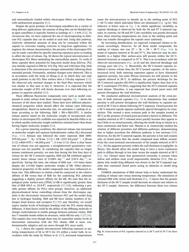
Simulations were performed for varying PCL capsule thicknesses asshown in Fig. 7. Note that the thickness of 79.6 μm corresponds to the59 °C-sintered sample, while the thickness of 154.2 μm corresponds tothe 58 °C-sintered sample. This simulation shows that the increasedthickness of the 58 °C capsule would reduce the rate of dye release but
not to the extent shown by the experimental data.The capsules sintered at 56 and 57 °C contained residual porosity
(Fig. 2a–d). The porosity was calculated to be 80.0% for the 56 °Csample and 67.3% for the 57 °C sample. These porosity values weredetermined by comparing the diametric shrinkage versus that of thefully dense 59 °C sample. The added porosity was assumed to be in-terconnected and filled with PBS, providing the dominant path for dyemotion. All other forms of diffusion were considered to be relativelynegligible. The effective diffusivity was modeled using the Millington-Quirk approximation.
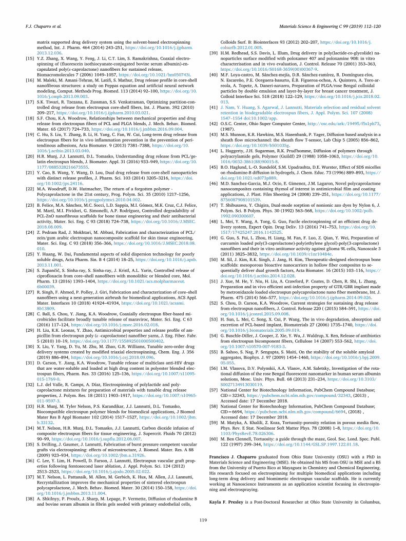
Release simulations were carried out for samples sintered at 56 and57 °C by considering both their respective porosities (80.0 and 67.3%)and the wall thicknesses (424.9 and 264.0 μm). These results are shownin Fig. 8. Note that the 57 °C capsule is observed to have a slightly fasterrelease profile despite its relatively lower porosity.
4. Discussion
Electrospun fibers show great utility as carriers for various drugrelease applications [11,15–21,26–30,48]. Typically, drugs are eitherblended into electrospun fibers, located within the core of core-shellfibers, conjugated to the surface of spun fibers, or contained withinnanocarriers that are then blended into electrospun fibers [48]. Each ofthose loading schemes shows distinct advantages for specific drugs/applications; electrospun drug delivery shows promise in a variety ofbiomedical applications [48].] Curcumin-loaded electrospun fibershave been examined for use as localized anti-cancer treatments [49],nanocarriers embedded within electrospun fibers have been shown toachieve delayed release to aid in tissue engineering applications [50],
Fig. 5. Cumulative Rose Bengal release through PCL sintered at differenttemperatures. (For interpretation of the references to colour in this figure le-gend, the reader is referred to the web version of this article.)
Fig. 6. Cumulative BSA-FITC release through PCL sintered at different tem-peratures.
Fig. 7. COMSOL simulations of cumulative Rhodamine B release from densePCL capsules having variable thicknesses.
Fig. 8. COMSOL simulations of cumulative Rhodamine B release through thewall of PCL capsules having interconnected porosity produced by sintering atdifferent temperatures.
F.J. Chaparro et al. Materials Science & Engineering C 99 (2019) 112–120
116
and metronidazole loaded within electrospun fibers can imbue themwith antibacterial properties [51].
Despite the great promise of electrospun nanofibers for a variety ofdrug delivery applications, the amount of drug that can be blended intoas-spun nanofibers is typically limited to loadings of< 1wt% [52]. Tocircumvent this, we have explored the use of electrospinning to fabri-cate PCL capsules that can be sealed at each end. Using this pathway,relatively substantial amounts of drug can be contained within thecapsule to overcome loading concerns in long-term applications. Toengineer the release characteristics, the porosity of the electrospun PCLcan be easily controlled by specific sintering exposures prior to loadingto affect the rate of release. Fig. S2 shows the typical microstructure ofelectrospun PCL fibers mimicking the extracellular matrix. To verify ifthese capsules show potential for long-term model drug delivery, PCLwas initially exposed to PBS for 315 (Fig. S2c) and 1042 days (Fig. S2d)to determine if the microstructure changes substantially following theseextended periods. Fortunately, minimal changes were observed. This isin accordance with the study of Zhang et al. in which they saw onlysmall pinholes in the PCL fiber surface after a 176-day exposure [15].We observed only minimal changes in the final fiber structure whichwas similar to that seen at day 0. However, it is expected that themolecular weight of PCL will slowly decrease over time following ex-posure to aqueous solution [53].
Three different fluorescent compounds were used as model com-pounds to study release from these sintered capsules. Fig. 1 shows thestructure of the three dyes studied. These dyes have different physico-chemical properties which should affect the release rates followingencapsulation. Based on molecular size, we anticipate a ranking of re-lease according to the following: RhB > RB≫ BSA-FITC. A similarrelease pattern based on the molecular weight of incorporated anti-biotics in electrospun PCL scaffolds was reported by Buschle-Diller et al.in which smaller molecular weight antibiotics released faster than thosehaving higher molecular weights [54].
For a given sintering condition, the observed release rate increasedas molecular weight and aqueous hydrodynamic radius (Rh) decreased(Figs. 4–6). Release rate followed the order expected: RhB (MW:479.02 gmol−1, Rh: 0.78 nm [55])>RB (1017.64 gmol−1, 1.28 nm[56]) ≫BSA-FITC (~66,000 gmol−1, 5.4 nm [55]). Since a clear linearrate of release was not apparent, a straightforward quantitative com-parison was not possible. In considering the capsules that no longerpossess continuous porosity, we note that during the first four days ofrelease for the 58 °C-sintered capsules, RhB and RB exhibited approxi-mately linear release rates of 13.68% day−1 and 2.81% day−1, re-spectively. During this time, the release of RhB was ~4.9 times fasterdespite the 2.4-fold higher molecular weight of RB. Other physico-chemical properties of the model compounds may have influenced re-lease rate. This difference in elution could be connected to the relativeaffinity of RB versus that of RhB for the underlying PCL substratesuggesting a slightly greater affinity for the RB. We note that the to-pological polar surface-area (TPSA) of RB is significantly greater thanthat of RhB (89.5 vs. 52.8 Å2, respectively [57,58]), indicating a pos-sibly greater affinity for PCL's ester groups. However, an additionalphysicochemical factor controlling release from the electrospun cap-sules is the electrostatic interaction between PCL and the model drugdue to hydrogen bonding. RhB and RB have similar numbers of hy-drogen bond donors and acceptors [57,58] and, therefore, we wouldexpect similar levels of hydrogen bonding with the PCL chains duringdiffusion. A final factor could be the ability of the diffusing molecule tochange conformations to enable passage between polymer chains. RhBhas 7 rotatable bonds within its structure, while RB has only 1 [57,58].This implies that even though there may be somewhat similar levels ofelectrostatic interaction with the PCL chains, RhB is more con-formationally adaptive and can move through PCL more easily.
Fig. 2 shows the capsule microstructure following exposure to sin-tering temperatures of 56 to 59 °C for 3 h within a water bath. In ac-cordance with the study by Nelson et al. [37], increased temperatures
cause the microstructure to densify up to the melting point of PCL(~60 °C) after which individual fibers are eliminated (Fig. 2g–h). Thisbehavior is better seen in Fig. 3 in which significant variability incapsule shrinkage is observed for both the 56 and 57 °C-sintered cap-sules. In contrast, for 58 and 59 °C the variability was greatly decreasedsince these sintering temperatures are close to the melting point andheat can transfer throughout the capsule more uniformly.
By increasing densification, we expected compound release to de-crease accordingly. However, for all three model compounds, theranking of release rate was 57 > 56 > 59 > 58 °C (Figs. 4-6). Interms of capsules sintered at 56 and 57 °C, this behavior may be attri-butable to a higher fiber surface-area still present within the 56 °C-sintered structure as compared to 57 °C. This is in accordance with theobserved microstructures (Fig. 2a–d) and the observed shrinkage andaverage pore size (Fig. 3). Meanwhile, the 58 °C-treated capsules ex-hibited a slower release than the 59 °C-treated capsules. At the micro-structural level, both sintering exposures appear to have eliminatedapparent porosity, but some fibrous structures are still present in thecapsule sintered at 58 °C. In addition, the percent shrinkage for the59 °C-sintered capsules (83.94%) is significantly higher than that of its58 °C counterpart (79.35%), suggesting that the 59 °C-sintered capsuleswere denser. Therefore, it was expected that closed pores were stillpresent throughout the wall thickness.
We conducted additional microstructure analysis of the cross-sec-tion of both sintered capsules (Fig. 9) to demonstrate that residualporosity is still present throughout the wall thickness in capsules sin-tered at 58 °C but is absent following 59 °C exposure. Closed porosity fora 58 °C-sintered capsule appears uniformly spaced throughout its cross-section. This created a more tortuous path in the samples treated at58 °C as the presence of closed pores provided a barrier to diffusion. Thesamples sintered at 59 °C released more quickly because they appear tohave little or no closed porosity, allowing the model drug to release in amore continuous and faster rate. Matyka et al. numerically studied therelation of different porosities and diffusion pathways, demonstratingthat at higher porosities the diffusion pathway is less tortuous [59].However, for the 58 °C-sintered capsule, the porosity is low and consistsonly of closed pores that the model compound must diffuse around.From the cross-sectional microstructure of the sample sintered at 59 °C(Fig. 9b) the apparent porosity within the wall thickness is negligible. Intheory, this should allow the model drug to have a more continuouspath to diffuse through in less time versus the sample sintered at 58 °C(Fig. 9a). Clennel states that geometrical tortuosity is present wheninflow and outflow must avoid impermeable obstacles [60]. This ex-plains why model drug diffusion was slower in the 58 °C-exposed cap-sules that contained closed pores acting as obstacles to model drugdiffusion.
COMSOL simulations of RhB release help to better understand theranking of release rate versus sintering temperature. The simulation ofreleased RhB with varied wall thickness (Fig. 7) shows that the greaterthickness of the 58 °C sample would decrease its release rate relative tothe 59 °C sample. However, the difference between these two release
Fig. 9. Cross-section of PCL capsules sintered at a) 58 °C and b) 59 °C for threehours.
F.J. Chaparro et al. Materials Science & Engineering C 99 (2019) 112–120
117
profiles was not as great as noted experimentally (Fig. 4). Therefore, itappears that there is another factor involved. The cross-section SEMimage (Fig. 9a) of the 58 °C sample exhibits a great deal of small, closedporosity, which would create a more torturous pathway for diffusion aspreviously discussed. RhB would have to diffuse around these pores totravel through the capsule wall; in this manner, these pores create amore torturous diffusion pathway. The increased thickness and pre-sence of residual porosity have a synergistic effect that decreases therelease rate.
The experimental results (Fig. 4) show a faster release for 57 °C-sintered capsules versus 56 °C-sintered samples. The COMSOL simula-tions of release exhibit this same trend (Fig. 8). Despite the higherporosity of the 56 °C sample, the 57 °C capsule is assumed to exhibitfaster release due to its reduced wall thickness. However, the experi-mental release curves appear to be more separated than those observedin the simulations. We suspect that release from the 56 °C-treatedcapsule may be further slowed via adsorption within its increased in-ternal surface-area. However, we did not attempt to model this effectsince the adsorption parameters for RhB on PCL are unknown andwould be expected to change with time as the polymer becomes in-creasingly hydrated.
More generally speaking, biphasic release patterns are experimen-tally observed for all 56 and 57 °C sintered samples (Figs. 4-6). Initially,a burst release is seen during the first 4 h followed by a gradual releaseuntil steady state is reached. While RhB is released almost completely,RB and BSA-FITC are only able to release up to 58 and 52%, respec-tively, from the capsules sintered at 57 °C. This is likely caused both bythe relative hydrodynamic radii of the diffusing molecules combinedwith their affinity for the matrix.
Due to the size of BSA-FITC, the burst release phenomenon is spreadout over 4 days for the 56 °C capsule and 6 days for the 57 °C capsule;the release rate then slowly decreases until no additional release can beobserved. The percentage of unreleased model drug could be partlycontrolled by entrapment in the wall thickness [21].
Encapsulating a compound in the 56 and 57 °C sintered structurescan be beneficial if biphasic release is desired. More controlled releaserate can be obtained by 58 or 59 °C-sintered capsules, but this sacrificesthe external ECM-like structure. However, the compositional versatilityof electrospinning can result in an inner dense layer and an outer ECM-like layer if a more sintering-resistant polymer is chosen for the capsuleexterior.
5. Conclusions
The release of various model compounds from electrospun PCLcapsules could be tightly controlled by prior sintering exposures. Themicrostructure of as-spun capsules was altered dramatically by sin-tering at different temperatures associated with a gradual decrease ofporosity. The release rate appears to be dependent on the physico-chemical properties of the model compounds. In essence, RB and RhBhad somewhat similar electrostatic interactions with PCL but the moreconformationally adaptive RhB can move through the PCL matrix moreeasily. For RhB and RB, biphasic release from capsules treated at 56 and57 °C was observed; a more controlled release occurred from capsulestreated at 58 and 59 °C. COMSOL simulations examining the observedrelease rankings (57 > 56 > 59 > 58 °C) effectively rationalized theeffects of capsule thickness and porosity. Differences in relative ad-sorption (56/57 °C) and closed porosity (58/59 °C) were hypothesizedto also influence the observed rates of release. The size and hydro-philicity of BSA-FITC rendered it unable to diffuse through the 58 and59 °C-sintered capsules. In conclusion, sintering was used to controldrug release from electrospun scaffolds following model drug en-capsulation. This state-of-the-art drug delivery approach can achievehigher drug loadings than current methods involving electrospunscaffolds and could be valuable for long-term release applications.
Acknowledgements
Research reported in this publication was supported by the Bureauof Land Management under award number L15AC00146. The content issolely the responsibility of the authors and does not necessarily re-present the official views of the Bureau of Land Management. This workwas also supported by the United States National Science Foundation(Grant No. EEC-0425626). Any opinions, findings, and conclusions orrecommendations expressed in this material are those of the authorsand do not necessarily reflect the views of the National ScienceFoundation. KP acknowledges the generous support of the Departmentof Defense (DoD) through the National Defense Science & EngineeringGraduate Fellowship (NDSEG) Program. KP would also like to thank theOhio Supercomputer Center for providing the resources necessary toperform the COMSOL simulations. There is no competing interest by theauthors.
Supplementary material contains: Schematics and photographs de-tailing capsule formation, SEM images of as-spun PCL after extendedPBS exposures, model compound release through as-spun PCL and ad-ditional modeling results.
Appendix A. Supplementary data
Supplementary material contains: Schematics and photographs de-tailing capsule formation, SEM images of as-spun PCL after extendedPBS exposures, model compound release through as-spun PCL and ad-ditional modeling results. Supplementary data to this article can befound online at doi: https://doi.org/10.1016/j.msec.2019.01.095.
References
[1] X. Shi, W. Zhou, D. Ma, Q. Ma, D. Bridges, Y. Ma, A. Hu, Electrospinning of na-nofibers and their applications for energy devices, J. Nanomater. 2015 (2015) 1–20,https://doi.org/10.1155/2015/140716.
[2] N.E. Zander, M. Gillan, D. Sweetser, Recycled PET nanofibers for water filtrationapplications, Materials (Basel) 9 (2016) 1–10, https://doi.org/10.3390/ma9040247.
[3] M. Zhu, J. Han, F. Wang, W. Shao, R. Xiong, Q. Zhang, H. Pan, Y. Yang, S.K. Samal,F. Zhang, C. Huang, Electrospun nanofibers membranes for effective air filtration,Macromol. Mater. Eng. 302 (2017) 1–27, https://doi.org/10.1002/mame.201600353.
[4] K. Presley, J. Hwang, S. Cheong, R. Tilley, J. Collins, M. Viapiano, J. Lannutti,Nanoscale upconversion for oxygen sensing, Mater. Sci. Eng. C Mater. Biol. Appl. 70(2017) 76–84, https://doi.org/10.1016/j.msec.2016.08.056.
[5] F.J. Chaparro, M.E. Matusicky, M.J. Allen, J.J. Lannutti, Biomimetic micro-structural reorganization during suture retention strength evaluation of electrospunvascular scaffolds, J Biomed Mater Res B Appl Biomater 104 (2016) 1525–1534,https://doi.org/10.1002/jbm.b.33493.
[6] N. Tucker, J.J. Stanger, M.P. Staiger, H. Razzaq, K. Hofman, The history of thescience and technology of electrospinning from 1600 to 1995, J. Eng. Fiber. Fabr. 7(2012) 63–73, https://doi.org/10.1177/155892501200702S10.
[7] T. Lu, Y. Li, T. Chen, Techniques for fabrication and construction of three-dimen-sional scaffolds for tissue engineering, Int. J. Nanomedicine 8 (2013) 337–350,https://doi.org/10.2147/IJN.S38635.
[8] N. Annabi, A. Fathi, S.M. Mithieux, A.S. Weiss, F. Dehghani, Fabrication of porousPCL/elastin composite scaffolds for tissue engineering applications, J. Supercrit.Fluids 59 (2011) 157–167, https://doi.org/10.1016/j.supflu.2011.06.010.
[9] W. Fu, Z. Liu, B. Feng, R. Hu, X. He, H. Wang, M. Yin, H. Huang, H. Zhang,W. Wang, Electrospun gelatin/PCL and collagen/PLCL scaffolds for vascular tissueengineering, Int. J. Nanomedicine 9 (2014) 2335–2344, https://doi.org/10.2147/IJN.S61375.
[10] W. Ji, Y. Sun, F. Yang, J.J.J.P. van den Beucken, M. Fan, Z. Chen, J.A. Jansen,Bioactive electrospun scaffolds delivering growth factors and genes for tissue en-gineering applications, Pharm. Res. 28 (2011) 1259–1272, https://doi.org/10.1007/s11095-010-0320-6.
[11] B.C. Geiger, M.T. Nelson, H.R. Munj, D.L. Tomasko, J.J. Lannutti, Dual drug releasefrom CO2-infused nanofibers via hydrophobic and hydrophilic interactions, J. Appl.Polym. Sci. 132 (2015) 1–10, https://doi.org/10.1002/app.42571.
[12] T.J. Sill, H.A. von Recum, Electrospinning: applications in drug delivery and tissueengineering, Biomaterials 29 (2008) 1989–2006, https://doi.org/10.1016/j.biomaterials.2008.01.011.
[13] C. He, W. Nie, W. Feng, Engineering of biomimetic nanofibrous matrices for drugdelivery and tissue engineering, J. Mater. Chem. B 2 (2014) 7828–7848, https://doi.org/10.1039/C4TB01464B.
[14] M. Hamori, S. Yoshimatsu, Y. Hukuchi, Y. Shimizu, K. Fukushima, N. Sugioka,A. Nishimura, N. Shibata, Preparation and pharmaceutical evaluation of nano-fiber
F.J. Chaparro et al. Materials Science & Engineering C 99 (2019) 112–120
118
matrix supported drug delivery system using the solvent-based electrospinningmethod, Int. J. Pharm. 464 (2014) 243–251, https://doi.org/10.1016/j.ijpharm.2013.12.036.
[15] Y.Z. Zhang, X. Wang, Y. Feng, J. Li, C.T. Lim, S. Ramakrishna, Coaxial electro-spinning of (fluorescein isothiocyanate-conjugated bovine serum albumin)-en-capsulated poly(ε-caprolactone) nanofibers for sustained release,Biomacromolecules 7 (2006) 1049–1057, https://doi.org/10.1021/bm050743i.
[16] M. Maleki, M. Amani-Tehran, M. Latifi, S. Mathur, Drug release profile in core-shellnanofibrous structures: a study on Peppas equation and artificial neural networkmodeling, Comput. Methods Prog. Biomed. 113 (2014) 92–100, https://doi.org/10.1016/j.cmpb.2013.09.003.
[17] S.K. Tiwari, R. Tzezana, E. Zussman, S.S. Venkatraman, Optimizing partition-con-trolled drug release from electrospun core-shell fibers, Int. J. Pharm. 392 (2010)209–217, https://doi.org/10.1016/j.ijpharm.2010.03.021.
[18] S.F. Chou, K.A. Woodrow, Relationships between mechanical properties and drugrelease from electrospun fibers of PCL and PLGA blends, J. Mech. Behav. Biomed.Mater. 65 (2017) 724–733, https://doi.org/10.1016/j.jmbbm.2016.09.004.
[19] C. Hu, S. Liu, Y. Zhang, B. Li, H. Yang, C. Fan, W. Cui, Long-term drug release fromelectrospun fibers for in vivo inflammation prevention in the prevention of peri-tendinous adhesions, Acta Biomater. 9 (2013) 7381–7388, https://doi.org/10.1016/j.actbio.2013.03.040.
[20] H.R. Munj, J.J. Lannutti, D.L. Tomasko, Understanding drug release from PCL/ge-latin electrospun blends, J. Biomater. Appl. 31 (2016) 933–949, https://doi.org/10.1177/0885328216673555.
[21] Y. Cao, B. Wang, Y. Wang, D. Lou, Dual drug release from core-shell nanoparticleswith distinct release profiles, J. Pharm. Sci. 103 (2014) 3205–3216, https://doi.org/10.1002/jps.24116.
[22] M.A. Woodruff, D.W. Hutmacher, The return of a forgotten polymer -Polycaprolactone in the 21st century, Prog. Polym. Sci. 35 (2010) 1217–1256,https://doi.org/10.1016/j.progpolymsci.2010.04.002.
[23] B. Felice, M.A. Sánchez, M.C. Socci, L.D. Sappia, M.I. Gómez, M.K. Cruz, C.J. Felice,M. Martí, M.I. Pividori, G. Simonelli, A.P. Rodríguez, Controlled degradability ofPCL-ZnO nanofibrous scaffolds for bone tissue engineering and their antibacterialactivity, Mater. Sci. Eng. C 93 (2018) 724–738, https://doi.org/10.1016/J.MSEC.2018.08.009.
[24] Z. Pedram Rad, J. Mokhtari, M. Abbasi, Fabrication and characterization of PCL/zein/gum arabic electrospun nanocomposite scaffold for skin tissue engineering,Mater. Sci. Eng. C 93 (2018) 356–366, https://doi.org/10.1016/J.MSEC.2018.08.010.
[25] Y. Huang, W. Dai, Fundamental aspects of solid dispersion technology for poorlysoluble drugs, Acta Pharm. Sin. B 4 (2014) 18–25, https://doi.org/10.1016/j.apsb.2013.11.001.
[26] S. Zupančič, S. Sinha-ray, S. Sinha-ray, J. Kristl, A.L. Yarin, Controlled release ofciprofloxacin from core–shell nanofibers with monolithic or blended core, Mol.Pharm. 13 (2016) 1393–1404, https://doi.org/10.1021/acs.molpharmaceut.6b00039.
[27] R. Singh, F. Ahmed, P. Polley, J. Giri, Fabrication and characterization of core–shellnanofibers using a next-generation airbrush for biomedical applications, ACS Appl.Mater. Interfaces 10 (2018) 41924–41934, https://doi.org/10.1021/acsami.8b13809.
[28] C. Ball, S. Chou, Y. Jiang, K.A. Woodrow, Coaxially electrospun fiber-based mi-crobicides facilitate broadly tunable release of maraviroc, Mater. Sci. Eng. C 63(2016) 117–124, https://doi.org/10.1016/j.msec.2016.02.018.
[29] H. Liu, K.K. Leonas, Y. Zhao, Antimicrobial properties and release profile of am-picillin from electrospun poly (ε -caprolactone) nanofiber yarns, J. Eng. Fiber. Fabr.5 (2010) 10–19, https://doi.org/10.1177/155892501000500402.
[30] X. Liu, Y. Yang, D. Yu, M. Zhu, M. Zhao, G.R. Williams, Tunable zero-order drugdelivery systems created by modified triaxial electrospinning, Chem. Eng. J. 356(2019) 886–894, https://doi.org/10.1016/j.cej.2018.09.096.
[31] D. Carson, Y. Jiang, K.A. Woodrow, Tunable release of multiclass anti-HIV drugsthat are water-soluble and loaded at high drug content in polyester blended elec-trospun fibers, Pharm. Res. 33 (2016) 125–136, https://doi.org/10.1007/s11095-015-1769-0.
[32] L.J. del Valle, R. Camps, A. Díaz, Electrospinning of polylactide and poly-caprolactone mixtures for preparation of materials with tunable drug releaseproperties, J. Polym. Res. 18 (2011) 1903–1917, https://doi.org/10.1007/s10965-011-9597-3.
[33] H.R. Munj, M. Tyler Nelson, P.S. Karandikar, J.J. Lannutti, D.L. Tomasko,Biocompatible electrospun polymer blends for biomedical applications, J BiomedMater Res B Appl Biomater 102 (2014) 1517–1527, https://doi.org/10.1002/jbm.b.33132.
[34] M.T. Nelson, H.R. Munj, D.L. Tomasko, J.J. Lannutti, Carbon dioxide infusion ofcomposite electrospun fibers for tissue engineering, J. Supercrit. Fluids 70 (2012)90–99, https://doi.org/10.1016/j.supflu.2012.06.007.
[35] S. Drilling, J. Gaumer, J. Lannutti, Fabrication of burst pressure competent vasculargrafts via electrospinning: effects of microstructure, J. Biomed. Mater. Res. A 88(2009) 923–934, https://doi.org/10.1002/jbm.a.31926.
[36] C. Lee, Y. Lim, H. Powell, D. Farson, J. Lannutti, Electrospun vascular graft prop-erties following femtosecond laser ablation, J. Appl. Polym. Sci. 124 (2012)2513–2523, https://doi.org/10.1016/j.ajodo.2005.02.022.
[37] M.T. Nelson, L. Pattanaik, M. Allen, M. Gerbich, K. Hux, M. Allen, J.J. Lannutti,Recrystallization improves the mechanical properties of sintered electrospunpolycaprolactone, J. Mech. Behav. Biomed. Mater. 30 (2014) 150–158, https://doi.org/10.1016/j.jmbbm.2013.11.004.
[38] A. Shkilnyy, P. Proulx, J. Sharp, M. Lepage, P. Vermette, Diffusion of rhodamine Band bovine serum albumin in fibrin gels seeded with primary endothelial cells,
Colloids Surf. B: Biointerfaces 93 (2012) 202–207, https://doi.org/10.1016/j.colsurfb.2012.01.005.
[39] H.M. Redhead, S.S. Davis, L. Illum, Drug delivery in poly(lactide-co-glycolide) na-noparticles surface modified with poloxamer 407 and poloxamine 908: in vitrocharacterisation and in vivo evaluation, J. Control. Release 70 (2001) 353–363,https://doi.org/10.1016/S0168-3659(00)00367-9.
[40] M.F. Loya-castro, M. Sánchez-mejía, D.R. Sánchez-ramírez, R. Domínguez-ríos,N. Escareño, P.E. Oceguera-basurto, É.B. Figueroa-ochoa, A. Quintero, A. Toro-ar-reola, A. Topete, A. Daneri-navarro, Preparation of PLGA/rose Bengal colloidalparticles by double emulsion and layer-by-layer for breast cancer treatment, J.Colloid Interface Sci. 518 (2018) 122–129, https://doi.org/10.1016/j.jcis.2018.02.013.
[41] J. Nam, Y. Huang, S. Agarwal, J. Lannutti, Materials selection and residual solventretention in biodegradable electrospun fibers, J. Appl. Polym. Sci. 107 (2008)1547–1554 doi:10.1002/app.
[42] O.S.C. Center, Ohio Super Computer Center, http://osc.edu/ark:/19495/f5s1ph73,(1987).
[43] M.S. Munson, K.R. Hawkins, M.S. Hasenbank, P. Yager, Diffusion based analysis in asheath flow microchannel: the sheath flow T-sensor, Lab Chip 5 (2005) 856–862,https://doi.org/10.1039/b501035g.
[44] L. Haggerty, J.H. Sugarman, R.K. Prud'homme, Diffusion of polymers throughpolyacrylamide gels, Polymer (Guildf) 29 (1988) 1058–1063, https://doi.org/10.1016/0032-3861(88)90015-8.
[45] B.O. Haglund, L.-O. Sundelöf, S.M. Upadrashta, D.E. Wurster, Effect of SDS micelleson rhodamine-B diffusion in hydrogels, J. Chem. Educ. 73 (1996) 889–893, https://doi.org/10.1021/ed073p889.
[46] M.D. Sanchez-Garcia, M.J. Ocio, E. Gimenez, J.M. Lagaron, Novel polycaprolactonenanocomposites containing thymol of interest in antimicrobial film and coatingapplications, J. Plast. Film Sheeting 24 (2008) 239–251, https://doi.org/10.1177/8756087908101539.
[47] T. Shibusawa, Y. Chigira, Dual-mode sorption of nonionic azo dyes by Nylon 6, J.Polym. Sci. B Polym. Phys. 30 (1992) 563–568, https://doi.org/10.1002/polb.1992.090300607.
[48] L. Mei, Y. Wang, A. Tong, G. Guo, Facile electrospinning of an efficient drug de-livery system, Expert Opin. Drug Deliv. 13 (2016) 741–753, https://doi.org/10.1517/17425247.2016.1142525.
[49] G. Guo, S. Fui, L. Zhou, H. Liang, M. Fan, F. Luo, Z. Qian, Y. Wei, Preparation ofcurcumin loaded poly(3-caprolactone)-poly(ethylene glycol)-poly(3-caprolactone)nanofibers and their in vitro antitumor activity against glioma 9L cells, Nanoscale 3(2011) 3825–3832, https://doi.org/10.1039/c1nr10484e.
[50] M. Sil, J. Kim, R.K. Singh, J. Jang, H. Kim, Therapeutic-designed electrospun bonescaffolds: mesoporous bioactive nanocarriers in hollow fiber composites to se-quentially deliver dual growth factors, Acta Biomater. 16 (2015) 103–116, https://doi.org/10.1016/j.actbio.2014.12.028.
[51] J. Xue, M. He, Y. Niu, H. Liu, A. Crawford, P. Coates, D. Chen, R. Shi, L. Zhang,Preparation and in vivo efficient anti-infection property of GTR/GBR implant madeby metronidazole loaded electrospun polycaprolactone nano fiber membrane, Int. J.Pharm. 475 (2014) 566–577, https://doi.org/10.1016/j.ijpharm.2014.09.026.
[52] S. Chou, D. Carson, K.A. Woodrow, Current strategies for sustaining drug releasefrom electrospun nanofibers, J. Control. Release 220 ( (2015) 584–591, https://doi.org/10.1016/j.jconrel.2015.09.008.
[53] H. Sun, L. Mei, C. Song, X. Cui, P. Wang, The in vivo degradation, absorption andexcretion of PCL-based implant, Biomaterials 27 (2006) 1735–1740, https://doi.org/10.1016/j.biomaterials.2005.09.019.
[54] G. Buschle-Diller, J. Cooper, Z. Xie, Y. Wu, J. Waldrup, X. Ren, Release of antibioticsfrom electrospun bicomponent fibers, Cellulose 14 (2007) 553–562, https://doi.org/10.1007/s10570-007-9183-3.
[55] B. Sahoo, S. Nag, P. Sengupta, S. Maiti, On the stability of the soluble amyloidaggregates, Biophys. J. 97 (2009) 1454–1460, https://doi.org/10.1016/j.bpj.2009.05.055.
[56] I.M. Vlasova, D.V. Polynskii, A.A. Vlasov, A.M. Saletsky, Investigation of the rota-tional diffusion of the rose Bengal fluorescent nanomarker in human serum albuminsolutions, Mosc. Univ. Phys. Bull. 68 (2013) 231–234, https://doi.org/10.3103/S0027134913030119.
[57] National Center for Biotechnology Information, PubChem Compound Database;CID=32343, https://pubchem.ncbi.nlm.nih.gov/compound/32343, (2013) ,Accessed date: 17 December 2018.
[58] National Center for Biotechnology Information, PubChem Compound Database;CID=6694, https://pubchem.ncbi.nlm.nih.gov/compound/6694, (2018) ,Accessed date: 17 December 2018.
[59] M. Matyka, A. Khalili, Z. Koza, Tortuosity-porosity relation in porous media flow,Phys. Rev. E Stat. Nonlinear Soft Matter Phys. 78 (2008) 1–8, https://doi.org/10.1103/PhysRevE.78.026306.
[60] M. Ben Clennell, Tortuosity: a guide through the maze, Geol. Soc. Lond. Spec. Publ.122 (1997) 299–344, https://doi.org/10.1144/GSL.SP.1997.122.01.18.
Francisco J. Chaparro graduated from Ohio State University (OSU) with a PhD inMaterials Science and Engineering (MSE). He obtained his MS from OSU in MSE and a BSfrom the University of Puerto Rico at Mayaguez in Chemistry and Chemical Engineering.His research focused on electrospinning for multiple biomedical applications includinglong-term drug delivery and biomimetic electrospun vascular scaffolds. He is currentlyworking at Nanoscience Instruments as an application scientist focusing in electrospin-ning and electrospraying.
Kayla F. Presley is a Post-Doctoral Researcher at Ohio State University in Columbus,
F.J. Chaparro et al. Materials Science & Engineering C 99 (2019) 112–120
119
Ohio. Her research is focused on the development of optical-based nanofiber sensors,particularly the advancement of such sensors across a range of biomedical applications.She was a National Defense Science and Engineering Graduate (NDSEG) Fellow and re-cently graduated with her PhD in Materials Science and Engineering from Ohio StateUniversity in Columbus, Ohio (2018). She received her BS and MS degrees in MaterialsScience and Engineering from Ohio State in 2013 and 2016, respectively.
Marco A. Coutinho da Silva attended Colorado State University where he obtained hisMS and PhD degrees in Reproductive Physiology. He became a Diplomate of the AmericanCollege of Theriogenologists in 2003. From 2006 to 2009, Dr. da Silva was a facultymember in the College of Veterinary Medicine at Cornell University. In the summer of2009, Dr. da Silva joined The Ohio State University in the Theriogenology and
Reproductive Medicine Service, with the objective of developing a comprehensive as-sisted-reproduction program. His research has been focused in understanding severalaspects of mammalian fertilization, including oocyte maturation, sperm physiology, andembryo development.
John J. Lannutti received MS and BS degrees from the University of Florida in 1982 and1984, and his PhD from the University of Washington in 1990. He joined the MaterialsScience and Engineering Department at The Ohio State University in 1990.With> 20 years of combined experience in materials manufacturing in both SiliconValley and academia, Dr. Lannutti's career has recently focused on the fabrication andsynthesis of advanced materials and sensors directed toward biological applications. Heholds eight patents and has published> 200 refereed technical papers.
F.J. Chaparro et al. Materials Science & Engineering C 99 (2019) 112–120
120