measurement of lung diffusion and cardiac output with a ... · pdf filemeasurements of heart...
TRANSCRIPT

The Open Sports Medicine Journal, 2009, 3, 73-79 73
1874-3870/09 2009 Bentham Open
Open Access
Measurement of Lung Diffusion and Cardiac Output with a Single Breath Gas Absorption Method During Graded Exercise Testing: Reference Values for Clinical Testing
Albert Miller*,1
, Maqbool Murtuza2 and Thomas E. Bachman
3
1Center for Biology of Natural Systems, Queens College, City University of New York, Flushing, NY, USA
2St. Francis Hospital, Poughkeepsie, New York, USA
3California State University, San Bernardino, San Bernardino, California, USA
Abstract: Measurement of lung diffusion and cardiac output with a single breath gas absorption method during graded
exercise testing: reference values for clinical testing.
Objective: The evaluation of ventilation and gas exchange has become a standard part of clinical exercise testing. We
sought to assess the practicality of integrating measurements of lung diffusion for carbon monoxide (DLCO) and non-
invasive cardiac output (using pulmonary capillary blood flow QC) into our clinical graded exercise tests. Our objective
was to define the responses in normal subjects so that impairment could be detected in patients suspected of having
pulmonary or pulmonary vascular disease despite normal resting DLCO and QC. We conducted incremental exercise tests
on 20 normal volunteers at 6 levels of exercise ranging between rest and 75% of their measured maximum. The
investigational method is based on absorption of CO (for DLCO) and acetylene (for QC) into the pulmonary circulation
during a single slow exhalation. The subjects averaged 35 years of age with a maximum work capacity of 76% of
predicted maximum.
Results: The values increased linearly with workload (QC in L/min = 3.5 + 6.5*VO2 in L/min, and DLCO in
ml/min/mmHg = 18.7 + 10.2*VO2 in L/min). From baseline levels the QC increased more than two-fold and the DLCO
increased by 50%. The mean deviation of individual measurements from the patient's regressed response was 9.9% and
6.6% respectively.
Conclusions: We found the procedure easy to include in our standard graded exercise protocol and the single breath
technique readily performed. Normative values were obtained for measurements expressed as a percentage of individual
maximum and as actual VO2. At a moderate level of exercise (VO2 1.0 liter) the DLCO and QC should increase at least
20% and 65% respectively above baseline. Since the response of Qc and DLCO to progressive exercise offers
pathophysiological information of clinical interest, its application can now be characterized in patients with different
disorders including those with normal resting Qc and DLCO.
Keywords: Cardiopulmonary exercise testing, diffusing capacity of the lung, cardiac output, single breath gas absorption, pulmonary capillary blood flow.
INTRODUCTION
The concept of evaluating the integrated responses of ventilation, circulation and skeletal muscle to exercise is well accepted [1, 2]. During graded exercise testing (GXT), measurements of heart rate, ECG rhythm/morphology, ventilation, metabolic responses and gas exchange are now standard. Measurements of cardiac output and lung diffusion (DLCO) during GXT would be of great interest but are far less frequent because of technical difficulties. Cardiac output is frequently measured before and after many therapeutic interventions, usually by invasive techniques. Pulmonary capillary blood flow (QC) is similar to cardiac output in the
*Address correspondence to this author at the Center for Biology of Natural
Systems, Queens College, City University of New York, 163-03 Horace
Harding Expressway, Flushing, NY 11365, USA;
E-mail: [email protected]
absence of cardiac or pulmonary anatomical shunts. An easily performed non-invasive estimate of QC during graded exercise would be well suited to serial studies of patients treated medically for heart failure or after surgical procedures including left ventricular assist and heart transplant. Evaluation of the expected increase of DLCO with exercise can illuminate the mechanism for exercise intolerance in patients with normal or near normal DLCO at rest. This is a common problem in pulmonary and occupational medicine. In two large groups of patients with minimal interstitial lung disease (asbestosis [3] and sarcoidosis [4]) studies by the senior author found failure to decrease deadspace ventilation during GXT to be the most sensitive indicator of disease. This requires an arterial line and serial sampling during progressively more intense exercise. Serial non-invasive measurements of DLCO, demonstrating failure to recruit and expand the pulmonary capillary bed, would be similarly useful. Studies of QC and
74 The Open Sports Medicine Journal, 2009, Volume 3 Miller et al.
DLCO during GXT have suggested their promise as prognostic indicators [5-8].
A steady exhalation single breath method for measuring QC and DLCO is commercially available as a research tool (SensorMedics Vmax System, Yorba Linda, California). The integration of these measurements into a system used for graded cardiopulmonary exercise testing suggested the possibility of measuring changes in QC and DLCO throughout a standard GXT. Most single breath measurements of QC and DLCO have been made in steady state exercise. One such study tested subjects during successive stages following return to baseline, but two to three days were necessary to complete each study [9].
To evaluate this feasibility, GXTs using a bicycle ergometer were conducted incorporating the QC and DLCO measurements along with traditional ventilatory and gas exchange parameters in 20 normal volunteers. The increases in QC and DLCO at progressive workloads (expressed both as a percentage of maximum VO2 and as VO2 in L/min at standard levels) in these normal subjects provide guidelines for assessing these responses in patients.
MATERIALS AND METHODS
Description of Technique
The "intrabreath" (IB) method employed in this study has been compared to invasive methods [10-12] and its use demonstrated during steady state exercise [9, 13, 14]
and
ramp protocols [15]. The IB technique measures the absorption of acetylene (for QC) and carbon monoxide (for DLCO) continuously, using a rapid response infra-red analyzer, during a single steady exhalation. Readings are normalized for lung volume with the use of a third, inert gas, methane. The calculations are based on gas measurements from the alveolar plateau of exhalation, which is clearly
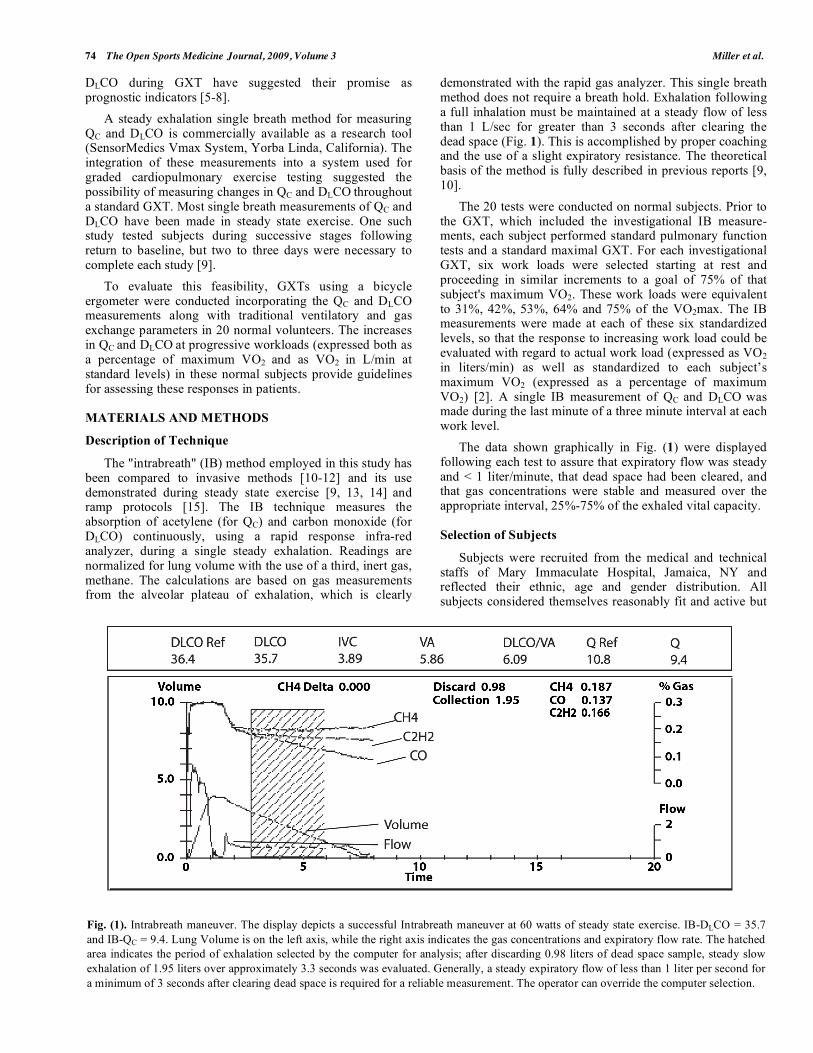
demonstrated with the rapid gas analyzer. This single breath method does not require a breath hold. Exhalation following a full inhalation must be maintained at a steady flow of less than 1 L/sec for greater than 3 seconds after clearing the dead space (Fig. 1). This is accomplished by proper coaching and the use of a slight expiratory resistance. The theoretical basis of the method is fully described in previous reports [9, 10].
The 20 tests were conducted on normal subjects. Prior to the GXT, which included the investigational IB measure-ments, each subject performed standard pulmonary function tests and a standard maximal GXT. For each investigational GXT, six work loads were selected starting at rest and proceeding in similar increments to a goal of 75% of that subject's maximum VO2. These work loads were equivalent to 31%, 42%, 53%, 64% and 75% of the VO2max. The IB measurements were made at each of these six standardized levels, so that the response to increasing work load could be evaluated with regard to actual work load (expressed as VO2 in liters/min) as well as standardized to each subject’s maximum VO2 (expressed as a percentage of maximum VO2) [2]. A single IB measurement of QC and DLCO was made during the last minute of a three minute interval at each work level.
The data shown graphically in Fig. (1) were displayed following each test to assure that expiratory flow was steady and < 1 liter/minute, that dead space had been cleared, and that gas concentrations were stable and measured over the appropriate interval, 25%-75% of the exhaled vital capacity.
Selection of Subjects
Subjects were recruited from the medical and technical staffs of Mary Immaculate Hospital, Jamaica, NY and reflected their ethnic, age and gender distribution. All subjects considered themselves reasonably fit and active but
Fig. (1). Intrabreath maneuver. The display depicts a successful Intrabreath maneuver at 60 watts of steady state exercise. IB-DLCO = 35.7
and IB-QC = 9.4. Lung Volume is on the left axis, while the right axis indicates the gas concentrations and expiratory flow rate. The hatched
area indicates the period of exhalation selected by the computer for analysis; after discarding 0.98 liters of dead space sample, steady slow
exhalation of 1.95 liters over approximately 3.3 seconds was evaluated. Generally, a steady expiratory flow of less than 1 liter per second for
a minimum of 3 seconds after clearing dead space is required for a reliable measurement. The operator can override the computer selection.
DLCO and QC During GXT The Open Sports Medicine Journal, 2009, Volume 3 75
not athletic and were free of any significant medical disorder. Informed consent was obtained and the study was conducted under the supervision of the Institutional Review Board of Mary Immaculate Hospital.
Evaluation Methods
Differences between means of QC, DLCO and VO2 at the six workloads were evaluated using t-tests, pooled or paired as appropriate. Linear regression equations describing the increase of QC and DLCO with exercise were calculated for each patient, and then averaged for the population. Variation among regression responses to exercise were evaluated using 95% confidence limits. A p<0.05 was prospectively defined as statistically significant. Unless indicated otherwise ± denotes standard deviation. All descriptive and comparative statistics were calculated using either Microsoft Excel (Microsoft Corporation, Redmond WA) or SPSS 11.0 (SPSS, Inc. Chicago, IL).
RESULTS
The demographics, pulmonary function, and GXT results of the subjects are described in Table 1. The subjects’ ages ranged between 25 and 62 years. Seven of the 20 subjects were female; 11 were of Asian origin. Subjects were of normal height and weight. Their pulmonary function was characterized as low normal [16, 17].
This is consistent with the ethnicity of a
majority of them since many Asian ethnic groups show low normal or slightly decreased values when compared to pulmonary function predicted values for Caucasians. Their fitness was typical of normal unconditioned adults with a maximum VO2 of approximately 27 ml/min/kg and a normal anaerobic threshold. Baseline data were complete for all subjects. Only 3 subjects did not complete the appropriate IB expiratory maneuver at one single stage of exercise; this was due to a temporary obstruction in the gas sample line.
Table 1. Characteristics of the 20 Subjects Performing
Graded Exercise Tests
Mean sd Range
Age 35 10 25-62
Gender % Female 35
Height (cm) 169 9 138-163
Weight (kg) 72 14 50-110
FVC (% predicted) [16] 94 12 78-107
FEV1 (% predicted) [16] 90 12 74-110
DLCO (% predicted) [17] 95 16 69-126
VO2 - max (ml/kg/min) 27 7 19-46
VO2 - max (L/min) 1.96 0.66 1.11-3.10
VO2 -max (% predicted)2 76 18 38-110
VO2-AT (ml/kg/min) 16 4 10-24
VO2- AT (L/min) 1.16 0.37 .75-1.99
VO2- AT (% predicted max) 2 46 11 22-69
HR max 167 14 150-194
AT= anaerobic threshold, HR= heart rate (beats/min).
The results of the investigational GXT for the six stages of exercise are described in Table 2. The tests covered a range from rest (VO2 = 0.35 ± 0.11 L/min) to 76% ± 13% of maximum (VO2 = 1.43 ± 0.38 L/min). There was a statistically significant increase in VO2 (p<0.001) and pulmonary capillary blood flow (p<0.05) between each successive stage of exercise. The increase in lung diffusion was significant (p<0.05) between all the stages except 2 & 3 and 4 & 5.
Table 2. Graded Exercise Test Results at Six Workloads
Stage Rest 2 3 4 5 6
% VO2 max
mean 19 32 43 55 64 76
sd 5 9 9 12 14 13
VO2 (L/min)
mean 0.35 0.59 0.81 1.02 1.20 1.43
sd 0.11 0.16 0.20 0.25 0.32 0.38
VO2 (ml/kg/min)
mean 5.0 8.4 11.4 14.4 16.9 20.2
sd 1.7 2.1 2.5 3.0 3.5 4.3
IB-QC (L/min)
mean 5.3 7.7 8.5 9.9 10.7 12.4
sd 1.5 2.2 1.9 2.1 2.5 3.1
IB-DLCO (ml/mmHg/min)
mean 21.5 25.5 26.5 29.1 30.5 32.3
sd 4.9 7.3 7.6 7.9 8.2 9.1
IB-QC= intrabreath pulmonary capillary blood flow, IB-DLCO= intrabreath lung
diffusion, sd= standard deviation.
The pulmonary capillary blood flow (QC) of the group increased more than two-fold from baseline values (95% confidence limits: 205% - 277%). The QC response to increased exercise, as determined by linear regression, was:
QC (L/min) = 3.5 + 6.5 * VO2 (L/min), (r = .81)
The lung diffusion increased about 50% above baseline (95% confidence limits: 139% - 163%). The DLCO response to increased exercise, as determined by linear regression, was:
DLCO (ml/mmHg/min) = 18.7 + 10.2 * VO2 (L/min), (r = .84)
Non linear regression did not improve the correlation for either parameter. There was no statistically significant difference in the regression with regard to gender, allowing pooling of the data. The standard error of the response to exercise (i.e., slope) for QC and DLCO was 0.6 L/min and 1.2 ml/mmHg/min respectively.
Because only one measurement of QC and DLCO could be made at each work load, we evaluated the reliability of individual measurements by comparing each of the six measurements to the regression for that subject. The mean variation from the regression for QC and DLCO were
76 The Open Sports Medicine Journal, 2009, Volume 3 Miller et al.
9.9% ± 4.6% and 6.6% ± 3.6% respectively. We also evaluated these differences to determine how many individual measurements were excessively deviant from the regression. Excessive deviation was defined to be 20% or 2 L/min for QC and 20% or 5 ml/mmHg/min for DLCO, whichever was larger. Only 4 measurements of both responses (<4% of the total) met the criteria for excessive deviation.
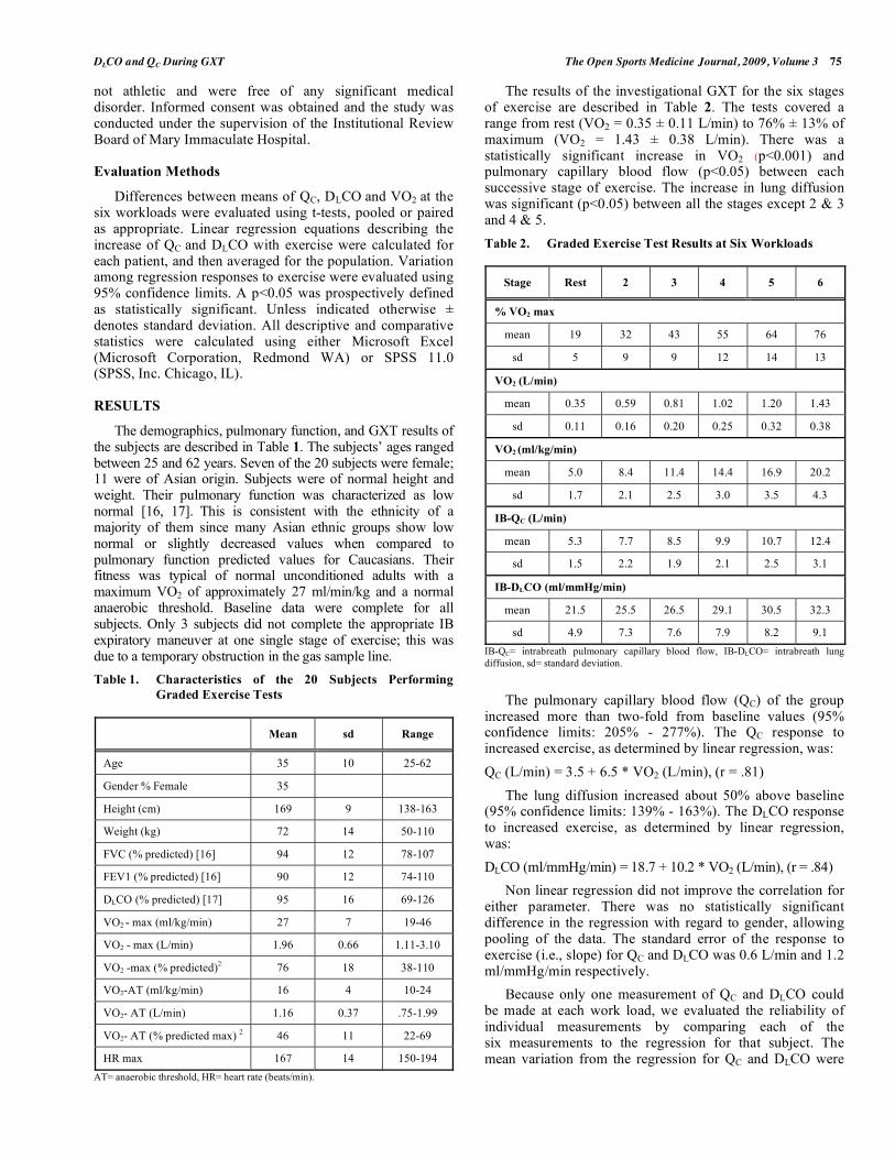
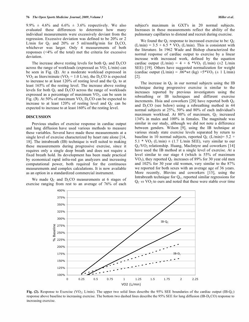
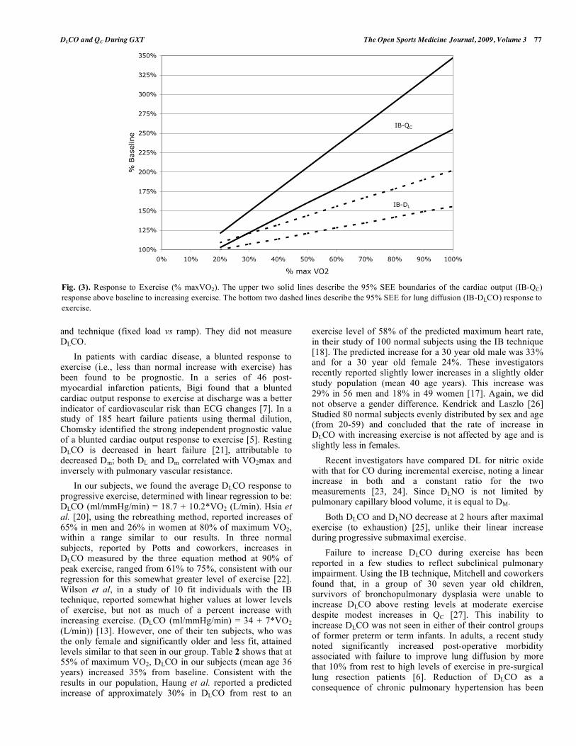
The increase above resting levels for both QC and DLCO across the range of workloads (expressed as VO2 L/min) can be seen in Fig. (2). At a moderate workload expressed in VO2 as liters/minute (VO2 = 1.0 L/m), the DLCO is expected to increase to at least 120% of resting level and the QC to at least 165% of the resting level. The increase above resting levels for both QC and DLCO across the range of workloads expressed as a percentage of maximum VO2, can be seen in Fig. (3). At 50% of maximum VO2, DLCO can be expected to increase to at least 120% of resting level and QC can be expected to increase to at least 160% of the resting level.
DISCUSSION
Previous studies of exercise response in cardiac output and lung diffusion have used various methods to measure these variables. Several have made these measurements at a single level of exercise characterized by heart rate alone [14, 18]. The intrabreath (IB) technique is well suited to making these measurements during progressive exercise, since it requires only a single deep breath and does not require a fixed breath hold. Its development has been made practical by economical rapid infra-red gas analyzers and increasing computational power, both required for the continuous measurements and complex calculations. It is now available as an option in a standardized commercial instrument.
We made QC and DLCO measurements at 6 stages of exercise ranging from rest to an average of 76% of each
subject's maximum in GXT's in 20 normal subjects. Increases in these measurements reflect the ability of the pulmonary capillaries to distend and recruit during exercise.
We found the QC response to increased exercise to be: QC (L/min) = 3.5 + 6.5 * VO2 (L/min). This is consistent with the literature. In 1962 Wade and Bishop characterized the normal response of cardiac output to exercise by a linear increase with increased work, defined by the equation cardiac output (L/min) = 4 + 6 *VO2 (L/min) (±2 L/min SEE) [19]. Others have suggested normalization for weight (cardiac output (L/min) = .06*wt (kg) +5*VO2 (± 1 L/min) [2].
The increase in QC in our normal subjects using the IB technique during progressive exercise is similar to the increases reported by previous investigators using the rebreathing or IB techniques and different exercise increments. Hsia and coworkers [20] have reported both QC and DLCO (see below) using a rebreathing method in 44 normal subjects at 25%, 50% and 80% of each individual's maximum workload. At 80% of maximum, QC increased 134% in males and 100% in females. The magnitude was similar in our study, although we did not note a difference between genders. Wilson
[9], using the IB technique at
various steady state exercise levels separated by return to baseline in 10 normal subjects, reported QC (L/min)= 5.2 + 5.1 * VO2 (L/min) ± (1.7 L/min SEE), very similar to our QC/VO2 relationship. Huang, MacIntyre and coworkers [14] have used the IB method at a single level of exercise. At a level similar to our stage 4 (which is 55% of maximum VO2), they reported QC increases of 89% for 30 year old men and 102% for 30 year old women, very similar to the 87% we reported for both sexes with an average age of 36 years. More recently, Blevins and coworkers [15], using the Intrabreath technique for QC, reported similar regressions for QC vs VO2 to ours and noted that these were stable over time
Fig. (2). Response to Exercise (VO2; L/min). The upper two solid lines describe the 95% SEE boundaries of the cardiac output (IB-QC)
response above baseline to increasing exercise. The bottom two dashed lines describe the 95% SEE for lung diffusion (IB-DLCO) response to
increasing exercise.
100%
125%
150%
175%
200%
225%
250%
275%
300%
325%
350%
375%
400%
0 0.25 0.5 0.75 1 1.25 1.5 1.75 2 2.25
VO2 (L/min)
% B
asel
ine
IB-QC
IB-DL
DLCO and QC During GXT The Open Sports Medicine Journal, 2009, Volume 3 77
and technique (fixed load vs ramp). They did not measure DLCO.
In patients with cardiac disease, a blunted response to exercise (i.e., less than normal increase with exercise) has been found to be prognostic. In a series of 46 post-myocardial infarction patients, Bigi found that a blunted cardiac output response to exercise at discharge was a better indicator of cardiovascular risk than ECG changes [7]. In a study of 185 heart failure patients using thermal dilution, Chomsky identified the strong independent prognostic value of a blunted cardiac output response to exercise [5]. Resting DLCO is decreased in heart failure [21], attributable to decreased Dm; both DL and Dm correlated with VO2max and inversely with pulmonary vascular resistance.
In our subjects, we found the average DLCO response to progressive exercise, determined with linear regression to be: DLCO (ml/mmHg/min) = 18.7 + 10.2*VO2 (L/min). Hsia et al. [20], using the rebreathing method, reported increases of 65% in men and 26% in women at 80% of maximum VO2, within a range similar to our results. In three normal subjects, reported by Potts and coworkers, increases in DLCO measured by the three equation method at 90% of peak exercise, ranged from 61% to 75%, consistent with our regression for this somewhat greater level of exercise [22]. Wilson et al, in a study of 10 fit individuals with the IB technique, reported somewhat higher values at lower levels of exercise, but not as much of a percent increase with increasing exercise. (DLCO (ml/mmHg/min) = 34 + 7*VO2 (L/min)) [13]. However, one of their ten subjects, who was the only female and significantly older and less fit, attained levels similar to that seen in our group. Table 2 shows that at 55% of maximum VO2, DLCO in our subjects (mean age 36 years) increased 35% from baseline. Consistent with the results in our population, Haung et al. reported a predicted increase of approximately 30% in DLCO from rest to an
exercise level of 58% of the predicted maximum heart rate, in their study of 100 normal subjects using the IB technique [18]. The predicted increase for a 30 year old male was 33% and for a 30 year old female 24%. These investigators recently reported slightly lower increases in a slightly older study population (mean 40 age years). This increase was 29% in 56 men and 18% in 49 women [17]. Again, we did not observe a gender difference. Kendrick and Laszlo [26] Studied 80 normal subjects evenly distributed by sex and age (from 20-59) and concluded that the rate of increase in DLCO with increasing exercise is not affected by age and is slightly less in females.
Recent investigators have compared DL for nitric oxide with that for CO during incremental exercise, noting a linear increase in both and a constant ratio for the two measurements [23, 24]. Since DLNO is not limited by pulmonary capillary blood volume, it is equal to DM.
Both DLCO and DLNO decrease at 2 hours after maximal exercise (to exhaustion) [25], unlike their linear increase during progressive submaximal exercise.
Failure to increase DLCO during exercise has been reported in a few studies to reflect subclinical pulmonary impairment. Using the IB technique, Mitchell and coworkers found that, in a group of 30 seven year old children, survivors of bronchopulmonary dysplasia were unable to increase DLCO above resting levels at moderate exercise despite modest increases in QC [27]. This inability to increase DLCO was not seen in either of their control groups of former preterm or term infants. In adults, a recent study noted significantly increased post-operative morbidity associated with failure to improve lung diffusion by more that 10% from rest to high levels of exercise in pre-surgical lung resection patients [6]. Reduction of DLCO as a consequence of chronic pulmonary hypertension has been
Fig. (3). Response to Exercise (% maxVO2). The upper two solid lines describe the 95% SEE boundaries of the cardiac output (IB-QC)
response above baseline to increasing exercise. The bottom two dashed lines describe the 95% SEE for lung diffusion (IB-DLCO) response to
exercise.
100%
125%
150%
175%
200%
225%
250%
275%
300%
325%
350%
0% 10% 20% 30% 40% 50% 60% 70% 80% 90% 100%
% max VO2
% B
asel
ine
IB-QC
IB-DL
78 The Open Sports Medicine Journal, 2009, Volume 3 Miller et al.
identified as an important factor in the limitation of peak exercise in patients with heart failure [21].
Individual intrabreath DLCO and QC measurements have considerable variability. Similar variability is found in cardiac output measured by other techniques during exercise. Average coefficient of variations of QC of 8.7% and 12% using the intrabreath method at rest were reported by Elkayam [10] and Blevins [15]. This is comparable to our experience during exercise, an average deviation from the regression of 9.9% and 6.6% for QC and DLCO respectively. We also found that less than 4% of the individual measurements were excessively deviant. Unfortunately only a single IB measurement of DLCO and QC can be made at each level of exercise when using incremental protocols with stages of 3 minutes or less. Variability can be addressed in two ways. It can be averaged out by looking at the trend in repeated single measurements with increasing exercise as was done in this study or by averaging repeated measurements at fewer more prolonged workloads. Some bias will exist between the IB QC, which measures unshunted pulmonary blood flow, and true cardiac output. This bias should be considered in evaluating the results in abnormal populations with the potential for intra cardiac and pulmonary shunts.
We presented our DLCO and QC results in three different metrics. The first approach presented the actual DLCO and QC values increasing as a function of the metabolic workload (VO2 L/min). The other two approaches compared percentage increases from baseline in DLCO and QC with incremental exercise to absolute workload (VO2 L/min) and to percent of individual maximum. The latter approach was undertaken to adjust for differences in exercise capacity associated with age, size and gender. Use of response above baseline, while appealing, is limited by the need for careful baseline measurement.
Based on these limitations it would appear that assessment of the slope of the response to exercise (QC/VO2 and DLCO/VO2) would be the most useful approach to interpretation. This approach is well suited to making single repeated measurements during an incremental GXT as used in this investigation and by Chomsky [5] or at several levels of steady state exercise as advocated by Jones [2]. Alternatively measurements at rest and at one level of moderate exercise might be used as a simple assessment of response in these variables. Our data show that DLCO increased by at least 20% and QC by at least 50% from resting levels at 50% of maximum, suggesting that we can apply these guidelines to simplify exercise testing. These findings are consistent with the two studies by the Duke group [15, 18]. Using measured VO2 rather than percentage maximum, similar increases can be expected at 0.9 L/min VO2. Several measurements can be made at such a workload to eliminate an aberrant value. Furthermore, these workloads are generally below the anaerobic threshold and thus easily tolerated by most subjects.
CONCLUSIONS
We found the intrabreath procedure easy to integrate into our clinical GXT. Furthermore our results with normal volunteers during incremental exercise, a methodology well suited to clinical practice, were consistent with published
expectations from research protocols using steady state exercise requiring multiple days to complete. Our findings are acceptably reliable to use for clinical reference. While a strong physiological rationale supports the likely prognostic potential of these measurements, there is a paucity of clinical data. We intend, and recommend to others, to include these measurements in the standard diagnostic GXT so that information on pulmonary blood flow and lung diffusion response to exercise in various abnormal populations can be fully characterized.
CONFLICT OF INTEREST
Statistical analysis was supported by VIASYS Healthcare Corporation, Yorba Linda CA through a grant to Mr. Bachman.
REFERENCES
[1] Wasserman K, Whipp B. Exercise physiology in health and
disease. Am Rev Respir Dis 1975; 112: 219-49. [2] Jones N. Clinical exercise testing. 4th ed. Philadelphia PA: WB
Saunders Company 1997. [3] Miller A, Bhuptani A, Sloane MF, Brown LK, Teirstein AS.
Cardiorespiratory responses to incremental exercise in patients with asbestos-related pleural thickening and normal or near normal lung
function. Chest 1993; 103: 1045-50. [4] Miller A, Brown LK, Sloane MF, Bhuptani A, Teirstein AS.
Cardiorespiratory responses to incremental exercise in sarcoidosis patients with normal spirometry. Chest 1995; 107: 323-29.
[5] Chomsky D, Lang C, Rayos G, et al. Hemodynamic exercise testing, a valuable tool in the selection of cardiac transplant
candidates. Circulation 1996; 94: 3176-83. [6] Wang JS, Abboud RT, Evans KG, Finley RJ, Graham BL. Role of
diffusing capacity during exercise in preoperative evaluation for lung resection. Am J Respir Crit Care Med 2000; 162: 1435-44.
[7] Bigi R, Desideri A, Rambaldi R, Cortigiani L, Sponzilli C, Fiorentini C. Angiographic and prognostic correlates of cardiac
output by cardiopulmonary exercise testing in patients with anterior myocardial infarction. Chest 2001; 120:825-33.
[8] Williams SG, Cooke GA, Wright DJ, et al. Peak exercise cardiac power output; a direct indicator of cardiac function strongly
predictive of prognosis in chronic heart failure. Eur Heart J 2001; 22: 1496-03.
[9] Wilson AF, Savariryan S, James N, Mukai D, Nishimura E. Almost simultaneous measurement of cardiovascular and gas exchange
variables during maximal exercise. Med Sci Sports Exer 1996; 28: 436-43.
[10] Elkayam U, Wilson A, Morrison J, et al. Non-invasive measurement of cardiac output by a single breath constant
expiratory technique. Thorax 1984; 39: 107-13. [11] Zenger M, Brenner M, Haruno M, Mahon D, Wilson A.
Measurement of cardiac output by an automated single breath technique and simultaneous comparison with thermal dilution and
Fick methods in cardiac patients. Am J Cardiol 1993; 71:105-9. [12] Sadeh JS, Miller A, Kukin M. Noninvasive measurement of cardiac
output by acetylene uptake technique and simultaneous comparison with thermal dilution in ICU patients. Chest 1997; 111: 1295-300.
[13] Ramage JE Jr, Coleman RE, MacIntyre NR. Rest and exercise cardiac output and diffusing capacity assessed by a single slow
exhalation of methane, acetelyne and carbon monoxide. Chest 1987; 92:44-50.
[14] Huang YT, Helms MJ, MacIntyre NR. Normal values for single exhalation diffusing capacity and pulmonary capillary blood flow
in sitting, supine positions and during mild exercise. Chest 1994; 105: 501-8.
[15] Blevins JS, Herbert WG, Zedalis D, Cross L. Reliability of a single-breath acetylene cardiac output technique during exercise:
ramping vs fixed load protocols. Am J Med Sports 2003; 5: 256-61. [16] Miller A, Thornton JC, Warshaw R, Bernstein J, Teirstein AS,
Selikoff IJ. Mean and instantaneous expiratory flows, FVC and FEV1: prediction equations for non smokers and smokers from a
DLCO and QC During GXT The Open Sports Medicine Journal, 2009, Volume 3 79
random sample of Michigan, a large industrial state. Bull
Physiopath Respir 1986; 22: 589-97. [17] Miller A, Thornton JC, Warshaw R, Anderson H, Selikoff IJ,
Teirstein AS. Single breath diffusing capacity in a representative sample of the population of Michigan, a large industrial state:
predicted values, lower limits of normal, frequencies of abnormality by smoking history. Am Rev Respir Dis 1983; 127;
270-77. [18] Huang YT, O'Brien SR, MacIntyre NR. Intrabreath diffusing
capability of the lung in healthy individuals at rest and during exercise. Chest 2002; 122: 177-85.
[19] Wade OL, Bishop JM. Cardiac output and regional blood flow. FA Davis Philadelphia: Blackwell Scientific Publication 1962.
[20] Hsia CW, McBrayer DG, Ramanathan M. Reference values of pulmonary diffusing capacity during exercise by a rebreathing
technique. Am J Respir Crit Care Med 1995; 152: 658-65. [21] Puri S, Baker BL, Dutka DP, Oakley CM, Hughes JMB, Cleland
JGF. Reduced alveolar-capillary membrane diffusing capacity in chronic heart failure: its pathophysiological relevance and
relationship to exercise performance. Circulation 1995; 91: 2769 -
74. [22] Potts J, Abboud R, Graham B. The use of the three equation
method to measure DLCOSB during exercise in normal healthy adults. Am J Respir Crit Care Med 1996; 153: A650 [abstract].
[23] Zavorsky GS, Quiron KB, Massarelli PS, et al. The relationship between single-breath diffusion capacity of the lung for nitric oxide
and carbon monoxide during various exercise intensities. Chest 2004; 125: 1019-27.
[24] Borland C, Mist B, Zammit M, Vuylsteke A. Steady-state measurement of NO and CO lung diffusing capacity on moderate
exercise in men. J Appl Physiol 2001; 90: 538-44. [25] Zavorsky GS, Lands LC. Lung diffusion capacity for nitric oxide
and carbon monoxide is impaired similarly following short-term graded exercise. Nitric Oxide 2005; 12: 31-8.
[26] Kendrick AH, Laszlo G. CO Transfer factor on exercise: age and sex differences. Eur Respir J 1990; 3: 323-8.
[27] Mitchell S, Teague W. Reduced gas transfer at rest and during exercise in school aged survivors of bronchopulmonary dysplasia.
Am J Respir Crit Care Med 1998; 157: 1406-12.
Received: June 19, 2009 Revised: August 3, 2009 Accepted: October 8, 2009
© Miller et al.; Licensee Bentham Open.
This is an open access article licensed under the terms of the Creative Commons Attribution Non-Commercial License (http://creativecommons.org/licenses/by-nc/3.0/) which permits unrestricted, non-commercial use, distribution and reproduction in any medium, provided the work is properly cited.