mechanical operation and intersubunit …liu.rockefeller.edu/assets/file/liu_bj_2014.pdfthe chemical...
TRANSCRIPT

1844 Biophysical Journal Volume 106 May 2014 1844–1858
Mechanical Operation and Intersubunit Coordination of Ring-ShapedMolecular Motors: Insights from Single-Molecule Studies
Shixin Liu,†‡ Gheorghe Chistol,†§ and Carlos Bustamante†‡§{*†Jason L. Choy Laboratory of Single-Molecule Biophysics, ‡California Institute for Quantitative Biosciences, §Department of Physics, and{Department of Molecular and Cell Biology, Department of Chemistry, Howard Hughes Medical Institute, and Kavli Energy NanoSciencesInstitute, University of California, Berkeley, and Lawrence Berkeley National Laboratory, Berkeley, California
ABSTRACT Ring NTPases represent a large and diverse group of proteins that couple their nucleotide hydrolysis activity to amechanical task involving force generation and some type of transport process in the cell. Because of their shape, these en-zymes often operate as gates that separate distinct cellular compartments to control and regulate the passage of chemical spe-cies across them. In this manner, ions and small molecules are moved across membranes, biopolymer substrates aresegregated between cells or moved into confined spaces, double-stranded nucleic acids are separated into single strands toprovide access to the genetic information, and polypeptides are unfolded and processed for recycling. Here we review the recentadvances in the characterization of these motors using single-molecule manipulation and detection approaches. We describethe various mechanisms by which ring motors convert chemical energy to mechanical force or torque and coordinate the activ-ities of individual subunits that constitute the ring. We also examine how single-molecule studies have contributed to a betterunderstanding of the structural elements involved in motor-substrate interaction, mechanochemical coupling, and intersubunitcoordination. Finally, we discuss how these molecular motors tailor their operation—often through regulation by othercofactors—to suit their unique biological functions.
INTRODUCTION
Cells are nonequilibrium systems that constantly consumeand dissipate energy to maintain or undergo transitionsamong steady states. Most essential cellular processes areunidirectional and are carried out by nanometer-scalemachine-like devices that operate as molecular motors,coupling a downhill chemical reaction to a mechanicaltask (1–3). The chemical free energy may come fromsubstrate binding, bond hydrolysis, or electrochemicalgradients. These microscopic engines differ from theirmacroscopic counterparts in that they operate at energiesonly marginally higher than the energy of the thermalbath, and thus, thermal fluctuations play an important rolein their operation. How these molecular machines carryout their cellular functions is of great interest to biologistsand biophysicists.
Many molecular motors display ring-shaped architecturesand consist of multiple subunits. In these ring motors,each individual subunit can couple its mechanical func-tion to the energy source and one central question is todetermine how the activities of these subunits are coordi-nated during the operation of the ring. A major fraction ofring motors belong to the Additional Strand ConservedE (ASCE) superfamily of NTPases, utilizing the energyfrom nucleotide binding and hydrolysis to catalyze a
Submitted January 31, 2014, and accepted for publication March 19, 2014.
*Correspondence: [email protected]
Shixin Liu and Gheorghe Chistol contributed equally to this work.
Gheorghe Chistol’s present address is Department of Biological Chemistry
and Molecular Pharmacology, Harvard Medical School, Boston, MA.
Editor: E. Ostap.
� 2014 by the Biophysical Society
0006-3495/14/05/1844/15 $2.00
diversity of cellular activities, including ATP synthesis,DNA translocation, duplex unwinding, and protein unfold-ing (4,5) (Fig. 1).
Various experimental approaches have been used toinvestigate the operating mechanisms of molecularmotors. High-resolution structures, especially those con-taining both the nucleotide-bound motor and its mechani-cal substrate, provide detailed albeit static snapshots ofmotor operation (6–9). Bulk biochemical methods canoffer dynamic information about motor function, butthey average the molecular signals over ensembles. Thisaveraging often obscures crucial mechanistic featuresof the motor such as transient intermediates or parallelreaction pathways. Single-molecule techniques haveyielded tremendous insights into various biological pro-cesses by enabling researchers to follow the dynamics ofindividual motor complexes in real time. Quantitiesdescribing molecular machines, such as displacement,force, torque, and rate, can be directly measured with sin-gle-molecule methods, making them ideally suited formotor studies.
There are two broad classes of single-molecule tech-niques: fluorescence-based detection (single particletracking, single-molecule fluorescence resonance energytransfer, DNA curtains, etc.), and force-based manipulation(optical tweezers, magnetic tweezers, atomic force micro-scopy, flow stretching, etc.). Comprehensive reviews ofthese methods can be found in the literature (10–19).Recently, high-speed atomic force microscopy in anaqueous environment was added to the single-moleculetoolbox (20), and this new methodology has already begun
http://dx.doi.org/10.1016/j.bpj.2014.03.029

E1 Helicase Rho Helicase
F1-ATPaseClpX Unfoldase
D5
BC4
A21 3
WA
RF
WB
CE
2 nm
A
B
DnaB Helicase FtsK Translocase
ADP
AMP-PNP
Empty
αβ
FIGURE 1 The architecture of ring-shapedmotors from the ASCE super-
family of NTPases. (A) A crystal structure gallery of representative ASCE
ring motors. The bovine papillomavirus replicative helicase E1 with a
ssDNA substrate (PDB:2GXA) (7); The E. coli transcription termination
factor Rho helicase with ssRNA (PDB: 3ICE) (8); The Bacillus stearother-
mophilus replicative helicase DnaB with ssDNA (PDB: 4ESV) (9); The mo-
tor domain of the Pseudomonas aeruginosa dsDNA translocase FtsK (PDB:
2IUU) (94); The E. coli protein unfoldase and polypeptide translocase ClpX
(PDB: 3HWS) (110); The bovine mitochondrial F1-ATPase with the three
catalytic sites encircled (a-subunits shown in green, b in red, and g in
blue) (PDB: 1BMF) (6). In all structures, nucleotides and their analogs
are bound at the interface between adjacent subunits (black). Note that
with the exception of the F1-ATPase, each ring consists of identical subunits
that are colored differently for clarity. (B) Diagram of the core ASCE fold
(modified from Lyubimov et al. (28)). WA, Walker A motif; WB, Walker B
motif; CE, catalytic glutamate; RF, arginine finger. Note that the positions
of CE and RF may vary in different motors and only the most common lo-
cations are shown.
Single-Molecule Studies on Ring Motors 1845
to provide remarkable real-time movies of motor proteins inaction (21–23). Moreover, new hybrid instruments thatcombine single-molecule fluorescence and force micro-scopy carry great potential for simultaneously observingmultiple aspects of motor dynamics (24–26).
The structure and function of ring-shaped molecularmotors have been extensively reviewed (5,27–30). We willlimit the scope of this review to single-molecule studieson ASCE ring NTPases. In particular, we will cover thefollowing motors in detail:
1. Packaging motors that pump viral genomes into pre-formed proteinaceous capsids during the assemblyof double-stranded DNA (dsDNA) viruses (31,32)(Fig. 2 A);
2. Replicative helicases, ubiquitous enzymes that catalyzestrand separation of basepaired nucleic acids at the repli-cation fork (33) (Fig. 2 B);
3. FtsK and SpoIIIE, membrane-bound and septum-local-ized DNA translocases involved in bacterial chromo-some segregation in Escherichia coli and Bacillussubtilis, respectively (34) (Fig. 2 C);
4. F1-ATPase, the soluble portion of the F0F1-ATP synthasethat utilizes a trans-membrane proton gradient to pro-duce ATP through the rotation of the central g-subunitinside a cylinder made of three ab dimers (35,36)(Fig. 2 D); and
5. ClpX, a bacterial unfoldase that unravels proteins andtranslocates unfolded polypeptides into the associatedpeptidase ClpP for degradation (37) (Fig. 2 E).
BASIC MOTOR PROPERTIES
Velocity
The velocity of a molecular motor, v, is given by v ¼ d �kcat � ε, where d is the step size, kcat is the rate of chemicalcatalysis, and ε is the coupling coefficient, i.e., the pro-bability that the motor takes a mechanical step per chem-ical cycle. For NTPases, kcat, and hence v, depends onthe nucleotide concentration. The motor velocity alsodepends on externally applied loads in single-moleculeforce-manipulation assays, which can shed light on theforce-generating mechanism of the motor (discussed laterin this article).
The velocity varies greatly among different motors. FordsDNA translocases, the fastest known motor is FtsK, whichtranslocates DNA at ~5 kbp,s�1 at room temperature(38,39), going up to a remarkable ~17 kbp,s�1 at 37�C(40). The bacteriophage 429 packaging motor has amaximum velocity of ~120 bp,s�1 at room temperature(41,42). By comparison, the bacteriophage l motor pack-ages DNA at ~600 bp,s�1 (43), whereas the bacteriophageT4 motor can reach 2 kbp,s�1 (44). The translocation veloc-ity of viral packaging motors scales roughly with the
Biophysical Journal 106(9) 1844–1858
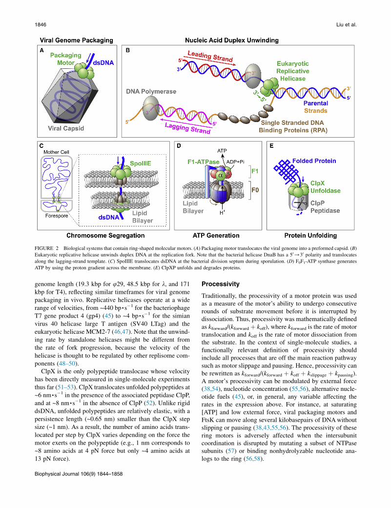
FIGURE 2 Biological systems that contain ring-shaped molecular motors. (A) Packaging motor translocates the viral genome into a preformed capsid. (B)
Eukaryotic replicative helicase unwinds duplex DNA at the replication fork. Note that the bacterial helicase DnaB has a 50/30 polarity and translocates
along the lagging-strand template. (C) SpoIIIE translocates dsDNA at the bacterial division septum during sporulation. (D) F0F1-ATP synthase generates
ATP by using the proton gradient across the membrane. (E) ClpXP unfolds and degrades proteins.
1846 Liu et al.
genome length (19.3 kbp for 429, 48.5 kbp for l, and 171kbp for T4), reflecting similar timeframes for viral genomepackaging in vivo. Replicative helicases operate at a widerange of velocities, from ~440 bp,s�1 for the bacteriophageT7 gene product 4 (gp4) (45) to ~4 bp,s�1 for the simianvirus 40 helicase large T antigen (SV40 LTag) and theeukaryotic helicase MCM2-7 (46,47). Note that the unwind-ing rate by standalone helicases might be different fromthe rate of fork progression, because the velocity of thehelicase is thought to be regulated by other replisome com-ponents (48–50).
ClpX is the only polypeptide translocase whose velocityhas been directly measured in single-molecule experimentsthus far (51–53). ClpX translocates unfolded polypeptides at~6 nm,s�1 in the presence of the associated peptidase ClpP,and at ~8 nm,s�1 in the absence of ClpP (52). Unlike rigiddsDNA, unfolded polypeptides are relatively elastic, with apersistence length (~0.65 nm) smaller than the ClpX stepsize (~1 nm). As a result, the number of amino acids trans-located per step by ClpX varies depending on the force themotor exerts on the polypeptide (e.g., 1 nm corresponds to~8 amino acids at 4 pN force but only ~4 amino acids at13 pN force).
Biophysical Journal 106(9) 1844–1858
Processivity
Traditionally, the processivity of a motor protein was usedas a measure of the motor’s ability to undergo consecutiverounds of substrate movement before it is interrupted bydissociation. Thus, processivity was mathematically definedas kforward/(kforward þ koff), where kforward is the rate of motortranslocation and koff is the rate of motor dissociation fromthe substrate. In the context of single-molecule studies, afunctionally relevant definition of processivity shouldinclude all processes that are off the main reaction pathwaysuch as motor slippage and pausing. Hence, processivity canbe rewritten as kforward/(kforward þ koff þ kslippage þ kpausing).A motor’s processivity can be modulated by external force(38,54), nucleotide concentration (55,56), alternative nucle-otide fuels (45), or, in general, any variable affecting therates in the expression above. For instance, at saturating[ATP] and low external force, viral packaging motors andFtsK can move along several kilobasepairs of DNAwithoutslipping or pausing (38,43,55,56). The processivity of thesering motors is adversely affected when the intersubunitcoordination is disrupted by mutating a subset of NTPasesubunits (57) or binding nonhydrolyzable nucleotide ana-logs to the ring (56,58).

Single-Molecule Studies on Ring Motors 1847
Directionality
Ring motors often exhibit directionality in the movementrelative to their substrate. For single-stranded nucleic acidtranslocases, the intrinsic asymmetry of the substrate cangive rise to the unidirectional translocation, and the struc-tural basis of these motors’ directionality has begun to beunderstood (8,28). Double-stranded nucleic acid translo-cases and helicases can acquire directionality by trackingonly one of the two strands. It is possible that this direction-ality is dictated by the initial orientation in which the motoris loaded onto the substrate track, using in some cases the ds/ss junction (e.g., the E1 helicase) (59) or specific recognitionsequences (e.g., KOPS for FtsK, SRS for SpoIIIE) (60–62).
Interestingly, FtsK and SpoIIIE can reverse directionalityduring translocation (38–40). One possible model toexplain such reversals is that each motor complex consistsof two coaxial rings arranged opposite to each other on theDNA. In this model, only one ring is active at any giventime; a direction reversal would then require turning offthe active ring and switching on the partner ring uponencountering a recognition sequence with a nonpermissiveorientation (63). Alternatively, direction can be reversed bya single ring dissociating and then immediately rebindingin the opposite orientation to resume translocation (60).Notably, protein unfoldases like ClpX do not possess anintrinsic translocation polarity and can efficiently movetheir polypeptide substrates in either direction (64). Thepolarity of ClpX is dictated by the placement of an 11-aarecognition tag (ssrA) on proteins targeted for degradation(65,66).
Stall force
The stall force of a molecular motor, Fstall, is defined as theforce at which the motor velocity drops to zero. Someparticularly strong motors, such as viral packaging motorsand FtsK, can operate against forces as high as ~60 pN(39,41,43,44), forces beyond which B-form DNA undergoesan overstretching phase transition (67). We note that themeasured stalled forces are often not the thermodynamicstall forces (i.e., the maximum force that the motor can exertgiven the free energy available for a particular step size), butoperational stall forces at which the substrate deforms or themotor slips or pauses indefinitely. In principle, the true ther-modynamic stall force can be determined by measuring theforce beyond which the motor reaction runs in reverse, asdemonstrated by studies in which F1-ATPase was turnedinto an ATP synthase by mechanically forcing the g-subunitto rotate in the opposite direction, generating ATP fromADP and Pi (68,69). The mechanochemical reversibilitymay not be always experimentally attainable due to limitsfor the maximum force that can be sustained by the motorand/or limits for the maximum [NDP] and [Pi] in thesolution.
Step size
The step size of a motor, d, is defined as its net displace-ment along the substrate per catalytic event. Step sizesof several ASCE ring NTPases were inferred fromatomic-resolution structures of the motor cocrystallizedwith its substrate and bound nucleotides (7–9). Recentadvances in single-molecule techniques allow the directobservation of discrete steps for many molecular motors.The step size of the rotary F1-ATPase was measured to be120� per ATP (70). The 429 packaging motor was foundto translocate DNA in 10-bp bursts, each consisting offour 2.5-bp steps (71). The T7 gp4 helicase unwinds duplexDNA in 2–3 bp bursts, likely with an elementary stepsize of 1 nt per nucleotide hydrolysis (72). Motor stepsize can be modulated by external load (73) or other regu-latory factors (74).
Although the step size of ultrafast translocases such asFtsK is not directly measurable with current techniques,several alternative strategies can be employed to overcomethis limitation:
1. Single-molecule noise spectra were used to infer FtsK’sstep size at 12 5 2 bp (38). Unfortunately, this estimaterelies on untested assumptions and must be interpretedwith caution.
2. Thermodynamic arguments can be used to estimate anupper bound for a motor’s step size. Because FtsK canexert at least 60 pN of force and the free energy ofATP hydrolysis is ~100 pN,nm in the standard transloca-tion buffer, FtsK’s step size cannot exceed ~1.7 nm(~5 bp).
3. Step size can be inferred by measuring the DNA translo-cation and ATP consumption rates in bulk, which yieldsan estimate of ~2 bp per ATP for FtsK (62). However,this ensemble method may lead to an underestimate ofthe step size if the motor undergoes frequent slippingor futile hydrolysis events.
MECHANOCHEMICAL COUPLING
The term ‘‘mechanochemistry’’ represents the mechanismby which a molecular motor couples chemical energy tomechanical force and displacement. Below we discuss thecommon experimental strategies used in single-moleculestudies to dissect the mechanochemical coupling of molec-ular motors.
Force-velocity dependence
Like chemical products, force can also be considered aproduct of the reaction. Thus, an opposing external forcecan function as an inhibitor of the reaction, whereasan assisting force can function as an activator. Underconditions where the translocation step (by definition
Biophysical Journal 106(9) 1844–1858

1848 Liu et al.
associated with a force-dependent rate constant) becomesrate-limiting for the reaction, the motor velocity becomesforce-sensitive. For a mechanochemical process with asingle force-dependent step per cycle, the force-velocityrelationship follows a Boltzmann equation:
vðFÞ ¼ v0 � ð1þ AÞ=½1þ A � expðFd=kBTÞ�:Here v0 is the velocity at zero force, kBT is the thermal energy,A is a constant describing the degree to which the force-
dependent transition is rate-limiting at zero force (if A � 1,the force-independent transitions are rate-limiting; if A R1, the force-dependent step is rate-limiting), and d is the dis-tance from the ground state to the transition state (75). Thevalue d can be thought of as the distance the motor mustmove along the translocation coordinate before committingto a step. By fitting force-velocity data to the Boltzmannequation, d was estimated at ~0.1 nm for the 429 packagingmotor (56) and at ~0.7 nm for ClpX (51)— corresponding to~13% and 70% of the respective motor’s step size.Interestingly, some motors display a complicated force-velocity dependence that cannot be explained by a singleforce-dependent step. For example, FtsK exhibits a velocityplateau at small forces, with increasing velocities at largeassisting forces and decreasing velocities at large opposingforces (38); the velocity of the bacteriophage l packagingmotor is force-sensitive at low forces but becomes force-insensitive at high forces (43). To rationalize these observa-tions, one must consider mechanochemical models withmultiple force-generating transitions or multiple kineticroutes.
TABLE 1 Dependence of Michaelis-Menten parameters on
perturbations such as an external force applied to the motor
Michaelis-Menten kinetics
A motor’s force-velocity dependence at various concentra-tions of nucleotide and hydrolysis products provides valu-able insight into its force-generating mechanism. Thereaction pathway of an NTPase typically includes NTPbinding, hydrolysis, product release, and mechanical move-ment that is coupled to certain chemical steps. When theoverall reaction is largely irreversible, the average NTPasevelocity follows the Michaelis-Menten equation,
v ¼ Vmax � ½NTP�=ðKm þ ½NTP�Þ;where Vmax is the maximum velocity at saturating [NTP]
(see also Eq. 1 in text)
Location of the affected kinetic transition Vmax Km Vmax/Km
ATP docking (k51) Same Modified Modified
Connected to docking
(k52, . ki, k5(jþ1), . k5n)
Modified Modified Modified
Separate kinetic block (k5(iþ1), . kj) Modified Modified Same
A general chemical cycle is separated into different kinetic blocks by irre-
versible transitions (ki and kj). How Vmax, Km, and Vmax/Km change with
force depends on where the modified kinetic transition is located within
the cycle.
and Km is the nucleotide concentration at which the velocityis half of Vmax. As shown below, a general chemical cyclecan be divided into distinct kinetic blocks—regions ofcontiguous reversible kinetic transitions separated by irre-versible kinetic transitions:
M1 þ T4k1½ATP�k�1
M24k2
k�2
/Mi!ki Miþ14kiþ1
k�ðiþ1Þ
/Mj!kj
Mjþ14kjþ1
k�ðjþ1Þ/Mn4
kn
k�n
M1:(1)
Biophysical Journal 106(9) 1844–1858
It can be shown thatKm is a function of all of the kinetic ratesin the cycle, Vmax is a function of all of the rates except theNTP docking and undocking rates, and Vmax/Km is a functionof only the rates within the block that contains NTP docking(76). By quantifying how the application of mechanicalforce affects Vmax, Km, and Vmax/Km, one can determinewhether the force-dependent transition is ATP docking,within the same kinetic block as ATP docking, or in aseparate kinetic block (Table 1). This analysis can alsobe used to determine which portion of the cycle is affectedby other perturbations such as a point mutation.
This analysis was applied to study the mechanochemicalcoupling in the 429 packaging motor and the ClpX unfol-dase (53,56). In both cases, Vmax and Km decrease withincreasing force, yet the ratio Vmax/Km remains constant.As a result, in conditions where ATP binding is rate-limiting([ATP] < Km), the velocity (v z (Vmax/Km) � [ATP]) doesnot depend on force. These observations rule out a schemein which ATP docking drives translocation. The observationthat Vmax/Km is independent of force also suggests that theforce-generating chemical transition is not within thesame kinetic block as ATP docking, which includes the irre-versible ATP tight-binding step and the reversible ADPrelease step (53,56,58,71). Thus, force generation is coupledto either ATP hydrolysis or Pi release. It is unlikely thatnucleotide hydrolysis results in a significant free energydrop along the reaction coordinate (77), whereas the releaseof phosphate involves a large change in free energy and isessentially irreversible because high concentrations of Pihave no measurable effect on velocity (53,56). Therefore,Pi release is the most likely candidate to power substratetranslocation in the 429 ATPase gp16 and ClpX. In contrast,F1-ATPase executes two power strokes in each hydrolysiscycle: ATP binding drives the initial 80� rotation of each120� step and Pi release drives the remaining 40� rotation(77–79).
Passive and active unwinding mechanisms ofhelicases
Helicases are enzymes that separate double-stranded nucleicacids into single strands and play crucial roles during all

Single-Molecule Studies on Ring Motors 1849
stages of nucleic acid metabolism (80–82). The mechanismby which a helicase unwinds a duplex is classified as‘‘passive’’ or ‘‘active’’ (82–84). Passive helicases rectifythermally driven fluctuations of the nucleic acid duplex bytranslocating and binding to the newly exposed singlestrand. In contrast, active helicases destabilize the duplexnear the junction and shift the close4open duplex equilib-rium. The activeness of a helicase can be quantified by theamount of destabilization energy it contributes per basepairunwound, DGd (85). DGd ¼ 0 for a purely passive helicaseand DGd ¼ 3.4 kBT (G–C base-pairing energy) for an opti-mally active helicase. Optical tweezers and magnetic twee-zers were used to analyze how unwinding velocity dependson external force and substrate sequence (G–C versusA–T/U). Activeness varies widely among different heli-cases, from the mostly passive phage T4 gp41 (DGd ¼0.05 kBT) (86), the partially active phage T7 gp4 (DGd ¼1–2 kBT) (87) and E. coli DnaB (DGd ¼ 0.5–1.6 kBT)(54), to the highly active non-ring-shaped helicases suchas hepatitis C virus NS3 (DGd ¼ 2.5 kBT) (88). However,caution should be exercised when interpreting these variednumbers as the reflection of distinct unwinding mecha-nisms, because they may also be due to differences in themodeling approaches and experimental geometries used indifferent studies (54,81,89).
INTERSUBUNIT COORDINATION
The coordination between the catalytic cycles of individ-ual subunits is a key feature in the operation of ringNTPases. Whereas each ring subunit is often capable ofhydrolyzing NTP and undergoing the associated mechani-cal transition, the functionality of the ring depends on themanner in which the activities of individual subunits areorchestrated. Structural and bulk biochemical studieshave suggested various models of intersubunit coordina-tion for ring motors. In the sequential model, subunitsbind and hydrolyze nucleotides in a well-defined order,most likely ordinally around the ring (7,8,90–95). In theconcerted model, all subunits simultaneously bind andhydrolyze nucleotides and release products, such thatthey are always at the same nucleotide state (96). In thestochastic model, each subunit undergoes its own catalyticcycle independently of the others (97). Single-moleculestudies, as presented in detail below, have provided freshinsights into the existing models and revealed novel coor-dination schemes.
F1-ATPase
Rotation of the g-subunit was first observed via an opticalmicroscope by attaching an actin filament to the g-rotor(98). At sufficiently low ATP concentrations, the rotationwas found to proceed in 120� steps (70) (Fig. 3 A), consis-tent with the threefold symmetry of the (ab)3 hexamer.
Using a 40-nm gold bead that generates a much lower hy-drodynamic drag than the actin filament, the 120� stepwas further resolved into 80� and 40� substeps (79). Theduration of the dwells preceding the 80� substeps isinversely proportional to [ATP] (79), indicating that ATPbinding occurs during these dwells.
Hydrolysis of ATP likely occurs in the dwell precedingthe 40� substeps, because this dwell is prolonged in a hydro-lysis-deficient mutant (b-E190D) or in the presence of ATP-g-S in the solution (99). Furthermore, experiments withCy3-ATP, a fluorescent ATP analog that is presumably hy-drolyzed slowly, allowed the simultaneous observation ofrotation and ATP binding. It suggested that the ATP thatwas bound when the g-subunit is at 0� is hydrolyzed whenthe g-subunit points at 200� (100). Thus, the dwells beforethe 80� substep and the 40� substep are termed the ‘‘ATP-binding dwell’’ and the ‘‘catalytic dwell’’, respectively(Fig. 3A).
The addition of Pi to the reaction mixture specificallyprolongs the catalytic dwell, and very high [Pi] induces fre-quent reversals of 40� substeps (78). These results stronglyindicate that Pi release drives the 40
� rotation. However, theexact timing of Pi release cannot be determined unambigu-ously: it may occur either immediately after hydrolysis dur-ing the same catalytic dwell (scenario 1), or during the nextcatalytic dwell after 120� rotation (scenario 2). A later studyshowed that there is an open nucleotide-binding site(asterisk in Fig. 3 A) freely accessible to ATP in the solutionand that free Pi from the solution competes with the nucle-otide binding, inconsistent with scenario 2 in which productPi is retained in this site and thus supporting scenario 1(101).
Finally, experiments monitoring the binding and releaseof Cy3-ATP (78), together with experiments monitoringthe site occupancy (number of nucleotides bound to thethree catalytic sites) (101), suggested that ADP release oc-curs during the 80� substep from 240� to 320� and possiblyhelps drive the 80� rotation, in addition to ATP binding.
To summarize, for each catalytic site, ATP hydrolysis, Pirelease, and ADP release occur at 200�, 200�, and 240� afterATP binding, respectively (Fig. 3 A). Although chemicalevents in the three catalytic sites occur sequentially, theF1-ATPase employs a much more intricate coordinationscheme than the canonical sequential model. All three cata-lytic sites participate to drive each 120� rotation of the g-subunit, and each site is involved in three consecutive120� steps (102). The model for F1 is the first and still oneof the most comprehensively characterized intersubunit co-ordination schemes for a ring NTPase. However, this modelis not without controversies. Some studies suggested that Piis released at 320� instead of 200�, thus after ADP isreleased (103,104). Additionally, under certain conditionswith a slow hydrolysis mutant, rotation was found to pro-ceed in steps of 120� and no 80� or 40� substeps wereobserved (105), a phenomenon also found in the related
Biophysical Journal 106(9) 1844–1858
B
ATP BindingADP Release
ATP HydrolysisPi Release 120 (binding angle)o
80 (catalytic angle)o
Time
Ang
leR
otor
A
ATP BindingADP Release
ATP
ADP
ADPATP
ATP
ADPPi. *
PiADP
ATPATP
ADP
ATP
*
ADPPi.
Pi
ATP
ATP
ADP
ADP *ATP
ADPPi.
Pi
0 (binding angle)o
Time
Teth
erD
NA
Leng
th
ADPADP
ADP
ADP
ADP
ATP
ADP ATP ADP
ATP
ADP
ADP
ADP
ADP
ATP
ADP
ADP
ADP
ATP
ATPADP
ADP
ATP
ATP
ATPADPATP
ATPATP
ATPATP
ATPATP
ATP
ATPADP ATP
ADP
ADP
ATP
ADPPi.
ADPPi.
ADPPi. ADP
Pi.
ADPPi.
Pi
Pi
Pi
Pi
ATP
ATPATP
ATP
ATPATP
ATP
ADP
ATP
ATP
ADPADP
ADP
ATP
ADPADP
ADP
ADPADP
ADP
ADP Release Interlaced with ATP Binding
2.5 bp
2.5 bp
2.5 bp
2.5 bp
Special SubunitEngages DNA
Pi
Special SubunitDis-Engages DNA
Translocating Subunits Hydrolyze ATP
C
[ATP] < KM [ATP] >> KM
Dwell ATP
ATPADP
Pi.
ADPPi.
Pi
Pi
Hydrolysis Cascadeis Triggered
ADP ReleaseATP Binding
ADP
ADP
ATP
ATP
~1 nm~1 nm
Dwell ATP
ATP
ATP
ATPADP
Pi.
ADPPi.
Pi
Pi
ADPPi.
ADPPi.
PiPi
Hydrolysis Cascadeis Triggered
ADP ReleaseATP Binding
ADP
ADPADP
ADP
ATP
ATPATP
ATP
~1 nm~1 nm
~1 nm~1 nm
TimeExt
ensi
onP
olyp
eptid
e
F1-A
TPas
eϕ2
9 Pa
ckag
ing
Mot
orC
lpX
Unf
olda
se
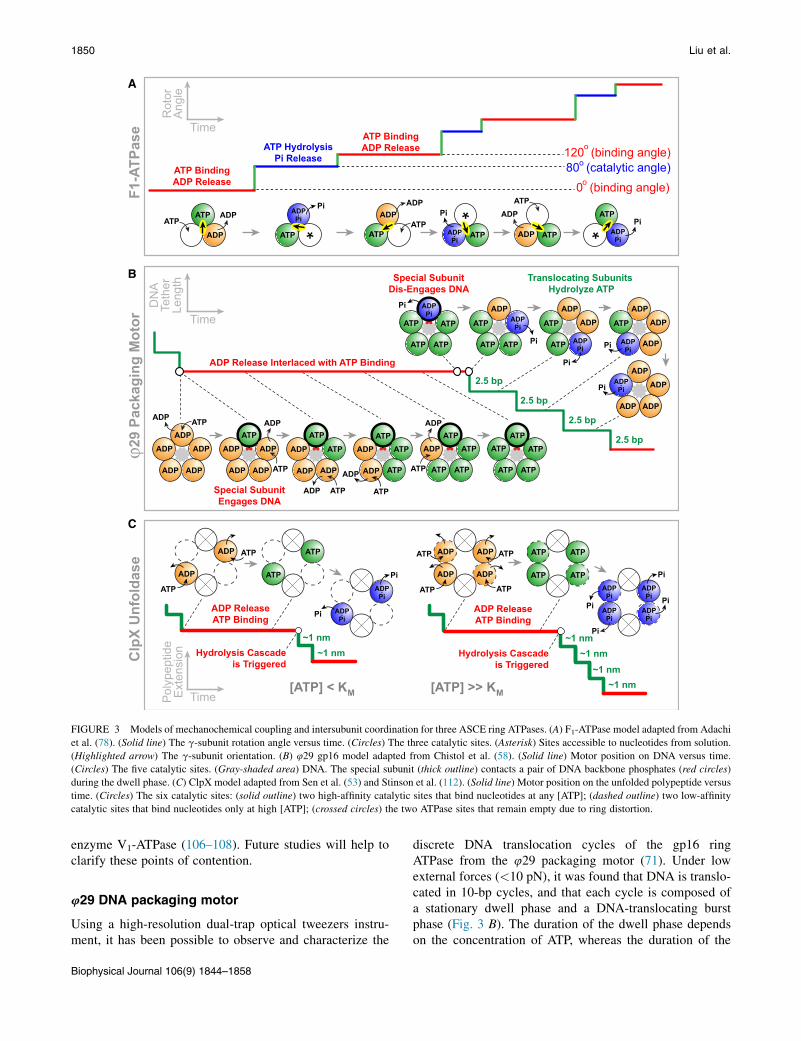
FIGURE 3 Models of mechanochemical coupling and intersubunit coordination for three ASCE ring ATPases. (A) F1-ATPase model adapted from Adachi
et al. (78). (Solid line) The g-subunit rotation angle versus time. (Circles) The three catalytic sites. (Asterisk) Sites accessible to nucleotides from solution.
(Highlighted arrow) The g-subunit orientation. (B) 429 gp16 model adapted from Chistol et al. (58). (Solid line) Motor position on DNA versus time.
(Circles) The five catalytic sites. (Gray-shaded area) DNA. The special subunit (thick outline) contacts a pair of DNA backbone phosphates (red circles)
during the dwell phase. (C) ClpX model adapted from Sen et al. (53) and Stinson et al. (112). (Solid line) Motor position on the unfolded polypeptide versus
time. (Circles) The six catalytic sites: (solid outline) two high-affinity catalytic sites that bind nucleotides at any [ATP]; (dashed outline) two low-affinity
catalytic sites that bind nucleotides only at high [ATP]; (crossed circles) the two ATPase sites that remain empty due to ring distortion.
1850 Liu et al.
enzyme V1-ATPase (106–108). Future studies will help toclarify these points of contention.
429 DNA packaging motor
Using a high-resolution dual-trap optical tweezers instru-ment, it has been possible to observe and characterize the
Biophysical Journal 106(9) 1844–1858
discrete DNA translocation cycles of the gp16 ringATPase from the 429 packaging motor (71). Under lowexternal forces (<10 pN), it was found that DNA is translo-cated in 10-bp cycles, and that each cycle is composed ofa stationary dwell phase and a DNA-translocating burstphase (Fig. 3 B). The duration of the dwell phase dependson the concentration of ATP, whereas the duration of the

Single-Molecule Studies on Ring Motors 1851
burst phase does not. These results suggest that ATP bindingoccurs during the dwell but not during the actual transloca-tion of the substrate.
Under high external forces (30–40 pN), the translocationburst slows down as expected, revealing that each 10-bpburst is composed of four 2.5-bp steps that occur in rapidsuccession (71). As mentioned above, translocation ispowered by the release of Pi in the 429 motor. Thus, Pirelease most likely coincides with or immediately precedeseach 2.5-bp step.
Nonhydrolyzable ATP analogs, ATP-g-S and AMP-PNP,were used to determine the timing of ATP hydrolysis in thedwell-burst cycle (58). The analog-induced pausing eventsinterrupt the burst phase, suggesting that hydrolysis occursduring the burst, with each subunit hydrolyzing ATP beforetaking a 2.5-bp step. To determine where in the cycle ADPrelease occurs, the packaging experiments were repeated inthe presence of orthovanadate, a Pi analog that forms a com-plex with ADP, delaying the dissociation of ADP from thebinding pocket (58). In contrast to the ATP-g-S case,ADP-vanadate-induced pausing events specifically prolongthe dwell phase, suggesting that ADP release occurs duringthe dwell.
To investigate the coordination between ATP binding andADP release events, which both take place during the dwell,ADP was added to the reaction mixture. It was found thatincreasing [ADP] gradually lengthens all dwells in a linearfashion (58). The linear dependence indicates that at anygiven time only one binding site is available for ADP inhi-bition, consistent with a model in which ADP release andATP binding occur in an alternating fashion.
Thus, the 429 packaging motor operates as a highly coor-dinated machine in which ADP release and ATP bindingoccur in an interlaced fashion during the dwell phase,whereas ATP hydrolysis, Pi release, and DNA translocationoccur in an ordinal manner, one subunit after another, duringthe burst phase (Fig. 3 B). A crucial question remained:given that the homo-pentameric motor only generates foursteps per cycle, what is the function of the special nontrans-locating subunit during the cycle? An interesting observa-tion shed light on this question: it was found that while ananalog nucleotide remains bound to the ring, the motorcan still stochastically take a few 10-bp bursts separatedby pauses much longer than regular dwells, resulting inwhat was termed a ‘‘pause cluster’’ (58). This phenomenonrequires that while the analog remains bound to one subunit,the remaining four subunits bind and spontaneously hydro-lyze ATP to translocate DNA by 2.5 bp each, albeit muchslower than the normal operation of the motor. This observa-tion suggests that the long pauses in a pause cluster reflectthe behavior of the motor when the nontranslocating subunitcannot hydrolyze the analog nucleotide. Hence, proper ATPturnover by the special, nontranslocating subunit is alsonecessary for the normal activity of the ring. Therefore,the function of this special subunit is regulatory, ensuring
the timed and proper firing of the other four translocatingsubunits. This finding reveals an unprecedented division oflabor among subunits in a homomeric ring ATPase: onlyfour of the five subunits translocate the substrate, whereasthe special one plays a critical regulatory role.
Is the regulatory role always performed by the samespecial subunit, or is that task passed around the ring inconsecutive cycles? The discrepancy between the 10-bpburst size of the motor and the 10.4-bp helical pitch ofB-form DNA suggests that the motor may rotate the DNAto bring the DNA into register with the motor after everycycle. If so, various scenarios for the identity of the specialsubunit predict different direction and/or magnitude ofDNA rotation. A rotor-bead assay was developed to monitorchanges of the DNA angle around its helical axis whilesimultaneously observing DNA translocation (74). Thisassay clearly showed that the DNA is indeed rotated bythe motor in a left-handed direction. In the absence ofDNA organization inside the capsid, the motor rotatesDNA by 1.5 5 0.2� per bp packaged. This measurementsupports a model in which the same special subunit makesregulatory contacts with the DNA in successive cycles: aftera 10-bp burst, the DNA backbone winds by 346�; thus a 14�
rotation realigns the DNA with the subunit that contactedthe DNA in the previous cycle, yielding a rotation densityof 1.4�/bp.
T7 replicative helicase
Unlike the other ring NTPases discussed here, the T7 gp4hexameric DNA helicase utilizes dTTP instead of ATP topower its operation (109). Single-molecule optical tweezersexperiments showed that ATP also supports unwinding, butdoes so much less efficiently than dTTP, because ATP in-duces frequent slipping that ultimately prevents unwindingover any significant distance (45). This unique propertyenabled the investigation of the T7 gp4 subunit coordinationmechanism by measuring the unwinding rate and processiv-ity with different amounts of ATP and dTTP. It was foundthat ATP and dTTP compete for the same nucleotide bindingsites and the unwinding rate is simply determined by ATPand dTTP concentrations and their respective Vmax andKm. The helicase processivity, defined here as the lengthunwound between slips, does not follow a simple competi-tive behavior; instead, it shows a cooperative dependenceon the ratio of [dTTP] and [ATP]. In other words, a smallfraction of dTTP mixed with ATP significantly increasesthe processivity. These results are inconsistent with arandom model in which the subunits function indepen-dently, and support a model in which most of the sixsubunits act together to coordinate nucleotide binding/hydrolysis and DNA binding to ensure processivity. Similarto the 429 packaging motor, only one subunit in the T7helicase ring is poised to bind a nucleotide at a time whilethe other subunits are nucleotide-occupied.
Biophysical Journal 106(9) 1844–1858

1852 Liu et al.
ClpX unfoldase/translocase
Structural and single-molecule data suggest a step size of~1 nm per ClpX subunit (51,52,110). Recent single-mole-cule work found that ClpX translocates polypeptides in in-crements of 2, 3, and 4 nm, suggesting that multiple ClpXsubunits (between two and four) fire in a coordinated fashion(53). Although ClpX translocates its substrate through adwell-burst mechanism similar to the one employed by429 gp16, the reduction in velocity at low ATP concentra-tion is achieved by ClpX and 429 gp16 through verydifferent means. At low ATP concentrations the 429 motorslows down by lengthening the mean dwell duration whilemaintaining a constant mean burst size (71). In contrast, atlow [ATP], ClpX slows down by decreasing its average burstsize while maintaining a constant mean dwell duration (53).ClpX’s mechanism of slowing down at low [ATP] suggeststhe existence of a type of internal clock that triggers themotor to fire a burst regardless of how many ClpX subunitsare bound to ATP (Fig. 3 C). At [ATP] % Km, the ClpXtranslocation bursts are predominantly 2–3 nm in size,whereas at [ATP] [ Km, the burst sizes are mostly 3–4 nm and do not exceed 4 nm. These results support a modelin which the ClpX hexamer contains two high-affinity andtwo low-affinity nucleotide binding sites, with the othertwo sites remaining empty, presumably due to large defor-mations in the ring (110,111). At low [ATP] only the twohigh-affinity sites bind ATP before the internal-clock mech-anism triggers the ring to fire, resulting in a 2-nm burst size(Fig. 3 C). At saturating [ATP], up to four sites can loadnucleotides before the burst is triggered, resulting in amaximum burst size of 4 nm. It is not known what servesas ClpX’s internal clock and triggers the burst phase.
A recent study suggests that ClpX periodically undergoesring-resetting independent of ATP hydrolysis (112). In theresetting events, the hexameric ring isomerizes and reas-signs individual subunits with apparently-new nucleotide-binding properties. Stinson et al. (112) proposed that thisresetting would make it possible for ClpX to avoid stallingduring the unfolding of very stable proteins, or to circum-vent temporary subunit inhibition by ADP in an ADP-richcellular environment. Such spontaneous resetting representsa plausible candidate for the internal-clock mechanism thatgives rise to the [ATP]-independent dwell duration. The pro-posed operation model for ClpX bears some similarity to themodel proposed for the 429 packaging motor, in that one (inthe case of 429 gp16) or two (in the case of ClpX) subunitinterfaces in the ATPase ring are different from the rest,implying a functional asymmetry in these homomeric rings(Fig. 3, B and C).
STRUCTURAL BASIS OF MOTOR OPERATION
The complete elucidation of a motor’s mechanism requiresa detailed understanding of the structural motifs in the
Biophysical Journal 106(9) 1844–1858
motor-substrate complex that are responsible for its func-tional dynamics. High-resolution structures have undoubt-edly shed much light on the operating mechanism of ringNTPases (6–9,91,93–95,110,113). Here we will discussthe complementary and emerging method of combiningsingle-molecule experiments with targeted mutagenesis ofthe motor or specific modifications of the substrate to dissectthe physical basis of motor operation.
Motor mutants
The ASCE motors share a variety of conserved sequenceelements, including the phosphate-binding Walker A motif(also known as P-loop), the metal-binding Walker B motif,the catalytic glutamate residue that activates a water mole-cule for nucleophilic attack on the bound NTP, and thearginine finger that protrudes into the NTP binding pocketfrom an adjacent subunit and participates in hydrolysisand intersubunit communication (5) (Fig. 1 B). The NTPbinding site is typically located at the interface betweentwo adjacent subunits. The roles of these elements in motoroperation have started to be characterized at the single-molecule level. Tsay et al. (114,115) generated a series ofpoint mutations in the bacteriophage l packaging motor.For example, a mutation (Y46F) in the Q-motif—a putativeadenine-binding motif upstream of Walker A—not onlydecreased the velocity but also showed increased forcesensitivity and impaired processivity. These results suggestthat the Q-motif is an important part of the mechanicalpathway of force generation, coupling changes in theATP-binding pocket to DNA substrate propulsion.
With high-resolution single-molecule measurements, it ispossible to pinpoint where the mutation takes effect withinthe mechanochemical cycle of the motor. For example, thecatalytic glutamate mutant in F1-ATPase, b-E190D, specif-ically prolonged the 80� dwells and was used to determinethe timing of hydrolysis (99). Moreover, to investigate theintersubunit coordination and functional asymmetry withina ring motor, it is advantageous to mutate only one or afew of the ring subunits rather than create a homomericmutant ring. This task can be achieved by mixing wild-type and mutant monomers or engineering covalently linkeddimers or trimers (57,102). Ideally, one would covalentlyconnect all subunits to form a single-chain ring motor,allowing precise control of the mutation sites within thering and ensuring sample homogeneity (97).
Substrate modifications
To determine the nature of motor-DNA interaction for the429 packaging motor, the motor was challenged with avariety of substrate modifications, including DNA witha neutral backbone, abasic DNA, single-stranded gapsand bulges, and even nonbiological polymers (116). Thedata indicate that the motor-DNA interaction is remarkably

Single-Molecule Studies on Ring Motors 1853
promiscuous, involving a wide variety of contacts with thenucleic acid moieties as well as contacts not specific toDNA. Moreover, it was found that the type of interactionchanges during the course of the mechanochemical cycle:
1. During the ATP-loading dwell phase, the motor makesspecific electrostatic contacts with a pair of adjacentbackbone phosphates every 10 bp. These ionic contactshave both a load-bearing role and a sensory or regulatoryrole, coupling mechanical and chemical events. They arealso likely responsible for rectifying the symmetrymismatch between the motor and the DNA at the endof each cycle via DNA rotation. It is plausible that thesespecific contacts are themselves responsible for breakingthe symmetry of the ring and for conferring the regula-tory role on the subunit that makes these interactions.
2. During the DNA-translocating burst phase, the motormakes nonspecific DNA contacts that are most likelysteric. These nonspecific interactions may be the reasonwhy the step size of 429 gp16 is a noninteger numberof basepairs.
Substrate modifications have also been used to investigatethe structural basis of g rotation in F1-ATPase. Wang andOster (117) proposed a push-pull mechanism based onstructural data, in which a nucleotide-bound b-subunit isbent toward, and pushes, the axle section of the g-subunit,whereas an empty b-subunit retracts and pulls g. This modelrequires a rigid g-axle pivoted at the bottom. However, itwas later found that an axleless g-mutant still supports rota-tion in the correct direction (118). Other truncation studiesfurther suggested that no part of the g-subunit is requiredfor rotation, even though some parts are needed for the gen-eration of full speed and full torque (22,119–121). Thus, itappears that rotation does not depend on specific interac-tions between g and the (ab)3 hexamer. In this regard, asKinosita (122) has pointed out, it would be fascinating toinvestigate whether F1-ATPase is able to rotate a completelyforeign object such as DNA.
BIOLOGICAL FUNCTIONS
Motor proteins are often part of larger multicomponentassemblies in vivo. When carrying out their biological tasks,these motors need to adjust their operation upon interactionwith accessory factors as well as other environmentalchanges such as fluctuating chemical concentrations andvarying mechanical loads. Below wewill discuss the biolog-ical functions of several ring motors and some experimentalefforts to examine how their activities are regulated duringthese tasks.
ATP synthesis and hydrolysis
The F0F1-ATP synthase is composed of two rotary motors—the ATP-hydrolysis-driven F1 and the proton-flow-driven F0.
Depending on the metabolic state of the cell, the coupledenzyme can either synthesize ATP using the free energyfrom a trans-membrane proton gradient, or pump protonsacross the membrane using the free energy from ATP hydro-lysis (Fig. 2 D). Through a series of elegant single-moleculeexperiments, it has been shown that the g-subunit rotates inopposite directions during ATP hydrolysis and synthesis(123), and that the isolated F1 can produce ATP when g isforced to rotate in reverse via mechanical manipulation(68,69). Nonetheless, the rotary mechanism for ATP syn-thesis is still poorly understood compared to that for ATPhydrolysis. Although reasonable as a first-order approxima-tion, it remains to be tested whether the mechanochemicalpathway for synthesis is the exact reversal of the pathwayfor hydrolysis (Fig. 3 A). Moreover, the largely constantoutput torque as a function of rotation angle implies anelastic power transmission between F1 and F0, which isconsidered essential for the high kinetic efficiency androbust operation of the holoenzyme consisting of two coun-teracting and symmetry-mismatched (C3 versus C10–14)stepping motors (124,125). The detailed coupling mecha-nism between these two motors is eagerly anticipated.
Viral genome packaging
In dsDNA viruses, the packaging motor needs to overcomemajor energetic barriers to compact the stiff and highlycharged genome to near-crystalline densities inside a smallcapsid (Fig. 2 A). It has long been known that packagingslows down as DNA fills the capsid and generates an inter-nal pressure working against the motor (41,43). However,the details of how capsid filling affects the operation ofthe packaging motor remained unclear. Recently the 429packaging dynamics was examined at different stages ofcapsid filling (74). It was found that 429 gp16 employs acomplex throttle control mechanism in which multipleparameters of the motor’s mechanochemical cycle aremodified in response to the increasing capsid filling.Specifically, high filling causes the motor to bind ATPmore slowly, enter long-lived pausing states more fre-quently, take smaller steps per cycle (~10 bp at low fillingversus ~9 bp at high filling), and translocate DNA moreslowly due to an effective internal force. The internal forcegenerated by the compressed DNA can be inferred fromthe measurement of the burst phase duration as a functionof force and as a function of capsid filling. This approachrevealed that the internal force reaches ~20 pN at100% filling, which corresponds to an internal pressure of~20 atm (74).
Remarkably, despite all the changes that cause the netvelocity to drop by two orders of magnitude at high filling,the motor maintains its strict subunit coordination and divi-sion of labor (74). This feat is achieved by the simultaneousadjustments of the step size and the amount of DNA rotationper cycle. Thus the 429 packaging motor possesses a
Biophysical Journal 106(9) 1844–1858

1854 Liu et al.
remarkable operational flexibility in the context of a fixedmechanism of mechanochemical coupling and intersubunitcoordination to allow robust activity as the genome becomesmore and more densely packed. It will be interesting toinvestigate whether or not such strategy is shared with otherdsDNA viruses, a group that includes most tailed bacterio-phages and some human pathogens such as herpes andadenoviruses.
Bacterial chromosome segregation
During cell division, FtsK and SpoIIIE are involved insegregating the circular bacterial genome between daughtercells (in the case of FtsK) or between a mother cell and aforespore (in the case of SpoIIIE) (Fig. 2 C). A central ques-tion in the field is how these DNA translocases ensure theefficient and directed segregation of a circular DNA througha division septum. As discussed above, FtsK and SpoIIIEcan reverse their translocation direction in vitro and in vivo(38–40,63). If not properly regulated, such direction rever-sals during chromosome segregation are detrimental to thecell. It is possible that the interaction with other regulatoryfactors, the skewed distribution of the orientation of therecognition sequences (KOPS, SRS) in the genome, or theorganization of the motor at the division septum itself,rectifies the motion of the translocase and maintains itsdirectionality. It should be pointed out that the functionaloligomeric form (hexameric versus dodecameric) ofSpoIIIE is still under debate (126,127). Future studiesshould clarify this point.
Roadblock displacement
Many ring translocases and helicases possess the ability ofdisplacing roadblocks bound to their substrates. Roadblockdisplacement activity is essential for replicative helicasesbecause it ensures unimpeded fork progression along adsDNA template bound with nucleosomes, RNA polymer-ases, and transcription factors. Single-molecule replicationexperiments with MCM2-7 and SV40 LTag revealed thatthe rate of replication fork progression is the same forsparsely and densely chromatinized DNA, suggesting thatboth helicases efficiently displace histones and other pro-teins bound to DNA (46,47). SV40 LTag was also shownto evict streptavidin from biotinylated DNA, thus disruptingone of the strongest noncovalent interactions (47). Consid-ering the diversity of DNA-bound obstacles encounteredby these helicases, their roadblock displacement activity ismost likely achieved via steric extrusion. Interestingly,SV40 LTag is also capable of bypassing protein barrierscross-linked to DNA (47), presumably by cracking its ringto form a lock-washer-like structure. It remains to be deter-mined whether MCM2-7 can use the same mechanism tobypass impediments cross-linked to DNA.
Biophysical Journal 106(9) 1844–1858
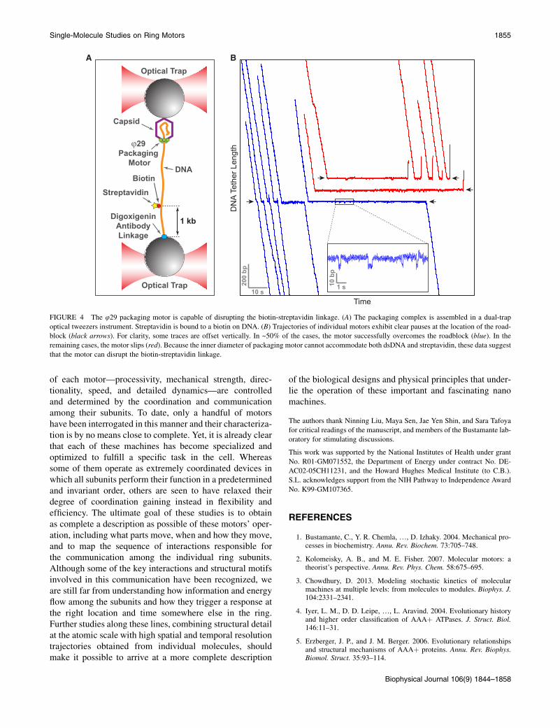
DsDNA translocases are also capable of displacing road-blocks. Both SpoIIIE and FtsK were shown to efficientlydisplace ssDNA from DNA triplexes (61,63). FtsK candisrupt the biotin-streptavidin interaction (57). SpoIIIEcan strip histone-like proteins and RNA polymerases fromthe DNA, which facilitates DNA compaction in the fore-spore, and helps to maintain the compartment-specificgene expression program in the mother cell and theforespore (128). To ensure maximal genome compaction,viral packaging motors also must displace DNA-bound pro-teins. Preliminary experiments suggest that the 429 pack-aging motor can disrupt the biotin-streptavidin linkage(Fig. 4, G. Chistol, C. Bustamante, unpublished data).
Protein unfolding and translocation
Protein unfoldases/translocases such as ClpX have evolveddistinct features to cope with unique challenges (Fig. 2 E).Compared to nucleic acid translocases, protein translocasesact on a substrate that is much more heterogeneous—poly-peptides that can contain as many as 20 amino acids withdifferent physicochemical properties. Thus, it is plausiblethat enzymes like ClpX interact with their polypeptide sub-strate via nonspecific steric interactions (110). Thesenonspecific contacts may be essential to ClpX’s ability toprocessively and unidirectionally translocate irregular anddiverse substrates from either the N-terminus or the C-ter-minus (64).
To efficiently unravel stably folded protein substrates,unfoldases must perform not only the thermodynamic taskof destabilizing the native state, but also the kinetic taskof quickly capturing the unraveled polypeptide before itcan refold. For example, to successfully unfold GFP, ClpXmust first quickly take a 4-nm burst that dislodges and trapsthe b-strand 11 before this segment can refold onto the GFPbarrel (53). At saturating [ATP], the time required for un-folding GFP is determined by the time that passes beforethe motor makes a 4-nm burst. At low [ATP], hydrolysisis often triggered before four ATP molecules can bind tothe motor and thus the motor rarely succeeds in unfoldingGFP. These findings are consistent with the observednonlinear decrease of substrate unfolding probability withATPase rate. The presence of the ClpP peptidase drasticallyreduces the slip frequency of ClpX—probably by providingadditional contacts with unfolded polypeptides—therebyenhancing the motor’s processivity and its ability to unfoldand translocate substrates (51,52).
CONCLUDING REMARKS
Organized as homo- or hetero-oligomers, ring NTPasescoordinate the catalytic and mechanical activity of theirmonomeric constituents during operation. Through theapplication of single-molecule methods, scientists havebegun to address and delineate how the unique properties
A B
Biotin
Streptavidin
DigoxigeninAntibodyLinkage
1 kb
ϕ29Packaging
Motor
Capsid
DNA
Optical Trap
Optical Trap 10 b
p
1 s200
bp
10 s
DN
A Te
ther
Len
gth
FIGURE 4 The 429 packaging motor is capable of disrupting the biotin-streptavidin linkage. (A) The packaging complex is assembled in a dual-trap
optical tweezers instrument. Streptavidin is bound to a biotin on DNA. (B) Trajectories of individual motors exhibit clear pauses at the location of the road-
block (black arrows). For clarity, some traces are offset vertically. In ~50% of the cases, the motor successfully overcomes the roadblock (blue). In the
remaining cases, the motor slips (red). Because the inner diameter of packaging motor cannot accommodate both dsDNA and streptavidin, these data suggest
that the motor can disrupt the biotin-streptavidin linkage.
Single-Molecule Studies on Ring Motors 1855
of each motor—processivity, mechanical strength, direc-tionality, speed, and detailed dynamics—are controlledand determined by the coordination and communicationamong their subunits. To date, only a handful of motorshave been interrogated in this manner and their characteriza-tion is by no means close to complete. Yet, it is already clearthat each of these machines has become specialized andoptimized to fulfill a specific task in the cell. Whereassome of them operate as extremely coordinated devices inwhich all subunits perform their function in a predeterminedand invariant order, others are seen to have relaxed theirdegree of coordination gaining instead in flexibility andefficiency. The ultimate goal of these studies is to obtainas complete a description as possible of these motors’ oper-ation, including what parts move, when and how they move,and to map the sequence of interactions responsible forthe communication among the individual ring subunits.Although some of the key interactions and structural motifsinvolved in this communication have been recognized, weare still far from understanding how information and energyflow among the subunits and how they trigger a response atthe right location and time somewhere else in the ring.Further studies along these lines, combining structural detailat the atomic scale with high spatial and temporal resolutiontrajectories obtained from individual molecules, shouldmake it possible to arrive at a more complete description
of the biological designs and physical principles that under-lie the operation of these important and fascinating nanomachines.
The authors thank Ninning Liu, Maya Sen, Jae Yen Shin, and Sara Tafoya
for critical readings of the manuscript, and members of the Bustamante lab-
oratory for stimulating discussions.
This work was supported by the National Institutes of Health under grant
No. R01-GM071552, the Department of Energy under contract No. DE-
AC02-05CH11231, and the Howard Hughes Medical Institute (to C.B.).
S.L. acknowledges support from the NIH Pathway to Independence Award
No. K99-GM107365.
REFERENCES
1. Bustamante, C., Y. R. Chemla, ., D. Izhaky. 2004. Mechanical pro-cesses in biochemistry. Annu. Rev. Biochem. 73:705–748.
2. Kolomeisky, A. B., and M. E. Fisher. 2007. Molecular motors: atheorist’s perspective. Annu. Rev. Phys. Chem. 58:675–695.
3. Chowdhury, D. 2013. Modeling stochastic kinetics of molecularmachines at multiple levels: from molecules to modules. Biophys. J.104:2331–2341.
4. Iyer, L. M., D. D. Leipe, ., L. Aravind. 2004. Evolutionary historyand higher order classification of AAAþ ATPases. J. Struct. Biol.146:11–31.
5. Erzberger, J. P., and J. M. Berger. 2006. Evolutionary relationshipsand structural mechanisms of AAAþ proteins. Annu. Rev. Biophys.Biomol. Struct. 35:93–114.
Biophysical Journal 106(9) 1844–1858

1856 Liu et al.
6. Abrahams, J. P., A. G. Leslie, ., J. E. Walker. 1994. Structure at2.8 A resolution of F1-ATPase from bovine heart mitochondria.Nature. 370:621–628.
7. Enemark, E. J., and L. Joshua-Tor. 2006. Mechanism of DNA trans-location in a replicative hexameric helicase. Nature. 442:270–275.
8. Thomsen, N. D., and J. M. Berger. 2009. Running in reverse: thestructural basis for translocation polarity in hexameric helicases.Cell. 139:523–534.
9. Itsathitphaisarn, O., R. A. Wing,., T. A. Steitz. 2012. The hexamerichelicase DnaB adopts a nonplanar conformation during translocation.Cell. 151:267–277.
10. Kapanidis, A. N., and T. Strick. 2009. Biology, one molecule at a time.Trends Biochem. Sci. 34:234–243.
11. van Oijen, A. M. 2007. Honey, I shrunk the DNA: DNA length as aprobe for nucleic-acid enzyme activity. Biopolymers. 85:144–153.
12. Greenleaf, W. J., M. T. Woodside, and S. M. Block. 2007. High-res-olution, single-molecule measurements of biomolecular motion.Annu. Rev. Biophys. Biomol. Struct. 36:171–190.
13. Joo, C., H. Balci, ., T. Ha. 2008. Advances in single-moleculefluorescence methods for molecular biology. Annu. Rev. Biochem.77:51–76.
14. Moffitt, J. R., Y. R. Chemla, ., C. Bustamante. 2008. Recentadvances in optical tweezers. Annu. Rev. Biochem. 77:205–228.
15. De Vlaminck, I., and C. Dekker. 2012. Recent advances in magnetictweezers. Annu. Rev. Biophys. 41:453–472.
16. Finkelstein, I. J., and E. C. Greene. 2011. Supported lipid bilayers andDNA curtains for high-throughput single-molecule studies. MethodsMol. Biol. 745:447–461.
17. Puchner, E. M., and H. E. Gaub. 2009. Force and function: probingproteins with AFM-based force spectroscopy. Curr. Opin. Struct.Biol. 19:605–614.
18. Walter, N. G., C.-Y. Huang, ., M. A. Sobhy. 2008. Do-it-yourselfguide: how to use the modern single-molecule toolkit. Nat. Methods.5:475–489.
19. Veigel, C., and C. F. Schmidt. 2011. Moving into the cell: single-molecule studies of molecular motors in complex environments.Nat. Rev. Mol. Cell Biol. 12:163–176.
20. Ando, T., T. Uchihashi, and N. Kodera. 2013. High-speed AFMand applications to biomolecular systems. Annu. Rev. Biophys. 42:393–414.
21. Kodera, N., D. Yamamoto, ., T. Ando. 2010. Video imaging ofwalking myosin V by high-speed atomic force microscopy. Nature.468:72–76.
22. Uchihashi, T., R. Iino,., H. Noji. 2011. High-speed atomic force mi-croscopy reveals rotary catalysis of rotorless F1-ATPase. Science.333:755–758.
23. Noi, K., D. Yamamoto, ., T. Ogura. 2013. High-speed atomic forcemicroscopic observation of ATP-dependent rotation of the AAAþchaperone p97. Structure. 21:1992–2002.
24. Heller, I., G. Sitters, ., G. J. Wuite. 2013. STED nanoscopy com-bined with optical tweezers reveals protein dynamics on denselycovered DNA. Nat. Methods. 10:910–916.
25. Comstock, M. J., T. Ha, and Y. R. Chemla. 2011. Ultrahigh-resolutionoptical trap with single-fluorophore sensitivity. Nat. Methods. 8:335–340.
26. Long, X., J. W. Parks,., M. D. Stone. 2013. Mechanical unfolding ofhuman telomere G-quadruplex DNA probed by integrated fluores-cence and magnetic tweezers spectroscopy. Nucleic Acids Res.41:2746–2755.
27. Singleton, M. R., M. S. Dillingham, and D. B. Wigley. 2007. Structureand mechanism of helicases and nucleic acid translocases. Annu. Rev.Biochem. 76:23–50.
28. Lyubimov, A. Y., M. Strycharska, and J. M. Berger. 2011. The nutsand bolts of ring-translocase structure and mechanism. Curr. Opin.Struct. Biol. 21:240–248.
Biophysical Journal 106(9) 1844–1858
29. Enemark, E. J., and L. Joshua-Tor. 2008. On helicases and other motorproteins. Curr. Opin. Struct. Biol. 18:243–257.
30. Iino, R., and H. Noji. 2013. Intersubunit coordination and cooperativ-ity in ring-shaped NTPases. Curr. Opin. Struct. Biol. 23:229–234.
31. Hetherington, C. L., J. R. Moffitt, ., C. Bustamante. 2012. ViralDNA packaging motors. In Molecular Motors and Motility.Y. E. Goldman and E. M. Ostap, editors. Elsevier, Oxford, UK,pp. 420–446.
32. Rao, V. B., and M. Feiss. 2008. The bacteriophage DNA packagingmotor. Annu. Rev. Genet. 42:647–681.
33. Patel, S. S., M. Pandey, and D. Nandakumar. 2011. Dynamic couplingbetween the motors of DNA replication: hexameric helicase, DNApolymerase, and primase. Curr. Opin. Chem. Biol. 15:595–605.
34. Kaimer, C., and P. L. Graumann. 2011. Players between the worlds:multifunctional DNA translocases. Curr. Opin. Microbiol. 14:719–725.
35. Kinosita, Jr., K., K. Adachi, and H. Itoh. 2004. Rotation of F1-ATPase:how an ATP-driven molecular machine may work. Annu. Rev.Biophys. Biomol. Struct. 33:245–268.
36. Boyer, P. D. 1997. The ATP synthase—a splendid molecular machine.Annu. Rev. Biochem. 66:717–749.
37. Baker, T. A., and R. T. Sauer. 2012. ClpXP, an ATP-powered unfold-ing and protein-degradation machine. Biochim. Biophys. Acta.1823:15–28.
38. Saleh, O. A., C. Perals,., J.-F. Allemand. 2004. Fast, DNA-sequenceindependent translocation by FtsK in a single-molecule experiment.EMBO J. 23:2430–2439.
39. Pease, P. J., O. Levy, ., N. R. Cozzarelli. 2005. Sequence-directedDNA translocation by purified FtsK. Science. 307:586–590.
40. Lee, J. Y., I. J. Finkelstein, ., E. C. Greene. 2012. Single-moleculeimaging of DNA curtains reveals mechanisms of KOPS sequence tar-geting by the DNA translocase FtsK. Proc. Natl. Acad. Sci. USA.109:6531–6536.
41. Smith, D. E., S. J. Tans, ., C. Bustamante. 2001. The bacteriophagestraight 429 portal motor can package DNA against a large internalforce. Nature. 413:748–752.
42. Rickgauer, J. P., D. N. Fuller, ., D. E. Smith. 2008. Portal motorvelocity and internal force resisting viral DNA packaging in bacterio-phage 429. Biophys. J. 94:159–167.
43. Fuller, D. N., D. M. Raymer,., D. E. Smith. 2007. Measurements ofsingle DNA molecule packaging dynamics in bacteriophage l revealhigh forces, high motor processivity, and capsid transformations.J. Mol. Biol. 373:1113–1122.
44. Fuller, D. N., D. M. Raymer, ., D. E. Smith. 2007. Single phage T4DNA packaging motors exhibit large force generation, high velocity,and dynamic variability. Proc. Natl. Acad. Sci. USA. 104:16868–16873.
45. Sun, B., D. S. Johnson, ., M. D. Wang. 2011. ATP-induced helicaseslippage reveals highly coordinated subunits. Nature. 478:132–135.
46. Yardimci, H., A. B. Loveland, ., J. C. Walter. 2010. Uncoupling ofsister replisomes during eukaryotic DNA replication. Mol. Cell.40:834–840.
47. Yardimci, H., X. Wang, ., J. C. Walter. 2012. Bypass of a proteinbarrier by a replicative DNA helicase. Nature. 492:205–209.
48. Delagoutte, E., and P. H. von Hippel. 2001. Molecular mechanisms ofthe functional coupling of the helicase (gp41) and polymerase (gp43)of bacteriophage T4 within the DNA replication fork. Biochemistry.40:4459–4477.
49. Stano, N. M., Y.-J. Jeong, ., S. S. Patel. 2005. DNA synthesis pro-vides the driving force to accelerate DNA unwinding by a helicase.Nature. 435:370–373.
50. Pandey, M., S. Syed, ., S. S. Patel. 2009. Coordinating DNA repli-cation by means of priming loop and differential synthesis rate.Nature. 462:940–943.

Single-Molecule Studies on Ring Motors 1857
51. Aubin-Tam, M.-E., A. O. Olivares,., M. J. Lang. 2011. Single-mole-cule protein unfolding and translocation by an ATP-fueled proteolyticmachine. Cell. 145:257–267.
52. Maillard, R. A., G. Chistol,., C. Bustamante. 2011. ClpXP generatesmechanical force to unfold and translocate its protein substrates. Cell.145:459–469.
53. Sen, M., R. A. Maillard, ., C. Bustamante. 2013. The ClpXP prote-ase unfolds substrates using a constant rate of pulling but differentgears. Cell. 155:636–646.
54. Ribeck, N., D. L. Kaplan,., O. A. Saleh. 2010. DnaB helicase activ-ity is modulated by DNA geometry and force. Biophys. J. 99:2170–2179.
55. Kottadiel, V. I., V. B. Rao, and Y. R. Chemla. 2012. The dynamicpause-unpackaging state, an off-translocation recovery state of aDNA packaging motor from bacteriophage T4. Proc. Natl. Acad.Sci. USA. 109:20000–20005.
56. Chemla, Y. R., K. Aathavan,., C. Bustamante. 2005. Mechanism offorce generation of a viral DNA packaging motor. Cell. 122:683–692.
57. Crozat, E., A. Meglio, ., D. J. Sherratt. 2010. Separating speed andability to displace roadblocks during DNA translocation by FtsK.EMBO J. 29:1423–1433.
58. Chistol, G., S. Liu, ., C. Bustamante. 2012. High degree of coordi-nation and division of labor among subunits in a homomeric ringATPase. Cell. 151:1017–1028.
59. Lee, S. J., S. Syed, ., L. Joshua-Tor. 2014. Dynamic look at DNAunwinding by a replicative helicase. Proc. Natl. Acad. Sci. USA.111:E827–E835.
60. Bigot, S., O. A. Saleh,., F.-X. Barre. 2006. Oriented loading of FtsKon KOPS. Nat. Struct. Mol. Biol. 13:1026–1028.
61. Levy, O., J. L. Ptacin, ., N. R. Cozzarelli. 2005. Identification ofoligonucleotide sequences that direct the movement of the Escheri-chia coli FtsK translocase. Proc. Natl. Acad. Sci. USA. 102:17618–17623.
62. Graham, J. E., D. J. Sherratt, and M. D. Szczelkun. 2010. Sequence-specific assembly of FtsK hexamers establishes directional transloca-tion on DNA. Proc. Natl. Acad. Sci. USA. 107:20263–20268.
63. Ptacin, J. L., M. Nollmann, ., C. Bustamante. 2008. Sequence-directed DNA export guides chromosome translocation during sporu-lation in Bacillus subtilis. Nat. Struct. Mol. Biol. 15:485–493.
64. Barkow, S. R., I. Levchenko, ., R. T. Sauer. 2009. Polypeptidetranslocation by the AAAþ ClpXP protease machine. Chem. Biol.16:605–612.
65. Lee, C., M. P. Schwartz, ., A. Matouschek. 2001. ATP-dependentproteases degrade their substrates by processively unraveling themfrom the degradation signal. Mol. Cell. 7:627–637.
66. Hoskins, J. R., K. Yanagihara, ., S. Wickner. 2002. ClpAP andClpXP degrade proteins with tags located in the interior of the primarysequence. Proc. Natl. Acad. Sci. USA. 99:11037–11042.
67. Bustamante, C., Z. Bryant, and S. B. Smith. 2003. Ten years of ten-sion: single-molecule DNA mechanics. Nature. 421:423–427.
68. Itoh, H., A. Takahashi,., K. Kinosita, Jr. 2004. Mechanically drivenATP synthesis by F1-ATPase. Nature. 427:465–468.
69. Rondelez, Y., G. Tresset,., H. Noji. 2005. Highly coupled ATP syn-thesis by F1-ATPase single molecules. Nature. 433:773–777.
70. Yasuda, R., H. Noji,., M. Yoshida. 1998. F1-ATPase is a highly effi-cient molecular motor that rotates with discrete 120� steps. Cell.93:1117–1124.
71. Moffitt, J. R., Y. R. Chemla, ., C. Bustamante. 2009. Intersubunitcoordination in a homomeric ring ATPase. Nature. 457:446–450.
72. Syed, S., M. Pandey, ., T. Ha. 2014. Single-molecule fluorescencereveals the unwinding stepping mechanism of replicative helicase.Cell Rep. 6:1037–1045.
73. Mallik, R., B. C. Carter, ., S. P. Gross. 2004. Cytoplasmic dyneinfunctions as a gear in response to load. Nature. 427:649–652.
74. Liu, S., G. Chistol,., C. Bustamante. Aviral packaging motor variesits DNA rotation and step size to preserve subunit coordination as thecapsid fills. Cell. http://dx.doi.org/10.1016/j.cell.2014.02.034.
75. Wang, M. D., M. J. Schnitzer,., S. M. Block. 1998. Force and veloc-ity measured for single molecules of RNA polymerase. Science.282:902–907.
76. Keller, D., and C. Bustamante. 2000. The mechanochemistry ofmolecular motors. Biophys. J. 78:541–556.
77. Oster, G., and H. Wang. 2000. Reverse engineering a protein: themechanochemistry of ATP synthase. Biochim. Biophys. Acta.1458:482–510.
78. Adachi, K., K. Oiwa, ., K. Kinosita, Jr. 2007. Coupling of rotationand catalysis in F1-ATPase revealed by single-molecule imagingand manipulation. Cell. 130:309–321.
79. Yasuda, R., H. Noji, ., H. Itoh. 2001. Resolution of distinct rota-tional substeps by submillisecond kinetic analysis of F1-ATPase.Nature. 410:898–904.
80. Patel, S. S., and K. M. Picha. 2000. Structure and function of hexame-ric helicases. Annu. Rev. Biochem. 69:651–697.
81. Yodh, J. G., M. Schlierf, and T. Ha. 2010. Insight into helicase mech-anism and function revealed through single-molecule approaches.Q. Rev. Biophys. 43:185–217.
82. Lohman, T. M., and K. P. Bjornson. 1996. Mechanisms of helicase-catalyzed DNA unwinding. Annu. Rev. Biochem. 65:169–214.
83. Singleton, M. R., and D. B. Wigley. 2002. Modularity and specializa-tion in superfamily 1 and 2 helicases. J. Bacteriol. 184:1819–1826.
84. von Hippel, P. H., and E. Delagoutte. 2001. A general model fornucleic acid helicases and their ‘‘coupling’’ within macromolecularmachines. Cell. 104:177–190.
85. Betterton, M. D., and F. Julicher. 2005. Opening of nucleic-acid dou-ble strands by helicases: active versus passive opening. Phys. Rev. EStat. Nonlin. Soft Matter Phys. 71:011904.
86. Lionnet, T., M. M. Spiering,., V. Croquette. 2007. Real-time obser-vation of bacteriophage T4 gp41 helicase reveals an unwinding mech-anism. Proc. Natl. Acad. Sci. USA. 104:19790–19795.
87. Johnson, D. S., L. Bai,., M. D.Wang. 2007. Single-molecule studiesreveal dynamics of DNA unwinding by the ring-shaped T7 helicase.Cell. 129:1299–1309.
88. Cheng, W., S. Dumont, ., C. Bustamante. 2007. NS3 helicaseactively separates RNA strands and senses sequence barriers aheadof the opening fork. Proc. Natl. Acad. Sci. USA. 104:13954–13959.
89. Manosas, M., X. G. Xi, ., V. Croquette. 2010. Active and passivemechanisms of helicases. Nucleic Acids Res. 38:5518–5526.
90. Adelman, J. L., Y.-J. Jeong, ., S. S. Patel. 2006. Mechanochemistryof transcription termination factor Rho. Mol. Cell. 22:611–621.
91. Sun, S., K. Kondabagil, ., V. B. Rao. 2008. The structure of thephage T4 DNA packaging motor suggests a mechanism dependenton electrostatic forces. Cell. 135:1251–1262.
92. Crampton, D. J., S. Mukherjee, and C. C. Richardson. 2006. DNA-induced switch from independent to sequential dTTP hydrolysis inthe bacteriophage T7 DNA helicase. Mol. Cell. 21:165–174.
93. Mancini, E. J., D. E. Kainov,., D. I. Stuart. 2004. Atomic snapshotsof an RNA packaging motor reveal conformational changes linkingATP hydrolysis to RNA translocation. Cell. 118:743–755.
94. Massey, T. H., C. P. Mercogliano,., J. Lowe. 2006. Double-strandedDNA translocation: structure and mechanism of hexameric FtsK.Mol.Cell. 23:457–469.
95. Singleton, M. R., M. R. Sawaya, ., D. B. Wigley. 2000. Crystalstructure of T7 gene 4 ring helicase indicates a mechanism for sequen-tial hydrolysis of nucleotides. Cell. 101:589–600.
96. Gai, D., R. Zhao, ., X. S. Chen. 2004. Mechanisms of conforma-tional change for a replicative hexameric helicase of SV40 large tu-mor antigen. Cell. 119:47–60.
Biophysical Journal 106(9) 1844–1858

1858 Liu et al.
97. Martin, A., T. A. Baker, and R. T. Sauer. 2005. Rebuilt AAAþmotorsreveal operating principles for ATP-fueled machines. Nature. 437:1115–1120.
98. Noji, H., R. Yasuda, ., K. Kinosita, Jr. 1997. Direct observation ofthe rotation of F1-ATPase. Nature. 386:299–302.
99. Shimabukuro, K., R. Yasuda, ., M. Yoshida. 2003. Catalysis androtation of F1 motor: cleavage of ATP at the catalytic site occurs in1 ms before 40� substep rotation. Proc. Natl. Acad. Sci. USA.100:14731–14736.
100. Nishizaka, T., K. Oiwa, ., K. Kinosita, Jr. 2004. Chemomechanicalcoupling in F1-ATPase revealed by simultaneous observation ofnucleotide kinetics and rotation. Nat. Struct. Mol. Biol. 11:142–148.
101. Shimo-Kon, R., E. Muneyuki, ., K. Kinosita, Jr. 2010. Chemo-mechanical coupling in F1-ATPase revealed by catalytic site occu-pancy during catalysis. Biophys. J. 98:1227–1236.
102. Ariga, T., E. Muneyuki, and M. Yoshida. 2007. F1-ATPase rotates byan asymmetric, sequential mechanism using all three catalytic sub-units. Nat. Struct. Mol. Biol. 14:841–846.
103. Okazaki, K., and G. Hummer. 2013. Phosphate release coupled torotary motion of F1-ATPase. Proc. Natl. Acad. Sci. USA. 110:16468–16473.
104. Watanabe, R., R. Iino, and H. Noji. 2010. Phosphate release in F1-ATPase catalytic cycle follows ADP release. Nat. Chem. Biol.6:814–820.
105. Shimabukuro, K., E. Muneyuki, and M. Yoshida. 2006. An alternativereaction pathway of F1-ATPase suggested by rotation without 80
�/40�substeps of a sluggish mutant at low ATP. Biophys. J. 90:1028–1032.
106. Imamura, H., M. Takeda,., K. Yokoyama. 2005. Rotation scheme ofV1-motor is different from that of F1-motor. Proc. Natl. Acad. Sci.USA. 102:17929–17933.
107. Furuike, S., M. Nakano, ., K. Yokoyama. 2011. Resolving steppingrotation in Thermus thermophilus Hþ-ATPase/synthase with an essen-tially drag-free probe. Nat. Commun. 2:233.
108. Minagawa, Y., H. Ueno, ., R. Iino. 2013. Basic properties of rotarydynamics of the molecular motor Enterococcus hirae V1-ATPase.J. Biol. Chem. 288:32700–32707.
109. Donmez, I., and S. S. Patel. 2006. Mechanisms of a ring shaped heli-case. Nucleic Acids Res. 34:4216–4224.
110. Glynn, S. E., A. Martin, ., R. T. Sauer. 2009. Structures of asym-metric ClpX hexamers reveal nucleotide-dependent motions in aAAAþ protein-unfolding machine. Cell. 139:744–756.
111. Hersch, G. L., R. E. Burton,., R. T. Sauer. 2005. Asymmetric inter-actions of ATP with the AAAþ ClpX6 unfoldase: allosteric control ofa protein machine. Cell. 121:1017–1027.
112. Stinson, B. M., A. R. Nager,., R. T. Sauer. 2013. Nucleotide bindingand conformational switching in the hexameric ring of a AAAþmachine. Cell. 153:628–639.
Biophysical Journal 106(9) 1844–1858
113. Strycharska, M. S., E. Arias-Palomo, ., J. M. Berger. 2013. Nucleo-tide and partner-protein control of bacterial replicative helicase struc-ture and function. Mol. Cell. 52:844–854.
114. Tsay, J. M., J. Sippy, ., D. E. Smith. 2009. The Q motif of a viralpackaging motor governs its force generation and communicatesATP recognition to DNA interaction. Proc. Natl. Acad. Sci. USA.106:14355–14360.
115. Tsay, J. M., J. Sippy,., D. E. Smith. 2010. Mutations altering a struc-turally conserved loop-helix-loop region of a viral packaging motorchange DNA translocation velocity and processivity. J. Biol. Chem.285:24282–24289.
116. Aathavan, K., A. T. Politzer,., C. Bustamante. 2009. Substrate inter-actions and promiscuity in a viral DNA packaging motor. Nature.461:669–673.
117. Wang, H., and G. Oster. 1998. Energy transduction in the F1 motor ofATP synthase. Nature. 396:279–282.
118. Furuike, S., M. D. Hossain, ., K. Kinosita, Jr. 2008. Axle-less F1-ATPase rotates in the correct direction. Science. 319:955–958.
119. Hossain, M. D., S. Furuike,., K. Kinosita, Jr. 2008. Neither helix inthe coiled coil region of the axle of F1-ATPase plays a significant rolein torque production. Biophys. J. 95:4837–4844.
120. Muller, M., O. Panke,., S. Engelbrecht. 2002. F1-ATPase, the C-ter-minal end of subunit g is not required for ATP hydrolysis-driven rota-tion. J. Biol. Chem. 277:23308–23313.
121. Kohori, A., R. Chiwata, ., K. Kinosita, Jr. 2011. Torque generationin F1-ATPase devoid of the entire amino-terminal helix of the rotorthat fills half of the stator orifice. Biophys. J. 101:188–195.
122. Kinosita, Jr., K. 2012. F1-ATPase: a prototypical rotary molecularmotor. In Viral Molecular Machines. M. G. Rossmann andV. B. Rao, editors. Springer, New York, pp. 5–16.
123. Diez, M., B. Zimmermann, ., P. Graber. 2004. Proton-powered sub-unit rotation in single membrane-bound F0F1-ATP synthase. Nat.Struct. Mol. Biol. 11:135–141.
124. Panke, O., D. A. Cherepanov, ., W. Junge. 2001. Viscoelastic dy-namics of actin filaments coupled to rotary F-ATPase: angular torqueprofile of the enzyme. Biophys. J. 81:1220–1233.
125. Junge, W., H. Sielaff, and S. Engelbrecht. 2009. Torque generationand elastic power transmission in the rotary F0F1-ATPase. Nature.459:364–370.
126. Fleming, T. C., J. Y. Shin, ., K. Pogliano. 2010. Dynamic SpoIIIEassembly mediates septal membrane fission during Bacillus subtilissporulation. Genes Dev. 24:1160–1172.
127. Fiche, J.-B., D. I. Cattoni, ., M. Nollmann. 2013. Recruitment,assembly, and molecular architecture of the SpoIIIE DNA pumprevealed by superresolution microscopy. PLoS Biol. 11:e1001557.
128. Marquis, K. A., B. M. Burton, ., D. Z. Rudner. 2008. SpoIIIE stripsproteins off the DNA during chromosome translocation. Genes Dev.22:1786–1795.