mechanisms of immunity
DESCRIPTION
CLS 420 Clinical Immunology & Molecular Diagnostics. Mechanisms of Immunity. Part One Types of Immunity. Objectives. Differentiate innate (natural) immunity and acquired (adaptive) immunity. Describe the significance of the following factors included in the first line of defense: - PowerPoint PPT PresentationTRANSCRIPT

Mechanisms of Immunity
Part One
Types of Immunity
CLS 420Clinical Immunology &Molecular Diagnostics

Objectives
1. Differentiate innate (natural) immunity and acquired (adaptive) immunity.
2. Describe the significance of the following factors included in the first line of defense:
a. Skin and mucous membranes
b. Age
c. Metabolism
d. Environment
e. Normal flora
f. Concomitant disease
Objectives
3. Compare specific versus non-specific immunity as it relates to:a. Phagocytosisb. Inflammationc. Cellular immunityd. Humoral immunitye. Memoryf. Opsonization
4. Differentiate humoral and cellular immunity.5. Compare active and passive immunity, including
examples of each.

Immunity
The state of protection from infectious disease.
Immunity
• Protection against:– Pathogens– Toxins – Tumors
• Recognition of foreign vs. self
• Two branches:– Innate– Adaptive (acquired)
Immune function is influenced by:
• Age • Nutritional status• Stress• Fatigue

What happens when foreign matter enters the body?
Innate ImmunityThe First Line Of Defense

Innate Immunity
• Available at birth (innate or natural).
• Generalized, nonspecific reaction.
• Response to invader is rapid.
• No prior exposure to foreign substance required.
• Response doesn’t change with further exposure.

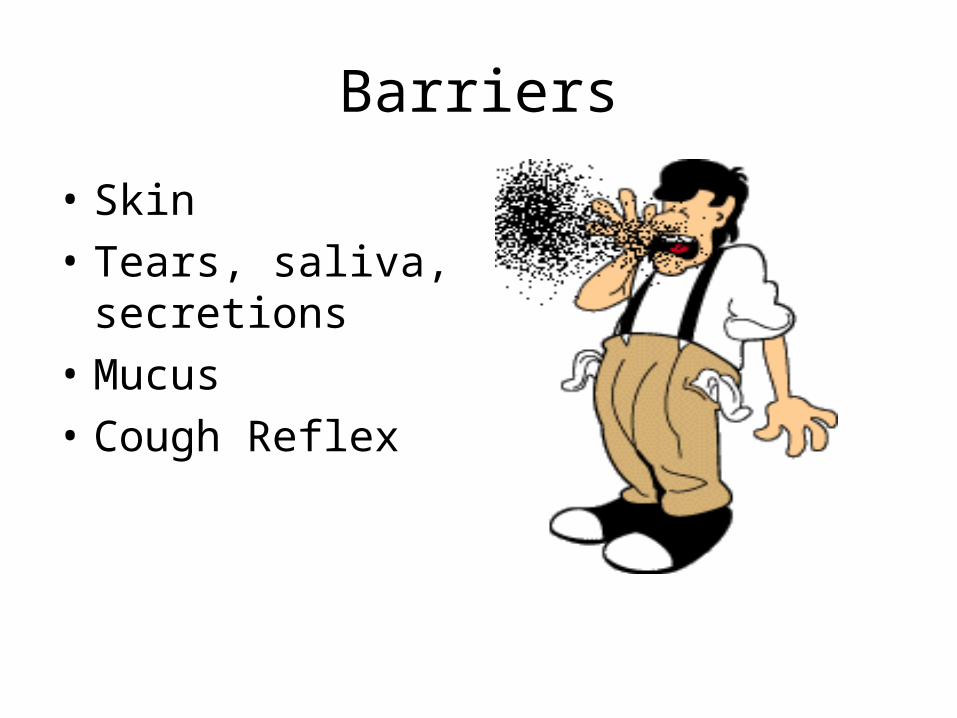
Barriers
• Skin
• Tears, saliva, secretions
• Mucus
• Cough Reflex
Environment
• pH
• Normal flora
• Temperature


Pathogen Recognition
• Toll-like receptors
• Complement – Alternative Pathway
– Mannose Binding Lectin Pathway

Chemicals
• Chemicals include:– Complement– Histamine– Cytokines– Acute Phase Reactants– Kinins
• Destroy pathogens • Expand the immune response• Neutralize the effects of other chemicals


Inflammation
• Capillaries dilate and blood accumulates in the affected area.– Redness– Increased temperature
• Fluid accumulates in tissue (edema).
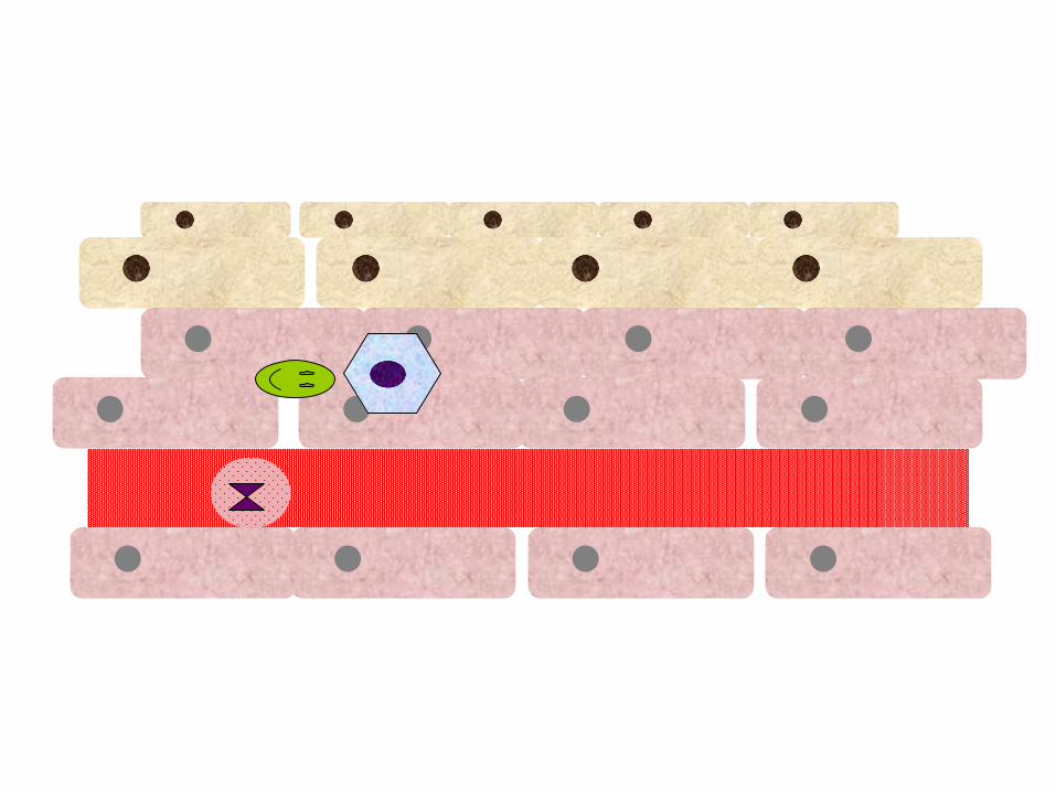

Inflammation
• Chemokines attract phagocytes into affected area.
• Phagocytes migrate into the tissue (extravasation) and destroy invading pathogens.
• Other chemicals are released that cause:– Fever– Increase WBC adhesion to vascular endothelium– Stimulate clotting/fibrinolysis cascades

Inflammation
• Pus formation
• Macrophages clean up debris.
• Release chemicals that enhance the inflammatory response.
• Growth factors rebuild tissue and blood vessels.

Inflammation – Acute vs. Chronic
• Well defined onset and resolution
• Mediated by macrophages– Fever– Increase WBC production– Acute phase reactants
released from liver
• Phagocytosis by neutrophils
• Ongoing inflammatory process
• Inability to clear pathogen• Less defined onset &
resolution• Macrophages and
lymphocytes involved• Loss of tissue function
– Granuloma formation– Scar tissue
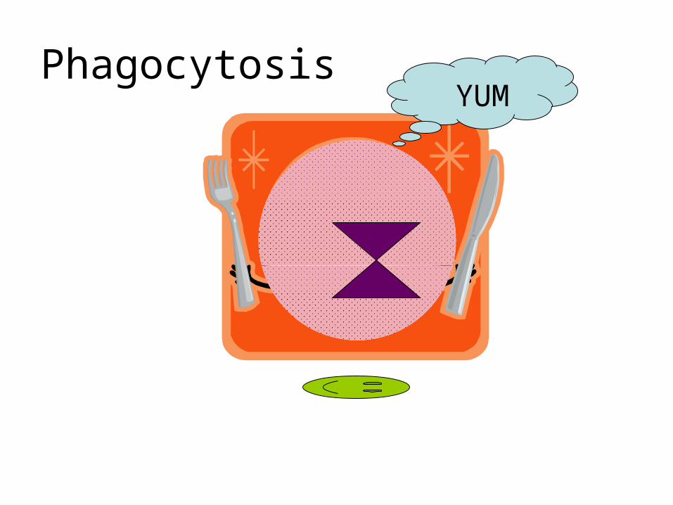
PhagocytosisYUM

Phagocytes
• Dendritic cells
• Neutrophils
• Macrophages
• Monocytes
• Eosinophils
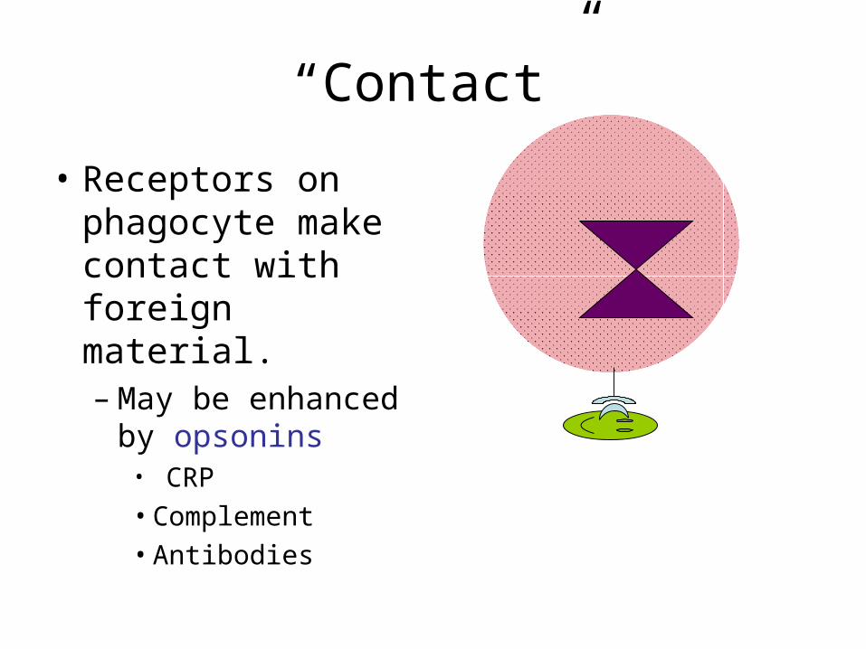
“Contact”
• Receptors on phagocyte make contact with foreign material.– May be enhanced
by opsonins• CRP• Complement• Antibodies
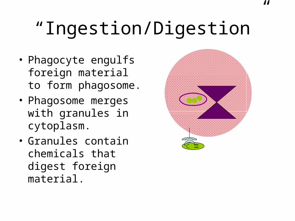
“Ingestion/Digestion”
• Phagocyte engulfs foreign material to form phagosome.
• Phagosome merges with granules in cytoplasm.
• Granules contain chemicals that digest foreign material.
Chemicals
• Respiratory burst – membrane bound oxidase converts oxygen to superoxide anion.
• Nitric oxide synthetase- oxides arginine releasing nitric oxide.
• Lysozyme- antimicrobial enzyme
• Defensins – cysteine rich peptides that attack bacterial cell membranes.
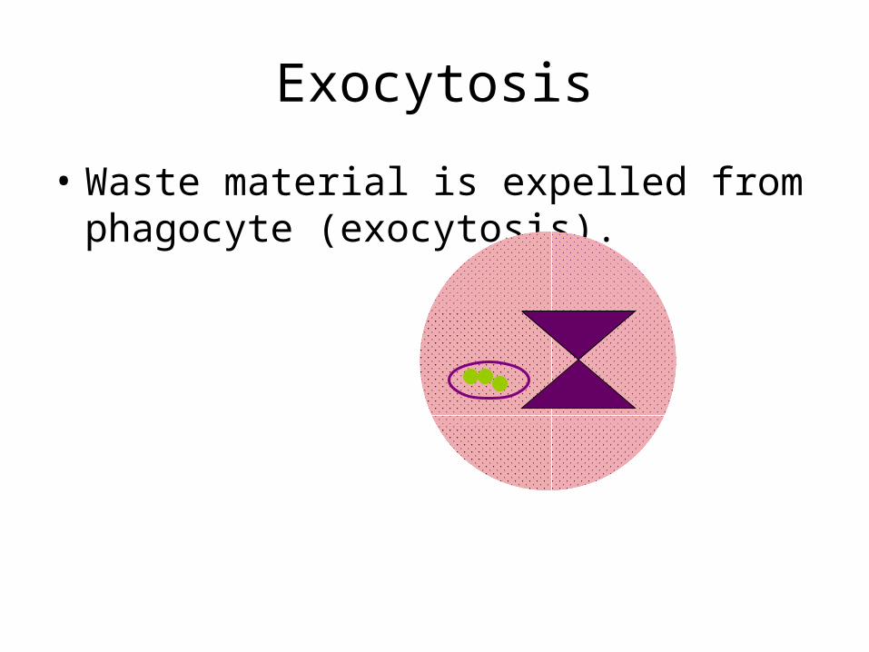
Exocytosis
• Waste material is expelled from phagocyte (exocytosis).

Cells Alive!
• Please go to http://www.cellsalive.com/ouch1.htm
• View “Anatomy of a Splinter”.
How can long-lasting protection be developed?
Acquired Immunity
a.k.a.
Adaptive Immunity

Characteristics• Forms only after exposure to foreign
substance.• Response is specific to the foreign
stimulus.• Recognizes self vs. non-self.• Displays memory.• 2 forms:
– Cell mediated– Humoral

Cell Mediated Immunity
• Controlled by T lymphocytes.– Influence other parts of the immune system
(including the humoral response) through the release of cytokines.
• Responsible for such processes as delayed hypersensitivity, transplant immunity, and tumor rejection.

Cell Mediated Process
• Foreign matter is broken down into small peptides.
• Peptides combine with the Major Histocompatibility Complex (MCH) molecules on the cell membrane.

Major Histocompatibility Complex
• Found on the membrane of every nucleated cell.
• Three classes:– I (A, B, C loci)– II (DR, DQ, DP loci)– III (complement , Tumor Necrosis Factor
[TNF])
• MHC has antigen binding groove which carries foreign peptides.
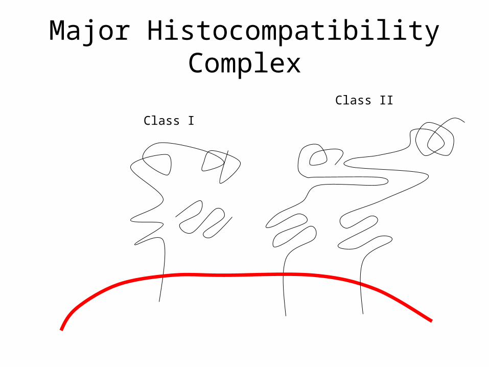
Major Histocompatibility Complex
Class I
Class II

Cell Mediated Process
• Class I MHC antigens bind peptides that are produced within the cell
• Class II MHC molecules are found on Antigen Presenting Cells (APC)– Monocytes, macrophages, dendritic cells, and
B lymphocytes
• Class II molecules bind foreign peptides that have been processed by the APC.

Recognition
• T lymphocytes possess receptors (TCR) that recognize foreign antigen when the antigen is bound to MHC antigen.
Signaling
• TCR is coupled to CD3.
• CD3 signals the interior of the T cell that antigen is present.
• T cell produces cytokines in response (effector cell).
• Some T cells mature into memory T cells.
T Lymphocyte Subsets
• T helper cells (TH)– Make up 2/3’s of the T cell population– CD4 positive– React with MHC class II proteins– Assist in initiating cytotoxic response– Stimulate humoral response
• T cytotoxic cells (TC)– CD8 positive– React with MHC class I proteins

Cytotoxic Response
• Tc cell recognizes foreign peptides associated with Class I MHC antigen.
• Cytotoxic T Lymphocyte (CTL) binds to target cell
• Release perforin and granzymes, leading to apoptosis of target cell.
• Cytokines TNF and interferon released to prevent spread of virus.– May injure healthy tissue.
Cells Alive!
• Please go to http://www.cellsalive.com/ctl.htm
• View “The Cytotoxic T Lymphocyte”.
Humoral Immunity
• Major cell involved is the B lymphocyte.– Develops into plasma cells and memory cells.
• Influenced by T helper cells.

Humoral Response
• Foreign antigen is introduced to host.
• APCs process antigen into peptide fragments, then present peptide to the TH lymphocyte.
• The TH recognizes the foreign antigen through its TCR and CD4.
Humoral Response
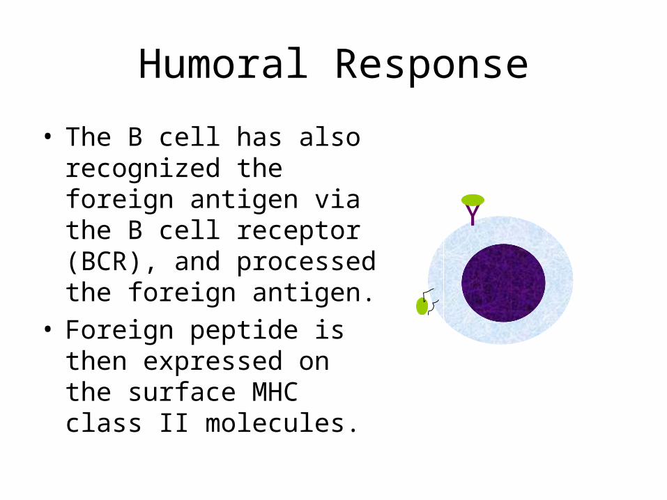
• The B cell has also recognized the foreign antigen via the B cell receptor (BCR), and processed the foreign antigen.
• Foreign peptide is then expressed on the surface MHC class II molecules.
Y
Humoral Response
• TH lymphocyte recognizes that the peptide seen by its TCR and the peptide presented by the B cell are the same.
• TH cell releases cytokines to stimulate B cell.

B Lymphocytes
• B cells divide into plasma cells or memory B cells.
• Plasma cells produce and secrete antibody.– The antibody produced is
specific for one epitope!
• Memory cells recall previous encounter with foreign antigen. – Respond quickly when
antigen is encountered again.
T Helper Lymphocytes
• Signals from the TH cell influence the class of immunoglobulin produced by the B cell and the ability to switch classes.– Interleukins– Transforming growth factor

Alternate Humoral Response
• B cells may be influenced to produce antibody independent of T cells.– Rapid response– Few memory cells – No switch to other classes of immunoglobulin– Additional nonspecific antibody may be
produced (autoantibody)
Cells Alive!
• Please go to http://www.cellsalive.com/antibody.htm
• View “Making Antibodies”.

Active vs. Passive Immunity
• In active immunity, the host produces an immune response to foreign matter following exposure.
• Long lasting.• Displays memory. • Examples include:
– Vaccinations– Immunity following infection
• In passive immunity, the host acquires antibody that was produced in another being.
• Short term protection.• No memory.• Examples include:
– Maternal transfer to fetus– Immune Serum Globulin
used to treat hypogammaglobulinemia

STRETCH
Take a break before moving on to Part II