mechanisms of polarized organelle distribution in neurons · mechanisms of polarized organelle...
TRANSCRIPT
MINI REVIEWpublished: 31 March 2016
doi: 10.3389/fncel.2016.00088
Mechanisms of Polarized OrganelleDistribution in NeuronsDylan J. Britt , Ginny G. Farías , Carlos M. Guardia and Juan S. Bonifacino *
Cell Biology and Neurobiology Branch, Eunice Kennedy Shriver National Institute of Child Health and Human Development,National Institutes of Health, Bethesda, MD, USA
Edited by:Hansen Wang,
University of Toronto, Canada
Reviewed by:Andres Couve,
University of Chile, ChileChristophe Leterrier,
CRN2M CNRS-AMU UMR7286,France
Susan E. Brockerhoff,University of Washington, USA
*Correspondence:Juan S. Bonifacino
Received: 19 February 2016Accepted: 21 March 2016Published: 31 March 2016
Citation:Britt DJ, Farías GG, Guardia CM andBonifacino JS (2016) Mechanisms of
Polarized OrganelleDistribution in Neurons.
Front. Cell. Neurosci. 10:88.doi: 10.3389/fncel.2016.00088
Neurons are highly polarized cells exhibiting axonal and somatodendritic domains withdistinct complements of cytoplasmic organelles. Although some organelles are widelydistributed throughout the neuronal cytoplasm, others are segregated to either theaxonal or somatodendritic domains. Recent findings show that organelle segregationis largely established at a pre-axonal exclusion zone (PAEZ) within the axon hillock.Polarized sorting of cytoplasmic organelles at the PAEZ is proposed to depend mainlyon their selective association with different microtubule motors and, in turn, with distinctmicrotubule arrays. Somatodendritic organelles that escape sorting at the PAEZ can besubsequently retrieved at the axon initial segment (AIS) by a microtubule- and/or actin-based mechanism. Dynamic sorting along the PAEZ-AIS continuum can thus explain thepolarized distribution of cytoplasmic organelles between the axonal and somatodendriticdomains.
Keywords: neurons, polarized sorting, organelle distribution, axon initial segment, pre-axonal exclusion zone,microtubules, kinesins, axonal transport
INTRODUCTION
Among the first properties of neurons described by neuroanatomists was their polarization intodistinct compartments—an axon, dendrites, and cell body or soma (Figure 1A; Deiters, 1865). Asearly as the first decade of the 20th century, Ramón y Cajal (1906) proposed that this structuralpolarity could explain the unidirectional transmission of information within and between neurons,a property he famously termed ‘‘dynamic polarization’’. Although the intervening century has seena revision of this one-way theory of neurotransmission, the notion of cellular structure as a criticaldeterminant of function has become a core principle in biology.
Despite the enormous heterogeneity in neuronal form and function, a given neuron consistsof a roughly spherical soma and one or more narrow membranous extensions, or neurites(Peters et al., 1991; Raine, 1999). Neurites are further subdivided into branched, taperingdendrites and a single thin axon, which may extend for long distances before branchinginto terminals (Peters et al., 1991; Raine, 1999). Although the soma and dendrites differ inshape, in terms of protein and organelle distribution the two are often treated as a unifiedsomatodendritic domain. The somatodendritic and axonal domains may be differentiatedby the presence of subdomains with unique roles in neurotransmission. The transmissionof information between neurons most often occurs at synapses formed by juxtapositionof a postsynaptic site on the somatodendritic domain and a presynaptic axon terminal.The plasma membrane of postsynaptic terminals is enriched in neurotransmitter receptorsunderlain by a complex scaffold of structural and signaling proteins (Ziff, 1997; Collins et al.,2006; Lasiecka et al., 2008). In contrast, the plasma membrane of presynaptic terminals is
Frontiers in Cellular Neuroscience | www.frontiersin.org 1 March 2016 | Volume 10 | Article 88
Britt et al. Polarized Organelle Distribution in Neurons
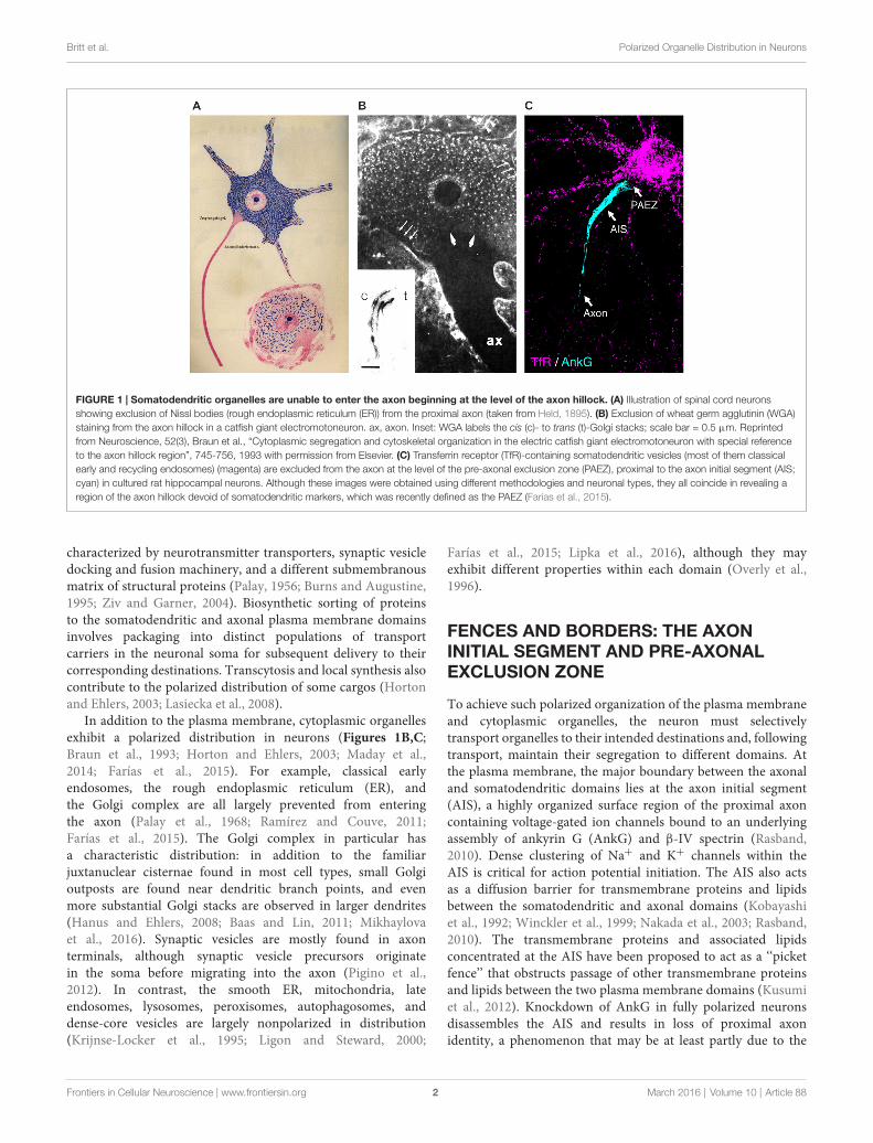
FIGURE 1 | Somatodendritic organelles are unable to enter the axon beginning at the level of the axon hillock. (A) Illustration of spinal cord neuronsshowing exclusion of Nissl bodies (rough endoplasmic reticulum (ER)) from the proximal axon (taken from Held, 1895). (B) Exclusion of wheat germ agglutinin (WGA)staining from the axon hillock in a catfish giant electromotoneuron. ax, axon. Inset: WGA labels the cis (c)- to trans (t)-Golgi stacks; scale bar = 0.5 µm. Reprintedfrom Neuroscience, 52(3), Braun et al., “Cytoplasmic segregation and cytoskeletal organization in the electric catfish giant electromotoneuron with special referenceto the axon hillock region”, 745-756, 1993 with permission from Elsevier. (C) Transferrin receptor (TfR)-containing somatodendritic vesicles (most of them classicalearly and recycling endosomes) (magenta) are excluded from the axon at the level of the pre-axonal exclusion zone (PAEZ), proximal to the axon initial segment (AIS;cyan) in cultured rat hippocampal neurons. Although these images were obtained using different methodologies and neuronal types, they all coincide in revealing aregion of the axon hillock devoid of somatodendritic markers, which was recently defined as the PAEZ (Farías et al., 2015).
characterized by neurotransmitter transporters, synaptic vesicledocking and fusion machinery, and a different submembranousmatrix of structural proteins (Palay, 1956; Burns and Augustine,1995; Ziv and Garner, 2004). Biosynthetic sorting of proteinsto the somatodendritic and axonal plasma membrane domainsinvolves packaging into distinct populations of transportcarriers in the neuronal soma for subsequent delivery to theircorresponding destinations. Transcytosis and local synthesis alsocontribute to the polarized distribution of some cargos (Hortonand Ehlers, 2003; Lasiecka et al., 2008).
In addition to the plasma membrane, cytoplasmic organellesexhibit a polarized distribution in neurons (Figures 1B,C;Braun et al., 1993; Horton and Ehlers, 2003; Maday et al.,2014; Farías et al., 2015). For example, classical earlyendosomes, the rough endoplasmic reticulum (ER), andthe Golgi complex are all largely prevented from enteringthe axon (Palay et al., 1968; Ramírez and Couve, 2011;Farías et al., 2015). The Golgi complex in particular hasa characteristic distribution: in addition to the familiarjuxtanuclear cisternae found in most cell types, small Golgioutposts are found near dendritic branch points, and evenmore substantial Golgi stacks are observed in larger dendrites(Hanus and Ehlers, 2008; Baas and Lin, 2011; Mikhaylovaet al., 2016). Synaptic vesicles are mostly found in axonterminals, although synaptic vesicle precursors originatein the soma before migrating into the axon (Pigino et al.,2012). In contrast, the smooth ER, mitochondria, lateendosomes, lysosomes, peroxisomes, autophagosomes, anddense-core vesicles are largely nonpolarized in distribution(Krijnse-Locker et al., 1995; Ligon and Steward, 2000;
Farías et al., 2015; Lipka et al., 2016), although they mayexhibit different properties within each domain (Overly et al.,1996).
FENCES AND BORDERS: THE AXONINITIAL SEGMENT AND PRE-AXONALEXCLUSION ZONE
To achieve such polarized organization of the plasma membraneand cytoplasmic organelles, the neuron must selectivelytransport organelles to their intended destinations and, followingtransport, maintain their segregation to different domains. Atthe plasma membrane, the major boundary between the axonaland somatodendritic domains lies at the axon initial segment(AIS), a highly organized surface region of the proximal axoncontaining voltage-gated ion channels bound to an underlyingassembly of ankyrin G (AnkG) and β-IV spectrin (Rasband,2010). Dense clustering of Na+ and K+ channels within theAIS is critical for action potential initiation. The AIS also actsas a diffusion barrier for transmembrane proteins and lipidsbetween the somatodendritic and axonal domains (Kobayashiet al., 1992; Winckler et al., 1999; Nakada et al., 2003; Rasband,2010). The transmembrane proteins and associated lipidsconcentrated at the AIS have been proposed to act as a ‘‘picketfence’’ that obstructs passage of other transmembrane proteinsand lipids between the two plasma membrane domains (Kusumiet al., 2012). Knockdown of AnkG in fully polarized neuronsdisassembles the AIS and results in loss of proximal axonidentity, a phenomenon that may be at least partly due to the
Frontiers in Cellular Neuroscience | www.frontiersin.org 2 March 2016 | Volume 10 | Article 88

Britt et al. Polarized Organelle Distribution in Neurons
removal of the lateral diffusion barrier (Hedstrom et al., 2008;Sobotzik et al., 2009; Song et al., 2009; Jenkins et al., 2015).
In addition to its role in segregating plasma membraneproteins, the AIS has been proposed to function as a selectivefilter for cytoplasmic organelles (Song et al., 2009; Al-Bassamet al., 2012; Watanabe et al., 2012). A model developed instudies of vesicular transport carriers originating from the somaposits that carriers intended for the axon are freely able to passthrough this filter, while somatodendritic carriers are blockedby an actin-dependent mechanism (Lewis et al., 2009; Songet al., 2009; Al-Bassam et al., 2012; Watanabe et al., 2012).In this model, the AIS functions either as a physical barrierthat prevents axonal entry of somatodendritic carriers or as ascaffold for myosin Va-mediated retrieval of somatodendriticcarriers that enter this segment. This view of the AIS as asieve for cytoplasmic organelles, however, has been challengedby several findings. First, polarized organelle transport arisesprior to AIS formation during neuronal development (Bradkeand Dotti, 1997; Petersen et al., 2014; Farías et al., 2015).Moreover, axonal exclusion of somatodendritic carriers isachieved even when subsequent AIS formation is preventedby AnkG knockdown (Farías et al., 2015), and AnkG-nullneurons maintain axonal identity as few as 50 µm fromthe soma (Jenkins et al., 2015). In terms of the proposedstructure, super-resolution microscopy and platinum replicaelectron microscopy have shown that actin in the AIS existsas rings or sparse filaments, and not the dense or polarizedstructures required by the filter model (Xu et al., 2013; Joneset al., 2014; Leterrier et al., 2015). A key result cited insupport of the AIS as an actin-based filter is the disruptionof somatodendritic polarity when actin-depolymerizing drugsare applied to neurons (Lewis et al., 2009; Song et al., 2009).However, actin depolymerization has also been shown to causemissorting of somatodendritic proteins into axonal carriersat the Golgi complex (Petersen et al., 2014). Kuijpers et al.(2016) have recently proposed a different mechanism for theretrieval of somatodendritic vesicles that enter the AIS throughAnkG-dependent recruitment of the proteins NDEL1 and LIS1,which activate the minus end-directed microtubule motordynein on these vesicles for retrograde transport to the soma.Therefore, polarized organelle distribution can be achieved inthe absence of the AIS, although myosin- and/or dynein-basedretrieval mechanisms may fine-tune this distribution upon AISassembly.
Although most studies to date have focused on the AIS as theboundary for somatodendritic and axonal organelles, a recentstudy has shown that in cultured hippocampal neurons mostsomatodendritic-specific organelles, such as somatodendriticcarriers, the Golgi complex, and the rough ER, are preventedfrom entering the axon at a more proximal ‘‘pre-axonal exclusionzone’’ (PAEZ) present in the axon hillock or at the baseof axons that emanate from dendrites (Farías et al., 2015;Figures 1A–C). This zone is defined at its proximal edge bya sharp decrease in the abundance of somatodendritic-specificorganelles at the cytoplasmic transition from the perikaryonto the axon hillock. The distal border of the PAEZ occurswhere the expression of AIS proteins begins, following the
narrowing of the axon. Exclusion at the PAEZ applies specificallyto organelles in the cytoplasm, as somatodendritic-specificproteins are found in the plasma membrane up to the AIS(Winckler et al., 1999; Farías et al., 2015). Further experimentsshowed that when somatodendritic transmembrane proteins areappended with a kinesin-1-binding peptide, vesicular carrierscontaining those proteins are able to traverse the PAEZ intothe axon (Farías et al., 2015). Polarized sorting, then, may relynot on a filter-like exclusion of certain classes of organelles,but rather on the selective attachment of those organelles toappropriately targeted microtubule motors. These findingssupport an alternative model in which the main determinantof polarized transport is the ability of organelles to interactwith specific microtubule motors that drive movement alongdifferent microtubule tracks (Braun et al., 1993; Nakata andHirokawa, 2003; Jacobson et al., 2006; Konishi and Setou,2009; Hammond et al., 2010; Nakata et al., 2011; Farías et al.,2015).
MANY PATHS TO TREAD: THE POLARIZEDNEURONAL CYTOSKELETON
In building a model of polarized transport, it is importantto consider the key ways in which neurons differ fromother cells in their cytoskeletal architecture. Althoughactin and intermediate filaments are found throughoutthe cytoplasm, long-range organelle movement in neuronsrelies primarily on the microtubule cytoskeleton (Madayet al., 2014). Microtubules in mature neurons do not arisefrom a central organizing center (Horton and Ehlers, 2003;Kapitein and Hoogenraad, 2015), allowing the establishmentof arrays with either uniform or mixed orientations andthus an additional form of cytoskeletal polarization acrossdomains (Akhmanova and Hoogenraad, 2015; Yau et al.,2016).
Perhaps unsurprisingly, axonal and somatodendriticdomains in neurons differ substantially in their microtubulearrangement (Kapitein and Hoogenraad, 2015; Yau et al.,2016). Although axonal microtubules are non-centrosomal,they are oriented uniformly, with more stable minus endsproximal to the nucleus and highly dynamic plus endsextending distally (Burton and Paige, 1981; Heidemannet al., 1981; Akhmanova and Hoogenraad, 2015). Theminus end-binding protein CAMSAP2 is required for thestability of microtubule arrays throughout the neuron andis enriched at minus ends proximal to the AIS (Yau et al.,2014). In addition, the microtubule-associated protein (MAP)TRIM46 plays a critical role in organizing parallel microtubulebundles spanning the PAEZ and extending into the AIS (vanBeuningen et al., 2015). During neuronal development, TRIM46localizes to the one neurite destined to become the axon andpromotes formation of microtubule bundles prior to eitheraxon specification or AIS assembly (van Beuningen et al.,2015).
Dendrites exhibit less microtubule polarity, with a roughlybalanced mixture of plus end-out and minus end-outmicrotubules (Baas et al., 1988; Yau et al., 2016). Early studies
Frontiers in Cellular Neuroscience | www.frontiersin.org 3 March 2016 | Volume 10 | Article 88
Britt et al. Polarized Organelle Distribution in Neurons
reported an increasing proportion of plus end-out microtubulestoward the distal end of the dendrite (Baas et al., 1988, 1989);however, a recent analysis has challenged this finding, suggestingthat orientations are equally mixed throughout the dendrite (Yauet al., 2016).
THE LONG WALK: MICROTUBULEMOTORS AND POLARIZED TRANSPORT
Long-range organelle movement in neurons is dominatedby the action of microtubule-based motors (Maday et al.,2014). Numerous kinesin families exist and are subdividedon the basis of structure and directionality of movement.The majority of kinesins possess N-terminal motor domainsand walk toward microtubule plus ends, while a few withC-terminal motor domains move toward minus ends(Hirokawa et al., 2009). A given kinesin family containsone or more genes encoding kinesin heavy chains (KIFs),which may interact with a variety of adaptors (Hirokawaet al., 2009). The main minus end-directed microtubulemotor in neurons is, however, a structurally distinct protein,dynein (Kapitein et al., 2010; Maday et al., 2014). Given theuniform orientation of axonal microtubules, most kinesins withN-terminal motor domains drive anterograde transport inthe axon, while dynein mediates retrograde axonal transport(Maday et al., 2014). In dendrites, a given motor may moveanterogradely or retrogradely with respect to the soma,depending on the orientation of the microtubule to which itbinds. A relay mechanism involving sequential interactions withdifferent kinesins and dynein may be required for transportto distal regions of dendrites (Welte et al., 1998; Levi et al.,2006).
A number of features of kinesin movement have beencharacterized. Certain plus end-directed kinesins move only intothe axon, including members of the kinesin-1 family, whichmediate axonal transport of synaptic vesicle precursors, carriersfor plasma membrane proteins, and mitochondria (Jacobsonet al., 2006; DeBoer et al., 2008; Hirokawa et al., 2009; Huangand Banker, 2012; Maday et al., 2014). Other kinesins, suchas members of the kinesin-3 family, can drive transport ofearly endosomes and carriers for various presynaptic proteinsinto dendrites (Hirokawa et al., 2009; Huang and Banker,2012; Farkhondeh et al., 2015; Lipka et al., 2016), wherethey may move bidirectionally, given the mixed orientationof dendritic microtubules (Baas et al., 1989; Yau et al.,2016). Of great interest in the study of neuronal polarity isthe preferential binding of kinesins to unique microtubulepopulations and thus the determination of domain specificityfor a given motor. A useful technique in locating initialkinesin binding sites has been the expression of ‘‘rigor’’kinesin mutants, which bind microtubules without walkingalong them (Nakata and Hirokawa, 1995, 2003). For example,rigor mutants of the kinesin-1 family members KIF5A andKIF5B preferentially localize to microtubule bundles spanningthe PAEZ, suggesting that a unique feature of these bundlesfavors binding of KIF5 and thus allows polarized transport
into the axon (Nakata and Hirokawa, 2003; Farías et al.,2015).
The ability of a specific kinesin to recognize and binda unique subset of microtubules may depend on MAPs orposttranslational modifications (PTMs) of tubulin (Nakata andHirokawa, 2003; Jacobson et al., 2006; Hammond et al., 2010;Nakata et al., 2011; Farías et al., 2015). Such preferences canbe domain-specific; for example, binding of the MAP DCLK1to dendritic microtubules is required for dendritic dense-core vesicle trafficking mediated by KIF1A-C, members of thekinesin-3 family (Lipka et al., 2016). Binding of KIF5A, on theother hand, occurs preferentially along GTP- and acetylatedtubulin-rich microtubule bundles spanning the PAEZ, andoverexpression of an acetylation-mimic tubulin mutant or thehMB11 intrabody to GTP-tubulin disrupts the selective bindingof KIF5 to axonal microtubules (Nakata et al., 2011; Faríaset al., 2015). PTMs implicated in polarized movement intothe axon may provide an upstream mechanism for recruitingspecific motors, and by extension their associated cargos,to the axon (Hammond et al., 2010). A broader ‘‘MAP-PTM-kinesin code’’ is speculated to regulate kinesin bindingto microtubule subpopulations (Liu et al., 2012; Athertonet al., 2013), although the diversity of proteins involvedleaves large numbers of potential interactions yet to betested.
Cargos themselves also modulate kinesin movement andcan, in effect, steer motors into a particular domain (Setouet al., 2002). Certain kinesin-organelle associations aredirect, through interactions between kinesin tail domainsand transmembrane cargos or membrane lipids (Hirokawaet al., 2009). Other interactions require adaptor and scaffoldproteins, for example the various kinesin light chains (KLCs)(Gyoeva et al., 2004). KLCs display both cargo and motorspecificity, and isoforms targeting heavy chains to the Golgi,mitochondria, and other organelles have been identified(Khodjakov et al., 1998; Gyoeva et al., 2000). Beyond theKLCs, a large assortment of adaptors, GTPases and theireffectors, and other regulatory proteins controls the binding,movement, and unloading of cargo (Hirokawa et al., 2009;Maday et al., 2014). In a microtubule motor-based view ofneuronal polarization, all of these proteins work in concert todetermine the distribution of organelles between the axonal andsomatodendritic domains.
OUTLOOK: A TRANSPORT-FOCUSEDVIEW OF POLARIZED ORGANELLEDISTRIBUTION IN NEURONS
In conclusion, a growing body of evidence supports a model inwhich the main determinants of polarized organelle distributionin neurons are differential interactions of cytoplasmic organelleswith various cargo adaptors, microtubule motors, andmicrotubule tracks (Figure 2). At its simplest, this modelpropounds that binding of organelles to axonally-directedkinesins drives transport to the axon, whereas bindingto dendritically-directed kinesins and/or dynein promotesmovement within the soma and into the dendrites. The exact
Frontiers in Cellular Neuroscience | www.frontiersin.org 4 March 2016 | Volume 10 | Article 88

Britt et al. Polarized Organelle Distribution in Neurons
FIGURE 2 | Schematic representation of microtubule motor-based polarized distribution of organelles in neurons. In this model, the ability of an organelleto move along microtubule tracks to the axonal or somatodendritic domain is determined by the microtubule motors to which it binds. Organelles that bindaxonally-directed kinesins are capable of crossing the PAEZ and moving into the axon. On the other hand, organelles that bind dendritically-directed kinesins ordynein do not traverse the PAEZ and are instead directed to the dendrites. A fraction of somatodendritic organelles that escape sorting at the PAEZ can be retrievedby dynein- or myosin-dependent retrograde transport at the AIS. Inset: detailed view of interactions mediating plus end-directed organelle transport. Adaptor orscaffold proteins determine the binding of organelles to specific microtubule motors. Shown are generic adaptors mediating interactions of kinesin light chain (KLC)with a transmembrane cargo or the organelle membrane. Some kinesin heavy chains (KIFs) interact directly with transmembrane cargos or organelle membraneswithout the need for adaptors. Microtubule-associated proteins (MAPs) and posttranslational modifications (PTMs) of tubulin are asymmetrically distributed inneurons and regulate the binding of specific kinesins to subpopulations of axonal and somatodendritic microtubules. While segregation of axonal andsomatodendritic organelles is mostly established at the level of the PAEZ, separation of axonal and somatodendritic plasma membrane proteins and lipids occurs atthe surface of the AIS.
combinations of factors that contribute to these differentialinteractions, however, remain to be fully elucidated formost organelles. This divergence is particularly manifestat the PAEZ, highlighting this region as the cytoplasmicboundary between the axonal and somatodendritic domains(Farías et al., 2015). Interestingly, this same boundary wasapparent in previous morphological studies of differentneuronal types, some dating back to the 19th century(Figures 1A,B; Held, 1895; Braun et al., 1993). Whilemost studies to date have focused on vesicular transportcarriers, we speculate that other organelles such as classicalearly endosomes, the Golgi complex, and the rough ERare also excluded from the PAEZ and the axon by theirfailure to associate with axonal kinesins and/or their earlyassociation with dendritic kinesins or dynein. For someof these organelles, stable anchoring to other cytoskeletalstructures may also prevent unwanted transport into the axon(Gurel et al., 2014). This model is compatible with the AISplaying a complementary role by supporting the retrieval ofsomatodendritic organelles that escape sorting at the PAEZ.This role of the AIS could depend on the same microtubule
motor-based mechanism that operates at the PAEZ (Faríaset al., 2015), and/or the AIS-specific recruitment of dyneinto the escaped somatodendritic organelles (Kuijpers et al.,2016). Myosin motors could also contribute to this retrievalthrough association with actin structures tethered to thesubmembranous AIS scaffold (Lewis et al., 2009; Al-Bassamet al., 2012; Watanabe et al., 2012). Dynamic sorting along thePAEZ-AIS continuum may thus be sufficient to determine thepolarized distribution of cytoplasmic organelles between thesomatodendritic and axonal domains in the absence of a physicalfilter.
The understanding of polarized organelle transport isstill evolving, and the findings discussed here warrant moredetailed structural, biochemical, and imaging studies of organelledynamics in and around the proximal axon. At a broaderscope, a fuller description of neuronal polarity will requirethe coupling of these findings to knowledge of the pathwaysregulating expression of adaptors, motors, and the machinery fortubulin modifications. Given the relevance of neuronal polarityto human development and disease, further work should alsoevaluate this model in the context of the multiple modes of
Frontiers in Cellular Neuroscience | www.frontiersin.org 5 March 2016 | Volume 10 | Article 88

Britt et al. Polarized Organelle Distribution in Neurons
polarization exhibited by neurons, particularly in higher-ordersystems such as tissues and whole organisms (Namba et al.,2015).
AUTHOR CONTRIBUTIONS
DJB prepared the preliminary draft of the manuscript. Allauthors (DJB, GGF, CMG, JSB) contributed to the further writing
and revision of the manuscript and the associated review of theliterature.
ACKNOWLEDGMENTS
Work in the authors’ laboratory is funded by the IntramuralProgram of National Institute of Child Health and HumanDevelopment, National Institues of Health (ZIA HD001607).
REFERENCES
Akhmanova, A., and Hoogenraad, C. C. (2015). Microtubule minus-end-targetingproteins. Curr. Biol. 25, R162–R171. doi: 10.1016/j.cub.2014.12.027
Al-Bassam, S., Xu, M., Wandless, T. J., and Arnold, D. B. (2012). Differentialtrafficking of transport vesicles contributes to the localization of dendriticproteins. Cell Rep. 2, 89–100. doi: 10.1016/j.celrep.2012.05.018
Atherton, J., Houdusse, A., and Moores, C. (2013). MAPping out distributionroutes for kinesin couriers. Biol. Cell 105, 465–487. doi: 10.1111/boc.201300012
Baas, P. W., Black, M. M., and Banker, G. A. (1989). Changes inmicrotubule polarity orientation during the development of hippocampalneurons in culture. J. Cell Biol. 109, 3085–3094. doi: 10.1083/jcb.109.6.3085
Baas, P. W., Deitch, J. S., Black, M. M., and Banker, G. A. (1988). Polarityorientation of microtubules in hippocampal neurons: uniformity in the axonand nonuniformity in the dendrite. Proc. Natl. Acad. Sci. U S A 85, 8335–8339.doi: 10.1073/pnas.85.21.8335
Baas, P. W., and Lin, S. (2011). Hooks and comets: the story of microtubulepolarity orientation in the neuron. Dev. Neurobiol. 71, 403–418. doi: 10.1002/dneu.20818
Bradke, F., and Dotti, C. G. (1997). Neuronal polarity: vectorial cytoplasmicflow precedes axon formation. Neuron 19, 1175–1186. doi: 10.1016/s0896-6273(00)80410-9
Braun, N., Schikorski, T., and Zimmerman, H. (1993). Cytoplasmic segregationand cytoskeletal organization in the electric catfish giant electromotoneuronwith special reference to the axon hillock region. Neuroscience 52, 745–756.doi: 10.1016/0306-4522(93)90423-d
Burns, M. E., and Augustine, G. J. (1995). Synaptic structure and function:dynamic organization yields architectural precision. Cell 83, 187–194. doi: 10.1016/0092-8674(95)90160-4
Burton, P. R., and Paige, J. L. (1981). Polarity of axoplasmic microtubules in theolfactory nerve of the frog. Proc. Natl. Acad. Sci. U S A 78, 3269–3273. doi: 10.1073/pnas.78.5.3269
Collins, M. O., Husi, H., Yu, L., Brandon, J. M., Anderson, C. N. G., Blackstock,W. P., et al. (2006). Molecular characterization and comparison of thecomponents and multiprotein complexes in the postsynaptic proteome.J. Neurochem. 97, 16–23. doi: 10.1111/j.1471-4159.2005.03507.x
DeBoer, S. R., You, Y., Szodorai, A., Kaminska, A., Pigino, G., Nwabuisi, E., et al.(2008). Conventional kinesin holoenzymes are composed of heavy and lightchain homodimers. Biochemistry 47, 4535–4543. doi: 10.1021/bi702445j
Deiters, O. (1865). ‘‘Über die bindesubstanz in den centralapparaten desnervensystems,’’ in Untersuchungen über Gehirn und Rückenmark desMenschen und der Säugethiere (Braunschweig: Friedrich Vieweg und Sohn),27–52.
Farías, G. G., Guardia, C. M., Britt, D. J., Guo, X., and Bonifacino, J. S. (2015).Sorting of dendritic and axonal vesicles at the pre-axonal exclusion zone. CellRep. 13, 1221–1232. doi: 10.1016/j.celrep.2015.09.074
Farkhondeh, A., Niwa, S., Takei, Y., and Hirokawa, N. (2015). CharacterizingKIF16B in neurons reveals a novel intramolecular ‘‘stalk inhibition’’mechanism that regulates its capacity to potentiate the selectivesomatodendritic localization of early endosomes. J. Neurosci. 35, 5067–5086.doi: 10.1523/JNEUROSCI.4240-14.2015
Gurel, P. S., Hatch, A. L., and Higgs, H. N. (2014). Connecting the cytoskeletonto the endoplasmic reticulum and Golgi. Curr. Biol. 24, R660–R672. doi: 10.1016/j.cub.2014.05.033
Gyoeva, F. K., Bybikova, E. M., and Minin, A. A. (2000). An isoform of kinesinlight chain specific for the Golgi complex. J. Cell Sci. 113, 2047–2054.
Gyoeva, F. K., Sarkisov, D. V., Khodjakov, A. L., and Minin, A. A. (2004). Thetetrameric molecule of conventional kinesin contains identical light chains.Biochemistry 43, 13525–13531. doi: 10.1021/bi049288l
Hammond, J. W., Huang, C.-F., Kaech, S., Jacobson, C., Banker, G., and Verhey,K. J. (2010). Posttranslational modifications of tubulin and the polarizedtransport of kinesin-1 in neurons. Mol. Biol. Cell 21, 572–583. doi: 10.1091/mbc.E09-01-0044
Hanus, C., and Ehlers, M. D. (2008). Secretory outposts for the local processingof membrane cargo in neuronal dendrites. Traffic 9, 1437–1445. doi: 10.1111/j.1600-0854.2008.00775.x
Hedstrom, K. L., Ogawa, Y., and Rasband, M. N. (2008). AnkyrinG is required formaintenance of the axon initial segment and neuronal polarity. J. Cell Biol. 183,635–640. doi: 10.1083/jcb.200806112
Heidemann, S. R., Landers, J. M., and Hamborg, M. A. (1981). Polarity orientationof axonal microtubules. J. Cell Biol. 91, 661–665. doi: 10.1083/jcb.91.3.661
Held, H. (1895). Beiträge zur struktur der nervenzellen und ihrer fortsätze. Arch.Anat. Physiol. Anat. Abt. 1, 396–416.
Hirokawa, N., Noda, Y., Tanaka, Y., and Niwa, S. (2009). Kinesin superfamilymotor proteins and intracellular transport. Nat. Rev. Mol. Cell Biol. 10,682–696. doi: 10.1038/nrm2774
Horton, A. C., and Ehlers, M. D. (2003). Neuronal polarity and trafficking.Neuron40, 277–295. doi: 10.1016/s0896-6273(03)00629-9
Huang, C. F., and Banker, G. (2012). The translocation selectivity of the kinesinsthat mediate neuronal organelle transport. Traffic 13, 549–564. doi: 10.1111/j.1600-0854.2011.01325.x
Jacobson, C., Schnapp, B., and Banker, G. A. (2006). A change in the selectivetranslocation of the kinesin-1 motor domain marks the initial specification ofthe axon. Neuron 49, 797–804. doi: 10.1016/j.neuron.2006.02.005
Jenkins, P. M., Kim, N., Jones, S. L., Tseng, W. C., Svitkina, T. M., Yin, H. H., et al.(2015). Giant ankyrin-G: a critical innovation in vertebrate evolution of fastand integrated neuronal signaling. Proc. Natl. Acad. Sci. U S A 112, 957–964.doi: 10.1073/pnas.1416544112
Jones, S. L., Korobova, F., and Svitkina, T. (2014). Axon initial segmentcytoskeleton comprises a multiprotein submembranous coat containing sparseactin filaments. J. Cell Biol. 205, 67–81. doi: 10.1083/jcb.201401045
Kapitein, L. C., and Hoogenraad, C. C. (2015). Building the neuronal microtubulecytoskeleton. Neuron 87, 492–506. doi: 10.1016/j.neuron.2015.05.046
Kapitein, L. C., Schlager, M. A., Kuijpers, M., Wulf, P. S., van Spronsen, M.,MacKintosh, F. C., et al. (2010). Mixed microtubules steer dynein-drivencargo transport into dendrites. Curr. Biol. 20, 290–299. doi: 10.1016/j.cub.2009.12.052
Khodjakov, A., Lizunova, E. M., Minin, A. A., Koonce, M. P., and Gyoeva,F. K. (1998). A specific light chain of kinesin associates with mitochondria incultured cells.Mol. Biol. Cell 9, 333–343. doi: 10.1091/mbc.9.2.333
Kobayashi, T., Storrie, B., Simons, K., and Dotti, C. G. (1992). A functional barrierto movement of lipids in polarized neurons. Nature 359, 647–650. doi: 10.1038/359647a0
Konishi, Y., and Setou, M. (2009). Tubulin tyrosination navigates the kinesin-1motor domain to axons. Nat. Neurosci. 12, 559–567. doi: 10.1038/nn.2314
Krijnse-Locker, J., Parton, R. G., Fuller, S. D., Griffiths, G., and Dotti, C. G.(1995). The organization of the endoplasmic reticulum and the intermediatecompartment in cultured rat hippocampal neurons. Mol. Biol. Cell 6,1315–1332. doi: 10.1091/mbc.6.10.1315
Frontiers in Cellular Neuroscience | www.frontiersin.org 6 March 2016 | Volume 10 | Article 88
Britt et al. Polarized Organelle Distribution in Neurons
Kuijpers, M., van de Willige, D., Freal, A., Chazeau, A., Franker, M. A., Hofenk,J., et al. (2016). Dynein regulator NDEL1 controls polarized cargo transportat the axon initial segment. Neuron 89, 461–471. doi: 10.1016/j.neuron.2016.01.022
Kusumi, A., Fujiwara, T. K., Chadda, R., Xie, M., Tsunoyama, T. A., Kalay, Z., et al.(2012). Dynamic organizing principles of the plasma membrane that regulatesignal transduction: commemorating the fortieth anniversary of Singer andNicolson’s fluid-mosaic model. Annu. Rev. Cell Dev. Biol. 28, 215–250. doi: 10.1146/annurev-cellbio-100809-151736
Lasiecka, Z. M., Yap, C. C., Vakulenko, M., and Winckler, B. (2008).Compartmentalizing the neuronal plasma membrane: from axon initialsegments to synapses. Int. Rev. CellMol. Biol. 272, 303–389. doi: 10.1016/S1937-6448(08)01607-9
Leterrier, C., Potier, J., Caillol, G., Debarnot, C., and Dargent, B. (2015). Nanoscalearchitecture of the axon initial segment reveals an organized and robustscaffold. Cell Rep. 13, 2781–2793. doi: 10.1016/j.celrep.2015.11.051
Levi, V., Serpinskaya, A. S., Gratton, E., and Gelfand, V. (2006). Organelletransport along microtubules in Xenopus melanophores: evidence forcooperation between multiple motors. Biophys. J. 90, 318–327. doi: 10.1529/biophysj.105.067843
Lewis, T. L., Mao, T., Svoboda, K., and Arnold, D. B. (2009). Myosin-dependenttargeting of transmembrane proteins to neuronal dendrites. Nat. Neurosci. 12,568–576. doi: 10.1038/nn.2318
Ligon, L. A., and Steward, O. (2000). Role of microtubules and actin filamentsin the movement of mitochondria in the axons and dendrites of culturedhippocampal neurons. J. Comp. Neurol. 427, 351–361. doi: 10.1002/1096-9861(20001120)427:3<351::aid-cne3>3.0.co;2-r
Lipka, J., Kapitein, L. C., Jaworski, J., and Hoogenraad, C. C. (2016). Microtubule-binding protein doublecortin-like kinase 1 (DCLK1) guides kinesin-3-mediated cargo transport to dendrites. EMBO J. 35, 302–318. doi: 10.15252/embj.201592929
Liu, J. S., Schubert, C. R., Fu, X., Fourniol, F. J., Jaiswal, J. K., Houdusse, A., et al.(2012). Molecular basis for specific regulation of neuronal kinesin-3 motors bydoublecortin family proteins.Mol. Cell 47, 707–721. doi: 10.1016/j.molcel.2012.06.025
Maday, S., Twelvetrees, A. E., Moughamian, A. J., and Holzbaur, E. L. F. (2014).Axonal transport: cargo-specific mechanisms of motility and regulation.Neuron 84, 292–309. doi: 10.1016/j.neuron.2014.10.019
Mikhaylova, M., Bera, S., Kobler, O., Frischknecht, R., and Kreutz, M. R. (2016).A dendritic golgi satellite between ERGIC and retromer. Cell Rep. 14, 189–199.doi: 10.1016/j.celrep.2015.12.024
Nakada, C., Ritchie, K., Oba, Y., Nakamura, M., Hotta, Y., Iino, R., et al. (2003).Accumulation of anchored proteins forms membrane diffusion barriers duringneuronal polarization. Nat. Cell Biol. 5, 626–632. doi: 10.1038/ncb1009
Nakata, T., and Hirokawa, N. (1995). Point mutation of adenosine triphosphate-binding motif generated rigor kinesin that selectively blocks anterogradelysosome membrane transport. J. Cell Biol. 131, 1039–1053. doi: 10.1083/jcb.131.4.1039
Nakata, T., and Hirokawa, N. (2003). Microtubules provide directional cues forpolarized axonal transport through interaction with kinesin motor head. J. CellBiol. 162, 1045–1055. doi: 10.1083/jcb.200302175
Nakata, T., Niwa, S., Okada, Y., Perez, F., and Hirokawa, N. (2011). Preferentialbinding of a kinesin-1 motor to GTP-tubulin-rich microtubules underliespolarized vesicle transport. J. Cell Biol. 194, 245–255. doi: 10.1083/jcb.201104034
Namba, T., Funahashi, Y., Nakamuta, S., Xu, C., Takano, T., and Kaibuchi, K.(2015). Extracellular and intracellular signaling for neuronal polarity. Physiol.Rev. 95, 995–1024. doi: 10.1152/physrev.00025.2014
Overly, C. C., Rieff, H. I., and Hollenbeck, P. J. (1996). Organelle motility andmetabolism in axons vs. dendrites of cultured hippocampal neurons. J. Cell Sci.109, 971–980.
Palay, S. L. (1956). Synapses in the central nervous system. J. Biophys. Biochem.Cytol. 2, 193–202. doi: 10.1083/jcb.2.4.193
Palay, S. L., Sotelo, C., Peters, A., and Orkand, P. M. (1968). The axon hillock andthe initial segment. J. Cell Biol. 38, 193–201. doi: 10.1083/jcb.38.1.193
Peters, A., Palay, S. L., and Webster, H. deF. (1991). ‘‘General morphology ofthe neuron,’’ in The Fine Structure of the Nervous System: Neurons and TheirSupporting Cells, (New York, NY: Oxford University Press), 3–13.
Petersen, J. D., Kaech, S., and Banker, G. (2014). Selective microtubule-basedtransport of dendritic membrane proteins arises in concert with axonspecification. J. Neurosci. 34, 4135–4147. doi: 10.1523/JNEUROSCI.3779-13.2014
Pigino, G., Morfini, G. A., and Brady, S. T. (2012). ‘‘Intracellular trafficking,’’in Basic Neurochemistry: Principles of Molecular, Cellular and MedicalNeurobiology, eds S. T. Brady, G. J. Siegel, R. W. Albers and D. L. Price (Oxford:Academic Press), 119–145.
Raine, C. S. (1999). ‘‘Characteristics of the neuron,’’ in Basic Neurochemistry:Molecular, Cellular and Medical Aspects, eds G. J. Siegel, B. W. Agranoff, R. W.Albers, S. K. Fisher and M. D. Uhler (Philadelphia: Lippincott-Raven).
Ramírez, O. A., and Couve, A. (2011). The endoplasmic reticulum and proteintrafficking in dendrites and axons. Trends Cell Biol. 21, 219–227. doi: 10.1016/j.tcb.2010.12.003
Ramón y Cajal, S. (1906). The structure and connexions of neurons. Nobel Lect.Physiol. Med. 220–253.
Rasband, M. N. (2010). The axon initial segment and the maintenance of neuronalpolarity. Nat. Rev. Neurosci. 11, 552–562. doi: 10.1038/nrn2852
Setou, M., Seog, D.-H., Tanaka, Y., Kanai, Y., Takei, Y., Kawagishi, M., et al.(2002). Glutamate-receptor-interacting protein GRIP1 directly steers kinesinto dendrites. Nature 417, 83–87. doi: 10.1038/nature743
Sobotzik, J.-M., Sie, J. M., Politi, C., Del Turco, D., Bennett, V., Deller, T., et al.(2009). AnkyrinG is required to maintain axo-dendritic polarity in vivo. Proc.Natl. Acad. Sci. U S A 106, 17564–17569. doi: 10.1073/pnas.0909267106
Song, A.-H., Wang, D., Chen, G., Li, Y., Luo, J., Duan, S., et al. (2009). A selectivefilter for cytoplasmic transport at the axon initial segment. Cell 136, 1148–1160.doi: 10.1016/j.cell.2009.01.016
van Beuningen, S. F. B., Will, L., Harterink, M., Chazeau, A., van Battum,E. Y., Frias, C. P., et al. (2015). TRIM46 controls neuronal polarity and axonspecification by driving the formation of parallel microtubule arrays. Neuron88, 1208–1226. doi: 10.1016/j.neuron.2015.11.012
Watanabe, K., Al-Bassam, S., Miyazaki, Y., Wandless, T. J., Webster, P., andArnold, D. B. (2012). Networks of polarized actin filaments in the axon initialsegment provide a mechanism for sorting axonal and dendritic proteins. CellRep. 2, 1546–1553. doi: 10.1016/j.celrep.2012.11.015
Welte, M. A., Gross, S. P., Postner, M., Block, S. M., and Wieschaus, E. F. (1998).Developmental regulation of vesicle transport in Drosophila embryos: forcesand kinetics. Cell 92, 547–557. doi: 10.1016/s0092-8674(00)80947-2
Winckler, B., Forscher, P., and Mellman, I. (1999). A diffusion barrier maintainsdistribution of membrane proteins in polarized neurons. Nature 397, 698–701.doi: 10.1038/17806
Xu, K., Zhong, G., and Zhuang, X. (2013). Actin, spectrin, and associated proteinsform a periodic cytoskeletal structure in axons. Science 339, 452–456. doi: 10.1126/science.1232251
Yau, K. W., Schätzle, P., Tortosa, E., Pagès, S., Holtmaat, A., Kapitein, L. C.,et al. (2016). Dendrites in vitro and in vivo contain microtubules ofopposite polarity and axon formation correlates with uniform plus-end-outmicrotubule orientation. J. Neurosci. 36, 1071–1085. doi: 10.1523/JNEUROSCI.2430-15.2016
Yau, K. W., van Beuningen, S. F. B., Cunha-Ferreira, I., Cloin, B. M. C., vanBattum, E. Y., Will, L., et al. (2014). Microtubule minus-end binding proteinCAMSAP2 controls axon specification and dendrite development. Neuron 82,1058–1073. doi: 10.1016/j.neuron.2014.04.019
Ziff, E. B. (1997). Enlightening the postsynaptic density. Neuron 19, 1163–1174.doi: 10.1016/s0896-6273(00)80409-2
Ziv, N. E., and Garner, C. C. (2004). Cellular and molecular mechanisms ofpresynaptic assembly. Nat. Rev. Neurosci. 5, 385–399. doi: 10.1038/nrn1370
Conflict of Interest Statement: The authors declare that the research wasconducted in the absence of any commercial or financial relationships that couldbe construed as a potential conflict of interest.
Copyright © 2016 Britt, Farías, Guardia and Bonifacino. This is an open-accessarticle distributed under the terms of the Creative Commons Attribution License (CCBY). The use, distribution and reproduction in other forums is permitted, providedthe original author(s) or licensor are credited and that the original publication in thisjournal is cited, in accordance with accepted academic practice. No use, distributionor reproduction is permitted which does not comply with these terms.
Frontiers in Cellular Neuroscience | www.frontiersin.org 7 March 2016 | Volume 10 | Article 88