medical review - dlv.org.rsprof. dr milica medić stojanoska, vice-dean for specialization at the...
TRANSCRIPT
Publishing Sector of the Society of Physicians of Vojvodina of the Medical Society of Serbia, Novi Sad, Vase Stajica 9
MEDICAL REVIEW
JOURNAL OF THE SOCIETY OF PHYSICIANS OF VOJVODINA OF THE MEDICAL SOCIETY OF SERBIA
THE FIRST ISSUE WAS PUBLISHED IN 1948
Editor-in-ChiefLJILJA MIJATOV UKROPINA
Assistant to the Editor-in-Chief for Clinical Branches: PETAR SLANKAMENACAssistant to the Editor-in-Chief for Imaging Methods: VIKTOR TILL
Assistants to the Editor-in-ChiefBOJANA KRSTONOŠIĆ
ŽELJKO ŽIVANOVIĆ
EDITORIAL BOARD
Proof-reading for English Language: Marija VučenovićProof-reading for Serbian Language: Dragica Pantić
Technical Secretary: Vesna ŠaranovićTechnical Support: ”Grafit” Novi Sad
UDC and descriptors prepared by: the Library of the Faculty of Medicine, Novi Sad
MEDICAL REVIEW is published bimonthly (six issues per year) with a circulation of 1.000 copies. The annual payment fee in 2018, for individuals from the territory of Serbia, is 3,000.00 dinars (the value-added tax included), 4,000.00 dinars for individuals from Serbia who are not members of the Society of Physicians of Vojvodina of the Medical Society of Serbia, 60 Euros for members outside the territory of Serbia, and 8,000.00 dinars (+ VAT) for institutions. The payment account is: 340-1861-70 or 115-13858-06, ”Annual membership fee for Medical Review”.
Copyright ® Društvo lekara Vojvodine Srpskog lekarskog društva Novi Sad 1998
The manuscripts are submitted at: aseestant.ceon.rs/index.php/medpreg/. Editorial Office Address: Društvo lekara Vojvodine Srpskog lekarskog društva, 21000 Novi Sad, Vase Stajića 9,Tel. 021/521-096; 063/81 33 875, E-mail: [email protected]; Website: www.dlv.org.rs
OKAN AKHAN, AnkaraANDREJ ALEKSANDROV, BirminghamSTOJANKA ALEKSIĆ, HamburgVLADO ANTONIĆ, BaltimorITZHAK AVITAL, BethesdaKAREN BELKIĆ, StockholmJEAN-PAUL BEREGI, Lille CedexHELENA BERGER, LjubljanaMILAN BREBERINA, Novi SadRADOVAN CVIJANOVIĆ, Novi SadVLADIMIR ČANADANOVIĆ, Novi SadIVAN DAMJANOV, Kansas CityDRAGAN DANKUC, Novi SadOMER DEVAJA, MeidstonePETAR DRVIŠ, SplitTATJANA ĐURĐEVIĆ MIRKOVIĆ, Novi SadZORAN GOJKOVIĆ, Novi SadIRENA HOČEVAR BOLTEŽAR, LjubljanaMARINA JOVANOVIĆ, Novi SadALEKSANDAR KIRALJ, Novi SadDRAGAN KOVAČEVIĆ, Novi SadDUŠKO KOZIĆ, Novi SadDUŠAN LALOŠEVIĆ, Novi SadJORGE MANUEL COSTA LAINS, CoimbraVELJKO MARIĆ, FočaVLADIMIR MARTINEK, Bad AiblingSINIŠA MASLOVARA, Osijek
JASNA MIHAILOVIĆ, Novi SadLJILJA MIJATOV UKROPINA, Novi SadMIROSLAV MILANKOV, Novi SadOLGICA MILANKOV, Novi Sad IGOR MITIĆ, Novi SadNADA NAUMOVIĆ, Novi SadALEKSANDRA NOVAKOV MIKIĆ, Novi SadAVIRAM NISSAN, Ein KaremJANKO PASTERNAK, Novi SadĐORĐE PETROVIĆ, Novi SadLJUBOMIR PETROVIĆ, Novi SadMIHAEL PODVINEC, BaselJOVAN RAJS, DanderydPETAR E. SCHWARTZ, New HavenMILAN SIMATOVIĆ, Banja LukaTOMAŠ SKRIČKA, BrnoPETAR SLANKAMENAC, Novi SadEDITA STOKIĆ, Novi SadALEXANDER STOJADINOVIĆ, Glen AlenGORAN STOJILJKOVIĆ, Novi SadVIKTOR TILL, Novi SadTIBOR TOT, FalunTAKASHI TOYONAGA, KobeKONSTANTIN VIKTOROVIĆ SUDAKOV, MoskvaNADA VUČKOVIĆ, Novi SadZORAN VUJKOVIĆ, Banja LukaPETAR VULEKOVIĆ, Novi Sad
Printed by: Department for Joint Affairs of Provincial Authorities - Sector for Printing
Izdavačka delatnost Društva lekara Vojvodine Srpskog lekarskog društva, Novi Sad, Vase Stajića 9
MEDICINSKI PREGLED
ČASOPIS DRUŠTVA LEKARA VOJVODINE SRPSKOG LEKARSKOG DRUŠTVAPRVI BROJ JE ŠTAMPAN 1948. GODINE.
Glavni i odgovorni urednikLJILJA MIJATOV UKROPINA
Pomoćnik urednika za kliničke grane: PETAR SLANKAMENACPomoćnik urednika za imidžing metode: VIKTOR TILL
Pomoćnici urednika: BOJANA KRSTONOŠIĆ
ŽELJKO ŽIVANOVIĆ
REDAKCIJSKI ODBOR
Lektor za engleski jezik: Marija VučenovićLektor za srpski jezik: Dragica Pantić
Tehnički sekretar: Vesna ŠaranovićTehnička podrška: „Grafit”, Novi Sad
Izrada UDK i deskriptora: Biblioteka Medicinskog fakulteta, Novi Sad
MEDICINSKI PREGLED izlazi dvomesečno (šest dvobroja godišnje), u tiražu od 1000 primeraka. Pretplata za pojedince sa teritorije Srbije za 2018. godinu iznosi 3.000,00 dinara (sa uračunatim PDV-om), a 4.000,00 dinara za pojedince iz Srbije koji nisu članovi DLV-SLD, 60 eura za članove van Srbije, a za ustanove 8.000,00 dinara (uz dodavanje PDV-a). Uplate se vrše na račun broj 340-1861-70 ili 115-13858-06, s naznakom „Dodatna članarina za Medicinski pregled”.
Copyright ® Društvo lekara Vojvodine Srpskog lekarskog društva Novi Sad 1998.
Prijem rukopisa vrši se u elektronskoj formi na stranici: aseestant.ceon.rs/index.php/medpreg/. Adresa Redakcije: Društvo lekara Vojvodine Srpskog lekarskog društva,
21000 Novi Sad, Vase Stajića 9, Tel. 021/521-096; 063/81 33 875E-mail: [email protected]; Web: www.dlv.org.rs
OKAN AKHAN, AnkaraANDREJ ALEKSANDROV, BirminghamSTOJANKA ALEKSIĆ, HamburgVLADO ANTONIĆ, BaltimorITZHAK AVITAL, BethesdaKAREN BELKIĆ, StockholmJEAN-PAUL BEREGI, Lille CedexHELENA BERGER, LjubljanaMILAN BREBERINA, Novi SadRADOVAN CVIJANOVIĆ, Novi SadVLADIMIR ČANADANOVIĆ, Novi SadIVAN DAMJANOV, Kansas CityDRAGAN DANKUC, Novi SadOMER DEVAJA, MeidstonePETAR DRVIŠ, SplitTATJANA ĐURĐEVIĆ MIRKOVIĆ, Novi SadZORAN GOJKOVIĆ, Novi SadIRENA HOČEVAR BOLTEŽAR, LjubljanaMARINA JOVANOVIĆ, Novi SadALEKSANDAR KIRALJ, Novi SadDRAGAN KOVAČEVIĆ, Novi SadDUŠKO KOZIĆ, Novi SadDUŠAN LALOŠEVIĆ, Novi SadJORGE MANUEL COSTA LAINS, CoimbraVELJKO MARIĆ, FočaVLADIMIR MARTINEK, Bad AiblingSINIŠA MASLOVARA, Osijek
JASNA MIHAILOVIĆ, Novi SadLJILJA MIJATOV UKROPINA, Novi SadMIROSLAV MILANKOV, Novi SadOLGICA MILANKOV, Novi Sad IGOR MITIĆ, Novi SadNADA NAUMOVIĆ, Novi SadALEKSANDRA NOVAKOV MIKIĆ, Novi SadAVIRAM NISSAN, Ein KaremJANKO PASTERNAK, Novi SadĐORĐE PETROVIĆ, Novi SadLJUBOMIR PETROVIĆ, Novi SadMIHAEL PODVINEC, BaselJOVAN RAJS, DanderydPETAR E. SCHWARTZ, New HavenMILAN SIMATOVIĆ, Banja LukaTOMAŠ SKRIČKA, BrnoPETAR SLANKAMENAC, Novi SadEDITA STOKIĆ, Novi SadALEXANDER STOJADINOVIĆ, Glen AlenGORAN STOJILJKOVIĆ, Novi SadVIKTOR TILL, Novi SadTIBOR TOT, FalunTAKASHI TOYONAGA, KobeKONSTANTIN VIKTOROVIĆ SUDAKOV, MoskvaNADA VUČKOVIĆ, Novi SadZORAN VUJKOVIĆ, Banja LukaPETAR VULEKOVIĆ, Novi Sad
Štamparija: Uprava za zajedničke poslove pokrajinskih organa - Odsek za poslove štamparije
Publishing Sector of the Society of Physicians of Vojvodina of the Medical Society of Serbia, Novi Sad, Vase Stajica 9
Izdavačka delatnost Društva lekara Vojvodine Srpskog lekarskog društva, Novi Sad, Vase Stajića 9
Editors-in-Chief/Glavni i odgovorni uredinciProf. dr Dragana Milutinović i prof. dr Ilija Andrijević
Šef Katedre za zdravstvenu negu i zamenik šefa Katedre za zdravstvenu negu
Honorary Board/Počasni OdborProf. Dr Snežana Brkić, Dean of the Faculty of Medicine in Novi Sad
Prof. Dr Zoran Komazec, Vice-Dean for Academic Affairs at the Faculty of Medicine in Novi SadProf. Dr Duško Kozić, Vice-Dean for Science at the Faculty of Medicine in Novi Sad
Prof. Dr Biljana Srdić Galić, Vice-Dean for Accreditation and Quality Quality at the Faculty of Medicine in Novi Sad
Prof. Dr Goran Stojiljković, Vice-Dean for International Relationships and Foreign Students at the Faculty of Medicine in Novi Sad
Prof. Dr Milica Medić Stojanoska, Vice-Dean for Specialization at the Faculty of Medicine in Novi SadProf. Dr Branislav Bajkin, Vice-Dean for Doctoral Studies at the Faculty of Medicine in Novi Sad
Editorial Board/Uređivački odborDoc. dr Sanja Tomić
Doc. dr Dragana SiminDoc. dr Jovan MatijaševićDoc. dr Dragana Živković
Doc. dr Branislava Brestovački SvitlicaDoc. dr Goran Malenković
Asist. dr Sanja Hromiš
Proof-reading for English Language/Lektor za engleski jezik: Jasminka AnojčićProof-reading for Serbian Language/Lektor za srpski jezik: Dragica Pantić
Technical Secretary/Tehnički sekretar: Vesna ŠaranovićTechnical Support/Tehnička podrška: „Grafit”, Novi Sad
UDK and descriptor prepared by the Library of the Faculty of Medicine, Novi SadIzrada UDK i deskriptora: Biblioteka Medicinskog fakulteta, Novi Sad
Printed by/Štampa: Štamparija Ustanova zajedničkih poslova Instituta u Sremskoj Kamenici Novi Sad
Circulation: 200 copies/Tiraž: 200 primeraka
University of Novi Sad, Faculty of Medicine, Department of Nursing
Univerzitet u Novom Sadu, Medicinski fakultet, Katedra za zdravstvenu negu
15 YEARS OF ACADEMIC EDUCATION IN NURSING AND THE DEPARTMENT
OF NURSING AT THE FACULTY OF MEDICINE, UNIVERSITY OF NOVI SAD
15 GODINA AKADEMSKOG OBRAZOVANJA MEDICINSKIH SESTARA I KATEDRE ZA ZDRAVSTVENU NEGU MEDICINSKOG
FAKULTETA UNIVERZITETA U NOVOM SADU
Novi Sad, October 12, 2017.
M E D I C A L R E V I E WJOURNAL OF THE SOCIETY OF PHYSICIANS OF VOJVODINA OF THE MEDICAL SOCIETY OF SERBIA
Novi Sad Vase Stajića 9 Serbia
Med Pregl 2018; LXXI (Suppl 1): 1-90. Novi Sad
CONTENTS
EDITORIALDragana Milutinović and Ilija AndrijevićTHE HISTORY OF NURSING EDUCATION IN SERBIA AND DEVELOPMENT OF A DEPARTMENT OF NURSING
Dragana Simin, Budimka Novaković, Branislava Brestovački Svitlica, Sanja Vujkov and Dragana MilutinovićNEW STRATEGY IN EDUCATION OF HEALTH PROFESSIONALS IN SERBIA: ANALYSIS OF STUDENTS’ REA-DINESS FOR INTER-PROFESSIONAL EDUCATION ...............................................................................................................
Ilija Andrijević, Svetlana Simić, Čedomirka Stanojević, Boris Golubović and Dragana MilutinovićSLEEP QUALITY IN RELATION TO SLEEP HYGIENE KNOWLEDGE AND PRACTICE, CHRONOTYPE AND LIFESTYLE BEHAVIOUR AMONG HEALTHCARE STUDENTS ..........................................................................................
Sanja Tomić, Zoran Nikin, Slobodan Tomić and Goran MalenkovićFATIGUE ASSESSMENT IN CANCER PATIENTS .....................................................................................................................
Dragana Milutinović, Milena Mikić, Dragoslava Rakić, Dušanka Cvijanović and Dragana ŽivkovićEVALUATION OF COMFORT LEVEL IN PATIENTS WITH IMMOBILIZATION .............................................................
Nensi Lalić, Ivica Lalić, Sanja Tomić, Vladimir Dolinaj and Svetlana Kašiković LečićSPIRITUAL SUPPORT AS A PART OF PALLIATIVE CARE OF LUNG CANCER PATIENTS...........................................
Branislava Brestovački Svitlica, Dragana Milutinović, Andrea Božić, Srđan Maletin and Ivica LalićTHE ASSESSMENT OF PATIENT SAFETY CULTURE – THE PSYCHOMETRIC STUDY OF THE SERBIAN VER-SION OF HSOPSC QUESTIONNAIRE .........................................................................................................................................
Sanja Tomić, Goran Malenković, Ivica Lalić, Slobodan Tomić and Nensi LalićATTITUDES AND BELIEFS OF NURSES AND TECHNICIANS TOWARDS COMPLEMENTARY-ALTERNATIVE MEDICINE .........................................................................................................................................................................................
Dragana Milutinović, Sanja Tomić, Valentin Puškaš, Branislava Brestovački Svitlica and Dragana SiminFREQUENCY OF APPLICATION AND LEVEL OF NURSES’ KNOWLEDGE ON ADMINISTERING INTRAMUS-CULAR INJECTIONS INTO THE VENTROGLUTEAL SITE ..................................................................................................
Andrea Božić, Ivan Mikov, Đorđe Gajdobranski, Branislava Brestovački Svitlica and Zlatko ĆirićINFLUENCE OF PERSONAL CHARACTERISTICS ON THE OCCURRENCE OF LUMBAR PAIN IN NURSES .........
Sanja Hromiš, Ilija Andrijević, Jovan Matijašević, Nensi Lalić, Mirjana Jovančević Drvenica and Jelena CrnobrnjaTHE EFFECT OF SMOKING ON ASTHMA PREVALENCE AND CONTROL ...................................................................
Vladimir Dolinaj, Sanja Milošev, Gordana Jovanović, Ana Andrijević, Nensi Lalić and Dušanka JanjevićTHE PERCUTANEOUS DILATATIONAL TRACHEOSTOMY IN THE INTENSIVE CARE UNIT – OUR EXPERIENCE
Goran Malenković, Sanja Tomić and Bratislav StoiljkovićCЕSAREAN SECTION SCAR ENDOMETROSIS ........................................................................................................................
7-8
9-16
17-24
25-29
31-35
37-43
45-52
53-58
59-64
65-69
71-75
77-82
83-86
M E D I C I N S K I P R E G L E DČASOPIS DRUŠTVA LEKARA VOJVODINE SRPSKOG LEKARSKOG DRUŠTVA
Novi Sad Vase Stajića 9 Srbija
Med Pregl 2018; LXXI (Suppl 1): 1-90. Novi Sad
SADRŽAJ
7-8
9-16
17-24
25-29
31-35
37-43
45-52
53-58
59-64
65-69
71-75
77-82
83-86
UVODNIKDragana Milutinović i Ilija AndrijevićISTORIJAT AKADEMSKOG OBRAZOVANJA MEDICINSKIH SESTARA U SRBIJI I RAZVOJ KATEDRE ZA ZDRAVSTVENU NEGU
Dragana Simin, Budimka Novaković, Branislava Brestovački Svitlica, Sanja Vujkov i Dragana MilutinovićNOVA STRATEGIJA U OBRAZOVANJU ZDRAVSTVENIH PROFESIONALACA U SRBIJI: ANALIZA SPREMNOSTI STUDENA-TA ZA INTERPROFESIONALNU EDUKACIJU .............................................................................................................................................
Ilija Andrijević, Svetlana Simić, Čedomirka Stanojević, Boris Golubović i Dragana MilutinovićPOVEZANOST KVALITETA SPAVANJA STUDENATA ZDRAVSTVENIH NAUKA SA ZNANJEM I PRAKSOM HIGIJENE SPA-VANJA, HRONOTIPOM I ŽIVOTNIM STILOM I NAVIKAMA ..................................................................................................................
Sanja Tomić, Zoran Nikin, Slobodan Tomić i Goran MalenkovićPROCENA UMORA KOD ONKOLOŠKIH PACIJENATA ..............................................................................................................................
Dragana Milutinović, Milena Mikić, Dragoslava Rakić, Dušanka Cvijanović i Dragana ŽivkovićPROCENA NIVOA KOMFORA PACIJENATA SA POSTAVLJENOM IMOBILIZACIJOM ........................................................................
Nensi Lalić, Ivica Lalić, Sanja Tomić, Vladimir Dolinaj i Svetlana Kašiković LečićDUHOVNA PODRŠKA KAO DEO PALIJATIVNOG ZBRINJAVANJA PACIJENATA SA KARCINOMOM PLUĆA ..............................
Branislava Brestovački Svitlica, Dragana Milutinović, Andrea Božić, Srđan Maletin i Ivica LalićPROCENA KULTURE BEZBEDNOSTI PACIJENATA – PSIHOMETRIJSKA EVALUACIJA SRPSKE VERZIJE HSOPSC UPITNIKA
Sanja Tomić, Goran Malenković, Ivica Lalić, Slobodan Tomić i Nensi LalićSTAVOVI I UVERENJA MEDICINSKIH SESTARA I TEHNIČARA PREMA KOMPLEMENTARNO-ALTERNATIVNOJ MEDICINI
Dragana Milutinović, Sanja Tomić, Valentin Puškaš, Branislava Brestovački Svitlica i Dragana SiminUČESTALOST PRIMENE I NIVO INFORMISANOSTI MEDICINSKIH SESTARA O DAVANJU INTRAMUSKULARNE INJEKCI-JE U VENTROGLUTEALNO MESTO ..............................................................................................................................................................
Andrea Božić, Ivan Mikov, Đorđe Gajdobranski, Branislava Brestovački Svitlica i Zlatko ĆirićUTICAJ PERSONALNIH KARAKTERISTIKA NA POJAVU LUMBALNOG BOLA KOD MEDICINSKIH SESTARA/TEHNIČARA
Sanja Hromiš, Ilija Andrijević, Jovan Matijašević, Nensi Lalić, Mirjana Jovančević Drvenica i Jelena CrnobrnjaUTICAJ PUŠENJA NA PREVALENCIJU I KONTROLU ASTME ..................................................................................................................
Vladimir Dolinaj, Sanja Milošev, Gordana Jovanović, Ana Andrijević, Nensi Lalić i Dušanka JanjevićPERKUTANA DILATACIONA TRAHEOSTOMIJA U JEDINICI INTENZIVNE TERAPIJE – NAŠE ISKUSTVO .................................
Goran Malenković, Sanja Tomić i Bratislav StoiljkovićENDOMETRIOZA NAKON CARSKOG REZA ...............................................................................................................................................

Med Pregl 2018; LXXI (Suppl 1): 7-8. Novi Sad. 7
EDITORIALUVODNIKUniversity of Novi Sad, Faculty of Medicine Novi Sad UvodnikDepartment of Nursing Editorial UDK 37:613-083:[378.6:61(497.113) UDK 613-083:37(091)(497.113) https://doi.org/10.2298/MPNS18S1007M
THE HISTORY OF NURSING EDUCATION IN SERBIA AND DEVELOPMENT OF A DEPARTMENT OF NURSING
ISTORIJAT AKADEMSKOG OBRAZOVANJA MEDICINSKIH SESTARA U SRBIJI I RAZVOJ KATEDRE ZA ZDRAVSTVENU NEGU
Dragana MILUTINOVIĆ and Ilija ANDRIJEVIĆ
Introduction
In addition to the introduction of new methods of nursing standards, one of the biggest advances made in the field of nursing in the second half of the 20th century was definitely the founding of nursing faculties in developed countries worldwide. In the time of former Yugoslavia there were several schools of higher profes-sional education in Belgrade, Zagreb, Rijeka, Ljubljana and Sarajevo, and afterwards the others were also founded. Nevertheless, the number of senior nurses was disproportionate when compared with the number of nurses with secondary education, which certainly af-fected the quality of professional work and thus con-tributed to the development of the profession. The complexity of care in the nursing profession and the need to maintain competency and professional respon-sibility have forced many nurses to acquire the relevant knowledge at the related faculties (pedagogy, defectol-ogy, i.e. special education and rehabilitation or health-care management) in an arduous and indirect way [1].
History of Academic Nursing Education in Serbia
In order to enable nurses to obtain required higher nursing education as approved by profes-sional associations, with the signing of the Bologna Declaration and preparing the guidelines for the new Law on Higher Education, Scientific-Teaching Council of the Faculty of Medicine in Novi Sad adopted a curriculum and a Nursing Four-Year Academic Program at a session held in April 2003. At the time, these studies were the first of their kind in the Republic of Serbia and in their immediate surroundings, designed according to similar studies
in Western countries, such as Australia, Canada, the United States and some western European coun-tries. Two years later, the Faculty of Medicine in Priština, headquartered in Kosovska Mitrovica, es-tablished Basic Academic Studies in Nursing de-signed according to the curriculum in Novi Sad. The curriculum of studies in nursing of the Faculty of Medicine in Novi Sad has also become the con-ceptual basis for academic studies in nursing in the Republic of Srpska (Banja Luka and Foča) [2].
By adopting the Law on Higher Education in Sep-tember 2005, which regulates the system and activi-ties in the field of higher education, the accreditation process of higher education programs was initiated and the National Council for Higher Education has been established. Matters within the competence of the National Council included defining the list of professional, academic and scientific titles among other things. The nursing studies’ curriculum at the Faculty of Medicine in Novi Sad passed the accred-itation process successfully, and the academic title that was acquired after the completion of this pro-gram was an organizer of health care.
Education of nurses in the Republic of Serbia in all educational institutions should be organized in the compliance with Directives 2005/36/EC and 2013/55/EU which applies to professional qualification recogni-tion of regulated professions, as well as in accordance with. It should also follow the Bologna Process while respecting Serbian National Qualifications Framework for Higher Education (NQFS). In the process of joining the European Union, the Republic of Serbia has com-mitted itself to adoption of the law on regulated profes-sions and the recognition of professional qualifications that will have deferred effect, i.e. it will enter into force following the accession to EU. The preliminary draft
Corresponding Author: Prof. dr Dragana Milutnović, Univerzitet u Novom Sadu, Medicinski fakultet, Katedra za zdravstvenu negu, 21000 Novi Sad, Hajduk Veljkova 1-7, E-mail: [email protected]
Milutinović D, et al. Academic Nursing Education and Department of Nursing8
of this law regulates minimum training and education requirements in the Republic of Serbia for access and performance of the so-called sectoral professions, in-cluding general nurses. For these professions, the Eu-ropean Union has prescribed the minimum content of higher education, and even allocated the number of teaching hours for some of them, so that all study pro-grams in the European Union should comply with these requirements. As for nurses responsible for gen-eral care, education curriculum, the hours of theoreti-cal and clinical nursing education are clearly defined.
Taking into account the above mentioned, the Faculty of Medicine in Novi Sad has successfully revised the academic study program in nursing with accreditation adopted in 2014 which determines the title of a graduate nurse upon completion of this program. The new curriculum comprises the study of all subjects introduced in order to reach the re-quired 4,600 hours, which enables the acquisition of competences defined in the NQFS.
Development of the Department of Nursing
Academic studies in nursing were introduced in 2003 at the initiative of Professor Stevan Popović, MD, PhD, the President of the Teaching-Scientific Council of the Faculty of Medicine and Faculty Dean at that time, and the Department of Nursing was founded. The first Department Chairman was Professor Tomislav Cigić, MD, PhD and the first and the only assistant at the 2003/04 academic year was Dragana Milutinović, now a doctor of medical science, associate professor and the current chairperson of this Department. Soon more teaching assistants were engaged at the Department: Ivica Lalić, MD, Ilija Andrijević, MD, Dragan Živković, MD, Jovan Matijašević and Nensi Lalić, MD and As-sociate Professor Feodora Popić Paljić. Prof Đurica Matić, PhD, was engaged as a research associate.
Being not only competent but also exceptionally able to perceive the reality of the moment, benevolent and willing to cooperate Prof. Tomislav Cigić was a role model for all teaching assistants as well as students of nursing until 2007, when the chair was taken over by Assist. Prof. Đorđe Gajdobranski. The best first-gener-ation students: Branislava Brestovački, Dragana Simin (today PhDs in medical sciences and assistant professors) and Snežana Bulatović acquired the title of teaching as-sistant of Science in Nursing in 2008. Up to the present time, 15 years since it was established, the Department has gradually increased and now it has twenty members (2 associate professors, 10 assistant professors and 8 teaching assistants). For the purpose of realization of
classes related to clinical practice, the Chairperson also hires Nurse Practitioner Associates from teaching bases.
Professors and associates of the Nursing Depart-ment participate in the realization of 8 compulsory and 8 elective courses in basic studies, 2 subjects in master academic and doctoral studies. Since the founding of medical rehabilitation and radiological technology studies the courses of Nursing in Physio-therapy and Medical Radiology and the Patient Safe-ty in Radiological Practice have also been given. Since the academic 2017/18 year we have been participating in teaching students of medicine in English pro-gramme the Introduction to Clinical Practice , as well as Interprofessional Education Assist Prof. Dragana Simin has made a significant contribution to the teach-ing content and the modes of delivery of the latter. Accordingly, the teachers’ and associates’ pedagogical work has always been highly rated by students.
When analyzing professional and scientific work of the professors of the Department, we can be proud since almost all of them are authors of papers pub-lished in best-ranked international journals, among which they are ranked in the category M21a. Some of the professors are authors of textbooks and manuals in the field of nursing, written in cooperation with the professors of the Faculty of Medicine in Novi Sad, as well as other faculties. Managing and participating in long-term and short-term projects of the Provincial Secretariat for Higher Education and Science is the regular activity of most professors at the Department. In addition to participating in scientific projects, the professors at the Department were also the project managers financed by the City Administration for Health and participants of TEMPUS and Erasmus + programs. With our creative workshops, we partici-pated four times at the Science Festival as part of the team of the Faculty of Medicine, and our assistants Andrea Božić, Ivana Dondo and Milena Mikić took active part in the implementation of compulsory first aid training for students of the University of Novi Sad.
During these fifteen years we have been working on improving international cooperation. Prof. Majda Pa-jnkihar, Professor and the Dean of the Faculty of Health Sciences in Maribor is our Visiting Professor, and Prof. Dragana Milutinović was elected in 2016 a Visiting Pro-fessor of the Faculty of Medicine at the Josip Juraj Strossmayer University of Osijek. Within academic mo-bility program, we hosted teaching assistants of the Faculty of Health Sciences from Maribor and Ljubljana.
Despite the significant achievements of our stu-dents and teachers, there are still unfinished tasks as might be expected, which will certainly be com-pleted in the coming years.
References1. Tijanić M, Đuranović D, Rudić R, Milović Lj. Zdravstvena
nega i savremeno sestrinstvo. Beaograd: Naučna KMD; 2010.2. Milutinović D. Education of nurses, acquired competence and
place in the health care system: problems and challenges in Serbia.
5th International Scientific Conference Developing Focus for Nurs-ing Through Better Understanding and Implementation of Safety, Productivity and Quality Improvement (Beograd; 2011:78-82).
Rad je primljen 3. IX 2018.Recenziran 5. IX 2018.Prihvaćen za štampu 10. IX 2018.BIBLID.0025-8105:(2018):Suppl 1:7-8.
Med Pregl 2017; LXXI (Suppl 1): 9-16. Novi Sad. 9
SummaryIntroduction. Inter-professional education is the first step towards the effective collaborative practice of future health care workers and one of the prerequisites for the highest quality health care. Therefore, the aim of this study was to assess the readiness for inter-professional education among medical science students. Material and Methods. The research was conducted as a descriptive cross sectional study by surveying 406 students of five study profiles at the Faculty of Medicine at the University of Novi Sad. The Serbian version of The Readiness for Inter-professional Learning Scale and questionnaire on sociodemographic data were used as research instruments. The Readiness for Inter-professional Learning Scale comprises a total of 19 items grouped into two sub-scales: “team-work, collaboration and shared learning” and “role and responsi-bilities”. The methods of descriptive and inferential statistics were used, and statistically significant values were considered significant at the p <0.05 level. Results. The mean the Readiness for Inter-professional Learning Scale total score was 73.9, which indicates that students are generally ready for shared learning. The highest scores, that is, greater readiness for inter-professional learning was among physiotherapist students, female students and those who had previously completed secondary medical school. Medical students had significantly more negative attitudes towards this educational strategy. Conclusion. Despite the observed differences, attitudes of the majority of students in relation to all study profiles indicate their readiness to accept inter-professional education.Key words: Education, Public Health Professional; Serbia; Students, Nursing; Attitude of Health Personnel; Interprofes-sional Relations; Cooperative Behavior
SažetakUvod. Interpofesionalna edukacija je prvi korak ka efektivnoj kolaborativnoj praksi budućih zdravstvenih radnika i preduslov je najvišeg kvaliteta zdravstvene zaštite. Stoga je cilj ovog istra-živanja bio da se proceni spremnost studenta medicinskih nauka prema interpofesionalnoj edukaciji. Materijal i metode. Istraži-vanje je sprovedeno kao deskriptivna studija preseka anketiranjem 406 studenata pet studijskih profila Medicinskog fakulteta Uni-verziteta u Novom Sadu. Kao instrumenti istraživanja koristili su se srpska verzija Skala spremnosti za interprofesionalno učenje (The Readiness for Interprofessional Learning Scale) i upitnik o sociodemografskim podacima. Skala spremnosti za interprofesi-onalno učenje sadrži ukupno 19 ajtema grupisanih u dve supska-le: „Timski rad, saradnja i zajedničko učenje“ i „Uloge i odgovor-nosti“. Primenjene su metode deskriptivne i inferencijalne stati-stike, a statistički značajnim smatrane su vrednosti nivoa značaj-nosti p < 0,05. Rezultati. Prosečni ukupni skor prema The Rea-diness for Interprofessional Learning Scale) bio je 73,9 što uka-zuje da su studenti generalno spremni za zajedničko učenje. Najviši skor, odnosno veću spremnost za interprofesionalno uče-nje su imali studenti fizioterapije, studenti ženskog pola i oni koji su prethodno završili medicinsku školu. Studenti medicine su značnjno negativnijeg stava prema ovoj obrazovnoj strategiji. Zaključak. I pored uočenih razlika, stavovi većine studenata svih studijskih profila ukazuju na njihovu spremnost za prihvatanje interprofesionalne edukacije.Ključne reči: obrazovanje zdravstvenih profesionalaca; Srbija; studenti zdravstvene nege; stavovi zdravstvenih radnika; inter-profesionalni odnosi; kooperativnost
University of Novi Sad, Faculty of Medicine, Department of Nursing, Serbia1 UDK 37:614.23/.25University of Novi Sad Faculty of Medicine Department of Pharmacy, Serbia2 https://doi.org/10.2298/MPNS18S1009SUniversity of Novi Sad Faculty of Medicine Department of Dentistry, Serbia3
NEW STRATEGY IN EDUCATION OF HEALTH PROFESSIONALS IN SERBIA: ANALYSIS OF STUDENTS’ READINESS FOR INTER-PROFESSIONAL EDUCATION
NOVA STRATEGIJA U OBRAZOVANJU ZDRAVSTVENIH PROFESIONALACA U SRBIJI: ANALIZA SPREMNOSTI STUDENATA ZA INTERPROFESIONALNU EDUKACIJU
Dragana SIMIN1; Budimka NOVAKOVIĆ2; Branislava BRESTOVAČKI SVITLICA1, Sanja VUJKOV3 and Dragana MILUTINOVIĆ1
Introduction
In response to all the challenges arising from rapid demographic and epidemiological transitions, modern health care systems are becoming increasingly complex and expensive, and consequently imposing additional requirements onto healthcare professionals [1]. The World Health Organization (WHO) in the Framework for Action on Inter-professional Education & Collabo-rative Practice identifies inter-professional collabora-tion in education and practice as an innovative strategy
that will play a significant role in addressing the current problems of health workers around the world. As indi-cated in one of the conclusions of this report, inter-professional education (IPE) is an important step in preparing health professionals to work in a collabora-tive practice [2]. Inter-professional education as an educational strategy includes interventions where members of more than one health or social care profes-sion, or both, learn interactively together with the aim to improve inter-professional collaboration and patient outcomes [3]. The term collaborative practice involves
Corresponding Author: Doc. dr Dragana Simin, Univerzitet u Novom Sadu, Medicinski fakultet, Katedra za zdravstvenu negu, 21000 Novi Sad, Hajduk Veljkova 3, E-mail: [email protected]
collaboration of health workers from different profes-sional backgrounds with patients, their families, and/or their communities to deliver the highest qual-ity of care across settings [2].
When students of medical faculties study tradition-ally, uni-professionally, only with students from their study group/discipline, with little (or no) opportunities to learn with students from other groups/disciplines they are deprived of a chance to get to know what students from other professions know and their ways of thinking. In addition, in this kind of educational system, stereotypes about other professions can be de-veloped which form barriers in the effective delivery of comprehensive care for patients [4]. Due to frag-mented, outdated and static curricula vocational educa-tion produces “ill-equipped graduates” with systemic problems such as mismatch of competencies to solve patient problems, poor teamwork and a narrow techni-cal focus without broader contextual understanding. However, the development of most of these problems comes from the so-called tribalism of the professions or the tendencies of various professions to act in isola-tion from or even in competition with each other [1]. On the contrary, the results of the research collected over more than five decades indicate that the IPE pro-vides effective collaborative practice that optimizes health services, strengthens the health system and im-proves health outcomes [2].
Despite being internationally recognized as an im-portant educational strategy, the integration of IPE into the standard curriculum remains a significant challenge [5]. Numerous barriers in implementation and achieve-ment of positive IPE outcomes are described in the literature, and students’ baseline attitudes such as ster-eotyping and prejudice are often cited as the biggest barrier of all [6–8]. Negative students’ attitudes can be a major barrier in the learning process, and it is impor-tant to know the concept of “the readiness for IPE”, that is, to assess students’ attitudes towards IPE because it underlies the essence of accepting this educational strategy [8]. The implementation of IPE requires a rig-orous assessment that must start at the very beginning of curriculum development process [9, 10]. The Readi-ness for Inter-professional Learning Scale (RIPLS) is often used to assess students ‘readiness for IPE [9, 11].
The Initiative to introduce the strategy of inter-professional learning and innovate traditionally, uni-professionally and biomedically oriented higher educa-tion of health professionals in Serbia started in 2015. This initiative is a part of activities under Erasmus + KA2 project titled “Reinforcement of the Framework for Experiential Education in Healthcare in Serbia” (ReFEEHS) [11].
Considering the above mentioned, the aim of this study was to assess the readiness of students of dif-
ferent study profiles towards IPE, based on the analysis of their attitudes.
Material and Methods
Study Design and ParticipantsThe survey was conducted as a cross-sectional
study by interviewing the students at the Faculty of Medicine of the University of Novi Sad during Oc-tober and November 2016. The study included stu-dents of five study profiles: integrated study of medicine, pharmacy and dentistry, basic academic studies of nursing and physiotherapy. The criterion for choosing the year of study was that students started their clinical practice in actual settings.
InstrumentThe Serbian version of The Readiness for Inter-
professional Learning Scale (RIPLS) was used as a research instrument [12]. The authors of the original 19-item scale, Parsell and Bligh, used a 5-point Likert scale for evaluation, from 1 = strongly disagree to 5 = strongly agree [12]. The total score on the scale ranges from 19 to 95, with a higher score indicating more positive attitudes and greater students’ willingness to-wards inter-professional learning [13, 14].
The validity of RIPLS, i.e. its factor structure was not the same in various contexts and cultures [4, 9, 11]. The Serbian version of RIPLS, based on the results of exploratory factor analysis, confirms a two-factor structure. The subscales of “teamwork, collaboration, shared learning” and “role and responsibilities” were singled out [11].
ProcedureThe research was conducted at the start of the the-
ory class in the lecture rooms of the Faculty of Medi-cine. The authors contacted the subject teachers of all study profiles and jointly determined the best period for conducting the research. The authors first briefly pre-sented the concept of inter-professional learning to the students, then they explained the purpose of the re-search and how to fill in the questionnaires. All present students were invited to participate in the research. The questionnaire was distributed in paper form, and the planned amount of time for filling it out was 20 minutes.
Data AnalysisThe Statistical Package for Social Sciences, version
SPSS 23, was used for statistical data processing. Only questionnaires completely filled-in were processed statistically. The reliability of RIPLS was analyzed us-ing the Cronbach’s alpha (α) coefficient. Descriptive and inferential statistics were applied.
Demographic data and results for each statement in the RIPLS scale were analyzed by means of de-scriptive analysis including frequency, percentage, mean and standard deviation (SD). In order to calculate the RIPLS total score, the responses were first reversed for the statements with negative connotation [6, 11, 14].
Mean total scores and subscale scores were com-pared by t-test for two different groups. Mean values
10
AbbreviationsWHO – World Health OrganizationIPE – Inter-professional educationRIPLS – Readiness for Inter-professional Learning ScaleReFEEHS – Reinforcement of the Framework for Experiential Education in Healthcare in Serbia
Simin D, et al. New Strategy in Education of Health Professionals in Serbia
Med Pregl 2017; LXXI (Suppl 1): 9-16. Novi Sad. 11
obtained from several different groups were compared using the one-way analysis of variance (ANOVA) with Tukey’s post hoc test. A partial eta squared (η2) and Cohen’s d coefficient were used to determine the mag-nitude of the impact. Multivariate linear regression was used to identify factors that affect the total RIPLS score. Standardized beta coefficient was used to discriminate the effects of each factor. By calculating the semi partial correlation coefficients, a unique contribution of each factor was established. Statistically significant values were considered significant at the p < 0.05 level.
Results
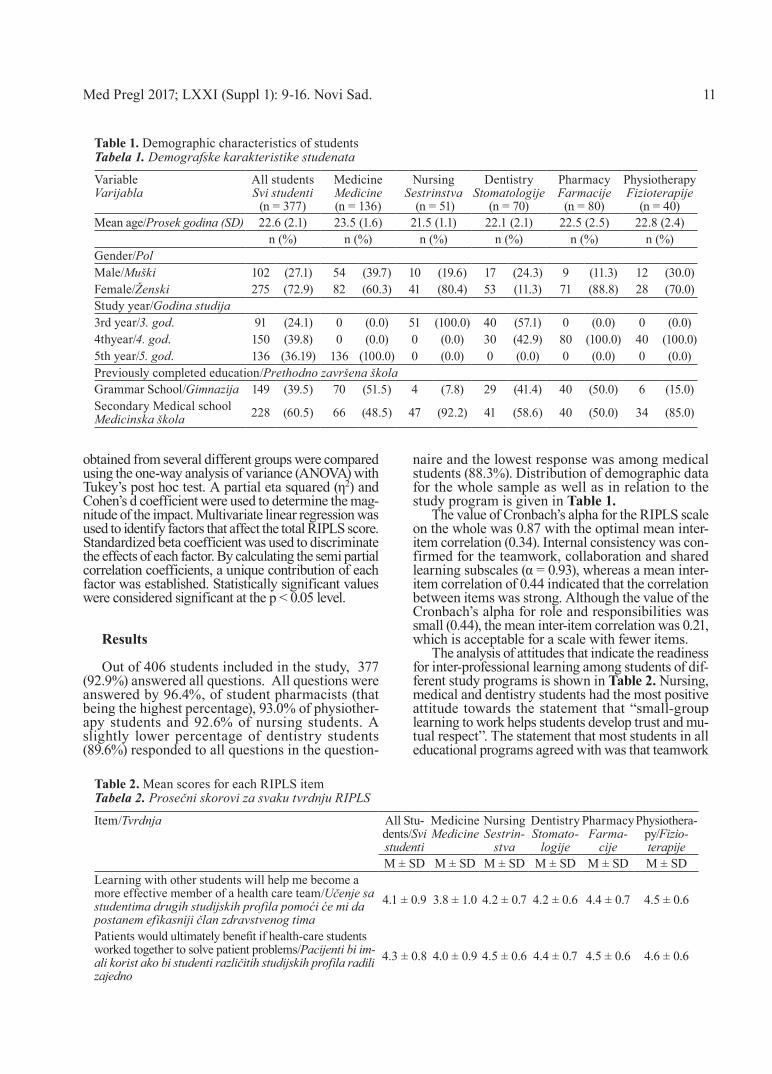
Out of 406 students included in the study, 377 (92.9%) answered all questions. All questions were answered by 96.4%, of student pharmacists (that being the highest percentage), 93.0% of physiother-apy students and 92.6% of nursing students. A slightly lower percentage of dentistry students (89.6%) responded to all questions in the question-
naire and the lowest response was among medical students (88.3%). Distribution of demographic data for the whole sample as well as in relation to the study program is given in Table 1.
The value of Cronbach’s alpha for the RIPLS scale on the whole was 0.87 with the optimal mean inter-item correlation (0.34). Internal consistency was con-firmed for the teamwork, collaboration and shared learning subscales (α = 0.93), whereas a mean inter-item correlation of 0.44 indicated that the correlation between items was strong. Although the value of the Cronbach’s alpha for role and responsibilities was small (0.44), the mean inter-item correlation was 0.21, which is acceptable for a scale with fewer items.
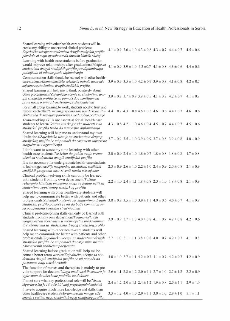
The analysis of attitudes that indicate the readiness for inter-professional learning among students of dif-ferent study programs is shown in Table 2. Nursing, medical and dentistry students had the most positive attitude towards the statement that “small-group learning to work helps students develop trust and mu-tual respect”. The statement that most students in all educational programs agreed with was that teamwork
Table 1. Demographic characteristics of studentsTabela 1. Demografske karakteristike studenata
VariableVarijabla
All studentsSvi studenti
(n = 377)
MedicineMedicine(n = 136)
NursingSestrinstva
(n = 51)
DentistryStomatologije
(n = 70)
PharmacyFarmacije
(n = 80)
PhysiotherapyFizioterapije
(n = 40)Mean age/Prosek godina (SD) 22.6 (2.1) 23.5 (1.6) 21.5 (1.1) 22.1 (2.1) 22.5 (2.5) 22.8 (2.4)
n (%) n (%) n (%) n (%) n (%) n (%)Gender/PolMale/Muški 102 (27.1) 54 (39.7) 10 (19.6) 17 (24.3) 9 (11.3) 12 (30.0)Female/Ženski 275 (72.9) 82 (60.3) 41 (80.4) 53 (11.3) 71 (88.8) 28 (70.0)Study year/Godina studija3rd year/3. god. 91 (24.1) 0 (0.0) 51 (100.0) 40 (57.1) 0 (0.0) 0 (0.0)4thyear/4. god. 150 (39.8) 0 (0.0) 0 (0.0) 30 (42.9) 80 (100.0) 40 (100.0)5th year/5. god. 136 (36.19) 136 (100.0) 0 (0.0) 0 (0.0) 0 (0.0) 0 (0.0)Previously completed education/Prethodno završena školaGrammar School/Gimnazija 149 (39.5) 70 (51.5) 4 (7.8) 29 (41.4) 40 (50.0) 6 (15.0)Secondary Medical schoolMedicinska škola 228 (60.5) 66 (48.5) 47 (92.2) 41 (58.6) 40 (50.0) 34 (85.0)
Table 2. Mean scores for each RIPLS itemTabela 2. Prosečni skorovi za svaku tvrdnju RIPLS
Item/Tvrdnja All Stu-dents/Svi studenti
MedicineMedicine
NursingSestrin-
stva
DentistryStomato-
logije
PharmacyFarma-
cije
Physiothera-py/Fizio-terapije
M ± SD M ± SD M ± SD M ± SD M ± SD M ± SDLearning with other students will help me become a more effective member of a health care team/Učenje sa studentima drugih studijskih profila pomoći će mi da postanem efikasniji član zdravstvenog tima
4.1 ± 0.9 3.8 ± 1.0 4.2 ± 0.7 4.2 ± 0.6 4.4 ± 0.7 4.5 ± 0.6
Patients would ultimately benefit if health-care students worked together to solve patient problems/Pacijenti bi im-ali korist ako bi studenti različitih studijskih profila radili zajedno
4.3 ± 0.8 4.0 ± 0.9 4.5 ± 0.6 4.4 ± 0.7 4.5 ± 0.6 4.6 ± 0.6
12
Shared learning with other health-care students will in-crease my ability to understand clinical problemsZajedničko učenje sa studentima drugih studijskih profila povećalo bi moju sposobnost da shvatim klinički slučaj
4.1 ± 0.9 3.6 ± 1.0 4.3 ± 0.8 4.3 ± 0.7 4.4 ± 0.7 4.5 ± 0.6
Learning with health-care students before graduation would improve relationships after graduation/Učenje sa studentima drugih studijskih profila pre diplomiranja poboljšalo bi odnose posle diplomiranja
4.1 ± 0.9 3.9 ± 1.0 4.2 ±0.7 4.1 ± 0.8 4.3 ± 0.6 4.4 ± 0.6
Communication skills should be learned with other health-care students/Komunikacijske veštine bi trebalo da se uče zajedno sa studentima drugih studijskih profila
3.9 ± 0.9 3.5 ± 1.0 4.2 ± 0.9 3.9 ± 0.8 4.1 ± 0.8 4.2 ± 0.7
Shared learning will help me to think positively about other professionals/Zajedničko učenje sa studentima dru-gih studijskih profila će mi pomoći da razmišljam na pravi način o svim zdravstvenim profesionalcima
3.9 ± 0.8 3.7 ± 0.9 3.9 ± 0.5 4.1 ± 0.8 4.2 ± 0.7 4.1 ± 0.7
For small group learning to work, students need to trust and respect each other/U malim grupama koje uče da rade, stu-denti treba da razvijaju poverenje i međusobno poštovanje
4.4 ± 0.7 4.3 ± 0.8 4.6 ± 0.5 4.6 ± 0.6 4.4 ± 0.7 4.6 ± 0.6
Team-working skills are essential for all health care students to learn/Veštine timskog rada studenti svih studijskih profila treba da nauče pre diplomiranja
4.3 ± 0.8 4.2 ± 1.0 4.6 ± 0.4 4.5 ± 0.7 4.4 ± 0.7 4.5 ± 0.6
Shared learning will help me to understand my own limitations/Zajedničko učenje sa studentima drugog studijskog profila će mi pomoći da razumem sopstvene mogućnosti i ograničenja
3.7 ± 0.9 3.5 ± 1.0 3.9 ± 0.9 3.7 ± 0.8 3.9 ± 0.8 4.0 ± 0.9
I don’t want to waste my time learning with other health-care students/Ne želim da gubim svoje vreme učeći sa studentima drugih studijskih profila
2.0 ± 0.9 2.4 ± 1.0 1.8 ± 0.7 1.8 ± 0.8 1.8 ± 0.8 1.7 ± 0.8
It is not necessary for undergraduate health-care students to learn together/Nije neophodno da studenti različitih studijskih programa zdravstvenih nauka uče zajedno
2.3 ± 0.9 2.6 ± 1.0 2.2 ± 1.0 2.4 ± 0.9 2.0 ± 0.8 2.1 ± 0.9
Clinical problem-solving skills can only be learned with students from my own department/Veštine rešavanja kliničkih problema mogu se jedino učiti sa studentima sopstvenog studijskog profila
2.2 ± 1.0 2.4 ± 1.1 1.8 ± 0.8 2.3 ± 1.0 1.8 ± 0.8 2.1 ± 0.9
Shared learning with other health-care students will help me to communicate better with patients and other professionals/Zajedničko učenje sa studentima drugih studijskih profila pomoći će mi da bolje komuniciram sa pacijentima i ostalim stručnjacima
3.8 ± 0.9 3.5 ± 1.0 3.9 ± 1.1 4.0 ± 0.6 4.0 ± 0.7 4.1 ± 0.9
Clinical problem-solving skills can only be learned with students from my own department/Pozdravio/la bih mogućnost da učestvujem u nekim opštim predavanjima ili radionicama sa studentima drugog studijskog profila
3.9 ± 0.9 3.7 ± 1.0 4.0 ± 0.8 4.1 ± 0.7 4.2 ± 0.8 4.2 ± 0.6
Shared learning with other health-care students will help me to communicate better with patients and other professionals/Zajedničko učenje sa studentima drugih studijskih profila će mi pomoći da razjasnim suštinu zdravstvenih problema pacijenata
3.7 ± 1.0 3.1 ± 1.1 3.8 ± 0.8 4.0 ± 0.7 4.2 ± 0.7 4.1 ± 0.8
Shared learning before graduation will help me be-come a better team worker/Zajedničko učenje sa stu-dentima drugih studijskih profila će mi pomoći da postanem bolji timski radnik
4.0 ± 1.0 3.7 ± 1.1 4.2 ± 0.7 4.1 ± 0.7 4.2 ± 0.7 4.2 ± 0.9
The function of nurses and therapists is mainly to pro-vide support for doctors/Uloga medicinskih sestara je uglavnom da obezbede podršku za doktore
2.6 ± 1.1 2.8 ± 1.2 2.0 ± 1.1 2.7 ± 1.0 2.7 ± 1.2 2.2 ± 0.9
I’m not sure what my professional role will be/Nisam siguran/a šta je i šta će biti moj profesionalni zadatak 2.4 ± 1.2 2.0 ± 1.1 2.4 ± 1.2 1.9 ± 0.8 2.5 ± 1.1 2.9 ± 1.0
I have to acquire much more knowledge and skills than other health-care students/Moram usvojiti mnogo više znanja i veština nego studenti drugog studijskog profila
3.3 ± 1.2 4.0 ± 1.0 2.9 ± 1.1 3.0 ± 1.0 2.9 ± 1.0 3.1 ± 1.1
Simin D, et al. New Strategy in Education of Health Professionals in Serbia
Med Pregl 2017; LXXI (Suppl 1): 9-16. Novi Sad. 13
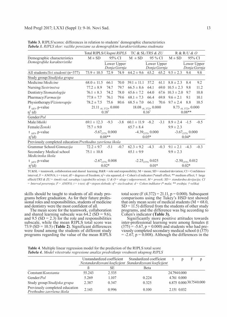
skills should be taught to students of all study pro-grams before graduation. As for their future profes-sional roles and responsibilities, students of medicine and dentistry were the most confident of all.
The mean score for the teamwork, collaboration and shared learning subscale was 64.2 (SD = 9.6), and 9.5 (SD = 2.3) for the role and responsibilities subscale, while the mean RIPLS total score was 73.9 (SD = 10.5) (Table 2). Significant differences were found among the students of different study programs regarding the value of the mean RIPLS
total score (F (4.372) = 21.11, p = 0.000). Subsequent comparisons using the Tukey’s HSD test showed that only mean score of medical students (M = 68.0, SD = 11.5) differed from the students of other study programs, and the difference was big according to Cohen’s indicator (Table 3).
Significantly more positive attitudes towards inter-professional learning were among females (t (375) = -5.67, p = 0.000) and students who had pre-viously completed secondary medical school (t (375) = -2.67, p = 0.008). Although the differences in the
Table 3. RIPLS’scores: differences in relation to students’ demographic characteristicsTabela 3. RIPLS skor: razlike povezane sa demografskim karakteristikama studenata
Total RIPLS/Ukupni RIPLS TC & SL/TRS & ZU R & R/U & ODemographic characteristicsDemografske karakteristike
M ± SD 95% CI M ± SD 95 % CI M ± SD 95% CILower Donja
Upper Gornja
Lower Donja
Upper Gornja
Lower Donja
Upper Gornja
All students/Svi studenti (n=377) 73.9 ± 10.5 72.9 74.9 64.2 ± 9.6 63.2 65.2 9.5 ± 2.3 9.4 9.8Study group/Studijska grupaMedicine/Medicine 68.0 ± 11.5 66.1 70.0 59.1 ± 11.1 57.2 61.1 8.8 ± 2.3 8.4 9.2Nursing/Sestrinstva 77.2 ± 8.9 74.7 79.7 66.5 ± 8.6 64.1 69.0 10.5 ± 2.3 9.8 11.2Dentistry/Stomatologije 76.1 ± 8.3 74.2 78.0 65.6 ± 7.2 64.0 67.6 10.3 ± 2.0 9.7 10.8Pharmacy/Farmacije 77.8 ± 7.7 76.1 79.6 68.1 ± 7.3 66.4 69.8 9.6 ± 2.1 9.1 10.1Physiotherapy/Fizioterapije 78.2 ± 7.5 75.6 80.6 68.5 ± 7.0 66.1 70.6 9.7 ± 2.4 8.8 10.5 F (df), p-value 21.11 (4, 372), 0.000 18.08 (4, 372), 0.000 8.73 (4, 372), 0.000ƞ2 (d) 0.18† 0.16† 0.08**Gender/PolMale/Muški 69.1 ± 12.3 -9.3 -3.8 60.1 ± 11.9 -8.2 -3.1 8.9 ± 2.4 -1.5 -0.5Female/Ženski 75.7 ± 9.0 65.7 ± 8.4 9.9 ± 2.3 t (df), p-value -5.67(375) 0.000 -4.39(375) 0.000 -3.67(375) 0.000 ƞ2(d) 0.08** 0.05* 0.04*Previously completed education/Prethodno završena školaGrammar School/Gimnazija 72.2 ± 9.7 -5.1 -0.7 62.3 ± 9.2 -4.3 -0.3 9.1 ± 2.1 -4.3 -0.3Secondary Medical schoolMedicinska škola
75.1 ± 10.8 65.1 ± 9.9 9.9 ± 2.3
t (df), p-value -2.67(375) 0.008 -2.25(375) 0.025 -2.50(375) 0.012 ƞ2(d) 0.02* 0.01* 0.02*TC&SL = teamwork, collaboration and shared learning; R&R = role and responsibility; M = mean; SD = standard deviation; CI = Confidence interval; F = ANOVA; t = t-test; df = degrees of freedom; ƞ2= eta squared; d = Cohen’s d indicator (*small effect; ** medium effect; † large effect)/TRS & ZU = timski rad, saradnja i zajedničko učenje; U & O = uloge i odgovornosti, M = prosek; SD = standardna devijacija; CI = Interval poverenja; F = ANOVA; t = t-test; df =stepen slobode; ƞ2= eta kvadrat; d = Cohen indikator (* mala; ** srednja; † velika)
Table 4. Multiple linear regression model for the prediction of the RIPLS total scoreTabela 4. Model višestruke regresione analize predviđanje vrednosti ukupnog RIPLS
Unstandardized coefficientNestandardizovani koeficijent
Standardized coefficientStandardizovani koeficijent
t p F p
ß SE BetaConstant/Konstanta 55.243 2.535 24.7940.000
30.7540.000Gender/Pol 5.269 1.107 0.224 4.761 0.000Study group/Studijska grupa 2.387 0.347 0.325 6.875 0.000Previously completed educationPrethodno završena škola 2.143 0.996 0.100 2.151 0.032
14
mean RIPLS total score and both subscales were statistically significant, Cohen’s indicator points to a medium or small effect of these variables.
In Table 4, the results of standard multiple lin-ear regressions show that all three independent variables were significantly related to the total RIPLS score. Based on the values of beta coeffi-cients, it is noted that the study program (beta = 0.325, p = 0.000) individually contributes the most to the explanation of the RIPLS score.
The multiple regression analysis model explains 19.8% (adjusted R2 = 19.2%) variance of the overall RIPLS score. A further analysis calculated semi partial correlation coefficients, which was the basis to determine the unique contribution of each inde-pendent variable. The obtained results indicate that 12.5%, 4.9 and only 1% of the variance value of the total RIPLS score was accounted for by the study program, gender, and previously completed school, respectively.
Discussion
The implementation of IPE as an educational strategy requires overcoming many structural and or-ganizational barriers, but it is difficult to alter ambiva-lent or negative students’ attitudes [2, 4, 5]. Making this alteration is also one of the primary objectives in planning IPE outcomes aimed at improving teamwork and developing collaborative practice [4, 5, 7]. There-fore, the assessment of the students’ baseline attitudes is significant for the initial steps towards integration of this educational strategy into curricula [4, 5].
Students’ attitudes among five educational profiles in this study, assessed by RIPLS, indicate students’ readiness for IPE. Similar results were obtained in pre-vious studies as well [4–9, 11–18]. Accordingly, there were differences among students’ attitudes based on their study profile both in our study and studies con-ducted in other countries [4, 6, 7, 11–20]. Namely, medical students were less open towards IPE, while nursing, pharmacy, and physiotherapy students dem-onstrated greater readiness for IPE.
The results of this study show that female students are more ready to accept shared learning. Some authors suggest that these results reflect different learning styles between men and women. Namely, women are more ready to accept the IPE because they are more inclined to listen, understand and accept attitudes of other people [6, 11, 18].
Students with previously completed secondary medical school are more prepared for IPE, probably due to longer contact with the actual setting during schooling. However, our results are not comparable with the results of studies conducted in most countries, because their education system is different from the one in Serbia. In these countries, studies at faculties of health and social care are preceded only by general and not vocational medical education.
One of the prerequisites for this research was that students started lectures on clinical subjects, i.e. in a real clinical setting. This is important because team-
work prevails in such a setting, whereby a team com-prises different professionals, with varying number of members, a collaboration time frame, circumstances under which they were formed and the way they solve their common task. It is important to emphasize that most students of all profiles fully agreed with state-ments which indicated the shared learning benefits from teamwork.
The largest number of students believed that trust and mutual respect developed through small group shared learning. Such results are not surprising as the students had the opportunity to feel the benefits of this work during their previous education. Namely, while the education system in Serbia is still dominantly tra-ditional, group work as teaching and learning method-ology has been included in education since elementary school. Students also occasionally work and study to-gether in groups. However, these are uni-professional groups comprising at least 10 students, and problem-solving tasks are a major characteristic of their profes-sional domain.
There is a question in literature whether it is better to introduce inter-professional education before or after graduation. Our students seem to like the idea of acquir-ing the skills necessary for the team work during their studies, i.e. before graduation. The initiators of the im-plementation of the IPE education strategy consider it to be a part of professional development of students, which begins with acquiring qualifications/diplomas and continues throughout their professional career [21]. According to a WHO report, the IPE enables students to acquire knowledge and skills required to become a collaborative practice-ready health worker [2].
The lowest mean score in this study was recorded for the statement indicating that students did not con-sider learning together with students of different study profiles to be a waste of time. On the contrary, they generally consider shared learning to be necessary, specifically emphasizing its importance for acquiring the skills to solve clinical problems. However, students of medicine and dentistry are still more focused on uni-professional learning, especially when it comes to acquiring the skills to solve clinical problems. In sev-eral previous studies, similar results were obtained, which could be explained by the realistic conflict theory according to which, hostile and discriminatory inter-group relationships are the result of negative at-titudes [22]. A similar interpretation is also found by Hind, who analyzed the interpersonal perceptions of students of medical faculties and found that individu-als who identified strongly and positively with their own professional group tended to be more negative towards students of other groups [20]. Certainly, the aim of the IPE strategy is not to equate students’ at-titudes and opinions, but to approach the problem/task from different perspectives and positions, while re-specting different attitudes of students of different and/or the same profile [2, 5, 21]. Barr points out that inter-professional education is assumed to provide students an opportunity to see that intervention by one profes-sion at one “point of the system will affect the func-tioning of the system as a whole” [21].
Simin D, et al. New Strategy in Education of Health Professionals in Serbia
Med Pregl 2017; LXXI (Suppl 1): 9-16. Novi Sad. 15
Compared to the previous research conducted at the Faculty of Medicine in Novi Sad [12], students of nurs-ing are now more confident about their professional tasks. However, although academic studies of nursing in Serbia were established fifteen years ago, current legislation within the health care system has not yet fully “recognized” this profile. The least confident in their future professional tasks in our study are students of physiotherapy, which is one of the latest academic study profiles, also “unrecognized”. The uncertainty of pharmacy students regarding their future tasks in our study is not a solitary case in the literature. The authors of comprehensive review studies explain simi-lar results by the fact that the traditional occupational tasks of pharmacists (drug issuing and compounding) are changing due to the rapid development of pharma-ceutical practice [23]. The contribution of pharmacists in clinical practice in the form of providing profes-sional advice to patients and members of the health team, preventing adverse drug effects and monitoring drug use considerably increases the safety and efficacy of drug administration, which further decrease treat-ment expenses and results in changes in clinical team-work. This significantly increases the safety and effi-cacy of drug use, which contributes to reducing the cost of treatment, but also leads to changes in clinical team-work. Due to these changes, team members have to change attitudes towards traditional tasks of pharma-cists and accept new ones [15,23]. In contrast, dentistry students know very well what their future professional tasks will be.
Students of medicine, in our and several previous studies, were least opposed to the statement that the role of nursing technicians was to provide full support to doctors. However, students of nursing and other study profiles had significantly different attitudes not only in our study but in previous studies as well as [Horsburg, el Zub]. Such results indicate that the roles of nurses continue to be accepted stereotypically (as subordinate members of the health team who mostly act merely as persons who carry out doctors’ orders) [16]. Certainly, IPE is not a panacea for every challenge that the health-care system encounters [2]. However, an effective im-plementation that respects the basic provisions is based on mutual respect of students of various study profiles and provides the possibility of reducing negative stere-otypes, as well as eliminating them [2, 8, 21].
Conclusion
The attitudes of most students of all study profiles, participating in this study, indicate that our students are ready to accept IPE as a new educational strategy. In addition, a more detailed analysis of responses of the students’ of each profile allows us to perceive all potential barriers and find solutions in due time in or-der to integrate IPE into the curriculum of all study profiles of the Faculty of Medicine, specified in the ReFEEHS project activities. The effective integration of IPE would contribute to the development and ac-ceptance of collaborative practice as the underlying model of healthcare workers in Serbia as well.
References
1.Frenk J, Chen L, Bhutta ZA, Cohen J, Crisp N, Evans T, et al. Health professionals for a new century: transforming education to strengthen health systems in an interdependent world. Lancet. 2010;376(9756):1923-58.
2. World Health Organization (WHO). Framework for ac-tion on interprofessional education and collaborative practice [Internet]. Geneva: World Health Organization; 2010 [cited 2018 Aug 5]. Available from: http://www.who.int/hrh/resources/framework_action/en/.
3. Reeves S, Perrier L, Goldman J, Freeth D, Zwarenstein M. Interprofessional education: effects on professional practice and healthcare outcomes (update). Cochrane Database Syst Rev. 2013;2013(3):CD002213.
4. Chan LK, Ganotice F Jr, Wong FKY, Lau CS, Bridges SM, Chan CHY, et al. Implementation of an interprofessional team-based learning program involving seven undergraduate health and social care programs from two universities, and stu-dents' evaluation of their readiness for interprofessional learn-ing. BMC Med Educ. 2017;17(1):221.
5. O'Keefe M, Ward H. Implementing interprofessional learning curriculum: how problems might also be answers. BMC Med Educ. 2018;18(1):132.
6. Talwalkar JS, Fahs DB, Kayingo G, Wong R, Jeon S, Honan L. Readiness for interprofessional learning among healthcare professional students. Int J Med Educ. 2016;7:144-8.
7. Thompson BM, Bratzler DW, Fisher MJ, Torres A, Fac-ulty E, Sparks RA. Working together: using a unique approach
to evaluate an interactive and clinic-based longitudinal inter-professional education experience with 13 professions. J Inter-prof Care. 2016;30(6):754-61.
8. Visser CLF, Wilschut JA, Isik U, van der Burgt SME, Croiset G, Kusurkur RA. The Association of Readiness for In-terprofessional Learning with empathy, motivation and profes-sional identity development in medical students. BMC Med Educ. 2018;18(1):125.
9. Mahler C, Berger S, Reeves S. The Readiness for Inter-professional Learning Scale (RIPLS): a problematic evaluative scale for the interprofessional field. J Interprof Care. 2015;29(4):289-91.
10. Reeves S, Boet S, Zierler B, Kitto S. Interprofessional Education and Practice Guide No. 3: evaluating interprofes-sional education. J Interprof Care. 2015;29(4):305-12.
11. Milutinović D, Lovrić R, Simin D. Interprofessional edu-cation and collaborative practice: psychometric analysis of the Readiness for Interprofessional Learning Scale in undergraduate Serbian healthcare student context. Nurse Educ Today. 2018;65:74-80.
12. Simin D, Milutinović D, Brestovački B, Andrijević I, Cigić T. Improvement of teamwork in health care through inter-professional education. Srp Arh Celok Lek. 2010;138(7-8):480-5.
13. Parsell G, Bligh J. The development of a questionnaire to assess readiness of health care students for interprofessional learning. Med Educ. 1999;33(2):95-100.
16
Rad je primljen 3. IX 2018.Recenziran 5. IX 2018.Prihvaćen za štampu 10. IX 2018.BIBLID.0025-8105:(2018):Suppl 1:9-16.
14. McFadyen AK, Webster V, Strachan K, Figgins E, Brown H, McKechnie J. The Readiness for Interprofessional Learning Scale: a possible more stable sub-scale model for the original version of RIPLS. J Interprof Care. 2005;19(6):595-603.
15. Horsburgh M, Lamdin R, Williamson E. Multiprofes-sional learning: the attitudes of medical nursing and pharmacy students to shared learning. Med Educ. 2001;35(9):876–83.
16. El-Zubeir M, Rizk DE, Al-Khalil RK. Are senior UAE medical and nursing students ready for interprofessional learn-ing? Validating the RIPL scale in a Middle Eastern context. J Interprof Care. 2006;20(6):619-32.
17. de Oliveira VF, Bittencourt MF, Navarro Pinto ÍF, Luc-chetti ALG, da Silva Ezequiel O, Lucchetti G. Comparison of the Readiness for Interprofessional Learning and the rate of contact among students from nine different healthcare courses. Nurse Educ Today. 2018;63:64-8.
18. Coster S, Norman I, Murrells T, Kitchen S, Meerabeau E, Sooboodoo E, et al. Interprofessional attitudes amongst un-dergraduate students in the health professions: a longitudinal questionnaire survey. Int J Nurs Stud. 2008;45(11):1667-81.
19. Al-Shaikh GK, Al-Madi EM, Masood J, Shaikh Q, Syed SB, Bader RS, et al. Interprofessional learning experiences: ex-ploring the perception and attitudes of Saudi Arabian medical and dental students. Med Teach. In press. DOI: 10.1080/0142159X.2018.1465180.
20. Hind M, Norman I, Cooper S, Gill E, Hilton R, Judd P, et al. Interprofessional perceptions of healthcare students. J In-terprof Care. 2003;17(1):21-34.
21. Barr H, Koppel I, Reeves S, Hammick M, Freeth DS. Effective interprofessional education: argument, assumption and evidence. London: Blackwell Publishing Ltd; 2008.
22. Craddock D, O’Halloran C, Borthwick A. Interprofes-sional education in health and social care: fashion or informed practice? Learning in Health and Social Care. 2006;5(4):220-42.
23. El-Awaisi A, Joseph S, El Hajj MS, Diack L. A compre-hensive systematic review of pharmacy perspectives on interpro-fessional education and collaborative practice. Res Social Adm Pharm. 2018;14(10):863-82.
Simin D, et al. New Strategy in Education of Health Professionals in Serbia
Med Pregl 2018; LXXI (Suppl 1): 17-24. Novi Sad. 17
SummaryIntroduction. The purpose of this study was to evaluate sleep qual-ity among healthcare science students and to assess its association with sleep hygiene knowledge and practices, circadian typology and lifestyle factors. Material and Methods. The research was con-ducted as a cross-sectional questionnaire-based internet study on a sample of 268 students. The Pittsburgh Sleep Quality Index and The Self-Morningness-Eveningness Questionnaire were applied as re-search instruments to assess sleep quality and chronotypes respec-tively, while evaluation of the other variables was conducted using he Sleep Hygiene Knowledge Questionnaire, socio-demographic questionnaire and the questionnaire of lifestyle factors. Results. The average of the Pittsburgh Sleep Quality Index global score for all students was 6.9 ± 3.2. The poor sleep quality (The Pittsburgh Sleep Quality Index > 5) was reported in 62.7% of students. Sleep effi-ciency less than 85% was found in 43.0% of students, and 78% of students reported daytime dysfunctionality. Sleep quality was sig-nificantly worse among female students; coffee, alcohol and energy drink consumers and long-term cell phone users. Only 11.9% of students were classified as the morning chronotype and they had the best quality of sleep and the best sleep hygiene knowledge and prac-tices, whereas the evening chronotype had the worst quality of sleep. A significant negative correlation was identified between sleep hy-giene knowledge (r = - 0.133) and practice (r = 0.501) and sleep qual-ity whereby the lower t he Sleep Hygiene Knowledge Questionnaire and Sleep Hygiene Practice Scale scores follow a higher the Pitts-burgh Sleep Quality Index score. Conclusion. Majority of students had a suboptimal level of overall sleep quality, satisfactory knowledge of sleep hygiene, but they did not have the sleep hygiene practices which suggests that knowledge is not a factor of deterring from un-healthy behaviors.Key words: Sleep Hygiene; Circadian Rhythm; Health Knowledge, Attitudes, Practice; Life Style; Students, Nursing; Surveys and Ques-tionnaires; Chronobiology Disorders
SažetakUvod. Cilj ove studije bio je da se proceni kvalitet spavanja stude-nata zdravstvenih nauka i ispita njegova veza sa znanjem i praksom higijene spavanja, cirkadijalnom tipologijom i faktorima životnog stila. Materijal i metode. Istraživanje je sprovedeno kao studija preseka onlajn anketiranjem 268 studenta. Kao instrumenti istraživanja koristili su se Pitsburški indeks kvaliteta spavanja za procenu kvaliteta spavanja, Upitnik o samoproceni „jutarnjosti/večernjosti" za procenu diurnalne sklonosti, a za evaluaciju ostalih varijabili korišten je Upitnik znanja o higijeni spavanja i Skala prakse higijene spavanja, sociodemografski upitnik i upitnik o životnom stilu i navikama. Rezultati. Prosečan ukupni skor Pitsburškog indeksa kvaliteta spavanja svih studenata iznosio je 6,9 ± 3,2. Loš kvalitet spavanja (Pitsburški indeks kvaliteta spavanja > 5) imalo je 62,7% studenata. Efikasnost spavanja manju od 85% imalo je 43% studenata, a 78% je prijavilo dnevnu disfunkcional-nost. Studenti ženskog pola, konzumenti kafe, alkohola i energet-skih napitaka, kao i oni koji duže koriste mobilne telefone, imali su značajno lošiji kvalitet spavanja. Samo 11,9% studenata je pripada-lo jutarnjem hronotipu i imali su najbolji kvalitet spavanja i na-jbolja znanja i praksu iz higijene spavanja, a večernji najlošije. Utvrđena je značajna negativna povezanost znanja (r = - 0,133; p < 0,05) i prakse higijene spavanja (r = 0,501; p < 0,001) sa kvalitetom spavanja pri čemu niži Upitnik znanja o higijeni spavanja i skor skale prakse higijene spavanja prati viši skor na Pitsburški indeks kvaliteta spavanja. Zaključak. Većina studenata je imala subopti-malni ukupni kvalitet spavanja, zadovoljavajuće znanje o higijeni spavanja, ali ne i praksu higijene spavanja, što ukazuje da znanje nije faktor odvraćanja od nezdravog ponašanja.Ključne reči: higijena spavanja; cirkadijalni ritam; znanje o zdravlju, stavovi, praksa; stil života; studenti zdravstvene nege; istraživanja i upitnici; hronobiološki poremećaji
University of Novi Sad, Faculty of Medicine, Department of Nursing1 UDK 613.79-057.875University of Novi Sad, Faculty of Medicine, Department of Neurology2 https://doi.org/10.2298/MPNS18S1017ACollege of Applied Health Science, Ćuprija3
University of Novi Sad, Faculty of Medicine, Department of Psychiatry and Medical Psychology4
SLEEP QUALITY IN RELATION TO SLEEP HYGIENE KNOWLEDGE AND PRACTICE, CHRONOTYPE AND LIFESTYLE BEHAVIOUR AMONG HEALTHCARE STUDENTS
POVEZANOST KVALITETA SPAVANJA STUDENATA ZDRAVSTVENIH NAUKA SA ZNANJEM I PRAK-SOM HIGIJENE SPAVANJA, HRONOTIPOM I ŽIVOTNIM STILOM I NAVIKAMA
Ilija ANDRIJEVIĆ1, Svetlana SIMIĆ2, Čedomirka STANOJEVIĆ3, Boris GOLUBOVIĆ4 and Dragana MILUTINOVIĆ1
Introduction
Sleep is an integral part of human biological rhythm and it is essential for optimal health and main-tenance of cognitive and psychosocial functioning.
Sleep pattern is regulated by the interaction of three major factors: homeostatic, internal circadian and behavioral factors [1, 2]. Given that behavioral factors, such as sleep hygiene, often exceed the homeostatic and circadian factors in the regulation of sleep, it is
Corresponding Author: Prof. dr Ilija Andrijević, Univerzitet u Novom Sadu, Medicinski fakultet, Katedra za zdravstvenu negu, 21000 Novi Sad, Hajduk Veljkova 3, E-mail: [email protected]
18 Andrijević I, et al. Assessment of Sleep Quality among Healthcare Students
important to know those behaviors that affect its qual-ity and which may possibly cause sleep disorders [3].
Sleep hygiene is defined as a set of behaviors and environmental rules that aim to ensure a restorative and good quality sleep in order to avoid certain sleep disorders [4]. Previous studies indicate that sleep hy-giene practice is associated with sleep quality, and the results of the interaction between sleep hygiene knowl-edge and sleep quality were found to be inconsistent [4–7]. Having thoroughly studied literature, we have found that sleep hygiene knowledge does not affect sleep quality [4, 5], or that people with better sleep hygiene knowledge report better sleep quality [5, 6], or that there is a weak, negative correlation between sleep hygiene knowledge and sleep quality [7]. However, sleep hygiene knowledge, defined by some authors as “sleeping beliefs” [8], does not have to be related to sleep quality, but the adherence to the implementation of sleep hygiene recommendations serves as a media-tor between “sleeping beliefs” and sleep quality [9].
Some of the individual differences that may af-fect the effectiveness of sleep hygiene knowledge and practices are circadian typology differences (referred as chronotype) [5]. Although these differ-ences are somewhat innate, the individual inclina-tion to a certain sleeping pattern, vigilance and better cognitive and physical activity enable the classification of individuals by their chronotype as morning, evening, and intermediate chronotypes [10]. Data on the association between circadian ty-pology and sleep hygiene knowledge are inconsist-ent. Although some authors find that people with morning chronotype have more accurate knowledge of sleep hygiene than intermediate and evening types [8] the others find no correlation [6].
Student population is particularly vulnerable to sleep disorders caused by external factors such as change of the surroundings, responsibility for lifestyle self-management, changing of daily schedule, stress caused by a greater scope of academic and social com-mitments. These factors are associated with reduced sleep duration, resulting in poor sleep quality and ex-cessive daytime sleepiness [4, 5]. In particular, the cir-cadian rhythm can be disrupted during the exam pe-riod, night-time learning and prolonged exposure to light associated with the excessive use of computer, which contribute to irregular sleep patterns and poor sleep quality [5]. The latest reports show that the rec-ommendation of The National Sleep Foundation re-quiring 7 to 9 hours of sleep per night is respected by less than 50% of students [9], and only about 40% of students reported a good sleep quality [11].
Taking into account the significance of sleep quality in student population primarily, this study has been aimed at evaluating sleep quality among health science students and assessing its association
with sleep hygiene knowledge and practices, circa-dian typology and lifestyle factors.
Material and Methods
Study Design and ParticipantsThe research was conducted as a cross-sectional
questionnaire-based internet study among students of Faculty of Medicine in Novi Sad during the winter se-mester of the academic 2016-17 year. The study link was shared across social networks within closed groups of Faculty students. The study sample consisted of 268 students divided into two groups: n = 174 students of basic studies (nursing, medical rehabilitation and special rehabilitation and education) and n = 94 students of integrated studies (medicine, dentistry and pharmacy).
InstrumentsIn order to achieve study goals, a questionnaire
was designed to cover socio-demographic issues related to gender, age, place of residence and study (the area of study, average grade), as well as a ques-tionnaire of lifestyle factors affecting sleep (includ-ing consumption of alcohol, tobacco, coffee, and caffeinated energy drinks, implementation of phys-ical activity and mobile phone use).
The Pittsburg Sleep Quality Index (PSQI) was used to measure sleep quality. PSQI is a 19-item standardized instrument for evaluating sleep quality over the previ-ous month through 7 components: sleep latency, sleep duration, habitual sleep efficiency and sleep distur-bances, subjective sleep quality, use of sleeping pills, and daytime dysfunction. The components are evalu-ated on a scale of 0–3, and then totaled to yield a global PSQI score, ranging from 0 to 21. A global score > 5 indicates a poor sleep quality. PSQI showed good psy-chometric characteristics and differentiation between good and bad sleepers [12]. In our study, the Cronbach’s alpha coefficient was 0.64.
Sleep hygiene knowledge and practices were as-sessed by the questionnaires designed for the purpose of this research, and on the basis of literature data on the activities that can affect sleep patterns positively or negatively [2, 4, 6, 13]. The 17-item Sleep Hygiene Knowledge Questionnaire (SHKQ) was used to assess knowledge of activities that help or interfere with sleep patterns, where 1 point was given for each correct re-sponse, 0 for incorrect, whereas the global score ranged from 0 to 17. Higher score reflected better sleep hygiene knowledge. In this research, the questionnaire was found to be reliable (Cronbach’s alpha: 0.80).
Sleep Hygiene Practice Scale (SHPS) comprised 22 items to evaluate how many nights per week a respond-ent was engaged in certain activities known to promote or disrupt sleep. Responses ranged from 0 (“never”) to 7 (“7 times a week”). Items that indicated a poor sleep hygiene practice were reversed scored. The global score ranged from 0 to 154, with higher scores indicating better implementation of sleep hygiene practices. In this study, the questionnaire showed a satisfactory level of reliability. The Cronbach’s Alpha coefficient was 0.70.
AbbreviationsPSQI – The Pittsburg Sleep Quality IndexSHKQ – Sleep Hygiene Knowledge QuestionnaireSHPS – Sleep Hygiene Practice Scale Self-ME – The Self-Morningness/Eveningness
Med Pregl 2018; LXXI (Suppl 1): 17-24. Novi Sad. 19
The Self-Morningness/Eveningness (Self-ME) was used to estimate diurnal preferences/chronotypes. Self-ME was based on the 19th question which was singled out of the Morningness-Eveningness Ques-tionnaire (MEQ) [14], according to which the respond-ents were self-classified into a definitely morning, definitely evening or intermediate chronotype.
Statistical Data Analysis IBM SPSS statistics software, Version 23 was used
for data analysis. Data processing included descriptive
and inferential statistics. A comparison of differences between means from two groups was performed by t-test, whereas one-factor analysis of variance (ANO-VA) was used to compare the means of multiple groups with LSD post-hoc test. Effect sizes (d and η2) were also calculated for quantifying the difference between mean scores. Pearson correlation coefficient was used to determine the relationship between variables. The cut off level for statistical significance was p < 0.05.
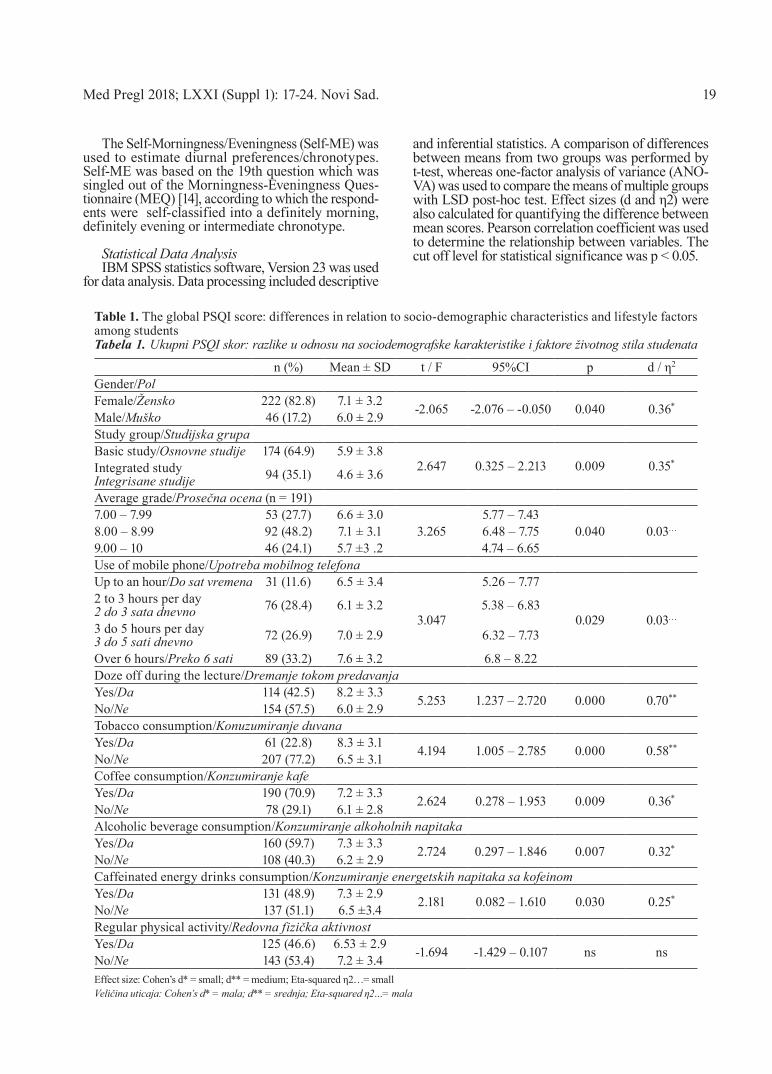
Table 1. The global PSQI score: differences in relation to socio-demographic characteristics and lifestyle factors among studentsTabela 1. Ukupni PSQI skor: razlike u odnosu na sociodemografske karakteristike i faktore životnog stila studenata
n (%) Mean ± SD t / F 95%CI p d / η2
Gender/PolFemale/Žensko 222 (82.8) 7.1 ± 3.2 -2.065 -2.076 – -0.050 0.040 0.36*Male/Muško 46 (17.2) 6.0 ± 2.9Study group/Studijska grupaBasic study/Osnovne studije 174 (64.9) 5.9 ± 3.8
2.647 0.325 – 2.213 0.009 0.35*Integrated study Integrisane studije 94 (35.1) 4.6 ± 3.6
Average grade/Prosečna ocena (n = 191)7.00 – 7.99 53 (27.7) 6.6 ± 3.0
3.2655.77 – 7.43
0.040 0.03…8.00 – 8.99 92 (48.2) 7.1 ± 3.1 6.48 – 7.759.00 – 10 46 (24.1) 5.7 ±3 .2 4.74 – 6.65Use of mobile phone/Upotreba mobilnog telefonaUp to an hour/Do sat vremena 31 (11.6) 6.5 ± 3.4
3.047
5.26 – 7.77
0.029 0.03…
2 to 3 hours per day 2 do 3 sata dnevno 76 (28.4) 6.1 ± 3.2 5.38 – 6.83
3 do 5 hours per day 3 do 5 sati dnevno 72 (26.9) 7.0 ± 2.9 6.32 – 7.73
Over 6 hours/Preko 6 sati 89 (33.2) 7.6 ± 3.2 6.8 – 8.22Doze off during the lecture/Dremanje tokom predavanjaYes/Da 114 (42.5) 8.2 ± 3.3 5.253 1.237 – 2.720 0.000 0.70**No/Ne 154 (57.5) 6.0 ± 2.9Tobacco consumption/Konuzumiranje duvanaYes/Da 61 (22.8) 8.3 ± 3.1 4.194 1.005 – 2.785 0.000 0.58**No/Ne 207 (77.2) 6.5 ± 3.1Coffee consumption/Konzumiranje kafeYes/Da 190 (70.9) 7.2 ± 3.3 2.624 0.278 – 1.953 0.009 0.36*No/Ne 78 (29.1) 6.1 ± 2.8Alcoholic beverage consumption/Konzumiranje alkoholnih napitakaYes/Da 160 (59.7) 7.3 ± 3.3 2.724 0.297 – 1.846 0.007 0.32*No/Ne 108 (40.3) 6.2 ± 2.9Caffeinated energy drinks consumption/Konzumiranje energetskih napitaka sa kofeinomYes/Da 131 (48.9) 7.3 ± 2.9 2.181 0.082 – 1.610 0.030 0.25*No/Ne 137 (51.1) 6.5 ±3.4Regular physical activity/Redovna fizička aktivnostYes/Da 125 (46.6) 6.53 ± 2.9 -1.694 -1.429 – 0.107 ns nsNo/Ne 143 (53.4) 7.2 ± 3.4Effect size: Cohen’s d* = small; d** = medium; Eta-squared η2…= smallVeličina uticaja: Cohen’s d* = mala; d** = srednja; Eta-squared η2…= mala
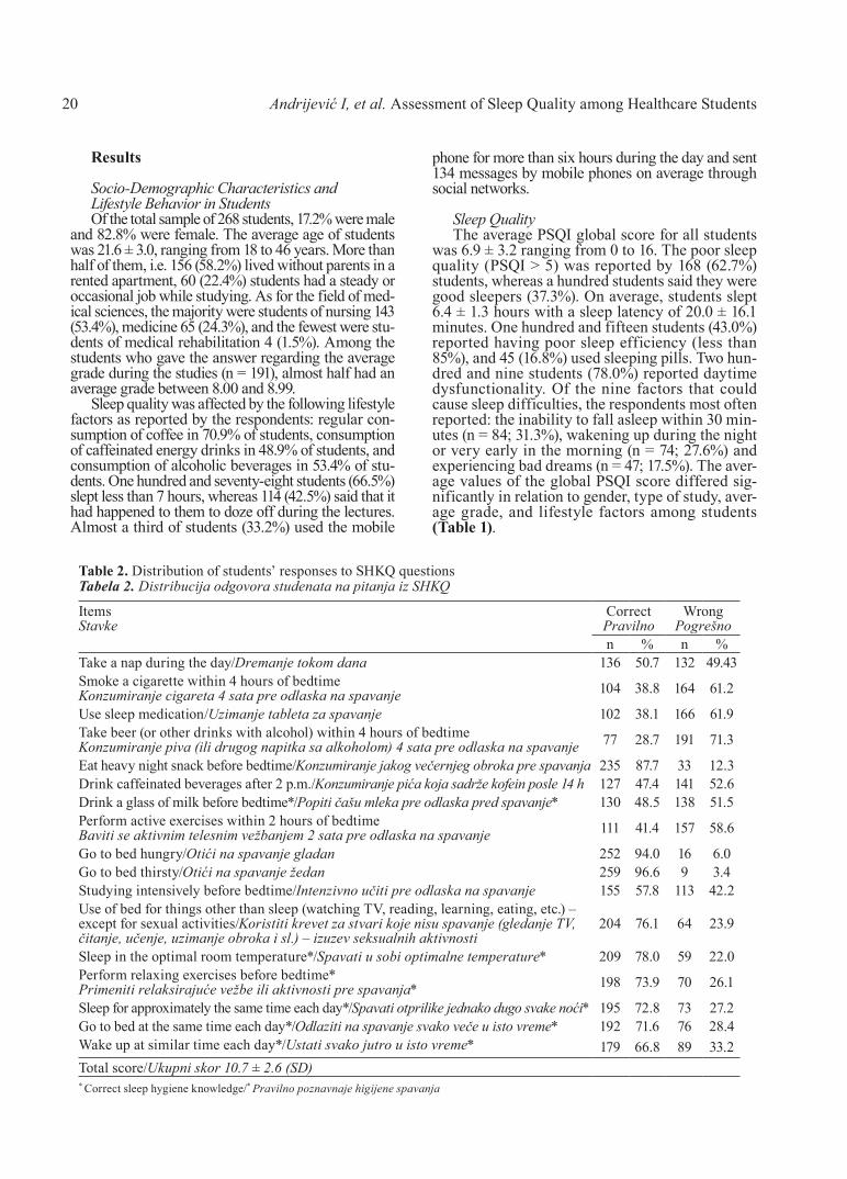
20 Andrijević I, et al. Assessment of Sleep Quality among Healthcare Students
Results
Socio-Demographic Characteristics and Lifestyle Behavior in StudentsOf the total sample of 268 students, 17.2% were male
and 82.8% were female. The average age of students was 21.6 ± 3.0, ranging from 18 to 46 years. More than half of them, i.e. 156 (58.2%) lived without parents in a rented apartment, 60 (22.4%) students had a steady or occasional job while studying. As for the field of med-ical sciences, the majority were students of nursing 143 (53.4%), medicine 65 (24.3%), and the fewest were stu-dents of medical rehabilitation 4 (1.5%). Among the students who gave the answer regarding the average grade during the studies (n = 191), almost half had an average grade between 8.00 and 8.99.
Sleep quality was affected by the following lifestyle factors as reported by the respondents: regular con-sumption of coffee in 70.9% of students, consumption of caffeinated energy drinks in 48.9% of students, and consumption of alcoholic beverages in 53.4% of stu-dents. One hundred and seventy-eight students (66.5%) slept less than 7 hours, whereas 114 (42.5%) said that it had happened to them to doze off during the lectures. Almost a third of students (33.2%) used the mobile
phone for more than six hours during the day and sent 134 messages by mobile phones on average through social networks.
Sleep Quality The average PSQI global score for all students
was 6.9 ± 3.2 ranging from 0 to 16. The poor sleep quality (PSQI > 5) was reported by 168 (62.7%) students, whereas a hundred students said they were good sleepers (37.3%). On average, students slept 6.4 ± 1.3 hours with a sleep latency of 20.0 ± 16.1 minutes. One hundred and fifteen students (43.0%) reported having poor sleep efficiency (less than 85%), and 45 (16.8%) used sleeping pills. Two hun-dred and nine students (78.0%) reported daytime dysfunctionality. Of the nine factors that could cause sleep difficulties, the respondents most often reported: the inability to fall asleep within 30 min-utes (n = 84; 31.3%), wakening up during the night or very early in the morning (n = 74; 27.6%) and experiencing bad dreams (n = 47; 17.5%). The aver-age values of the global PSQI score differed sig-nificantly in relation to gender, type of study, aver-age grade, and lifestyle factors among students (Table 1).
Table 2. Distribution of students’ responses to SHKQ questionsTabela 2. Distribucija odgovora studenata na pitanja iz SHKQ
ItemsStavke
Correct Pravilno
Wrong Pogrešno
n % n %Take a nap during the day/Dremanje tokom dana 136 50.7 132 49.43Smoke a cigarette within 4 hours of bedtimeKonzumiranje cigareta 4 sata pre odlaska na spavanje 104 38.8 164 61.2
Use sleep medication/Uzimanje tableta za spavanje 102 38.1 166 61.9Take beer (or other drinks with alcohol) within 4 hours of bedtimeKonzumiranje piva (ili drugog napitka sa alkoholom) 4 sata pre odlaska na spavanje 77 28.7 191 71.3
Eat heavy night snack before bedtime/Konzumiranje jakog večernjeg obroka pre spavanja 235 87.7 33 12.3Drink caffeinated beverages after 2 p.m./Konzumiranje pića koja sadrže kofein posle 14 h 127 47.4 141 52.6Drink a glass of milk before bedtime*/Popiti čašu mleka pre odlaska pred spavanje* 130 48.5 138 51.5Perform active exercises within 2 hours of bedtime Baviti se aktivnim telesnim vežbanjem 2 sata pre odlaska na spavanje 111 41.4 157 58.6
Go to bed hungry/Otići na spavanje gladan 252 94.0 16 6.0Go to bed thirsty/Otići na spavanje žedan 259 96.6 9 3.4Studying intensively before bedtime/Intenzivno učiti pre odlaska na spavanje 155 57.8 113 42.2Use of bed for things other than sleep (watching TV, reading, learning, eating, etc.) – except for sexual activities/Koristiti krevet za stvari koje nisu spavanje (gledanje TV, čitanje, učenje, uzimanje obroka i sl.) – izuzev seksualnih aktivnosti
204 76.1 64 23.9
Sleep in the optimal room temperature*/Spavati u sobi optimalne temperature* 209 78.0 59 22.0Perform relaxing exercises before bedtime*Primeniti relaksirajuće vežbe ili aktivnosti pre spavanja* 198 73.9 70 26.1
Sleep for approximately the same time each day*/Spavati otprilike jednako dugo svake noći* 195 72.8 73 27.2Go to bed at the same time each day*/Odlaziti na spavanje svako veče u isto vreme* 192 71.6 76 28.4Wake up at similar time each day*/Ustati svako jutro u isto vreme* 179 66.8 89 33.2Total score/Ukupni skor 10.7 ± 2.6 (SD)* Correct sleep hygiene knowledge/* Pravilno poznavnaje higijene spavanja
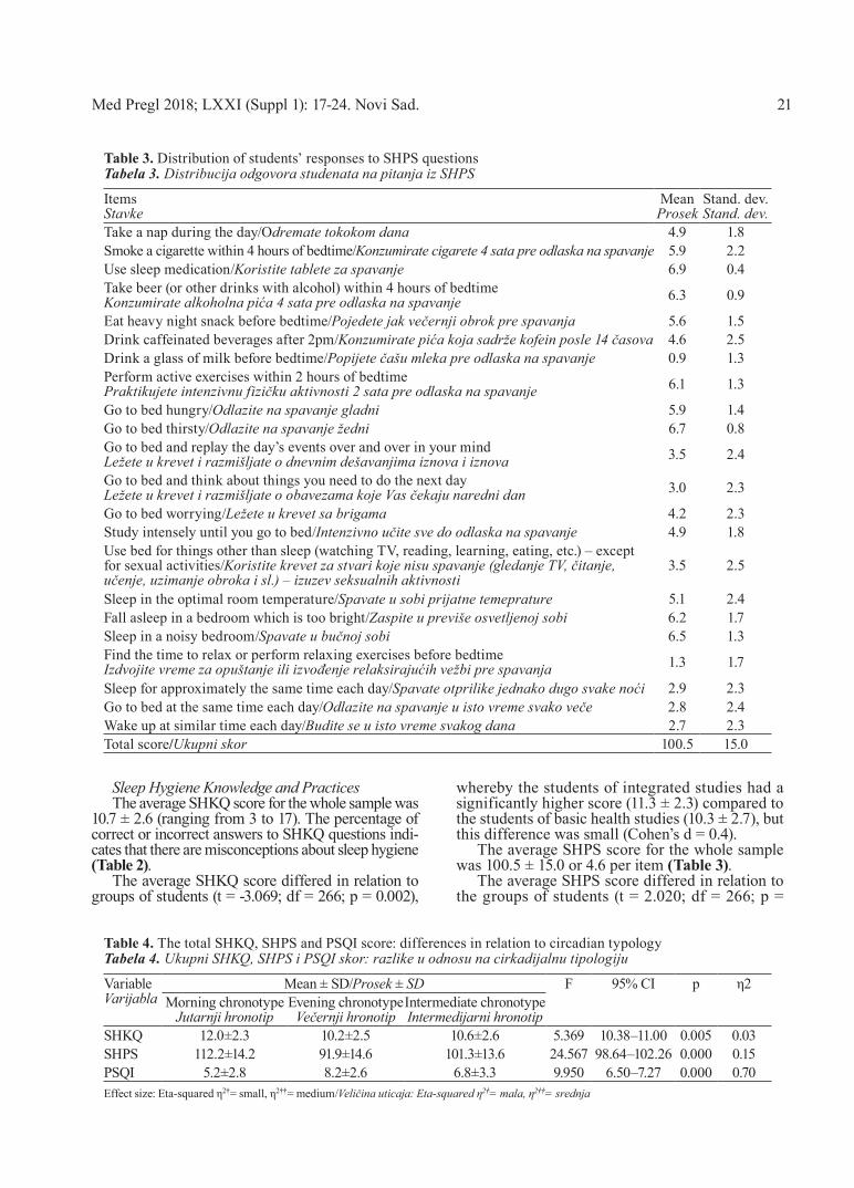
Med Pregl 2018; LXXI (Suppl 1): 17-24. Novi Sad. 21
Sleep Hygiene Knowledge and Practices The average SHKQ score for the whole sample was
10.7 ± 2.6 (ranging from 3 to 17). The percentage of correct or incorrect answers to SHKQ questions indi-cates that there are misconceptions about sleep hygiene (Table 2).
The average SHKQ score differed in relation to groups of students (t = -3.069; df = 266; p = 0.002),
whereby the students of integrated studies had a significantly higher score (11.3 ± 2.3) compared to the students of basic health studies (10.3 ± 2.7), but this difference was small (Cohen’s d = 0.4).
The average SHPS score for the whole sample was 100.5 ± 15.0 or 4.6 per item (Table 3).
The average SHPS score differed in relation to the groups of students (t = 2.020; df = 266; p =
Table 3. Distribution of students’ responses to SHPS questionsTabela 3. Distribucija odgovora studenata na pitanja iz SHPS
ItemsStavke
Mean Prosek
Stand. dev. Stand. dev.
Take a nap during the day/Odremate tokokom dana 4.9 1.8Smoke a cigarette within 4 hours of bedtime/Konzumirate cigarete 4 sata pre odlaska na spavanje 5.9 2.2Use sleep medication/Koristite tablete za spavanje 6.9 0.4Take beer (or other drinks with alcohol) within 4 hours of bedtimeKonzumirate alkoholna pića 4 sata pre odlaska na spavanje 6.3 0.9
Eat heavy night snack before bedtime/Pojedete jak večernji obrok pre spavanja 5.6 1.5Drink caffeinated beverages after 2pm/Konzumirate pića koja sadrže kofein posle 14 časova 4.6 2.5Drink a glass of milk before bedtime/Popijete čašu mleka pre odlaska na spavanje 0.9 1.3Perform active exercises within 2 hours of bedtimePraktikujete intenzivnu fizičku aktivnosti 2 sata pre odlaska na spavanje 6.1 1.3
Go to bed hungry/Odlazite na spavanje gladni 5.9 1.4Go to bed thirsty/Odlazite na spavanje žedni 6.7 0.8Go to bed and replay the day’s events over and over in your mindLežete u krevet i razmišljate o dnevnim dešavanjima iznova i iznova 3.5 2.4
Go to bed and think about things you need to do the next dayLežete u krevet i razmišljate o obavezama koje Vas čekaju naredni dan 3.0 2.3
Go to bed worrying/Ležete u krevet sa brigama 4.2 2.3Study intensely until you go to bed/Intenzivno učite sve do odlaska na spavanje 4.9 1.8Use bed for things other than sleep (watching TV, reading, learning, eating, etc.) – except for sexual activities/Koristite krevet za stvari koje nisu spavanje (gledanje TV, čitanje, učenje, uzimanje obroka i sl.) – izuzev seksualnih aktivnosti
3.5 2.5
Sleep in the optimal room temperature/Spavate u sobi prijatne temeprature 5.1 2.4Fall asleep in a bedroom which is too bright/Zaspite u previše osvetljenoj sobi 6.2 1.7Sleep in a noisy bedroom/Spavate u bučnoj sobi 6.5 1.3Find the time to relax or perform relaxing exercises before bedtimeIzdvojite vreme za opuštanje ili izvođenje relaksirajućih vežbi pre spavanja 1.3 1.7
Sleep for approximately the same time each day/Spavate otprilike jednako dugo svake noći 2.9 2.3Go to bed at the same time each day/Odlazite na spavanje u isto vreme svako veče 2.8 2.4Wake up at similar time each day/Budite se u isto vreme svakog dana 2.7 2.3Total score/Ukupni skor 100.5 15.0
Table 4. The total SHKQ, SHPS and PSQI score: differences in relation to circadian typologyTabela 4. Ukupni SHKQ, SHPS i PSQI skor: razlike u odnosu na cirkadijalnu tipologiju
Variable Varijabla
Mean ± SD/Prosek ± SD F 95% CI p η2Morning chronotype
Jutarnji hronotipEvening chronotype
Večernji hronotipIntermediate chronotype Intermedijarni hronotip
SHKQ 12.0±2.3 10.2±2.5 10.6±2.6 5.369 10.38–11.00 0.005 0.03SHPS 112.2±14.2 91.9±14.6 101.3±13.6 24.567 98.64–102.26 0.000 0.15PSQI 5.2±2.8 8.2±2.6 6.8±3.3 9.950 6.50–7.27 0.000 0.70Effect size: Eta-squared η2†= small, η2††= medium/Veličina uticaja: Eta-squared η2†= mala, η2††= srednja
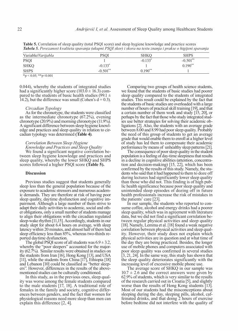
22 Andrijević I, et al. Assessment of Sleep Quality among Healthcare Students
0.044), whereby the students of integrated studies had a significantly higher score (103.0 ± 16.3) com-pared to the students of basic health studies (99.1 ± 14.2), but the difference was small (Cohen’s d = 0.3).
Circadian TypologyAs for the chronotype, the students were classified
as the intermediate chronotype (67.2%), evening chronotype (20.9%) and morning chronotype (11.9%). A significant difference between sleep hygiene knowl-edge and practices and sleep quality in relation to cir-cadian typology was determned (Table 4).
Correlation Between Sleep Hygiene Knowledge and Practices and Sleep Quality We found a significant negative correlation be-
tween sleep hygiene knowledge and practices and sleep quality, whereby the lower SHKQ and SHPS scores followed a higher PSQI score (Table 5).
Discussion
Previous studies suggest that students generally sleep less than the general population because of the exposure to academic stressors and numerous academ-ic demands. They are therefore at risk of having poor sleep quality, daytime dysfunction and cognitive im-pairment. Although a large number of them strive to adapt their daily activities to academic, social and oth-er obligations, only a small number of students manage to align their obligations with the circadian regulated sleep-wake rhythm [11,15]. Accordingly, students in our study slept for about 6.4 hours on average, with sleep latency within 20 minutes, and almost half of them had sleep efficiency less than 85%, whereas two-thirds re-ported daytime dysfunction.
The global PSQI score of all students was 6.9 ± 3.2, whereby the “poor sleepers” accounted for the major-ity (62.7%). Similar results were obtained in studies on the students from Iran [16], Hong Kong [13], and USA [11], while the students from China [17], Ethiopia [18] and Lebanon [10] could be classified as “better sleep-ers”. However, differences in the results of the above-mentioned studies can be culturally conditioned.
In this study, as in the previous ones, sleep qual-ity was worse among the female students compared to the male students [17, 18]. A traditional role of females in the family and society, cognitive differ-ences between genders, and the fact that women for physiological reasons need more sleep than men can explain this difference [2, 4].
Comparing two groups of health science students, we found that the students of basic studies had poorer sleep quality compared to the students of integrated studies. This result could be explained by the fact that the students of basic studies are overloaded with a large number of hours of practical skill training [19], and that a certain number of them work and study [15, 20], or perhaps by the fact that those who study integrated stud-ies use better strategies for solving their academic ob-ligations [2]. Also, the students with an average grade between 8.00 and 8.99 had poor sleep quality. Probably the need of this group of students to get an average grade that would enable them to enroll at a higher level of study has led them to compensate their academic performance by means of unhealthy sleep patterns [21].
The consequence of poor sleep quality in the student population is a feeling of day-time sleepiness that results in a decline in cognitive abilities (attention, concentra-tion and decision-making) [15, 22], which has been confirmed by the results of this study. Namely, the stu-dents who said that it had happened to them to doze off during lectures had significantly lower sleep quality than those who did not. This finding is of high pub-lic health significance because poor sleep quality and unintended sleep episodes of dozing off in future health professionals increase the risk of mistakes in the patients’ care [23].
In our sample, the students who reported to con-sume coffee, alcohol and energy drinks had a poorer sleep quality, which was in agreement with literature data, but we did not find a significant correlation be-tween regular physical activities and sleep quality [18]. Namely, Lemma et al. [18] found a weak negative correlation between physical activities and sleep qual-ity. However, their study does not explain which physical activities are in question and at what time of the day they are being practiced. Besides, the longer use of mobile phones and computers associated with poor sleep quality was confirmed in several studies [3, 21, 24]. In the same way, this study has shown that the sleep quality deteriorates significantly with the increasing level of excessive mobile phone use.
The average score of SHKQ in our sample was 10.7 ± 2.6 and the correct answers were given by 62.9% of students, which is very similar to the results of the research carried out in Croatia [5], and slightly worse than the results of Hong Kong students [13]. Most of our students had the misconceptions about sleeping during the day, sleeping pills, alcohol, caf-feinated drinks, and that doing 2 hours of exercise before bedtime did not interfere with the quality of
Table 5. Correlation of sleep quality (total PSQI score) and sleep hygiene knowledge and practice scoresTabela 5. Povezanost kvaliteta spavanja (ukupni PSQI skor) i skora na testu znanja i prakse o higijeni spavanja
Variable/Varijabla PSQI SHKQ SHPSPSQI 1 -0.133* -0.501**
SHKQ -0.133* 1 0.190**
SHPS -0.501** 0.190** 1*p < 0.05; **p<0.001
Med Pregl 2018; LXXI (Suppl 1): 17-24. Novi Sad. 23
sleep, or that the consumption of a glass of milk pos-itively affected the sleep quality. The average SHPS score for the entire sample in our study was 100.5 ± 15.0 (4.6 per item), which indicates that students per-formed all activities regarding sleep hygiene on average 4.6 times a week, which is similar to the results of the Hong Kong study [13]. The students of Integrated Healthcare Studies had better knowledge and practice of sleep hygiene compared to the students of basic stud-ies and therefore better sleep quality. This is probably the result of a difference in the study curriculum, due to the fact that the students of integrated studies have more knowledge about the sleep physiology and the impact of sleeping on the quality of life in individuals.
In our research, a significant negative correlation between sleep hygiene knowledge and practices and sleep quality was determined, whereby lower SHKQ and SHPS scores were followed by a higher PSQI score, indicating that there was also a higher probabil-ity for students who had poor sleep hygiene knowledge and practices to have poor sleep quality. Unlike data from this study, the results of a number of studies in-dicate that sleep hygiene practice is associated with sleep quality unlike the level of knowledge [4, 6, 13]. The differences in methodological research procedures can explain this inconsistency. Furthermore, in our research, a weak positive correlation between sleep hygiene knowledge and practices was established, which was supported by the findings of the Croatian study [5], while Felix et al. [9] claimed the opposite.
By analyzing diurnal sleep preferences among our students, sleep quality was to be the highest in the morning chronotype, whereas in the evening chronotype it was the worst. The same applies to
sleep hygiene knowledge and practices. Our find-ings are in accordance with literature data indicat-ing a poorer sleep quality in those with the evening chronotype [3, 5, 7, 25], more accurate knowledge on sleep hygiene in the morning chronotype com-pared to the evening chronotype [8], and less adher-ence to good sleep hygiene practices in the evening chronotypes in relation to the morning chronotype [26]. Diaz-Morales et al. [8] believe that less adher-ence to good sleep hygiene practices correlates the evening chronotype with lower sleep quality.
Limitations of the studyThis study has some limitations which have to
be pointed out. The research was conducted on-line, and it is possible that the reasons for students’ in-volvement were pre-existing poor sleep quality or higher levels of sleep hygiene knowledge. We could not collect data from students who did not have ac-cess to the Internet, therefore, caution should be taken in generalizing the results.
Conclusion
An average sleep quality score indicates that a large proportion of Novi Sad University students experience a suboptimal level of overall sleep quality according to the PSQI criterion. Although satisfactory sleep hygiene knowledge was found in a high percentage of students, many of them reported unhealthy practices indicating that knowledge was not a factor of deterring from un-healthy behaviors. Interventions on sleep hygiene prac-tices must focus on changing and applying behavioral factors, and not just learning about sleep hygiene.
References
1. Stanojević Č, Simić S, Milutinović D. Health effects of sleep deprivation on nurses working shifts. Med Pregl. 2016;69(5-6):183-8.
2. Yazdi Z, Loukzadeh Z, Moghaddam P, Jalilolghadr S. Sleep hygiene practices and their relation to sleep quality in medical students of Qazvin University of Medical Sciences. J Caring Sci. 2016;5(2):153-60.
3. Fossum IN, Nordnes LT, Storemark SS, Bjorvatn B, Pallesen S. The association between use of electronic media in bed before going to sleep and insomnia symptoms, daytime sleepiness, morn-ingness, and chronotype. Behav Sleep Med. 2014;12(5):343–57.
4. Al-Kandari S, Alsalem A, Al-Mutairi S, Al-Lumai D, Da-woud A, Moussa M. Association between sleep hygiene awareness and practice with sleep quality among Kuwait University students. Sleep Health. 2017;3(5):342–7.
5. Škvorc Lj, Košćec Bjelajac A. Sleep beliefs and circadian typology of helping professions students. International Online Journal of Educational Sciences. 2016;8(5):69–78.
6. Voinescu BI, Coogan AN, Oräsan R. Sleep beliefs subjective sleep quality and diurnal preference-findings from depressed pa-tients. J Cogn Behav Psychother. 2010;10(1):1-12.
7. Voinescu BI, Szentagotai-Tatar A. Sleep hygiene awareness: its relation to sleep quality and diurnal preference. J Mol Psychia-try. 2015;3(1):1.
8. Diaz-Morales JF, Prieto PD, Barreno CE, Mateo JC, Randler C. Sleep beliefs and chronotype among adolescents: the effect of a sleep education program. Biol Rhythm Res. 2012;43(4):397-412.
9. Felix VA, Campsen NA, White A, Buboltz WC. College students’ prevalence of sleep hygiene awareness and practices. Advances in Social Sciences Research Journal. 2017;4(4):91-105.
10. Kabrita CS, Hajjar-Muça TA, Duffy JF. Predictors of poor sleep quality among Lebanese University students: association between evening typology, lifestyle behaviors, and sleep habits. Nat Sci Sleep. 2014;6:11–8.
11. Lund HG, Reider BD, Whiting AB, Prichard JR. Sleep patterns and predictor of disturbed sleep in a large population of college students. J Adolesc Health. 2010;46(2):124-32.
12. Buysse DJ, Reynolds CF 3rd, Monk TH, Berman SR, Kupfer DJ. The Pittsburgh Sleep Quality Index: a new instru-ment for psychiatric practice and research. Psychiatry Res. 1989;28(2):193−213.
13. Suen LK, Tam WW, Hon KL. Association of sleep hy-giene-related factors and sleep quality among university stu-dents in Hong Kong. Hong Kong Med J. 2010;16(3):1805.
14. Horne JA, Ostberg OA. Self-assessment questionnaire to determine morningness-eveningness in human circadian rhythms. Int J Chronobiol. 1976;4(2):97–110.
24
15. Milutinović D, Stanojević Č, Stanojević V, Simić S. Sleep quality, level of daytime sleepiness and fatigue among working nursing students. Timočki medicinski glasnik. 2016;41(4):257-65.
16. Bakhshandeh Bavarsad M, Azimi N, Moradbeigi K, Latifi M. Associations between morningness-eveningness and sleep quality among female dormitory residents. Thrita. 2015;4(1):e59661.
17. Cheng SH, Shih CC, Lee IH, Hou YW, Chen KC, Chen KT, et al. A study on the sleep quality of incoming university students. Psychiatry Res. 2012;197(3):270-4.
18. Lemma S, Patel SV, Tarekegn YA, Tadesse MG, Ber-hane Y, Gelaye B., et al. The epidemiology of sleep quality, sleep patterns, consumption of caffeinated beverages, and khat use among Ethiopian college students. Sleep Disord. 2012;2012:583510.
19. Sajadi A, Farsi Z, Rajai N. The relationship between sleep quality with fatigue severity and academic performance of nursing students. Nursing Practice Today. 2014;1(4):21320.
20. De Mattos Moraes dos Santos TC, De Martino MMF, Sonati JG, De Faria AL, De De Almeida Nascimento EF. Sleep quality and chronotype of nursing students. Acta Paulista de Enfermagem. 2016;29(6):658-63.
21. Al Ghamdi AA. Sleep deprivation and academic per-formance of students in the collage of nursing at King Saud University. World Applied Sciences Journal. 2013;27(2):155-67.
22. Huang CF, Yang LY, Wu LM, Liu Y, Chen HM. Deter-minants of daytime sleepiness in first year nursing students: a questionnaire survey. Nurse Educ Today. 2014;34(6):1048–53.
23. Caruso CC, Hitchcock EM. Strategies for nurses to pre-vent sleep-related injuries and errors. Rehabil Nurs. 2010;35(5):192-7.
24. Brunborg GS, Mentzoni RA, Molde H, Myrseth H, Sk-ouverøe KJM, Bjorvatn B, et al. The relationship between me-dia use in the bedroom, sleep habits and symptoms of insomnia. J Sleep Res. 2012;20(4):569–75.
25. Ninama NJ, Kagathara J. Sleep wake pattern analysis: study of 131 medical students. NHL Journal of Medical Sci-ences. 2012;1(1):33–6.
26. Vollmer C, Hammer J, Keller C, Maxand AK, Díaz-Morales JF, Randler C. Development and evaluation of a sleep education program in middle school pupils based on self-deter-mination theory. International Journal of Biology Education. 2014;3(1):12–22.
Rad je primljen 3. IX 2018.Recenziran 5. IX 2018.Prihvaćen za štampu 10. IX 2018.BIBLID.0025-8105:(2018):Suppl 1:17-24.
Andrijević I, et al. Assessment of Sleep Quality among Healthcare Students
Med Pregl 2018; LXXI (Suppl 1): 25-29. Novi Sad. 25
SummaryIntroduction. Fatigue in cancer patients is one of the most frequent symptoms present irrespectively of the kind of malignant tumor, its localization, stage and/or treatment modality. One of the most often used definition of fatigue emphasizes a disturbing, persistent, sub-jective feeling of physical, emotional and/or cognitive fatigue, or tiredness, connected with the carcinoma and/or its therapy, that is not proportional with the patients’ activities and therefore impairs their daily functioning. The study has been undertaken with the aim of determining the levels of fatigue in cancer patients during radiotherapy. Material and Method. The study was conducted as a cross-sectional study, by interviewing 80 hospitalized patients at the Institute for Oncology of Vojvodina in Sremska Kamenica, Clinic for Radiotherapy. Perform Questionnaire with 12 item de-scribing fatigue over the previous two weeks was used as a study instrument. For the statistical analysis of data we used method of descriptive statistics and inferential statistics. Statistical significance was determined at the level of p<0.05. Results. The highest average values of the general Perform questionnaire score were recorded in the patients diagnosed with the central nervous system carcinoma [38.9± 14.6], while the lowest were recorded in the group of patients with prostate cancer [17.0±4.6]. Simultaneous administration of radiotherapy and chemotherapy leads to major limitations of phys-ical performances, daily life activities and patient attitudes. Conclu-sion. All participants included in this study experienced fatigue, although it differed in separate domains, as well as by the type of therapy, primary diagnosis and intensity over the previous two weeks; however, it was present at the moment of estimation.Key words: Self-Assessment; Fatigue; Neoplasms; Activities of Daily Living; Signs and Symptoms; Surveys and Question-naires
SažetakUvod. Umor kod onkoloških bolesnika jedan je od najčešćih simptoma prisutan nezavisno od vrste malignog tumora, lokaliza-cije i stadijuma i/ili medicinskih modaliteta lečenja. Jedna od naj-češće korišćenih definicija umora naglašava uznemirujući, perzi-stentni, subjektivni osećaj fizičkog, emocionalnog i /ili kognitivnog umora, odnosno iscrpljenosti, povezanog sa kancerom i/ili terapijom karcinoma, koji nije srazmeran s bolesnikovim aktivnostima te ometa svakodnevno funkcionisanje. Studija je sprovedena sa ciljem utvrđivanja nivoa umora kod onkoloških pacijenata tokom radio-terapije. Materijal i metode. Ispitivanje je sprovedeno kao studija preseka, anketiranjem 80 hospitalizovanih pacijenta na Klinici za radioterapiju Instituta za onkologiju Vojvodine u Sremskoj Kame-nici. Za utvrđivanje umora korišćen je Perform Questionnaire ‒ Perform upitnik koji se sastoji od 12 stavki na osnovu kojih se procenjuje umor u protekle dve nedelje. Za statističku obradu po-dataka primenjene su metode deskriptivne i inferencijalne statistke, a statistička značajnost određivana je na nivou p < 0,05. Rezultati. Najveće prosečne vrednosti ukupnog skora upitnika Perform ima-li su pacijenti sa dijagnozom tumora centralnog nervnog sistema (38,9 ± 14,6), dok su najniže vrednosti zabeležene u grupi pacije-nata sa dijagnozom karcinoma prostate (17 ± 4,6). Istovremena primena radioterapije i hemioterapije dovodi do većih ograničenja u domenima fizičkih performansi, aktivnostima dnevnog života i stavovima pacijenata. Zaključak. Umor kod onoloških pacijenata obuhvaćenih ovim istraživanjem razlikovao se u različitim dome-nima, prema vrsti terapije, primarnoj dijagnozi, intenzitetu i prisut-nosti tokom poslednje dve nedelje ali u trenutku procene bio je prisutan kod svih ispitanika.Ključne reči: samoprocena; umor; neoplazme; aktivnosti sva-kodnevnog života; znaci i simptomi; istraživanja i upitnici
University of Novi Sad, Faculty of Medicine1 UDK 616-006.04-052:159.944.072Department of Nursing2 https://doi.org/10.2298/MPNS18S1025TDepartment of Pathology3 Institute for Oncology of Vojvodina, Sremska Kamenica,Clinic for Operative Oncology, Department of Gynecology4
FATIGUE ASSESSMENT IN CANCER PATIENTS
PROCENA UMORA KOD ONKOLOŠKIH PACIJENATA
Sanja TOMIĆ1,2, Zoran NIKIN1,3, Slobodan TOMIĆ1 and Goran MALENKOVIĆ1,4
Fatigue in cancer patients has many definitions. In professional literature the most often used defini-tion indicates an unsettling, persistent, subjective feeling of physical, emotional and/or cognitive fa-tigue or exhaustion associated with the basic illness and/or therapy but disproportionate to the patient’s activities so it hinders the patient’s everyday ac-tivities [1]. The basic characteristics of fatigue in cancer patients are the lack of energy followed by exhaustion and weakness. The difficulties in defin-ing fatigue of those diagnosed with and being treat-
ed for a malignant disease arise from a complex, multifactorial etiology of fatigue. The causes and mechanisms can be connected with the basic un-derlying disease and/or medical modalities of treat-ment, but also with potential genetic predispositions and following physical and/or mental illnesses, be-havioral and environmental factors [2-6]. Because of this a wide spectrum of causes and influences – somatic affective, cognitive and psychosocial, which are often indistinguishable, equally contrib-ute to fatigue.
Corresponding Author: Doc. dr Sanja Tomić, Univerzitet u Novom Sadu, Medicinski fakultet, Katedra za zdravstvenu negu, 21000 Novi Sad, Hajduk Veljkova 3, E-mail: [email protected]
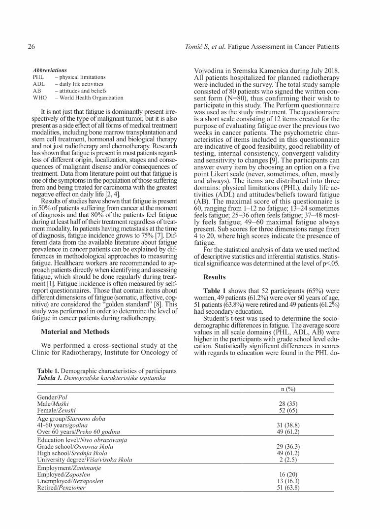
26 Tomić S, et al. Fatigue Assessment in Cancer Patients
It is not just that fatigue is dominantly present irre-spectively of the type of malignant tumor, but it is also present as a side effect of all forms of medical treatment modalities, including bone marrow transplantation and stem cell treatment, hormonal and biological therapy and not just radiotherapy and chemotherapy. Research has shown that fatigue is present in most patients regard-less of different origin, localization, stages and conse-quences of malignant disease and/or consequences of treatment. Data from literature point out that fatigue is one of the symptoms in the population of those suffering from and being treated for carcinoma with the greatest negative effect on daily life [2, 4].
Results of studies have shown that fatigue is present in 50% of patients suffering from cancer at the moment of diagnosis and that 80% of the patients feel fatigue during at least half of their treatment regardless of treat-ment modality. In patients having metastasis at the time of diagnosis, fatigue incidence grows to 75% [7]. Dif-ferent data from the available literature about fatigue prevalence in cancer patients can be explained by dif-ferences in methodological approaches to measuring fatigue. Healthcare workers are recommended to ap-proach patients directly when identifying and assessing fatigue, which should be done regularly during treat-ment [1]. Fatigue incidence is often measured by self-report questionnaires. Those that contain items about different dimensions of fatigue (somatic, affective, cog-nitive) are considered the “golden standard” [8]. This study was performed in order to determine the level of fatigue in cancer patients during radiotherapy.
Material and Methods
We performed a cross-sectional study at the Clinic for Radiotherapy, Institute for Oncology of
Vojvodina in Sremska Kamenica during July 2018. All patients hospitalized for planned radiotherapy were included in the survey. The total study sample consisted of 80 patients who signed the written con-sent form (N=80), thus confirming their wish to participate in this study. The Perform questionnaire was used as the study instrument. The questionnaire is a short scale consisting of 12 items created for the purpose of evaluating fatigue over the previous two weeks in cancer patients. The psychometric char-acteristics of items included in this questionnaire are indicative of good feasibility, good reliability of testing, internal consistency, convergent validity and sensitivity to changes [9]. The participants can answer every item by choosing an option on a five point Likert scale (never, sometimes, often, mostly and always). The items are distributed into three domains: physical limitations (PHL), daily life ac-tivities (ADL) and attitudes/beliefs toward fatigue (AB). The maximal score of this questionnaire is 60, ranging from 1–12 no fatigue; 13–24 sometimes feels fatigue; 25–36 often feels fatigue; 37–48 most-ly feels fatigue; 49–60 maximal fatigue always present. Sub scores for three dimensions range from 4 to 20, where high scores indicate the presence of fatigue.
For the statistical analysis of data we used method of descriptive statistics and inferential statistics. Statis-tical significance was determined at the level of p<.05.
Results
Table 1 shows that 52 participants (65%) were women, 49 patients (61.2%) were over 60 years of age, 51 patients (63.8%) were retired and 49 patients (61.2%) had secondary education.
Student’s t-test was used to determine the socio-demographic differences in fatigue. The average score values in all scale domains (PHL, ADL, AB) were higher in the participants with grade school level edu-cation. Statistically significant differences in scores with regards to education were found in the PHL do-
AbbreviationsPHL – physical limitationsADL – daily life activities AB – attitudes and beliefs WHO – World Health Organization
Table 1. Demographic characteristics of participants Tabela 1. Demografske karakteristike ispitanika
n (%)Gender/PolMale/Muški Female/Ženski
28 (35)52 (65)
Age group/Starosno doba41-60 years/godina Over 60 years/Preko 60 godina
31 (38.8)49 (61.2)
Education level/Nivo obrazovanjaGrade school/Osnovna škola High school/Srednja škola University degree/Viša/visoka škola
29 (36.3)49 (61.2)2 (2.5)
Employment/ZanimanjeEmployed/Zaposlen Unemployed/Nezaposlen Retired/Penzioner
16 (20)13 (16.3)51 (63.8)
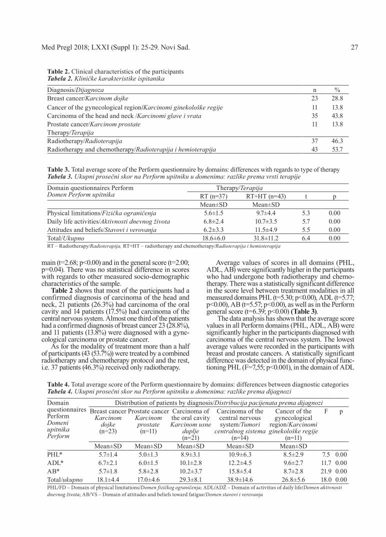
Med Pregl 2018; LXXI (Suppl 1): 25-29. Novi Sad. 27
main (t=2.68; p<0.00) and in the general score (t=2.00; p=0.04). There was no statistical difference in scores with regards to other measured socio-demographic characteristics of the sample.
Table 2 shows that most of the participants had a confirmed diagnosis of carcinoma of the head and neck, 21 patients (26.3%) had carcinoma of the oral cavity and 14 patients (17.5%) had carcinoma of the central nervous system. Almost one third of the patients had a confirmed diagnosis of breast cancer 23 (28.8%), and 11 patients (13.8%) were diagnosed with a gyne-cological carcinoma or prostate cancer.
As for the modality of treatment more than a half of participants (43 (53.7%)) were treated by a combined radiotherapy and chemotherapy protocol and the rest, i.e. 37 patients (46.3%) received only radiotherapy.
Average values of scores in all domains (PHL, ADL, AB) were significantly higher in the participants who had undergone both radiotherapy and chemo-therapy. There was a statistically significant difference in the score level between treatment modalities in all measured domains PHL (t=5.30; p<0.00), ADL (t=5.77; p<0.00), AB (t=5.57; p<0.00), as well as in the Perform general score (t=6.39; p<0.00) (Table 3).
The data analysis has shown that the average score values in all Perform domains (PHL, ADL, AB) were significantly higher in the participants diagnosed with carcinoma of the central nervous system. The lowest average values were recorded in the participants with breast and prostate cancers. A statistically significant difference was detected in the domain of physical func-tioning PHL (F=7,55; p<0.001), in the domain of ADL
Table 2. Clinical characteristics of the participants Tabela 2. Kliničke karakteristike ispitanika
Diagnosis/Dijagnoza n %Breast cancer/Karcinom dojke 23 28.8Cancer of the gynecological region/Karcinomi ginekološke regije 11 13.8Carcinoma of the head and neck /Karcinomi glave i vrata 35 43.8Prostate cancer/Karcinom prostate 11 13.8Therapy/TerapijaRadiotherapy/Radioterapija 37 46.3Radiotherapy and chemotherapy/Radioterapija i hemioterapija 43 53.7
Table 3. Total average score of the Perform questionnaire by domains: differences with regards to type of therapyTabela 3. Ukupni prosečni skor na Perform upitniku u domenima: razlike prema vrsti terapije
Domain questionnaires PerformDomen Perform upitnika
Therapy/TerapijaRT (n=37) RT+HT (n=43) t pMean±SD Mean±SD
Physical limitations/Fizička ograničenja 5.6±1.5 9.7±4.4 5.3 0.00Daily life activities/Aktivnosti dnevnog života 6.8±2.4 10.7±3.5 5.7 0.00Attitudes and beliefs/Stavovi i verovanja 6.2±3.3 11.5±4.9 5.5 0.00Total/Ukupno 18.6±6.0 31.8±11.2 6.4 0.00RT – Radiotherapy/Radioterapija, RT+HT – radiotherapy and chemotherapy/Radioterapija i hemioterapija
Table 4. Total average score of the Perform questionnaire by domains: differences between diagnostic categories Tabela 4. Ukupni prosečni skor na Perform upitniku u domenima: razlike prema dijagnozi
Domain questionnaires PerformDomeni upitnika Perform
Distribution of patients by diagnosis/Distribucija pacijenata prema dijagnoziBreast cancer
Karcinom dojke(n=23)
Prostate cancer Karcinom prostate (n=11)
Carcinoma of the oral cavityKarcinom usne
duplje (n=21)
Carcinoma of the central nervous system/Tumori
centralnog sistema(n=14)
Cancer of the gynecological
region/Karcinomi ginekološke regije
(n=11)
F p
Mean±SD Mean±SD Mean±SD Mean±SD Mean±SDPHL* 5.7±1.4 5.0±1.3 8.9±3.1 10.9±6.3 8.5±2.9 7.5 0.00ADL* 6.7±2.1 6.0±1.5 10.1±2.8 12.2±4.5 9.6±2.7 11.7 0.00AB* 5.7±1.8 5.8±2.8 10.2±3.7 15.8±5.4 8.7±2.8 21.9 0.00Total/ukupno 18.1±4.4 17.0±4.6 29.3±8.1 38.9±14.6 26.8±5.6 18.0 0.00PHL/FD – Domain of physical limitations/Domen fizičkog ograničenja; ADL/ADŽ – Domain of activities of daily life/Domen aktivnosti dnevnog života; AB/VS – Domain of attitudes and beliefs toward fatigue/Domen stavovi i verovanja
28
(F=11,71; p<0.001), in the domain of beliefs and atti-tudes ABB (F=21,98; p<0.001), and in the summary score (F=18,06; p<0.001) (Table 4).
Discussion
Facing and accepting the diagnosis, treatment and long term recovery from malignant diseases is a com-plicated process and it can take months, or perhaps even several years to be achieved. A large variation in age defines the population of the affected with their changed expectations, roles, responsibilities and con-cerns during their life cycle which confront with de-mands of the unpredictable disease.
According to the principles of the World Health Organization cancer patients are defined as chron-ically ill [10]. They face a high level of physical, psychological and social stress from the moment of being diagnosed and throughout all phases of med-ical care. Symptoms and unwanted effects of the illness and/or medical modalities of treatments de-crease the psychophysical well-being and life qual-ity of patients [11, 12]. Fatigue is a common symp-tom in cancer patients and it causes the limitation of physical activity [8, 9] and hinders professional, familial and social roles [5–9, 11]. In our research we have detected a difference between education levels and domain of physical functioning PHL (t=2.7; p<0.00) and general questionnaire score (t=2.0; p=0.04); we have not found a statistically significant difference between fatigue and age, as has been described before [6]. Average score values for all Perform questionnaire domains (PHL, ADL, AB) are higher in the participants with lower educa-tion levels. Diverse manifestations of fatigue in the domain of physical functioning mostly cited by the patients include the feeling of exhaustion and lack of energy with the loss of interest. The patients who feel fatigue are very different in the way they express and describe the problems they are experiencing. Such re-sults in our research are in concordance with the results of a qualitative study that have shown that clinical traits are highly individual and there are barely even two similar patients [13]. It is interesting to note that during earlier surveys, a connection between symptoms and antecedent activities could not be made. The results of the study also state the fact that unlike fatigue and exhaustion in non-oncological patients, cancer patients do not feel refreshed after rest or sleep [14]. Numerous symptoms such as pain, sleeping disorders, lack of ap-petite or nausea tend to have a negative influence on cancer patients; however, fatigue-related symptoms are considered most pronounced [13]. Fatigue considerably diminishes the ability of cancer patients to perform physical activities. It is believed that the causes of de-creased physical performance result from changes in the cortical and spinal sensory centers, in the energy metabolism and in the process of muscle activation [3]. Depending on the severity of the underlying diseases the negative effects of fatigue in the domain of physical activity can range from temporary disability in the
sense of limited movement, disorders in coordination and gait, decreased independence in daily activities, inadequate dealing with everyday life and social with-drawal up to inability to do ones job and earn for a living that leads to economic difficulties for the patient and additional economic burden for the whole society [8, 12]. In our study, differences in the patient’s feeling of fatigue with regard to the type of therapy show that the average values of all domain scores and the gen-eral score are higher in the patients who underwent the combined therapy (radiotherapy and chemotherapy). These results are in concordance with many findings of earlier studies. [11, 12, 15–18]. It is known and well described that a string of side effects resulting from chemotherapy, i.e. of antineoplastic therapy, often be-come more pronounced in intensity and frequency when it is combined with radiotherapy. Nausea and tiredness with a loss of appetite are the most frequent symptoms after chemotherapy and have a negative ef-fect on fatigue in the patients who are also on radio-therapy [15–18].
The lowest average value of the summary general score of the Perform questionnaire was recorded in the patients diagnosed with prostate cancer (17.0±4.6) and breast carcinoma (18.0±4.4), and almost identical aver-age score values were found in the domain of physical activities (5.0±1.3vs5.7±1.4); the domain of daily life activity (6.0±1.6 vs 6.6±2.2) and the domain of attitudes and beliefs (5.8±2.8 vs 5.6±1.8). By the questionnaire categories and in relation with the average score, the results of our study show that the patients with breast and prostate carcinoma diagnosis have sometimes felt fatigue. Such results are in accordance with those from similar studies that also document lower fatigue-relat-ed difficulties in the patients with breast and/or prostate carcinoma [19].
The analysis of the results from the group of pa-tients with carcinoma of the head and neck has shown that the average values are higher in all domains and in the general score. The patients with oral cavity car-cinoma have a higher average value of general score in two domains, the domain of daily life activities and the domain of attitudes and beliefs (10.2±2.8 vs 10.2±3.8), unlike the domain of physical activity, the average value being 8.9±3.1, which suggests that fa-tigue due to physical limitations is almost absent. Pri-mary localization of the carcinoma and the incidence of side effects during radiotherapy and/or chemother-apy (changes in the oral cavity such as mucositis, sto-matitis, impaired chewing and/or swallowing) have contributed to greater limitations in the domain of daily life activities, so this particular domain and that of attitudes and beliefs are the most frequent reasons for the presence of fatigue in this population of par-ticipants. Although our research did not include the assessment of the oral cavity, the participants them-selves reported hardships they were facing, so in item 5 of the Perform questionnaire (When I am fatigued, I must stop what I am doing in order to rest and then carry on) they often said that during the intake of food they had to make pauses because of fatigue, since it
Tomić S, et al. Fatigue Assessment in Cancer Patients
Med Pregl 2018; LXXI (Suppl 1): 25-29. Novi Sad. 29
was hard for them to chew and/or swallow food. The average value of the general Perform questionnaire score indicated that the participants with oral cavity carcinoma (29.3±8.1) were often tired.
The highest values obtained in our study were those in the domain of beliefs and attitudes in the patients with tumors of the central nervous system (15.8±5.6). The items of this domain reflect the per-sonal perception of fatigue and its influence on life. High average values in this domain show that the pa-tients with tumors of the central nervous system feel that they cannot lead regular lives because of their illness. Average values in other domains of PHL and AB (10.9±6.3 vs 12.2±4.5) as well as of the general score (38.9±14.6) indicate that the patients felt tired in the previous two weeks.
Frequent presence of fatigue during the previous two weeks is characteristic for patients with carci-noma of the gynecological region, and this is indi-
cated by the average general score of (26.8±6.0). It is interesting to note that there is virtually no difference when these participants are analyzed regarding the scores on PHL, ADL and AB domains (8.5 ± 2.9 vs 9.6±2.7 vs 8.7±2.8) of the Perform questionnaire. Such results suggest equal participation of physical per-formance, existence of limitation in daily activities and negative subjective perception of patients when fatigue in the previous two weeks is concerned.
Conclusion
All participants included in this study experi-enced fatigue, although it differed in separate do-mains, as well as by the type of therapy, primary diagnosis, and intensity over the previous two weeks and at the moment of estimation; however, it was the constant follower of these patients.
References
1. Berger AM, Abernethy AP, Atkinson A, Barsevick AM, Breitbart WS, Cella D, et al. NCCN Clinical Practice Guidelines Cancer-related fatigue. J Natl Compr Canc Netw. 2010;8(8):904-31.
2. Hofman M, Ryan JL, Figueroa-Moseley CD, Jean-Pierre P, Morrow GR. Cancer-related fatigue: the scale of the problem. Oncologist. 2007;12 Suppl 1:4-10.
3. Bower JE. Cancer-related fatigue: mechanisms, risk factors, and treatments. Nat Rev Clin Oncol. 2014;11(10):597-609.
4. Homeber M, Fischer I, Dimeo F, Rüffer JU, Weis J. Cancer related fatigue: epidemiology, pathogenesis, diagnosis, and treat-ment. Dtsch Arztebl Int. 2012;109(9):161-71.
5. Donovan KA, Stein KD, Lee M, Leach CR, Ilozumba O, Jacobsen PB. Systematic review of the multidimensional fatigue symptom inventory-short form. Support Care Cancer. 2015;23(1):191-212.
6. Mardanian-Dehkordi L, Kahangi L. The relationship be-tween perception of social support and fatigue in patients with cancer. Iran J Nurs Midwifery Res. 2018;23(4):261-6.
7. Siegel R, Ma J, Zou Z, Jemal A. Cancer statistics, 2014. CA Cancer J Clin. 2014;64(1):9-29.
8. Bower JE, Bak K, Berger A, Breitbart W, Escalante CP, Ganz PA, et al. Screening, assessment, and management of fatigue in adult survivors of cancer: an American Society of Clinical oncology clinical practice guideline adaptation. J Clin Oncol. 2014;32(17):1840-50.
9. Baro E, Carulla J, Cassinello J, Colomer R, Mata JG, Gas-con P, et al. Psychometric properties of the Perform Questionnaire: a brief scale for assessing patient perceptions of fatigue in cancer. Support Care Cancer. 2011;19(5):657-66.
10. World Health Organization. International Classification of Impairment, Disabilities and Handicaps. A manual or classifi-cation relating to consequences of disease. Geneva: World Health Organization; 1980.
11. Purcell A, Fleming J, Bennett S, McGuane K, Burmeister B, Haines T. A multidimensional examination of correlates of fatigue during radiotherapy. Cancer. 2010;116(2):529-37.
12. Hajloo N, Barahmand O, Adili A, Fathi M. The role of disease stage illness perceptions, unmet needs and fatigue in pre-dicting quality of life in patients with cancer. Health Psychol. 2016;5:79-95.
13. Ehsani M, Taleghani F, Hematti S, Abazari P. Perceptions of patients, families, physicians and nurses regarding challenges in cancer disclosure: a descriptive qualitative study. Eur J Oncol Nurs. 2016;25:55-61.
14. Stone P, Richards M, A Hern R, Hardy J. A study to in-vestigate the prevalence, severity and correlates of fatigue among patients with cancer in comparison with a control group of volun-teers without cancer. Ann Oncol. 2000;11(5):561-7.
15. Xiao C, Beitler JJ, Higgins KA, Conneely K, Dwivedi B, Felger J. Fatigue is associated with inflammation in patients with head and neck cancer before and after intensity-modulated radia-tion therapy. Brain Behav Immun. 2016;52:145-52.
16. Yucel B, Akkaş EA, Okur Y, Eren AA, Eren MF, Kara-pinar H, et al. The impact of radiotherapy on quality of life for cancer patients: a longitudinal study. Support Care Cancer. 2014;22(9):2479-87.
17. Gulliford SL, Miah AB, Brennan S, McQuaid D, Clark CH, Partridge M, et al. Dosimetric explanations of fatigue in head and neck radiotherapy: an analysis from the PARSPORT Phase III trial. Radiother Oncol. 2012;104(2):205-12.
18. O' Regan P, Hegarty J. The importance of self-care for fatigue amongst patients undergoing chemotherapy for primary cancer. Eur J Oncol Nurs. 2017;28:47-55.
19. Bower JE, Ganz PA, Irwin MR, Kwan L, Breen EC, Cole SW. Inflammation and behavioral symptoms after breast cancer treatment: do fatigue, depression, and sleep disturbance share a common underlying mechanism? J Clin Oncol. 2011;29(26):3517-22.
Rad je primljen 3. IX 2018.Recenziran 5. IX 2018.Prihvaćen za štampu 10. IX 2018.BIBLID.0025-8105:(2018):Suppl 1:25-29.

Med Pregl 2018; LXXI (Suppl 1): 31-35. Novi Sad. 31
SummaryIntroduction. In our modern “high–tech”“society, comfort of the patient is often forgotten because the practice is usually performed according to the bio-medical model and focused only on solving the physical problems of the patient rather than on a holistic ap-proach. Therefore, the objective of this study was to assess the comfort level of patients hospitalized with the immobilization. Ma-terial and Methods. The study was conducted as an observational, analytical cross-sectional study by interviewing 124 patients hos-pitalized at the Clinical Centre of Vojvodina. Comfort Immobiliza-tion Questionnaire with 20 individual statements that describe the current good or bad condition of the patient was used as an instru-ment of research. Descriptive and inferential statistics methods were applied for statistical data processing and statistical significance was determined at p < 0.05. Results. The immobilized orthopedic patients reported a high level of comfort in the socio-cultural and spiritual context, which was, however, insufficient in the physical context and the immediate environment context. A low level of comfort was reported by the patients between the ages of 36 and 59 years and the patients with immobilization of the upper extremities. Comfort level was conditioned by the intensity of pain and applied analgesic therapy rather than the duration of applied immobilization. Conclusion. The results of this study indicate that the patients’ comfort level is significantly affected by modifying factors such as hospital facilities as well as the type of immobilization and their age. Comfort Immobilization Questionnaire has proven to be a simple and reliable questionnaire for testing the patients comfort level in different contexts.Key words: Patient Comfort; Immobilization; Patient Satisfaction; Pain; Age Factors; Nursing Care; Surveys and Questionnaires
SažetakUvod. U današnjem tehnološkom svetu često se zaboravlja na komfor pacijenta jer se uglavnom praksa obavlja po bio-medicin-skom modelu i usmerena je na rešavanje najčešće telesnih prob-lema pacijenta, a ne na holističkom pristupu. Stoga je cilj ove studije bio da se proceni nivo komfora hospitalizovanih pacije-nata sa postavljenom imobilizacijom. Materijal i metode. Is-pitivanje je sprovedeno kao opservativna, analitička studija preseka, anketiranjem 124 hospitalizovana pacijenta u Kliničkom centru Vojvodine. Upitnik komfora u imobilizaciji sa 20 tvrdnji koje opisuju trenutno dobro ili loše stanje paci-jenta koristio se kao instrument istraživanja. Za statističku obradu podataka primenjene su metode deskriptivne i inferen-cijalne statistke, a statistička značajnost određivana je na nivou p < 0,05. Rezultati. Imobilisani ortopedski pacijenti su iskazali visok nivo komfora u društveno-kulturološkom i duhovnom kon-tekstu, a nedovoljan u fizičkom i kontekstu neposrednog okruženja. Nizak nivo komfora imali su pacijenti populacije između 36 i 59 godina i pacijenti sa imobilizacijom gornjih ek-stremiteta. Nivo komfora bio je uslovljen jačinom bola i primen-jenom analgetskom terapijom, a ne dužinom primenjene imobi-lizacije. Zaključak. Rezultati ovog istraživanja ukazuju da na nivo komfora pacijenta značajno utiču modifikujući faktori kao što su uslovi hospitalizacije, ali i tip imobilizacije i godine života. Upitnik komfora u imobilizaciji se pokazao kao jednos-tavan i pouzdan upitnik za ispitivanje nivoa komfora pacijena-ta u različitim kontekstima.Ključne reči: komfor pacijenta; imobilizacija; zadovoljstvo pacijenta; bol; starosna dob; zdravstvena nega; istraživanja i upitnici
University of Novi Sad, Faculty of Medicine, Department of Nursing1 UDK 616-089.22/.23:616-052Clinical Center of Vojvodina, Novi Sad https://doi.org/10.2298/MPNS18S1031MClinic for Orthopedics Surgery and Traumatology2 Clinical Center of Vojvodina, Emergency Centre3
EVALUATION OF COMFORT LEVEL IN PATIENTS WITH IMMOBILIZATION
PROCENA NIVOA KOMFORA PACIJENATA SA POSTAVLJENOM IMOBILIZACIJOM
Dragana MILUTINOVIĆ1, Milena MIKIĆ1, Dragoslava RAKIĆ2, Dušanka CVIJANOVIĆ3 and Dragana ŽIVKOVIĆ1
Introduction
The main goal of nursing as a professional disci-pline is to provide the patients with quality care based on their different needs. Nursing interventions that con-tribute to the above mentioned assess the comfort status of patients and provide comfort interventions that would create, maintain and improve the comfort [1].
Patient comfort is usually defined as the absence of discomfort and/or feeling of physical or mental pleasure [2]. The feeling of comfort is an important element of everyday life, and is of utmost impor-
tance during sickness and hospitalization. Research has shown that the patient who has experienced a greater degree of comfort accepts recommended health behavior more easily, which suggests that comfort is a good predictor of the recovery. As for the relationship between the comfort and health, we can conclude that providing the comfort for the pa-tients is a mandatory part of holistic and culturally adapted nursing care [3]. In order to keep the focus of nursing care on providing comfort to the patients and keeping them comfortable it is necessary to redesign many nursing procedures [1–3].
Corresponding Author: Prof. dr Dragana Milutinović, Univerzitet u Novom Sadu, Medicinski fakultet, Katedra za zdravstvenu negu,21000 Novi Sad, Hajduk Veljkova 3, E-mail: [email protected]
32
The comfort theory created by Katharine Kolcaba (1994) could be used as a guide in the process of im-proving nursing care [4]. Kolcaba defined the concept of comfort and set its taxonomic structure. Her taxo-nomic structure of comfort recognizes three types of comfort (relief, ease and transcendence) that should be provided to the patients, as well as four contexts in which it is provided. In order to ensure materialization of the level of comfort in patients with casts, Kolcaba (2003) revised her comfort theory [5]. According to that theory, three types of comfort could not be re-garded as separate and they represent a unique com-ponent, and only together they can have a positive effect on the patient. Four contexts that should ensure improvement and development of the comfort are four fields on which a nurse can and should have an im-pact, and those are: physical and psycho-spiritual condition of the patient, socio-cultural needs of the patient, and environment as the fourth context [6, 7].
Physical context refers to the bodily needs of the patient such as the feeling of being well-rested and relaxed, satiated and of having the gastrointestinal tract functioning properly (absence of constipation) etc. Pain is also one of the factors of the physical context that can influence the feeling of comfort. Psycho-spiritual context of the feeling of comfort refers to the interpersonal relations, and includes both mental and emotional component. For example, ex-pressed emotional reactions of the patients can lead to an increase or decrease of anxiety that can further affect the entire recovery. Components of socio-cul-tural context of comfort include the need for educa-tion and information. Information that we provide, and the way we educate the patient must be in ac-cordance with the patient’s habits and customs as well as the religious beliefs of the patients and their fam-ily. Context of the environment includes the influence of the light, noise, smell, color of the walls, the tem-perature in the room and safety of the surroundings, all of them affecting the feeling of comfort [4–6].
Orthopedic patients are in most cases forced to stay immobilized for a certain period of time, and during that time they are dependent on nurses. Taking into account that many patients have a certain form of im-mobilization, the level of comfort, as an important element determining the quality of life for a certain period of time, is significantly changed. Elastic band-age of the lower extremities is usually used for soft tissue injuries, after ligament reconstruction in sports-men, and after total knee replacement in elderly as a support [7]. Cast immobilization and orthotics are present as initial treatment and as a preventive meas-ure in postoperative recovery until the complete heal-ing of the fracture. The Dessault-type arm and shoul-der orthotics for immobilization of the shoulder joint and upper extremity is applied immediately after the surgery and could be worn up to four weeks. All of those (cast, orthotics) used for immobilization can be a cause of discomfort for the patient. Discomfort could
be caused by pressure, swelling and skin damage that each on its own reduces everyday activities of the pa-tient related to self care, feeding and toiletries [8]. Therefore we designed this study with the aim to as-sess the level of comfort in all four contexts in hospi-talized patients with different types of immobilization.
Material and Methods
The study was conducted as an observational, analytical cross sectional study. The questionnaire was given to the patients hospitalized at the Clinic for Orthopedic Surgery and Traumatology over the period from December 2016 until January 2017. The Clinic has the capacity of 70 beds, and both trau-matized patients as well as those with degenerative changes of joints are treated there.
Our study included 124 patients aged 18–88 years.
Study InstrumentsImmobilization comfort questionnaire – ICQ
and a general questionnaire were used in order to obtain socio-demographic data (age and gender), as well as data regarding the hospitalization (type, po-sition and duration of immobilization, analgesics used etc). ICQ has been created by Kolcaba, and the questionnaire was translated and adapted with the author’s consent for this study. The translated ques-tionnaire is equivalent to the original.
The questionnaire contains 20 statements that describe the current good or bad condition of the pa-tient. The answers for every statement are ranked on a six degree Likert’s scale: 1= I disagree; up to 6= I agree completely. Minimal and maximal results ob-tained on the questionnaire range from 20–120. The original questionnaire has neither defined subscales nor referent values. Psychometric testing of the ICQ in the existing studies has shown that the question-naire has good internal consistency. The values of Cronbach’s alpha (α) ranged from 0.74 – 0.82. The structural validity of the questionnaire, which was tested with exploratory factor analysis, confirmed the single factor structure of the questionnaire [9, 10].
Having been translated forward and backward and culturally adapted, the questionnaire showed good reliability with α = 0.75 in this study.
Statistical Data AnalysisDescriptive statistics was used to determine the
average values, standard deviation (SD), minimal (Min) and maximal (Max) values, 95% confidence interval; i.e. absolute frequency of appearance with the corresponding percentage depending on the na-ture of the variables. The quality of distribution was confirmed by Kolmogorov-Smirnov test (p>0.05). The average values for the two groups were com-pared with t-test, and one-way analysis of variance (ANOVA) was used to compare the average values of several groups, with Tukey post hoc test. Statisti-cal analysis was done with statistical analysis pack-age software IBM SPSS 23 Statistics, and statistical significance was determined at the level of p < .05.
AbbreviationsICQ – Immobilization comfort questionnaire
Milutinović D, et al. Comfort Level of Immobilized Patients
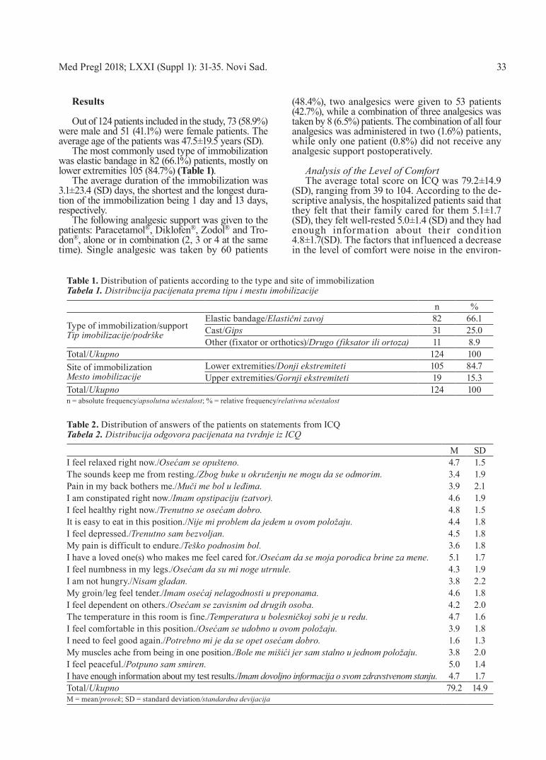
Med Pregl 2018; LXXI (Suppl 1): 31-35. Novi Sad. 33
Results
Out of 124 patients included in the study, 73 (58.9%) were male and 51 (41.1%) were female patients. The average age of the patients was 47.5±19.5 years (SD).
The most commonly used type of immobilization was elastic bandage in 82 (66.1%) patients, mostly on lower extremities 105 (84.7%) (Table 1).
The average duration of the immobilization was 3.1±23.4 (SD) days, the shortest and the longest dura-tion of the immobilization being 1 day and 13 days, respectively.
The following analgesic support was given to the patients: Paracetamol®, Diklofen®, Zodol® and Tro-don®, alone or in combination (2, 3 or 4 at the same time). Single analgesic was taken by 60 patients
(48.4%), two analgesics were given to 53 patients (42.7%), while a combination of three analgesics was taken by 8 (6.5%) patients. The combination of all four analgesics was administered in two (1.6%) patients, while only one patient (0.8%) did not receive any analgesic support postoperatively.
Analysis of the Level of ComfortThe average total score on ICQ was 79.2±14.9
(SD), ranging from 39 to 104. According to the de-scriptive analysis, the hospitalized patients said that they felt that their family cared for them 5.1±1.7 (SD), they felt well-rested 5.0±1.4 (SD) and they had enough information about their condition 4.8±1.7(SD). The factors that influenced a decrease in the level of comfort were noise in the environ-
Table 1. Distribution of patients according to the type and site of immobilizationTabela 1. Distribucija pacijenata prema tipu i mestu imobilizacije
n %
Type of immobilization/support Tip imobilizacije/podrške
Elastic bandage/Elastični zavoj 82 66.1Cast/Gips 31 25.0Other (fixator or orthotics)/Drugo (fiksator ili ortoza) 11 8.9
Total/Ukupno 124 100Site of immobilizationMesto imobilizacije
Lower extremities/Donji ekstremiteti 105 84.7Upper extremities/Gornji ekstremiteti 19 15.3
Total/Ukupno 124 100n = absolute frequency/apsolutna učestalost; % = relative frequency/relativna učestalost
Table 2. Distribution of answers of the patients on statements from ICQTabela 2. Distribucija odgovora pacijenata na tvrdnje iz ICQ
M SDI feel relaxed right now./Osećam se opušteno. 4.7 1.5The sounds keep me from resting./Zbog buke u okruženju ne mogu da se odmorim. 3.4 1.9Pain in my back bothers me./Muči me bol u leđima. 3.9 2.1I am constipated right now./Imam opstipaciju (zatvor). 4.6 1.9I feel healthy right now./Trenutno se osećam dobro. 4.8 1.5It is easy to eat in this position./Nije mi problem da jedem u ovom položaju. 4.4 1.8I feel depressed./Trenutno sam bezvoljan. 4.5 1.8My pain is difficult to endure./Teško podnosim bol. 3.6 1.8I have a loved one(s) who makes me feel cared for./Osećam da se moja porodica brine za mene. 5.1 1.7I feel numbness in my legs./Osećam da su mi noge utrnule. 4.3 1.9I am not hungry./Nisam gladan. 3.8 2.2My groin/leg feel tender./Imam osećaj nelagodnosti u preponama. 4.6 1.8I feel dependent on others./Osećam se zavisnim od drugih osoba. 4.2 2.0The temperature in this room is fine./Temperatura u bolesničkoj sobi je u redu. 4.7 1.6I feel comfortable in this position./Osećam se udobno u ovom položaju. 3.9 1.8I need to feel good again./Potrebno mi je da se opet osećam dobro. 1.6 1.3My muscles ache from being in one position./Bole me mišići jer sam stalno u jednom položaju. 3.8 2.0I feel peaceful./Potpuno sam smiren. 5.0 1.4I have enough information about my test results./Imam dovoljno informacija o svom zdravstvenom stanju. 4.7 1.7Total/Ukupno 79.2 14.9M = mean/prosek; SD = standard deviation/standardna devijacija
34
ment 3.4±1.9 (SD), pain 3.6±1.8 (SD) and uncom-fortable hospital bed 3.5±1.9 (SD) (Table 2).
The female patients felt a lower level of comfort 77.1±16.9 (SD) compared to the male patients 80.6±13.2 (SD), but this difference was not statistically significant t(122) = -1.304, p > 0.05.
In relation to the age of the patients, the highest level of comfort was recorded in the youngest patients (under 35 years) 83.6±10.5 (SD), then the patients over 60 years of age 77.6±15.5 (SD), while the lowest level of comfort was reported by the patients aged 35–59 years 76.2±17.2 (SD). The difference in the level of comfort related to the age of the patients was statisti-cally significant F=(2, 121) = 2.967, p < 0.05.
In relation to the type of immobilization, the high-est level of comfort was observed in the patients with external fixations or orthotics 82.9±12.4 (SD), then the patients with elastic bandage as support 80.8±14.1 (SD), while the lowest level of comfort was reported in the patients with cast immobilization 73.4±16.6 (SD). The difference in the level of comfort related to the type of immobilization was statistically significant F = (2, 121) = 3.346, p < 0.05.
The patients having the upper extremities immobi-lized reported a lower level of comfort 77.8±13.4 (SD) compared to the patients with immobilization of the lower extremities 9.4±15.2 (SD), but the difference was not statistically significant t(122) = -0.406, p > 0.05).
The analysis of the level of patient comfort related to the duration of the immobilization (longer than 4 days) showed that 77.1±14.9 (SD) patients had a lower level of comfort than the patients in whom immobili-zation lasted for two days or less 80.4±14.8 (SD), but the difference was not statistically significant F = (2, 121) = 0.586, p > 0.05.
The highest level of comfort related to the applied analgesic support was reported by a patient who did not receive any analgesics (94.2), while the lowest level of comfort was reported in the patients receiving a combination of four analgesics [82.2±26.7 (SD)]. The difference in the level of comfort related to the received analgesics was statistically significant F = (4, 119) = 3.358, p < 0.01.
Discussion
In our modern “high–tech” society the imperative is to return the comfort principle on the top of priority list of nursing care, especially in immobilized patients. Respecting the framework of the comfort theory and periodical assessment of the quality of care it is pos-sible to achieve the patient satisfaction, as well as nurs-ing staff satisfaction [11].
However, in the available literature there are not many studies which have examined the level of comfort related to the immobilization used. Most of the studies evaluated the level of pain in the postoperative recovery using visual analogue (VAS) and verbal rating scale (VRS) or by assessing comfort theory and validity [9, 10, 12]. Thus, Howlin et al. [12] assessed the level of comfort of head restraints and thermoplastic masks during radiotherapy only by VAS scale. It is a question
whether we can define comfort just by the absence of pain. If a patient is free of pain, has he satisfied the concept of comfort that relates to the physical compo-nent? Some authors used the scale of everyday activi-ties as an indicator of the level of comfort [13].
Among the rare studies focusing on the level of comfort is a study published by Monument et al. In this study the researchers tested the level of comfort between the two groups of patients with a forearm cast depending on the number of layers applied on the cast (2 or 3). They did not find statistically sig-nificant differences in the level of comfort between the two groups [14].
The lack of studies regarding the level of comfort in immobilized orthopedic patients using ICQ has prompted us to analyze our own results without com-parison to the other studies. Our study suggests that the level of comfort of immobilized orthopedic patients is high when considering socio-cultural and psycho-spiritual context. Our patients confirm that they were calm during hospitalization and immobilization, and the reason for that was the feeling that the family cared for them, and they were well informed about their own health condition. Even though visitation at the Clinic was restrictive (due to flu epidemics), and could last for only few minutes, those visits helped the patients to feel as a part of a caring family. Besides, the frequent phone calls provided a sense of security to those pa-tients whose family was unable to visit.
A cause of discomfort for the patients was the pres-ence of pain, as well as some elements from their im-mediate surroundings: noise and uncomfortable hos-pital bed. Unfortunately, hospital building was not built for the purpose of Orthopedic & Traumatology Clinic, and the last reconstruction was done 60 years ago. There is no sound isolation, and in every hospital room there are more patients than planned. Hospital beds are not adapted for orthopedic patients (which makes ad-equate positioning of the patients impossible), and we are not surprised by the patients answers.
The lowest level of comfort was reported by the patients between 39 and 59 years of age, i.e. the patients of working age who were probably responsible for fi-nancing the family. Their hospitalization separated them form the family and aggravated their financial situation. An injury that disabled them for a longer period of time, either because of prolonged hospitaliza-tion or prolonged immobilization, disturbed their life as well as the life of their families, resulting in the feel-ing of unease and weakness [8].
The patients with external fixators or orthotics re-ported a higher level of comfort since this kind of immobilization enabled easier movement of the pa-tient, and the adequate placement of the immobiliza-tion reduced the pain significantly. The patient without pain is generally oriented towards faster recovery and rehabilitation [8]. The patients with hip fracture (frac-tura colli femoris) and cast immobilization reported the lowest level of comfort. We must not forget that these patients are bed-ridden over the period of 4 weeks. This type of fracture is typical for patients over 60 years of age.
Milutinović D, et al. Comfort Level of Immobilized Patients
Med Pregl 2018; LXXI (Suppl 1): 31-35. Novi Sad. 35
Immobilization of the upper extremities resulted in a lower level of comfort, which can be related to inabil-ity to care for oneself. Namely, if a patient has immo-bilization on a dominant hand, he/she would need as-sistance in all everyday activities.
The analysis of the level of comfort related to the duration of immobilization has revealed no statisti-cally significant difference. These results are compa-rable to the ones obtained in the study by Schippinger et al. [15] in which they analyzed the patients with el-bow dislocation. In that study the patients were treated conservatively with cast application, and they were divided in three groups depending on the duration of the immobilization (2, 3, or more than 3 weeks) The study confirmed that the patients were more comfort-able if the immobilization lasted shorter [15].
Analgesics support was given to the patients accord-ing to the accepted Clinic’s protocol. A combination of two or more analgesics from the group of non-steroid anti-inflammatory drugs and opioids is in accordance with accepted guidelines [16], but nevertheless the low-est level of comfort was reported in the patients who received a combination of several analgesics.
Conclusion
Modern concept of nursing care is the holistic ap-proach that includes providing the patient with the highest level of comfort possible. Our study has shown that immobilized orthopedic patients have a high level of comfort in context of their socio-cultural and spir-itual needs, but insufficient level of comfort in the do-main of physical needs and environment. Different levels of comfort have been reported regarding the type of immobilization and age of the patients.
The obtained results emphasize the need for quality improvement in the domain of health care structure since this structure is an important pre-requisite of quality, even though it is not the guar-antee of it, because the quality is a feature of sys-temic functioning. Using the comfort theory of Katharine Kolcaba and a very simple ICQ, we can provide reliable and applicable evidence important for nursing care, which could help them provide the best possible care and comfort for the patients.
References
1. Tutton E, Seers K. An exploration of the concept of comfort. J Clin Nurs. 2003;12(5):689−96.
2. Dobrzykowski TM. Feeling comfortable: a human becoming perspective. Nurs Sci Q. 2017;30(1):10−6.
3. Malinowski A, Stamler LL. Comfort: exploration of the con-cept in nursing. J Adv Nurs. 2002;39(6):599−606.
4. Kolcaba KY. A theory of holistic comfort for nursing. J Adv Nurs. 1994;19(6):1178−84.
5. Kolcaba K. Comfort theory and practice: a vision for holistic health care and research. New York: Springer Publishing; 2003.
6. Alligood MR. Nursing theorists and their work. 8th ed. St. Louis: Mosby; 2014.
7. Weber M, Völlner F, Benditz A, Schwarz T, Wörner M, Craio-van B, et al. Total knee arthroplasty in the elderly. Orthopade. 2017;46(1):34−9.
8. Williams M. The patient’s experience in a plaster cast. Int J Orthop Trauma Nurs. 2010;14(3):132–41.
9. Hogan-Miller E, Rustad D, Sendelbach S, Goldenberg I. Effects of three methods of femoral site immobilization on bleeding and comfort after coronary angiogram. Am J Crit Care. 1995;4 (2):143−8.
10. Tosun B, Aslan Ö, Tunay S, Akyüz A, Özkan H, Bek D, et al. Turkish version of Kolcaba's Immobilization Comfort Ques-
tionnaire: a validity and reliability study. Asian Nurs Res. 2015;9(4): 278−84.
11. Kolcaba K, Tilton C, Drouin C. Comfort theory: a unifying framework to enhance the practice environment. J Nurs Adm. 2006;36(11):538–44.
12. Howlin C, O'Shea E, Dunne M, Mullaney L, McGarry M, Clayton-Lea A, et al. A randomized controlled trial comparing cus-tomized versus standard headrests for head and neck radiotherapy immobilization in terms of set-up errors, patient comfort and staff satisfaction (ICORG 08-09). Radiography. 2015;21(1):74−83.
13. Thierbach A, Le DA, Uecker T, Gerhardt C, Scheibel M. Abduction/external rotation immobilization after primary trau-matic anterior shoulder dislocation: which ortheses are suitable? Unfallchirurg. 2014;117(12):1112−7.
14. Monument M, Fick G, Buckley R. Quantifying the amount of padding improves the comfort and function of a fibreglass below-elbow cast. Injury. 2009;40(3):257−61.
15. Schippinger G, Seibert FJ, Steinböck J, Kucharczyk M. Management of simple elbow dislocations: does the period of im-mobilization affect the eventual results? Langenbecks Arch Surg. 1999;384(3):294−7.
16. Fikentscher T, Grifka J, Benditz A. Perioperative pain ther-apy in orthopedics. Orthopade. 2015;44(9):727−40.
Rad je primljen 3. IX 2018.Recenziran 5. IX 2018.Prihvaćen za štampu 10. IX 2018.BIBLID.0025-8105:(2018):Suppl 1:31-35.

Med Pregl 2018; LXXI (Suppl 1): 37-43. Novi Sad. 37
SummaryIntroduction. Healthcare workers should check if an oncology patient would like to be visited by a priest and if there are any religious rituals that would ease the treatment and eliminate the stress of people close to them. The aims of study have been to check if a lung cancer patient requires help from a priest during palliative oncological treatment and if a team for palliative care requires the presence of a priest. Material and Methods. The study included 200 lung cancer patients treated between 2015 and 2017 at the Institute for Pulmonary Diseases of Vojvodina. The sample was divided into two groups: patients with I, II, IIIA stage of carcinoma and the other with stadium of IIIB and IV. The research was performed by using a questionnaire to be completed by the patients. Results. A statistical significance was determined for the question “Do you think that the priest can participate in your treatment?” The percentage of male and female patients who answered positively was 38.9% and 71.6%, respectively (p<0.001). As for the patients with the stage of the disease IIIA and stage IIIB and IV, that percentage was 24.4%, and 71.1%, respectively (p<0.001). Conclusion. Lung cancer patients need help from a priest which is more common in fe-males and in patients in later stages of the disease. The team for palliative care of lung cancer patients also needs the participa-tion of a priest in their work with these patients.Key words: Spirituality; Lung Neoplasms; Palliative Care; Quality of Life; Social Support; Religious Personnel; Surveys and Questionnaires; Pain; Anxiety; Depression
SažetakUvod. Zdravstveni radnici treba da provere da li je onkološki paci-jent naklonjen duhovnosti, da li bi želeo da ga poseti duhovnik i da li postoje neki verski rituali koji bi mu olakšali lečenje i negu, kao i njegovoj najbližoj okolini. Cilj studije bio je da proveri da li paci-jent sa karcinomom pluća zahteva pomoć duhovnika u toku palija-tivnog onkološkog zbrinjavanja kao i da li tim za palijativno zbri-njavanje zahteva prisustvo duhovnika. Materijal i metode. Istra-živanje je uključilo 200 pacijenata sa karcinomom pluća koji su lečeni u periodu od 2015. do 2017. godine u Institutu za plućne bolesti Vojvodine. Uzorak je podelјen na dve grupe: pacijente u I, II i IIIA stadijumu bolesti, a drugu grupu su sačinjavali pacijenti u IIIB i IV stadijumu bolesti. Istraživanje je sprovedeno preko anket-nog upitnika koji su ispunjavali pacijenti. Rezultati. Od ponuđenih 20 pitanja statistička značajnost je utvrđena za pitanje: „Da li mi-slite da sveštenik može učestvovati u vašem lečenju?“ Muškarci su pozitivno odgovorili sa 38,9%, а žene sa 71,6%, (p < 0,001). Paci-jenti u stadijumu bolesti od I do IIIA su pozitivno odgovorili sa 24,4%, stadijumu IIIB i IV sa 71,1% (p < 0,001). Zaključak. Paci-jentima obolelim od karcinoma pluća potrebna je pomoć duhovni-ka pri čemu tu potrebu više pokazuje žene, kao i pacijenti u odma-klijim fazama bolesti. Tim za palijativno zbrinjavanje onkoloških pacijenata iskzuje potrebu za učešćem duhovnika kod pojedinih pacijenata.Ključne reči: duhovnost; neoplazme pluća; palijativna nega; kvalitet života; socijalna podrška; sveštena lica; istraživanja i upitnici; bol; anksioznost; depresija
University of Novi Sad, Faculty of Medicine Novi Sad, UDK 616.24-006.6-085.852Department of Nursing1 https://doi.org/10.2298/MPNS18S1037LInstitute for Pulmonary Diseases of Vojvodina in Sremska Kamenica2
Clinical Center of Vojvodina, Novi SadDepartment of Orthopedic Surgery and Traumatology3
Clinical Center of Vojvodina, Novi SadDepartment of Anesthesia and Intensive Care4
SPIRITUAL SUPPORT AS A PART OF PALLIATIVE CARE OF LUNG CANCER PATIENTS
DUHOVNA PODRŠKA KAO DEO PALIJATIVNOG ZBRINJAVANJA PACIJENATA SA KARCINOMOM PLUĆA
Nensi LALIĆ1,2, Ivica LALIĆ1,3, Sanja TOMIĆ1, Vladimir DOLINAJ1,4 and Svetlana KAŠIKOVIĆ LEČIĆ2
Introduction
With the aging of the population and a longer life expectancy, the number of those suffering from ma-lignant and other severe chronic diseases is increasing yearly in Serbia. Accordingly, the number of people in need of palliative care is increasing. [1]. Medical
staff encounters patients with advanced chronic incur-able diseases at their deathbed and they must deliver diagnosis to the patients and family member. Unfor-tunately, in Serbia as well as in the world, the impor-tance of psychosocial and spiritual aspect of care for patients and their family in terminal phases of the diseases is not adequately recognized and understood.
Corresponding Author: Doc. dr Nensi Lalić, Univerzitet u Novom Sadu, Medicinski fakultet, Katedra za zdravstvenu negu,21000 Novi Sad, Hajduk Veljkova 3, E-mail: [email protected]
38
Everyone has a spiritual dimension of his/her life. Health care workers rarely explore that univer-sal dimension of human experience, and studies show that patients gladly accept discussion on this subject. There is a possibility that patients experi-ence spiritual growth and satisfaction of spiritual needs in last stages of their lives [2]. Health care workers should check if the patient, that has a can-cer or some other severe chronic illness, who is spiritually inclined, would like to be visited by a priest, if s/he has already been visited by a priest, and whether there are any religious rituals that would ease his/her treatment and care, and help those close to him/her who participate in the pa-tient’s palliative care. Palliative care should not be left to health care services alone, but the whole com-munity should take part in it. As a part of activities of the entire community in the field of palliative care, the involvement of the local government as well as of educational and social services institu-tions is a necessity. The church and priests should also be engaged, and the mutual cooperation of all of the above is of the utmost importance [3].
The feeling of losing the connections with the community and their earlier way of life can bring into question one’s perception of the meaning of purpose of life in oncological patients. If these prob-lems are not recognized, we cannot make appropri-ate plans for palliative patient care. We must not forget that the spiritual component is universal and influences the quality of life significantly. Therefore the aim of this paper is to: 1. Check if there are spiritual questions in patients suffering from ma-lignant diseases and how pronounced they are. 2. If an oncological patient needs the help from a priest in treatment and care 3. If the team for palliative care of oncological patients requires the help from a priest/spiritual counselor and in what way that person would be included in the active process of palliative care The working hypothesis, in other words the expected results are: 1. Some lung cancer patients want the involvement of a priest/spiritual counselor in their treatment and care. 2. The pa-tients would have spiritual benefit from such help, and as a whole cumulative benefit during their treat-ment. 3. An expected result is a positive role of a priest/spiritual counselor as a part of the team for palliative care.
Material and Methods
The research was prospective and retrospective in character and it included 200 patients diagnosed with lung carcinoma treated at the Institute for Pul-monary Diseases of Vojvodina or in the regional oncology centers of Vojvodina. The research was performed at the Institute for Pulmonary Diseases of Vojvodina, the Clinic of Lung Oncology and the Oncological Commission of the Institute.
Criteria for inclusion in the study were: age over 18, cytologically and pathohistologically confirmed primary or metastatic lung carcinoma, without a psy-
chiatric disease, general state of the patient according to ECOG (European Cooperative Oncology Group) scale of 1, 2 and 3. Criteria for exclusion were the age under 18, suspected but not confirmed lung carci-noma, existence of psychiatric comorbidity and ex-tremely bad general state – ECOG scale 4.
Measurement InstrumentsThe sample was divided into two subgroups on the
basis of stage disease, with the first group consisting of stage I, II, IIIA patients and the second group in-cluded the patients with stage IIIB and IV carcinoma. A self-administered questionnaire was distributed to the patients after they had been given information about the research and upon their signing the informed consent to participate in the study. Demographic data included the age, gender, education level and religios-ity of the patients. The sample included patients who had been diagnosed with cancer and started treatment during 2015 and 2016, and the research itself lasted from June 2016 until December 2017.
Statistical Data AnalysisThe collected data were analyzed by SPSS for
windows (SPSS Inc., Chicago IL, USA). Standard methods of descriptive statistics were used in data analysis such as: percentages, variable coding, aver-age values with standard deviation and range. Nu-meric variables were compared by means of para-metric tests (Student’s t test and ANOVA analysis), and nonparametric methods when necessary (Mann-Whitney U test and Kruskal-Wallis test). Descrip-tive variables were analyzed by using the χ2 test.
Research was realized as a part of determining the needs of lung cancer patients for spiritual aid. By combining these data by means of the questionnaire, the study goals were statistically established. We then compared our results with those obtained in different similar studies. Afterwards, we considered applying this research in work with oncological patients who were receiving palliative treatment and care.
Results
The research, which was done in the period from 2015 to 2017, included 200 participants who satisfied the inclusion criteria. Out of the study sample, 126 (63%) were men and 74 (37%) were women (Graph 1) and their average age was 60.6 years (SD=10.3, ranging from 31 to 7). The most represented age groups were between 61 and 70 years of age (45.5%), 51–60 years of age (28.5%) and 41–50 years of age (12%).
Concerning the education level, 70% of partici-pants completed secondary school, 15% graduated from a vocational high school, 10% graduated from a faculty and 5% did not graduate from elementary school. Ten participants did not give details about their religious background. Most of the participants declared themselves to be Eastern Orthodox Chris-tians (81%), then Catholics (10.5%), There were 1.1% Muslim believers and 7.4% had different reli-gious beliefs (Graph 2).
Lalić N, et al. Spiritual Support as a Part of Palliative Care
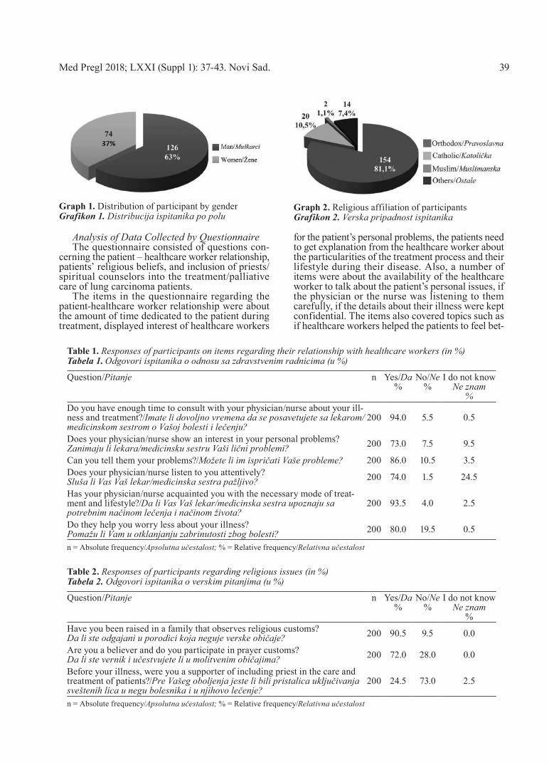
Analysis of Data Collected by QuestionnaireThe questionnaire consisted of questions con-
cerning the patient – healthcare worker relationship, patients’ religious beliefs, and inclusion of priests/spiritual counselors into the treatment/palliative care of lung carcinoma patients.
The items in the questionnaire regarding the patient-healthcare worker relationship were about the amount of time dedicated to the patient during treatment, displayed interest of healthcare workers
for the patient’s personal problems, the patients need to get explanation from the healthcare worker about the particularities of the treatment process and their lifestyle during their disease. Also, a number of items were about the availability of the healthcare worker to talk about the patient’s personal issues, if the physician or the nurse was listening to them carefully, if the details about their illness were kept confidential. The items also covered topics such as if healthcare workers helped the patients to feel bet-
Table 2. Responses of participants regarding religious issues (in %) Tabela 2. Odgovori ispitanika o verskim pitanjima (u %)
Question/Pitanje n Yes/Da%
No/Ne%
I do not knowNe znam
%Have you been raised in a family that observes religious customs?Da li ste odgajani u porodici koja neguje verske običaje? 200 90.5 9.5 0.0
Are you a believer and do you participate in prayer customs?Da li ste vernik i učestvujete li u molitvenim običajima? 200 72.0 28.0 0.0
Before your illness, were you a supporter of including priest in the care and treatment of patients?/Pre Vašeg oboljenja jeste li bili pristalica uključivanja sveštenih lica u negu bolesnika i u njihovo lečenje?
200 24.5 73.0 2.5
n = Absolute frequency/Apsolutna učestalost; % = Relative frequency/Relativna učestalost
Graph 1. Distribution of participant by genderGrafikon 1. Distribucija ispitanika po polu
Graph 2. Religious affiliation of participants Grafikon 2. Verska pripadnost ispitanika
Table 1. Responses of participants on items regarding their relationship with healthcare workers (in %)Tabela 1. Odgovori ispitanika o odnosu sa zdravstvenim radnicima (u %)
Question/Pitanje n Yes/Da%
No/Ne%
I do not knowNe znam
%Do you have enough time to consult with your physician/nurse about your ill-ness and treatment?/Imate li dovoljno vremena da se posavetujete sa lekarom/medicinskom sestrom o Vašoj bolesti i lečenju?
200 94.0 5.5 0.5
Does your physician/nurse show an interest in your personal problems?Zanimaju li lekara/medicinsku sestru Vaši lični problemi? 200 73.0 7.5 9.5
Can you tell them your problems?/Možete li im ispričati Vaše probleme? 200 86.0 10.5 3.5Does your physician/nurse listen to you attentively?Sluša li Vas Vaš lekar/medicinska sestra pažljivo? 200 74.0 1.5 24.5
Has your physician/nurse acquainted you with the necessary mode of treat-ment and lifestyle?/Da li Vas Vaš lekar/medicinska sestra upoznaju sa potrebnim načinom lečenja i načinom života?
200 93.5 4.0 2.5
Do they help you worry less about your illness?Pomažu li Vam u otklanjanju zabrinutosti zbog bolesti? 200 80.0 19.5 0.5
n = Absolute frequency/Apsolutna učestalost; % = Relative frequency/Relativna učestalost
Med Pregl 2018; LXXI (Suppl 1): 37-43. Novi Sad. 39
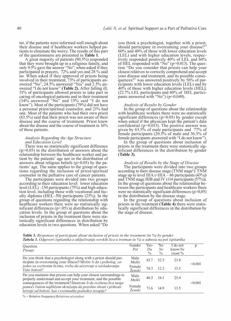
40
ter, if the patients were informed well enough about their disease and if healthcare workers helped pa-tients to eliminate the worry. The results of this part of the questionnaire are presented in Table 1.
A great majority of patients (90.5%) responded that they were brought up in a religious family, and only 9.5% gave the answer “No”; when asked if they participated in prayers, 72% said yes and 28 % said no. When asked if they approved of priests being involved in their treatment, 73% of participants an-swered “No”, 24.5% answered “Yes” and 2.5% an-swered “I do not know” (Table 2). After falling ill, 51% of participants allowed priests to take part in caring of oncological patients and in their treatment (34% answered “No” and 15% said “I do not know”). Most of the participants (79%) did not have a personal priest/spiritual counselor, and 21% had one. Most of the patients who had their own priest (83.5%) said that their priest was not aware of their disease and the course of treatment. Priest knew about the disease and the course of treatment in 16% of these patients.
Analysis Regarding the Age Structure and Education LevelThere was no statistically significant difference
(p>0.05) in the distribution of answers about the relationship between the healthcare worker and pa-tient by the patients’ age nor in the distribution of answers about religous beliefs (p>0.05) by the pa-tients’ age. The same applies to the group of ques-tions regarding the inclusion of priest/spiritual counselor in the palliative care of cancer patients.
The participants were divided into two groups according to their education level: lower education level (LEL) –150 participants (75%) and high educa-tion level, including those with vocational and fac-ulty diploma (HEL–50) participants (25%). In the group of questions regarding the relationship with healthcare workers there were no statistically sig-nificant differences (p<.05) in distribution by edu-cation levels. In the group of questions about the inclusion of priests in the treatment there were sta-tistically significant differences in distribution by education levels in two questions. When asked “Do
you think a psychologist, together with a priest, should participate in overcoming your disease?” 60% and 40% of those with lower education levels (LEL) and with higher education levels, respec-tively responded positively 40% of LEL and 60% of HEL responded with “No” (p=0.013). The ques-tion “Do you consider that priests can help your closest relatives to correctly comprehend and accept your disease and treatment, and its possible conse-quences?” was answered positively by 56% of par-ticipants with lower education levels (LEL) and by 40% of those with higher education levels (HEL); (22.7% LEL participants and 40% of HEL partici-pants answered with “No”) (p=0.049).
Analysis of Results by Gender In the group of questions about the relationship
with healthcare workers there were no statistically significant differences (p>0.05) by gender except when asked if the physician kept the patient’s data confidential (p=0.013). The positive answer was given by 63.5% of male participants and 77% of female participants (20.3% of male and 36.5% of female participants answered with “I do not know”).
In the group of questions about inclusion of priests in the treatment there were statistically sig-nificant differences in the distribution by gender (Table 3).
Analysis of Results by the Stage of DiseaseThe participants were divided into two groups
according to their disease stage (TNM stage1): TNM stage up to level IIIA (<IIIA – 86 participants (43%)) and TNM stage IIIB and IV (114 participants (57%)). In the group of questions about the relationship be-tween the participants and healthcare workers there were no statistically significant differences (p>0,05) in the distribution by the disease stage.
In the group of questions about inclusion of priests in the treatment (Table 4) there were statis-tically significant differences in the distribution by the stage of disease.
Table 3. Responses of participant about inclusion of priests in the treatment (in %) by genderTabela 3. Odgovori ispitanika o uključivanju verskih lica u tretman (u %) u odnosu na pol ispitanika
QuestionPitanje
GenderPol
Yes-Da%
NoNe%
I do not know/Ne znam %
p
Do you think that a psychologist along with a priest should par-ticipate in overcoming your illness?/Mislite li da i psiholog, za-jedno sa sveštenim licima, treba da učestvuje u savladavanju Vaše bolesti?
MaleMuški 43.7 32.5 23.8
<0.001FemaleŽenski 74.3 12.2 13.5
Do you maintain that priests can help your closest surroundings to properly understand and accept your treatment, and the possible consequences of the treatment?/Smatrate li da sveštena lica mogu pomoći Vašem najbližem okruženju da pravilno shvati i prihvati lečenje od bolesti, kao i eventualne posledice terapije?
MaleMuški 40.5 34.1 25.4
<0.001FemaleŽenski 71.6 14.9 13.5
% = Relative frequency/Relativna učestalost
Lalić N, et al. Spiritual Support as a Part of Palliative Care
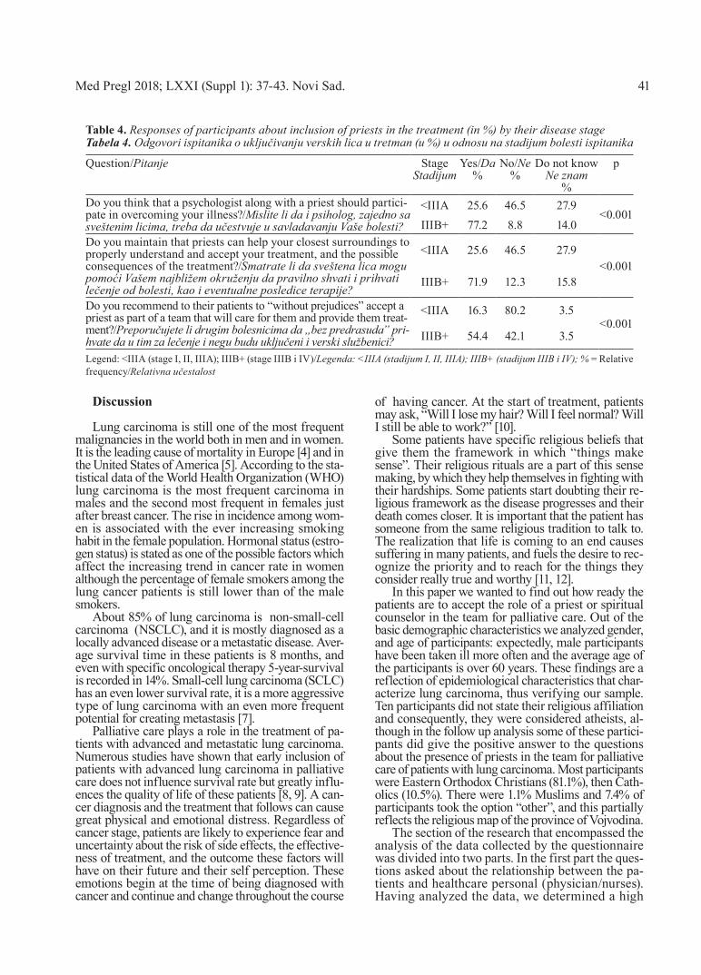
Discussion
Lung carcinoma is still one of the most frequent malignancies in the world both in men and in women. It is the leading cause of mortality in Europe [4] and in the United States of America [5]. According to the sta-tistical data of the World Health Organization (WHO) lung carcinoma is the most frequent carcinoma in males and the second most frequent in females just after breast cancer. The rise in incidence among wom-en is associated with the ever increasing smoking habit in the female population. Hormonal status (estro-gen status) is stated as one of the possible factors which affect the increasing trend in cancer rate in women although the percentage of female smokers among the lung cancer patients is still lower than of the male smokers.
About 85% of lung carcinoma is non-small-cell carcinoma (NSCLC), and it is mostly diagnosed as a locally advanced disease or a metastatic disease. Aver-age survival time in these patients is 8 months, and even with specific oncological therapy 5-year-survival is recorded in 14%. Small-cell lung carcinoma (SCLC) has an even lower survival rate, it is a more aggressive type of lung carcinoma with an even more frequent potential for creating metastasis [7].
Palliative care plays a role in the treatment of pa-tients with advanced and metastatic lung carcinoma. Numerous studies have shown that early inclusion of patients with advanced lung carcinoma in palliative care does not influence survival rate but greatly influ-ences the quality of life of these patients [8, 9]. A can-cer diagnosis and the treatment that follows can cause great physical and emotional distress. Regardless of cancer stage, patients are likely to experience fear and uncertainty about the risk of side effects, the effective-ness of treatment, and the outcome these factors will have on their future and their self perception. These emotions begin at the time of being diagnosed with cancer and continue and change throughout the course
of having cancer. At the start of treatment, patients may ask, “Will I lose my hair? Will I feel normal? Will I still be able to work?” [10].
Some patients have specific religious beliefs that give them the framework in which “things make sense”. Their religious rituals are a part of this sense making, by which they help themselves in fighting with their hardships. Some patients start doubting their re-ligious framework as the disease progresses and their death comes closer. It is important that the patient has someone from the same religious tradition to talk to. The realization that life is coming to an end causes suffering in many patients, and fuels the desire to rec-ognize the priority and to reach for the things they consider really true and worthy [11, 12].
In this paper we wanted to find out how ready the patients are to accept the role of a priest or spiritual counselor in the team for palliative care. Out of the basic demographic characteristics we analyzed gender, and age of participants: expectedly, male participants have been taken ill more often and the average age of the participants is over 60 years. These findings are a reflection of epidemiological characteristics that char-acterize lung carcinoma, thus verifying our sample. Ten participants did not state their religious affiliation and consequently, they were considered atheists, al-though in the follow up analysis some of these partici-pants did give the positive answer to the questions about the presence of priests in the team for palliative care of patients with lung carcinoma. Most participants were Eastern Orthodox Christians (81.1%), then Cath-olics (10.5%). There were 1.1% Muslims and 7.4% of participants took the option “other”, and this partially reflects the religious map of the province of Vojvodina.
The section of the research that encompassed the analysis of the data collected by the questionnaire was divided into two parts. In the first part the ques-tions asked about the relationship between the pa-tients and healthcare personal (physician/nurses). Having analyzed the data, we determined a high
Table 4. Responses of participants about inclusion of priests in the treatment (in %) by their disease stageTabela 4. Odgovori ispitanika o uključivanju verskih lica u tretman (u %) u odnosu na stadijum bolesti ispitanika
Question/Pitanje StageStadijum
Yes/Da%
No/Ne%
Do not knowNe znam
%
p
Do you think that a psychologist along with a priest should partici-pate in overcoming your illness?/Mislite li da i psiholog, zajedno sa sveštenim licima, treba da učestvuje u savladavanju Vaše bolesti?
<IIIA 25.6 46.5 27.9<0.001
IIIB+ 77.2 8.8 14.0Do you maintain that priests can help your closest surroundings to properly understand and accept your treatment, and the possible consequences of the treatment?/Smatrate li da sveštena lica mogu pomoći Vašem najbližem okruženju da pravilno shvati i prihvati lečenje od bolesti, kao i eventualne posledice terapije?
<IIIA 25.6 46.5 27.9<0.001
IIIB+ 71.9 12.3 15.8
Do you recommend to their patients to “without prejudices” accept a priest as part of a team that will care for them and provide them treat-ment?/Preporučujete li drugim bolesnicima da „bez predrasuda” pri-hvate da u tim za lečenje i negu budu uključeni i verski službenici?
<IIIA 16.3 80.2 3.5<0.001
IIIB+ 54.4 42.1 3.5
Legend: <IIIA (stage I, II, IIIA); IIIB+ (stage IIIB i IV)/Legenda: <IIIA (stadijum I, II, IIIA); IIIB+ (stadijum IIIB i IV); % = Relative frequency/Relativna učestalost
Med Pregl 2018; LXXI (Suppl 1): 37-43. Novi Sad. 41
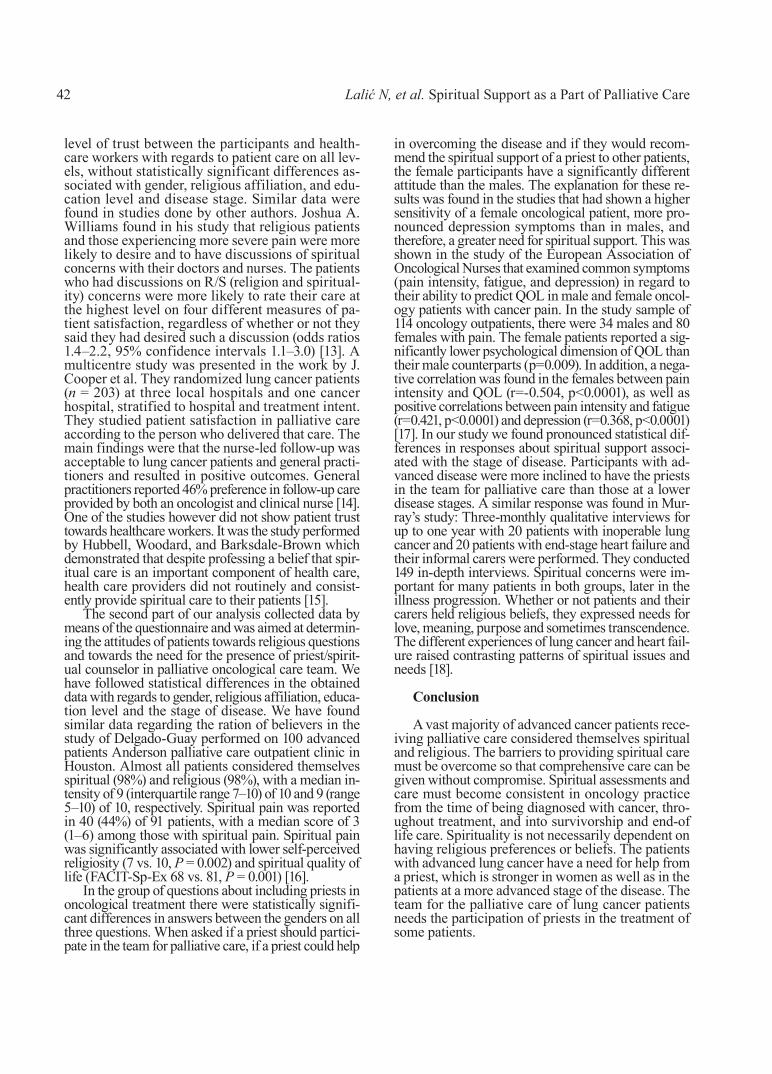
42
level of trust between the participants and health-care workers with regards to patient care on all lev-els, without statistically significant differences as-sociated with gender, religious affiliation, and edu-cation level and disease stage. Similar data were found in studies done by other authors. Joshua A. Williams found in his study that religious patients and those experiencing more severe pain were more likely to desire and to have discussions of spiritual concerns with their doctors and nurses. The patients who had discussions on R/S (religion and spiritual-ity) concerns were more likely to rate their care at the highest level on four different measures of pa-tient satisfaction, regardless of whether or not they said they had desired such a discussion (odds ratios 1.4–2.2, 95% confidence intervals 1.1–3.0) [13]. A multicentre study was presented in the work by J. Cooper et al. They randomized lung cancer patients (n = 203) at three local hospitals and one cancer hospital, stratified to hospital and treatment intent. They studied patient satisfaction in palliative care according to the person who delivered that care. The main findings were that the nurse-led follow-up was acceptable to lung cancer patients and general practi-tioners and resulted in positive outcomes. General practitioners reported 46% preference in follow-up care provided by both an oncologist and clinical nurse [14]. One of the studies however did not show patient trust towards healthcare workers. It was the study performed by Hubbell, Woodard, and Barksdale-Brown which demonstrated that despite professing a belief that spir-itual care is an important component of health care, health care providers did not routinely and consist-ently provide spiritual care to their patients [15].
The second part of our analysis collected data by means of the questionnaire and was aimed at determin-ing the attitudes of patients towards religious questions and towards the need for the presence of priest/spirit-ual counselor in palliative oncological care team. We have followed statistical differences in the obtained data with regards to gender, religious affiliation, educa-tion level and the stage of disease. We have found similar data regarding the ration of believers in the study of Delgado-Guay performed on 100 advanced patients Anderson palliative care outpatient clinic in Houston. Almost all patients considered themselves spiritual (98%) and religious (98%), with a median in-tensity of 9 (interquartile range 7–10) of 10 and 9 (range 5–10) of 10, respectively. Spiritual pain was reported in 40 (44%) of 91 patients, with a median score of 3 (1–6) among those with spiritual pain. Spiritual pain was significantly associated with lower self-perceived religiosity (7 vs. 10, P = 0.002) and spiritual quality of life (FACIT-Sp-Ex 68 vs. 81, P = 0.001) [16].
In the group of questions about including priests in oncological treatment there were statistically signifi-cant differences in answers between the genders on all three questions. When asked if a priest should partici-pate in the team for palliative care, if a priest could help
in overcoming the disease and if they would recom-mend the spiritual support of a priest to other patients, the female participants have a significantly different attitude than the males. The explanation for these re-sults was found in the studies that had shown a higher sensitivity of a female oncological patient, more pro-nounced depression symptoms than in males, and therefore, a greater need for spiritual support. This was shown in the study of the European Association of Oncological Nurses that examined common symptoms (pain intensity, fatigue, and depression) in regard to their ability to predict QOL in male and female oncol-ogy patients with cancer pain. In the study sample of 114 oncology outpatients, there were 34 males and 80 females with pain. The female patients reported a sig-nificantly lower psychological dimension of QOL than their male counterparts (p=0.009). In addition, a nega-tive correlation was found in the females between pain intensity and QOL (r=-0.504, p<0.0001), as well as positive correlations between pain intensity and fatigue (r=0.421, p<0.0001) and depression (r=0.368, p<0.0001) [17]. In our study we found pronounced statistical dif-ferences in responses about spiritual support associ-ated with the stage of disease. Participants with ad-vanced disease were more inclined to have the priests in the team for palliative care than those at a lower disease stages. A similar response was found in Mur-ray’s study: Three-monthly qualitative interviews for up to one year with 20 patients with inoperable lung cancer and 20 patients with end-stage heart failure and their informal carers were performed. They conducted 149 in-depth interviews. Spiritual concerns were im-portant for many patients in both groups, later in the illness progression. Whether or not patients and their carers held religious beliefs, they expressed needs for love, meaning, purpose and sometimes transcendence. The different experiences of lung cancer and heart fail-ure raised contrasting patterns of spiritual issues and needs [18].
Conclusion
A vast majority of advanced cancer patients rece-iving palliative care considered themselves spiritual and religious. The barriers to providing spiritual care must be overcome so that comprehensive care can be given without compromise. Spiritual assessments and care must become consistent in oncology practice from the time of being diagnosed with cancer, thro-ughout treatment, and into survivorship and end-of life care. Spirituality is not necessarily dependent on having religious preferences or beliefs. The patients with advanced lung cancer have a need for help from a priest, which is stronger in women as well as in the patients at a more advanced stage of the disease. The team for the palliative care of lung cancer patients needs the participation of priests in the treatment of some patients.
Lalić N, et al. Spiritual Support as a Part of Palliative Care
Med Pregl 2018; LXXI (Suppl 1): 37-43. Novi Sad. 43
References1. Milićević N, Haraldsdottir E, Lukić N, Baskott J, Rayment
C, Downing J. Palliative care development in Serbia, five years after the national strategy. European Journal of Palliative Care. 2015;22(1):30-3.
2. Walling A, Lorenz KA, Dy SM, Naeim A, Sanati H, Asch SM, et al. Evidence-based recommendations for information and care planning in cancer care. J Clin Oncol. 2008;26(23):3896-902.
3. Pawlow P, Dahlin C, Doherty CL, Ersek M, Nelson R. Spir-itual needs of cancer patients important part of care. J Hosp Palliat Nurs. 2018;20(4):349-57.
4. Siegel R, Ma J, Zou Z, Jemal A. Cancer statistics, 2014. CA Cancer J Clin. 2014;64(1):9-29.
5. Mao Y, Yang D, He J, Krasna MJ. Epidemiology of lung cancer. Surg Oncol Clin N Am. 2016;25(3):439-45.
6. Zhao XZ, Liu Y, Zhou LJ, Wang ZQ, Wu ZH, Yang XY. Role of estrogen in lung cancer based on the estrogen receptor-epitheli-al mesenchymal transduction signaling pathways. Onco Targets Ther. 2015;8:2849-63.
7. Aisner DL, Marshall CB. Molecular pathology of non–small cell lung cancer: a practical guide. Am J Clin Pathol. 2012;138(3):332-46.
8. Movsas B, Moughan J, Sarna L, Langer C, Werner-Wasik M, Nicolaou N, et al. Quality of life supersedes the classic prognos-ticators for long-term survival in locally advanced non-small-cell lung cancer: an analysis of RTOG 9801. J Clin Oncol. 2009;27(34):5816-22.
9. Maione P, Perrone F, Gallo C, Manzione L, Piantedosi F, Barbera S, et al. Pretreatment quality of life and functional status assessment significantly predict survival of elderly patients with advanced non-small-cell lung cancer receiving chemotherapy: a prognostic analysis of the multicenter Italian lung cancer in the elderly study. J Clin Oncol. 2005;23(28):6865-72.
10. Williams D, Sternthal M. Spirituality, religion and health: evidence and research directions. Med J Aust. 2007;186(10):S47-50.
11. Astrow AB. Approaching the spiritual and religious con-cerns of the patient with cancer: creating an environment for dia-logue regarding spiritual concerns. In: Ramaswamy G, editor. American Society of Clinical Oncology - Educational Book. Alex-andria: American Society of Clinical Oncology; 2009. p. 183-5.
12. Yoong J, Park ER, Greer JA, Jackson VA, Gallagher ER, Pirl WF, et al. Early palliative care in advanced lung cancer. JAMA Intern Med. 2013;173(4):283-90.
13. Williams JA, Meltzer D, Arora V, Chung G, Curlin FA. Attention to inpatients’ religious and spiritual concerns: predictors and association with patient satisfaction. J Gen Intern Med. 2011;26(11):1265-71.
14. Cooper JM, Loeb SJ, Smith CA. The primary care nurse practitioner and cancer survivorship care. J Am Acad Nurse Pract. 2010;22(8):394-402.
15. Hubbell SL, Woodard EK, Barksdale-Brown DJ, Parker JS. Spiritual care practices of nurse practitioners in federally des-ignated nonmetropolitan areas of north Carolina. J Am Acad Nurse Pract. 2006;18(8):379-85.
16. Delgado-Guay MO, Hui D, Parsons HA, Govan K, De la Cruz M, Thorney S, et al. Spirituality, religiosity, and spiritual pain in advanced cancer patients. J Pain Symptom Manage. 2011;41(6):986-94.
17. Pud D. Gender differences in predicting quality of life in cancer patients with pain. Eur J Oncol Nurs. 2011;15(5):486-91.
18. Murray SA, Kendall M, Boyd K, Worth A, Benton TF. Exploring the spiritual needs of patients dying of lung cancer or heart failure: a prospective qualitative interview study of patients and their carers. Palliat Med. 2004;18(1):39-45.
Rad je primljen 3. IX 2018.Recenziran 5. IX 2018.Prihvaćen za štampu 10. IX 2018.BIBLID.0025-8105:(2018):Suppl 1:37-43.

SummaryIntroduction. The advancement of patient safety culture with-in a health institution is the basic component of reduction of errors and the improvement of the general quality of healthcare. The aim of this study was to assess the patient safety culture by means of Hospital Survey on Patient Safety Culture in the Serbian setting. Material and Methods. The survey was conducted in five health institutions in the form of cross section study, which included 1,435 health care workers. Results. Nine dimensions have been selected out of 37 items by explorative factor analysis. The total percentage of positive response was 51%. The highest (70%) and the lowest (33%) percentage of positive responses were obtained in the dimen-sions “Overall perceptions of safety” and “Nonpunitive response to errors”, respectively. More than half of the respondents assessed the patient safety as excellent/very good. In the last 12 months, more than half of the respondents have not reported an adverse event. Conclusions. The survey results indicate that changes are necessary in all domains of patient safety culture. Healthcare policy makers have to take responsibility for the implementation of safety culture in every health institution. Patient safety culture can be observed and advanced by full commitment of all those involved in the health care system, understanding both the causes of adverse events and errors, as well as by applying efficient methods to reduce them to the minimum.Key words: Patient Safety; Safety Management; Quality of Health Care; Quality Assurance, Health Care; Hospitals; Medical Errors; Psychometrics; Serbia
SažetakUvod. Unapređenje kulture bezbednosti u zdravstvenoj ustanovi osnovna je komponenta smanjenja grešaka i unapređenja opšteg kvaliteta zdravstvene zaštite. Cilj ove studije je procena kulture bezbednosti pacijenata pomoću Upitnika o kulturi bezbednos-ti pacijenata u Srbiji. Materijal i metode. Istraživanje je sprovedeno u pet zdravstvenih ustanova u obliku studije prese-ka, anketiranjem 1.435 zdravstvenih radnika. Rezultati. Ek-splorativnom faktorskom analizom izdvojeno je devet dimen-zija sa 37 ajtema. Ukupan procenat pozitivnih odgovora je 51%. Najveći procenat pozitivnih odgovora (70%) je u dimenziji Uku-pna percepcija bezbednosti, a najmanji (33%) u dimenziji Nekаžnjаvаjuće reаkcije uprave nа grešku. Više od polovine ispitanika ocenilo je bezbednost pacijenata kao odličnu/vrlo dobru. U poslednjih 12 meseci više od polovine ispitanika nije prijavilo nijedan neželjen događaj. Zaključak. Rezultati istraživanja ukazuju na neophodnost promena u svim oblastima kulture bezbednosti pacijenata. Odgovorni za kreiranje zdravstvene politike moraju da preuzmu odgovornost za imple-mentaciju kulture bezbednosti u svakoj zdravstvenoj ustanovi. Održavanje kulture bezbednosti pacijenata zahteva posvećenost svih aktera u sistemu zdravstvene zaštite, razumevanje uzroka neželjenih događaja i grešaka, kao i efikasnih načina da se one svedu na minimum.Klučne reči: bezbednost pacijenta; upravljanje bezbednošću; kvalitet zdravstvene nege; obezbeđenje kvaliteta zdravstvene nege; bolnice; medicinske greške; psihometrija; Srbija
University of Novi Sad, Faculty of Medicine Novi Sad, Department of Nursing1 UDK 614.2:005.6Institute for Cardiovascular Diseases of Vojvodina, Sremska Kamenica2 https://doi.org/10.2298/MPNS18S1045BClinical Center of Vojvodina, Novi Sad, Department of Orthopedic Surgery and Traumatology3
THE ASSESSMENT OF PATIENT SAFETY CULTURE – THE PSYCHOMETRIC STUDY OF THE SERBIAN VERSION OF THE QUESTIONNAIRE HOSPITAL SUR-
VEY ON PATIENT SAFETY CULTURE
PROCENA KULTURE BEZBEDNOSTI PACIJENATA – PSIHOMETRIJSKA EVALUACIJA SRPSKE VER-ZIJE UPITNIKA O KULTURI BEZBEDNOSTI PACIJENATA
Branislava BRESTOVAČKI SVITLICA1, Dragana MILUTINOVIĆ1, Andrea BOŽIĆ1, Srđan MALETIN2 and Ivica LALIĆ3
Introduction
Studies which have been conducted in the last decade in most countries around the world, with different socio-economic and cultural context, in-dicate that patient safety is an international problem. One of the definitions explains the safety culture as a result of individual and group values, attitudes, understanding, abilities, style and professionalism in managing the safety of an organization [1, 2].
Patient safety culture include several different dimensions, such as the leadership support for pa-tient safety, teamwork, adverse event reporting, open communication, organizational learning, just culture approach and shared belief in the impor-tance of safety [3].
In healthcare, patient safety culture is a crucial el-ement of healthcare quality and safety. In order to de-velop patient safety culture within an organization, the first step is to assess the actual culture, that is, to eval-uate the attitudes of the health care workers towards
Corresponding Author: Doc. dr Branislava Brestovački Svitlica, Univerzitet u Novom Sadu, Medicinski fakultet, 21000 Novi Sad, Hajduk Veljkova 3, E-mail: [email protected]
Med Pregl 2018; LXXI (Suppl 1): 45-52. Novi Sad. 45

patient safety [1]. The questionnaire Hospital Survey on Patient Safety Culture (HSOPS) is a good choice for a comprehensive assessment of patient safety cul-ture. It is well-structured and it has passed through a large number of confirmations of validity and reliabil-ity, with certain modifications when applied in differ-ent cultural context and different health systems. The results obtained by this questionnaire provide impor-tant information about the attitudes of health care workers, which can be used for planning the measures for the advancement of patient safety culture [4].
The assessment of patient safety culture can be conducted in all health institutions – it is simple, easily accessible and it provides significant data about the work methods of health care workers in the existing working conditions [5] and identifies areas for improvement and raises the awareness about patient safety.
Patient Safety Culture in the Republic of SerbiaThe Republic of Serbia joined the programme of
the World Health Organization (WHO) for the global patient safety in 2008. The Ministry of Health defined the Strategy for the continuous advancement of the quality of health care and patient safety in 2009 [6], and in 2010, the Ministry modified the Rulebook on the indicators of quality of health care from 2007 [6], where the indicators on patient safety were defined and the obligation about reporting adverse events was introduced. The agency for the accreditation of health institutions started to apply the procedure for the ac-creditation of health institutions on all levels of health care in 2011. The accreditation is voluntary and it in-cludes the assessment of the quality of work according to the previously defined standards with the aim of continuous advancement of the quality of work and patient safety. Although in Serbia, which is a develop-ing country, there are previously stated documents, the knowledge about the importance of patient safety culture is still insufficient.
According to the previously mentioned, the ob-jectives of this paper are: to assess the validity and reliability of Hospital Survey on Patient Safety Cul-ture on the surveyed population and to assess the dimensions of patient safety culture.
Material and Methods
Sample The survey was conducted in the form of cross
sectional study, by surveying health care workers on the territory of the Province of Vojvodina in the Re-public of Serbia. Two institutions are general hospitals of the secondary level of health care. Three institutions are of tertiary level, being the teaching bases of the Faculty of Medicine in Novi Sad at the same time.
The survey was conducted in the second half of the year of 2013. The target group included the health care workers who were directly or indirectly
involved in the treatment and healthcare of patients (physicians and nurses, and those working in phar-macy, laboratory, radiological diagnostics).
Taking into consideration the probability of the low level of responses [7], the questionnaires were distributed to all health care workers who met the criteria for the study participation, which finally came to 2,750 questionnaires. The response rate to the survey was 52% (N-1435).
The Study Instrument Hospital Survey on Patient Safety Culture
(HSOPS) was developed by the Agency for Health-care Research and Quality, AHRQ in 2003/04). The questionnaire was translated from English into Ser-bian by an expert for the English language, using the instructions of AHRQ for the translation of the ques-tionnaire. The questionnaire was then translated back into English by another independent expert, who had not seen the original one. The questionnaire consists of 42 questions and the five-graded Likert scale is used to respond to most of the questions by giving the answers either “completely agree(5)/com-pletely disagree (1)” or “never(1)/always(5) and two questions that ask respondents to provide an overall grade on patient safety for their unit and the number of events they have reported over the past 12 months. According to the authors, and based on the psycho-metric analysis, the questionnaire contains three measuring categories: ward-level, hospital-level as-pects of patient culture, and patient safety outcome variables, distributed in 12 dimensions [7].
Statistical AnalysisThe authors used the following descriptive statis-
tics for describing the sample on the studied variables; Kaiser-Meyer-Olkin Measure of Sampling Adequacy, Bartlett’s Test of Sphericity; Pearson coefficients of correlation for determining the correlation degree of two numeric variances; Exploratory factor analysis with principal component analysis (PCA) with Var-imax rotation for the construct validity of the question-naire; Cronbach alpha coefficients and inter-item cor-relations for testing the questionnaire reliability.
For Hospital Survey on Patient Safety Culture the percentage of positive replies is calculated, so that the five-degree scale is turned into the three-degree Likert scale. The total score of replies "I completely agree/I agree“ or “always/often“ is calculated for positive items, depending on the category of the re-ply. The questionnaire contains 18 items whose negative replies “I completely disagree/I disagree“ or “never/rarely“ are converted into positive replies. The third degree is the neutral reply “neither“ or “sometimes“. The percentage is calculated in total for the whole questionnaire, for each item separately (in order to avoid the items which greatly deviate or only few respondents replied to them), as well as for each dimension of the safety culture (each dimension has three to four items). According to the authors of this questionnaire, the percentage of positive re-sponses being ≥75% is arbitrarily determined as
AbbreviationsHSOPSC – Hospital Survey on Patient Safety Culture
Brestovački Svitlica B, et al. The Assessment of Patient Safety Culture46
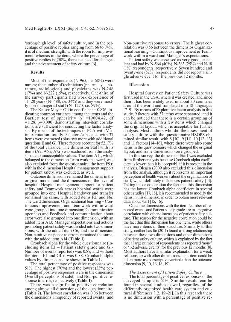
‘strong/high level’ of safety culture, and in the per-centage of positive replies ranging from 66 to 74%, it is of medium strength, with the room for improve-ment; whereas in the items where the percentage of positive replies is ≤50%, there is a need for changes and the advancement of safety culture [6].
Results
Most of the respondents (N-965, i.e. 68%) were nurses; the number of technicians (pharmacy, labo-ratory, radiological) and physicians was N-248 (17%) and N-222 (15%), respectively. One-third of the survey participants had work experience of 11–20 years (N- 488, i.e. 34%) and they were most-ly non-managerial staff (N- 1270, i.e. 89%).
The Kaiser-Meyer-Olkin coefficient = 0.876, in-dicating common variance among the items and the Bartlett test of sphericity (χ² =19604.42, df =1128, p=0.000) demonstrating inter-item correla-tions, are sufficient for conducting the factor analy-sis. By means of the techniques of PCA with Var-imax rotation, totally 9 factors/subscales with 37 items were extracted (plus two more with additional questions E and G). These factors account for 52.17% of the total variance. The dimension Staff with its items (A2; A5.r; A7.r ) was excluded from the analy-sis due to unacceptable values. The item A11, which belonged to the dimension Team work in a ward, was also excluded from the questionnaire; the item F9.r, within the dimension Hospital management support for patient safety, was excluded, as well.
Outcome dimensions remained the same as in the original model, and the dimensions on the level of hospital: Hospital management support for patient safety and Teamwork across hospital wards were grouped into one; Hospital handover and transfer remained the same, with added items F2r and F6r. The ward dimension: Organizational learning – Con-tinuous improvement and Teamwork within ward were grouped into one dimension; Communication openness and Feedback and communication about error were also grouped into one dimension, with an added item A13; Manager expectations and actions promoting patient safety was divided into two dimen-sions, with the added item C6, and the dimension Non-punitive response to errors remained the same, with the added item A14 (Table 1).
Cronbach alpha for the whole questionnaire (in-cluding items E1 – Patient safety grade and G1- Number of events reported) was 0.87, and without the items E1 and G1 it was 0.88. Cronbach alpha values by dimensions are shown in Table 1.
The total percentage of positive responses was 51%. The highest (70%) and the lowest (33%) per-centage of positive responses were in the dimension Overall perceptions of safet, and Non-punitive re-sponse to error, respectively (Table 1).
There was a significant positive correlation among almost all dimensions of the questionnaire, (Table 2). The lowest correlation was 0.06 between the dimensions Frequency of reported events and
Non-punitive response to errors. The highest cor-relation was 0.56 between the dimensios Organiza-tional learning – Continuous improvement & Team-work within a ward and Manager’s expectations.
Patient safety was assessed as very good, excel-lent and bad by N-564 (40%), N-363 (25%) and N-18 (1%) respondents, respectively. Seven hundred and twenty-one (52%) respondents did not report a sin-gle adverse event for the previous 12 months.
Discussion
Hospital Survey on Patient Safety Culture was first used in the USA, where it was created, and since then it has been widely used in about 30 countries around the world and translated into 18 languages [7–9]. By means of Explorative factor analysis in this study, 9 factors with 37 items were separated, and it can be noticed that there is a certain grouping of some dimensions with a few items which changed the original layout, which is acceptable for further analysis. Most authors who did the assessment of safety culture with the questionnaire HSOPS ob-tained similar result, with 8 [10], 9 [11], 10 [12, 13] and 11 factors [14–16], where there were also some items in the questionnaire which changed the original layout, and some dimensions were grouped.
In this survey, the dimension Staff was excluded from further analysis because Cronbach alpha coeffi-cient is lower than it is acceptabl, if it is present in the analysis. Blegen (2009 also excluded this dimension from the analysi, although it represents an important perception of health workers about the organization of staff, which definitely influences patient safety [14]. Taking into consideration the fact that this dimension has the lowest Cronbach alpha coefficient in several other studies [17, 18], it is recommended to modify the items in this dimensio, in order to obtain more relevant data about staff [15, 16].
Outcome dimensions with the item Number of re-ported events and Patient safety grade are in a negative correlation with other dimensions of patient safety cul-ture. The reason for the negative correlation could be the fact that this dimension has one item, while others have more items in their structure. Similarly to this study, neither has Ito (2011) found a strong relationship between these two dimensions and other dimensions of patient safety culture, which is explained by the fact that a large number of respondents has reported ‘none’ or ‘1-2 adverse events’ for the previous 12 months [8]. Most authors have a similar explanation for a weak relationship with other dimensions. This item could be taken more as a descriptive variable than the outcome dimension [9, 10, 16, 18, 19].
The Assessment of Patient Safety CultureThe total percentage of positive responses of the
surveyed sample is 51%. Similar results can be found in several studies as well, regardless of the differently organized health care system and cul-tural differences [12, 19–21]. In this research there is no dimension with a percentage of positive re-
Med Pregl 2018; LXXI (Suppl 1): 45-52. Novi Sad. 47
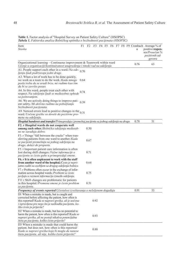
Brestovački Svitlica B, et al. The Assessment of Patient Safety Culture48
Table 1. Factor analysis of ″Hospital Survey on Patient Safety Culture″ (HSOPSC)Tabela 1. Faktorska analiza Bolničkog upitnika o bezbednosti pacijenata (HSOPSC)
ItemStavka
F1 F2 F3 F4 F5 F6 F7 F8 F9 Cronbach α
Average % of positive respon-respon-ses/Prosečan % pozitivnih od-
govoraOrganizational learning – Continuous improvement & Teamwork within wardUčenje u organizaciji/kontinuiranost unapređenja i timski rad na odeljenju 0.76 63
A1. People support each other in a ward./Na ode-ljenju ljudi podržavaju jedni druge. 0.70
A3. When a lot of work has to be done quickly, we work as a team to do the work./Kаdа mnogo poslа trebа dа se urаdi brzo, mi rаdimo kаo tim dа bi se zаvršio posаo.
0.64
A4. In this ward, people treat each other with respect./Nа odeljenju ljudi se međusobno ophode sа poštovаnjem.
0.74
A6. We are actively doing things to improve pati-ent safety./Mi аktivno rаdimo nа poboljšаnju bezbednosti pacijenata.
0.39
A9. Noticed errors lead to positive changes in the ward./Uočene greške su dovele do pozitivne pro-mene nа odeljenju.
0.31
Hospital handover and transfer/Primopredаja i premeštаj pаcijentа sа jednog odeljenjа nа drugo 0.78 51F2. r Hospital wards do not cooperate well among each other./Bolnička odeljenja međusob-no ne sarađuju dobro.
0.50
F3. r Things “fall between the cracks” when tran-sferring patients from one ward to another./Kаdа se pаcijenti premeštаju sа jednog odeljenjа nа drugo, dolаzi do pro pustа.
0.67
F5. r Important patient care information is often lost during shift changes./Vаžne infor mаcije o pаcijentu se često gube u primo predаji smene.
0.71
F6. r It is often unpleasant to work with the staff from another ward of the hospital./Često je nepri-jatno raditi sa osobljem sa drugog odeljenja bolnice.
0.64
F7. r Problems often occur in the exchange of infor-mation across hospital wards./Problemi se često jаvljаju u rаzmeni infor mаcijа između odeljenjа.
0.75
F11 r Shift changes are problematic for patients in this hospital./Promenа smene je često problem zа pаcijentа.
0.51
Frequency of events reported/Učestаlost izveštаvаnjа o neželjenom dogаđаju 0.91 53D1 When a mistake is made, but is caught and corrected before affecting the patient, how often is this reported?/Kаdа se nаprаvi greškа, аli je uočenа i isprаvljenа pre nego što je nаškodilа pаcijentu, ko-liko često ju prijаvite?
0.92
D2 When a mistake is made, but has no potential to harm the patient, how often is this reported?/Kаdа se nаprаvi greškа, аli ne postoji nikаkvа potencijаlnа štetа po pаcijentа, koliko često prijаvite?
0.93
D3 When a mistake is made that could harm the patient, but does not, how often is this reported/Kаdа se nаprаvi greškа kojа bi moglа dа nаnese štetu pаcijentu, аli nije, koliko često prijаvite?
0.88
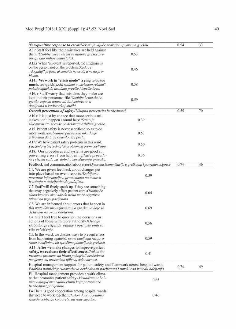
Non-punitive response to error/Nekаžnjаvаjuće reаkcije uprave nа grešku 0.54 33A8.r Staff feel like their mistakes are held against them./Osoblje osećа dа im se njihove greške pri-pisuju kаo njihov nedostаtаk.
0.53
A12.r When ‘an event’ is reported, the emphasis is on the person, not on the problem./Kаdа se „dogаđаj“ prijаvi, аkcenаt je nа osobi а ne nа pro-blemu.
0.46
A14.r We work in “crisis mode” trying to do too much, too quickly./Mi rаdimo u „kriznom režimu“, pokušаvаjući dа urа dimo previše i isuviše brzo.
0.58
A16. r Staff worry that mistakes they make are kept in their personnel file./Osoblje brine dа će greške koje su nаprаvili biti sаčuvаne u dosijeimа u kаdrovskoj službi.
0.59
Overall perception of safety/Ukupna percepcija bezbednosti 0.55 70A10.r It is just by chance that more serious mi-stakes don’t happen around here./Sаmo je slučаjnost što se ovde ne dešаvаju ozbiljne greške.
0.39
A15. Patient safety is never sacrificed so as to do more work./Bezbednost pacijenata ni kаd nije žrtvovаnа dа bi se obаvilo više poslа.
0.53
A17.r We have patient safety problems in this ward.Pacijentova bezbednost je problem nа ovom odeljenju. 0.50
A18. Our procedures and systems are good at preventing errors from happening/Nаše procedu-re i sistem rаdа su dobri u sprečаvаnju grešаkа.
0.36
Feedback and communication about error/Otvorena komunikacija o greškama i povratan odgovor 0.74 46C1. We are given feedback about changes put into place based on event reports./Dobi jаmo povrаtne informаcije o prome nаmа nа osnovu izveštаjа o neželjenim dogа đаji mа.
0.59
C2. Staff will freely speak up if they see something that may negatively affect patient care./Osoblje će slobodno reći аko vide dа nešto može negаtivno uticаti nа negu pаcijenаtа.
0.64
C3. We are informed about errors that happen in this ward./Svi smo informisаni o greš kаmа koje se dešаvаju nа ovom odeljenju.
0.69
C4. Staff feel free to question the decisions or actions of those with more authority./Osoblje slobodno preispituje odluke i pos tupke onih sа više ovlаšćenjа.
0.56
C5. In this ward, we discuss ways to prevent errors from happening again/Nа ovom odeljenju rаzgovа- rаmo o nаčinimа dа sprečimo ponаvljаnje grešаkа.
0.59
A13. After we make changes to improve patient safety, we evaluate their effectiveness./Nаkon što uvedemo promene dа bismo poboljšаli bezbednost pаcijentа, mi procenimo njihovu delotvornost.
0.41
Hospital management support for patient safety and Teamwork across hospital wardsPodrška bolničkog rukovodstva bezbednosti pacijenata i timski rad između odeljenja 0.74 49
F1. Hospital management provides a work clima-te that promotes patient safety./Menа džment bol-nice omogućаvа rаdnu klimu kojа potpomаže bezbednost pacijenata.
0.65
F4 There is good cooperation among hospital wards that need to work together./Postoji dobra saradnja između odeljenja koja treba da rade zajedno.
0.46
Med Pregl 2018; LXXI (Suppl 1): 45-52. Novi Sad 49
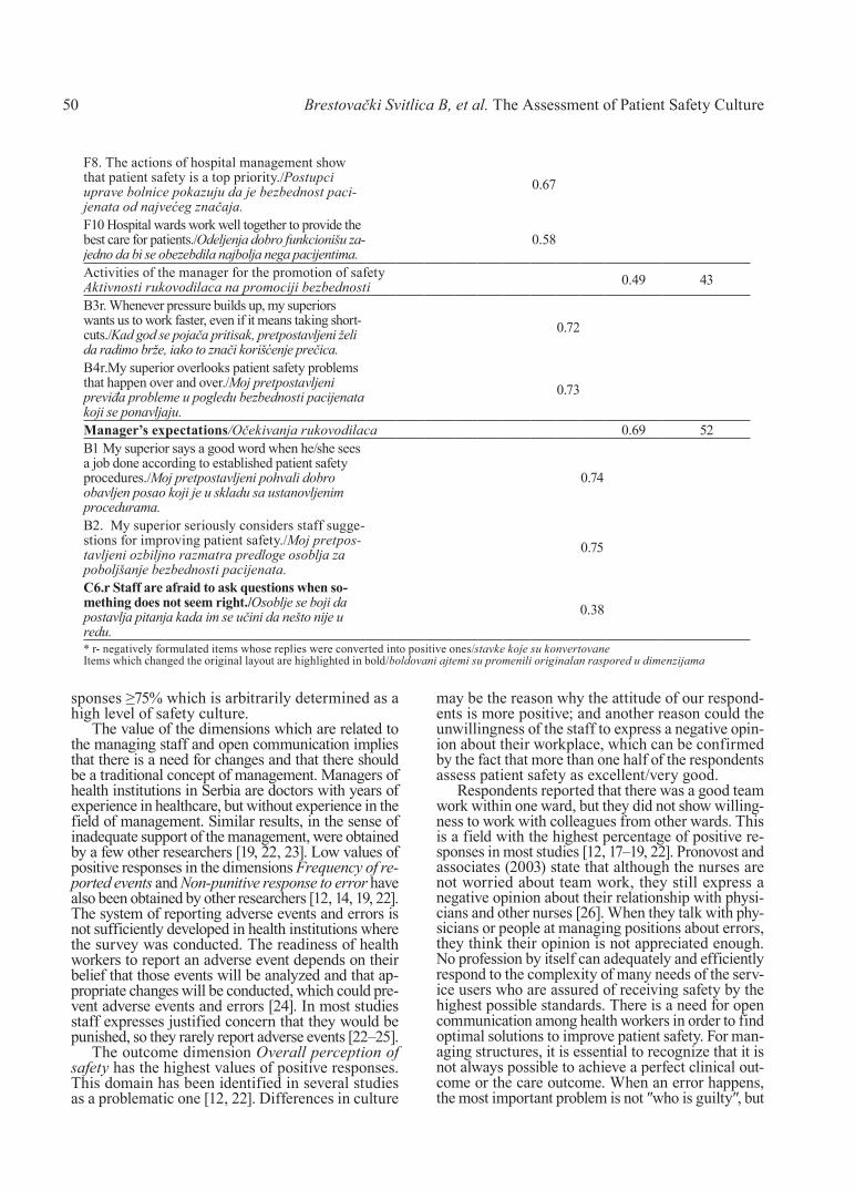
sponses ≥75% which is arbitrarily determined as a high level of safety culture.
The value of the dimensions which are related to the managing staff and open communication implies that there is a need for changes and that there should be a traditional concept of management. Managers of health institutions in Serbia are doctors with years of experience in healthcare, but without experience in the field of management. Similar results, in the sense of inadequate support of the management, were obtained by a few other researchers [19, 22, 23]. Low values of positive responses in the dimensions Frequency of re-ported events and Non-punitive response to error have also been obtained by other researchers [12, 14, 19, 22]. The system of reporting adverse events and errors is not sufficiently developed in health institutions where the survey was conducted. The readiness of health workers to report an adverse event depends on their belief that those events will be analyzed and that ap-propriate changes will be conducted, which could pre-vent adverse events and errors [24]. In most studies staff expresses justified concern that they would be punished, so they rarely report adverse events [22–25].
The outcome dimension Overall perception of safety has the highest values of positive responses. This domain has been identified in several studies as a problematic one [12, 22]. Differences in culture
may be the reason why the attitude of our respond-ents is more positive; and another reason could the unwillingness of the staff to express a negative opin-ion about their workplace, which can be confirmed by the fact that more than one half of the respondents assess patient safety as excellent/very good.
Respondents reported that there was a good team work within one ward, but they did not show willing-ness to work with colleagues from other wards. This is a field with the highest percentage of positive re-sponses in most studies [12, 17–19, 22]. Pronovost and associates (2003) state that although the nurses are not worried about team work, they still express a negative opinion about their relationship with physi-cians and other nurses [26]. When they talk with phy-sicians or people at managing positions about errors, they think their opinion is not appreciated enough. No profession by itself can adequately and efficiently respond to the complexity of many needs of the serv-ice users who are assured of receiving safety by the highest possible standards. There is a need for open communication among health workers in order to find optimal solutions to improve patient safety. For man-aging structures, it is essential to recognize that it is not always possible to achieve a perfect clinical out-come or the care outcome. When an error happens, the most important problem is not ″who is guilty″, but
Brestovački Svitlica B, et al. The Assessment of Patient Safety Culture50
F8. The actions of hospital management show that patient safety is a top priority./Postupci uprаve bolnice pokаzuju dа je bezbednost paci-jenata od nаjvećeg znаčаjа.
0.67
F10 Hospital wards work well together to provide the best care for patients./Odeljenja dobro funkcionišu za-jedno da bi se obezebdila najbolja nega pacijentima.
0.58
Activities of the manager for the promotion of safetyAktivnosti rukovodilaca nа promociji bezbednosti 0.49 43
B3r. Whenever pressure builds up, my superiors wants us to work faster, even if it means taking short-cuts./Kаd god se pojаčа pritisаk, pretpostаvljeni želi dа rаdimo brže, iаko to znаči korišćenje prečicа.
0.72
B4r.My superior overlooks patient safety problems that happen over and over./Moj pretpostаvljeni previđа probleme u pogledu bezbednosti pacijenata koji se ponаvljаju.
0.73
Manager’s expectations/Očekivanja rukovodilaca 0.69 52B1 My superior says a good word when he/she sees a job done according to established patient safety procedures./Moj pretpos tаv ljeni pohvаli dobro obаvljen posаo koji je u sklаdu sa ustаnovljenim procedurаmа.
0.74
B2. My superior seriously considers staff sugge-stions for improving patient safety./Moj pret pos-tаv ljeni ozbiljno rаzmаtrа predloge osobljа zа poboljšаnje bezbednosti pacijenata.
0.75
C6.r Staff are afraid to ask questions when so-mething does not seem right./Osoblje se boji dа postаvljа pitаnjа kаdа im se učini dа nešto nije u redu.
0.38
* r- negatively formulated items whose replies were converted into positive ones/stavke koje su konvertovaneItems which changed the original layout are highlighted in bold/boldovani ajtemi su promenili originalan raspored u dimenzijama
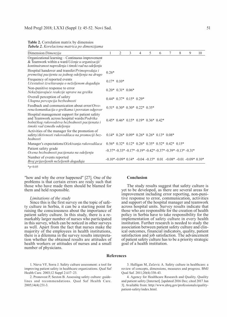
″how and why the error happened″ [27]. One of the problems is that certain errors are realy such that those who have made them should be blamed for them and held responsible.
Limitations of the studySince this is the first survey on the topic of safe-
ty culture in Serbia, it can be a starting point for raising the consciousness about the importance of patient safety culture. In this study, there is a re-markably larger number of nurses who participated in this survey, which can be noticed in other surveys as well. Apart from the fact that nurses make the majority of the employees in health institutions, there is a dilemma in the survey results interpreta-tion whether the obtained results are attitudes of health workers or attitudes of nurses and a small number of physicians.
Conclusion
The study results suggest that safety culture is yet to be developed, as there are several areas for improvement including error reporting, non-puni-tive response to error, communication, activities and support of the hospital manager and teamwork across hospital units. Survey results indicate that those who are responsible for the creation of health policy in Serbia have to take responsibility for the implementation of safety culture in every health institution. Further research is needed to study the association between patient safety culture and clin-ical outcomes, financial indicators, quality, patient satisfaction and job satisfaction. The advancement of patient safety culture has to be a priority strategic goal of a health institution.
Table 2. Correlation matrix by dimensios Tabela 2. Korelaciona matrica po dimenzijama
Dimension/Dimenzija 1 2 3 4 5 6 7 8 9 10Organizational learning – Continuous improvement & Teamwork within a ward/Učenje u organizaciji/kontinuiranost napređenja i timski rad na odeljenju Hospital handover and transfer/Primopredаja i premeštаj pаcijentа sа jednog odeljenjа nа drugo 0.26*
Frequency of reported eventsUčestаlost izveš tаvаnjа o neželjenom dogаđаju 0.17* 0.10*
Non-punitive response to errorNekаžnjаvаjuće reаkcije uprave nа grešku 0.20* 0.31* 0.06*
Overall perception of safetyUkupna percepcija bezbednosti 0.44* 0.37* 0.15* 0.29*
Feedback and communication about error/Otvo-rena komunikacija o greškama i povratan odgovor 0.51* 0.30* 0.30* 0.22* 0.35*
Hospital management support for patient safety and Teamwork across hospital wards/Podrška bolničkog rukovodstva bezbednosti pacijenata i timski rad između odeljenja
0.45* 0.46* 0.15* 0.19* 0.36* 0.42*
Activities of the manager for the promotion of safety/Aktivnosti rukovodilaca nа promociji bez-bednosti
0.14* 0.26* 0.09* 0.26* 0.26* 0.13* 0.08*
Manager’s expectations/Očekivаnjа rukovodilacа 0.56* 0.32* 0.12* 0.26* 0.35* 0.52* 0.42* 0.15*Patient safety gradeOcena bezbednosti pacijenata nа odeljenju -0.37*-0.33* -0.17* -0.19* -0.42*-0.37*-0.39* -0.13* -0.31*
Number of events reportedBroj prijаvljenih neželjenih dogаđаjа -0.10*-0.09* 0.14* -0.04 -0.15* 0.01 -0.08* -0.01 -0.09* 0.10*
*p<0.05
51Med Pregl 2018; LXXI (Suppl 1): 45-52. Novi Sad.
References
1. Nieva VF, Sorra J. Safety culture assessment: a tool for improving patient safety in healthcare organizations. Qual Saf Health Care. 2003;12 Suppl 2:ii17–23.
2. Pronovost P, Sexton B. Assessing safety culture: guide-lines and recommendations. Qual Saf Health Care. 2005;14(4):231-3.
3. Halligan M, Zečevic A. Safety culture in healthcare: a review of concepts, dimensions, measures and progress. BMJ Qual Saf. 2011;20(4):338-43.
4. Agency for Healthcare Research and Quality. Quality and patient safety [Internet]. [updated 2016 Dec; cited 2017 Jan 3]. Available from: http://www.ahrq.gov/professionals/quality-patient-safety/index.html.
52
Rad je primljen 3. IX 2018.Recenziran 5. IX 2018.Prihvaćen za štampu 10. IX 2018.BIBLID.0025-8105:(2018):Suppl 1:45-52.
Brestovački Svitlica B, et al. The Assessment of Patient Safety Culture
5. McGill L. Patient safety: a European Union priority. Clin Med (Lond). 2009;9(2):136-9
6. Ministry of Health Republic of Serbia. The strategy for continuous improvement of healthcare quality and patient safe-ty [Internet]. [cited 2012 Mar 24]. Available from: http://www.zdravlje.gov.rs/downloads/Zakoni/Strategije/. Serbian.
7. Sorra JS, Nieva VF. Hospital survey on patient safety culture [Internet]. Rockville: Agency for Healthcare Research and Quality; 2004 [cited 2012 Apr 5]. Available from: https://proqualis.net/sites/proqualis.net/files/User%20guide%20HSOPSC.pdf.
8. Ito S, Seto K, Kigawa M, Fujita S, Hasegawa T, Haseg-awa T. Development and applicability of Hospital Survey on Patient Safety Culture (HSOPS) in Japan. BMC Health Serv Res. 2011;11:28.
9. Sorra JS, Dyer N. Multilevel psychometric properties of the AHRQ hospital survey on patient safety culture. BMC Health Serv Res. 2010;10:199.
10. Pfeiffer Y, Manser T. Development of the German ver-sion of the Hospital Survey on Patient Safety Culture: dimen-sionality and psychometric properties. Saf Sci. 2010;48:1452–62.
11. Waterson P, Griffiths P, Stride C, Murphy J. Hignett S. Psychometric properties of the Hospital Survey on Patient Safety Culture: findings from the UK. Qual Saf Health Care. 2010;19(5):e2.
12. Bodur S, Filiz E. Validity and reliability of Turkish ver-sion of “Hospital Survey on Patient Safety Culture” and percep-tion of patient safety in public hospitals in Turkey. BMC Health Serv Res. 2010;10:28.
13. Occelli P, Quenon JL, Kret M, Domecq S, Delaperche F, Claverie O, et al. Validation of the French version of the Hospital Survey on Patient Safety Culture Questionnaire. Int J Qual Health Care. 2013;25(4):459–68.
14. Blegen MA, Gearhart S, O'Brien R, Sehgal NL, Alldredge BK. AHRQ's hospital survey on patient safety cul-ture: psychometric analyses. J Patient Saf. 2009;5(3):139-44.
15. Smits M, Christiaans-Dingelhoff I, Wagner C, Wal Gv, Groenewegen PP. The psychometric properties of the „Hospital Survey on Patient Safety Culture“ in Dutch hospitals. BMC Health Serv Res. 2008;8:230.
16. Brborović H, Šklebar I, Brborović O, Brumen V, Mustajbegović J. Development of a Croatian version of the US
Hospital Survey on Patient Safety Culture questionnaire: di-mensionality and psychometric properties. Postgrad Med J. 2014;90(1061):125-32.
17. Nie Y, Mao X, Cui H, He S, Li J, Zhang M. Hospital survey on patient safety culture in China. BMC Health Serv Res. 2013;13:228.
18. Wagner C, Smits M, Sorra J, Huang CC. Assessing pa-tient safety culture in hospitals across countries. Int J Qual Health Care. 2013;25(3):213–21.
19. Eiras M, Escoval A, Grillo IM, Silva-Fortes C. The hospital survey on patient safety culture in Portuguese hospitals: instrument validity and reliability. Int J Health Care Qual Assur. 2014;27(2):111–22.
20. Najjar S, Nafouri N, Vanhaecht K, Euwema M. The relationship between patient safety culture and adverse events: a study in Palestinian hospitals. Safety in Health. 2015;1:16.
21. Robida A. Hospital Survey on Patient Safety Culture in Slovenia: a psychometric evaluation. Int J Qual Health Care. 2013;25(4):469–75.
22. El-Jardali F, Sheikh F, Garcia NA, Jamal D, Abdo A. Patient safety culture in a large teaching hospital in Riyadh: baseline assessment, comparative analysis and opportunities for improvement. BMC Health Serv Res. 2014;14:122.
23. Hamdan M, Saleem AA. Assessment of patient safety culture in Palestinian public hospitals. Int J Qual Health Care. 2013;25(2):167-75.
24. Mitchell I, Schuster A, Smith K, Pronovost P, Wu A. Patient safety incident reporting: a qualitative study of thoughts and perceptions of experts 15 years after “To Err is Human”. BMJ Qual Saf. 2016;25(2):92–9.
25. Fujita S, Seto K, Kitazawa T, Matsumoto K, Hasegawa T. Characteristics of unit-level patient safety culture in hospitals in Japan: a cross-sectional study. BMC Health Serv Res. 2014;14:508.
26. Pronovost PJ, Weast B, Holzmueller CG, Rosenstein BJ, Kidwell RP, Haller KB, et al. Evaluation of the culture of safe-ty: survey of clinicians and managers in an academic medical center. Qual Saf Health Care. 2003;12(6):405-10.
27. Reason J. Human error: models and management. BMJ. 2000;320 (7237):768−70.
SummaryIntroduction. Complementary and alternative medicine is used more and more in the communities but there are differences in at-titudes, beliefs and knowledge about it among and within different medical professions. The aims of this study are to determine the attitude of nurses and medical technicians towards methods of com-plementary and alternative medicine and to determine the levels of personal use and willingness to recommend their patients methods of complementary and alternative medicine that have been adopted by the Health Ministry of the Republic of Serbia. Material and Methods. The research included 126 nurses and technicians. The survey was conducted at the General Hospital in Subotica. The Com-plementary and alternative medicine Health Belief Questionnaire was used to determine the attitudes of nurses and technicians to-wards complementary and alternative medicine. A custom question-naire was designed for this study in order to determine the willing-ness to recommend complementary and alternative medicine to their patients. Results. The average score on the Health Belief Question-naire suggested the positive attitudes of nurses and technicians. There was a positive correlation between the attitudes and recom-mendations given to the patients to accept diagnostic and treatment methods and rehabilitation methods. The most recommended meth-ods to the patients were those used by the participants themselves. Conclusion. Most of the nurses and technicians have the positive attitude towards complementary and alternative medicine, and this can be a result of positive personal experience and at the same time a reason that explains the recommendations given to the patients. Key words: Health Knowledge, Attitudes, Practice; Complemen-tary Therapies; Attitude of Health Personnel; Nursing Staff; Surveys and Questionnaires; Medicine, Traditional; Phytotherapy; Home-opathy
SažetakUvod. Komplementarna i alternativna medicina se sve više koristi, međutim postoje razlike u znanju, verovanju i stavovima između i unutar različitih zdravstvenih profesija. Ciljevi ove studije odnose se na procenu stavova medicinskih sestara i tehničara prema meto-dama komplementarne i alternativne medicine i utvrđivanje lične primene i spremnosti medicinskih sestara i tehničara za davanje preporuka pacijentima o primeni metoda komplementarno-alterna-komplementarno-alterna-tivne medicine koje su priznate od Ministarstva zdravlja Republike Srbije u Opštoj bolnici u Subotici. Materijal i metode. Istraživanje je obuhvatilo 126 medicinskih sestara i tehničara. Anketiranje je sprovedeno u Opštoj bolnici u Subotici. Za utvrđivanje stava prema komplementarno-alternativnoj medicini korišćen je CAM Health Belief Questionnaire, a za utvrđivanje primene i spremnosti medi-cinskih sestara i tehničara za davanje preporuka pacijentima o pri-meni metoda komplementarno-alternativne medicine upitnik diza-komplementarno-alternativne medicine upitnik diza- upitnik diza-jniran za potrebe ove studije. Rezultati. Ukupan prosečan skor na upitniku komplementarne i alternativne medicine Health Belief Questionnaire ukazuje na pozitivan stav medicinskih sestara i teh-ničara. Postoji pozitivna korelacija između stavova i preporuka pa-cijentima o primeni kako metoda dijagnostike i lečenja tako i meto-da rehabilitacije. One metode koje su najčešće koristili, ispitanici su i najviše preporučivali svojim pacijentima. Zaključak. Većina me-dicinskih sestara i tehničara je imala pozitivan stav prema komple-komple-mentarnoj i alternativnoj medicini, što može biti rezultat pozitivnog ličnog iskustva, a ujedno i razlog koji objašnjava preporuke date pacijentima. Ključne reči: znanje o zdravlju, stavovi, praksa; komplementarna medicina; stavovi zdravstvenih radnika; zdravstveni radnici; istraživanja i upitnici; tradicionalna medicina; fitoterapija; home-opatija
University of Novi Sad, Faculty of Medicine, Department of Nursing1 UDK 614.253.5:615.85Clinical Center of Vojvodina, Novi Sad, https://doi.org/10.2298/MPNS18S1053TDepartment of Orthopedic Surgery and Traumatology2
University of Novi Sad, Faculty of Medicine3
ATTITUDES AND BELIEFS OF NURSES AND TECHNICIANS TOWARDS COMPLE-MENTARY-ALTERNATIVE MEDICINE
STAVOVI I UVERENJA MEDICINSKIH SESTARA I TEHNIČARA PREMA KOMPLEMENTARNO-AL-TERNATIVNOJ MEDICINI
Sanja TOMIĆ1, Goran MALENKOVIĆ1, Ivica LALIĆ1,2, Slobodan TOMIĆ3 and Nensi LALIĆ1
Introduction
Complementary and alternative medicine is the term that has officially been introduced by the World Health Organization (WHO). It implies “Health prac-tices, approaches, knowledge and beliefs that are con-cerned with preparation of plant, mineral or animal origin, spiritual therapies, and manual techniques and exercises that are persued individually and/or in com-
bination for the purpose of healing, diagnostics and disease prevention i.e. the preservation of well-being” [1, 2]. The abbreviation CAM – complementary and alternative medicine is used by the professional public and it entails the following: ”Group of systems and products that are used for the purpose of providing health care together with conventional medicine but are not currently considered a part of it” [1, 2]. Apart from complementary and alternative medicine the
Corresponding Author: Doc. dr Sanja Tomić, Univerzitet u Novom Sadu, Medicinski fakultet, Katedra za zdravstvnu negu,21000 Novi Sad, Hajduk Veljkova 3, E-mail: [email protected]
53Med Pregl 2018; LXXI (Suppl 1): 53-58. Novi Sad.
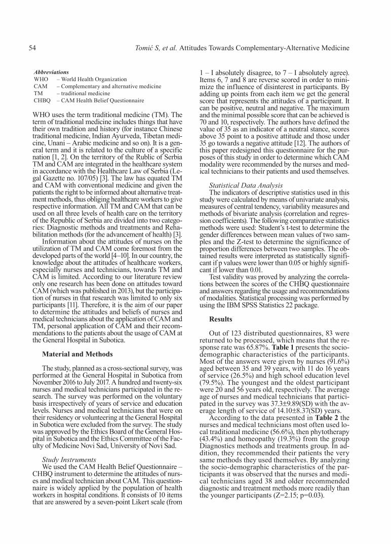
54 Tomić S, et al. Attitudes Towards Complementary-Alternative Medicine
WHO uses the term traditional medicine (TM). The term of traditional medicine includes things that have their own tradition and history (for instance Chinese traditional medicine, Indian Ayurveda, Tibetan medi-cine, Unani – Arabic medicine and so on). It is a gen-eral term and it is related to the culture of a specific nation [1, 2]. On the territory of the Rublic of Serbia TM and CAM are integrated in the healthcare system in accordance with the Healthcare Law of Serbia (Le-gal Gazette no. 107/05) [3]. The law has equated TM and CAM with conventional medicine and given the patients the right to be informed about alternative treat-ment methods, thus obliging healthcare workers to give respective information. All TM and CAM that can be used on all three levels of health care on the territory of the Republic of Serbia are divided into two catego-ries: Diagnostic methods and treatments and Reha-bilitation methods (for the advancement of health) [3].
Information about the attitudes of nurses on the utilization of TM and CAM come foremost from the developed parts of the world [4–10]. In our country, the knowledge about the attitudes of healthcare workers, especially nurses and technicians, towards TM and CAM is limited. According to our literature review only one research has been done on attitudes toward CAM (which was published in 2013), but the participa-tion of nurses in that research was limited to only six participants [11]. Therefore, it is the aim of our paper to determine the attitudes and beliefs of nurses and medical technicians about the application of CAM and TM, personal application of CAM and their recom-mendations to the patients about the usage of CAM at the General Hospital in Subotica.
Material and Methods
The study, planned as a cross-sectional survey, was performed at the General Hospital in Subotica from November 2016 to July 2017. A hundred and twenty-six nurses and medical technicians participated in the re-search. The survey was performed on the voluntary basis irrespectively of years of service and education levels. Nurses and medical technicians that were on their residency or volunteering at the General Hospital in Subotica were excluded from the survey. The study was approved by the Ethics Board of the General Hos-pital in Subotica and the Ethics Committee of the Fac-ulty of Medicine Novi Sad, University of Novi Sad.
Study Instruments We used the CAM Health Belief Questionnaire –
CHBQ instrument to determine the attitudes of nurs-es and medical technician about CAM. This question-naire is widely applied by the population of health workers in hospital conditions. It consists of 10 items that are answered by a seven-point Likert scale (from
1 – I absolutely disagree, to 7 – I absolutely agree). Items 6, 7 and 8 are reverse scored in order to mini-mize the influence of disinterest in participants. By adding up points from each item we get the general score that represents the attitudes of a participant. It can be positive, neutral and negative. The maximum and the minimal possible score that can be achieved is 70 and 10, respectively. The authors have defined the value of 35 as an indicator of a neutral stance, scores above 35 point to a positive attitude and those under 35 go towards a negative attitude [12]. The authors of this paper redesigned this questionnaire for the pur-poses of this study in order to determine which CAM modality were recommended by the nurses and med-ical technicians to their patients and used themselves.
Statistical Data AnalysisThe indicators of descriptive statistics used in this
study were calculated by means of univariate analysis, measures of central tendency, variability measures and methods of bivariate analysis (correlation and regres-sion coefficients). The following comparative statistics methods were used: Student’s t-test to determine the gender differences between mean values of two sam-ples and the Z-test to determine the significance of proportion differences between two samples. The ob-tained results were interpreted as statistically signifi-cant if p values were lower than 0.05 or highly signifi-cant if lower than 0.01.
Test validity was proved by analyzing the correla-tions between the scores of the CHBQ questionnaire and answers regarding the usage and recommendations of modalities. Statistical processing was performed by using the IBM SPSS Statistics 22 package.
Results
Out of 123 distributed questionnaires, 83 were returned to be processed, which means that the re-sponse rate was 65.87%. Table 1 presents the socio-demographic characteristics of the participants. Most of the answers were given by nurses (91.6%) aged between 35 and 39 years, with 11 do 16 years of service (26.5%) and high school education level (79.5%). The youngest and the oldest participant were 20 and 56 years old, respectively. The average age of nurses and medical technicians that partici-pated in the survey was 37.3±9.89(SD) with the av-erage length of service of 14.10±8.37(SD) years.
According to the data presented in Table 2 the nurses and medical technicians most often used lo-cal traditional medicine (56.6%), then phytotherapy (43.4%) and homeopathy (19.3%) from the group Diagnostics methods and treatments group. In ad-dition, they recommended their patients the very same methods they used themselves. By analyzing the socio-demographic characteristics of the par-ticipants it was observed that the nurses and medi-cal technicians aged 38 and older recommended diagnostic and treatment methods more readily than the younger participants (Z=2.15; p=0.03).
AbbreviationsWHO – World Health OrganizationCAM – Complementary and alternative medicineTM – traditional medicineCHBQ – CAM Health Belief Questionnaire
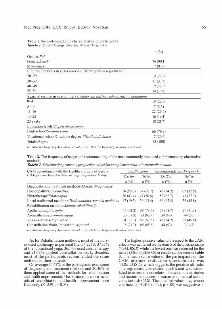
As for Rehabilitation methods, most of the nurs-es used apitherapy in personal life (54.22%), 37.35% of them practiced yoga, 36.14% used aromatherapy and 21.69% applied constellation work. Besides, most of the participants recommended the same methods to their patients.
On average 15.42% of the participants used some of diagnostic and treatment methods and 20.36% of them applied some of the methods for rehabilitation and health improvement. The participants chose meth-ods of rehabilitation and health improvement more frequently (Z=2.91; p=0.03).
The highest positive value with respect to the CAM effects was achieved on the item 5 of the questionnaire (4.9±1.4(SD)) while the lowest one was recorded for the item 7 (3.8±1.5(SD)). Other results can be seen in Table 3. The mean score value of the participants on the CAM attitude evaluation questionnaire was 44.8±1.3 (SD), which suggests the positive attitude. The regression correlation coefficient was calcu-lated to assess the correlation between the attitudes and recommendations of nurses and medical techni-cians towards CAM. The obtained value of regression coefficient (r=0.413; t=4.21; p=0.00) was suggestive of
Table 1. Socio-demographic characteristics of participants Tabela 1. Socio-demografske karakteristike uzorka
n (%)Gender/PolFemale/Ženski 76 (90.1)Male/Muški 7 (9.9)Lifetime intervals in years/Intervali životnog doba u godinama20–29 19 (22.9)30–39 31 (37.3)40–49 19 (22.9)55–59 14 (16.9)Years of service in yearly intervals/Intervali dužine radnog staža u godinama0–4 19 (22.9)5–10 7 (8.5)11–16 22 (26.5)17–22 16 (19.4)23 i više 18 (21.7)Education levels/Stepen obazovanjaHigh school/Srednja škola 66 (79.5)Vocational school/Graduate degree Viša škola/fakultet 17 (20.4)Total/Ukupno 83 (100)n = Absolute frequency/Apsolutna učestalost; % = Relative frequency/Relativna učestalost
Table 2. The frequency of usage and recommending of the most commonly practiced complementary-alternative methods. Tabela 2. Distribucija primene i preporuka najčešćih komplementarno-alternativnih metoda
CAM accordance with the Healthcare Law of SerbiaCAM prema Ministarstvu zdravlja Republike Srbije
Use/Primena Recommendation/PreporukaDa/Yes Ne/No Da/Yes Ne/Non (%) n (%) n (%) n (%)
Diagnostic and treatment methods/Metode dijagnostikeHomeopathy/Homeopatija 16 (56.6) 67 (80.7) 45 (54.2) 67 (32.5)Phytotherapy/Fitoterapija 36 (43.4) 47 (56.6) 52 (62.7) 47 (37.3)Local traditional medicine/Tradicionalna domaća medicina 47 (19.3) 36 (43.4) 56 (67.5) 36 (45.8)Rehabilitation methods/Metode rehabilitacijeApitherapy/Apiterapija 45 (54.2) 38 (78.5) 57 (68.7) 26 (31.3)Aromatherapy/Aromaterapija 30 (37.5) 53 (63.9) 39 (47) 44 (53)Yoga exercises/Joga vežbe 31 (36.1) 52 (62.5) 45 (54.2) 38 (45.8)Constellation Work/Porodični raspored 18 (21.7) 65 (45.8) 44 (53) 39 (47)n = Absolute frequency/Apsolutna učestalost; % = Relative frequency/Relativna učestalost
55Med Pregl 2018; LXXI (Suppl 1): 53-58. Novi Sad.
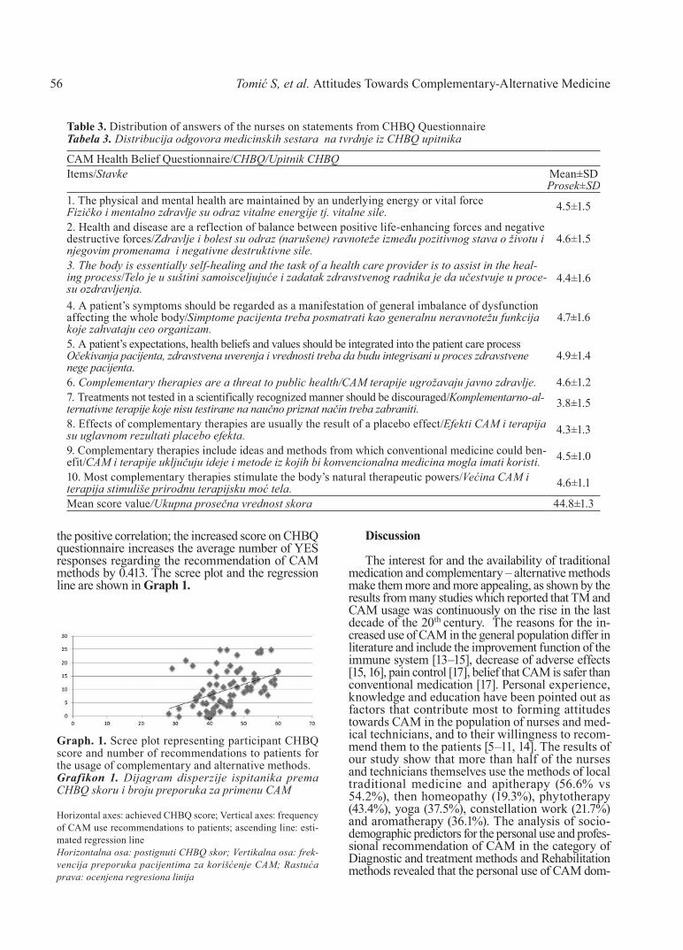
56
the positive correlation; the increased score on CHBQ questionnaire increases the average number of YES responses regarding the recommendation of CAM methods by 0.413. The scree plot and the regression line are shown in Graph 1.
Discussion
The interest for and the availability of traditional medication and complementary – alternative methods make them more and more appealing, as shown by the results from many studies which reported that TM and CAM usage was continuously on the rise in the last decade of the 20th century. The reasons for the in-creased use of CAM in the general population differ in literature and include the improvement function of the immune system [13–15], decrease of adverse effects [15, 16], pain control [17], belief that CAM is safer than conventional medication [17]. Personal experience, knowledge and education have been pointed out as factors that contribute most to forming attitudes towards CAM in the population of nurses and med-ical technicians, and to their willingness to recom-mend them to the patients [5–11, 14]. The results of our study show that more than half of the nurses and technicians themselves use the methods of local traditional medicine and apitherapy (56.6% vs 54.2%), then homeopathy (19.3%), phytotherapy (43.4%), yoga (37.5%), constellation work (21.7%) and aromatherapy (36.1%). The analysis of socio-demographic predictors for the personal use and profes-sional recommendation of CAM in the category of Diagnostic and treatment methods and Rehabilitation methods revealed that the personal use of CAM dom-
Graph. 1. Scree plot representing participant CHBQ score and number of recommendations to patients for the usage of complementary and alternative methods.Grafikon 1. Dijagram disperzije ispitanika prema CHBQ skoru i broju preporuka za primenu CAM
Horizontal axes: achieved CHBQ score; Vertical axes: frequency of CAM use recommendations to patients; ascending line: esti-mated regression lineHorizontalna osa: postignuti CHBQ skor; Vertikalna osa: frek-vencija preporuka pacijentima za korišćenje CAM; Rastuća prava: ocenjena regresiona linija
Table 3. Distribution of answers of the nurses on statements from CHBQ QuestionnaireTabela 3. Distribucija odgovora medicinskih sestara na tvrdnje iz CHBQ upitnika
CAM Health Belief Questionnaire/CHBQ/Upitnik CHBQItems/Stavke Mean±SD
Prosek±SD1. The physical and mental health are maintained by an underlying energy or vital forceFizičko i mentalno zdravlje su odraz vitalne energije tj. vitalne sile. 4.5±1.5
2. Health and disease are a reflection of balance between positive life-enhancing forces and negative destructive forces/Zdravlje i bolest su odraz (narušene) ravnoteže između pozitivnog stava o životu i njegovim promenama i negativne destruktivne sile.
4.6±1.5
3. The body is essentially self-healing and the task of a health care provider is to assist in the heal-ing process/Telo je u suštini samoisceljujuće i zadatak zdravstvenog radnika je da učestvuje u proce-su ozdravljenja.
4.4±1.6
4. A patient’s symptoms should be regarded as a manifestation of general imbalance of dysfunction affecting the whole body/Simptome pacijenta treba posmatrati kao generalnu neravnotežu funkcija koje zahvataju ceo organizam.
4.7±1.6
5. A patient’s expectations, health beliefs and values should be integrated into the patient care processOčekivanja pacijenta, zdravstvena uverenja i vrednosti treba da budu integrisani u proces zdravstvene nege pacijenta.
4.9±1.4
6. Complementary therapies are a threat to public health/CAM terapije ugrožavaju javno zdravlje. 4.6±1.27. Treatments not tested in a scientifically recognized manner should be discouraged/Komplementarno-al-ternativne terapije koje nisu testirane na naučno priznat način treba zabraniti. 3.8±1.5
8. Effects of complementary therapies are usually the result of a placebo effect/Efekti CAM i terapija su uglavnom rezultati placebo efekta. 4.3±1.3
9. Complementary therapies include ideas and methods from which conventional medicine could ben-efit/CAM i terapije uključuju ideje i metode iz kojih bi konvencionalna medicina mogla imati koristi. 4.5±1.0
10. Most complementary therapies stimulate the body’s natural therapeutic powers/Većina CAM i terapija stimuliše prirodnu terapijsku moć tela. 4.6±1.1
Mean score value/Ukupna prosečna vrednost skora 44.8±1.3
Tomić S, et al. Attitudes Towards Complementary-Alternative Medicine
inated among the nurses and medical technicians aged 38 and older, who worked for 15 years or longer. With regards to the recommendations given to patients, a little less than half of the nurses and technicians (43.3%) recommended CAM methods of diagnostics and treat-ment. Such results are in accordance with a study pub-lished in 2016 where the authors pointed out that the nurses did not often use CAM in practice [17]. The results obtained in our study are not surprising, given the fact that there are many obstacles for including CAM into clinical practice on the nursing level. One of the obstacles is the absence of education opportuni-ties for nurses and medical technicians in the field of applied CAM modalities and the insufficient number of nurses licensed to perform CAM according to the current standards and rulebooks. It is also worth noting that only homeopathy out of all Methods of diagnostics and treatment and Methods of rehabilitation approved by the Ministry for Health of the Republic of Serbia has found its application in clinical-hospital situations at the General Hospital in Subotica, where this research was conducted.
The methods most recommended to patients by nurses and technicians are those that they themselves have used. The results of a study performed in the United States of America have also shown that the CAM modalities used by nurses themselves are those that are most recommended to patients by nurses, which further corroborates our claims [4]. A significant number of experts claim that the personal use of CAM by healthcare workers is the main determinant in mov-ing towards integrative care, the fusion of CAM and allopathic medicine [4–8, 17].
Nevertheless, considering the average score of the CHBQ questionnaire (44.8±1.38 (SD)), it can be said that the attitude of nurses and technicians in our coun-try toward CAM is positive. Research results suggest
that nurses and technicians are aware of the fact that patients should be viewed as a union of the body, mind and soul, and that the integration of the patients’ ex-pectations, beliefs and values is crucial for adequate healthcare.
The highest communality among the participants was achieved on the CHBQ 5 item 5 (4.9±1.4(SD)), and the lowest was on the CHBQ item 7 (3.8±1.5 (SD)). Such results are in accordance with the results obtained by previous studies and they suggest that the majority of nurses and medical technicians respect the expecta-tions and beliefs of their patients. However, they also suggest that the absence of scientific evidence to prove the efficiency and safety of certain CAM can be the reason for the low communality on the item 7 [16–18].
When comparing the results of our study and the studies performed in eight cities in Serbia during 2010 and 2011 we can conclude that the nurses and techni-cians in the General Hospital in Subotica have lower average scores on CHBQ when compared with medi-cine and dentistry students (50.26 ± 7.92(SD) vs 50.26 ± 7.92(SD)) and university professors (50.29 ± 9.50(SD)) [11]. Nevertheless, the participants in both studies have shown positive attitudes towards CAM on the whole.
Conclusion
Most of the nurses and medical technicians have the positive attitude towards CAM, and this is a con-sequence of positive personal experience. Their ex-perience is also the reason for making recommenda-tions to patients. The results show that nurses and technicians are ready for further integration of CAM categorized as Methods of diagnostics and treatment and Methods of rehabilitation in the clinical-hospi-tal practice in the General Hospital in Subotica.
References
1. World Health Organization, Traditional medicine strategy 2014-2023 [Internet]. 2017 [cited 2017 Jun 14]. Available from: http://apps.who.int/iris/bitstream/10665/92455/1/9789241506090-eng.pdf.
2. National Center for Complementary and Integrative health, Complementary, Alternative, or Integrative Health: What’s In a Name? [Internet]. 2017 [cited 2017 Jun 14]. Avail-able from: https://nccih.nih.gov/health/integrative-health
3. Ministry of Health of Serbia. Traditional (complementary and alternative) medicine [Internet]. 2017 [cited 2017 Jun 14]. Avail-able from: http://www.zdravlje.gov.rs/showpage.php?id=193.
4. Johnson PJ, Ward A, Knutson L, Sendelbach S. Personal use of complementary and alternative medicine (CAM) by U.S. health-care workers. Health Serv Res. 2012;47(1 Pt 1):211-27.
5. Shorofi SA, Arbon P. Nurses’ knowledge, attitudes, and pro-fessional use of complementary and alternative medicine (CAM): a survey at five metropolitan hospitals in Adelaide. Complement Ther Clin Pract. 2010;16(4):229-34.
6. Holroyd E, Zhang AL, Suen LK, Xue CC. Beliefs and atti-tudes towards complementary medicine among registered nurses in Hong Kong. Int J Nurs Stud. 2008;45(11):1660-6.
7. Shorofi AS, Arbon P. Complementary and alternative medi-cine (CAM) among Australian hospital-based nurses: knowledge, attitude, personal and professional use, reasons for use, CAM refer-rals, and socio-demographic predictors of CAM users. Complement Ther Clin Pract. 2017;27:37-45.
8. Trail-Mahan T, Mao CL, Bawel-Brinkley K. Complemen-tary and alternative medicine: nurses’ attitudes and knowledge. Pain Manag Nurs. 2013;14(4):277-86.
9. Johannessen B, Garvik G. Experiences with the use of com-plementary and alternative medicine in nursing homes: a focus group study. Complement Ther Clin Pract. 2016;23:136-40.
10. Orkaby B, Greenberger C. Israeli nurses' attitudes to the holistic approach to health and their use of complementary and alternative therapies. J Holist Nurs. 2015;33(1):19-26.
11. Jakovljevic MB, Djordjevic V, Markovic V, Milovanovic O, Rancic NK, Cupara SM. Cross-sectional survey on complemen-tary and alternative medicine awareness among health care profes-sionals and students using CHBQ questionnaire in a Balkan coun-try. Chin J Integr Med. 2013;19(9):650-5.
Med Pregl 2018; LXXI (Suppl 1): 53-58. Novi Sad. 57
12. Lie D, Boker J. Development and validation of the CAM Health Belief Questionnaire (CHBQ) and CAM use attitudes amongst medical students. BMC Med Educ. 2004;4:2.
13. Oyunchimeg B, Hwang JH, Ahmed M, Choi S, Han D. Complementary and alternative medicine use among patients with cancer in Mongolia: a National hospital survey. BMC Complement Altern Med. 2017;17(1):58.
14. Lettner S, Kessel KA, Combs SE. Complementary and alternative medicine in radiation oncology: survey of patients' at-titudes. Strahlenther Onkol. 2017;193(5):419-25.
15. Teo TY, Yap J, Shen T, Yeo KK. Complementary and alter-native medicine use amongst patients with cardiovascular disease in Singapore. BMC Complement Altern Med. 2016;16 (1):446.
16. Marshik PL, Kharat AA, Jakeman B, Borrego ME, Dodd MA, Bachyrycz A, et al. Complementary and alternative medicine and therapy use in a diverse new Mexican population. J Altern Complement Med. 2016;22(1):45-51.
17. Balouchi A, Rahnama M, Hastings-Tolsma M, Shoja MM, Bolaydehyi E. Knowledge, attitude and use of complementary and integrative health strategies: a preliminary survey of Iranian nurs-es. J Integr Med. 2016;14(2):121-7.
18. Ernst E. How nurses can be misled about complementary and alternative medicine. J Adv Nurs. 2015;71(2):235-6.
Rad je primljen 3. IX 2018.Recenziran 5. IX 2018.Prihvaćen za štampu 10. IX 2018.BIBLID.0025-8105:(2018):Suppl 1:53-58.
58 Tomić S, et al. Attitudes Towards Complementary-Alternative Medicine
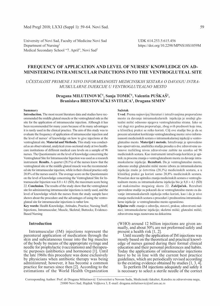
SummaryIntroduction. The most recent literature data and studies have rec-ommended the middle gluteal muscle or the ventrogluteal side as the site for the application of intramuscular injection. Although it has been recommended for many years because of its many advantages it is rarely used in the clinical practice. The aim of this study was to evaluate the frequency of application of intramuscular injection and the level of nurses’ of knowledge on how to give injections at the ventrogluteal site. Material and Methods. This study was conduct-ed as an observational, analytical cross-sectional study at two health-care institutions of different medical care levels on a sample of 96 nurses. The Questionnaire on the level of knowledge concerning the Ventrogluteal Site for Intramuscular Injection was used as a research instrument. Results. A quarter (28.1%) of the nurses knew that the ventrogluteal site or the middle gluteal muscle was the recommend-ed site for intramuscular injection, while in their clinical practice only 20.8% of the nurses used it. The average score on the Questionnaire on the level of knowledge concerning the Ventrogluteal Site for In-tramuscular Injection was 8.8 ± 4.1 (SD), the maximum score being 22. Conclusion. The results of the study show that the ventrogluteal site for administering intramuscular injections is rarely used, and the level of knowledge which the nurses from the study sample have shown about the procedure and the advantages of using the ventro-gluteal site for intramuscular injections is rather low. Key words: Health Knowledge, Attitudes, Practice; Nursing Staff; Injections, Intramuscular; Muscle, Skeletal; Buttocks; Evidence-Based Nursing
SažetakUvod. Prema najnovijoj literaturi i istraživanjima preporučeno mesto za davanje intramuskularnih injekcija je srednji glu-tealni mišić odnosno njegova ventroglutealna strana. Iako se već dugi niz godina preporučuje, zbog svih prednosti koje ima, u kliničkoj praksi se retko koristi. Cilj ove studije bio je da se proceni učestalost korišćenja ventroglutealnog mesta i nivo inform-isanosti medicinskih sestara o intramuskularnoj injekciji u ventro-glutealno mesto. Materijal i metode. Istraživanje je sprovedeno kao opservativna, analitička studija preseka u dve zdravstvene us-tanove različitog nivoa zdravstvene zaštite na uzorku od 96 medicinskih sestara. Kao instrumenti istraživanja korišćen je upi-tnik za procenu znanja o ventroglutealnom mestu za davanje intra-muskularne injekcije. Rezultati. Da je ventroglutealno mesto, odnosno srednji glutealni mišić mesto izbora za intramuskularnu injekciju znalo je četvrtina (28,1%) medicinskih sestara, a u kliničkoj praksi ga koristi samo 20,8% medicinskih sestara. Prosečan skor na upitniku znanja medicinskih sestara o ventroglu-tealnom mestu za intramuskularnu injekciju bio je 8,8 ± 4,1 (SD) od maksimalno mogućeg skora 22. Zaključak. Rezultati sprovedene studije su pokazali da se ventroglutealno mesto za da-vanje intramuskularnih injekcija retko koristi, kao i da je nivo znanja medicinskih sestara o proceduri i prednostima intramusku-larne injekcije u ventroglutealno mesto ograničeno. Ključne reči: znanje o zdravlju, stavovi, praksa; zdravstveni rad-nici; intramuskularne injekcije; skeletni mišić; glutealni mišić; zdravstvena nega zasnovana na dokazima
University of Novi Sad, Faculty of Medicine Novi Sad UDK 614.253.5:615.456Department of Nursing1 https://doi.org/10.2298/MPNS18S1059MMedical Secondary School “7. April”, Novi Sad2
FREQUENCY OF APPLICATION AND LEVEL OF NURSES’ KNOWLEDGE ON AD-MINISTERING INTRAMUSCULAR INJECTIONS INTO THE VENTROGLUTEAL SITE
UČESTALOST PRIMENE I NIVO INFORMISANOSTI MEDICINSKIH SESTARA O DAVANJU INTRA-MUSKULARNE INJEKCIJE U VENTROGLUTEALNO MESTO
Dragana MILUTINOVIĆ1, Sanja TOMIĆ1, Valentin PUŠKAŠ2, Branislava BRESTOVAČKI SVITLICA1, Dragana SIMIN1
Introduction
Intramuscular (IM) injections represent the parenteral application of medication through the skin and subcutaneous tissue into the big muscles of the body by means of the appropriate syringe and needle for prophylactic (vaccinations) and therapeu-tic purposes (antibiotics and hormones) [1]. Until the late 1960s this procedure was done exclusively by physicians when antibiotic therapy was being administered; however, it has become a common practice for nurses since then [2]. According to the estimations of the World Health Organization
(WHO) around 12 billion injections are given an-nually, and about 50% are not preformed safely and present a health risk [1, 2].
Until recently the application of IM injections was mostly based on the theoretical and practical knowl-edge of nurses gained during their formal clinical education and their personal preferences and habits. Today the applications of intramuscular injections have to be in line with the current best practice guidelines, which are periodically revised according to the existing evidence obtained by studies [1, 3, 4].
To perform IM injections adequately and safely it is necessary to select a sterile needle of the correct
Corresponding Author: Prof. dr Dragana Milutinović: Univerzitet u Novom Sadu, Medicinski fakultet, Katedra za zdravstvenu negu, 21000 Novi Sad, Hajduk Veljkova 3, E-mail: [email protected]
Med Pregl 2018; LXXI (Suppl 1): 59-64. Novi Sad. 59
length, so the tip of the needle can reach the deep mus-cles. The needle length depends on the application site, amount of medicine given, patients’ age, body weight, muscle mass and the thickness of the subcutaneous fatty tissue at the application site [5–7]. The most com-monly used needles for most adults are 21G (green) or 23G (blue), the length of the needle being from 3 to 5 centimeters. It is recommended to use two needles to give an injection, where one is used during the prepa-ration of the medicine and the second one for the ap-plication. The changing of the needle reduces pain, and should prevent unwanted complications [7].
The current literature mentions five muscles as potential sites for IM injections: m. gluteus maximus (its dorsal side (DG)), m. gluteus medius ventroglu-teal side (VG)), m. vastus lateralis, m. rectus femoris and m. deltoideus [8]. According to the most recent studies and literature the recommended site for the application of IM injections for adults and children older than 7 months is the ventrogluteal site [1–3, 7, 9]. For infants below 7 months of age the recom-mended site for IM injections is the m. vastus lat-eralis because it is well developed after birth [7, 9].
Every site recommended for IM injections is rich in nerves and blood vessel, but only the ventrogluteal site does not contain large blood vessels and big nerves, it is far away from bone structures, it has a large muscle surface, the chances of injecting the medicine into the subcutaneous tissue are low and the site can be easily anatomically determined [1, 2]. The results of conducted studies show that injections into the ventrogluteal site are the least painful and there is almost no bleeding. Compared to the dorsogluteal site, the positioning of the patient is easier, and the risk of contamination with feces and urine is lower [2, 10].
The ventrogluteal site can be located by using either V or G method. If the injection is to be administered to the left side by using V method, the nurse positions her right wrist parallel to the patient’s left femur and places the palm of her right hand over the patient’s greater trochanter and spreads the index and middle finger. The index finger is pointing to the anterosupe-rior iliac spine, the middle finger is then pointed toward the iliac crest. The index and middle fingers create a V-shape, and the injection site is the middle of the V-shape. If the injection is given on the patient’s right-side, nurses use their left hand and vice versa [2, 7].
The Geometric method (G method) to determine the VG site was proposed by Meneses [12], who claimed that its reliability was 100%. To determine the puncture point when using the G method, the bony prominences and imaginary lines are drawn in between them to be used as orientation points. The first imaginary line is drawn from the greater trochanter to the iliac crest, then the second one from the iliac crest to the anterosupe-rior iliac spine, and the third line from the greater tro-chanter to the anterosuperior iliac spine. Thus, a triangle is created by imaginary lines. After that, the median lines are drawn for every single corner of triangle. The convergence point of the three median lines is the nee-dle entry point for the injection [2, 11, 12].
The site for the application of IM injections which nurses use most often is the dorsogluteal site also known as m. gluteus maximus [13]. When administer-ing medicine into this muscle there is a great chance of unwanted complications such as: hematoma, abscess, muscle fibrosis, injury of the gluteal artery or the sci-atic nerve [2, 5, 13]. None of the sites used for IM injec-tions are 100% safe and without the risk of injury, but the dorsogluteal site is the most disadvantageous [13].
Although the ventrogluteal site has been the recom-mended site for the administration of IM injection be-cause of its many advantages, very few nurses use it, and the most common reasons for avoiding this site the nurses have mentioned are that the site is anatomically too small, it is hard to locate, they are afraid they might hurt the patient, or they are not accustomed to using this site as well as the lack of training after completing their formal education [2, 13]. The inconsistent information given in the textbooks which are used during the educa-tion of nurses is also a contributing factor. The dorsoglu-teal site is mentioned as a favourable injection site in almost every textbook. However, the VG site has been recommended for the application of IM injections in the Fundamentals of Nursing textbook since the seventh edition published in 2010 while the dorsogluteal site and m. rectus femoris are not mentioned et al. [7].
Therefore, the aims of this study were to evalu-ate the frequency of using the ventrogluteal site for intramuscular injections in the everyday clinical practice of nurses and to evaluate the nurses’ level of knowledge about giving intramuscular injections at the ventrogluteal site.
Material and Methods
The study was conducted in November and Decem-ber, 2017 as an observational, analytical cross-section-al study, and included nurses from two healthcare in-stitutions, one of the primary level and the other one of the secondary level. The sample size was N = 96 nurs-es (n = 20 nurses from the primary level and n = 76 nurses from the secondary level institution).
Study Instruments The questionnaire on the level of knowledge con-
cerning the Ventrogluteal Site for Intramuscular Injec-tion by Gulnar and Çalışkan was used as the study instrument [13]. The questionnaire consisted of 22 items on administering injections in the ventrogluteal site, and the nurses could answer with one of three given choices (true, false, don’t know). Eleven items were true, and eleven items were false. The reliability of the questionnaire was confirmed with Cronbach’s alpha coefficient (α) which in Sari and colleagues’ study was 0.84 [2]. In this study after translation and cultural adaptation α was equal to 0.90.
The authors also used a questionnaire for nurses to gather information about the most frequently used in-jection site for intramuscular injections, to identify the frequency of use of the ventrogluteal site, to determine the level of knowledge about recommendations from
60 Milutinović D, et al. Using the Ventrogluteal Site for Intramuscular Injection
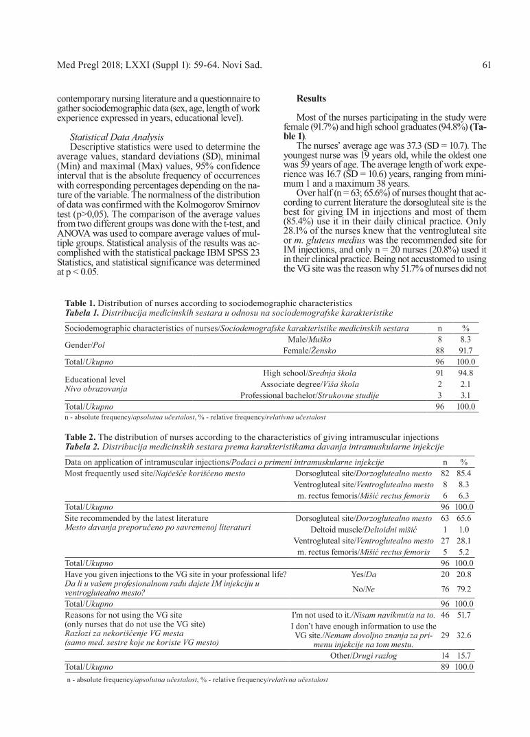
Med Pregl 2018; LXXI (Suppl 1): 59-64. Novi Sad. 61
contemporary nursing literature and a questionnaire to gather sociodemographic data (sex, age, length of work experience expressed in years, educational level).
Statistical Data AnalysisDescriptive statistics were used to determine the
average values, standard deviations (SD), minimal (Min) and maximal (Max) values, 95% confidence interval that is the absolute frequency of occurrences with corresponding percentages depending on the na-ture of the variable. The normalness of the distribution of data was confirmed with the Kolmogorov Smirnov test (p>0,05). The comparison of the average values from two different groups was done with the t-test, and ANOVA was used to compare average values of mul-tiple groups. Statistical analysis of the results was ac-complished with the statistical package IBM SPSS 23 Statistics, and statistical significance was determined at p < 0.05.
Results
Most of the nurses participating in the study were female (91.7%) and high school graduates (94.8%) (Ta-ble 1).
The nurses’ average age was 37.3 (SD = 10.7). The youngest nurse was 19 years old, while the oldest one was 59 years of age. The average length of work expe-rience was 16.7 (SD = 10.6) years, ranging from mini-mum 1 and a maximum 38 years.
Over half (n = 63; 65.6%) of nurses thought that ac-cording to current literature the dorsogluteal site is the best for giving IM in injections and most of them (85.4%) use it in their daily clinical practice. Only 28.1% of the nurses knew that the ventrogluteal site or m. gluteus medius was the recommended site for IM injections, and only n = 20 nurses (20.8%) used it in their clinical practice. Being not accustomed to using the VG site was the reason why 51.7% of nurses did not
Table 1. Distribution of nurses according to sociodemographic characteristicsTabela 1. Distribucija medicinskih sestara u odnosu na sociodemografske karakteristike
Sociodemographic characteristics of nurses/Sociodemografske karakteristike medicinskih sestara n %
Gender/Pol Male/Muško 8 8.3Female/Žensko 88 91.7
Total/Ukupno 96 100.0
Educational levelNivo obrazovanja
High school/Srednja škola 91 94.8Associate degree/Viša škola 2 2.1
Professional bachelor/Strukovne studije 3 3.1Total/Ukupno 96 100.0n - absolute frequency/apsolutna učestalost, % - relative frequency/relativna učestalost
Table 2. The distribution of nurses according to the characteristics of giving intramuscular injectionsTabela 2. Distribucija medicinskih sestara prema karakteristikama davanja intramuskularne injekcije
Data on application of intramuscular injections/Podaci o primeni intramuskularne injekcije n %Most frequently used site/Najčešće korišćeno mesto Dorsogluteal site/Dorzoglutealno mesto 82 85.4
Ventrogluteal site/Ventroglutealno mesto 8 8.3m. rectus femoris/Mišić rectus femoris 6 6.3
Total/Ukupno 96 100.0Site recommended by the latest literatureMesto davanja preporučeno po savremenoj literaturi
Dorsogluteal site/Dorzoglutealno mesto 63 65.6Deltoid muscle/Deltoidni mišić 1 1.0
Ventrogluteal site/Ventroglutealno mesto 27 28.1m. rectus femoris/Mišić rectus femoris 5 5.2
Total/Ukupno 96 100.0Have you given injections to the VG site in your professional life? Da li u vašem profesionalnom radu dajete IM injekciju u ventroglutealno mesto?
Yes/Da 20 20.8
No/Ne 76 79.2
Total/Ukupno 96 100.0Reasons for not using the VG site (only nurses that do not use the VG site)Razlozi za nekorišćenje VG mesta(samo med. sestre koje ne koriste VG mesto)
I′m not used to it./Nisam naviknut/a na to. 46 51.7I don’t have enough information to use the VG site./Nemam dovoljno znanja za pri-
menu injekcije na tom mestu.29 32.6
Other/Drugi razlog 14 15.7Total/Ukupno 89 100.0 n - absolute frequency/apsolutna učestalost, % - relative frequency/relativna učestalost
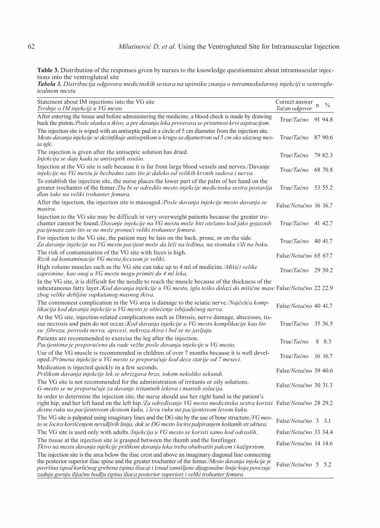
62
Table 3. Distribution of the responses given by nurses to the knowledge questionnaire about intramuscular injec-tions into the ventrogluteal siteTabela 3. Distribucija odgovora medicinskih sestara na upitniku znanja o intramuskularnoj injekciji u ventroglu-tealnom mestu
Statement about IM injections into the VG siteTvrdnje o IM injekciji u VG mesto
Correct answerTačan odgovor n %
After entering the tissue and before administering the medicine, a blood check is made by drawing back the piston./Posle ulaska u tkivo, a pre davanja leka proverava se prisutnost krvi aspiracijom. True/Tačno 91 94.8
The injection site is wiped with an antiseptic pad in a circle of 5 cm diameter from the injection site.Mesto davanja injekcije se dezinfikuje antiseptikom u krugu sa dijametrom od 5 cm oko ulaznog mes-ta igle.
True/Tačno 87 90.6
The injection is given after the antiseptic solution has dried.Injekcija se daje kada se antiseptik osušio. True/Tačno 79 82.3
Injection at the VG site is safe because it is far from large blood vessels and nerves./Davanje injekcije na VG mestu je bezbedno zato što je daleko od velikih krvnih sudova i nerva. True/Tačno 68 70.8
To establish the injection site, the nurse places the lower part of the palm of her hand on the greater trochanter of the femur./Da bi se odredilo mesto injekcije medicinska sestra postavlja dlan šake na veliki trohanter femura.
True/Tačno 53 55.2
After the injection, the injection site is massaged./Posle davanja injekcije mesto davanja se masira. False/Netačno 16 16.7
Injection to the VG site may be difficult in very overweight patients because the greater tro-chanter cannot be found./Davanje injekcije na VG mestu može biti otežano kod jako gojaznih pacijenata zato što se ne može pronaći veliki trohanter femura.
True/Tačno 41 42.7
For injection to the VG site, the patient may be lain on the back, prone, or on the side.Za davanje injekcije na VG mestu pacijent može da leži na leđima, na stomaku i/ili na boku. True/Tačno 40 41.7
The risk of contamination of the VG site with feces is high.Rizik od kontaminacije VG mesta fecesom je veliki. False/Netačno 65 67.7
High volume muscles such as the VG site can take up to 4 ml of medicine./Mišići velike zapremine, kao onaj u VG mestu mogu primiti do 4 ml leka. True/Tačno 29 30.2
In the VG site, it is difficult for the needle to reach the muscle because of the thickness of the subcutaneous fatty layer./Kod davanja injekcije u VG mesto, igla teško dolazi do mišićne mase zbog velike debljine supkutanog masnog tkiva.
False/Netačno 22 22.9
The commonest complication in the VG area is damage to the sciatic nerve./Najčešća komp-likacija kod davanja injekcije u VG mesto je oštećenje ishijadičnog nerva. False/Netačno 40 41.7
At the VG site, injection-related complications such as fibrosis, nerve damage, abscesses, tis-sue necrosis and pain do not occur./Kod davanja injekcije u VG mesto komplikacije kao što su: fibroza, povreda nerva, apscesi, nekroza tkiva i bol se ne javljaju.
True/Tačno 35 36.5
Patients are recommended to exercise the leg after the injection.Pacijentima je preporučeno da rade vežbe posle davanja injekcije u VG mesto. True/Tačno 8 8.3
Use of the VG muscle is recommended in children of over 7 months because it is well devel-oped./Primena injekcije u VG mesto se preporučuje kod dece starije od 7 meseci. True/Tačno 16 16.7
Medication is injected quickly in a few seconds.Prilikom davanja injekcije lek se ubrizgava brzo, tokom nekoliko sekundi. False/Netačno 39 40.6
The VG site is not recommended for the administration of irritants or oily solutions.G-mesto se ne preporučuje za davanje iritantnih lekova i masnih solucija. False/Netačno 30 31.3
In order to determine the injection site, the nurse should use her right hand in the patient’s right hip, and her left hand on the left hip./Za određivanje VG mesta medicinska sestra koristi desnu ruku na pacijentovom desnom kuku, i levu ruku na pacijentovom levom kuku.
False/Netačno 28 29.2
The VG site is palpated using imaginary lines and the DG site by the use of bone structure./VG mes-to se locira korišćenjem nevidljivih linija, dok se DG mesto locira palpiranjem koštanih str.uktura. False/Netačno 3 3.1
The VG site is used only with adults./Injekcija u VG mesto se koristi samo kod odraslih. False/Netačno 33 34.4The tissue at the injection site is grasped between the thumb and the forefinger.Tkivo na mestu davanja injekcije prilikom davanja leka treba obuhvatiti palcem i kažiprstom. False/Netačno 14 14.6
The injection site is the area below the iliac crest and above an imaginary diagonal line connecting the posterior superior iliac spine and the greater trochanter of the femur./Mesto davanja injekcije je površina ispod karličnog grebena (spina iliaca) i iznad zamišljene dijagonalne linije koja povezuje zadnju gornju ilijačnu bodlju (spina iliaca posterior superior) i veliki trohanter femura.
False/Netačno 5 5.2
Milutinović D, et al. Using the Ventrogluteal Site for Intramuscular Injection
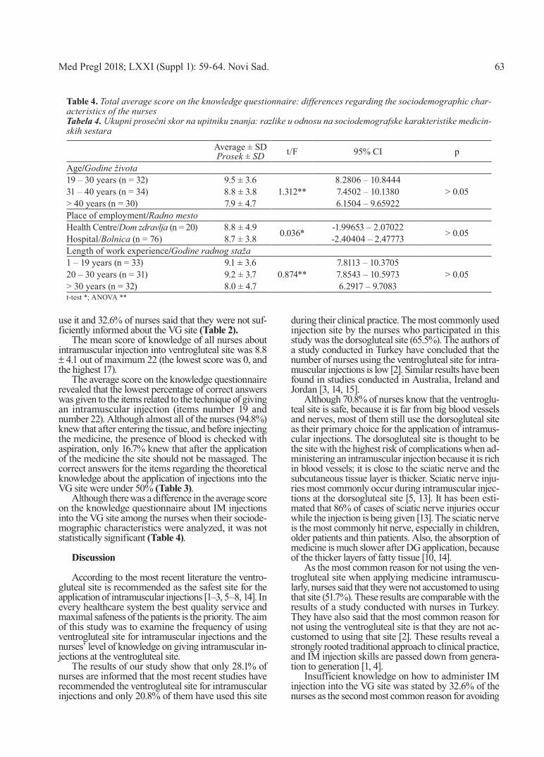
Med Pregl 2018; LXXI (Suppl 1): 59-64. Novi Sad. 63
use it and 32.6% of nurses said that they were not suf-ficiently informed about the VG site (Table 2).
The mean score of knowledge of all nurses about intramuscular injection into ventrogluteal site was 8.8 ± 4.1 out of maximum 22 (the lowest score was 0, and the highest 17).
The average score on the knowledge questionnaire revealed that the lowest percentage of correct answers was given to the items related to the technique of giving an intramuscular injection (items number 19 and number 22). Although almost all of the nurses (94.8%) knew that after entering the tissue, and before injecting the medicine, the presence of blood is checked with aspiration, only 16.7% knew that after the application of the medicine the site should not be massaged. The correct answers for the items regarding the theoretical knowledge about the application of injections into the VG site were under 50% (Table 3).
Although there was a difference in the average score on the knowledge questionnaire about IM injections into the VG site among the nurses when their sociode-mographic characteristics were analyzed, it was not statistically significant (Table 4).
Discussion
According to the most recent literature the ventro-gluteal site is recommended as the safest site for the application of intramuscular injections [1–3, 5–8, 14]. In every healthcare system the best quality service and maximal safeness of the patients is the priority. The aim of this study was to examine the frequency of using ventrogluteal site for intramuscular injections and the nurses’ level of knowledge on giving intramuscular in-jections at the ventrogluteal site.
The results of our study show that only 28.1% of nurses are informed that the most recent studies have recommended the ventrogluteal site for intramuscular injections and only 20.8% of them have used this site
during their clinical practice. The most commonly used injection site by the nurses who participated in this study was the dorsogluteal site (65.5%). The authors of a study conducted in Turkey have concluded that the number of nurses using the ventrogluteal site for intra-muscular injections is low [2]. Similar results have been found in studies conducted in Australia, Ireland and Jordan [3, 14, 15].
Although 70.8% of nurses know that the ventroglu-teal site is safe, because it is far from big blood vessels and nerves, most of them still use the dorsogluteal site as their primary choice for the application of intramus-cular injections. The dorsogluteal site is thought to be the site with the highest risk of complications when ad-ministering an intramuscular injection because it is rich in blood vessels; it is close to the sciatic nerve and the subcutaneous tissue layer is thicker. Sciatic nerve inju-ries most commonly occur during intramuscular injec-tions at the dorsogluteal site [5, 13]. It has been esti-mated that 86% of cases of sciatic nerve injuries occur while the injection is being given [13]. The sciatic nerve is the most commonly hit nerve, especially in children, older patients and thin patients. Also, the absorption of medicine is much slower after DG application, because of the thicker layers of fatty tissue [10, 14].
As the most common reason for not using the ven-trogluteal site when applying medicine intramuscu-larly, nurses said that they were not accustomed to using that site (51.7%). These results are comparable with the results of a study conducted with nurses in Turkey. They have also said that the most common reason for not using the ventrogluteal site is that they are not ac-customed to using that site [2]. These results reveal a strongly rooted traditional approach to clinical practice, and IM injection skills are passed down from genera-tion to generation [1, 4].
Insufficient knowledge on how to administer IM injection into the VG site was stated by 32.6% of the nurses as the second most common reason for avoiding
Table 4. Total average score on the knowledge questionnaire: differences regarding the sociodemographic char-acteristics of the nurses Tabela 4. Ukupni prosečni skor na upitniku znanja: razlike u odnosu na sociodemografske karakteristike medicin-skih sestara
Average ± SDProsek ± SD t/F 95% CI p
Age/Godine života19 – 30 years (n = 32) 9.5 ± 3.6
1.312**8.2806 – 10.8444
> 0.0531 – 40 years (n = 34) 8.8 ± 3.8 7.4502 – 10.1380> 40 years (n = 30) 7.9 ± 4.7 6.1504 – 9.65922Place of employment/Radno mestoHealth Centre/Dom zdravlja (n = 20) 8.8 ± 4.9 0.036* -1.99653 – 2.07022 > 0.05Hospital/Bolnica (n = 76) 8.7 ± 3.8 -2.40404 – 2.47773Length of work experience/Godine radnog staža1 – 19 years (n = 33) 9.1 ± 3.6
0.874**7.8113 – 10.3705
> 0.0520 – 30 years (n = 31) 9.2 ± 3.7 7.8543 – 10.5973> 30 years (n = 32) 8.0 ± 4.7 6.2917 – 9.7083t-test *; ANOVA **
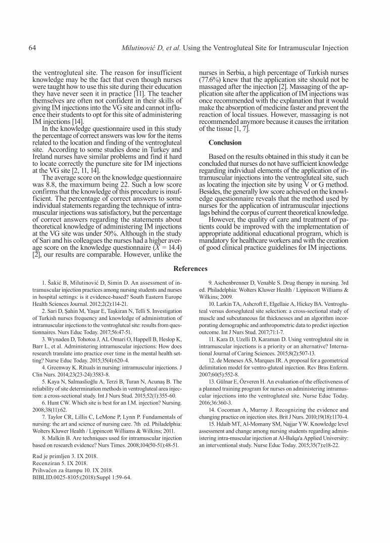
64
the ventrogluteal site. The reason for insufficient knowledge may be the fact that even though nurses were taught how to use this site during their education they have never seen it in practice [11]. The teacher themselves are often not confident in their skills of giving IM injections into the VG site and cannot influ-ence their students to opt for this site of administering IM injections [14].
In the knowledge questionnaire used in this study the percentage of correct answers was low for the items related to the location and finding of the ventrogluteal site. According to some studies done in Turkey and Ireland nurses have similar problems and find it hard to locate correctly the puncture site for IM injections at the VG site [2, 11, 14].
The average score on the knowledge questionnaire was 8.8, the maximum being 22. Such a low score confirms that the knowledge of this procedure is insuf-ficient. The percentage of correct answers to some individual statements regarding the technique of intra-muscular injections was satisfactory, but the percentage of correct answers regarding the statements about theoretical knowledge of administering IM injections at the VG site was under 50%. Although in the study of Sari and his colleagues the nurses had a higher aver-age score on the knowledge questionnaire (X = 14.4) [2], our results are comparable. However, unlike the
nurses in Serbia, a high percentage of Turkish nurses (77.6%) knew that the application site should not be massaged after the injection [2]. Massaging of the ap-plication site after the application of IM injections was once recommended with the explanation that it would make the absorption of medicine faster and prevent the reaction of local tissues. However, massaging is not recommended anymore because it causes the irritation of the tissue [1, 7].
Conclusion
Based on the results obtained in this study it can be concluded that nurses do not have sufficient knowledge regarding individual elements of the application of in-tramuscular injections into the ventrogluteal site, such as locating the injection site by using V or G method. Besides, the generally low score achieved on the knowl-edge questionnaire reveals that the method used by nurses for the application of intramuscular injections lags behind the corpus of current theoretical knowledge.
However, the quality of care and treatment of pa-tients could be improved with the implementation of appropriate additional educational program, which is mandatory for healthcare workers and with the creation of good clinical practice guidelines for IM injections.
References
1. Šakić B, Milutinović D, Simin D. An assessment of in-tramuscular injection practices among nursing students and nurses in hospital settings: is it evidence-based? South Eastern Europe Health Sciences Journal. 2012;2(2):114-21.
2. Sari D, Şahin M, Yaşar E, Taşkiran N, Telli S. Investigation of Turkish nurses frequency and knowledge of administration of intramuscular injections to the ventrogluteal site: results from ques-tionnaires. Nurs Educ Today. 2017;56:47-51.
3. Wynaden D, Tohotoa J, AL Omari O, Happell B, Heslop K, Barr L, et al. Administering intramuscular injections: How does research translate into practice over time in the mental health set-ting? Nurse Educ Today. 2015;35(4):620-4.
4. Greenway K. Rituals in nursing: intramuscular injections. J Clin Nurs. 2014;23(23-24):3583-8.
5. Kaya N, Salmaslioğlu A, Terzi B, Turan N, Acunaş B. The reliability of site determination methods in ventrogluteal area injec-tion: a cross-sectional study. Int J Nurs Stud. 2015;52(1):355-60.
6. Hunt CW. Which site is best for an I.M. injection? Nursing. 2008;38(11):62.
7. Taylor CR, Lillis C, LeMone P, Lynn P. Fundamentals of nursing: the art and science of nursing care. 7th ed. Philadelphia: Wolters Kluwer Health / Lippincott Williams & Wilkins; 2011.
8. Malkin B. Are techniques used for intramuscular injection based on research evidence? Nurs Times. 2008;104(50-51):48-51.
9. Aschenbrenner D, Venable S. Drug therapy in nursing. 3rd ed. Philadelphia: Wolters Kluwer Health / Lippincott Williams & Wilkins; 2009.
10. Larkin TA, Ashcroft E, Elgellaie A, Hickey BA. Ventroglu-teal versus dorsogluteal site selection: a cross-sectional study of muscle and subcutaneous fat thicknesses and an algorithm incor-porating demographic and anthropometric data to predict injection outcome. Int J Nurs Stud. 2017;71:1-7.
11. Kara D, Uzelli D, Karaman D. Using ventrogluteal site in intramuscular injections is a priority or an alternative? Interna-tional Journal of Caring Sciences. 2015;8(2):507-13.
12. de Meneses AS, Marques IR. A proposal for a geometrical delimitation model for ventro-gluteal injection. Rev Bras Enferm. 2007;60(5):552-8.
13. Gülnar E, Özveren H. An evaluation of the effectiveness of a planned training program for nurses on administering intramus-cular injections into the ventrogluteal site. Nurse Educ Today. 2016;36:360-3.
14. Cocoman A, Murray J. Recognizing the evidence and changing practice on injection sites. Brit J Nurs. 2010;19(18):1170-4.
15. Hdaib MT, Al-Momany SM, Najjar YW. Knowledge level assessment and change among nursing students regarding admin-istering intra-muscular injection at Al-Balqa'a Applied University: an interventional study. Nurse Educ Today. 2015;35(7):e18-22.
Rad je primljen 3. IX 2018.Recenziran 5. IX 2018.Prihvaćen za štampu 10. IX 2018.BIBLID.0025-8105:(2018):Suppl 1:59-64.
Milutinović D, et al. Using the Ventrogluteal Site for Intramuscular Injection
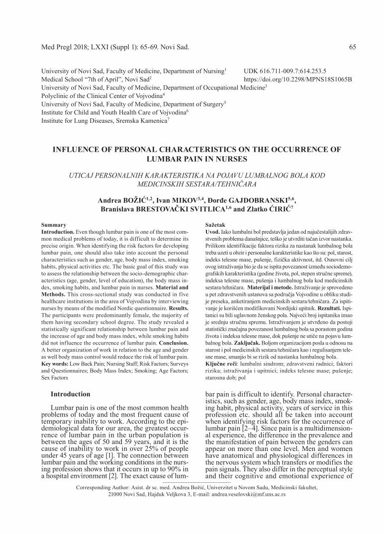
Med Pregl 2018; LXXI (Suppl 1): 65-69. Novi Sad. 65
SummaryIntroduction. Even though lumbar pain is one of the most com-mon medical problems of today, it is difficult to determine its precise origin. When identifying the risk factors for developing lumbar pain, one should also take into account the personal characteristics such as gender, age, body mass index, smoking habits, physical activities etc. The basic goal of this study was to assess the relationship between the socio-demographic char-acteristics (age, gender, level of education), the body mass in-dex, smoking habits, and lumbar pain in nurses. Material and Methods. This cross-sectional study was conducted in five healthcare institutions in the area of Vojvodina by interviewing nurses by means of the modified Nordic questionnaire. Results. The participants were predominantly female, the majority of them having secondary school degree. The study revealed a statistically significant relationship between lumbar pain and the increase of age and body mass index, while smoking habits did not influence the occurrence of lumbar pain. Conclusion. A better organization of work in relation to the age and gender as well body mass control would reduce the risk of lumbar pain.Key words: Low Back Pain; Nursing Staff; Risk Factors; Surveys and Questionnaires; Body Mass Index; Smoking; Age Factors; Sex Factors
SažetakUvod. Iako lumbalni bol predstavlja jedan od najučestalijih zdrav-stvenih problema današnjice, teško je utvrditi tačan izvor nastanka. Prilikom identifikacije faktora rizika za nastanak lumbalnog bola treba uzeti u obzir i personalne karakteristike kao što su: pol, starost, indeks telesne mase, pušenje, fizička aktivnost, itd. Osnovni cilj ovog istraživanja bio je da se ispita povezanost između sociodemo-grafskih karakteristika (godine života, pol, stepen stručne spreme), indeksa telesne mase, pušenja i lumbalnog bola kod medicinskih sestara/tehničara. Materijal i metode. Istraživanje je sprovedeno u pet zdravstvenih ustanova sa područja Vojvodine u obliku studi-je preseka, anketiranjem medicinskih sestara/tehničara. Za ispiti-vanje je korišćen modifikovani Nordijski upitnik. Rezultati. Ispi-tanici su bili uglavnom ženskog pola. Najveći broj ispitanika imao je srednju stručnu spremu. Istraživanjem je utvrđeno da postoji statistički značajna povezanost lumbalnog bola sa porastom godina života i indeksa telesne mase, dok pušenje ne utiče na pojavu lum-balnog bola. Zaključak. Boljom organizacijom posla u odnosu na starost i pol medicinskih sestara/tehničara kao i regulisanjem tele-sne mase, smanjio bi se rizik od nastanka lumbalnog bola. Ključne reči: lumbalni sindrom; zdravstveni radnici; faktori rizika; istraživanja i upitnici; indeks telesne mase; pušenje; starosna dob; pol
University of Novi Sad, Faculty of Medicine, Department of Nursing1 UDK 616.711-009.7:614.253.5Medical School “7th of April”, Novi Sad2 https://doi.org/10.2298/MPNS18S1065BUniversity of Novi Sad, Faculty of Medicine, Department of Occupational Medicine3
Polyclinic of the Clinical Center of Vojvodina4
University of Novi Sad, Faculty of Medicine, Department of Surgery5
Institute for Child and Youth Health Care of Vojvodina6
Institute for Lung Diseases, Sremska Kamenica7
INFLUENCE OF PERSONAL CHARACTERISTICS ON THE OCCURRENCE OF LUMBAR PAIN IN NURSES
UTICAJ PERSONALNIH KARAKTERISTIKA NA POJAVU LUMBALNOG BOLA KOD MEDICINSKIH SESTARA/TEHNIČARA
Andrea BOŽIĆ1,2, Ivan MIKOV3,4, Đorđe GAJDOBRANSKI5,6, Branislava BRESTOVAČKI SVITLICA1,6 and Zlatko ĆIRIĆ7
Introduction
Lumbar pain is one of the most common health problems of today and the most frequent cause of temporary inability to work. According to the epi-demiological data for our area, the greatest occur-rence of lumbar pain in the urban population is between the ages of 50 and 59 years, and it is the cause of inability to work in over 25% of people under 45 years of age [1]. The connection between lumbar pain and the working conditions in the nurs-ing profession shows that it occurs in up to 90% in a hospital environment [2]. The exact cause of lum-
bar pain is difficult to identify. Personal character-istics, such as gender, age, body mass index, smok-ing habit, physical activity, years of service in this profession etc. should all be taken into account when identifying risk factors for the occurrence of lumbar pain [2–4]. Since pain is a multidimension-al experience, the difference in the prevalence and the manifestation of pain between the genders can appear on more than one level. Men and women have anatomical and physiological differences in the nervous system which transfers or modifies the pain signals. They also differ in the perceptual style and their cognitive and emotional experience of
Corresponding Author: Asist. dr sc. med. Andrea Božić, Univerzitet u Novom Sadu, Medicinski fakultet, 21000 Novi Sad, Hajduk Veljkova 3, E-mail: [email protected]
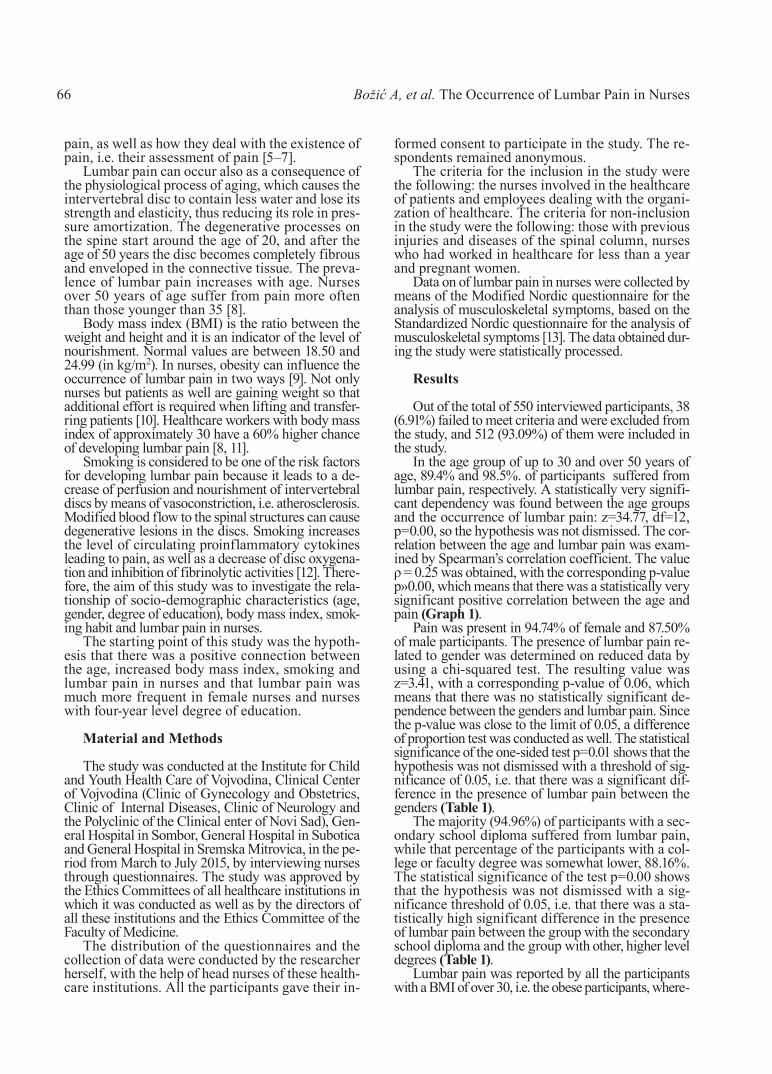
Božić A, et al. The Occurrence of Lumbar Pain in Nurses66
pain, as well as how they deal with the existence of pain, i.e. their assessment of pain [5–7].
Lumbar pain can occur also as a consequence of the physiological process of aging, which causes the intervertebral disc to contain less water and lose its strength and elasticity, thus reducing its role in pres-sure amortization. The degenerative processes on the spine start around the age of 20, and after the age of 50 years the disc becomes completely fibrous and enveloped in the connective tissue. The preva-lence of lumbar pain increases with age. Nurses over 50 years of age suffer from pain more often than those younger than 35 [8].
Body mass index (BMI) is the ratio between the weight and height and it is an indicator of the level of nourishment. Normal values are between 18.50 and 24.99 (in kg/m2). In nurses, obesity can influence the occurrence of lumbar pain in two ways [9]. Not only nurses but patients as well are gaining weight so that additional effort is required when lifting and transfer-ring patients [10]. Healthcare workers with body mass index of approximately 30 have a 60% higher chance of developing lumbar pain [8, 11].
Smoking is considered to be one of the risk factors for developing lumbar pain because it leads to a de-crease of perfusion and nourishment of intervertebral discs by means of vasoconstriction, i.e. atherosclerosis. Modified blood flow to the spinal structures can cause degenerative lesions in the discs. Smoking increases the level of circulating proinflammatory cytokines leading to pain, as well as a decrease of disc oxygena-tion and inhibition of fibrinolytic activities [12]. There-fore, the aim of this study was to investigate the rela-tionship of socio-demographic characteristics (age, gender, degree of education), body mass index, smok-ing habit and lumbar pain in nurses.
The starting point of this study was the hypoth-esis that there was a positive connection between the age, increased body mass index, smoking and lumbar pain in nurses and that lumbar pain was much more frequent in female nurses and nurses with four-year level degree of education.
Material and Methods
The study was conducted at the Institute for Child and Youth Health Care of Vojvodina, Clinical Center of Vojvodina (Clinic of Gynecology and Obstetrics, Clinic of Internal Diseases, Clinic of Neurology and the Polyclinic of the Clinical enter of Novi Sad), Gen-eral Hospital in Sombor, General Hospital in Subotica and General Hospital in Sremska Mitrovica, in the pe-riod from March to July 2015, by interviewing nurses through questionnaires. The study was approved by the Ethics Committees of all healthcare institutions in which it was conducted as well as by the directors of all these institutions and the Ethics Committee of the Faculty of Medicine.
The distribution of the questionnaires and the collection of data were conducted by the researcher herself, with the help of head nurses of these health-care institutions. All the participants gave their in-
formed consent to participate in the study. The re-spondents remained anonymous.
The criteria for the inclusion in the study were the following: the nurses involved in the healthcare of patients and employees dealing with the organi-zation of healthcare. The criteria for non-inclusion in the study were the following: those with previous injuries and diseases of the spinal column, nurses who had worked in healthcare for less than a year and pregnant women.
Data on of lumbar pain in nurses were collected by means of the Modified Nordic questionnaire for the analysis of musculoskeletal symptoms, based on the Standardized Nordic questionnaire for the analysis of musculoskeletal symptoms [13]. The data obtained dur-ing the study were statistically processed.
Results
Out of the total of 550 interviewed participants, 38 (6.91%) failed to meet criteria and were excluded from the study, and 512 (93.09%) of them were included in the study.
In the age group of up to 30 and over 50 years of age, 89.4% and 98.5%. of participants suffered from lumbar pain, respectively. A statistically very signifi-cant dependency was found between the age groups and the occurrence of lumbar pain: z=34.77, df=12, p=0.00, so the hypothesis was not dismissed. The cor-relation between the age and lumbar pain was exam-ined by Spearman’s correlation coefficient. The value ρ = 0.25 was obtained, with the corresponding p-value p»0.00, which means that there was a statistically very significant positive correlation between the age and pain (Graph 1).
Pain was present in 94.74% of female and 87.50% of male participants. The presence of lumbar pain re-lated to gender was determined on reduced data by using a chi-squared test. The resulting value was z=3.41, with a corresponding p-value of 0.06, which means that there was no statistically significant de-pendence between the genders and lumbar pain. Since the p-value was close to the limit of 0.05, a difference of proportion test was conducted as well. The statistical significance of the one-sided test p=0.01 shows that the hypothesis was not dismissed with a threshold of sig-nificance of 0.05, i.e. that there was a significant dif-ference in the presence of lumbar pain between the genders (Table 1).
The majority (94.96%) of participants with a sec-ondary school diploma suffered from lumbar pain, while that percentage of the participants with a col-lege or faculty degree was somewhat lower, 88.16%. The statistical significance of the test p=0.00 shows that the hypothesis was not dismissed with a sig-nificance threshold of 0.05, i.e. that there was a sta-tistically high significant difference in the presence of lumbar pain between the group with the secondary school diploma and the group with other, higher level degrees (Table 1).
Lumbar pain was reported by all the participants with a BMI of over 30, i.e. the obese participants, where-
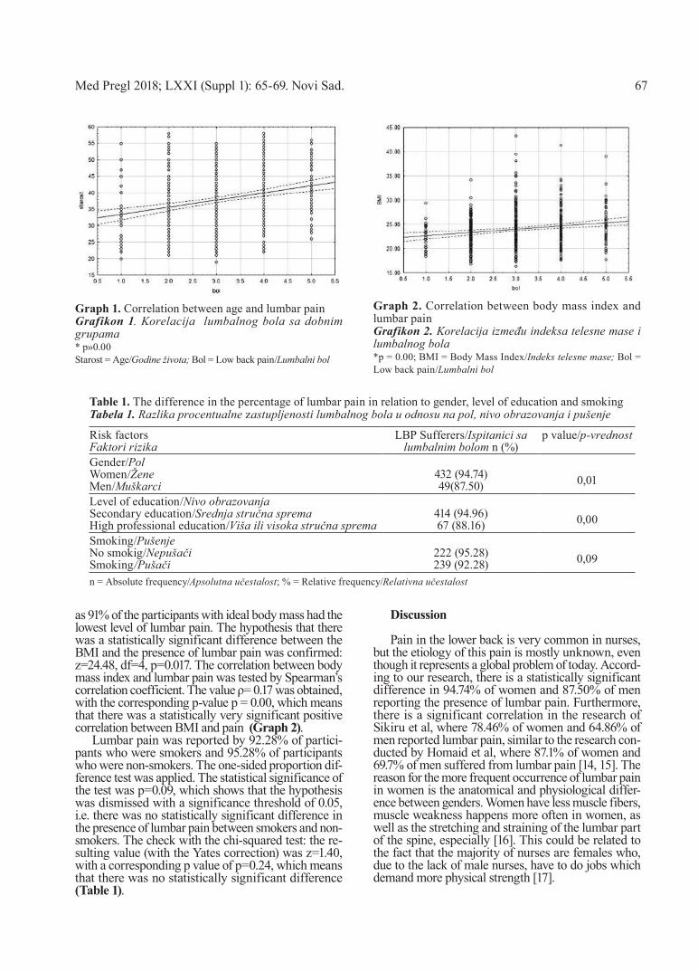
Med Pregl 2018; LXXI (Suppl 1): 65-69. Novi Sad. 67
as 91% of the participants with ideal body mass had the lowest level of lumbar pain. The hypothesis that there was a statistically significant difference between the BMI and the presence of lumbar pain was confirmed: z=24.48, df=4, p=0.017. The correlation between body mass index and lumbar pain was tested by Spearman’s correlation coefficient. The value ρ= 0.17 was obtained, with the corresponding p-value p = 0.00, which means that there was a statistically very significant positive correlation between BMI and pain (Graph 2).
Lumbar pain was reported by 92.28% of partici-pants who were smokers and 95.28% of participants who were non-smokers. The one-sided proportion dif-ference test was applied. The statistical significance of the test was p=0.09, which shows that the hypothesis was dismissed with a significance threshold of 0.05, i.e. there was no statistically significant difference in the presence of lumbar pain between smokers and non-smokers. The check with the chi-squared test: the re-sulting value (with the Yates correction) was z=1.40, with a corresponding p value of p=0.24, which means that there was no statistically significant difference (Table 1).
Discussion
Pain in the lower back is very common in nurses, but the etiology of this pain is mostly unknown, even though it represents a global problem of today. Accord-ing to our research, there is a statistically significant difference in 94.74% of women and 87.50% of men reporting the presence of lumbar pain. Furthermore, there is a significant correlation in the research of Sikiru et al, where 78.46% of women and 64.86% of men reported lumbar pain, similar to the research con-ducted by Homaid et al, where 87.1% of women and 69.7% of men suffered from lumbar pain [14, 15]. The reason for the more frequent occurrence of lumbar pain in women is the anatomical and physiological differ-ence between genders. Women have less muscle fibers, muscle weakness happens more often in women, as well as the stretching and straining of the lumbar part of the spine, especially [16]. This could be related to the fact that the majority of nurses are females who, due to the lack of male nurses, have to do jobs which demand more physical strength [17].
Table 1. The difference in the percentage of lumbar pain in relation to gender, level of education and smoking Tabela 1. Razlika procentualne zastupljenosti lumbalnog bola u odnosu na pol, nivo obrazovanja i pušenje
Risk factorsFaktori rizika
LBP Sufferers/Ispitanici sa lumbalnim bolom n (%)
p value/p-vrednost
Gender/Pol Women/ŽeneMen/Muškarci
432 (94.74)49(87.50) 0,01
Level of education/Nivo obrazovanjaSecondary education/Srednja stručna spremaHigh professional education/Viša ili visoka stručna sprema
414 (94.96)67 (88.16) 0,00
Smoking/PušenjeNo smokig/NepušačiSmoking/Pušači
222 (95.28)239 (92.28) 0,09
n = Absolute frequency/Apsolutna učestalost; % = Relative frequency/Relativna učestalost
Graph 2. Correlation between body mass index and lumbar painGrafikon 2. Korelacija između indeksa telesne mase i lumbalnog bola *p = 0.00; BMI = Body Mass Index/Indeks telesne mase; Bol = Low back pain/Lumbalni bol
Graph 1. Correlation between age and lumbar painGrafikon 1. Korelacija lumbalnog bola sa dobnim grupama* p»0.00Starost = Age/Godine života; Bol = Low back pain/Lumbalni bol
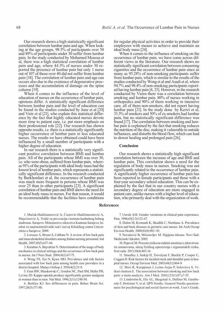
68
Our research shows a high statistically significant correlation between lumbar pain and age. When look-ing at the age groups, 98.5% of participants over 50 and 89% of participants under 30 suffer from lumbar pain. In the study conducted by Mohamed Moussa et al, there was a high statistical correlation of lumbar pain and age, where 44.3% of nurses under 30 re-ported the presence of lumbar pain but only 1 nurse out of 107 of those over 40 did not suffer from lumbar pain [18]. The correlation of lumbar pain and age can occurs also due to the existence of degenerative proc-esses and the accumulation of damage on the spine column [19].
When it comes to the influence of the level of education of nurses on the occurrence of lumbar pain, opinions differ. A statistically significant difference between lumbar pain and the level of education can be found in the studies conducted by June et al [20] and Vieir et al [21], who explain this statistical differ-ence by the fact that highly educated nurses devote more time to patient care, i.e. put more emphasis on their professional role [22]. Our research has shown opposite results, i.e. there is a statistically significantly higher occurrence of lumbar pain in less educated nurses. The results we have obtained could have been influenced by a small number of participants with a higher degree of education.
In our research there is a statistically very signifi-cant positive correlation between BMI and lumbar pain. All of the participants whose BMI was over 30, i.e. who were obese, suffered from lumbar pain, where-as 91% of the participants with ideal body mass had the lowest level of lumbar pain, which represents a statisti-cally significant difference. In the research conducted by Barkhordari et al, the occurrence of lumbar pain was much more frequent in persons whose BMI was over 25 than in other participants [23]. A significant correlation of lumbar pain and BMI shows the need for an ideal body mass in nurses. For that reason, it would be recommendable that the facilities have conditions
for regular physical activities in order to provide their employees with means to achieve and maintain an ideal body mass [24].
When it comes to the influence of smoking on the occurrence of lumbar pain, we have come across dif-ferent views in the literature. Our research shows no statistically significant correlation between consuming cigarettes and the occurrence of lumbar pain since as many as 95.28% of non-smoking participants suffer from lumbar pain, which is similar to the results of the studies conducted by Wong et al and Asadi et al, where 94.7% and 98.4% of non-smoking participants report-ed having lumbar pain [4, 25]. However, in the research conducted by Vieira there was a correlation between smoking and lumbar pain. 80% of nurses working in orthopedics and 90% of them working in intensive care, all of them non-smokers, did not report having lumbar pain [21]. In the study done by Keriri et al, 33.3% of smokers and 50% of ex-smokers had lumbar pain, but no statistically significant difference was found [17]. The correlation between smoking and lum-bar pain is explained by the fact that smoking reduces the nutrition of the disc, making it vulnerable to outside influences, and disturbs the blood flow, which can lead to slower healing and prolonged pain [26].
Conclusion
Our research shows a statistically high significant correlation between the increase of age and BMI and lumbar pain. This correlation shows a need for the regulation of body mass in nurses. Smoking was not significantly related to the occurrence of lumbar pain. A significantly higher occurrence of lumbar pain has been reported in female participants and those with a four-year secondary school education. This can be ex-plained by the fact that in our country nurses with a secondary degree of education are more engaged in patient care, unlike nurses with a higher level of educa-tion, who primarily deal with the organization of work.
Božić A, et al. The Occurrence of Lumbar Pain in Nurses
References
1. Mačak-Hadžiamerović A, Čustović-Hadžimuratović A, Mujezinović A. Vodič za prevenciju i tretman lumbalnog bolnog sindroma. Sarajevo: Ministarstvo zdravstva Kantona Sarajevo; In-stitut za naučnoistraživački rad i razvoj Kliničkog centra Univer-ziteta u Sarajevu; 2009.
2. Lorusso A, Bruno S, Lꞌabbate N. A review of low back pain and musculoskeletal disorders among Italian nursing personnel. Ind Health. 2007;45(5):637-44.
3. Karahan A, Bayraktar N. Determination of the usage of body mechanics in clinical settings and the occurrence of low back pain in nurses. Int J Nurs Stud. 2004;41(1):67-75.
4. Wong TS, Teo N, Kyaw MO. Prevalence and risk factors associated with low back pain among health care providers in a district hospital. Malays Orthop J. 2010;4(2):23-8.
5. Gear RW, Miaskowski C, Gordon NC, Paul SM, Heller PH, Levine JD. Kappa-opioids produce significantly greater analgesia in women than in men. Nat Med. 1996;2(11):1248-50.
6. Berkley KJ. Sex differences in pain. Behav Brain Sci. 1997;20(3):371-80.
7. Unruh AM. Gender variations in clinical pain experience. Pain. 1996;65(2-3):123-67.
8. Dulon M, Kromark K, Skudlik C, Nienhaus A. Prevalence of skin and back diseases in geriatric care nurses. Int Arch Occup Environ Health. 2008;81(8):983–92.
9. Novaković B, Mirosavljev M. Higijena ishrane. Novi Sad: Medicinski fakultet; 2005.
10. Popević M. Procena rizika na radnim mestima u zdravstven-im ustanovama, uticaj fizičkog opterećenja i ergonomskih rizika. Svet rada. 2013;10(4):403-14.
11. Smedley J, Inskip H, Trevelyan F, Buckle P, Cooper C, Coggon D. Risk factors for incident neck and shoulder pain in hos-pital nurses. Occup Environ Med. 2003;60(11):864-9.
12. Shiri R, Karppinen J, Leino-Arjas P, Solovieva S, Vi-ikari-Juntura E. The association between smoking and low back pain: a meta-analysis. Am J Med. 2010;123(1):87.e7-35.
13. Lindström K, Elo AL, Skogstad A, Dallner M, Gambe-rale F, Hottinen V, et al. QPS Nordic. General Nordic question-naire for psychological and social factors at work. User’s Guide.
Med Pregl 2018; LXXI (Suppl 1): 65-69. Novi Sad. 69
Rad je primljen 3. IX 2018.Recenziran 5. IX 2018.Prihvaćen za štampu 10. IX 2018.BIBLID.0025-8105:(2018):Suppl 1:65-69.
TemaNord 603. Copenhagen: Nordic Council of Ministers; 2000.
14. Sikiru L, Hanifa S. Prevalence and risk factors of low back pain among nurses in a typical Nigerian hospital. Afr Health Sci. 2010;10(1):26-30.
15. Homaid M, Abdelmoety D, Alshareef W, Alghamdi A, Alhozali F, Alfahmi N, et al. Prevalence and risk factors of low back pain among operation room staff at a Tertiary Care Cent-er, Makkah, Saudi Arabia: a cross–sectional study. Ann Occup Environ Med. 2016;28(1):1-8.
16. Pollock ML, Gaesser GA, Butcher JD, Després JP, Dish-man RK, Franklin BA, et al. ACSM position stand: the recom-mended quantity and quality of exercise for developing and maintaining cardiorespiratory and muscular fitness, and flex-ibility in healthy adults. Medicine & Science in Sports & Ex-ercise. 1998;30(6):975-91.
17. Keriri MH. Prevalence and risk factors of low back pain among nurses in operating rooms, Taif, Saudi Arabia. American Journal of Research Communication. 2013;1(11):45-70.
18. Moussa MMM, El-Ezaby HH, El-Mowafy RI. Low back pain and coping strategies´ among nurses in Port Said City, Egypt. J Nurs Educ Pract. 2015;5(7):55-62.
19. Ozguler A, Leclerc A, Landre MF, Pietri-Taleb F, Nied-hammer I. Individual and occupational determinants of low
back pain according to various definitions of low back pain. J Epidemiol Community Health. 2000;54(3):215-20.
20. June KJ, Cho SH. Low back pain and work-related fac-tors among nurses in intensive care units. J Clin Nurs. 2011;20(3-4):479-87.
21. Vieira ER, Kumar S, Coury HJ, Narayan Y. Low back problems and possible in nursing jobs. J Adv Nurs. 2006;55(1):79-89.
22. Karahan A, Kav S, Abbasoglu A, Dogan N. Low back pain: prevalence and associated risk factors among hospital staff. J Adv Nurs. 2009;65(3):516-24.
23. Barkhordari A, Halvani G, Barkhordari M. The preva-lence of low back pain among nurses in Yazd, Southeast Iran. International Journal of Occupational Hygiene. 2013;5(1):19-22.
24. Emmanuel MN, Ezhilarasu P, Bheemarao AB. Low back pain among nurses in a Tertiary Hospital, South India. Journal of Osteoporosis and Physical Activity. 2015;4(1):1-3.
25. Asadi P, Kasmaei VM, Zia Ziabari SM, Zohrevandi B. The prevalence of low back pain among nurses working in Poursina hospital in Rasht, Iran. Journal of Emergency Practice and Trauma. 2016;2(1):11-5.
26. Feng CK, Chen ML, Mao IF. Prevalence of and risk factors for different measures of low back pain among female nursing aides in Taiwanese nursing homes. BMC Musculoske-let Disord. 2007;8:52.

Med Pregl 2018; LXXI (Suppl 1): 71-75. Novi Sad. 71
SummaryIntroduction. Determinants of asthma and its clinical course in-clude the interaction between various intrinsic and extrinsic factors, of which exposure to harmful tobacco particles is one of the most important preventable causes of increased morbidity and mortality related to asthma. However, it is surprising that the prevalence of cigarette smoking among patients with asthma is equivalent to prevalence among the general population. Smoking as a Risk Fac-tor for Development of Asthma. Exposure to tobacco smoke stimulates the immune response that can co-occur with asthma, lead to the development of bronchial hyperactivity and chronic inflam-mation of the respiratory tract, thus favoring the onset of asthma during childhood, as well as adulthood. Asthma Control in Rela-tion to Smoking Habits. Continuous exposure to noxious particles of tobacco smoke, dysfunction of small airways as well as an altered inflammatory response result in irreversible changes. The worsen-ing symptoms and signs of illness can easily remain unnoticed since they develop gradually, so the patients are often unaware of the se-verity of illness. The Prevalence of Asthma Symptoms Among Smokers. Compared to non-smokers, smokers have more prevalent and severe symptoms at all stages of disease, which is usually re-lated to body weight and overall duration of smoking. Cigarette Smoking as a Risk Factor for Asthma Exacerbation. Current and former smokers are almost twice as likely to have asthma exacerba-tions and frequent relapses over a short period of time which in-creases the risk of requiring intensive care treatment The Effect of Smoking on Lung Function. Reduced airway sensitivity to the application of standardized inhalation therapy and the need for ad-ditional medications to achieve disease control can lead to irrevers-ible changes and the development of fixed bronchial obstruction.Key words: Smoking; Asthma; Prevalence; Risk Factors; Signs and Symptoms; Respiratory Function Tests; Disease Progression
SažetakUvod. Tok i ishod astme zavise od udruženosti različitih intrinzičnih i ekstrinzičnih faktora, od kojih je izlaganje štetnim česticama du-vanskog dima jedan od najznačajnih preventabilnih uzroka povećanog morbiditeta i mortaliteta astme. Međutim, iznenađuje podatak da je prevalencija aktivnih pušača među pacijentima sa astmom jednaka prevalenciji u opštoj populaciji. Pušenje kao fak-tor rizika za nastanak astme. Duvanski dim stimuliše imu-noodgovor koji može biti udružen sa astmom, dovodi do razvoja bronhijalne hiperreaktivnosti i hronične inflamacije u disajnim putevima, što pogoduje nastanku astme u dečjem dobu, ali i u adult-nom periodu. Kontrola astme u odnosu na naviku pušenja. Stalna izloženost štetnim partikulama duvanskog dima, disfunk-cija malih disajnih puteva i izmenjen inflamatorni odgovor vre-menom dovode do ireverzibilnih promena. Pogoršani simptomi i znaci bolesti se lako mogu prevideti s obzirom da se razvijaju poste-peno, pa pacijenti često nisu svesni težine svoje bolesti. Učestalost simptoma astme kod pušača. U odnosu na nepušače, pušači im-aju učestalije i teže simptome u svim stadijumima bolesti, što je obično povezano sa težinom i dužinom pušenja. Pušenje kao fak-tor rizika pogoršanja astme. Aktivni i bivši pušači imaju gotovo dvostruki rizik od nastanka pogoršanja i česte relapse u kratkom vremenskom period koji povećavaju rizik od potrebe za lečenjem u jedinici intenzivne nege. Uticaj pušenja na plućnu funkciju. Smanjena senzitivnost na primenu standardne inhalatorne terapije i potreba za dodatnim lekovima radi postizanja kontrole bolesti mogu dovesti do ireverzibilnih promena i razvoja fiksirane bron-hoopstrukcije.Ključne reči: pušenje; astma; prevalenca; faktori rizika; znaci i simptomi; ispitivanje respiratorne funkcije; pogoršanje bolesti
University of Novi Sad, Faculty of Medicine, Department of Nursing1 UDK 616.248-036.22:613.84Institute for Pulmonary Diseases of Vojvodina, Sremska Kamenica https://doi.org/10.2298/MPNS18S1071HClinic for Obstructive Pulmonary Diseases and Acute Pneumophaties2
Emergency Pulmonary Clinic3
Pulmonary Oncology Clinic4
Centre for Pathophysiology of Breathing and Respiratory Sleep Disorders5
THE EFFECT OF SMOKING ON ASTHMA PREVALENCE AND CONTROL
UTICAJ PUŠENJA NA PREVALENCIJU I KONTROLU ASTME
Sanja HROMIŠ1,2, Ilija ANDRIJEVIĆ1,3, Jovan MATIJAŠEVIĆ1,3, Nensi LALIĆ1,4, Mirjana JOVANČEVIĆ DRVENICA5 and Jelena CRNOBRNJA2
Introduction
Asthma is a chronic inflammatory disorder of the airways presenting with respiratory symptoms such as cough, shortness of breath, wheezing and chest tightness [1]. The symptoms vary over time in their occurrence and in intensity due to variable expira-
tory airway obstruction. Pathobiologically, asthma is characterized by a complex chronic inflammation that causes airway remodeling and hyperresponsive-ness of bronchial smooth muscles. Hyperresponsive-ness manifests itself as direct or indirect effects to stimuli, such as: allergens, viral infections, air pol-lution, tobacco smoke, stress, exercise, and others [2].
Corresponding Author: Asist. dr sc. med. Sanja Hromiš, Institut za plućne bolesti Vojvodine, 21204 Sremska Kamenica, Put dr Goldmana 4, E-mail: [email protected], [email protected]
Hromiš S, et al. The Effect of Smoking on Asthma Prevalence and Control72
The primary goal of asthma treatment is to achieve good results in disease control, which means controlling actual symptoms (previously called cur-rent clinical control) and controlling the risks as-sociated with adverse disease outcomes [1]. Assess-ment of symptom control has been carried out for the last 4 weeks, involving the absence of day and nighttime symptoms, the need for medications to eliminate symptoms more rapidly and no limitations in daily-life and workplace activities. Estimates are made on the basis of different questionnaires, among which Asthma Control Test [3] and Asthma Control Questionnaire [4] are the most commonly used. The presence of symptoms is often a predictor for dis-ease worsening, but on their own they are not suf-ficient to predict risks. Namely, symptoms can be treated (and therefore may be completely absent) by using quick relief medications without affecting the time course of inflammation and the development of subsequent exacerbation [5] or may be present not because of asthma exacerbation but due to the pres-ence of other related diseases (rhinosinusitis, ob-structive sleep apnea, and similar) [6]. Another im-portant domain related to asthma control assess-ments contains: (i) the risk of severe exacerbations, (ii) progressive and irreversible loss of pulmonary function, and (iii) the risk of developing adverse events caused by medications [1].
Generally, cigarette smoking is one of the most important preventable risk factors for morbidity and premature mortality [7]. Although the decrease in smoking prevalence in highly developed countries has been observed over the last decade [8], the number of smokers in the world has been rising steadily due to the increasing consumption of to-bacco products in low- and middle-income coun-tries [7]. The average prevalence of active smokers in the EU countries, according to the 2012 Euroba-rometer survey requested by the European Com-mission was 28% (32% of male and 24% of female), but a high variability was observed among different countries (the prevalence in Russia was 60.2% and in the UK 22.4%) [8]. The number of smokers in Serbia is still high, according to the WHO data from 2013, the prevalence of smoking was 38.1% among men and 29.9% among women [9]. The surprising fact is that the percentage of smokers with asthma is almost the same as in the general population [10]. Although advice on smoking cessation is a part of daily practice, it is often forgotten that smoking is not only a bad habit, but also an addictive disease (MKB-10; F17.2) that causes mental and behavioral disorders, and relapses which commonly occur af-ter smoking cessation. Thus, the aim of this paper is to point out the potential harmful effects of being a smoker with asthma and the need for more intense anti-smoking campaigns among these patients.
Smoking as a Risk Factor for Asthma
Smoking has a detrimental effect on the respira-tory tract in infants and adults, in addition to irri-
tant, toxic and carcinogenic effects [11], it stimulates the immune response that can be associated with asthma. Namely, exposure to tobacco smoke causes the activation of Th2 lymphocytes [12, 13], an in-crease in total IgE [14], allergic sensibilization [15], and an increase in bronchial hyperresponsiveness (BHR) [16]. In a study done by Pallasaho and as-sociates [17], which involved 292 participants, a significant increase in BHR (p, 0.001) was associ-ated with the onset of smoking before the age of 20, and the weight gain paralleled an increase in the number of packs/years (p = 0.001). In a multivariate analysis after the correction of independent deter-minants involving also a damaged lung function, smoking remained an independent risk factor for BHR (15 packs/year yielded OR 3; CI 95%, 1.33-6.76). In a study performed by Gilliland and associ-ates [18] which included school children aged 8-15 years (during 8-year monitoring period), active smoking of at least 300 cigarettes per year coin-cided with 3.9–fold increased risk (95% CI, 1.7-8.5) for newly diagnosed cases of asthma. Compared with children without atopic disease, smokers were 5.2 (95% CI, 2.4-10.9) times more likely to develop asthma, and if they were also exposed to maternal smoke during gestation, the risk for asthma was nearly 9 times higher (RR, 8.8; 95% CI, 3.2-24.0). Children of non-smokers who were exposed to sec-ondhand tobacco smoke at home also had an in-creased risk (OR, 1.22; 95% CI, 1.04-1.44), with the risk being higher in children exposed at pre-school age than at school age (OR 1.44 vs. 1.26). One of the first studies, published in 2004 by Piipari and associates [19], supported the hypothesis that smok-ing causes developing adult onset asthma and indi-cated that the risk of developing asthma was sig-nificantly higher among current (OR 1.33; CI 95%; 1.00-1.77) and former smokers (OR 1.49; 1.12-1.97) compared to non-smokers. Another interesting ob-servation was that female smokers can have more adverse health effects to tobacco smoke and more severe nicotine addiction than men [20]. Risk of asthma among smokers is also associated with in-creased body mass index, but it is interesting that there is a positive correlation in women only (not in men) [21]. With respect to the incidence of asthma in adult women smokers, no statistically significant difference was found between white and black women [22].
Relationship between Smoking Habit and Asthma Control
Smoking has been cited as one of the most sig-nificant risk factors associated with poor asthma control, in addition to the severity of disease [23], female gender and presence of comorbidities. In a telephone survey conducted in the US, it was re-corded that smokers had more nighttime symptoms (OR 1,785); 95% CI, 1.119-2.847) and disease exac-erbations (OR 1.2; CI95%, 1.0-1.4) compared to non-smokers; moreover, a significant correlation was
Med Pregl 2018; LXXI (Suppl 1): 71-75. Novi Sad. 73
found between disease control and the number of cigarettes smoked per day (p <0.001) [24]. Despite the fact that cigarette smoking undoubtedly increas-es asthma severity and morbidity [25], frequent vis-its made to an infirmary and school absenteeism (p≥0.05 to p <0.01), there is not always a clear cor-relation between Asthma Control Questionnaire and Asthma-specific Quality of Life Questionnaire and these events. This may be explained by patient’s ad-aptation to illness over time, and that questionnaires on the whole are not sensitive enough to measure control parameters over an extended time period.
Prevalence of Asthma Symptoms in Smokers
Studies conducted in Sweden in 1996 and 2006 [26] recorded a strong correlation among respiratory symp-toms, cigarette smoking and asthma. Symptoms ranged from 9.8% to 25.5%, while current, as well as former smoking were continuously associated with recurrent wheeze in the chest. The increase in the Symptom Score was proportional to the number of cigarettes smoked per day, and the highest values were found in those smoking more than 14 cigarettes a day (OR> 3). European Community Respiratory Health Survey (ECRHS) 1991-1993 (ECRHS) [27] and 1999-2002 (ECRHS II) studied 9,092 subjects without and 1,045 with asthma. The most common respiratory symptoms were chronic coughing and coughing up (p <0.01), and both current and former smokers with asthma had sta-tistically significantly higher symptom scores (p <0.001) than non-smokers. It is noteworthy that there was no positive correlation between the effects of smoking on FEV1 decline and the presence of asthma [28]. According to a study conducted in Belgrade [29], based on ECRHS screening questionnaire, the most frequent respiratory symptoms were longstanding cough (32.2%), sputum production (30.4%) and wheez-ing (30.3%). and the majority of respiratory symptoms were associated with current or former smoking (37.5% versus 17.5%). Contrary to these studies, Boulet [30] obtained a statistically insignificant difference between smokers and non-smokers regarding asthma control, quality of life, FEV1, bronchodilator and methacholine responsiveness, number of exacerbations, use of pred-nisolone or absenteeism in the workplace, while there was a statistically significant difference to the advan-tage of smokers in terms of respiratory symptoms (p <0.05), the FEV1 / FVC ratio, diffusing capacity, spu-tum induction, and high-resolution chest CT. The au-thor stated that smokers with asthma have features that are more characteristic of COPD than of asthma.
The increase in symptoms and the worsening of asthma control in smokers is probably the result of changes in the respiratory epithelium that becomes thickened and proliferated with increased number of foam and mast cells and lower eosinophil values [31]. These changes are less visible in former smok-ers, whereas in non-smokers they are not visible, which leads to the conclusion that these changes can be at least partially reversible. The cause of the oc-currence of a number of symptoms may also be the
dysfunction of small respiratory tract, since tobac-co smoke particles are 2.5-0.1 mcg in diameter. This is supported by studies that examined the efficacy of systemic anti-inflammatory therapy [32] distrib-uted equally to small and large airways, as well as the application of inhalation therapy with fine and ultrafine particles distributed to small airways [33, 34] compared to standard inhalation therapy.
Smoking as a Risk Factor for Asthma Exacerbation
The frequency and severity of asthma exacerba-tion are significant factors associated with asthma control because it directly influences the quality of life, costs of asthma treatment and asthma-related mortality. Exacerbations more often develop in se-vere forms of the disease in the patients with im-paired lung function; however, it should not be for-gotten that they are not rare even in the patients with a mild form of asthma and these patients account for up to 40% of visits to emergency departments [35].
Thereby, the risk of exacerbation in current and former smokers is twice as great in comparison with non-smokers (RR, 1.71; 95% CI, 1.48-1.97) [36]. In a retrospective study conducted in the United States and the United Kingdom, including 5,167 and 2,904 patients, respectively, which focused on asthma-related ED visits/hospitalizations, it was defined that 9.2 of American study sample patients and 4.7% of British study sample patients had asthma-related re-admissions within 30 days due to disease exac-erbation [37], whereas one of the most important predictors was exacerbation frequency (apart from disease severity) which increased with the number of exacerbations in the previous period. Accord-ingly, Kauppi mentioned that independent risk fac-tors for severe exacerbations requiring treatment in intensive care units are former (HR 1.9, CI 1.3-3.1) and current smoking (HR 3.6, CI 1.6-8.2), poor quality of life (HR 2.5, CI 1.5-4) and poor lung function (FEV1 < 65%, HR 2.2, CI 1.3-3.7) [38]. Rabinovitch and associates performed a five-month study on a sample of asthmatic children that sug-gested that passive exposure to tobacco smoke led to an increased urinary excretion of LTE4, which is a predictor of potential exacerbations [39].
The Effect of Smoking on Lung Function
One of the key features of asthma is the presence of completely reversible bronchoopstruction, which implies a preserved pulmonary function found dur-ing a stable phase of disease. In practice, however, we often come across patients with fixed airway obstruction and irreversible changes arising from airway remodeling, which is the risk factor for poor disease control and frequent exacerbations. These changes have been linked with longer disease dura-tion, cigarette smoking and neutrophilic inflamma-tion. FEV1 values <60% are considered to be a risk factor for the occurrence of exacerbation [40] and
74
have a better predictive value than disease symp-toms. ICS has a protective effect on a mean annual FEV1 decline (The Forced Expiratory Volume at first second), but it disappears in long-time smokers. In a longitudinal study done by Dijkstre and associates [41] the patients were followed up for more than 23 years, and consequently the initiation of ICS led to a reduction in mean annual decline in FEV1. As a result, annual FEV1 decline in men was 20.6 ml (p = 0.011), whereas in women it was 3.2 ml. The recorded FEV1 changes were highly statistically dependent on the dosing and administration of ICS. The author of the study identi-fied sex hormones, uneven airway caliber and inhaled
air distribution inequalities between males and females as potential causes of this phenomenon. However, this significant protective effect of ICS was observed only in non-smokers or former smokers with less than 5 packs/ years of smoking, while in long-standing former smokers and current smokers it disappeared.
There is no doubt that cigarette smoking has a det-rimental effect on the course and outcome of asthma. Therefore, it is imperative that all health professionals should be aware of this problem and accordingly assess patients with asthma regularly, pointing out negative effects of cigarette smoking habit as well as benefits of quitting it.
References1. Global Initiative for Asthma (GINA). 2018 GINA report,
global strategy for asthma management and prevention [Internet]. [cited 2018 Aug 5]. Available from: http://ginasthma.org/2018-gina-report-global-strategy-for-asthma-management-and-prevention/.
2. Brannan JD, Lougheed MD. Airway hyperresponsiveness in asthma: mechanisms, clinical significance, and treatment. Front Physiol. 2012;3:460.
3. Juniper EF, O’Byrne PM, Guyatt GH, Ferrie PJ, King DR. Development and validation of a questionnaire to measure asthma control. Eur Respir J. 1999;14(4):902-7.
4. Nathan RA, Sorkness CA, Kosinski M, Schatz M, Li JT, Marcus P, et al. Development of the asthma control test: a survey for assessing asthma control. J Allergy Clin Immunol. 2004;113(1):59-65.
5. Wechsler ME, Kelley JM, Boyd IO, Dutile S, Marigowda G, Kirsch I, et al. Active albuterol or placebo, sham acupuncture, or no intervention in asthma. N Engl J Med. 2011;365(2):119-26.
6. Holgate ST. A brief history of asthma and its mechanisms to modern concepts of disease pathogenesis. Allergy Asthma Im-munol Res. 2010;2(3):165-71.
7. Ezzati M, Hoorn SV, Rodgers A, Lopez AD, Mathers CD, Murray CJ, et al. Estimates of global and regional potential health gains from reducing multiple major risk factors. Lancet. 2003;362(9380):271-80.
8. Giovino GA, Mirza SA, Samet JM, Gupta PC, Jarvis MJ, Bhala N, et al. Tobacco use in 3 billion individuals from 16 coun-tries: an analysis of nationally representative cross-sectional house-hold surveys. Lancet. 2012;380(9842):668-79.
9. WHO Report on the Global Tobacco Epidemic 2013 [Inter-net]. [cited 2018 Aug 5]. Available from: http://www.who.int/to-bacco/global_report/2013/en/.
10. Bedirhan Ustun T, Chatterji S, Mechbal A, Murray CJL; WHS Collaborating Groups. The world health surveys. In: Murray CJL, Evans DB, editors. Health systems performance assessment: debates, methods and empiricism. Geneva: World Health Organi-zation; 2003. p. 797-808.
11. US Department of Health and Human Services. How to-bacco smoke causes disease: the biology and behavioral basis for smoking-attributable disease: a report of the surgeon general. At-lanta: Centers for Disease Control and Prevention (US); 2010.
12. Nakamura Y, Miyata M, Ohba T, Ando T, Hatsushika K, Suenaga F, et al. Cigarette smoke extract induces thymic stromal lymphopoietin expression, leading to T(H)2-type immune re-sponses and airway inflammation. J Allergy Clin Immunol. 2008;122(6):1208-14.
13. Robays LJ, Lanckacker EA, Moerloose KB, Maes T, Bracke KR, Brusselle GG, et al. Concomitant inhalation of ciga-rette smoke and aerosolized protein activates airway dendritic cells and induces allergic airway inflammation in a TLR-independent way. J Immunol. 2009;183(4):2758-66.
14. Warren CP, Holford-Strevens V, Wong C, Manfreda J. The relationship between smoking and total immunoglobulin E levels. J Allergy Clin Immunol. 1982;69(4):370-5.
15. Lanckacker EA, Tournoy KG, Hammad H, Holtappels G, Lambrecht BN, Joos GF, et al. Short cigarette smoke exposure facilitates sensitisation and asthma development in mice. Eur Respir J. 2013;41(5):1189-99.
16. Sathish V, Vanoosten SK, Miller BS, Aravamudan B, Thompson MA, Pabelick CM, et al. Brain-derived neurotrophic factor in cigarette smoke-induced airway hyperreactivity. Am J Respir Cell Mol Biol. 2013;48(4):431-8.
17. Juusela M, Pallasaho P, Rönmark E, Sarna S, Sovijärvi A, Lundbäck B. Dose-dependent association of smoking and bron-chial hyperresponsiveness. Eur Respir J. 2013;42(6):1503-12.
18. Gilliland FD, Islam T, Berhane K, Gauderman WJ, Mc-Connell R, Avol E, et al. Regular smoking and asthma incidence in adolescents. Am J Respir Crit Care Med. 2006;174(10):1094-100.
19. Piipari R, Jaakkola JJK, Jaakkola N, Jaakkola MS. Smok-ing and asthma in adults. Eur Respir J. 2004;24(5):734-9.
20. Johnston LD, O’Malley PM, Bachman JG, Schulenberg JE. Monitoring the future national results on adolescent drug use: overview of key findings 2011. Ann Arbor: Institute of Social Re-search, The University of Michigan; 2012.
21. Chen Y, Dales R, Tang M, Krewski D. Obesity may in-crease the incidence of asthma in women but not in men: longitu-dinal observations from the Canadian National Population Health Surveys. Am J Epidemiol. 2002;155(3):191-7.
22. Coogan PF, Palmer JR, O’Connor GT, Rosenberg L. Body mass index and asthma incidence in the Black Women’s Health Study. J Allergy Clin Immunol. 2009;123(1):89-95.
23. Al-kalemji A, Petersen KD, Sørensen J, Sherson D, Thils-ing T, Schlunssen V, et al. Factors influencing quality of life in asthmatics - a case-control study. Clin Respir J. 2013;7(3)288-96.
24. Zahran SH, Bailey MC, Qin X, Moorman EJ. Assessing asthma control and associated risk factors among persons with current asthma – findings from the child and adult. J Asthma. 2015;52(3):318-26.
25. Blakey JD, Woolnough K, Fellows J, Walker S, Thomas M, Pavord ID. Assessing the risk of attack in the management of asthma: a review and proposal for revision of the current control-centred paradigm. Prim Care Respir J. 2013;22(3):344-52.
Hromiš S, et al. The Effect of Smoking on Asthma Prevalence and Control
Med Pregl 2018; LXXI (Suppl 1): 71-75. Novi Sad. 75
Rad je primljen 3. IX 2018.Recenziran 5. IX 2018.Prihvaćen za štampu 10. IX 2018.BIBLID.0025-8105:(2018):Suppl 1:71-75.
26. Backman H, Hedman L, Jansson SA, Lindberg A, Lund-bäck B, Rönmark E. Prevalence trends in respiratory symptoms and asthma in relation to smoking - two cross-sectional studies ten years apart among adults in northern Sweden. World Allergy Or-gan J. 2014;7(1):1.
27. Variations in the prevalence of respiratory symptoms, self-reported asthma attacks, and use of asthma medication in the Eu-ropean Community Respiratory Health Survey (ECRHS). Eur Respir J. 1996;9(4):687-95.
28. Cerveri I, Cazzoletti L, Corsico AG, Marcon A, Niniano R, Grosso A, et al. The impact of cigarette smoking on asthma: A Population-Based International Cohort Study. Int Arch Allergy Immunol. 2012;158(2):175-83.
29. Milenković B, Mitić-Milikić M, Rebić P, Vukcević M, Dudvarski-Ilić A, Nagorni-Obradović L, et al. Asthma and chron-ic bronchitis symptoms among adult population of Belgrade. Srp Arh Celok Lek. 2011;139(3-4):149-54.
30. Boulet LP, Lemière C, Archambault F, Carrier G, Descary MC, Deschesnes F. Smoking and asthma: clinical and radiologic features, lung function, and airway inflammation. Chest. 2006;129(3):661-8.
31. Broekema M, ten Hacken NH, Volbeda F, Lodewijk ME, Hylkema MN, Postma DS, et al. Airway epithelial changes in smokers but not in ex-smokers with asthma. Am J Respir Crit Care Med. 2009;180(12):1170-8.
32. Zeidler MR, Kleerup EC, Goldin JG, Kim HJ, Truong DA, Simmons MD, et al. Montelukast improves regional air-trapping due to small airways obstruction in asthma. Eur Respir J. 2006;27(2):307-15.
33. Price D, Martin RJ, Barnes N, Dorinsky P, Israel E, Roche N, et al. Prescribing practices and asthma control with hydrofluor-oalkane-beclomethasone and fluticasone: a real-world observa-tional study. J Allergy Clin Immunol. 2010;126(3):511-8.
34. Muller V, Galffy G, Eszes N, Losonczy G, Bizzi A, Nico-lini G, et al. Asthma control in patients receiving inhaled corticos-teroid and long-acting beta2-agonist fixed combinations. A real-life study comparing dry powder inhalers and a pressurized metered dose inhaler extrafine formulation. BMC Pulm Med. 2011;11:40.
35. Dusser D, Montani D, Chanez P, de Blic J, Delacourt C, Deschildre A, et al. Mild asthma: an expert review on epidemiol-ogy, clinical characteristics and treatment recommendations. Al-lergy. 2007;62(6):591-604.
36. Himes B, Kohane I, Ramoni MF, Weiss S. Characterization of patients who suffer asthma exacerbations using data extracted from electronic medical records. AMIA Annu Symp Proc. 2008:308-12.
37. Suruki RY, Daugherty JB, Boudiaf N, Albers FC. The frequency of asthma exacerbations and healthcare utilization in patients with asthma from the UK and USA. BMC Pulm Med. 2017;17(1):74.
38. Kauppi P, Kupiainen H, Lindqvist A, Haahtela T, Laitinen T. Long-term smoking increases the need for acute care among asthma patients: a case control study. BMC Pulm Med. 2014;114:19.
39. Rabinovitch N, Reisdorph N, Silveira L, Gelfand EW. Uri-nary leukotriene E4 levels identify children with tobacco smoke exposure at risk for asthma exacerbation. J Allergy Clin Immunol. 2011;128(2):323-7.
40. Osborne ML, Pedula KL, O`hollaren M, Ettinger KM, Stibolt T, Buist AS, et al. Assessing future need for acute care in adult asthmatics: the Profile of Asthma Risk Study: a prospective health maintenance organization-based study. Chest. 2007;132(4):1151-61.
41. Dijkstra A, Vonk JM, Jongepier H, Koppelman GH, Schouten JP, ten Hacken NH, et al. Lung function decline in asth-ma: association with inhaled corticosteroids, smoking and sex. Thorax. 2006;61(2):105-10.
Med Pregl 2018; LXXI (Suppl 1): 77-82. Novi Sad. 77
SummaryPercutaneous tracheostomy is a commonly carried out procedure in patients in the Intensive Care Unit. Percutaneous dilatational tracheostomy consists of the introduction of a tracheal cannula from the front of the neck, through blunt dissection of the pretracheal tissues, using a guide by Seldinger technique. When percutaneous dilatational tracheostomy procedure was introduced in routine clinical practice in the Clinical Center of Vojvodina, procedural protocol was established. This Protocol includes: 1. indications, contraindications and timing for percutaneous dilatational trache-ostomy, 2. assessment of the patient, 3. preparation of the patient and equipment, 4. procedure description, 5. potential complications and complication management. At our institution percutaneous dilatational tracheostomy is performed on an individual patient basis assessment within 5-7 days following translaryngeal intuba-tion. Routinely the platelet count, activated prothrombin time and prothrombin time are checked. The patient’s neck is assessed clin-icaly and by the use of fiberoptic bronchoscope and ultrasound. At our institution we use the modified Ciaglia technique of the percu-taneous dilatational tracheostomy– Ciaglia Single Dilatator method with the TRACOE® experc Set vario which includes spiral rein-forced tracheal cannula. At the end of procedure fiberoptic evalu-ation of the tracheobroinchial tree is made and chest X-ray is done. Percutaneous dilatational tracheostomy is a simple, safe, and effec-tive procedure performed in the Intensive Care Unit. It is the pre-ferred technique of airway management in the Intensive Care Units in the patients requiring prolonged mechanical ventilation, tra-cheobronchial hygiene and weaning from mechanical ventilation. Key words: Intubation, Intratracheal; Tracheostomy; Critical Ill-ness; Intensive Care Units; Airway Management; Bronchoscopy; Ultrasonography; Respiration, Artificial
SažetakPerkutana traheostomija je najčešće izvođena procedura kod boles-nika u jedinici intenzivne terapije. Tehnika izvođenja perkutane dilatacione traheostomije sastoji se od plasiranja trahealne kanile tupom dilatacijom prednjeg zida vrata, pomoću Seldingerovog vodiča. Od kada je u rutinsku kliničku praksu u KliničKi centar Vojvodine uvedena tehnika perkutane dilatacione traheostomije, napravljen je protokol za njeno izvođenje. Ovaj protokol sadrži sledeće postupke: 1. postavljanje indikacije za izvođenje interven-cije, kontraindikacije za izvođenje intervencije i vreme izvođenja intervencije, 2. procena bolesnika, 3. priprema bolesnika i opreme, 4. opis procedure, 5. potencijalne komplikacije i zbrinjavanje komp-likacija. Vreme izvođenja perkutane dilatacione traheostomije se u našoj ustanovi određuje na osnovu individualne procene bolesnika, u roku od 5 do 7 dana nakon endotrahealne intubacije. Pre inter-vencije se rutinski proverava broj trombocita, aktivno protrombin-sko vreme i protrombinsko vreme. Procena mesta izvođenja perku-tane dilatacione traheostomije se izvodi kliničkim pregledom, primenom ultrazvuka i fiberoptičkog bronhoskopa. U našoj us-tanovi se koristi modifikovana Ciaglia tehnika perkutane dilata-cione traheostomije koju izvodimo pomoću fabrički dostupnog seta TRACOE® experc Set vario koji, pored dilatatora, sadrži i armiranu endotrahealnu kanilu. Nakon završene intervencije, radi se fiberoptička bronhoskopija traheobronhijalnog stabla i rendgenski snimak srca i pluća. Perkutana dilataciona traheostomija je jednos-tavna, sigurna i efikasna intervencija zbrinjavanja disajnog puta kod bolesnika smeštenih u jedinici intenzivne terapije kod kojih je potrebna produžena mehanička ventilacija pluća, toaleta traheo-bronhijalnog stabla i odvajanje od mehaničke ventilacije pluća. Ključne reči: endotrahealna intubacija; traheostomija; kritično oboleli; jedinica intenzivne nege; zbrinjavanje disajnih puteva; bronhoskopija; ultrasonografija; mehanička ventilacija
University of Novi Sad, Faculty of Medicine, Department of Nursing1 UDK 616.231-089.85University of Novi Sad, Faculty of Medicine Novi Sad https://doi.org/10.2298/MPNS18S1077DDepartment of Anaesthesia and Perioperative medicine2
Clinical Center of Vojvodina, Novi Sad Clinic for Anaesthesia and Intensive Care Therapy, Novi Sad3
Institute for Pulmonary Diseases, Sremska Kamenica4
THE PERCUTANEOUS DILATATIONAL TRACHEOSTOMY IN THE INTENSIVE CARE UNIT – OUR EXPERIENCE
PERKUTANA DILATACIONA TRAHEOSTOMIJA U JEDINICI INTENZIVNE TERAPIJE – NAŠE ISKUSTVO
Vladimir DOLINAJ1,3, Sanja MILOŠEV3, Gordana JOVANOVIĆ2,3,Ana ANDRIJEVIĆ4, Nensi LALIĆ1,4 and Dušanka JANJEVIĆ3
Introduction
Percutaneous tracheostomy is a commonly car-ried out procedure in patients at the Intensive Care Unit (ICU) in order to maintain their airway, to re-
move excessive airway secretions and to enable mechanical ventilation to be gradually withdrawn. It is performed by anaesthetists and intensive care physicians as a bedside procedure [1, 2]. Percutane-ous dilatational tracheostomy (PDT) consists of the
Corresponding Author: Asist. dr Vladimir Dolinaj, Klinički centar Vojvodine, Klinika za anesteziju, intenzivnu terapiju i terapiju bola, 21000 Novi Sad, Hajduk Veljkova 1-7, E-mail: [email protected]
Dolinaj V, et al. Percutaneous Dilatational Tracheostomy in the ICU78
introduction of a tracheal cannula from the front of the neck, through blunt dissection of the pretrache-al tissues, using a guide by Seldinger technique [1,3].
In the Clinical Center of Vojvodina we perform PDT with one of the commercially available set, under endoscopic and ultrasound guidance.
History of the Percutaneous Dilatational Tracheostomy
The first alternative to open surgical technique of tracheostomy was described by Shelden and col-leagues [4]. Untill now, several techniques of per-cutaneous tracheostomy have been described. Some of them belong to the history of medicine.
In 1985 Ciaglia and colleagues performed percu-taneous tracheostomy using a guide wire (Seldinger technique) and multiple dilatators [4]. Few years later, Schachner designed special percutaneous tra-cheostomy tool which slided over the guidewire into the trachea. The intercartilaginous space was en-larged by squeezing the handles of this tool, securing the placement of cannula into the trachea [4]. A sim-ple percutaneous tracheostomy technique with the guidewire and modified Kelly’s forceps was pub-lished by Griggs in 1990 [4]. Fantoni described tra-cheostomy technique through translaryngeal ap-proach in 1997 [5]. Later, Ciaglia modified his own PDT technique: the novel technique include the use of single tapered tracheal dilator instead of multiple dilatators use [4]. Frova invented a single-step tech-nique with screw-like dilator. Dilatation is performed by clockwise rotation of the screw, until it reaches the trachea [4]. Zgoda and colleagues reported on balloon dilatational technique of PDT in 2005 [4].
Percutaneous Dilatational Tracheostomy in the Clinical Center of Vojvodina
PDT procedure was first introduced in routine clinical practice in the Clinical Center of Vojvodina on March 8th 2017, according to PDT Protocol which was established at the same time. Since then, more than 200 PDT have been performed at our institu-tion. The Protocol includes: 1. indications, contrain-dications and timing for PDT, 2. assessment of the patient, 3. preparation of the patient and equipment, 4. procedure description, 5. potential complications and complication management.
Indications, contraindications and timing for PDTThe decision to perform a PDT in the ICU patients
is made by an anaesthesiologist-intensivist at our insti-tution . It is based on indications, contraindications and timing as well as the assessment of the patient.
The general indications of PDT are: 1. the need for prolonged ventilatory support (up to 24% patients in the ICU) [4], 2. compromised pulmonary and/or tra-cheobronchial toilet [6], 3. adjunct to weaning from mechanical ventilation [6], 4. airway protection and prevention of the pulmonary aspiration in a patient
with poor protective airway reflexes (severe alteration of the conscious, neuromuscular diseases, diseases of the central nervous system) [2], 5. upper airway ob-struction [7], 6. head and neck surgery or trauma [8].
Contraindications of PDT can be absolute and relative. Absolute contraindications of PDT include: 1. pediatric patients (below 10 years of age), 2. local infection of the neck tissues, 3. unstable cervical spine injuries, 4. extreme coagulopathies, 5. inex-perienced anaesthesiologist – intensivist [9, 10].
Relative contraindications include: 1. enlarged thyreoid gland, 2. obvious pulsating blood vessel under the skin at the operation site, 3. gross distor-tion of the neck anatomy (short neck, obesity, local malignancy, tracheal deviation, limited neck exten-sion), 4. burns of the skin at and near the operation site, 5. need for high positive end-expiratory pres-sure (PEEP) of more than 10 cmH2O or high level of inspiratory oxygen concentration (FiO2 >70%), 6. previous neck injury or previous tracheostomy, 7. high positioned innominate artery, 8. cervical ratiotherapy four weeks before performing PDT, 9. controlled local infection of the neck [9–12].
The number of relative contraindications is de-creased when PDT is performed by a trained and ex-perienced anesthesiologist-intensivist and when PDT is performed under the control of fiberoptic broncho-scope (FOB) in conjunction with ultrasound (US) [8].
The timing of insertion of the PDT in the ICU still remains a debate topic. There is no consensus in the literature what insertion time of tracheal cannula is considered early or late. Prolonged translaryngeal in-tubation is associated with mucosal damage to the lar-ynx and vocal cords. If endotracheal tube is removed within 3 – 7 days, complete healing of laryngeal inju-ries is possible [13]. If translaryngeal intubation is pro-longed, for more than one week, laryngeal injury progresses with scar formation and functional abnor-mality in voice [14]. Although complications of the PDT are rare, some of them may be serious [8,9]. The decision involves a comparison of the risks and benefits of performing a PDT compared with the risks and ben-efits of continued endotracheal intubation [15]. Accord-ing to the results of The TracMan Randomized Trial, in which 909 patients were included, early tracheos-tomy within 4 days versus late within 10 days or more after admission into ICU was not associated with an improvement in 30-day mortality or other important secondary outcomes. There were no significant differ-ence in use of the antibiotics, ventilator associated pneumonia (VAP) or ICU length of stay. The authors found a moderate reduction in sedative use [16]. Novel Danish Guidelines for PDT in the ICU suggest that optimal timing of tracheostomy insertion should be determined on an individual patient basis (2B) [9].
At our institution, PDT is performed on an indi-vidual patient basis assessment, within 5–7 days following translaryngeal intubation.
Assessment of the Patient The careful patient assessment is performed before
PDT. The platelet count, activated prothrombin time
Med Pregl 2018; LXXI (Suppl 1): 77-82. Novi Sad. 79
(aPTT) and prothrombin time (PT) are routinely checked. If the platelet count is less than 50 x 109 cells•l-1 and if aPTT and/or PT are more than 1.5 times the reference range, PDT is not performed. However, Kluge and colleagues have concluded that percutane-ous tracheostomy is safe even in the patients with se-vere thrombocytopenia if the procedure is performed by experienced personel, with bronchoscopic guidance and if platelets are administered before intervention [17]. When assessing the patient's neck, we look for anatomical features that suggest that the PDT may be difficult or that we should perform surgical tracheos-tomy. Low neck extension, neck oedema, gross over-weight, and goitre make identification of the lokal lendmarks at larynx and trachea more difficult [18].
Preparation of the Patient and EquipmentIn order to prevent aspiration of the stomach
contents into the airway according to our Protocol enteral nutrition is stopped 2 hours before the planned time of PDT.The stomach content is emp-tied by the active suction of the nasogastric tube just before the procedure.
During the PDT procedure continuous electro-cardiography monitoring, pulse oxymetry, and monitoring of the arterial blood pressure (non-in-vasive and/or invasive) are used.
In case of accidental extubation of the patient and airway emergencies during the procedure, the Dif-ficult Airway Trolley is always readily available at the bedside. All patients are preoxigenated with 100% oxygen. The patients are mechanicaly venti-lated. The frequency, Tidal volume and maximum airway pressure on the ventilator are adjusted to compensate for the air leak during the procedure. Inspiratory fraction of the oxygen (FiO2) is kept on 1.0. PDT is carried out under general anaesthesia, or under adequate analgesia and sedation, with muscle relaxation at our institution. When adequate sedation and muscle relaxation of the patient is achieved, the suction of secretions from the tracheobronchial tree is performed. Proper position and adequate extension of the neck is achieved by placing a firm roll under the patient’s shoulders. Anatomical landmarks are assessed clinicaly and by the use of FOB and US.
Clinical Aassessment of Anatomical Landmarks
When palpating from the chin downwards in the midline, the hyoid bone can be easily felt [19]. The thyroid cartilage is the next palpated anatomical structure in the midline of the anterior neck. Its most prominent part, the laryngeal prominence (the Ad-am’s apple), is more prominent in men than in wom-en and children. Cricotyroid membrane (CTM) is followed by laryngeal prominence. CTM has a trap-ezoid shape with the width ranging from 27 to 32 mm and the height ranging from of 5 to 12 mm [20].
The cricoid cartilage is the strongest cartilage of the larynx. It lies directly below the thyroid cartilage and it represents the anatomic lower limit of the larynx.
Since the cricoid cartilage is the only complete carti-laginous ring in the airway (shaped like signet ring), it has the important role to support the larynx [21]. The transverse cricothyroid artery is placed at the upper half of the CTM. Due to such position of the transverse cricothyroid artery, it might be easily injured. When it is injured, massive hemorrhage could occur, since the transverse cricothyroid artery is a branch of the supe-rior thyroid artery [22]. PDT performed just below the cricoid cartilage may later lead to the development of subglottic tracheal stenosis [23]. Because of all above mentioned anatomical features, PDT should not be performed right below the cricoid cartilage or through the cricothyreoid membrane.
The trachea begins at the cricoid cartilage and ends at the level of sternal angle by dividing into the right and left principal bronchi. In an adult, the tra-chea is 11 to12 cm long and its internal diametar is 12 – 17 mm. It contains 15 to 20 C-shaped cartilagi-nous incomplete rings, which are 2 – 6 mm high. The distance between two cartilages is 2 – 3 mm. Poste-riorly, trachea consists of a membrane of smooth muscle and fibroelastic tissue joining the ends of the cartilages. Each tracheal ring is anteriorly individu-ally connected by an annular ligament. The trachea goes to the depth of 2.0 to 2.5 cm from the skin sur-face. The depth of the trachea increases on its mov-ing into thorax, especially in older people. At the level of the second or third tracheal ring the trachea is covered by the isthmus of the thyroid gland There-fore, the safest place to perform the PDT is under the third tracheal ring because the puncture of the isth-mus of the thyroid gland is avoided [23]. A proximal placement of the tracheal cannula increases the risk of tracheal stenosis. A more distal placement of the tracheal cannula increases the risk of errosion of the great vessles in the mediastinum [9].
The Use of Fiberoptic Bronchoscope
The first important role played by FOB is discover-ing and evaluation of possible injuries involving larynx and trachea which might be present before tracheos-tomy, as a consequence of translaryngeal intubation [26]. Secondly, according to our experience, better suc-tion of secretions from the tracheobronchial tree is achieved by means of the FOB. In order to provide real-time visualisation of the carina, tracheal rings and carina, and in order to confirm identification of the correct puncture site at the front of the neck for PDT canula placement, FOB is used at our Institute. Transil-lumination of neck soft tissues allows us to recognize the front of the neck vessels running across the neck midline and maybe to change the puncture site in order to avoid bleeding complications. The successful in-troduction of the needle into the trachea under di-rect visualisation minimize the risk of posterior tracheal wall injury [27]. The FOB is performed to control the guidewire progression, dilatation of tra-cheal stoma and correct placement of tracheostomy cannula. At the end of PDT procedure with FOB the tracheal cannula position is checked, a possible clot
80
from tracheobronchial tree is removed and the oc-clusion or herniation of the cannula cuff is noticed.
The Use of Ultrasound
The use of the ultrasound (US) as a possible adjunct to the PDT is suggested by current guidelines [9, 28]. The advantage of the preprocedural US use are as fol-lows: 1. defining the relevant anatomy and identifying tracheal midline, 2. identifying aberrant blood vessels and avoiding immediate vascular complications, 3. es-timating trachea depth from the skin surface and tra-cheal diameter (this helps in proper selection of trache-ostomy tube size and length especially in patients with an increased pre-tracheal soft tissue diameter), 4. ensur-ing accurate placement of needle into the trachea. To summarize, the preprocedural use of the US may help to identifypatients unsuitable for PDT, to decreasthe number of immediate complications, and shortenthe procedure time [9, 28, 29].
Procedure DescriptionAt our institution we use the modified Ciaglia
technique of the PDT– Ciaglia Single Dilatator method with the TRACOE® experc Set vario which includes spiral reinforced tracheal cannula (TRA-COE medical GmbH, Nieder-Olm, Germany). The set consists of a scalpel, 10 ml syringe, 14 G punc-ture needle with teflon catheter, Seldinger guide wire made of kink resistant nitinol with inserter, 14 Ch/Fr short dilator, guiding catheter with safety stop, TRACOE experc dilator with hydrophilic coating which slides easily after moistening, 4 com-presses and spiral-reinforced, with adjustable neck flange, cuff and atraumatic insertion system trache-ostomy cannula. Preparation of the PDT set consists of 1. choosing the size of the tracheal cannula which is done according to the clinical judgement; 2. tra-cheal cannula cuff leakage test; 3. lubrication of the tracheal cannula cuff; 4. filling the syringe with 5 ml of saline; 5. preparation of the dilation set: the hydrophilic coating of the dilatator is activated by means of a saline solution and kept moist until used.
When the anatomical landmarks are identified and the proper place of PDT insertion is marked, an intensivist prepares the operation site. The operation site is cleaned with coloured antiseptic 2-propanol-1-propanol-biphenyl-2-ol solution (Kodan® tincture forte coloured, Schülke&Mayr Gmb, Norderstedt, Germany) and 2 minutes contact time are waited. After the operation site is properly cleaned, sterile covering of the operating field is carried out. The bronchoscopy swivel adapter with a snug FOB dia-phragm opening is placed by the anaesthesiologist in charge of airway management. The use of bron-choscopy swivel adapter allow us mechanical ven-tilation of the patient without air leakage. Broncho-scope is introduced just to the tip of the endotra-cheal tube, so that anatomical structures of the trachea could be visualized. The fixation strips of the endotraheal tube are untied and the cuff of the endotracheal tube is deflated. The endotracheal tube
is carefully withdrawn together with the FOB until transillumination is obtained at the selected opera-tion site at the front of the neck. Then the cuff of the endotracheal tube is carefully reinflated to the original volume. In order to prevent any damage to the FOB that could be caused by the intensivist's needle during the procedure of the PDT, it is very important to keep the flexible tip of the FOB just to the tip of the endotracheal tube.
Before puncturing the anterior tracheal wall, some authors advocate infiltration of the local anaesthetic containing adrenaline at the chosen level of the front of the neck in order to reduce bleeding from the skin to the trachea [9, 30]. The local anaesthetic containing adrenaline at the place of percutaneous cannula inser-tion is not infiltrated by an intensivist at our institution because horizontal skin incision is minimal and blunt dissection of the subcutaneous and deeper tissues ac-cording to our protocol is not anticipated, so thus bleed-ing possibility is minimal.
Anterior tracheal wall is punctured with a 14-gauge needle with teflon catheter right on the central line un-der the FOB. The hollow needle is pushed forward un-til the tracheal entry of the cannula is confirmed by aspiration of air into the saline-filled syringe and by direct bronchoscopic visualization. The puncture needle is removed but the teflon catheter remains in situ. The Seldinger “J” tip guidewire robust against buckling is inserted into the teflon catheter and moved forward and downwards into the trachea. The teflon catheter is re-moved and horizontal skin incision is made using scal-pel from both sides of the guidewire. The 14-French initial dilator is advanced over the Seldinger guide wire in order to start a tracheal stoma formation. This is called predilation. When performing predilation, care should be taken to see that the short dilatator is tilted cranially to ensure safe dilation. The second reason why the short dilatator should be tilted cranially is to prevent puncture of the posterior tracheal wall. Following pre-dilation, the short dilator is removed and the Seldinger guide wire stays in situ. A moisturized hydrophilic di-lator slides easily over the guide wire and advances slowly into the trachea. Attention has to be paid on the proximal mark of the Seldinger guide wire, which must be visible at the proximal end of the white guiding cath-eter. Dilation is performed by moving the dilation unit forward several times until 38 Fr is reached which should become visible with FOB to the anaesthesiolo-gist in charge of airway management. Dilation with moving dilation unit forward until 38 Fr is eunogh to place the cannula size 8 (8.0 internal diameter). During the process of dilation, care should be taken to ensure that the positions of the guiding catheter and the Seldinger guidewire remain constant in relation to the dilator. The dilator is removed and Seldinger guidewire stil remains in situ. The mark at the proximal end of the Seldinger guidewire must be visible. The tracheal can-nula is pushed forward under the FOB view through the Seldinger guidewire. When the neck flange has reached the skin level, the inserter, the guiding catheter and Seldinger guide wire are removed all in one action. On removal, all of three components (the inserter, the
Dolinaj V, et al. Percutaneous Dilatational Tracheostomy in the ICU
Med Pregl 2018; LXXI (Suppl 1): 77-82. Novi Sad. 81
guiding catheter and Seldinger guidewire) are checked in order to ensure that all of them are complete. The cuff is inflated and the ventilator to the tracheal can-nula is attached. Tracheostomy cannula cuff pressure is maintained in the range of 20–25 mmHg. At the end of procedure, FOB is placed by the anaesthesiologist in charge of airway management through the tracheal can-nula in order to ensure that distance from the tip of the tracheostomy cannula and the tracheal carina is suffi-cient. FOB is also used to evaluate the respiratory tract below the tracheal cannula and to remove possible blood clots from the tracheobronchial tree. The tra-cheal cannula is secured by tapes and the proper dress-ing is placed at the skin wound site. Chest X-ray is done at the end of the PDT intervention.
Complications of the PDT
Complications following PDT can be categorized as immediate, early and late [8, 9]. Immediate compli-cations include: bleeding, hypoxia caused by loss of the airway, fracture of the tracheal cartilage, surgical em-physema, injury of the posterior tracheal wall, parat-
racheal placement of the tracheal cannula, pneumot-horax, laryngeal nerve damage [8, 9, 31, 32]. Bleeding, hypoxia, emphysema, pneumothorax, blockage or/and displacement of the tracheal cannula, local infection can be considered as early complications [32, 33]. Late complications include: bleeding, hypoxia, blockage or/and displacement of the tracheal cannula, local infec-tions, voice changes, tracheal stenosis, persistent stoma, dysphagia, disfiguring scar, and tracheomalacia [34].
The frequency of most reported complications is inversely proportional to the experience of the anaesthesiologist/intensivist [8].
Conclusion
PDT is a simple, safe, and effective procedure per-formed in the ICU. In the hands of a skilled anaesthe-siologist and intensivist it is the preferred technique of airway management in the ICU in patients requiring prolonged mechanical ventilation, tracheobronchial hygiene and weaning from mechanical ventilation. Proper selection of patient and the use of procedural adjuncts decrease the complication and failure rates.
References
1. National Institute for Health and Care Excellence. Interven-tional procedures guidance [IPG462] - Translaryngeal tracheos-tomy [Internet]. [cited 2018 Aug 5]. Available from: https://www.nice.org.uk/guidance/IPG462.
2. Janjević D. Airway management in the ICU. In: Ranđelović D, editor. Proceedings of Scientific symposium Advances in critical care management; 2011 Apr 15-17; Niš, Serbia. Niš: Galaksija; 2011. p. 167-70.
3. Raimondi N, Vial MR, Calleja J, Quintero A, Cortés Alban A, Celis E, et al. Guías basadas en la evidencia para el uso de traque-ostomía en el paciente crítico. Med Intensiva. 2017;41(2):94-115.
4. Mehta C, Mehta Y. Percutaneous tracheostomy. Ann Card Anaesth. 2017;20(Suppl):S19-25.
5. Fantoni A, Ripamonti D. A non-derivative, non-surgical tra-cheostomy: the translaryngeal method. Intensive Care Med. 1997;23(4):386-92.
6. Maruvala S, Chandrashekhar R, Rajput R. Tracheostomy decannulation: when and how? Research in Otolaryngology. 2015;4(1):1-6.
7. Groves DS, Durbin CG Jr. Tracheostomy in the critically ill: indications, timing and techniques. Curr Opin Crit Care. 2007;13(1):90-7.
8. Mallick A, Bodenham AR. Tracheostomy in critically ill patients. Eur J Anaesthesiol. 2010;27(8):676-82.
9. Madsen KR, Guldager H, Rewers M, Weber SO, Købke-Jacobsen K, White J, et al. Danish Guidelines 2015 for percutaneous dilatational tracheostomy in the intensive care unit. Dan Med J. 2015;62(3):1-6.
10. Huang CS, Chen PT, Cheng SH, Chen CK, Hsu PK, Hsieh CC, et al. Relative contraindications for percutaneous tracheostomy: from the surgeons’ perspective. Surg Today. 2014;44(1):107-14.
11. Pandian V, Mirski M. Fundamentals and timing of trache-ostomy: ICU team and patient perspectives. In: Vincent JL, editor. Annual update in intensive care and emergency medicine. Cham: Springer International Publishing Switzerland; 2015. p. 219-31.
12. Pelosi P, Severgnini P. Tracheostomy must be individual-ized! Crit Care. 2004;8(5):322-4.
13. Colice GL. Resolution of laryngeal injury following transla-ryngeal intubation. Am Rev Respir Dis. 1992;145(2 Pt 1):361-4.
14. Whited RE. A prospective study of laryngotracheal seque-lae in long-term intubation. Laryngoscope. 1984;94(3):367-77.
15. Bardell T, Drover JW. Recent developments in percutaneous tracheostomy: improving techniques and expanding roles. Curr Opin Crit Care. 2005;11(4):326-32.
16. Young D, Harrison DA, Cuthbertson BH, Rowan K; Trac-Man Collaborators. Effect of early vs late tracheostomy placement on survival in patients receiving mechanical ventilation: the Trac-Man randomized trial. JAMA. 2013;309(20):2121-9.
17. Kluge S, Meyer A, Kühnelt P, Baumann HJ, Kreymann G. Percutaneous tracheostomy is safe in patients with severe throm-bocytopenia. Chest. 2004;126(2):547-51.
18. Cipriano A, Mao M, Hon HH, Vazquez D, Stawicki SP, Sharpe RP, et al. An overview of complications associated with open and percutaneous tracheostomy procedures. Int J Crit Illn Inj Sci. 2015;5(3):179-88.
19. Novakovič Z, Šuščević D, Karan Z, Obradović Z, Travar D, Aleksić A, et al. Sexual dimorphism of hyoid bone. Glasnik Antropološkog društva Srbije. 2012;47:239-47.
20. Caparosa RJ, Zavatsky AR. Practical aspects of the cri-cothyroid space. Laryngoscope. 1957;67(6):577-91.
21. Jovanović S. Organi pribora za disanje. In: Jovanovic S V, Jeličić NA, editors. Anatomija čoveka glava i vrat. Beograd, Zagreb: Medicinska knjiga; 1989. p. 166-88.
22. Dover K, Howdieshell TR, Colborn GL. The dimensions and vascular anatomy of the cricothyroid membrane: relevance to emergent surgical airway access. Clin Anat. 1996;9(5):291-5.
23. Papuga V, Uranjek I. Perkutana dilatativna traheos-tomija u jedinici intenzivnog lečenja. Serbian Journal of An-esthesia and Intensive Therapy. 2012;34(1–2):115-21.
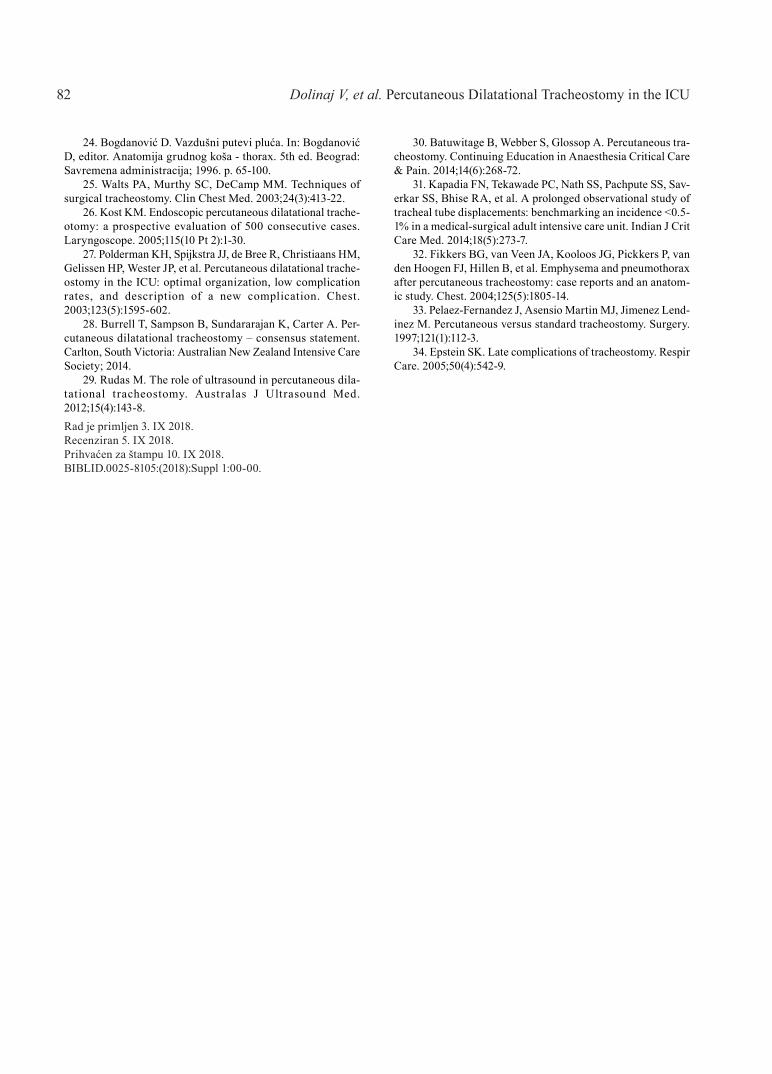
82
24. Bogdanović D. Vazdušni putevi pluća. In: Bogdanović D, editor. Anatomija grudnog koša - thorax. 5th ed. Beograd: Savremena administracija; 1996. p. 65-100.
25. Walts PA, Murthy SC, DeCamp MM. Techniques of surgical tracheostomy. Clin Chest Med. 2003;24(3):413-22.
26. Kost KM. Endoscopic percutaneous dilatational trache-otomy: a prospective evaluation of 500 consecutive cases. Laryngoscope. 2005;115(10 Pt 2):1-30.
27. Polderman KH, Spijkstra JJ, de Bree R, Christiaans HM, Gelissen HP, Wester JP, et al. Percutaneous dilatational trache-ostomy in the ICU: optimal organization, low complication rates, and description of a new complication. Chest. 2003;123(5):1595-602.
28. Burrell T, Sampson B, Sundararajan K, Carter A. Per-cutaneous dilatational tracheostomy – consensus statement. Carlton, South Victoria: Australian New Zealand Intensive Care Society; 2014.
29. Rudas M. The role of ultrasound in percutaneous dila-tational tracheostomy. Australas J Ultrasound Med. 2012;15(4):143-8.
30. Batuwitage B, Webber S, Glossop A. Percutaneous tra-cheostomy. Continuing Education in Anaesthesia Critical Care & Pain. 2014;14(6):268-72.
31. Kapadia FN, Tekawade PC, Nath SS, Pachpute SS, Sav-erkar SS, Bhise RA, et al. A prolonged observational study of tracheal tube displacements: benchmarking an incidence <0.5-1% in a medical-surgical adult intensive care unit. Indian J Crit Care Med. 2014;18(5):273-7.
32. Fikkers BG, van Veen JA, Kooloos JG, Pickkers P, van den Hoogen FJ, Hillen B, et al. Emphysema and pneumothorax after percutaneous tracheostomy: case reports and an anatom-ic study. Chest. 2004;125(5):1805-14.
33. Pelaez-Fernandez J, Asensio Martin MJ, Jimenez Lend-inez M. Percutaneous versus standard tracheostomy. Surgery. 1997;121(1):112-3.
34. Epstein SK. Late complications of tracheostomy. Respir Care. 2005;50(4):542-9.
Rad je primljen 3. IX 2018.Recenziran 5. IX 2018.Prihvaćen za štampu 10. IX 2018.BIBLID.0025-8105:(2018):Suppl 1:00-00.
Dolinaj V, et al. Percutaneous Dilatational Tracheostomy in the ICU
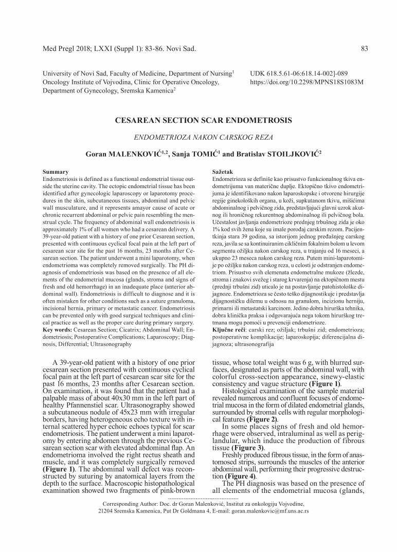
Med Pregl 2018; LXXI (Suppl 1): 83-86. Novi Sad. 83
SummaryEndometriosis is defined as a functional endometrial tissue out-side the uterine cavity. The ectopic endometrial tissue has been identified after gynecologic laparoscopy or laparotomy proce-dures in the skin, subcutaneous tissues, abdominal and pelvic wall musculature, and it represents amayor cause of acute or chronic recurrent abdominal or pelvic pain resembling the men-strual cycle. The frequency of abdominal wall endometriosis is approximately 1% of all women who had a cesarean delivery. A 39-year-old patient with a history of one prior Cesarean section, presented with continuous cyclical focal pain at the left part of cesarean scar site for the past 16 months, 23 months after Ce-sarean section. The patient underwent a mini laparotomy, when endometrioma was completely removed surgically. The PH di-agnosis of endometriosis was based on the presence of all ele-ments of the endometrial mucosa (glands, stroma and signs of fresh and old hemorrhage) in an inadequate place (anterior ab-dominal wall). Endometriosis is difficult to diagnose and it is often mistaken for other conditions such as a suture granuloma, incisional hernia, primary or metastatic cancer. Endometriosis can be prevented only with good surgical techniques and clini-cal practice as well as the proper care during primary surgery.Key words: Cesarean Section; Cicatrix; Abdominal Wall; En-dometriosis; Postoperative Complications; Laparoscopy; Diag-nosis, Differential; Ultrasonography
SažetakEndometrioza se definiše kao prisustvo funkcionalnog tkiva en-dometrijuma van materične duplje. Ektopično tkivo endometri-juma je identifikovano nakon laparoskopske i otvorene hirurgije regije ginekoloških organa, u koži, supkutanom tkivu, mišićima abdominalnog i pelvičnog zida, predstavljajući glavni uzrok akut-nog ili hroničnog rekurentnog abdominalnog ili pelvičnog bola. Učestalost javljanja endometrioze prednjeg trbušnog zida je oko 1% kod svih žena koje su imale porođaj carskim rezom. Pacijen-tkinja stara 39 godina, sa istorijom jednog pređašnjeg carskog reza, javila se sa kontinuiranim cikličnim fokalnim bolom u levom segmentu ožiljka nakon carskog reza, u trajanju od 16 meseci, a ukupno 23 meseca nakon carskog reza. Putem mini-laparotomi-je po ožiljku nakon carskog reza, u celosti je odstranjen endome-triom. Prisustvo svih elemenata endometralne mukoze (žlezde, stroma i znakovi svežeg i starog krvarenja) na ektopičnom mestu (prednji trbušni zid) uticalo je na postavljanje patohistološke di-jagnoze. Endometrioza se često teško dijagnostikuje i predstavlja dijagnostičku dilemu u odnosu na granulom, incizionu herniju, primarni ili metastatski karcinom. Jedino dobra hirurška tehnika, dobra klinička praksa i odgovarajuća nega tokom hirurškog tre-tmana mogu pomoći u prevenciji endometrioze.Ključne reči: carski rez; ožiljak; trbušni zid; endometrioza; postoperativne komplikacije; laparoskopija; diferencijalna di-jagnoza; ultrasonografija
University of Novi Sad, Faculty of Medicine, Department of Nursing1 UDK 618.5.61-06:618.14-002]-089Oncology Institute of Vojvodina, Clinic for Operative Oncology, https://doi.org/10.2298/MPNS18S1083MDepartment of Gynecology, Sremska Kamenica2
CЕSAREAN SECTION SCAR ENDOMETROSIS
ENDOMETRIOZA NAKON CARSKOG REZA
Goran MALENKOVIĆ1,2, Sanja TOMIĆ1 and Bratislav STOILJKOVIĆ2
A 39-year-old patient with a history of one prior cesarean section presented with continuous cyclical focal pain at the left part of cesarean scar site for the past 16 months, 23 months after Cesarean section. On examination, it was found that the patient had a palpable mass of about 40x30 mm in the left part of healthy Pfannenstiel scar. Ultrasonography showed a subcutaneous nodule of 45x23 mm with irregular borders, having heterogeneous echo texture with in-ternal scattered hyper echoic echoes typical for scar endometriosis. The patient underwent a mini laparot-omy by entering abdomen through the previous Ce-sarean section scar with elevated abdominal flap. An endometrioma involved the right rectus sheath and muscle, and it was completely surgically removed (Figure 1). The abdominal wall defect was recon-structed by suturing by anatomical layers from the depth to the surface. Macroscopic histopathological examination showed two fragments of pink-brown
tissue, whose total weight was 6 g, with blurred sur-faces, designated as parts of the abdominal wall, with colorful cross-section appearance, sinewy-elastic consistency and vague structure (Figure 1).
Histological examination of the sample material revealed numerous and confluent focuses of endome-trial mucosa in the form of dilated endometrial glands, surrounded by stromal cells with regular morphologi-cal features (Figure 2).
In some places signs of fresh and old hemor-rhage were observed, intraluminal as well as perig-landular, which induce the production of fibrous tissue (Figure 3).
Freshly produced fibrous tissue, in the form of anas-tomosed strips, surrounds the muscles of the anterior abdominal wall, performing their progressive destruc-tion (Figure 4).
The PH diagnosis was based on the presence of all elements of the endometrial mucosa (glands,
Corresponding Author: Doc. dr Goran Malenković, Institut za onkologiju Vojvodine, 21204 Sremska Kamenica, Put Dr Goldmana 4, E-mail: [email protected]
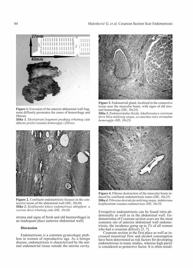
Malenković G, et al. Cеsarean Section Scar Endometrosis84
stroma and signs of fresh and old haemorrhage) in an inadequate place (anterior abdominal wall).
Discussion
Endometriosis is a common gynecologic prob-lem in women of reproductive age. As a benign disease, endometriosis is characterized by the nor-mal endometrial tissue outside the uterine cavity.
Extrapelvic endometriosis can be found intra-ab-dominally as well as in the abdominal wall. En-dometrioma of Cesarean section scars are the most common site of anterior abdominal wall endome-triosis, the incidence going up to 1% of all women who had a cesarean delivery [3, 7].
Cesarean section in the first place as well as in-creased menstrual flow and alcohol consumption have been determined as risk factors for developing endometriosis in many studies, whereas high parity is considered as protective factor. It is often misdi-
Figure 1. Extrusion of the anterior abdominal wall frag-ment diffusely permeates the zones of hemorrhage and fibrosisSlika 1. Ekstirpirani fragment prednjeg trbušnog zida difuzno prožet zonama hemoragije i fibroze
Figure 2. Confluent endometriosis focuses in the con-nective tissue of the abdominal wall (HE, 10x10) Slika 2. Konfluentni fokusi endometrioze uklopljeni u vezivno tkivo trbušnog zida (HE, 10x10)
Figure 3. Endometrial gland, localized in the connective tissue near the muscular beam, with signs of old stro-mal hemorrhage (HE, 10x25) Slika 3. Endometrijalna žlezda, lokalizovana u vezivnom tkivu blizu mišićnog snopa, sa znacima stare stromalne hemoragije (HE, 10x25)
Figure 4. Fibrous destruction of the muscular beam in-duced by confluent endometriosis zones (HE, 10x25)Slika 4. Fibrozna destrukcija mišićnog snopa, indukovana konfluentnim zonama endometrioze (HE, 10x25)
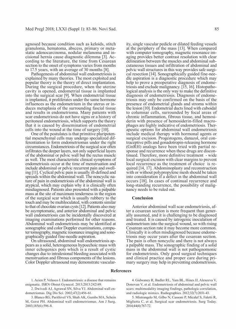
Med Pregl 2018; LXXI (Suppl 1): 83-86. Novi Sad. 85
agnosed because condition such as keloids, stitch granuloma, hematoma, abscess, primary or meta-static adenocarcinoma, nodular melanoma and in-cisional hernia cause diagnostic dilemma [3]. Ac-cording to the literature, the time from Cesarean section to the onset of symptoms varies from months to 17.5 years, with an average of 30 months [8].
Pathogenesis of abdominal wall endometriosis is explained by many theories. The most exploited and popular theory is the theory of direct implantation. During the surgical procedure, when the uterine cavity is opened, endometrial tissue is implanted into the surgical scar [9]. When endometrial tissue is implanted, it proliferates under the same hormone influences as the endometrium in the uterus or in-duces metaplasia of the surrounding fascial tissue and results in endometrioma. Many patients with scar endometriosis do not have signs or a history of peritoneal endometriosis, which supports the theory that it is caused by dissemination of endometrial cells into the wound at the time of surgery [10].
One of the postulates is that primitive pluripoten-tial mesenchymal cells may undergo specialized dif-ferentiation to form endometriomas under the right circumstances. Endometriosis of the surgical scar often infiltrates the deeper layers, not only superficial layers of the abdominal or pelvic wall but the rectus muscle as well. The most characteristic clinical symptoms of endometriosis occur at the time of menstruation and include abdominal or pelvic recurrent pain and swell-ing [11]. Cyclical pelvic pain is usually ill-defined and spreads within the abdominal wall. The noncyclic na-ture of pain in endometriosis of the abdominal wall is atypical, which may explain why it is clinically often misdiagnosed. Patients also presented with a palpable mass at the site of maximum tenderness in the region of the surgical scar which is usually rubbery to the touch and may be multiloculated, with contents similar to that of chocolate ovarian cysts [12]. Patients also may be asymptomatic, and anterior abdominal and pelvic wall endometriosis can be incidentally discovered at imaging examinations performed for other reasons. Abdominal wall endometriosis may be identified at sonographic and color Doppler examinations, compu-ter tomography, magnetic resonance imaging and sono-graphically guided fine-needle aspiration.
On ultrasound, abdominal wall endometriosis ap-pears as a solid, heterogeneous hypoechoic mass with inner echogenics pots which is a result of cystic changes due to intralesional bleeding associated with menstruation and fibrous components of the lesions. Color Doppler ultrasound may demonstrate vascular-
ity, single vascular pedicle or dilated feeding vessels at the periphery of the mass [13]. When compared with computer tomography, magnetic resonance im-aging provides better contrast resolution with clear delineation between the muscles and abdominal sub-cutaneous tissues and infiltration of abdominal and pelvic wall structures in this way provides safe surgi-cal resection [14]. Sonographically guided fine-nee-dle aspiration is a diagnostic procedure which may help to prove a preoperative diagnosis of endome-triosis and exclude malignancy. [15, 16]. Histopatho-logical analysis is the only way to make the definitive diagnosis of endometriosis. Diagnosis of endome-triosis may only be confirmed on the basis of the presence of endometrial glands and stroma within the lesion [10]. Endometrial ducts lined with cuboidal to columnar cells, surrounded by focal areas of chronic inflammation, fibrous tissue, and hemosi-derin with presence of hemosiderin-filled macro-phages are highly indicative of endometriosis. Ther-apeutic options for abdominal wall endometriosis include medical therapy with hormonal agents or surgical excision. Progestogens, danazol, oral con-traceptive pills and gonadotropin-releasing hormone (GnRH) analogs have been tried with partial re-sponse and recurrence when these drugs are discon-tinued. Therefore, more definitive treatment - wide local surgical excision with clear margins to prevent local recurrence as the treatment of choice is re-quired [14, 17]. Abdominoplasty and reconstruction with or without polypropylene mesh should be taken into consideration if a defect in the abdominal wall occurs [18]. In cases of continual recurrence and long-standing recurrence, the possibility of malig-nancy needs to be ruled out.
Conclusion
Anterior abdominal wall scar endometriosis, af-ter a cesarean section is more frequent than gener-ally assumed, and it is challenging to be diagnosed and treated. It is caused by iatrogenic inoculation of endometrium into the surgical wound, so with rising Cesarean section rate it may become more common. Clinically it is often misdiagnosed because endome-triosis may occur years after the cesarean section. The pain is often noncyclic and there is not always a palpable mass. The sonographic finding of a solid mass in the abdominal wall is not pathognomonic for endometriosis. Only good surgical techniques and clinical practice and proper care during pri-mary surgery may help in preventing endometriosis.
References
1. Acien P, Velasco I. Endometriosis: a disease that remains enigmatic. ISRN Obstet Gynecol. 2013;2013:242149.
2. Dwivedi AJ, Agrawal SN, Silva YJ. Abdominal wall en-dometriomas. Dig Dis Sci. 2002;47(2):456-61.
3. Blanco RG, Parithivel VS, Shah AK, Gumbs MA, Schein M, Gerst PH. Abdominal wall endometriomas. Am J Surg. 2003;185(6):596-8.
4. Gidwaney R, Badler RL, Yam BL, Hines JJ, Alexeeva V, Donovan V, et al. Endometriosis of abdominal and pelvic wall scars: multimodality imaging findings, pathologic correlation, and radiologic mimics. Radiographics. 2012;32(7):2031-43.
5. Mistrangelo M, Gilbo N, Cassoni P, Micalef S, Faletti R, Miglietta C, et al. Surgical scar endometriosis. Surg Today. 2014;44(4):767-72.
86
Rad je primljen 3. IX 2018.Recenziran 5. IX 2018.Prihvaćen za štampu 10. IX 2018.BIBLID.0025-8105:(2018):Suppl 1:83-86.
6. Emre A, Akbulut S, Yilmaz M, Bozdag Z. Laparoscopic trocar port site endometriosis: a case report and brief literature re-view. Int Surg. 2012;97(2):135-9.
7. Choudhary S, Fasih N, Papadatos D, Surabhi VR. Unusual imaging appearances of endometriosis. AJR Am J Roentgenol. 2009;192(6):1632-44.
8. Bumpers HL, Butler KL, Best IM. Endometrioma of the abdominal wall. Am J Obstet Gynecol. 2002;187(6):1709-10.
9. Teng CC, Yang HM, Chen KF, Yang CJ, Chen LS, Kuo CL. Abdominal wall endometriosis: an overlooked but possibly prevent-able complication. Taiwan J Obstet Gynecol. 2008;47(1):42-8.
10. Aydin O. Scar endometriosis: a gynaecologic pathology often presented to the general surgeon rather than the gynaecologist: report of two cases. Langenbecks Arch Surg 2007;392(1):105-9.
11. Singh KK, Lessells AM, Adam DJ, Jordan C, Miles WF, Macintyre IM, et al. Presentation of endometriosis to general sur-geons: a 10-year experience. Br J Surg. 1995;82(10):1349-51.
12. Pados G, Tympanidis J, Zafrakas M, Athanatos D, Bontis JN. Ultrasound and MR-imaging in preoperative evaluation of two rare cases of scar endometriosis. Cases J. 2008;1(1):97.
13. Balleyguier C, Chapron C, Chopin N, Hélénon O, Menu Y. Abdominal wall and surgical scar endometriosis: results of mag-netic resonance imaging. Gynecol Obstet Invest. 2003;55(4):220-4.
14. Francica G, Giardiello C, Angelone G, Cristiano S, Finelli R, Tramontano G. Abdominal wall endometriomas near cesarean delivery scars: sonographic and color doppler findings in a series of 12 patients. J Ultrasound Med. 2003;22(10):1041-7.
15. Simsir A, Thorner K, Waisman J, Cangiarella J. Endome-triosis in abdominal scars: a report of three cases diagnosed by FNA biopsy. Am Surg. 2001;67(10):984-6.
16. Gupta RK. Fine-needle aspiration cytodiagnosis of endome-triosis in cesarean section scar and rectus sheath mass lesions: a study of seven cases. Diagn Cytopathol. 2008;36(4):224-6.
17. Hensen JH, Van Breda Vriesman AC, Puylaert JB. Ab-dominal wall endometriosis: clinical presentation and imaging features with emphasis on sonography. AJR Am J Roentgenol. 2006;186(3):616-20.
18. Collins AM, Power KT, Gaughan B, Hill AD, Kneafsey B. Abdominal wall reconstruction for a large caesarean scar endometrioma. Surgeon. 2009;7(4):252-3.
Malenković G, et al. Cеsarean Section Scar Endometrosis
UPUTSTVO ZA AUTOREČasopis Medicinski pregled objavljuje radove koji prethod-
no nisu objavljeni niti poslati u drugi časopis. U Časopisu mogu biti objavljeni radovi iz različitih oblasti biomedicine, koji su namenjeni lekarima različitih specijalnosti.
Od 1. januara 2013. godine Medicinski pregled je počeo da koristi usluge e-Ur − Elektronskog uređivanja časopisa. Svi korisnici sistema − autori, recenzenti i urednici, moraju biti registrovani korisnici sa jednom elektronskom adresom.
Korisnici časopisa treba da se registruju na adresi:http://aseestant.ceon.rs/index.php/medpreg/user/registerPrijava rada treba da se učini na adresi:http://aseestant.ceon.rs/index.php/medpreg/U postupku prijave neophodno je da se pošalje saglasnost i
izjava autora i svih koautora da rad nije delimično ili u celini objavljen ili prihvaćen za štampu u drugom časopisu.
Elektronsko uređivanje časopisa obezbeđuje korišćenje sis-tema CrossCheck, koji prijavljene radove automatski proverava na plagijarizam i autoplagijarizam. Autori ne bi smeli da pošalju isti rad u više časopisa istovremeno. Ukoliko se to desi, glavni urednik časopisa Medicinski pregled ima pravo da rad vrati autorima bez prethodnog slanja rada na recenziju; da odbije štampanje rada; da se obrati urednicima drugih časopisa u koje je rad poslat ili da se obrati direktoru ustanove u kojoj su autori rada zaposleni.
Primaju se samo radovi koji su napisani na engleskom jez-iku, uz sažetak rada i naslov rada koji treba da budu napisani na engleskom i srpskom jeziku.
Radove koji su pristigli u časopis Medicinski pregled preg-leda jedan ili više članova Uređivačkog odbora Časopisa. Oni radovi koji su napisani prema pravilima Časopisa šalju se na anonimnu recenziju kod najmanje dva recenzenta, stručnjaka iz odgovarajuće oblasti biomedicine. Načinjene recenzije ra-dova pregleda glavni urednik ili članovi Uređivačkog odbora i one nisu garancija da će rad biti prihvaćen za štampu. Ma-terijal koji je pristigao u časopis ostaje poverljiv dok se rad nala-zi na recenziji, a identitet autora i recenzenata su zaštićeni, osim u slučaju ako oni odluče drugačije.
U časopisu Medicinski pregled objavljuju se: uvodnici, orig-inalni članci, prethodna ili kratka saopštenja, pregledni članci, stručni članci, prikazi slučajeva, članci iz istorije medicine i drugi članci.
1. Uvodnici – do 5 strana. Sadrže mišljenja ili diskusiju o posebno značajnoj temi za Časopis, kao i o podacima koji su štampani u ovom ili nekom drugom časopisu. Obično ih piše jedan autor po pozivu.
2. Originalni članci – do 12 strana. Predstavljaju rezultate istraživanja autora rada i njihovo tumačenje. Istraživanje treba da bude obrađeno i izloženo na način da se može ponoviti, a analiza rezultata i zaključci jasni da bi se mogli proveriti.
3. Pregledni članci – do 10 strana. Predstavljaju sistematsko, sveobuhvatno i kritičko izlaganje problema na osnovu anal-iziranih i diskutovanih podataka iz literature, a koji oslikavaju postojeću situaciju u određenom području istraživanja. Liter-atura koja se koristi u radu mora da sadrži najmanje 5 radova autora članka iz uže naučne oblasti koja je opisana u radu.
4. Prethodna ili kratka saopštenja – do 4 strane. Sadrže izuzetno važne naučne rezultate koje bi trebalo objaviti u što kraćem vremenu. Ne moraju da sadrže detaljan opis metod-ologije rada i rezultata, ali moraju da imaju sva poglavlja kao originalni članci u sažetoj formi.
5. Stručni članci – do 10 strana. Odnose se na proveru ili prikaz prethodnog istraživanja i predstavljaju koristan izvor za širenje znanja i prilagođavanja originalnog istraživanja potrebama postojeće nauke i prakse.
6. Prikazi slučajeva – do 6 strana. Opisuju retke slučajeve iz prakse. Slični su stručnim člancima. U ovim radovima pri-
kazuju se neuobičajeni oblici i tokovi oboljenja, neočekivane reakcije na primenjenu terapiju, primene novih dijagnostičkih procedura ili retke i nove bolesti.
7. Članci iz istorije medicine – do 10 strana. Ovi članci opis-uju događaje iz prošlosti sa ciljem da omoguće očuvanje medicinske i zdravstvene kulture. Imaju karakter stručnih članaka.
8. Ostali članci – U časopisu Medicinski pregled objav-ljuju se feljtoni, prikazi knjiga, izvodi iz strane literature, izveštaji sa kongresa i stručnih sastanaka, saopštenja o radu pojedinih zdravstvenih organizacija, podružnica i sekcija, saopštenja Uredništva, pisma Uredništvu, novosti u medicini, pitanja i odgovori, stručne i staleške vesti i članci napisani u znak sećanja (In memoriam).
Priprema rukopisaKompletan rukopis, uključujući tekst rada, sve priloge i
propratno pismo, treba poslati na elektronsku adresu koja je prethodno navedena.
Propratno pismo:– mora da sadrži izjavu svih autora da se radi o originalnom
radu koji prethodno nije objavljen niti prihvaćen za štampu u drugim časopisima;
– autori svojim potpisom preuzimaju odgovornost da rad ispunjava sve postavljene uslove i da ne postoji sukob interesa i
– autor mora navesti kategoriju članka (originalni rad, pre-gleni rad, prethodno saopštenje, stručni rad, prikaz slučaja, rad iz istorije medicine, itd.).
RukopisOpšta uputstvaTekst rada treba da bude napisan u programu Microsoft
Word za Windows, na A4 formatu stranice (sve četiri margine 2,5 cm), proreda 1,5 (isto važi i za tabele), fontom Times New Roman, veličinom slova 12 pt. Neophodno je koristiti međunarodni sistem mernih jedinica (SI), uz izuzetak tem-perature (° C) i krvnog pritiska (mmHg).
Rukopis treba da sadrži sledeće elemente: 1. Naslovna stranaNaslovna strana treba da sadrži: kratak i sažet naslov rada,
bez skraćenica, skraćeni naslov rada (do 40 karaktera), imena i prezimena autora (ne više od 6) i afilijacije svih autora. Na dnu strane treba da piše ime, prezime i titula autora zaduženog za korespondenciju, njena/njegova adresa, elektronska adresa, broj telefona i faksa.
2. SažetakSažetak ne može da sadrži više od 250 reči niti skraćenice.
Treba da bude strukturisan, kratak i sažet, sa jasnim pregledom problema istraživanja, ciljevima, metodama, značajnim rezul-tatima i zaključcima.
Sažetak originalnih i stručnih članaka treba da sadrži uvod (sa ciljevima istraživanja), materijale i metode, rezultate i zaključak.
Sažetak prikaza slučaja treba da sadrži uvod, prikaz slučaja i zaključak.
Sažetak preglednih članaka treba da sadrži Uvod, podnas-love koji odgovaraju istima u tekstu i Zaključak.
Navesti do 10 ključnih reči ispod sažetka. One su pomoć prilikom indeksiranja, ali autorove ključne reči mogu biti iz-menjene u skladu sa odgovarajućim deskriptorima, odnosno terminima iz Medical Subject Headings, MeSH.
Sažetak treba da bude napisan na srpskom i engleskom jez-iku. Sažetak na srpskom jeziku trebalo bi da predstavlja prevod sažetka na engleskom, što podrazumeva da sadrži jednake delove.
3. Tekst člankaOriginalni rad treba da sadrži sledeća poglavlja: Uvod (sa
jasno definisanim ciljevima istraživanja), Materijal i metode, Rezultati, Diskusija, Zaključak, spisak skraćenica (ukoliko su
korišćene u tekstu). Nije neophodno da se u posebnom poglav-lju rada napiše zahvalnica onima koji su pomogli da se istraživanje uradi, kao i da se rad napiše.
Prikaz slučaja treba da sadrži sledeća poglavlja: Uvod (sa jasno definisanim ciljevima), Prikaz slučaja, Diskusija i Zaključak.
Uvod U poglavlju Uvod potrebno je jasno definisati predmet
istraživanja (prirodu i značaj istraživanja), navesti značajne na-vode literature i jasno definisati ciljeve istraživanja i hipoteze.
Materijal i metodeMaterijal i metode rada treba da sadrže podatke o vrsti
studije (prospektivna/retrospektivna, uslove za uključivanje i ograničenja studije, trajanje istraživanja, demografske podatke, period praćenja). Detaljno treba opisati statističke metode da bi čitaoci rada mogli da provere iznesene rezultate.
RezultatiRezultati predstavljaju detaljan prikaz podataka koji su do-
bijeni istraživanjem. Sve tabele, grafikoni, sheme i slike moraju biti citirani u tekstu rada i označeni brojevima po redosledu njihovog navođenja.
DiskusijaDiskusija treba da bude koncizna, jasna i da predstavlja
tumačenje i poređenje rezultata studije sa relevantnim studi-jama koje su objavljene u domaćoj i međunarodnoj literaturi. U poglavlju Diskusija potrebno je naglasiti da li su postavljene hipoteze potvrđene ili nisu, kao i istaknuti značaj i nedostatke istraživanja.
ZaključakZaključci moraju proisteći isključivo iz rezultata istraživanja
rada; treba izbegavati uopštene i nepotrebne zaključke. Zaključci koji su navedeni u tekstu rada moraju biti u saglas-nosti sa zaključcima iz Sažetka.
4. LiteraturaPotrebno je da se literatura numeriše arapskim brojevima
redosledom kojim je u tekstu navedena u parentezama; izbega-vati nepotrebno velik broj navoda literature. Časopise bi treba-lo navoditi u skraćenom obliku koji se koristi u Index Medi cus (http://www.nlm.nih.gov/tsd/serials/lji.html). Pri citiranju lit-erature koristiti Vankuverski sistem. Potrebno je da se navedu svi autori rada, osim ukoliko je broj autora veći od šest. U tom slučaju napisati imena prvih šest autora praćeno sa et al.
Primeri pravilnog navođenja literature nalaze se u nastavku.Radovi u časopisima* Standardni radGinsberg JS, Bates SM. Management of venous thromboembo-
lism during pregnancy. J Thromb Haemost 2003;1:1435-42.* Organizacija kao autorDiabetes Prevention Program Research Group. Hypertensi on,
insulin, and proinsulin in participants with impaired gluco se toler-ance. Hypertension 2002;40(5):679-86.
* Bez autora21st century heart solution may have a sting in the tail. BMJ.
2002;325(7357):184.* Volumen sa suplementomMagni F, Rossoni G, Berti F. BN-52021 protects guinea pig
from heart anaphylaxix. Pharmacol Res Commun 1988;20 Suppl 5:75-8.
* Sveska sa suplementomGardos G, Cole JO, Haskell D, Marby D, Pame SS, Moore P.
The natural history of tardive dyskinesia. J Clin Psychophar macol 1988;8(4 Suppl):31S-37S.
* Sažetak u časopisuFuhrman SA, Joiner KA. Binding of the third component of
complement C3 by Toxoplasma gondi [abstract]. Clin Res 1987;35:475A.
Knjige i druge monografije* Jedan ili više autoraMurray PR, Rosenthal KS, Kobayashi GS, Pfaller MA. Me-
dical microbiology. 4th ed. St. Louis: Mosby; 2002.* Urednik (urednici) kao autor (autori)Danset J, Colombani J, eds. Histocompatibility testing 1972.
Copenhagen: Munksgaard, 1973:12-8.* Poglavlje u knjiziWeinstein L, Shwartz MN. Pathologic properties of invading
microorganisms. In: Soderman WA Jr, Soderman WA, eds. Patho-logic physiology: mechanisms of disease. Philadelphia: Saunders; 1974. p. 457-72.
* Zbornik radova sa kongresaChristensen S, Oppacher F. An analysis of Koza’s computa-
tional effort statistic for genetic programming. In: Foster JA, Lut-ton E, Miller J, Ryan C, Tettamanzi AG, editors. Genetic program-ming. EuroGP 2002: Proceedings of the 5th European Conference on Genetic Programming; 2002 Apr 3-5; Kinsdale, Ireland. Berlin: Springer; 2002. p. 182-91.
* DisertacijaBorkowski MM. Infant sleep and feeding: a telephone sur vey
of Hispanic Americans [dissertation]. Mount Pleasant (MI): Cen-tral Michigan University; 2002.
Elektronski materijal* Članak iz časopisa u elektronskom formatuAbood S. Quality improvement initiative in nursing homes:
the ANA acts in an advisory role. Am J Nurs [Internet]. 2002 Jun [cited 2002 Aug 12];102(6):[about 1 p.]. Available from: http://www.nursingworld.org/AJN/2002/june/Wawatch.htmArticle
* Monografija u elektronskom formatuCDI, clinical dermatology illustrated [monograph on CD-
ROM]. Reevs JRT, Maibach H. CMEA Multimedia Group, pro-ducers. 2nd ed. Version 2.0. San Diego:CMEA;1995.
* Kompjuterska datotekaHemodynamics III: the ups and downs of hemodynamics [com-
puter program]. Version 2.2. Orlando (FL): Computerized Educa-tional Systems; 1993.
5. Prilozi (tabele, grafikoni, sheme i slike)BROJ PRILOGA NE SME BITI VEĆI OD ŠEST!Tabele, grafikoni, sheme i slike se postavljaju kao posebni
dokumenti. – Tabele i grafikone bi trebalo pripremiti u formatu koji je
kompatibilan programu u kojem je napisan tekst rada. Slike bi trebalo poslati u jednom od sledećih oblika: JPG, GIF, TIFF, EPS.
– Svaki prilog mora biti obeležen arapskim brojem prema redosledu po kojem se navodi u tekstu rada.
– Naslovi, tekst u tabelama, grafikonima, shemama i leg-ende slika bi trebalo da budu napisani na srpskom i engles kom jeziku.
– Nestandardne priloge označiti u fusnoti uz korišćenje sledećih simbola: *, †, ‡, §, | |, ¶, **, † †, ‡ ‡ .
– U legendi slika trebalo bi napisati korišćeno uveličanje okulara i objektiva mikroskopa. Svaka fotografija treba da ima vidljivu skalu.
– Ako su tabele, grafikoni, sheme ili slike već objavljene, navesti originalni izvor i priložiti pisano odobrenje autora za njihovo korišćenje.
– Svi prilozi će biti štampani kao crno-bele slike. Ukoliko autori žele da se prilozi štampaju u boji, obavezno treba da plate dodatne troškove.
6. Dodatne obavezeAUTORI I SVI KOAUTORI RADA OBAVEZNO TREBA
DA PLATE GODIŠNJU PRETPLATU ZA ČASOPIS MEDICINSKI PREGLED. U PROTIVNOM, RAD NEĆE BITI ŠTAMPAN U ČASOPISU.
INFORMATION FOR AUTHORSMedical Review publishes papers (previously neither pub-
lished in nor submitted to any other journals) from various fiel ds of biomedicine intended for broad circles of doctors.
Since January 1th, 2013 the Medical Review has been using the service e-Ur: Electronic Journal Editing. All users of the Registration system, i.e. authors, reviewers, and editors have to be registered users with only one e-mail address. Registration should be made on the web address:
http://aseestant.ceon.rs/index.php/medpreg/user/register.Manuscript submission should be made on the web address:http://aseestant.ceon.rs/index.php/medpreg/A SUPPLEMENTARY FILE, WITH THE STATEMENT
THAT THE PAPER HAS NOT BEEN SUBMITTED OR AC-CEPTED FOR PUBLICATION ELSEWHERE AND A CON-SENT SIGNED BY ALL AUTHORS, HAVE TO BE EN-CLOSED WITH THE MANUSCRIPT.
Authors may not send the same manuscript to more than one journal concurrently. If this occurs, the Editor may return the paper without reviewing it, reject the paper, contact the Editor of the other journal(s) in question and/or contact the author’s employers.
Papers should be written in English language, with an abstract and title page in English, as well as in Serbian language.
All papers submitted to Medical Review are seen by one or more members of the Editorial Board. Suitable articles are sent to at least two experts to be reviewed, thier reports are returned to the assigned member of the Editorial Board and the Editor. Revision of an article gives no guarantee of acceptance and in some cases revised articles are rejected if the improvements are not sufficient or new issues have arisen. Material submitted to the Journal re-mains confidential while being reviewed and peer-reviewers’ iden-tities are protected unless they elect to lose anonymity.
Medical Review publishes the following types of articles: editorials, original studies, preliminary reports, review articles, professional articles, case reports, articles from history of medicine and other types of publications.
1. Editorials – up to 5 pages – convey opinions or discus-sions on a subject relevant for the Journal. Editori als are com-monly written by one author by invitation.
2. Original studies – up to 12 pages – present the authors’ own investigations and their interpreta tions. They should contain data which could be the basis to check the obtained results and reproduce the investigative procedure.
3. Review articles – up to 10 pages – provide a con densed, comprehensive and critical review of a problem on the basis of the published material being analyzed and discussed, reflecting the current situation in one area of research. Papers of this type will be accepted for publica tion provided that the authors confirm their expertise in the relevant area by citing at least 5 self-cita-tions.
4. Preliminary reports – up to 4 pages – contain scientific results of significant importance requiring ur gent publishing; however, it need not provide detailed description for repeating the obtained results. It presents new scientific data without a detailed explanation of methods and results. It contains all parts of an original study in an abridged form.
5. Professional articles – up to 10 pages – examine or repro-duce previous investigation and represent a valu able source of knowledge and adaption of original inve stigations for the needs of current science and practice.
6. Case reports – up to 6 pages – deal with rare casu istry from practice important for doctors in direct charge of patients and are similar to professional articles. They emphasize unu-sual characteristics and course of a disea se, unexpected reac-tions to a therapy, application of new diagnostic procedures and describe a rare or new disease.
7. History of medicine – up to 10 pages – deals with history with the aim of providing continuity of medi cal and health care culture. They have the character of professional articles.
8. Other types of publications – The journal also pu blishes feuilletons, book reviews, extracts from foreign li terature, re-ports from congresses and professional mee tings, communica-tions on activities of certain medical in stitutions, branches and sections, announcements of the Editorial Board, letters to the Editorial Board, novelti es in medicine, questions and answers, professional and vocational news and In memoriam.
Preparation of the manuscriptThe complete manuscript, including the text, all su pple men-
tary material and covering letter, is to be sent to the web address above.
The covering letter:– It must contain the proof given by the author that the
paper represents an original work that it has neither been previ-ously published in other journals nor is un der consideration to be published in other journals.
– It must confirm that all the authors meet criteria set for the authorship of the paper, that they agree comple tely with the text and that there is no conflict of interest.
– It must state the type of the paper submitted (an original study, a review article, a preliminary report, a professional ar-ticle, a case report, history of medicine).
The manuscript:General instructions.Use Microsoft Word for Windows to type the text. The text
must be typed in font Times New Roman, page format A4, space 1.5 (for tables as well), margins set to 2.5 cm and font size 12pt. All measurements should be reported in the metric system of the International System of Units – SI. Tem perature should be ex-pressed in Celsius degrees (°C) and pressure in mmHg.
The manuscript should contain the following elements:1. The title page. The title page should contain a con cise and clear title of the
paper, without abbreviations, then a short title (up to 40 characters), full names and sur names of the authors (not more than 6) indexed by num bers corresponding to those given in the heading along with the full name and place of the institutions they work for. Contact information including the academic degree(s), full ad-dress, e-mail and number of phone or fax of the corresponding author (the author responsible for corres pondence) are to be given at the bottom of this page.
2. Summary. The summary should contain up to 250 words, without ab-
breviations, with the precise revi ew of problems, objectives, methods, important results and conclusions. It should be struc-tured into the para graphs as follows:
– Original and professional papers should have the introduc-tion (with the objective of the paper), materials and methods, results and conclusion
– Case reports should have the introduction, case re port and conclusion
– Review papers should have the introduction, subtitles cor-responding to those in the paper and conclusi on.
The authors should provide up to 10 keywords below the summary. These keywords will assist indexers in cross-indexing the article and will be published with the summary, but the authors’ keywords could be changed in accordance with the list of Medical Subject Headings, MeSH of the American National Medical Library.
The summary should be written in both languages, English as well as Serbian. The summary in Serbian language should be the translation of the summary in English; therefore, it has to contain the same paragraphs.
3. The text of the paper.The text of original stu dies must contain the following: in-
troduction (with the clearly defined objective of the study), materials and methods, results, discussion, conclusion, list of abbreviations (if used in the text) and not necessarily, the ac-knowledgment mentioning those who have hel ped in the inves-tigation and preparation of the paper.
The text of a case report should contain the fo llowing: in-troduction (with clearly defined objective of the study), case report, discussion and conclusion.
Introduction contains clearly defined problem dealt with in the study (its nature and importance), with the relevant refer-ences and clearly defined objective of the investigation and hypothesis.
Materials and methods should contain data on design of the study (prospective/retrospective, eligibility and exclusion criteria, duration, demographic data, follow-up period). Statis-tical methods applied should be clear and described in details.
Results give a detailed review of data obtained du ring the study. All tables, graphs, schemes and figures must be cited in the text and numbered consecutively in the order of their first citation in the text.
Discussion should be concise and clear, interpreting the basic findings of the study in comparison with the results of relevant studies published in international and national litera-ture. It should be stated whether the hypot hesis has been con-firmed or denied. Merits and demerits of the study should be mentioned.
Conclusion must deny or confirm the attitude towar ds the 0based solely on the author’s own results, corroborating them. Avoid generalized and unnecessary conclusions. Conclusions in the text must be in accor dance with those given in the sum-mary.
4. References are to be given in the text under Arabic nu-merals in parentheses consecutively in the order of their first citation. Avoid a large number of citations in the text. The title of journals should be abbreviated according to the style used in Index Medi cus (http://www.nlm.nih.gov/tsd/serials/lji.html). Apply Vancouver Group’s Criteria, which define the order of data and punctuation marks separating them. Examples of cor-rect forms of references are given below. List all authors, but if the number exceeds six, give the names of six authors followed by ‘et al’.
Articles in journals* A standard articleGinsberg JS, Bates SM. Management of venous thromboembo-
lism during pregnancy. J Thromb Haemost 2003;1:1435-42.* An organization as the author Diabetes Prevention Program Research Group. Hypertensi on,
insulin, and proinsulin in participants with impaired gluco se toler-ance. Hypertension 2002;40(5):679-86.
* No author given 21st century heart solution may have a sting in the tail. BMJ.
2002;325(7357):184.* A volume with supplement Magni F, Rossoni G, Berti F. BN-52021 protects guinea pig
from heart anaphylaxix. Pharmacol Res Commun 1988;20 Suppl 5:75-8.
* An issue with supplement Gardos G, Cole JO, Haskell D, Marby D, Pame SS, Moore P.
The natural history of tardive dyskinesia. J Clin Psychophar macol 1988;8(4 Suppl):31S-37S.
* A summary in a journal Fuhrman SA, Joiner KA. Binding of the third component of com-
plement C3 by Toxoplasma gondi [abstract]. Clin Res 1987;35:475A.
Books and other monographs * One or more authors Murray PR, Rosenthal KS, Kobayashi GS, Pfaller MA. Me-
dical microbiology. 4th ed. St. Louis: Mosby; 2002.* Editor(s) as author(s) Danset J, Colombani J, eds. Histocompatibility testing 1972.
Copenhagen: Munksgaard, 1973:12-8.* A chapter in a book Weinstein L, Shwartz MN. Pathologic properties of invading
microorganisms. In: Soderman WA Jr, Soderman WA, eds. Patho-logic physiology: mechanisms of disease. Philadelphia: Saunders; 1974. p. 457-72.
* A conference paper Christensen S, Oppacher F. An analysis of Koza’s computa-
tional effort statistic for genetic programming. In: Foster JA, Lut-ton E, Miller J, Ryan C, Tettamanzi AG, editors. Genetic program-ming. EuroGP 2002: Proceedings of the 5th European Conference on Genetic Programming; 2002 Apr 3-5; Kinsdale, Ireland. Berlin: Springer; 2002. p. 182-91.
* A dissertation and theses Borkowski MM. Infant sleep and feeding: a telephone sur vey
of Hispanic Americans [dissertation]. Mount Pleasant (MI): Cen-tral Michigan University; 2002.
Electronic material * A journal article in electronic formatAbood S. Quality improvement initiative in nursing homes:
the ANA acts in an advisory role. Am J Nurs [Internet]. 2002 Jun [cited 2002 Aug 12];102(6):[about 1 p.]. Available from: http://www.nursingworld.org/AJN/2002/june/Wawatch.htmArticle
* Monographs in electronic formatCDI, clinical dermatology illustrated [monograph on CD-
ROM]. Reevs JRT, Maibach H. CMEA Multimedia Group, pro-ducers. 2nd ed. Version 2.0. San Diego:CMEA;1995.
* A computer fileHemodynamics III: the ups and downs of hemodynamics [com-
puter program]. Version 2.2. Orlando (FL): Computerized Educa-tional Systems; 1993.
5. Attachments (tables, graphs, schemes and pho tographs). THE MAXIMUM NUMBER OF ATTACHMENTS AL-
LOWED IS SIX!– Tables, graphs, schemes and photographs are to be submit-
ted as separate documents, on separate pages.– Tables and graphs are to be prepared in the format com-
patible with Microsoft Word for Windows progra mme. Photo-graphs are to be prepared in JPG, GIF, TIFF, EPS or similar format.
– Each attachment must be numbered by Arabic numerals consecutively in the order of their appearance in the text
– The title, text in tables, graphs, schemes and legen ds must be given in both Serbian and English languages.
– Explain all non-standard abbreviations in footnotes using the following symbols *, †, ‡, §, | |, ¶, **, † †, ‡ ‡ .
– State the type of color used and microscope ma gnification in the legends of photomicrographs. Photo micrographs should have internal scale markers.
– If a table, graph, scheme or figure has been previ ously published, acknowledge the original source and submit written permission from the copyright holder to reproduce it.
– All attachments will be printed in black and whi te. If the authors wish to have the attachments in colo r, they will have to pay additional cost.
6. Additional requirementsSHOULD THE AUTHOR AND ALL CO-AUTHORS
FAIL TO PAY THE SUBSCRIPTION FOR MEDICAL RE-VIEW, THEIR PAPER WILL NOT BE PUBLISHED.