medicinal mushrooms inhibit full
TRANSCRIPT

7/30/2019 Medicinal Mushrooms Inhibit Full
http://slidepdf.com/reader/full/medicinal-mushrooms-inhibit-full 1/7
Medicinal mushroom extracts inhibit ras-induced cell transformationand the inhibitory effect requires the presence of normal cells
W.L.Wendy Hsiao1,2,4, You Quan Li2, Tin Lap Lee2,Ning Li2, Marilyn M.You3 and Shu-ting Chang3
1Biomedical Science, School of Chinese Medicine, Hong Kong Baptist
University, Hong Kong, 2Department of Biology, Hong Kong University of Science and Technology, Hong Kong and 3Department of Biology,Chinese University of Hong Kong, Hong Kong, China
4To whom correspondence should be addressed
Email: [email protected]
Previously, we developed a simple Rat 6 (R6) cell system bywhich the inhibitory effects of non-cytotoxic chemicals canbe assessed by focus formation assay upon transfection of ras oncogene to the host cells. Using this system, two wellstudied medicinal mushrooms Ganoderma lucidum and
Tricholoma lobayense with anticancer potential were exam-ined for their possible advert effects on cell transformationinduced by ras oncogene. Results indicated that both spe-cies of mushrooms yielded strong inhibitory effects on ras-induced cell transformation. Further study on T.lobayenseindicated that the DEAE-column-bound, polysaccharides(PS)-peptide enriched, but not the unbound fraction,showed strong inhibition in a dosage-dependent manner.Subsequent time course study revealed that the continuedpresence of the extract in the transfected cultures wasrequired for a maximum inhibitory effect. At the sametime, we also observed that significant levels of inhibitionoccurred even when the application of the extract wasdelayed until day 12 after transfection. Using a stable
transformed cell line, R6/GFP-Ras expressing green fluor-escent protein-ras fusion protein in a co-culture assay withnormal R6 cells, we demonstrated that R6/GFP-Ras cellsgrew into green fluorescent foci with striking transformingmorphology in the absence of extracts. However, in thepresence of extracts, R6/GFP-Ras cells, in most cases,remained as small colonies compiled with only a few greenfluorescent cells. Moreover, the inhibitory effect requiresthe presence of R6 cells. In our study, mushroom extractshave no effect on the growth of individually culturednormal and transformed R6 cells. It is noteworthy thatthe extracts do not affect the level, or the subcellular loca-lization of the Ras protein. Collectively, the data stronglysuggest that the inhibitory effect of the mushroom extracts
is not due to a direct killing of the transformed cells, rather,it may be mediated through the surrounding normal R6.While the general understanding of the antitumor effect of PS and PSPC is mediated through the cytokines releasedby activated marcrophages and T-lymphocytes, our datamay provide a novel alternative mechanism that the
mushroom PS peptides may exert anticancer effect bytargeting the ras-mediated signaling pathway.
Introduction
The antitumor effect of mushrooms has long been observed inAsia, especially in China and Japan. The analysis of variousspecies of mushrooms has resulted in the identification of afamily of high molecular weight, hot-water-soluble poly-saccharides (PS) and polysaccharideEEpeptide complexes(PSPC), which have tested positive for antitumor activities inanimal studies (1). NMR analysis reveals that the antitumor PS
are composed of a variety of linear and branched glucans. Theyappear in various conformations, and some are in the gel state.As a result of the complexity and heterogeneity of PS andPSPC and limited suitable bioassay systems, the mechanismof action of these PS remain obscure. Until now, research onthe antitumor activities is based mainly on the results derivedfrom experiments with implanted Sarcoma180 or chemicallyinduced tumors in mice (2EE6). Antimitotic tests in tumor celllines have been, so far, rather inconsistent or negative(1,2,4,7EE9). Aside from their antitumor effects, mushroom-derived PS and PSPC seem to function as immunomodulators;this was observed in animals as well as in cultured macro-phages and T-lymphocytes. It has been postulated that theantitumor effect of PS and PSPC may be mediated through
the cytokines released by activated macrophages and T-lymphocytes, instead of through direct cytocidal effects ontumor cells (4,8EE11). In order to gain a more comprehensiveunderstanding, it is necessary to investigate the antitumoractivity of mushrooms in a wider range of cell systems.
Previously, we have demonstrated that the establishedrodent cell line R6 is resistant to transformation induced by apotent c-H-ras (T24) oncogene in a focus formation assay. Thetransforming efficiency of T24, however, can be modulated bytreatment with various tumor promoters and factors (12,13).Using this R6/ ras assay system, we have assessed the inhibi-tory or enhancing effects of various chemicals on T24 induced-transformation (14EE16). In the current study, we explored theantitumor activity of mushrooms using the focus formation
assay built around the R6/ ras model system.In the study, we focused on two medicinal mushrooms:Ganoderma lucidum and Tricholoma lobayense. Both mush-rooms exhibit antitumor activities, based mainly on animalstudies (1EE4,17). Tricholoma lobayense is a native HongKong species. Ganoderma lucidum is an important traditionalmedicine in China and Japan, used for promoting health andtreatment of various diseases, including cancer. Ganodermalucidum is also a better documented natural product interms of its pharmacological and chemical properties. Ourresults showed that ras-induced transformed foci wereeffectively inhibited by the addition of extracts of G.lucidumand T.lobayense in dosage-dependent and time-dependent
Abbreviations: DMEM, Dulbecco's modified Eagle medium; D10CS,Dulbecco's modified Eagle medium supplemented with 10% calf serum;GFP, green fluorescent protein; PS, polysaccharides; PSPC,polysaccharideEEpeptide complexes; T24, activated human c-H-ras oncogene.
Carcinogenesis vol.25 no.7 # Oxford University Press 2004; all rights reserved. 1177
Carcinogenesis vol.25 no.7 pp.1177EE1183, 2004DOI: 10.1093/carcin/bgh119

7/30/2019 Medicinal Mushrooms Inhibit Full
http://slidepdf.com/reader/full/medicinal-mushrooms-inhibit-full 2/7
manners. Data also revealed that the PS fraction of T.lobayensewould only exert an inhibitory effect on Ras-transformed cellswhen cells were co-cultivated with normal R6 cells, suggestinga novel mechanism in which the inhibitory effect of PS ismediated through the surrounding normal R6 cells.
Materials and methods
Preparations of mushroom samples
Fruiting bodies of G.lucidum were homogenized and extracted with boilingdistilled water for 6 h to obtain the PS-enriched preparations. After centrifuga-tion to remove the insoluble portion, the water-soluble extracts were lyophil-ized, then kept at room temperature for later usage. Liquid mycelium culturesof G.lucidum were also used to obtain PS-extract. Prior to extraction, thecultures were filtered and precipitated with ethanol according to Liu et al.(17). The precipitates were dissolved in distilled water, centrifuged to removethe insoluble, then lyophilized and designated as mycelium filtrate. Tricho-loma lobayense was originally isolated and established in cultures byS.T.Chang's Laboratory at the Chinese University of Hong Kong, HongKong. Tricholoma lobayense was cultured in nutrient broth as described(17). Liquid cultures containing the secreted fungal PS were prepared asabove. The water-soluble, PS-enriched components were lyophilized anddesignated as mycelium filtrate. Part of the filtrate was further fractionatedinto the unbound Fraction A1 and the salt-eluted bound fraction A2 using a
DEAE-cellulose ion exchange column chromatography (17). Both fractionswere dialyzed against ddH2O and lyophilized for later usage.
Cell cultures and plasmids
The Rat 6 (R6) cell line was a subclone of the Fisher rat embryo fibroblastsoriginating from Freeman's Laboratory (18). R6/T24 cell line is a clonal R6cell line transformed by the activated human c-Ha-ras oncogene (12). The R6/ green fluorescent protein GFP-Ras cell line is a transformed clonal cell lineestablished from a transformed focus derived from R6 cultures transfected by aGFP-ras fusion vector in our lab. Cells were grown in Dulbecco's modifiedEagle medium (DMEM) supplemented with 10% calf serum (D10CS)(Invitrogen, Carlsbad, CA). Cultures were maintained in a humidified incuba-tor at 37C with 5% CO2 in air and fed twice a week with fresh medium.Plasmid pT24 contains a 6.4 kb BamHI fragment corresponding to the codingsequence of the human bladder c-Ha-ras oncogene. The plasmid pT24 wasobtained from M.Wigler's Laboratory.
Focus formation assay
The standard focus formation assay and treatment of the cultures were per-formed as described earlier (12). In brief, 5 Â 105 cells seeded in 90 mm platewere transfected with 1 mg T24 plasmid DNA and 20 mg R6 genomic DNA ascarrier DNA by DNA-mediated transfection procedure based on Bacchetti andGraham (19) and Wigler et al. (20), with slight modifications (12,13). Todetermine the effects of mushroom extracts on ras-induced focus formation,transfected cultures were fed with DMEM plus 5% fetal calf serum (D5FCS) inthe presence and absence of test sample on day 2 upon transfection, thencontinued feeding with each respective growth medium twice a week through-out the experiment. Lyophilized samples of mushroom preparations wereweighted, dissolved in boiling ddH2O, then centrifuged at 10 000 r.p.m. for 20 min to remove the residues. The supernatant was sterile filtered, then addedto the growth medium at designated concentrations. All experiments wereperformed in six replicate plates.
Cytotoxicity assay
The cytotoxic effect ofeach sample was performed on both R6 and R6/T24 cell
lines. Cells were seeded in triplicate at 104
/60 mm plate in D10CS. The nextday, the test sample was added to the cultures and kept for 5 days. At the end of treatments, cells were trypsinized and counted using a Coulter Counter Fullerton, CA. Cytotoxicity was expressed as percent survival, i.e. cell counts of the treated cultures divided by cell counts obtained from the untreated cultures.
Colony formation and co-culture assays
For a better quantitative assessment of the inhibitory effect for drug testing,we designed a co-culture assay to simulate the focus formation assay. Theassay was set up by seeding 500 ras-transformed R6 cells on a 90 mm plate intriplicate pre-seeded with 2.5 Â 105 normal R6 cells 24 h earlier. A dayafter the seeding of the transformed R6 cells, the test sample was added to theco-cultures of normal and transformed R6 cells grown in DMEM plus 5% calf serum (D5CS). At the end of 2 weeks, culture plates were fixed with 10%formaldehyde, stained with Giemsa stain and photographed. In order todistinguish the transformed from the neighboring normal R6 cells, a
transformed cell line, R6/GFP-Ras expressing green fluorescent GFP-Rasfusion protein was used in the co-culture assay. In a parallel experiment, thepossible toxic effect of mushroom extracts on the growth of R6 and R6/GFP-Ras cells were tested by seeding 500 of each cell line separately in D10CS inthe presence and absence of test chemical for the duration of 12 EE14 days.Cultures were stained and scored for total number of colonies per plate. All theexperimental cultures were fed twice a week with or without the mushroomextract.
Western blot analysis
R6 or R6/GFP-Ras-transformed cells were seeded 2.5 Â 105
per 90 mm platein DMEM plus D10CS. The next day, cultures were fed with fresh medium inthe presence and absence of Tricholoma filtrate A2 fraction (500 mg/ml) andfed twice a week. Cells were washed with cold PBS three times and lysed in400 ml NET buffer (150 mM NaCl, 50 mM Tris EEHCl, pH 7.4, 5 mM EDTA,pH 8.0, 1 mM APMSF, 1 mM E-64, 1 mM pepstatin, 100 mM NaVO5 and 10mg/ml aprotinin) plus 1% NP-40 on ice according to Lu et al. (21). For westernblotting analysis, 40 mg of protein extracts were loaded on a 12% SDSEEPAGE.After separation, the proteins were transferred to a Hybond-C nylon membrane(Amersham, Piscataway, NJ), reacted to either anti-Ras (Santa Cruz) or anti-GFP antibodies (Clontech, Palo Alto, CA) and visualized by the ECL detectionkit (Amersham) according to the manufacturer's manual. Blots were hybri-dized with anti-actin antibodies (Santa Cruz, Santa Cruz, CA) to normalize thegel loading.
Results
Effect of extracts of G.lucidum on ras-induced transformation
In the primary experiment, extracts of fruiting bodies andfiltrate of G.lucidum were tested. While both the fruitingbodies and mycelium filtrate were effective in inhibiting fociformation induced by c-Ha-ras oncogene, the latter seemed tobe more effective (Table I). At 200 mg (dry weight)/ml andabove (Table I,B), mycelium filtrate showed nearly 100%reduction of foci. On the other hand, the fruiting bodyextract-treated cultures exhibited 56% inhibition at 200 mg/mland 80% at a dose of 500 mg/ml (Table I,A). Neither samplesposted cytotoxicity on either the host R6 or R6/T24 cells(Table II). Thus, the inhibitory effect of the mushroom extractson ras-transformation is not due to a direct killing of cells
transformed by ras oncogene in the focus formation assay.
Effect of Tricholoma filtrate on ras-induced transformation
Both the total, and the DEAE-column-bound fraction of T.lobayense filtrate were tested in R6 cells upon transfectionof the ras oncogene. Data showed that the total filtrate and the
Table I. Effect of hot water extracts of G.lucidum on ras-inducedtransformation
Material tested Conc. (mg/ml) Number of foci Relative number of focia
A. Extract of fruiting bodiesMock control 0No treatment 0 14.7 Æ 4.0 1Extract 10 13.7 Æ 4.5 0.93
100 8.3 Æ 2.9 0.56500 3.0 Æ 1.0 0.2
B. Extract of mycelium filtrateMock control 0No treatment 0 18.3 Æ 2.3 1Extract 100 2.7 Æ 1.2 0.15
200 1.0 Æ 1.0 0.05500 1.3 Æ 1.5 0.07
aRelative number of foci; the ratio of foci obtained in the presence of drug to that in the absence of drug (i.e. no treatment control).
W.L.W.Hsiao et al.
1178

7/30/2019 Medicinal Mushrooms Inhibit Full
http://slidepdf.com/reader/full/medicinal-mushrooms-inhibit-full 3/7
DEAE-column-bound fraction A2 of T.lobayense markedlyinhibited ras-foci, while the unbound fraction produced noeffect (Table III and Figure 1). The preparations had no or slightly toxic effect on either normal or transformed R6 cells(Table IV). The nutrient broth used for the T.lobayense cul-tures alone presented no inhibitory effect.
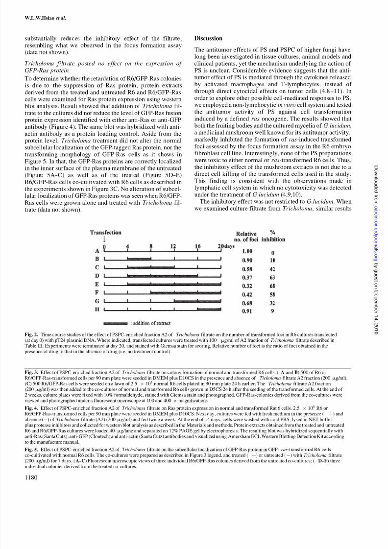
Dosage and time course studies of Tricholoma filtrate on
ras-induced transformationThe inhibitory effect of the Tricholoma filtrate was further explored with regard to dosage and duration of treatment.Results revealed that the inhibitory effect of Tricholoma fil-trate was dosage-dependent. Extracts in concentration as lowas 1 mg/ml exerted a 19% inhibitory effect on the formation of ras-foci (Table V). Time course study indicated that themaximal effect was obtained when transfected cultures were
treated with Tricholoma filtrate from days 4 to 20 after thetransfection. Interestingly, a 32% reduction in foci number wasstill obtained when the treatment was delayed until day 12 afterthe transfection of the ras oncogene (Figure 2).
Effect of Tricholoma filtrate on R6/GFP-Ras cellsco-cultivated with normal R6 cells
To further explore the nature of the inhibitory effect of
Tricholoma filtrate, we reconstituted the focus formationassay by seeding 500 R6/GFP-Ras cells on a 90 mm cultureplate pre-seeded with 2.5 Â 105 normal R6 cells 24 h earlier.The co-cultures were then maintained in D5CS medium inthe presence and absence of Tricholoma filtrate. The effect ofTricholoma on colony formations of individual R6 and R6/GFP-Ras cell lines were conducted in parallel with the co-culture assay. Results indicated that addition of Tricholomadid not affect the colony formation (Figure 3A and B), northe morphology of R6 or R6/GFP-Ras cells (data not shown).In the co-culture of R6 and R6/GFP-Ras (Figure 3C), the R6/GPF-Ras cells formed many dense transformed colonies on thetop of the monolayer of R6 cells in the absence of treatment,resembling the formation of transformed foci shown in
Figure 1. Addition of Tricholoma filtrate (200 mg/ml) effec-tively blocked the formation of the GFP-Ras transformedcolonies. Indeed, under the fluorescent microscope, thegreen fluorescent colonies, representing the R6/GFP-Rascells, were severely retarded in the presence of the Tricho-loma filtrate, while grown to sizable colonies in the absenceof the drug treatment (Figure 3). It is worthy to note thattreatment with Tricholoma does not affect the initial platingefficiency of R6/GFP-Ras cells as nearly the same number ofGFP-positive cells were observed on both the treatedand untreated plates under a fluorescent microscope, 24 hupon the addition of the filtrate. Additional evidencecame from the fact that early withdrawal of the treatment
Table V. Dosage effect of DEAE-column purified fraction of Tricholomafiltrate on ras-induced transformation
Test sample Conc. (mg/ml) Relative numberof foci
Mock control (no T24 DNA) 0 0No treatment 0 1DEAE-fraction A2a 1 0.81
5 0.7810 0.8720 0.5650 0.47
100 0.32
aDEAE A2 fraction was prepared as described in Table III legend.
Table II. Cytotoxicity of hot water extract of G.lucidum
Treatmenta Conc. (mg/ml) R6 cells %survivalb
R6/T24 cells %survivalb
No treatment 0 100 100Fruiting body 500 93 98Mycelium filtrate 500 89 98
aThe duration of the treatment was 5 days for all treatment groups.b% Survival cell counts of treated cultures/cell counts of untreatedcultures Â100%.
Table III. Effect of extracts of Tricholoma filtrate on ras-inducedtransformation
Material tested Conc. (mg/ml) Relative number of foci
Mock controlEE
0No treatment 0 1Total mycelium filtrate 100 0.04
200 0.10DEAE-column purified fraction A2a 100 0.04DEAE-unbound fraction A1a 50 0.96
100 0.91
aThe liquid cultures of T.lobayense was filtered, concentrated and precipitated
with ethanol. The precipitate was dissolved in distilled water. The water-soluble fraction was then applied to a DEAE-cellulose column. The unboundfraction A1 was eluted with distilled water, and the retained components(A2) were eluted with a salt gradient of NaCl (0 EE2 M) (5).
Table IV. Cytotoxicity of extracts of Tricholoma filtrate
Treatmenta Conc.(mg/ml)
R6 cells% survival
R6/T24 cells% survival
No treatment 0 100 100Total filtrate 400 98 91DEAE-column-bound fraction A2 100 80 93DEAE-column-unbound fraction A1 100 101 105
aThe duration of the treatment was 5 days for all groups.
Fig. 1. Giemsa stain of transformed foci in R6 cell cultures transfectedwith pT24 plasmid DNA. Transfected cultures were grown in normalmedium alone (A); normal medium with 100 mg/ml DEAE-column-boundfraction A2 (i.e. PSPC-enriched fraction) (B); or normal medium with100 mg/ml DEAE-column-unbound fraction A1 (C) of Tricholoma filtrates.Experiments were carried out as described in Table III.
Mushroom extracts inhibit cell transformation by ras
1179
7/30/2019 Medicinal Mushrooms Inhibit Full
http://slidepdf.com/reader/full/medicinal-mushrooms-inhibit-full 4/7
substantially reduces the inhibitory effect of the filtrate,resembling what we observed in the focus formation assay(data not shown).
Tricholoma filtrate posted no effect on the expression of GFP-Ras protein
To determine whether the retardation of R6/GFP-Ras coloniesis due to the suppression of Ras protein, protein extractsderived from the treated and untreated R6 and R6/GFP-Rascells were examined for Ras protein expression using westernblot analysis. Result showed that addition of Tricholoma fil-trate to the cultures did not reduce the level of GFP-Ras fusionprotein expression identified with either anti-Ras or anti-GFPantibody (Figure 4). The same blot was hybridized with anti-actin antibody as a protein loading control. Aside from theprotein level, Tricholoma treatment did not alter the normalsubcellular localization of the GFP-tagged Ras protein, nor thetransforming morphology of GFP-Ras cells as it shows inFigure 5. In that, the GFP-Ras proteins are correctly localizedin the inner surface of the plasma membrane of the untreated(Figure 5AEEC) as well as of the treated (Figure 5DEEE)R6/GFP-Ras cells co-cultivated with R6 cells as described inthe experiments shown in Figure 3C. No alteration of subcel-lular localization of GFP-Ras proteins was seen when R6/GFP-Ras cells were grown alone and treated with Tricholoma fil-trate (data not shown).
Discussion
The antitumor effects of PS and PSPC of higher fungi havelong been investigated in tissue cultures, animal models andclinical patients, yet the mechanism underlying the action of PS is unclear. Considerable evidence suggests that the anti-tumor effect of PS is mediated through the cytokines releasedby activated macrophages and T-lymphocytes, instead of through direct cytocidal effects on tumor cells (4,8EE11). In
order to explore other possible cell-mediated responses to PS,we employed a non-lymphocytic in vitro cell system and testedthe antitumor activity of PS against cell transformationinduced by a defined ras oncogene. The results showed thatboth the fruiting bodies and the cultured mycelia of G.lucidum,a medicinal mushroom well known for its antitumor activity,markedly inhibited the formation of ras-induced transformedfoci assessed by the focus formation assay in the R6 embryofibroblast cell line. Interestingly, none of the PS preparationswere toxic to either normal or ras-transformed R6 cells. Thus,the inhibitory effect of the mushroom extracts is not due to adirect cell killing of the transformed cells used in the study.This finding is consistent with the observations made inlymphatic cell system in which no cytotoxicity was detectedunder the treatment of G.lucidum (4,9,10).
The inhibitory effect was not restricted to G.lucidum. Whenwe examined culture filtrate from Tricholoma, similar results
Fig. 2. Time course studies of the effect of PSPC-enriched fraction A2 of Tricholoma filtrate on the number of transformed foci in R6 cultures transfected(at day 0) with pT24 plasmid DNA. Where indicated, transfected cultures were treated with 100 mg/ml of A2 fraction of Tricholoma filtrate described inTable III. Experiments were terminated at day 20, and stained with Giemsa stain for scoring. Relative number of foci is the ratio of foci obtained in thepresence of drug to that in the absence of drug (i.e. no treatment control).
Fig. 3. Effect of PSPC-enriched fraction A2 of Tricholoma filtrate on colony formation of normal and transformed R6 cells. ( A and B) 500 of R6 or R6/GFP-Ras-transformed cells per 90 mm plate were seeded in DMEM plus D10CS in the presence and absence of Ticholoma filtrate A2 fraction (200 mg/ml).
(C) 500 R6/GFP-Ras cells were seeded on a lawn of 2.5 Â 105
normal R6 cells plated in 90 mm plate 24 h earlier. The Tricholoma filtrate A2 fraction(200 mg/ml) was then added to the co-cultures of normal and transformed R6 cells grown in D5CS 24 h after the seeding of the transformed cells. At the end of 2 weeks, culture plates were fixed with 10% formaldehyde, stained with Giemsa stain and photographed. GFP-Ras-colonies derived from the co-cultures wereviewed and photographed under a fluorescent microscope at 100 and 400Â magnifications.
Fig. 4. Effect of PSPC-enriched fraction A2 of Tricholoma filtrate on Ras protein expression in normal and transformed Rat 6 cells. 2.5 Â 105 R6 or R6/GFP-Ras-transformed cells per 90 mm plate were seeded in DMEM plus D10CS. Next day, cultures were fed with fresh medium in the presence ( ) andabsence (À) of Tricholoma filtrate (A2) (200 mg/ml) and fed twice a week. At the end of 14 days, cells were washed with cold PBS, lysed in NET buffer plus protease inhibitors and collected for western blot analysis as described in the Materials and methods. Protein extracts obtained from the treated and untreatedR6 and R6/GFP-Ras cultures were loaded 40 mg/lane and separated on 12% PAGE gel by electrophoresis. The resulting blot was hybridized sequentially withanti-Ras (Santa Cutz), anti-GFP (Clontech) and anti-actin (Santa Cutz) antibodies and visualized using Amersham ECL Western Blotting Detection Kit accordingto the manufacture manual.
Fig. 5. Effect of PSPC-enriched fraction A2 of Tricholoma filtrate on the subcellular localization of GFP-Ras protein in GFP-ras-transformed R6 cellsco-cultivated with normal R6 cells. The co-cultures were prepared as described in Figure 3 legend, and treated () or untreated (À) with Tricholoma filtrate(200 mg/ml) for 7 days. (AEEC) Fluorescent microscopic views of three individual R6/GFP-Ras colonies derived from the untreated co-cultures; ( DEEF) threeindividual colonies derived from the treated co-cultures.
W.L.W.Hsiao et al.
1180

7/30/2019 Medicinal Mushrooms Inhibit Full
http://slidepdf.com/reader/full/medicinal-mushrooms-inhibit-full 5/7
Fig. 3.
Fig. 4.
Fig. 5.
Mushroom extracts inhibit cell transformation by ras
1181

7/30/2019 Medicinal Mushrooms Inhibit Full
http://slidepdf.com/reader/full/medicinal-mushrooms-inhibit-full 6/7
were obtained. In the previous study, an antitumor componentwas identified in the culture filtrate of T.lobayense (17). Theactive component, based on tumorigenesis studies in animals,was found to reside in the DEAE-cellulose ion exchangecolumn bound fraction, but not in the unbound fraction.Further characterization of the bound fraction showed thatthe fraction is a PSPC with a molecular weight of 154 kDa. Itis intriguing that the levels of inhibitory activity of the DEAE-
bound and -unbound fractions of Tricholoma assessed by focusformation assay were remarkably similar to those obtained byanimal tumorigenesis test studied by Liu et al. (17) (Table VI).The inhibitory activity of the bound fraction appeared to bedosage-dependent. Based on the time course study, early with-drawal of the component impaired the full activity of the PSPCas shown in Figure 2BEED. On the other hand, a 32% inhibitionwas still observed when the addition of the chemical wasdelayed until day 12 (Figure 2G). In fact, our study showsthat the duration, rather than the time of application, dictatesthe efficacy of the compound, as demonstrated by the relativenumber of foci of groups B and H, C and G and D and F. Eachgroup of the pair received the same duration, but different timeframe of treatment, yet each yielded a similar number of foci.
We also observed that the relative foci decreased from 0.32 to0.04 when the experiment was carried out for 24 (Table III),instead of 20 days (Figure 2). This result reiterates the tentativeconclusion that it is the length, not the time frame, of treatmentthat is more critical in determining the extent of inhibition of transformed foci.
The mechanism underlying the inhibitory effect of PS or PSPC against ras-foci remains unclear. In this study, the ras-transformed cells in focus formation were effectively inhibitedduring the early stage of transformation, and were equallyinhibited when the stably transformed cells were mixed withnormal cells, then treated with PS extract in the co-cultureassay (Figure 3C). Three key findings in this study may shedlight on the possible mode of inhibition against ras-foci. First,
treatment with the extracts posts no cytocidal effect on either normal or established ras-transformed cell line assessed by thecell proliferation test and colony formation assay (Table II andFigure 3A and B). Secondly, mushroom extracts do not blockthe expression (Figure 4), nor alter the membranous localiza-tion and the transforming activity of the ras oncoproteintagged with GFP displayed in R6/GFP-ras cells (Figure 5).Thirdly, the inhibitory effect of mushroom extracts againstras-transformed cells requires the presence of normal cells.The last point was well illustrated in the co-culture experiment,in which the colony formation of R6/GFP-ras cells was onlyinhibited in the present, but not in the absence of the co-cultured normal R6 cells (Figure 3C). As mentioned earlier,
treatment with Tricholoma does not affect cell adhesion as thenumber of seeded R6/GFP-Ras cells found in the treated cul-tures was similar to that found in the untreated cultures. Thus,the mushroom extract seems to exert its opposing effect on cellexpansion, rather than on cell adhesion of the transformedcells. An early report indicated that certain triterpenoids fromG.lucidum inhibited farnesyltransferase activity of Ras proteinand retarded the growth of k-ras transformed cells (22). In our
case, based on the clear display of the membranous GFP-tagged Ras protein under the treatment with Tricholoma fil-trate observed in vivo, the PS extract does not seem to act asfarnesyltransferase inhibitor. Taking all these observationstogether, our data suggest that the antitumor effect of PS or PSCP from G.lucidum and T.lobayense is very likely mediatedthrough the normal Rat 6 host cells, by direct or indirect cellcontact. Based on our preliminary investigation, however,inhibition of the Tricholoma filtrate on the growth of trans-formed cells was not apparent when normal and transformedcells were each grown on an individual chamber (upper andlower) separated by a microporous membrane using a Trans-well culture chamber system, suggesting that the inhibitoryeffect of mushroom filtrate may require a direct cell-to-cell
contact (data not shown). However, determining the precisetarget of the PS and PSPC requires further investigation. Our previous works indicate that the transforming ability of theactivated ras oncogene can be modulated by various factorsand compounds (14EE16). Early works by others suggested thatthe antitumor effect of fungal PS and PSPC is mediatedthrough the cytokine released from the host cells. Later,Wang et al. presented evidence that treatment with G.lucidumstimulated macrophages and T lymphocytes to release TNF-aand IFN-g, both of which were cytotoxic to HL-60 and U937(9,23). Other related studies that may shed light on themechanism of mushroom extract are the recent works onglucan, a natural PS product widely distributed in fungi.Glucan has been reported to act as immunomodulator and
cell response modifier. Binding of glucan to its specific glucanreceptors can elicit a serial cellular response through themodulating of activities of various factors including IgE, cyto-kines, chemokines, transcriptional factors and growth factors(24EE26). Interestingly, the bioactive glucan receptors are pre-sent in human fibroblasts (26). Whether a similar mechanismapplies to the inhibitory effect of mushroom extracts in our cellsystem warrants further investigation.
This study is the first to demonstrate that the PS- and PSCP-enriched mushroom extracts can inhibit cell transformationinduced by a defined oncogene through a novel non-cytocidalroute. Ras proteins play a pivotal role in regulating cell growthand the development of human cancer. The demonstration of
Table VI. Comparisons of anticancer effects of filtrates of T.lobayense cultures assessed by animal test versus in vitro focus formation assay
Material tested ICR male micea
(% of inhibition)Balb/c male micea
(% of inhibition)Focus formation assay(% of inhibition)
Crude extract 84% 66% 90% (100 mg/ml)DEAE-columnpurified fraction (A2)
96% 50% 96% (100 mg/ml)
DEAE-unbound fraction (A1) 40% 6% 9% (100 mg/ml)No treatment 0% 0% 0%
aMice were injected intraperitoneally for 10 consecutive days with 20 mg of each tested sample/kg/day or distilled water as a negative control.Antitumor activities of each sample were tested against the growth of solid S-180 tumor transplanted in both ICR and Balb/c male mice [aanimal data wasobtained from Liu et al. (17)].
W.L.W.Hsiao et al.
1182

7/30/2019 Medicinal Mushrooms Inhibit Full
http://slidepdf.com/reader/full/medicinal-mushrooms-inhibit-full 7/7
the inhibitory effect of mushroom extracts on ras-inducedtransformation in this current study may have broad implica-tions for cancer prevention and treatment and may provide abetter understanding of the underlying mechanism of thecancer inhibitory effect of mushroom PS.
References
1. Mizuno,T., Saito,H., Nishitoba,T. and Kawagishi,H. (1995) Antitumor-active substances from mushrooms. Food Rev. Int., 11, 23EE61.
2.Miyazaki,T. and Nishijima,M. (1981) Studies on fungal poly-saccharides. XXVII. Structural examination of a water-soluble, antitumor polysaccharide of Ganoderma lucidum. Chem. Pharm. Bull., 29,3611EE3616.
3. Zhang,J., Wang,G., Li,H., Zhuang,C., Mizuno,T., Ito,H., Mayuzumi,H.,Okamoto,H. and Li,J. (1994) Antitumor active protein-containing glycansfrom the Chinese mushroom songshan lingzhi, Ganoderma tsugaemycelium. Biosci. Biotech. Biochem., 58, 1202EE1205.
4.Wang,H.X., Liu,W.K., Ng,T.B., Ooi,V.E.C. and Chang,S.T. (1995)Immunomodulatory and antitumor activities of a polysaccharide-peptidecomplex from a mycelial culture of Tricholoma lobayense. A local ediblemushroom. Life Sci., 57, 269EE281.
5.Yun,T.K., Kim,S.H. and Lee,Y.S. (1995) Trial of a new medium-termmodel using benzo(a)pyrene induced lung tumor in newborn mice.
Anticancer Res., 15, 839EE
846.6. Lu,H., Kyo,E., Uesaka,T., Katoh,O. and Watanabe,H. (2002) Prevention
of development of N , N H-dimethylhydrazine-induced colon tumors by awater-soluble extract from cultured medium of Ganoderma lucidum(Rei-shi) mycelia in male ICR mice. Int. J. Mol. Med., 9, 113EE117.
7.Chen,T.W., Wong,Y.K. and Lee,S.S. (1991) In vitro cytotoxicity of Ganoderma lucidum on oral cancer cells. Chin. Med. J., 48, 54EE58.
8.Kim,R.S., Kim,H.W. and Kim,B.K. (1997) Suppressive effects of Ganoderma lucidum on proliferation of peripheral blood mononuclear cells. Mol. Cell., 7, 52EE57.
9. Wang,S.Y., Hsu,M.L., Hsu,H.C., Tzeng,C.H., Lee,S.S., Shiao,M.S. andHo,C.K. (1997) The anti-tumor effect of Ganoderma lucidum is mediatedby cytokines released from activated macrophages and T lymphocytes. Int.
J. Cancer , 70, 699EE705.10. Sakagami,H., Aoki,T., Simpson,A. and Tanuma,S. (1991) Induction of
immunopotentiation activity by a protein-bound polysaccharide, PSK. Anticancer Res., 11, 993EE999.
11. Wang,Y.Y., Khoo,K.H., Chen,S.T., Lin,C.C., Wong,C.H. and Lin,C.H.(2002) Studies on the immuno-modulating and antitumor activities of Ganoderma lucidum (Reishi) polysaccharides: functional and proteomicanalyses of a fucose-containing glycoprotein fraction responsible for theactivities. Bioorganic Med. Chem., 10, 1057EE1062.
12.Hsiao,W.-L.W., Wu,T. and Weinstein,I.B. (1986) Oncogene-inducedtransformation of rat embryo fibroblasts cell line is enhanced by tumorpromoters. Mol. Cell. Biol., 6, 1943EE1950.
13.Hsiao,W.-L.W., Lopez,C.A., Wu,T. and Weinstein,I.B. (1987) A factorpresent in fetal calf serum enhances oncogene-induced transformation ofrodent fibroblasts. Mol. Cell. Biol., 7, 3380EE3385.
14.Lopez,C.A., Hsiao,W.-L.W. and Weinstein,I.B. (1989a) Effects of variouschemical agents on the transformation of rat fibroblasts by an activatedc-Ha-ras oncogene. Mol. Carcinogen., 2, 81EE87.
15.Lopez,C.A., Hsiao,W.-L.W. and Weinstein,I.B. (1989b) Effects of
triiodothyronine (T3) and tamoxifen on cell transformation induced byan activated c-Ha-ras oncogene. Cancer Res., 49, 895EE939.
16.Hsiao,W.L.W., Pei,H., Matsui,M. and Weinstein,I.B. (1990) Effects ofspecific fatty acids on cell transformation induced by an activated c-H-rasoncogene. Oncogene, 5, 417EE421.
17.Liu,F., Ooi,V.E. and Chang,S.T. (1995) Anti-tumor components of theculture filtrates from Tricholoma sp. World J. Microbiol. Biotechnol., 11,486EE490.
18. Freeman,A.E., Black,P.H., Vanderpool,E.A., Henry,P.H., Austin,J.B. andHuebner,R.J. (1967) Transformation of primary rat embryo cells byadenovirus type 2. Proc. Natl Acad. Sci. USA, 58, 1205EE1212.
19. Bacchetti,S. and Graham,F.L. (1977) Transfer of the gene for thymidinekinase to thymidine kinase-deficient human cells by purified herpessimplex viral DNA. Proc. Natl Acad. Sci. USA, 74, 1590EE1594.
20.Wigler,M., Pellicer,A., Silverstein,S. and Axel,R. (1978) Biochemicaltransfer of single-copy eucaryotic genes using total cellular DNA as donor.Cell, 14, 725EE731.
21.Lu,X., Park,S.H., Thompson,T.C. and Lane,D.P. (1992) Ras-inducedhyperplasia occurs with mutation of p53, but activated ras and myctogether can induce carcinoma without p53 mutation. Cell, 70, 153EE161.
22.Lee,S., Park,S., Oh,J.W. and Yang C.H. (1998) Natural inhibitors forprotein prenyltransferase. Planta Medica, 64, 303EE308.
23.Lieu,C.W., Lee,S.S. and Wang,S.Y. (1992) The effect of Ganodermalucidum on induction of differentiation in leukemic U937 cells. Anticancer
Res., 12, 1211EE1215.24. Brown,G.D. and Gordon,S. (2003) Fungal beta-glucan and mammalian
immunity. Immunity, 19, 311EE315.25.Lebron,F., Vassallo,R., Puri,V. and Limper,A.H. (2003) Pneumo-
cystis carinii cell wall beta-glucans initiate macrophage inflammatoryresponses through NF-kappaB activation. J. Biol. Chem., 278,25001EE25008.
26.Wei,D., Williams,D. and Browder,W. (2002) Activation of Ap-1and SP1 correlates with wound growth factor gene expression inglucan-treated human fibroblasts. Int. Immunopharmacol., 2,
1163EE
1172.
Received November 25, 2002; revised December 17, 2003;accepted February 4, 2004
Mushroom extracts inhibit cell transformation by ras
1183