megha h. unhelkar hhs public access †,‡ vy t. duong
TRANSCRIPT

Structure prediction and network analysis of chitinases from the Cape sundew, Drosera capensis.
Megha H. Unhelkar†,‡, Vy T. Duong†,¶,‡, Kaosoluchi N. Enendu¶, John E. Kelly†, Seemal Tahir†, Carter T. Butts*,§,∥,⊥, Rachel W. Martin*,†,¶
†Department of Chemistry, University of California, Irvine, Irvine, CA 92697
‡These authors contributed equal work.
¶Department of Molecular Biology and Biochemistry, University of California, Irvine, Irvine CA 92697
§Department of Sociology, UC Irvine, Irvine, CA, 92697 USA
∥Department of Electrical Engineering and Computer Science, UC Irvine, Irvine, CA, 92697 USA
⊥Department of Statistics, UC Irvine, Irvine, CA, 92697 USA
Abstract
Background: Carnivorous plants possess diverse sets of enzymes with novel functionalities
applicable to biotechnology, proteomics, and bioanalytical research. Chitinases constitute an
important class of such enzymes, with future applications including human-safe antifungal agents
and pesticides. Here, we compare chitinases from the genome of the carnivorous plant Drosera capensis to those from related carnivorous plants and model organisms.
Methods: Using comparative modeling, in silico maturation, and molecular dynamics simulation,
we produce models of the mature enzymes in aqueous solution. We utilize network analytic
techniques to identify similarities and differences in chitinase topology.
Results: Here, we report molecular models and functional predictions from protein structure
networks for eleven new chitinases from D. capensis, including a novel class IV chitinase with two
active domains. This architecture has previously been observed in microorganisms but not in
plants. We use a combination of comparative and de novo structure prediction followed by
molecular dynamics simulation to produce models of the mature forms of these proteins in
aqueous solution. Protein structure network analysis of these and other plant chitinases reveal
characteristic features of the two major chitinase families.
General Significance: This work demonstrates how computational techniques can facilitate
quickly moving from raw sequence data to refined structural models and comparative analysis, and
to select promising candidates for subsequent biochemical characterization. This capability is
*To whom correspondence should be addressed [email protected]; [email protected], Phone: 1(949)-824-7959.Author ContributionsR.W.M. chose the protein set and oversaw the structural biology aspects of the study. C.T.B. performed the cluster analysis, molecular dynamics simulations, and network visualization and analysis. R.W.M., M.H.U., V.T.D., K.E., S.T., and J.E.K. performed sequence annotation and structural analysis. M.H.U., V.T.D, C.T.B. and R.W.M. wrote the manuscript.
HHS Public AccessAuthor manuscriptBiochim Biophys Acta Gen Subj. Author manuscript; available in PMC 2019 August 04.
Published in final edited form as:Biochim Biophys Acta Gen Subj. 2017 March ; 1861(3): 636–643. doi:10.1016/j.bbagen.2016.12.007.
Author M
anuscriptA
uthor Manuscript
Author M
anuscriptA
uthor Manuscript

increasingly important given the large and growing body of data from high-throughput genome
sequencing, which makes experimental characterization of every target impractical.
Keywords
chitinase; protein sequence analysis; protein structure prediction; protein structure network; molecular dynamics; in silico maturation
Introduction
Chitin, a polymer of β-(1,4)-N acetylglucosamine (GlcNAc), is the second-most abundant
biopolymer.1 Chitinases (EC 3.2.1.14) are ubiquitous even among organisms that do not
produce chitin, with the latter employing them for purposes of digestion and/or defense.
These enzymes cleave chitin at the β-1,4 linkage of N-acetyl glucosamine units, although
substantial variation in activity and substrate specificity exists. Some chitinases can also
cleave peptidoglycans at β-1,4 linkages between N-acetylmuramic acid and N-acetyl-D-
glucosamine, and chitodextrins between N-acetyl-D-glucosamine units. Plant chitinases
sometimes have multiple functionalities; some display lysozyme activity,2 while others have
a calcium storage function.3 In humans, chitinases are produced in response to fungal
infections, a feature of the innate immune system that is suppressed in immunocompromised
individuals, including AIDS patients, transplant recipients, and burn victims.4 These
enzymes and related chitin-binding proteins are expressed in human lung tissue, where they
are dysregulated in cystic fibrosis and asthma.5
In plants, these enzymes are expressed in response to environmental stress and pathogen or
pest infestation,6 driving efforts to overexpress particularly effective examples in transgenic
crop plants.7 Carnivorous plants use chitinases as part of the prey capture response: active
chitinases have been found in the pitcher fluid of Nepenthes,8,9 and in the digestive fluids of
the Venus flytrap.10 However, the extent to which chitin is used as a nitrogen source remains
controversial. Drosera capensis plants fed on chitin incorporate its nitrogen into their leaf
tissue; however nutrient uptake is less efficient than for plants fed on protein.11 Examination
of insect carcasses after digestion reveals that 40-60% of the total nitrogen is unused,12,13
consistent with the observation that the remains of insect exoskeletons appear mostly intact.14 However, chitinase expression is upregulated in the presence of prey in the related species
Nepenthes alata. In Drosera rotundifolia, an increase in both expression of chitinase mRNA
and chitinase activity was induced by addition of crustacean chitin with mechanical
stimulation of the traps.15 The prey-induced induction of chitinase activity, despite the low
efficiency of chitin use, may indicate that chitinases primarily function to inhibit fungal
growth in the traps, just as cytotoxic peptides discourage microbial growth in the fluid of
Nepenthes pitchers.16,17
Here, we compare novel chitinases recently discovered from the genome of the Cape sundew
(Drosera capensis),18 to those from other carnivorous plants in order Caryophylalles. The
conservation of the overall protein folds and active site architectures suggests that many of
the D. capensis chitinase sequences form functional enzymes. We use sequence analysis,
comparative modeling with all-atom refinement followed by in silico maturation,19 and
Unhelkar et al. Page 2
Biochim Biophys Acta Gen Subj. Author manuscript; available in PMC 2019 August 04.
Author M
anuscriptA
uthor Manuscript
Author M
anuscriptA
uthor Manuscript

investigation of protein structure networks to identify structurally distinct subgroups of
proteins for subsequent expression and biochemical characterization.
Results and Discussion
Two Distinct Families of Carnivorous Plant Chitinases Are Found
Gene sequences annotated as coding for chitinases using the MAKER-P (v2.31.8) pipeline20
and a BLAST search against SwissProt (downloaded 8/30/15) and InterProScan21 were
clustered by sequence similarity, along with chitinases previously identified from Dionaea muscipula10 and various species of Drosera and Nepenthes.22 Annotated sequence
alignments of the Family 18 and Family 19 chitinases are shown in Supplementary Figures
S1 and S2, respectively. We have identified four fragments ranging from 41%-100% identity
to the DcChit1_1 fragment previously found by Renner and Specht in D. capensis genomic
DNA22 (Supplementary Figure S3). Several well-characterized reference sequences (e.g
chitinases from Vitis vinifera, Brassica napus, and Hordeum vulgare) are also included for
comparison. Using the characterization scheme of the carbohydrate-active enzymes (CAZy)
database,23,24 the chitinases investigated here belong to Family 18 (orange) or Family 19
(green). Overall, the sequence identity among the Family 18 chitinases from Caryophylalles
carnivorous plants is much higher than that of Family 19, as illustrated in Figure 1A and B.
These two types of chitinases have different folds and are thought to have evolved
independently,,25,26 consistent with their separation into separate clusters (Figure 1C).
Family 18 contains types III and V, while types I, II and IV belong to Family 1910.
D. capensis Chitinases are Predicted to Adopt Folds Consistent with Active Enzymes
Family 18 chitinases, which retain the β-anomeric carbon stereochemistry from the substrate
to the product, adopt the (α-β)8 triosephosphateisomerase (TIM)-barrel fold,28,29 shown for
DCAP_0106 in Figure 2A. The in silico maturation process, which we have previously
described for cysteine proteases,19 is illustrated in Supplementary Figure S4. The active site
(Figure 2B), consists of a characteristic DXXDXDXE motif.28,29 The “tunnel” containing
the active site is shaped by an unusual structural feature, two non-proline cis peptide bonds
that are highly conserved, although the particular residues involved are somewhat variable.3,30 The cis peptide bonds (shown in black in Figure 2C), are captured by the molecular
models for all full-length Family 18 chitinases examined here. The shape of the tunnel and
the surface formed by the aromatic rings opposite the catalytic D and E residues acts to
guide the chitin polymer chains into the active site, leading to processive activity.31 The
ability of Family 18 chitinases to keep the strand that is currently being degraded from re-
encountering solid substrate is thought to be a key determinant of their ability to hydrolyze
crystalline polysaccharides.32
The Family 19 chitinases, all of which are characterized by an anomeric inverting
mechanism,33 have diverse structural features. Much of the structural and functional
diversity results from two highly variable regions, the C-rich chitin-binding domain and the
P-rich hinge,34,35 each of which may vary in length or be absent altogether. We have
identified two class I chitinases (DCAP_4817 and DCAP_5513) and one class IV chitinase
(DCAP_0533) from the D. capensis genome. Most of the sequences in this set contain N-
Unhelkar et al. Page 3
Biochim Biophys Acta Gen Subj. Author manuscript; available in PMC 2019 August 04.
Author M
anuscriptA
uthor Manuscript
Author M
anuscriptA
uthor Manuscript

terminal secretion signals, however two D. spatulata sequences (Q6IVX2_9CARY and
Q6IVX4_9CARY) and the reference sequence CHI2_BRANA contain short C-terminal
extensions indicating targeting to the vacuole, consistent with their playing a purely
defensive role. One sequence each from D. capensis (DCAP_5513), D. rotundifolia (Q6IV09_DRORT), and D. spatulata (Q6DUK0_9CARY) is missing one or more critical
active site residues; in other organisms, enzymatically non-functional chitinase homologs are
often present and can serve as chitin-binding proteins.36 The predicted structure after in silico maturation for a representative chitinase, VF-1 from D. muscipula (Figure 2) is in
good agreement overall with the homology model of Paszota et al.,10 with the active site
residues positioned in a shallow cleft on the surface of the active domain. The two models
do differ in the relative orientations of the domains; however examination of the other
models in this set suggests that the P-rich hinge is highly flexible (Supplementary Figure
S5).
Because sequence identity between our targets and proteins with solved structures is only
moderate (in the range of 30-50 %), comparative modeling with all-atom refinement was
used. The starting structures are predicted using the Robetta implementation37 of Rosetta38.
This approach uses a combination of fragment homology and de novo structure prediction,
and is regularly validated via CAMEO39. Our modeling approach, in which the starting
Rosetta structures are subjected to in silico maturation, was previously validated
experimentally when the x-ray structure of a cysteine protease we had previously predicted
was solved. The crystal structure of Dionain 1 (PDB ID 5A24),40 shows excellent agreement
with our predicted structure, with the prediction capturing all major secondary structural
elements and exhibiting only minor deviations in the flexible loop regions.19 For the
chitinases, fragment homology was the primary method used. Sequence alignments for the
target molecule with all of the template sequences used by Rosetta are shown for
representative members of Family 18 and Family 19 in Supplementary Figures S6 and S7,
respectively. For DCAP_2209 (Family 18), excluding the N-terminal signal sequence, 100%
of the sequence aligns with homologous regions in the 11 template sequences (tabulated in
Supplementary Table S1). For DCAP_5513, excluding the N-terminal signal sequence, only
one 6-residue stretch of the P-rich region is not directly homologous to at least one of the
template sequences (tabulated in Supplementary Table S2). As a further validation, a blind
structure prediction was performed for the reference sequence HORV2, in which the actual
pdb structure of this molecule (PDBID 1CNS, 2BAA)41 was excluded from the template set.
The predicted and experimental structures are shown overlaid in Supplementary Figure S8.
After equilibration, the backbone RMSD between these structures was 1.01 Å. All major
secondary structure elements are reproduced, with only minor differences in relative
orientation as well as some deviation in the loops and termini.
The Class IV Chitinase DCAP_0533 Has Two Functional Domains
We have identified a new class IV chitinase from Drosera capensis, DCAP_0533. A class IV
chitinase has previously been described as one of the most abundant proteins in the pitcher
fluid N. alata,16 where it preferentially hydrolyzes small GlcNAc oligomers over larger
polymeric substrates.42 Unlike other known plant chitinases, DCAP_0533 contains two class
IV catalytic domains. The N-terminal domain appears to be fully active, while the C-
Unhelkar et al. Page 4
Biochim Biophys Acta Gen Subj. Author manuscript; available in PMC 2019 August 04.
Author M
anuscriptA
uthor Manuscript
Author M
anuscriptA
uthor Manuscript

terminal domain lacks one of the active residues but containts a full complement of
substrate-binding residues(Figure 2E, Supplementary Figures S9-S10). Multidomain
chitinases containing dedicated substrate-binding domains have previously been observed in
microbes.43 For example, ChiA from the thermophilic archeon Pyrococcus kodakaraensis, has two chitinase domains and three catalytically inactive substrate binding domains,
allowing separate optimizaton of substrate binding and catalytic function. 44 AFM data
suggests the binding is mostly determined by interaction of the aromatic residues in the
binding site (orange in Figure 2E) with the pyranose rings of the substrate.45 This type of
functionality has not been previously observed in plants; we hypothesize that it is an
adaptation associated with carnivory, perhaps related to more effective breakdown of small
oligosaccharides to components that can be used as a nitrogen source.
Network Analysis Shows Substantial Topological Differences by Family and within Proteins
When selecting potential targets for biophysical characterization, it is useful to consider
general patterns of structural similarity or difference within and between families that may
correlate with functional differences. Protein structure networks are useful for this purpose,
as they directly encode the potential for direct physical interaction between functional
groups (rather than representing detailed structure through properties such as side chain
dihedral angles that can often vary substantially and dynamically without impacting protein
function). Here we employ the PSN representation of Benson and Daggett,47 where vertices
represent small moieties and edges represent the potential for direct interaction (as
determined by moiety-specific proximity constraints). Given two or more such PSNs, we
may compare their topology by the structural distance method of,48 identifying the smallest
number of edge changes (i.e. altered inter-moiety interactions) needed to make one PSN
isomorphic to the other. Figure 3 depicts respective hierarchical clusterings of the Family 18
(panel A) and Family 19 (panel B) chitinases based on this notion of structural similarity,
with distances normalized by the number of vertices to yield a metric with units of average
changed interactions per moiety. For Family 18, the pattern of topological similarity is
strikingly close to the pattern of sequence similarity, although somewhat more diversity can
be seen among structures than among sequences (compare with Figure 1). By contrast,
topological clustering of Family 19 chitinases shows substantial differences from the
sequence-based clustering. For instance, while DCAP_0533, A9ZMK1_NEPAL, and
Q6IV09_DRORT belong to an outlying but internally cohesive cluster with respect to
sequence similarity, the three show markedly different topologies (and, indeed, are split
between the two large structural clusters characterizing the family). More broadly, we find
that the Family 19 chitinases divide structurally into two primary clusters (rather than the
four obtained from sequence similarity), both of which are internally heterogeneous and
neither of which maps cleanly onto the clusters found by sequence similarity. The
relationship between sequence and structure is thus much more tightly coupled for Family
18 than Family 19.
Further insight into the structural differences between the two families can be obtained by
considering variation in the properties of their respective PSNs. Here, we examine four basic
graph-level indices (GLIs) related to protein network organization. Transitivity49 is defined
Unhelkar et al. Page 5
Biochim Biophys Acta Gen Subj. Author manuscript; available in PMC 2019 August 04.
Author M
anuscriptA
uthor Manuscript
Author M
anuscriptA
uthor Manuscript

as the fraction of (i,j,k) two-paths for which there exists an (i,k) edge, and is a standard
measure of triadic closure; in the PSN context, higher levels of transitivity are associated
with structures that are closely and uniformly packed, with few cavities or extended regions.
Degree is defined as the number of edges incident on a given vertex; for a PSN, this
corresponds to the number of other moieties with which a given chemical group is in
contact. The standard deviation of the degree distribution within a PSN then provides a
measure of the level of heterogeneity in local packing around chemical groups, and we
employ it here as a second GLI. At a somewhat less local level, the (degree) core number of
a given vertex50 provides a measure of the extent to which that vertex is embedded in a
region of high cohesion within the graph. More precisely, the k-th core (or k-core) of a graph
is defined as the maximum set of vertices having at least k neighbors within the set. The core
number of a vertex is then the number of the highest-order k-core to which it belongs.
Although each k-core is not necessarily cohesive as a whole, cores with k ≥ 2 are composed
of unions of cohesive subgraphs, such that all vertices with high core numbers necessarily
belong to highly cohesive subgroups. In a PSN context, cohesive subgroups of moieties are
joined by multiple, redundant paths and cannot be pulled apart without severing large
numbers of edges. At the level of the entire PSN, then, the standard deviation of the core
number serves as an indicator of the degree of heterogeneity in structural cohesion, and
distinguishes between highly organized structures and structures that combine rigidly and
loosely bound regions. Finally, we consider an indicator of the global path structure within
the PSN, which we call M-eccentricity. The eccentricity of a vertex is the maximum
geodesic distance from that vertex to any other vertex in the graph;51 we here refer to the
corresponding mean geodesic distance as the M-eccentricity. Vertices with high M-
eccentricity are on average peripheral to the graph structure, while those with low M-
eccentricity are relatively centrally located. At the level of the PSN as a whole, the standard
deviation of the M-eccentricity distinguishes between uniformly globular structures and
structures with deformations or other elongations, and we employ it as our fourth GLI.
Panel C of Figure 3 shows the distribution of the above GLI values for both chitinase
families. All GLIs were calculated using the sna library;52 to facilitate visualization, each
GLI was standardized across the combined set of PSNs by subtracting the mean and dividing
by the standard deviation prior to analysis. As is clear from Figure 3, the two families differ
markedly on these four characteristics. On average, the Family 18 structures are
substantially more homogeneous with respect to extended structure, local packing, and
cohesion, while also being less transitive (p < 0.001 for all measures, two-tailed t-test). With
respect to variation within family, the Family 18 structures show significantly less variability
in eccentricity heterogeneity and transitivity (permutation test of logged IQR ratios,
respective p values < 1e – 5 and 0.015), but more comparable variability with respect to
heterogeneity in local packing and cohesion (respectively p = 0.073 and p = 0.066, not
significant).
To provide an intuition for how these patterns play out in specific cases, Figure 4 shows
vertex-level core numbers and M-eccentricity scores for the structures of CF821_NEPMI
(Family 18) and DCAP_5513 (Family 19). These structures have low median distance to
each other structure in the family, and are hence broadly representative of the classes in
question. The core number visualizations of panels (a) and (b) clearly show that
Unhelkar et al. Page 6
Biochim Biophys Acta Gen Subj. Author manuscript; available in PMC 2019 August 04.
Author M
anuscriptA
uthor Manuscript
Author M
anuscriptA
uthor Manuscript

CF821_NEPMI is dominated be a large and uniformly cohesive core region, with few
vertices in the outer region (i.e., lower cores). By contrast, the highly irregular structure of
DCAP_5513 has numerous areas of low cohesion (including much of the C-rich domain) as
well as the highly cohesive region associated with the central helices (compare with Figure
2). Differences in global structure are brought into sharp relief by the M-eccentricity
visualizations of panels (c) and (d). The uniform and tightly connected topology of
CF821_NEPMI results in a large number of vertices with short path distances to nearly all
other chemical groups in the protein, and relatively little overall variation. Moieties in
DCAP_5513, on the other hand, may be at an average distance of more than 9 steps from the
rest of the protein, with large differences between the relatively central vertices in the helical
region and those in the outer portions of the C-rich domain or the P-rich hinge.
Taken together, these findings suggest substantial structural differences in the basic
organization of the Family 18 and Family 19 chitinases, with the former having more
internally homogeneous structures, and with structural differences being more closely
related to differences in sequence. Family 19 is on the whole more diverse, and contains
members that are on average less internally homogeneous. The presence of a higher volume
of low-cohesion regions in the Family 19 chitinases suggests that these enzymes may be
more prone to thermal denaturation than those in Family 18 (since low-cohesion regions
require fewer disrupted edges to pull apart), but may also have functional significance (e.g.,
by allowing enhanced flexibility). Such structural insights from PSN topology complement
those gained by studying specific features, and are more easily extended to analyzing large
numbers of sequences.
Materials and Methods
Sequence Alignment and Prediction of Putative Protein Structures
Sequences were aligned with ClustalOmega53 (gap open penalty = 10.0, gap extension
penalty = 0.05, hydrophilic residues = GPSNDQERK, weight matrix = BLOSUM).
Secretion signal sequences were predicted using SignalP 4.1.54 Structure prediction was
performed as in.18 In the first stage, the Robetta37 implementation of Rosetta38 was used to
produce an initial model for each protein. In the second stage, the model was subjected to
“in silico maturation.” Signal peptides were removed, and disulfide bonds identified by a
combination of homology and distance constraints. Protonation states of active site residues
were corrected to match literature values where necessary; for Family 18 chitinases, we
approximate the sharing of a proton between active site residues D1 and D2 by protonation
of D1 (which results in realistic side chain orientations and preserves the attractive
interaction between D1 and D2). In the third and final stage, we equilibrated each matured
enzyme model in explicit solvent (TIP3P water55) under periodic boundary conditions using
NAMD.56 Simulation was performed using the CHARMM36 forcefield,57 with each model
being energy-minimized for 10,000 iterations and then simulated at 293K for 500ps; the
final protein conformation was retained for subsequent analysis. For the one reference
sequence for which a structure was available (HORV2, PDB ID 2BAA,58), this was used as
the initial starting model (following removal of heteroatoms and protonation using
Unhelkar et al. Page 7
Biochim Biophys Acta Gen Subj. Author manuscript; available in PMC 2019 August 04.
Author M
anuscriptA
uthor Manuscript
Author M
anuscriptA
uthor Manuscript

REDUCE59). PDB files corresponding to the equilibrated structures for all the proteins
discussed in this manuscript are available in the supplemental information.
Network Modeling and Analysis
We mapped each equilibrated protein structure to a protein structure network (PSN) as
defined by the representation of47 using software tools from;18 these in turn make use of
VMD60 and the statnet toolkit61,62 within the R statistical computing system.63 To compare
PSNs, we use the structural distance approach of,48 which defines a metric on graph pairs
that is in our case equal to the number of edges in one graph that would need to be altered in
order to make it isomorphic to the other. (Isolate addition was performed when comparing
graphs with differing numbers of vertices.) To remove size effects, the raw distance between
each pair of PSNs was normalized by the number of vertices, yielding a metric
corresponding to edge changes per vertex. These normalized structural distances were
analyzed using hierarchical clustering using R. Additional network analysis and
visualization was performed using the network and sna libraries within statnet.52,62
Conclusion
Modeling and analysis of Family 18 and 19 chitinases from D. capensis and several related
species reveal a number of novel enzymes that present promising targets for subsequent
expression and biophysical characterization. These include what is to our knowledge the first
plant chitinase found with multiple active domains, as well as several proteins that differ in
more conventional ways from others in their class. Comparative network analysis of these
structures reveals within- and between-family differences in structural properties, with
Family 18 chitinases tending to be substantially more homogeneous in internal structure and
Family 19 chitinases showing variation in cohesion and packing with possible implications
for both function and thermal stability. These results also demonstrate the potential of in silico pipelines to move rapidly from genomic DNA to predictions of tertiary structure and
comparative analysis thereof. As the “genomic revolution” makes such data available at an
ever-increasing rate, such pipelines will become critical to our ability to exploit this
scientific resource.
Supplementary Material
Refer to Web version on PubMed Central for supplementary material.
Acknowledgement
This work was made possible, in part, through access to the Genomic High Throughput Facility Shared Resource of the Cancer Center Support Grant (CA-62203) at the University of California, Irvine and NIH shared instrumentation grants 1S10RR025496-01 and 1S10OD010794-01; this research was also supported by NSF award DMS-1361425. K.N.E was supported by the UCI UROP program. S.T. and R.W.M. acknowledge the California State Summer School for Math & Science (COSMOS) and NSF grant CHE-1308231.
References
(1). Khoushab F; Yamabhai M Chitin Research Revisited. Marine Drugs 2010, 8, 1988–2012. [PubMed: 20714419]
Unhelkar et al. Page 8
Biochim Biophys Acta Gen Subj. Author manuscript; available in PMC 2019 August 04.
Author M
anuscriptA
uthor Manuscript
Author M
anuscriptA
uthor Manuscript

(2). Rathore A; Gupta R Chitinases from Bacteria to Human: Properties, Applications, and Future Perspectives. Enzyme Research 2015, 2015, 1 – 8.
(3). Masuda T; Zhao G; Mikami B Crystal structure of class III chitinase from pomegranate provides the insight into its metal storage capacity. Bioscience, Biotechnology, and Biochemistry 2015, 79, 45–50.
(4). Vega K; Kalkum M Chitin, chitinase responses, and invasive fungal infections. International Journal of Microbiology 2012, 2012, Article ID 920459.
(5). Mack I; Hector A; Ballbach M; Kohlhäufl J; Fuchs KJ; Weber A; Mall MA; Hartl D The role of chitin, chitinases, and chitinase-like proteins in pediatric lung diseases. Molecular and Cellular Pediatrics 2015, 2, DOI 10.1186/s40348-015-0014-6.
(6). Busam G; Kassemeyer H-H; Matern U Differential Expression of Chitinases in Vitis vinifera L. Responding to Systemic Acquired Resistance Activators or Fungal Challenge. Plant Physiol. 1997, 15, 1029–1038.
(7). Karmakar S; Molla K; Chanda P; Sarkar S; Datta S; Datta K Green tissue-specific co-expression of chitinase and oxalate oxidase 4 genes in rice for enhanced resistance against sheath blight. Planta 2016, 243, 115–130. [PubMed: 26350069]
(8). Eilenberg H; Pnini-Cohen S; Schuster S; Movtchan A; Zilberstein A Isolation and characterization of chitinase genes from pitchers of the carnivorous plant Nepenthes khasiana. Journal of Experimental Botany 2006, 57, 2775–2784. [PubMed: 16829546]
(9). Rottloff S; Stieber R; Maischak H; Turini FG; Heubl G; Mithöfer A Functional characterization of a class III acid endochitinase from the traps of the carnivorous pitcher plant genus, Nepenthes. Journal of Experimental Botany 2011, 62, 4639–4647. [PubMed: 21633084]
(10). Paszota P; Escalante-Perez M; Thomsen LR; Risør MW; Dembski A; Sanglas L; Nielsen TA; Karring H; Thøgersen IB; Hedrich R et al. Secreted major Venus flytrap chitinase enables digestion of arthropod prey. Biochimica et Biophysica Acta (BBA) - Proteins and Proteomics 2014, 1844, 374–383. [PubMed: 24275507]
(11). Pavlovic A; Krausko M; Adamec L A carnivorous sundew plant prefers protein over chitin as a source of nitrogen from its traps. Plant Physiology and Biochemistry 2016, 104, 11–16. [PubMed: 26998942]
(12). Adamec L Leaf absorption of mineral nutrients in carnivorous plants stimulates root nutrient uptake. New Phytolologist 2002, 2155, 89–100.
(13). Pavlovic A; Krausko M; Libiakova M; Adamec L Feeding on prey increases photosynthetic efficiency in the carnivorous sundew Drosera capensis. Ann. Bot. 2014, 113, 69–78. [PubMed: 24201141]
(14). Juniper B; Robins R; Joel D The Carnivorous Plants; Academic Press: London, UK, 1989.
(15). Matusíková I; Salaj J; Moravcíková J; Mlynárová L; Nap J; Libantová J Tentacles of in vitro-grown round-leaf sundew (Drosera rotundifolia L.) show induction of chitinase activity upon mimicking the presence of prey. Planta 2005, 222, 1020–1027. [PubMed: 16049675]
(16). Hatano N; Hamada T Proteomic analysis of secreted protein induced by a component of prey in pitcher fluid of the carnivorous plant Nepenthes alata. Journal of Proteomics 2012, 75, 4844–4852. [PubMed: 22705321]
(17). Buch F; Rott M; Rottloff S; Paetz C; Hilke I; Raessler M; Mithöfer A Secreted pitfall-trap fluid of carnivorous Nepenthes plants is unsuitable for microbial growth. Annals of Botany 2013, 111.
(18). Butts CT; Bierma JC; Martin RW Novel proteases from the genome of the carnivorous plant Drosera capensis: structural prediction and comparative analysis. Proteins: Structure, Function, and Bioinformatics 2016, in press, doi:10.1002/prot.25095.
(19). Butts CT; Zhang X; Kelly JE; Roskamp KW; Unhelkar MH; Freites JA; Tahir S; Martin RW Sequence comparison, molecular modeling, and network analysis predict structural diversity in cysteine proteases from the Cape sundew, Drosera capensis. Computational and Structural Biotechnology Journal 2016, in press.
(20). Campbell M; Law M; Holt C; Stein J; Moghe G; Hufnagel D; Lei J; Achawanantakun R; Jiao D; Lawrence CJ et al. MAKER-P: A Tool-kit for the Rapid Creation, Management, and Quality Control of Plant Genome Annotations. Plant Physiology 2013, 164, 513–524. [PubMed: 24306534]
Unhelkar et al. Page 9
Biochim Biophys Acta Gen Subj. Author manuscript; available in PMC 2019 August 04.
Author M
anuscriptA
uthor Manuscript
Author M
anuscriptA
uthor Manuscript

(21). Quevillon E; Silventoinen V; Pillai S; Harte N; Mulder N; Apweiler R; Lopez R InterProScan: Protein Domains Identifier. Nucleic Acids Research 2005, 33, W116–W120. [PubMed: 15980438]
(22). Renner T; Specht CD Molecular and functional evolution of Class I chitinases for plant carnivory in the caryophyllales. Molecular Biology and Evolution 2012, 29, 2971–2985. [PubMed: 22490823]
(23). Cantarel BL; Coutinho PM; Rancurel C; Bernard T; Lombard V; Henrissat B The Carbohydrate-Active EnZymes database (CAZy): an expert resource for glycogenomics. Nucleic Acids Research 2009, 37, D233–D238. [PubMed: 18838391]
(24). Lombard V; Ramulu HG; Drula E; Coutinho PM; Henrissat B The carbohydrate-active enzymes database (CAZy) in 2013. Nucleic Acids Research 2014, 42, D490–D495. [PubMed: 24270786]
(25). Merzendorfer H The cellular basis of chitin synthesis in fungi and insects: Common principles and differences. European Journal of Cell Biology 2011, 90, 759 – 769. [PubMed: 21700357]
(26). Lukas Hartl SZ; Seidl-Seiboth V Fungal chitinases: diversity, mechanistic properties and biotechnological potential. Appl Microbiol Biotechnol 2011, 533 – 543. [PubMed: 22134638]
(27). Székely GJ; Rizzo ML Hierarchical clustering via joint between-within distances: Extending Ward’s minimum variance method. Journal of Classification 2005, 22, 151–183.
(28). Kesari P; Patil DN; Kumar P; Tomar S; Sharma AK; Kumar P Structural and functional evolution of chitinase-like proteins from plants. PROTEOMICS 2015, 15, 1693–1705. [PubMed: 25728311]
(29). Van Aalten D; Komander D; Synstad B; Gåseidnes S; Peter M; Eijsink V Structural insights into the catalytic mechanism of a family 18 exo-chitinase. Proceedings of the National Academy of Sciences 2001, 98, 8979–8984.
(30). Terwisscha van Scheltinga A; Kalk K; Beintema J; B.W. D Crystal structures of hevamine, a plant defence protein with chitinase and lysozyme activity, and its complex with an inhibitor. Structure 1994, 2, 1181–1189. [PubMed: 7704528]
(31). Horn SJ; Sikorski P; Cederkvist JB; Vaaje-Kolstad G; Sórlie M; Synstad B; Vriend G; Vårum KM; Eijsink VGH Costs and benefits of processivity in enzymatic degradation of recalcitrant polysaccharides. Proceedings of the National Academy of Sciences of the United States of America 2006, 103, 18089–18094. [PubMed: 17116887]
(32). von Ossowski I; J. S; Koivula A; Piens K; Becker D; Boer H; Harle R; Harris M; Divne C; Mahdi S et al. Engineering the exo-loop of Trichoderma reesei cellobiohydrolase, Cel7A. A comparison with Phanerochaete chrysosporium Cel7D. Journal of Molecular Biology 2003, 333, 817–829. [PubMed: 14568538]
(33). T. F Chitinolytic enzymes: catalysis, substrate binding, and their application. Current Protein and Peptide Science 2000, 1, 105–124. [PubMed: 12369923]
(34). Meins F; Fritig B; Linthorst H; Mikkelsen J; Neuhaus J; Ryals J Plant chitinase genes. Plant Molecular Biology Reporter 1994, 12, S22–S28.
(35). Neuhaus J; Fritig B; Linthorst H; Meins F; Mikkelsen J; Ryals J A revised nomenclature for chitinase genes. Plant Mol Biol Rep 1996, 14, 102–104.
(36). Purushotham P; Arun PVPS; Prakash JSS; Podile AR Chitin binding proteins act synergistically with chitinases in Serratia proteamaculans 568. PLoS ONE 2012, 7, e36714. [PubMed: 22590591]
(37). D.E. K; Chivian D; Baker D Protein Structure Prediction and Analysis Using the Robetta Server. Nucleic Acids Research 2004, 32, W526–31. [PubMed: 15215442]
(38). Raman S; Vernon R; Thompson J; Tyka M; Sadreyev R; Pei J; Kim D; Kellogg E; DiMaio F; Lange O et al. Structure prediction for CASP8 with all-atom refinement using Rosetta. Proteins 2009, 77, 89–99. [PubMed: 19701941]
(39). Haas J; Roth S; Arnold K; Kiefer F; Schmidt T; Bordoli L; Schwede T The Protein Model Portal - a comprehensive resource for protein structure and model information. Database (PMID: 23624946), 2013.
(40). Ris/or MW; Thomsen LR; Sanggaard KW; Nielsen TA; Th/ogersen IB; Lukassen MV; Rossen L; Garcia-Ferrer I; Guevara T; Scavenius C et al. Enzymatic and structural characterization of the
Unhelkar et al. Page 10
Biochim Biophys Acta Gen Subj. Author manuscript; available in PMC 2019 August 04.
Author M
anuscriptA
uthor Manuscript
Author M
anuscriptA
uthor Manuscript

major endopeptidase in the Venus flytrap digestion fluid. The Journal of Biological Chemistry 2016, 291, 2271–2287. [PubMed: 26627834]
(41). Song HK; Suh SW Refined structure of the chitinase from barley seeds at 2.0 Åresolution. Acta Crystallographica D: Biological Crystallography 1996, 52, 289–298. [PubMed: 15299702]
(42). Ishisaki K; Honda Y; Taniguchi H; Hatano N; Hamada T Heterogonous expression and characterization of a plant class IV chitinase from the pitcher of the carnivorous plant Nepenthes alata. Glycobiology 2012, 22, 345–351. [PubMed: 21930651]
(43). Talamantes D; Biabini N; Dang H; Abdoun K; Berlemont R Natural diversity of cellulases, xylanases, and chitinases in bacteria. Biotechnology for Biofuels 2016, 9, DOI: 10.1186/s13068-016-0538-6.
(44). Tanaka T; Fujiwara S; Nishikori S; Fukui T; Takagi M; Imanaka T A unique chitinase with dual active sites and triple substrate binding sites from the hyperthermophilic archaeon Pyrococcus kodakaraensis KOD1. Applied and Environmental Microbiology 1999, 65, 5338–5344. [PubMed: 10583986]
(45). Kikkawa Y; Fukuda M; Kashiwada A; Matsuda K; Kanesato M; Wada M; Imanaka T; Tanaka T Binding ability of chitinase onto cellulose: an atomic force microscopy study. Polymer Journal 2011, 43, 742–744.
(46). Si J; Yan R; Wang C; Zhang Z; ; Su X TIM-Finder: A new method for identifying TIM-barrel proteins. BMC Structural Biology 2009, 9, doi:10.1186/1472-6807-9-73.
(47). Benson NC; Daggett V A chemical group graph representation for efficient high-throughput analysis of atomistic protein simulations. Journal of Bioinformatics and Computational Biology 2012, 10, 1250008. [PubMed: 22809421]
(48). Butts CT; Carley KM Some Simple Algorithms for Structural Comparison. Computational and Mathematical Organization Theory 2005, 11, 291–305.
(49). Wasserman S; Faust K Social Network Analysis: Methods and Applications; Cambridge University Press: Cambridge, 1994.
(50). Seidman SB Network Structure and Minimum Degree. Social Networks 1983, 5, 269–287.
(51). West DB Introduction to Graph Theory; Prentice Hall: Upper Saddle River, NJ, 1996.
(52). Butts CT Social Network Analysis with sna. Journal of Statistical Software 2008, 24.
(53). Sievers F; Wilm A; Dineen D; Gibson TJ; Karplus K; Li W; Lopez R; McWilliam H; Remmert M; Söding J et al. Fast, scalable generation of high-quality protein multiple sequence alignments using Clustal Omega. Mol Syst Biol 2011, 7, 539–539, 21988835[pmid]. [PubMed: 21988835]
(54). Petersen T; Brunak S; von Heijne G; Henrik Nielsen H SignalP 4.0: discriminating signal peptides from transmembrane regions. Nature Methods 2011, 8, 785–786. [PubMed: 21959131]
(55). Jorgensen WL; Chandrasekhar J; Madura JD; Impey RW; Klein ML Comparison of simple potential functions for simulating liquid water. The Journal of chemical physics 1983, 79, 926–935.
(56). Phillips JC; Braun R; Wang W; Gumbart J; Tajkhorshid E; Villa E; Chipot C; Skeel RD; Kalé L; Schulten K Scalable molecular dynamics with NAMD. Journal of Computational Chemistry 2005, 26, 1781–1802. [PubMed: 16222654]
(57). Best RB; Zhu X; Shim J; Lopes PEM; Mittal J; Feig M; MacKerell AD Jr. Optimization of the additive CHARMM all-atom protein force field targeting improved sampling of the backbone ϕ, ψ, and side-chain χ1 and χ2 dihedral angles. Journal of Chemical Theory and Computation 2012, 8, 3257–3273. [PubMed: 23341755]
(58). Hart PJ; Pfluger HD; Monzingo AF; Hollis T; Robertus JD The refined crystal structure of an endochitinase from Hordeum vulgare L. seeds at 1.8Åresolutiontructure of an endochitinase from Hordeum vulgare L. seeds at 1.8Åresolution. Journal of Molecular Biology 1995, 248, 402. [PubMed: 7739049]
(59). Word JM; Lovell SC; Richardson JS; Richardson DC Asparagine and glutamine: using hydrogen atom contacts in the choice of side-chain amide orientation. Journal of Molecular Biology 1999, 285, 1735–1747. [PubMed: 9917408]
(60). Humphrey W; Dalke A; Schulten K VMD: visual molecular dynamics. Journal of Molecular Graphics 1996, 14, 33–38, 27–28. [PubMed: 8744570]
Unhelkar et al. Page 11
Biochim Biophys Acta Gen Subj. Author manuscript; available in PMC 2019 August 04.
Author M
anuscriptA
uthor Manuscript
Author M
anuscriptA
uthor Manuscript

(61). Handcock MS; Hunter DR; Butts CT; Goodreau SM; Morris M statnet: Software Tools for the Representation, Visualization, Analysis and Simulation of Network Data. Journal of Statistical Software 2008, 24, 1–11. [PubMed: 18612375]
(62). Butts CT network: a Package for Managing Relational Data in R. Journal of Statistical Software 2008, 24.
(63). R Core Team, R: A Language and Environment for Statistical Computing. R Foundation for Statistical Computing: Vienna, Austria, 2015.
Unhelkar et al. Page 12
Biochim Biophys Acta Gen Subj. Author manuscript; available in PMC 2019 August 04.
Author M
anuscriptA
uthor Manuscript
Author M
anuscriptA
uthor Manuscript

Highlights:
We report eleven new chitinases from the carnivorous plant Drosera capensis.
A novel two domain class IV chitinase similar to those found in microbes was found.
Protein structure prediction and comparison to other carnivorous plant chitinases reveals
commonalities.
Sequence and structural motifs are conserved among carnivorous plant chitinases.
Protein structure networks reveal structural differences and predict functionality.
Unhelkar et al. Page 13
Biochim Biophys Acta Gen Subj. Author manuscript; available in PMC 2019 August 04.
Author M
anuscriptA
uthor Manuscript
Author M
anuscriptA
uthor Manuscript

Figure 1: Clustering of chitinases identified from the D. capensis genome, compared with those from
other Caryophylalles carnivorous plants and well-characterized reference sequences. All of
the sequences examined belong to GH Families 18 or 19. The sequence dissimilarity used
here is the e-distance metric of Székely and Rizzo27 (with α = 1). This parameter is a
weighted function of within-cluster similarities and between-cluster differences with respect
to a user-specified reference metric, defined here as the raw sequence dissimilarity (1 -
(%identity)/100).
Unhelkar et al. Page 14
Biochim Biophys Acta Gen Subj. Author manuscript; available in PMC 2019 August 04.
Author M
anuscriptA
uthor Manuscript
Author M
anuscriptA
uthor Manuscript

Figure 2: Equilibrated structures of the mature sequences of chitinases from carnivorous plants. A.
DCAP_0106, a representative Family 18 chitinase, after in silico maturation. Numbering of
secondary structure elements follows the convention of Si et al.46 B. Notably, the tunnel
containing the active site has two surfaces with different chemical properties; the aromatic
rings (orange) hold the more hydrophobic face of the chitin polymer in place, while the
acidic residues (red) perform hydrolysis of the glycosidic linkages. C. Two conserved non-
proline cis peptide bonds (black) are critical to shaping the active site tunnel in Family 18
chitinases. D. Chitinase VF-1 from Dionaea muscipula V5TEI0_DIOMU,10 with important
sequence features and active site residues labeled (red: acidic active residue. blue: basic
active residue. yellow: disulfide bond). E. The two-domain chitinase DCAP_0533. Color
coding is as in D, with the addition of substrate-binding residues in orange.
Unhelkar et al. Page 15
Biochim Biophys Acta Gen Subj. Author manuscript; available in PMC 2019 August 04.
Author M
anuscriptA
uthor Manuscript
Author M
anuscriptA
uthor Manuscript

Figure 3: (a)-(b) Within-family clustering of chitinases by normalized structural distances. Ward’s
method (in the generalization of27) was employed to construct a hierarchical clustering of
Family 18 (a) and Family 19 (b) chitinases based on topological dissimilarity. Sequence
similarity is broadly recapitulated by the structural distances in Family 18, while Family 19
shows distinct patterns of variation. Differences between families are large, as illustrated in
(c), which shows distributions of M-eccentricity variation, degree variation, core number
variation, and transitivity by family. Family 19 chitinases tend to be markedly more
internally heterogeneous, with chemical groups whose local structural environments vary far
more than their counterparts in Family 18. Family 19 chitinases also show a higher overall
level of triadic closure, as captured by transitivity.
Unhelkar et al. Page 16
Biochim Biophys Acta Gen Subj. Author manuscript; available in PMC 2019 August 04.
Author M
anuscriptA
uthor Manuscript
Author M
anuscriptA
uthor Manuscript
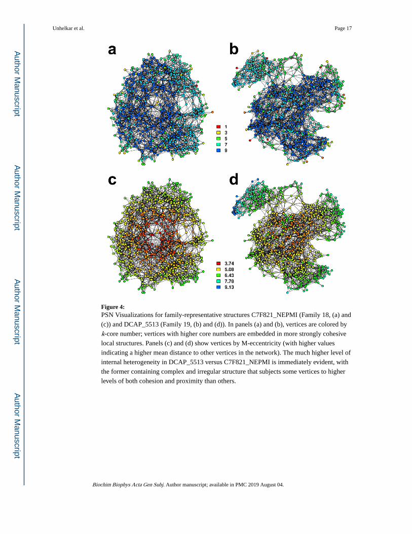
Figure 4: PSN Visualizations for family-representative structures C7F821_NEPMI (Family 18, (a) and
(c)) and DCAP_5513 (Family 19, (b) and (d)). In panels (a) and (b), vertices are colored by
k-core number; vertices with higher core numbers are embedded in more strongly cohesive
local structures. Panels (c) and (d) show vertices by M-eccentricity (with higher values
indicating a higher mean distance to other vertices in the network). The much higher level of
internal heterogeneity in DCAP_5513 versus C7F821_NEPMI is immediately evident, with
the former containing complex and irregular structure that subjects some vertices to higher
levels of both cohesion and proximity than others.
Unhelkar et al. Page 17
Biochim Biophys Acta Gen Subj. Author manuscript; available in PMC 2019 August 04.
Author M
anuscriptA
uthor Manuscript
Author M
anuscriptA
uthor Manuscript