membrane potential-dependent modulation of recurrent
TRANSCRIPT

Membrane Potential-Dependent Modulation ofRecurrent Inhibition in Rat NeocortexJie Zhu, Man Jiang, Mingpo Yang, Han Hou, Yousheng Shu*
Institute of Neuroscience, State Key Laboratory of Neuroscience, Shanghai Institutes for Biological Sciences, Chinese Academy of Sciences, Shanghai, P. R. China
Abstract
Dynamic balance of excitation and inhibition is crucial for network stability and cortical processing, but it is unclear how thisbalance is achieved at different membrane potentials (Vm) of cortical neurons, as found during persistent activity or slow Vm
oscillation. Here we report that a Vm-dependent modulation of recurrent inhibition between pyramidal cells (PCs)contributes to the excitation-inhibition balance. Whole-cell recording from paired layer-5 PCs in rat somatosensory corticalslices revealed that both the slow and the fast disynaptic IPSPs, presumably mediated by low-threshold spiking and fastspiking interneurons, respectively, were modulated by changes in presynaptic Vm. Somatic depolarization (.5 mV) of thepresynaptic PC substantially increased the amplitude and shortened the onset latency of the slow disynaptic IPSPs inneighboring PCs, leading to a narrowed time window for EPSP integration. A similar increase in the amplitude of the fastdisynaptic IPSPs in response to presynaptic depolarization was also observed. Further paired recording from PCs andinterneurons revealed that PC depolarization increases EPSP amplitude and thus elevates interneuronal firing and inhibitionof neighboring PCs, a reflection of the analog mode of excitatory synaptic transmission between PCs and interneurons.Together, these results revealed an immediate Vm-dependent modulation of cortical inhibition, a key strategy throughwhich the cortex dynamically maintains the balance of excitation and inhibition at different states of cortical activity.
Citation: Zhu J, Jiang M, Yang M, Hou H, Shu Y (2011) Membrane Potential-Dependent Modulation of Recurrent Inhibition in Rat Neocortex. PLoS Biol 9(3):e1001032. doi:10.1371/journal.pbio.1001032
Academic Editor: Alberto Bacci, European Brain Research Institute, Italy
Received October 2, 2010; Accepted February 9, 2011; Published March 22, 2011
Copyright: � 2011 Zhu et al. This is an open-access article distributed under the terms of the Creative Commons Attribution License, which permits unrestricteduse, distribution, and reproduction in any medium, provided the original author and source are credited.
Funding: This work was supported by the 973 Program (2011CBA00400 and 2006CB806600), the National Natural Science Foundation of China Project(31025012), the Shanghai Pujiang Program 07PJ14108, the Hundreds of Talents Program and the Knowledge Innovation Project from Chinese Academy ofSciences (KSCX2-YW-R-102). The funders had no role in study design, data collection and analysis, decision to publish, or preparation of the manuscript.
Competing Interests: The authors have declared that no competing interests exist.
Abbreviations: AP, action potential; EPSP, excitatory postsynaptic potential; FS, fast spiking interneuron; LTS, low-threshold spiking interneuron; PC, pyramidalcell; RMP, resting membrane potential; Vm, membrane potential
* E-mail: [email protected]
Introduction
The excitatory and inhibitory inputs received by cortical
neurons are normally under dynamic balance during cortical
functions [1–7]. The interaction among these inputs, together with
intrinsic membrane properties of cortical neurons, often results in
shifts of membrane potential (Vm) between different states [8–11],
which could regulate neuronal responsiveness to synaptic and
sensory inputs [1,12–17]. However, it is unclear how the balance
of excitation and inhibition is achieved when cortical neurons are
at different Vm levels. During global membrane oscillations
involving a large number of cortical neurons, excitation and
inhibition may be proportionally altered by the Vm shift, but the
underlying mechanisms remain unknown, in view of diversity of
connectivity and functions of local inhibitory interneurons [18,19].
In the case of persistent activities associated with some
behaviorally relevant conditions, e.g., during working memory, a
subpopulation of neurons undergoes changes in the Vm [20–24].
Microcircuits involving these active neurons also require dynamic
control of their excitation-inhibition balance. In this study, we
investigated how Vm changes of a cortical neuron may modulate
the efficacy of recurrent inhibition within the microcircuit.
It has been shown recently that cortical excitatory neurons
communicate not only through the generation of all-or-none
action potentials (APs, digital mode) but also through a presynaptic
Vm-dependent modulation of transmitter release (analog mode)
[25,26]. It remains unknown to what extent the analog-mode
communication influences the operation of local circuitry and has
a functional role in the cortex. Considering that interneurons
within the microcircuit are driven by excitatory neurons, leading
to recurrent inhibition, we hypothesized that the amount of
recurrent inhibition might be subjected to modulation in a manner
that depends on the level of depolarization of the excitatory
neuron. The Vm changes in the presynaptic excitatory neuron may
modulate the size of excitatory postsynaptic potentials (EPSPs) in
interneurons, leading to changes in their AP firing, which in turn
alter the efficacy of their inhibition on neighboring neurons.
Cortical inhibitory interneurons show a huge diversity in their
biochemical and physiological properties [18,19]. Two distinct
subtypes of interneurons, low-threshold spiking (LTS) neuron and
fast spiking (FS) neuron, mediate the slow and the fast recurrent
inhibition, respectively [27–29]. The LTS neuron receives EPSPs
that show facilitation in response to a train of high-frequency
stimuli of the presynaptic PC [30–32], generating APs with a long
onset latency and evoking late-onset (slow) disynaptic IPSPs in its
neighboring PCs [27,29,33]. The FS neuron (and some other
inhibitory interneurons) receives EPSPs that show depression
during high-frequency presynaptic stimulation [31,34]. However,
the high release probability of PC-FS synapses often allows
discharges of the FS neuron in response to single APs in the PC,
PLoS Biology | www.plosbiology.org 1 March 2011 | Volume 9 | Issue 3 | e1001032

leading to time-locked early onset (fast) IPSPs in neighboring PCs
[29,35]. In this study, we sought to examine whether the slow and
the fast recurrent inhibition meditated by these two distinct
microcircuits (PC-LTS-PC and PC-FS-PC) were subjected to
modulation in response to Vm changes of the presynaptic PC.
We performed paired whole-cell recording (PC-PC, PC-LTS,
and PC-FS) in rat somatosensory cortical slices and found that
both the slow and the fast recurrent inhibition were indeed
modulated by the presynaptic somatic Vm changes, and this
modulation resulted from analog-mode signaling in excitatory
synapses between PCs and interneurons. These results show an
important role of analog communication in controlling the
operation of cortical microcircuits and provide new insights into
the cellular mechanisms underlying dynamic balance of cortical
excitation and inhibition.
Results
Vm-Dependent Modulation of Slow Recurrent InhibitionWe performed paired whole-cell recording from nearby layer-5
pyramidal cells (PCs, ,100 mm apart) in acutely isolated rat
somatosensory cortical slices. In response to stimulation of the PC
with a burst of APs (70,200 Hz), disynaptic IPSPs were observed
in 19% of the pairs successfully tested (207/1,087 pairs), with 2%
(23/1,087) exhibiting reciprocal IPSPs (see Methods, Figure 1A).
Consistent with previous studies [27,29], these IPSPs had a peak
amplitude of 1.360.1 mV (s.e.m., n = 38 PC-PC pairs) and a long
but rather precise onset latency (11164 ms) following PC
stimulation (15 APs at 100 Hz). The IPSPs were detected only
when the presynaptic PC fired at a frequency higher than 50 Hz.
Bath application of either CNQX (10 mM) or picrotoxin (50 mM)
completely abolished these IPSPs (n = 8/8 PC pairs), consistent
with the involvement of both excitatory and inhibitory transmis-
sion in these disynaptic responses. In our PC-PC paired
recordings, a single AP or a burst of APs in one PC could evoke
monosynaptic EPSPs but never triggered firing in postsynaptic
PCs, suggesting no polysynaptic events involved in generating the
disynaptic IPSPs.
To examine the Vm-dependence of this slow (late-onset)
recurrent inhibition, we manipulated Vm by injecting repetitive
step-depolarizing currents (duration ,45 s, at 90 s intervals) to
induce subthreshold depolarization in the presynaptic PC. The
magnitudes of disynaptic IPSPs were measured by applying trains
of current pulses (duration: 1 ms) to evoke AP bursts (15 APs at
100 Hz) in the presynaptic PC at the resting and depolarized Vm
(Figure 1B and 1C). In the majority of PC-PC pairs tested with this
protocol (n = 38 pairs), we found that step presynaptic depolariza-
tion from a resting Vm (264.660.6 mV) to a level near the firing
threshold (247.260.6 mV) significantly increased the average
amplitude (n = 25/38 PC pairs tested, p,0.05) and integrated
voltage area (mV6s, n = 28/38, p,0.05) of the disynaptic IPSPs
and decreased the average onset latency and jitter (n = 23/38,
p,0.05). Cumulative frequency distribution of the average
amplitude (or total voltage area, unpublished data) showed a
highly significant difference between the two Vm levels (n = 38
pairs, Figure 1D, p,0.01, Kolmogorov-Smirnov test). In these
experiments, we noted that the extent of modulation of these
IPSPs varied greatly from pair to pair. Comparison of the IPSP
amplitude (or total voltage area) and onset latency at two different
Vm levels of the same pairs also showed highly significant
differences (Figure 1D and 1E, p,0.01, t test). Interestingly, the
percentage increase in the peak amplitude (or total voltage area) of
IPSPs found at the depolarized Vm levels decreased with increasing
average peak amplitude. As shown in Figure 1F, the data could be
well fitted by a hyperbolic function (y = 100%+0.67/x), which
predicts a linear relationship between the IPSP amplitudes at
depolarized Vm and those at resting Vm (y = x+0.53, with the slope
fixed at 1, see Figure S1 and Discussion). In 12 PC-PC pairs, we
varied the time difference between the depolarization onset and
the first AP burst during the depolarization in order to examine
the time course of the facilitation and found that the IPSP
amplitudes progressively increase after depolarization with a time
course t of 3.5 s (single exponential fit, Figure 1G), consistent with
the slow component of the EPSP facilitation induced by
presynaptic depolarization found in monosynaptically connected
PC pairs [26].
Further analysis revealed that the disynaptic IPSP facilitation
could be attributed in part to a decrease in its failure rate. In 33/
35 PC pairs tested, IPSP failures occurred at both depolarized (by
18.060.8 mV) and resting presynaptic Vm, but the average failure
rate was lower under depolarized Vm (0.0860.02) than that at
resting Vm (0.2560.03). Among all experiments (n = 123 PC pairs),
nine showed complete failure of disynaptic IPSPs at resting Vm but
detectable IPSPs at depolarized Vm (Figure 2A–C), suggesting that
such modulation could not only change but also turn on recurrent
inhibition. This abrupt appearance of disynaptic IPSPs and the
shortened onset latency associated with presynaptic depolarization
may narrow the time window of the integration of EPSPs. Indeed,
in PC connections that had both monosynaptic EPSPs and
disynaptic IPSPs, the EPSP summation time was shorter at
depolarized Vm in comparison with that at resting Vm (13669 ms
versus 175613 ms, p,0.01, n = 11, Figure 2D–G).
We next examined a range of presynaptic Vm in PC-PC pairs
that showed IPSP facilitation to determine the threshold
depolarization for inducing facilitation and the Vm-dependence
of facilitation (Figure 3A–B). None of the nine connections tested
showed facilitation when the presynaptic PC was depolarized by
only 3–5 mV. However, 5–10 mV depolarization resulted in IPSP
facilitation in 24% (n = 4/17) of the connections tested. The
percentage of pairs exhibiting facilitation increased to 100% for
Author Summary
Proper functioning of the neocortex requires a balancebetween excitation and inhibition. This balance can beachieved through the operation of cortical microcircuitsinterweaved by excitatory and inhibitory neurons. Sincethe membrane potentials (Vm) of cortical neurons fluctuateat different levels during cortical activities, it is importantto know how the balance of excitation and inhibition isdynamically maintained at different Vm. Recurrent inhibi-tion between excitatory pyramidal cells is mediated by twodistinct types of inhibitory interneurons. Here, we showthat the amount of recurrent inhibition depends on the Vm
levels of presynaptic pyramidal cells. Modest depolariza-tion of a pyramidal cell substantially increases, andsometimes turns on, disynaptic inhibition on its neighbor-ing pyramidal cells. We find that this effect is due to anincrease in the strength of synaptic connections from thepyramidal cell to inhibitory interneurons and a consequentelevation of interneuronal firing. The depolarization-induced increase in synaptic strength from the pyramidalcell therefore reflects ‘‘analog-mode’’ signaling in corticalexcitatory synapses. We thus reveal a profound impact ofanalog-mode signaling on the operation of corticalmicrocircuits and provide a new mechanism for dynamiccontrol of the balance of cortical excitation andinhibition.
Vm-Dependent Modulation of Cortical Inhibition
PLoS Biology | www.plosbiology.org 2 March 2011 | Volume 9 | Issue 3 | e1001032

depolarization more than 20 mV (n = 11/11, Figure 3C). The
IPSP amplitudes also increased and their onset latencies decreased
with increasing depolarization of the PC, as compared to those
observed at the resting Vm. As shown in Figure 3D–E, for pairs
that exhibited disynaptic facilitation (n = 20), the average IPSP
amplitude (including failures) increased progressively with presyn-
aptic depolarization (r = 0.95, 5.7% per mV), whereas the onset
latency decreased accordingly (r = 20.80, 0.8% per mV). Again,
the increase in IPSP amplitude was partially due to the decrease of
failure rate (Figure 3F).
Together, these results show that a relatively small Vm shift of 5–
10 mV of the presynaptic PC can alter the amplitude and the
onset latency of disynaptic IPSPs received by its neighboring
neurons, indicating a robust Vm-dependent modulation of slow
recurrent inhibition.
Role of LTS InterneuronsWe next investigated the mechanisms underlying this Vm-
dependent modulation. The late-onset disynaptic IPSP between
excitatory PCs is known to be mediated by LTS interneurons
[27,29]. In PC-LTS paired recordings, a train of high-frequency
APs in the PC results in facilitating EPSPs and AP generation in
the LTS neuron, which in turn triggers IPSPs in the PC [27,29].
We therefore examined whether the Vm changes in the presynaptic
PC could modulate the magnitude of summated EPSPs and
discharge probability of its postsynaptic LTS neuron.
Figure 1. Modulation of slow (late-onset) disynaptic IPSP by presynaptic somatic Vm. (A) Left, schematic diagram of the PC-PC pairedrecording (IN indicates those unidentified inhibitory interneurons that mediate the disynaptic IPSPs). Right, an AP burst (15 APs at 100 Hz) evoked bya train of current injection in PC1 induced a disynaptic response in PC2 with a long latency from the onset of the AP burst. * indicates individual IPSPs.(B) Example recording showing that presynaptic depolarization increased the amplitude of AP burst-induced disynaptic IPSPs. (C) Overlay of the IPSPsevoked at resting (blue) and depolarized (red) Vm in the presynaptic PC. Arrows indicate the onset of the AP train. Notice that presynapticdepolarization caused a reduction in failure, increased the amplitude, and shortened the latency of the disynaptic IPSPs. (D) Left, cumulativefrequency distribution of the tested connections (n = 38 PC-PC pairs) by the average amplitude of disynaptic IPSP at resting (blue) and depolarized Vm
(red); right, pooled results showing changes of the average amplitude at the two Vm levels in individual PC-PC pairs. (E) Pooled results (n = 38 pairs)showing that the onset latency of IPSPs was shortened by presynaptic depolarization. (F) The percentage increase was dependent on the averageamplitude of disynaptic IPSPs (n = 38 pairs). Red line, hyperbolic fit. (G) Average time course of the facilitation in PC-PC pairs that showed significantincrease in IPSP amplitude (n = 12 pairs tested). Error bars represent s.e.m. ** p,0.01.doi:10.1371/journal.pbio.1001032.g001
Vm-Dependent Modulation of Cortical Inhibition
PLoS Biology | www.plosbiology.org 3 March 2011 | Volume 9 | Issue 3 | e1001032

In PC-LTS paired recordings (Figure 4A, see also Figure S2A–
D), a burst of APs (15 APs at 100 Hz) initiated in the presynaptic
PC caused significant synaptic facilitation that triggered spiking of
the LTS cell (Figure 4B–C). Consistent with previous studies
[27,29], the spiking probability and the onset of LTS spiking
depended on the number and the frequency of presynaptic APs
(unpublished data). To reveal the effect of presynaptic Vm on the
summated EPSPs, we hyperpolarized the LTS cell to prevent its
spiking. Steady depolarization of the presynaptic PC from the
resting Vm to a level near the firing threshold significantly
increased the peak amplitude of the summated EPSPs (from
4.460.7 to 5.260.9 mV, n = 18, p,0.01) and the total integrated
area associated with the EPSPs (from 0.660.1 to 0.860.2 mV6s,
p,0.01). Close examination of the individual EPSPs revealed that
the failure rate of the 2nd to 5th EPSPs was significantly lower
when presynaptic PC was depolarized, as compared to that found
at the resting Vm (p,0.05, Figure 4D). Peak amplitudes (measured
from the baseline before the train) of individual EPSPs during the
train were also significantly increased (Figure 4E, Figure S3), as
reflected by the increased slope in the plot of normalized EPSP
amplitude versus the AP number (from 0.12 to 0.15 per AP, n = 18
PC-LTS pairs).
Next, we compared the spiking probability of the postsynaptic
LTS cell before and after the presynaptic Vm shift from resting to
depolarized levels. Summated EPSPs evoked by trains of
presynaptic APs (15 APs at 100 Hz) triggered AP generation in
some of the LTS neurons recorded at resting Vm (n = 6/22 PC-
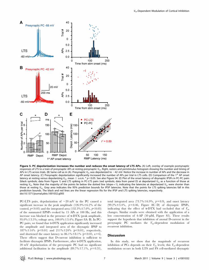
LTS pairs; Figure 4B–C and Figure 5). In these six pairs that
exhibited LTS spiking, the shift of Vm in the presynaptic PC from
resting to a level near the firing threshold resulted in an increase in
numbers of APs per trial in LTS cells (from 2.061.0 to 2.561.1,
p,0.05; Figure 5A–C) and a decrease in the onset latency (from
113.3616.4 to 99.7613.7 ms, p,0.05; Figure 5A–B and D) and
jitter of spiking (from 25.263.7 to 21.262.9 ms, p = 0.055).
Figure 2. Presynaptic depolarization turns on recurrent inhibition and shortens the integration time of EPSPs. (A) Example PC-PCpaired recording showing that the disynaptic IPSP only occurred at a depolarized Vm. (B and C) Parts in (A) were expanded for clarity. Insets, overlay ofthe somatic APs during the train indicating that no AP failure occurred. (D) Schematic drawing of the recordings (for E–G) from PC-PC pair that hadboth the monosynaptic excitatory connections and the disynaptic inhibitory connections. (E) An example showing that disynaptic IPSP (average of 33trials), which occurred only when the presynaptic Vm was depolarized, shortened the EPSP summation time (arrowheads). The arrow indicates afacilitated EPSP. (F) Similar example (average of 15 trials) as shown in (E) except that disynaptic IPSP occurred at both depolarized and restingpresynaptic Vm. Note the difference in the time window of EPSP summation (arrowheads). (G) Group data (n = 9 PC-PC pairs) indicated thatpresynaptic depolarization shortened the time window for EPSP integration. ** p,0.01.doi:10.1371/journal.pbio.1001032.g002
Vm-Dependent Modulation of Cortical Inhibition
PLoS Biology | www.plosbiology.org 4 March 2011 | Volume 9 | Issue 3 | e1001032

Similar results could be obtained even when presynaptic PC was
stimulated at low intensities (10 APs at 20 Hz, Figure S4). Plot of
the onset latency of LTS spiking and disynaptic IPSPs (shown in
Figure 1E) at depolarized Vm as a function of that at resting Vm
revealed a close correlation between them (Figure 5E), indicating
that the decrease in IPSP latency resulted from the early spiking of
LTS cells. Taken together, these results correlate well with the
findings on the Vm-dependent modulation of slow disynaptic IPSPs
described above (see Figure 1) and indicate that PC depolarization
may recruit more LTS cells and/or trigger more firing in these
cells, thus causing more inhibition in their neighboring PCs.
In these experiments, we also found that single APs in LTS cells
evoked IPSPs in PCs with a failure rate of 0.1960.04 and an
average amplitude of 20.4360.14 mV (n = 19 LTS-PC pairs) at a
holding potential of approximately 250 mV. Consistent with their
distal input location at the apical dendrite, these IPSPs had a
reversal potential of 280.261.9 mV (n = 6 pairs). The rise time
and the decay time constant (t) were 7.2560.82 and 58.668.3 ms,
respectively (n = 16 pairs). These basic kinetics were different from
those of FS-PC IPSPs (rise, 4.7660.64 ms; decay, 85.2615.5 ms;
n = 28 pairs), which had a reversal potential of 270.861.4 mV
(n = 4 pairs). In 13/52 LTS-PC pairs tested, we observed
reciprocal connections (Figure 4A), suggesting that the Vm-
dependent modulation of disynaptic IPSPs could directly influence
the feedback inhibition of the presynaptic PC, in addition to the
inhibition of other downstream PCs.
Vm-Dependent Modulation of Fast Recurrent InhibitionTo investigate whether the fast disynaptic inhibition mediated
by interneurons that receive depressing excitatory inputs is also
Vm-dependent, we analyzed the PC-PC pairs that exhibited fast
(early onset) IPSPs in response to a single presynaptic AP.
Consistent with previous reports [27–29], we found that the
probability of success in detecting fast disynaptic IPSPs was very
low. Only 7/1,103 PC-PC pairs tested bi-directionally exhibited
fast disynaptic IPSPs, and three out of these seven pairs showed
Vm-dependent modulation. Presynaptic depolarization of ,18 mV
from the resting Vm (263 mV) substantially reduced the failure
rate of evoking IPSPs in these three pairs (from 0.78 to 0.67, 0.85
to 0.78, and 0.63 to 0.36, respectively; Figure 6A–B). This reduced
failure rate is consistent with our hypothesis that the depolarized
Vm in the PC elevated the EPSP amplitude and increased the
spiking probability of the FS cell. When the average amplitude of
IPSPs (failure excluded) evoked by single APs was measured, we
found that it was unchanged in the first two pairs (from 1.4260.05
to 1.4460.04 mV, p = 0.4; from 2.060.1 to 2.160.1 mV, p = 0.2).
This supports the notion that disynaptic modulation was mainly
due to changes in the firing probability of the interneurons.
However, we found surprisingly that the average IPSP amplitude
observed in the third pair was significantly increased from
0.6260.02 to 0.7560.02 mV (p,0.01) even with failure excluded.
Given the low probability of observing fast disynaptic IPSPs
(n = 7/1,103 PC-PC pairs), the recruitment of two interneurons in
this case is unlikely. However, it is possible that triggering of two
APs (instead of one) in the interneuron during presynaptic
depolarization could account for the remaining increase in IPSP
amplitude after failure exclusion. The precise mechanism for these
observations remains to be further examined.
The interneurons that mediate these fast disynaptic IPSPs are
most likely FS neurons, which receive depressing EPSPs in
response to high-frequency presynaptic stimulation [31,34]. We
therefore recorded PC-FS pairs to examine whether EPSPs are
Figure 3. Vm-dependence of disynaptic IPSP facilitation. (A) Example PC-PC paired recording showing that the amplitude of disynaptic IPSPwas closely associated with presynaptic Vm levels. (B) Overlay of the averaged IPSPs at different presynaptic Vm levels. Notice the changes of the IPSPamplitude and onset latency. Same PC-PC pair as in (A). (C) Bar plot of percentage of pairs that showed facilitation of disynaptic IPSPs at differentlevels of presynaptic depolarization. Notice that depolarization progressively increased the percentage of pairs that exhibited facilitation. (D) Groupdata showing that the IPSP amplitude correlated closely with the level of depolarization. (E) Onset latency shortened with depolarizing Vm. (F) Failurerate of the disynaptic IPSPs decreased with depolarizing Vm. Red lines indicate linear fits of the data. Data were shown as mean 6 s.e.m.doi:10.1371/journal.pbio.1001032.g003
Vm-Dependent Modulation of Cortical Inhibition
PLoS Biology | www.plosbiology.org 5 March 2011 | Volume 9 | Issue 3 | e1001032

indeed subjected to modulation by the Vm levels of PCs. Consistent
with previous findings [25,26], presynaptic depolarization of 15–
20 mV significantly increased the average amplitude of the single
AP-triggered EPSPs (single AP at 1 Hz) in about one-third of the
pairs tested (n = 12/35 PC-FS pairs, Figure 6C–D). These elevated
EPSPs may increase the spiking probability of FS cells, thus
reducing the failure rate in evoking IPSPs in their postsynaptic
PCs. Together, these results show that fast recurrent inhibition is
also Vm-dependent, resulting from the Vm-dependent analog
signaling in excitatory synapses between PCs and interneurons.
Vm-Dependent Modulation of Monosynaptic IPSPSince cortical states affect both PCs and inhibitory interneurons
[8,11], we next examined whether this Vm-dependent analog
signaling occurs at inhibitory synapses. In the FS-PC and LTS-PC
pairs that showed monosynaptic inhibitory connections, we
depolarized the presynaptic FS or LTS cells from resting Vm
(,270 mV) to a level near the firing threshold and found that
monosynaptic IPSPs evoked by single APs (using similar protocol
as that shown in Figure 6A, also see [26]) showed no significant
change in basic kinetics of IPSPs (FS-PC: rise time from
4.7660.64 to 4.1660.50 ms and decay t from 85.2615.5 to
62.063.6 ms, n = 28 pairs; LTS-PC: rise time from 7.2560.82 to
7.0460.72 ms and decay t from 58.668.3 to 60.967.4 ms, n = 16
pairs; p.0.05); however, the average amplitude of IPSPs was
significantly enhanced in a small subpopulation of tested pairs.
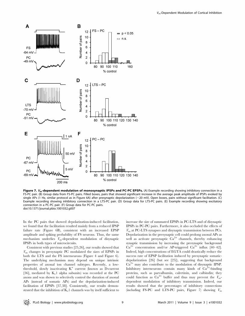
The percentages of FS-PC and LTS-PC pairs that showed IPSP
facilitation in response to presynaptic depolarization were 17.2%
(n = 5/29) and 10.5% (2/19), respectively, which were smaller than
that for PC-PC pairs (37.0%, n = 10/27) exhibiting EPSP
facilitation (Figure 7). The lower probability of finding Vm
modulation in LTS-PC pairs than in FS-PC pairs may result
from the differences in the location of the inhibitory synapses. The
LTS cells send their axons to superficial layers and form synapses
onto the distal apical dendrites of PCs, while FS cells mainly target
the perisomatic region of PCs. Thus, Vm changes at the soma may
decay more substantially when arriving at axon terminals in LTS
cells than FS cells to influence synaptic transmission. Taken
together, these results demonstrate that monosynaptic IPSPs are
also subjected to modulation by Vm changes in presynaptic
interneurons in a small subpopulation of inhibitory connections.
Role of Axonal Kv1 ChannelsThe above experiments showed that both fast and slow
recurrent inhibition were subjected to modulation by Vm changes
in PCs, resulting from the Vm-dependent analog-mode signaling in
PC-interneuron excitatory synapses. A rapidly activating but
slowly inactivating axonal K+ current, known as D-current [36],
has been shown to regulate the axonal AP duration and potentially
contribute to the Vm-dependent modulation of the EPSP
amplitude [37,38]. We thus further investigated the role of axonal
D-currents in Vm-dependent modulation of recurrent inhibition in
cortical microcircuits.
Consistent with previous findings, bath application of a-
dendrotoxin (a-DTX, 100 nM) blocked the PC depolarization-
induced facilitation of the summated EPSPs in LTS cells. In six
Figure 4. Vm-dependent modulation of the slow recurrent inhibition is mediated by LTS interneurons. (A) Left, schematic diagram ofthe PC-LTS paired recording. Right, the characteristic firing pattern of the LTS was shown at the bottom (in response to a square pulse of 0.2 nA);single AP (middle) in the LTS could evoke an IPSP (top) in the PC. (B) Example recording from the PC-LTS pair shown in (A). Presynaptic depolarization(,20 mV) increased the peak amplitude of the summated EPSPs (evoked by a burst of 15 APs at 100 Hz in the PC) and occasionally caused AP firingin the LTS. (C) Overlay of the summated EPSPs recorded at the LTS. Note the facilitated EPSPs. (D) The failure rate of the 2nd to 5th EPSPs decreasedafter presynaptic depolarization. Inset, example traces showing EPSP failures occurred at resting (blue) and depolarized Vm (red); arrow indicates theonset of the presynaptic AP train. (E) Group data (mean 6 s.e.m., including EPSP failures) indicating the facilitation of individual EPSPs afterpresynaptic depolarization. Peak amplitudes of individual EPSPs were normalized to the 6th EPSP at resting presynaptic Vm. See also Figure S3.doi:10.1371/journal.pbio.1001032.g004
Vm-Dependent Modulation of Cortical Inhibition
PLoS Biology | www.plosbiology.org 6 March 2011 | Volume 9 | Issue 3 | e1001032
PC-LTS pairs, depolarization of ,20 mV in the PC caused a
significant increase in the peak amplitude (126.9%66.2% of the
control, p,0.05) and the integrated area (132.3%67.0%, p,0.05)
of the summated EPSPs evoked by 15 APs at 100 Hz, and this
increase was blocked in the presence of a-DTX (peak amplitude,
95.0%63.5%; voltage area, 100.0%63.4%; Figure 8A–B). In PC-
PC pairs, we found that a-DTX application significantly increased
the amplitude and integrated area of the disynaptic IPSP to
187%614% (p,0.01) and 211%624% (p,0.01), respectively,
and shortened the onset latency to 86.1%68.1% (p,0.05, n = 6).
These effects suggest that D-current inhibition is sufficient to
facilitate disynaptic IPSPs. Furthermore, after a-DTX application,
20 mV depolarization of the presynaptic PC had no significant
additional facilitation in the amplitude (89.7%67.1%, p = 0.31),
total integrated area (73.7%66.9%, p = 0.9), and onset latency
(99.2%63.4%, p = 0.46, Figure 8C–D) of disynaptic IPSPs,
indicating that the effect of a-DTX had occluded that of Vm
changes. Similar results were obtained with the application of a
low concentration of 4-AP (50 mM, Figure S5). These results
support the hypothesis that inhibition of axonal D-current in the
presynaptic PC mediates the Vm-dependent modulation of
recurrent inhibition.
Discussion
In this study, we show that the magnitude of recurrent
inhibition of PCs depends on their Vm levels; this Vm-dependent
modulation occurs in both LTS and FS cell-mediated disynaptic
Figure 5. PC depolarization increases the number and reduces the onset latency of LTS APs. (A) Left, overlay of example postsynapticresponses of LTS to a train of presynaptic APs at resting presynaptic Vm. Right, rasters and peristimulus histogram showing the number and timing ofAPs in LTS across trials. (B) Same cell as in (A). Presynaptic Vm was depolarized to 242 mV. Notice the increase in number of APs and the decrease inAP onset latency. (C) Presynaptic depolarization significantly increased the number of APs per trial in LTS cells. (D) Comparison of the 1st AP onsetlatency at resting versus depolarizing Vm. mean 6 s.e.m. * p,0.05. See also Figure S4. (E) Plot of the onset latency of disynaptic IPSPs in PC-PC pairs(black symbols; data from Figure 1) and LTS spiking in PC-LTS pairs (red symbols; data from panel D) at depolarized Vm as a function of those atresting Vm. Note that the majority of the points lie below the dotted line (slope = 1), indicating the latencies at depolarized Vm were shorter thanthose at resting Vm. Gray area indicates the 95% prediction bounds for IPSP latencies. Note that the points for LTS spiking latencies fall in thisprediction bounds. The black and red lines are the linear regression fits for the IPSP and LTS spiking latencies, respectively.doi:10.1371/journal.pbio.1001032.g005
Vm-Dependent Modulation of Cortical Inhibition
PLoS Biology | www.plosbiology.org 7 March 2011 | Volume 9 | Issue 3 | e1001032

recurrent connections. We provide the first evidence showing that
presynaptic Vm-dependent analog communication occurs at
excitatory synapses between PCs and interneurons and mediates
the Vm-dependent modulation of recurrent inhibition. For slow
disynaptic IPSPs, depolarization in presynaptic PCs increases the
amplitude of AP burst-induced summated responses in postsyn-
aptic LTS neurons, thus increasing the number and decreasing the
latency of their AP discharges, which in turn enhance the
amplitude and reduce the onset latency and jitter of slow IPSPs
in neighboring PCs. Similarly, modest depolarization in PCs
enhanced single AP-induced fast disynaptic IPSPs in neighboring
PCs presumably due to a Vm-dependent increase in the size of
EPSPs in FS neurons. Together, these results reveal that PC Vm-
dependent modulation of cortical inhibition is a key strategy
through which the cortex efficiently and dynamically maintains
the excitation-inhibition balance, a condition critical for cortical
information processing.
Mechanisms for Vm-Dependent Modulation of InhibitionRecent studies have discovered that the PC-LTS-PC microcir-
cuit mediates the slow (late-onset) recurrent inhibition in
somatosensory and other cortices [27,29,39]. The somatostatin-
positive LTS interneuron is the key player in this microcircuit. It
receives EPSPs that show facilitation in response to a train of
presynaptic stimuli that may initiate firing of APs, which in turn
evoke IPSPs in its postsynaptic PCs [27,29,33]. Generation of
these disynaptic IPSPs depends on the number and frequency of
APs in PCs. Our results show that the presynaptic Vm is also a
powerful determinant for controlling the strength and timing of
disynaptic IPSPs—a few mV depolarization (.5 mV) can cause
substantial IPSP facilitation (Figure 3). More importantly, stronger
depolarization could not only modulate the amplitude of existing
disynaptic IPSPs but also turn on silent recurrent connections
(Figure 2). Further analysis showed a close relationship between
the magnitude of IPSP facilitation and the extent of presynaptic
depolarization in PCs, consistent with the requirement of
excitation-inhibition balance during elevated network activity
[1,3]. The abrupt occurrence and the facilitation of disynaptic
IPSPs may result from the increases in the spiking probability or
the number of APs in LTS interneurons (Figures 4 and 5). Since
the inhibitory synaptic strength at synapses between the newly
recruited LTS interneurons and the postsynaptic PC should not
depend on the amplitude of existing IPSPs, we expect that the net
increase does not depend on the baseline IPSP amplitude. This
was supported by the finding that the data shown in Figure 1F
were well fitted by a hyperbolic function, which predicts a linear
function between IPSP amplitudes at depolarized Vm and those at
resting Vm (see Figure S1).
The PC-FS-PC microcircuit is a potential candidate mediating
the fast (early-onset) recurrent inhibition [27,29]. In contrast to
LTS cells, FS neurons receive EPSPs that show depression in
response to presynaptic high-frequency APs [31,34]. A single
presynaptic AP can trigger the FS neuron to discharge once and
subsequently evoke a unitary IPSP at neighboring PCs [28,29].
Consistent with the findings reported previously [27,29], we
observed fast disynaptic IPSPs in PC pairs at a very low frequency.
Figure 6. Vm-dependent modulation of fast (early-onset) disynaptic IPSP. (A) Schematic diagram showing the PC-PC paired recording andthe protocol of stimulation. Brief injection (1 ms) of depolarizing current pulses to the presynaptic PC evoked single APs at a rate of 1 Hz, whileperiodic constant current injection caused ,20 mV depolarization from the resting Vm. The resting and depolarized periods were ,15 s each. Thedotted lines indicate a break in the time axis. (B) PC-PC paired recordings tested with the protocol shown in (A). Traces (average of at least 300 trials)from three pairs showing that the average disynaptic IPSP at depolarized Vm (red) were larger than that at resting Vm (blue) when IPSP failures wereincluded. (C) PC-FS paired recording. Left: recording configuration. Right: bottom trace showing the non-adapting fast-spiking pattern of therecorded FS neuron in response to a current pulse (0.4 nA); the middle trace indicates the depressing EPSPs in FS neuron in response to a train of APsin the PC (0.3 nA, top trace). (D) Group data from PC-FS pairs using similar protocol as in (A). Significant EPSP facilitation was observed in 12 out of 35pairs. Inset: example traces (average of at least 300 trials) from two PC-FS pairs.doi:10.1371/journal.pbio.1001032.g006
Vm-Dependent Modulation of Cortical Inhibition
PLoS Biology | www.plosbiology.org 8 March 2011 | Volume 9 | Issue 3 | e1001032
In the PC pairs that showed depolarization-induced facilitation,
we found that the facilitation resulted mainly from a reduced IPSP
failure rate (Figure 6B), consistent with an increased EPSP
amplitude and spiking probability of FS neurons. Thus, the same
mechanism underlies Vm-dependent modulation of disynaptic
IPSPs in both types of microcircuits.
Consistent with previous studies [25,26], our results showed that
Vm changes in presynaptic PC modulated the sizes of EPSPs in
both the LTS and the FS interneurons (Figure 4 and Figure 6).
The underlying mechanism may depend on unique intrinsic
properties of axonal ion channel subtypes. Recently, a low-
threshold, slowly inactivating K+ current (known as D-current
[36], mediated by Kv1 alpha subunits) was recorded at the PC
axons and was shown to selectively control the duration of axonal
APs (instead of somatic APs) and the depolarization-induced
facilitation of EPSPs [37,38]. Consistently, our results demon-
strated that the inhibition of Kv1 channels was by itself sufficient to
increase the size of summated EPSPs in PC-LTS and of disynaptic
IPSPs in PC-PC pairs. Furthermore, it also occluded the effects of
Vm at PC-LTS synapses and disynaptic transmission between PCs.
Depolarization in the presynaptic cell could prolong axonal APs as
well as activate presynaptic Ca2+ channels, thereby enhancing
synaptic transmission by increasing the presynaptic background
Ca2+ concentration and/or AP-triggered Ca2+ influx [40–42].
Indeed, high concentrations of EGTA could drastically reduce the
success rate of EPSP facilitation induced by presynaptic somatic-
depolarization [26] (but see [25]), suggesting that background
Ca2+ may also contribute to the modulation of disynaptic IPSP.
Inhibitory interneurons contain many kinds of Ca2+-binding
proteins, such as parvalbumin, calretinin, and calbindin; they
could function as Ca2+ buffer and thus may prevent the Vm-
dependent modulation of inhibitory transmission. Indeed, our
results showed that the percentages of inhibitory connections
(including FS-PC and LTS-PC pairs, Figure 7) showing Vm
Figure 7. Vm-dependent modulation of monosynaptic IPSPs and PC-PC EPSPs. (A) Example recording showing inhibitory connection in aFS-PC pair. (B) Group data from FS-PC pairs. Filled boxes, pairs that showed significant increase in the average peak amplitude of IPSPs evoked bysingle APs (1 Hz, similar protocol as in Figure 6A) after presynaptic depolarization (,20 mV). Open boxes, pairs without significant facilitation. (C)Example recording showing inhibitory connection in a LTS-PC pair. (D) Group data for LTS-PC pairs. (E) Example recording showing excitatoryconnection in a PC-PC pair. (F) Group data for PC-PC pairs.doi:10.1371/journal.pbio.1001032.g007
Vm-Dependent Modulation of Cortical Inhibition
PLoS Biology | www.plosbiology.org 9 March 2011 | Volume 9 | Issue 3 | e1001032

modulation are far less than those of excitatory connections
(including PC-PC and PC-interneuron pairs, Figures 6 and 7).
Whether the intracellular Ca2+-binding proteins may be respon-
sible for regulating analog-mode signaling at inhibitory synapses
remains to be further examined.
Subthreshold Vm changes in the soma spread down the axon
with a length constant of 400–800 mm [25,26,37,43]. Over 150
putative synaptic boutons are distributed at axon collaterals within
500 mm of the cell body of layer-5 PCs [26]. Boutons in remote
axon terminals may not be affected by somatic Vm changes. This
may explain why not all recurrent inhibitory connections were
subjected to the Vm-dependent modulation and why the
percentages of LTS-PC pairs showing Vm modulation are smaller
than those of FS-PC pairs. In comparison with FS cells that target
the perisomatic region of their neighboring PCs, LTS cells in layer
5 send their axons to superficial layers and innervate distal apical
dendrites of nearby PCs. Therefore, Vm modulation should be
weaker in LTS-PC than in FS-PC synapses. Whether such
modulation is indeed spatially confined to local circuits within the
range of axonal spread of somatic depolarization, or alternatively
only specific cortical microcircuits are modulated, remains to be
further determined.
Physiological SignificanceThe Vm-dependent modulation of recurrent inhibition described
here may serve several distinct functions. First, it may contribute to
maintaining a dynamic excitation-inhibition balance at different
cortical activity states appropriate for diverse behavioral condi-
tions [8,10,11,44]. For example, when Vm depolarizes during an
active but relatively stable cortical state, e.g., the ‘‘Up’’ state, the
inhibitory conductances due to recurrent connections increase to
match the elevated excitatory conductances [1,3,4]. A recent work
revealed that excitation-inhibition balance is also instantaneously
controlled with a millisecond precision during spontaneous and
sensory-evoked activities [2] and disruption of this balance causes
dysfunction of the network [3,9], leading to various disorders such
as epileptic seizures [45,46] and schizophrenia [47]. Second, the
Vm-dependent modulation of recurrent inhibition may also
contribute to rapid transitions between ‘‘Up’’ and ‘‘Down’’ states
[1,3] that are important for gain modulation of synaptic and
sensory inputs [1,12,13,16,17,48,49]. For example, transient
excitation-inhibition imbalance caused by abrupt changes in Vm-
dependent recurrent inhibition, e.g., unsilencing of recurrent
connections (Figure 2A–C) induced by Vm fluctuation at the
depolarized Vm levels, could cause a switch from ‘‘Up’’ to ‘‘Down’’
state. Third, the shortening of IPSP latency associated with Vm-
dependent facilitation of recurrent inhibition (Figure 2D–G) may
regulate the time window of integration of excitatory inputs,
therefore providing a mechanism for Vm-dependent feedback
control of the timing of spike initiation in PCs [50–52].
To conclude, we have shown that both slow and fast recurrent
inhibition is susceptible to modulation by the Vm changes of PCs.
These results demonstrate a circuit function of Vm-dependent
modulation of excitatory transmission (analog-mode signaling).
Whether such Vm-dependent modulation is universal among all
cortical circuits and whether it plays an important function in
regulating circuit dynamics in behaviorally relevant conditions
remain to be examined.
Figure 8. Role of Kv1 channels in the Vm-dependent modulation of disynaptic IPSPs. (A) Left, schematic diagram of the PC-LTS pairrecording. Right, an example recording showing that bath application of a-DTX (100 nM) increased the size of disynaptic IPSPs and occluded thefacilitation induced by PC depolarization. Blue and red traces are the averaged EPSPs evoked by trains of APs (same protocol as in Figure 4) at restingand depolarized Vm, respectively. Note the depolarization-induced facilitation of the summated EPSPs before a-DTX application. (B) Group data (n = 6)showing that a-DTX diminished the depolarization-induced increases in the peak amplitude and the integral of the summated EPSPs. (C) Similarrecording as shown in Figure 1. Note that a-DTX not only mimicked the depolarization-induced facilitation but also occluded the effect of Vm
changes. (D) Group data (n = 6) showing that a-DTX blocked the Vm shift-induced changes in the peak amplitude and the integral of disynaptic IPSPs.** p,0.01. See also Figure S5.doi:10.1371/journal.pbio.1001032.g008
Vm-Dependent Modulation of Cortical Inhibition
PLoS Biology | www.plosbiology.org 10 March 2011 | Volume 9 | Issue 3 | e1001032

Materials and Methods
Ethics StatementThe use and care of animals complied with the guidelines of the
Animal Advisory Committee at the Shanghai Institutes for
Biological Sciences.
Slice PreparationWe anesthetized the animal (15,18-d-old Sprague-Dawley rats)
with sodium pentobarbital (30 mg kg21) before decapitation. The
brain was quickly dissected out and immersed in an ice-cold
oxygenated (95% O2 and 5% CO2) slicing solution in which the
NaCl was substituted with sucrose (213 mM) and dextrose was
reduced to 10 mM. We cut parasagittal slices (350 mm) of
somatosensory cortex in this solution with a Leica microtome
(VT-1000S) and immediately transferred to an incubation beaker
filled with aerated normal artificial cerebrospinal fluid (ACSF)
containing (in mM): NaCl 126, KCl 2.5, MgSO4 2, CaCl2 2,
NaHCO3 26, NaH2PO4 1.25, and dextrose 25 (315,325 mOsm,
pH 7.3). Slices were incubated at 34.5uC for at least 45 min, then
at room temperature until use. Visualization of cortical layers and
neurons were made with an upright infrared-DIC microscope
(BX51WI, Olympus) equipped with an infrared camera (OLY-
150). All experiments were done at a temperature of 35.5–37uC.
Electrophysiological RecordingsWe obtained dual whole-cell recordings from layer-5 PCs and
inhibitory interneurons (LTS and FS neurons) using Multiclamp
700B amplifiers (Molecular Devices). Patch pipettes were prepared
with a P-97 microelectrode puller (Sutter Instruments) and filled
with an internal solution containing (in mM) KGluconate 140,
KCl 3, MgCl2 2, Na2ATP 2, BAPTA 0.025, and HEPES 10
(pH 7.2 with KOH, 280,290 mOsm). Patch electrodes had an
impedance of 3–6 MV. For tracing and labeling the recorded
neurons, we added Alexa Fluo 488 (100 mM) and biocytin (0.2%)
to the pipette solution. We identified the recorded layer-5 neurons
through their unique somatic and dendritic morphology under the
DIC and fluorescent microscope and their distinct firing patterns.
The total time the cell was exposed to fluorescence was kept to less
than 10 s to minimize cell damage. The PCs could be easily
distinguished from interneurons because of their thick apical
dendrite and large pyramid-shaped somata. The FS neurons were
identified through their non-adapting and fast-spiking (300–
500 Hz in response to current stimulation, Figure 6 and Figure
S2) firing properties and their ‘‘noisy’’ resting Vm constantly
bombarded with large-amplitude EPSPs. The LTS neurons
(Figure 4) were classified through their low-threshold regular
firing patterns with initial accelerating then decelerating discharges
in response to step current injection (see also Figure S2). After
electrophysiological recording, the neurons were further identified
using DAB-staining. The intrinsic properties of individual neurons
and the properties of synaptic connections between PCs and FS
and LTS neurons (,100 mm apart) were examined as soon as a
dual or triple recording was achieved. We injected negative
(20.1,20.5 nA) and positive current pulses (0.1,1.5 nA,
500 ms) to examine the input resistance and firing patterns of
each neuron. To test for synaptic connections, we injected 1-ms
current pulses (10,20 pulses) at a frequency of 20,200 Hz to
each PC to evoke a train of APs every 15 s while monitoring the
Vm changes in other PCs or interneurons. Unless otherwise stated,
for data analysis and figures, we normally evoked a train of 15 APs
at 100 Hz through current injection in the presynaptic PC. In
most experiments, Cl2 concentration in the recording pipette was
7 mM, and the calculated reversal potential for Cl2 was 274 mV.
Disynaptic IPSPs recorded between PCs with this pipette solution
were hyperpolarizing potentials at a depolarized postsynaptic Vm
(normally depolarized from resting to ,246 mV with DC current
injection). We injected constant DC current to evoke intermittent
depolarizing and hyperpolarizing Vm levels (,10–20 mV, ,45 s
each) in the presynaptic PC while keeping the postsynaptic Vm
constant. In the recordings examining the monosynaptic connec-
tions between PC and FS neurons, we used a high concentration of
Cl2 (75 mM) in the pipette solution. To test for the presynaptic
Vm-dependent modulation of monosynaptic EPSPs in PC-FS pairs
and fast disynaptic IPSPs in PC pairs, we only injected brief pulses
to presynaptic PC to evoke single APs at 1 Hz on top of the
intermittent depolarizations and hyperpolarizations. In most of
our recordings, synaptic responses were stable and could be
recorded up to 1–2 h after obtaining whole-cell recordings without
any apparent rundown. Data were discarded if the evoked IPSPs
and EPSPs showed significant rundown, as shown by a statistically
significant change in the amplitude of the IPSP or EPSP between
the first and last third of the interspersed control periods (when the
presynaptic PC was at resting Vm). The Vm values were not
corrected for the liquid junction potential (15 mV).
During the whole period of recording, access resistance was
monitored frequently; recordings with access resistance higher
than 25 MV were discarded. Bridge balance and capacitance
neutralization were carefully adjusted before and after every
experimental protocol. We collected the electrophysiological data
using a Micro 1401 digitizer and Spike 2 software (Cambridge
Electronic Design, Cambridge, UK). After a recording was
completed, the slice was transferred to 4% paraformaldehyde in
0.1 M phosphate buffer for subsequent immunostaining and
visualization.
CNQX (AMPA receptor antagonist), picrotoxin (PTX, GABAA
receptor antagonist), a–dendrotoxin (a–DTX, Kv1 channel
blocker), and 4-aminopyridine (4-AP, D-current blocker when
applied at low concentrations) were applied through bath
perfusion. Their concentrations were indicated in the text.
Data AnalysisWe performed all computations using Spike 2 and MATLAB
(MathWorks, Bethesda, MD). The significance of differences
between the cumulative frequency distributions was determined by
Kolmogorov-Smirnov test using original disynaptic IPSPs. We
used Student’s t test to test the significance of differences in peak
amplitude, integrated voltage area, and onset latency between
resting and depolarized presynaptic Vm in individual pairs. Values
were presented as mean 6 standard error in the figures as well as
in the main text.
For disynaptic IPSPs, the peak amplitude was the difference
between the peak value and the average baseline Vm (2 s prior to
the stimuli onset), whereas the integral voltage area was the curve
area underlying the responses. The onset latency was the time
difference between the response onset and the beginning of
presynaptic stimulation. For comparison of these values at
different Vm, we normalized the values to those obtained at the
baseline Vm for each pair and then performed the statistical tests.
To identify the EPSP failures in PC-LTS pairs, we first averaged
the EPSPs evoked by presynaptic AP trains and selected an EPSP
template from the average trace, then performed a correlation test
between the voltage trace after each AP and the template EPSP. A
failure was identified if the correlation coefficient was lower than
0.8. Considering that the first five EPSPs during the train had a
high failure rate and most of them showed significant differences in
the failure rates at different Vm (Figure 4D), we therefore chose the
6th EPSP amplitude (measured from the baseline before the train,
Vm-Dependent Modulation of Cortical Inhibition
PLoS Biology | www.plosbiology.org 11 March 2011 | Volume 9 | Issue 3 | e1001032

see Figure S3) as a reference for normalization. The peak
amplitude of each EPSP during the train was normalized to the
6th EPSP for each PC-LTS pair and then averaged for group data
presentation (Figure 4, Figure S3). For the monosynaptic
connections from PC to FS neurons (or from interneurons to
PCs), we calculated the average EPSP (or IPSP) and determined
the time of the peak. The amplitude of each evoked EPSP (or
IPSP) on single trials was taken as the difference between the
postsynaptic Vm at the peak time of the average EPSP (or IPSP)
after the AP and the Vm before onset of the current pulse evoking
the AP. We measured the baseline activity as the difference in Vm
over the same time delay, but without a presynaptic AP. The rise
time of the monosynaptic IPSP was measured as the time from
20% to 80% of the peak amplitude, and the decay time constant
was obtained through a single exponential fit to the decay phase.
Supporting Information
Figure S1 A linear relationship between amplitudes of disynap-
tic IPSPs at depolarized and resting Vm. Dashed line is the line of
y = x. Red line, a linear regression fit (y = x+0.53) with the slope
fixed at 1. This linear regression function was predicted by the
hyperbolic function (y = 100%+0.67/x) that fits the data shown in
Figure 1F well.
(TIF)
Figure S2 Identification of PC, LTS, and FS cells. (A) An
example image (DAB staining) of a PC-LTS pair. The LTS cell
was indicated by the arrowhead. Scale bar: 10 mm. (B) Distinct
firing patterns of the PC and the LTS cell. See also the Methods.
(C) PC depolarization enhanced the summated EPSPs (evoked by
AP burst at the presynaptic PC) at the LTS cell. (D) Averaged
EPSPs at the depolarized Vm (red) were larger than those at resting
Vm of the PC. Note that the LTS cell received facilitating EPSPs.
Panels A–D, same pair. (E) An example image of a PC-FS pair.
The FS cell was indicated by the arrowhead. Scale bar: 10 mm. (F)
Firing pattern of the FS cell. (G) PC depolarization significantly
increased the average amplitude of the EPSPs evoked by single
APs (p,0.001). Same protocol as shown in Figure 6A. Inset,
depressing EPSPs recorded at the FS cell in response to an AP
burst at the presynaptic PC. Panels E–G, same pair.
(TIF)
Figure S3 Measurements for peak amplitude of individual
EPSPs during an AP train. Top, a train of APs was evoked
through current injection in the presynaptic PC; bottom,
facilitating EPSPs recorded at an LTS interneuron. The peak
amplitude of individual EPSPs was obtained by measuring the
voltage difference between the peak of an EPSP and the baseline
Vm. The peak amplitudes were then normalized to the 6th EPSP
(while presynaptic Vm was at resting) and averaged (as shown in
Figure 4E).
(TIF)
Figure S4 PC depolarization increases the number and
decreases the onset latency of LTS APs. (A) Left, overlay of
example postsynaptic responses of LTS to a train of presynaptic
APs (10 APs fired at a relatively low frequency of 20 Hz) at resting
presynaptic Vm (265 mV). Right, rasters and peristimulus
histogram showing the number and timing of APs in LTS across
trials. (B) Same cell as in (A). Presynaptic Vm was depolarized to
242 mV. Notice the increase in number of APs and the decrease
in AP onset latency.
(TIF)
Figure S5 Vm-dependent modulation of recurrent inhibition was
dependent of the inhibition of D-current. (A) Example recording
from a PC-PC pair that had reciprocal disynaptic IPSPs. (B) Same
recording as shown in (A). In control condition, the size of
disynaptic IPSPs was larger after presynaptic depolarization (red)
in comparison with that at resting Vm. Blocking D-current with
bath application of a low concentration of 4-AP (50 mM) increased
the amplitude and the integrated voltage area and shortened the
onset latency of disynaptic IPSPs, but no additional changes were
observed after presynaptic depolarization. Arrows indicate the
onset of stimulation. (C) Group data (n = 7 connections in 6 PC-PC
pairs) showing that 4-AP abolished the presynaptic depolarization-
induced facilitation of disynaptic IPSPs. ** p,0.01.
(TIF)
Acknowledgments
We thank M. M. Poo for his valuable comments on this work.
Author Contributions
The author(s) have made the following declarations about their
contributions: Conceived and designed the experiments: YS. Performed
the experiments: JZ MJ. Analyzed the data: JZ MJ MY HH YS. Wrote the
paper: YS.
References
1. Haider B, Duque A, Hasenstaub AR, McCormick DA (2006) Neocortical
network activity in vivo is generated through a dynamic balance of excitation
and inhibition. J Neurosci 26: 4535–4545.
2. Okun M, Lampl I (2008) Instantaneous correlation of excitation and inhibition
during ongoing and sensory-evoked activities. Nat Neurosci 11: 535–537.
3. Shu Y, Hasenstaub A, McCormick DA (2003) Turning on and off recurrent
balanced cortical activity. Nature 423: 288–293.
4. Monier C, Chavane F, Baudot P, Graham LJ, Fregnac Y (2003) Orientation and
direction selectivity of synaptic inputs in visual cortical neurons: a diversity of
combinations produces spike tuning. Neuron 37: 663–680.
5. Wehr M, Zador AM (2005) Synaptic mechanisms of forward suppression in rat
auditory cortex. Neuron 47: 437–445.
6. Zhu Y, Stornetta RL, Zhu JJ (2004) Chandelier cells control excessive cortical
excitation: characteristics of whisker-evoked synaptic responses of layer 2/3
nonpyramidal and pyramidal neurons. J Neurosci 24: 5101–5108.
7. Fujisawa S, Matsuki N, Ikegaya Y (2006) Single neurons can induce phase
transitions of cortical recurrent networks with multiple internal States. Cereb
Cortex 16: 639–654.
8. Gentet LJ, Avermann M, Matyas F, Staiger JF, Petersen CC (2010) Membrane
potential dynamics of GABAergic neurons in the barrel cortex of behaving mice.
Neuron 65: 422–435.
9. Sanchez-Vives MV, McCormick DA (2000) Cellular and network mechanisms
of rhythmic recurrent activity in neocortex. Nat Neurosci 3: 1027–1034.
10. Steriade M, Nunez A, Amzica F (1993) A novel slow (,1 Hz) oscillation of
neocortical neurons in vivo: depolarizing and hyperpolarizing components.
J Neurosci 13: 3252–3265.
11. Steriade M, Timofeev I, Grenier F (2001) Natural waking and sleep states:
a view from inside neocortical neurons. J Neurophysiol 85: 1969–
1985.
12. Anderson J, Lampl I, Reichova I, Carandini M, Ferster D (2000) Stimulus
dependence of two-state fluctuations of membrane potential in cat visual cortex.
Nat Neurosci 3: 617–621.
13. Chance FS, Abbott LF, Reyes AD (2002) Gain modulation from background
synaptic input. Neuron 35: 773–782.
14. Haider B, Duque A, Hasenstaub AR, Yu Y, McCormick DA (2007)
Enhancement of visual responsiveness by spontaneous local network activity in
vivo. J Neurophysiol 97: 4186–4202.
15. Petersen CC, Hahn TT, Mehta M, Grinvald A, Sakmann B (2003) Interaction
of sensory responses with spontaneous depolarization in layer 2/3 barrel cortex.
Proc Natl Acad Sci U S A 100: 13638–13643.
16. Shu Y, Hasenstaub A, Badoual M, Bal T, McCormick DA (2003) Barrages of
synaptic activity control the gain and sensitivity of cortical neurons. J Neurosci
23: 10388–10401.
17. Timofeev I, Contreras D, Steriade M (1996) Synaptic responsiveness of cortical
and thalamic neurons during various phases of slow sleep oscillation in cat.
J Physiol 494(Pt 1): 265–278.
Vm-Dependent Modulation of Cortical Inhibition
PLoS Biology | www.plosbiology.org 12 March 2011 | Volume 9 | Issue 3 | e1001032

18. Conde F, Lund JS, Jacobowitz DM, Baimbridge KG, Lewis DA (1994) Local
circuit neurons immunoreactive for calretinin, calbindin D-28k or parvalbuminin monkey prefrontal cortex: distribution and morphology. J Comp Neurol 341:
95–116.
19. Markram H, Toledo-Rodriguez M, Wang Y, Gupta A, Silberberg G, et al.(2004) Interneurons of the neocortical inhibitory system. Nat Rev Neurosci 5:
793–807.20. Funahashi S, Bruce CJ, Goldman-Rakic PS (1989) Mnemonic coding of visual
space in the monkey’s dorsolateral prefrontal cortex. J Neurophysiol 61:
331–349.21. Fuster JM (1995) Memory in the Cerebral Cortex (MIT press, Cambridge,
Massachusetts).22. Goldman-Rakic PS (1995) Cellular basis of working memory. Neuron 14:
477–485.23. Aksay E, Gamkrelidze G, Seung HS, Baker R, Tank DW (2001) In vivo
intracellular recording and perturbation of persistent activity in a neural
integrator. Nat Neurosci 4: 184–193.24. Wang XJ (2001) Synaptic reverberation underlying mnemonic persistent
activity. Trends Neurosci 24: 455–463.25. Alle H, Geiger JR (2006) Combined analog and action potential coding in
hippocampal mossy fibers. Science 311: 1290–1293.
26. Shu Y, Hasenstaub A, Duque A, Yu Y, McCormick DA (2006) Modulation ofintracortical synaptic potentials by presynaptic somatic membrane potential.
Nature 441: 761–765.27. Kapfer C, Glickfeld LL, Atallah BV, Scanziani M (2007) Supralinear increase of
recurrent inhibition during sparse activity in the somatosensory cortex. NatNeurosci 10: 743–753.
28. Silberberg G (2008) Polysynaptic subcircuits in the neocortex: spatial and
temporal diversity. Curr Opin Neurobiol 18: 332–337.29. Silberberg G, Markram H (2007) Disynaptic inhibition between neocortical
pyramidal cells mediated by Martinotti cells. Neuron 53: 735–746.30. Kozloski J, Hamzei-Sichani F, Yuste R (2001) Stereotyped position of local
synaptic targets in neocortex. Science 293: 868–872.
31. Markram H, Wang Y, Tsodyks M (1998) Differential signaling via the sameaxon of neocortical pyramidal neurons. Proc Natl Acad Sci U S A 95:
5323–5328.32. Wang Y, Toledo-Rodriguez M, Gupta A, Wu C, Silberberg G, et al. (2004)
Anatomical, physiological and molecular properties of Martinotti cells in thesomatosensory cortex of the juvenile rat. J Physiol 561: 65–90.
33. Berger TK, Silberberg G, Perin R, Markram H (2010) Brief bursts self-inhibit
and correlate the pyramidal network. PLoS Biol 8(9): e1000473. doi:10.1371/journal.pbio.1000473.
34. Thomson AM, Deuchars J (1997) Synaptic interactions in neocortical localcircuits: dual intracellular recordings in vitro. Cereb Cortex 7: 510–522.
35. Pouille F, Scanziani M (2004) Routing of spike series by dynamic circuits in the
hippocampus. Nature 429: 717–723.
36. Storm JF (1988) Temporal integration by a slowly inactivating K+ current in
hippocampal neurons. Nature 336: 379–381.
37. Kole MH, Letzkus JJ, Stuart GJ (2007) Axon initial segment Kv1 channels
control axonal action potential waveform and synaptic efficacy. Neuron 55:
633–647.
38. Shu Y, Yu Y, Yang J, McCormick DA (2007) Selective control of cortical axonal
spikes by a slowly inactivating K+ current. Proc Natl Acad Sci U S A 104:
11453–11458.
39. Berger TK, Perin R, Silberberg G, Markram H (2009) Frequency-dependent
disynaptic inhibition in the pyramidal network: a ubiquitous pathway in the
developing rat neocortex. J Physiol 587: 5411–5425.
40. Awatramani GB, Price GD, Trussell LO (2005) Modulation of transmitter
release by presynaptic resting potential and background calcium levels. Neuron
48: 109–121.
41. Yu Y, Maureira C, Liu X, McCormick D (2010) P/Q and N channels control
baseline and spike-triggered calcium levels in neocortical axons and synaptic
boutons. J Neurosci 30: 11858–11869.
42. Christie JM, Chiu DN, Jahr CE (2011) Ca(2+)-dependent enhancement of
release by subthreshold somatic depolarization. Nat Neurosci 14: 62–68.
43. Scott R, Ruiz A, Henneberger C, Kullmann DM, Rusakov DA (2008) Analog
modulation of mossy fiber transmission is uncoupled from changes in
presynaptic Ca2+. J Neurosci 28: 7765–7773.
44. Cowan RL, Wilson CJ (1994) Spontaneous firing patterns and axonal
projections of single corticostriatal neurons in the rat medial agranular cortex.
J Neurophysiol 71: 17–32.
45. Cobos I, Calcagnotto ME, Vilaythong AJ, Thwin MT, Noebels JL, et al. (2005)
Mice lacking Dlx1 show subtype-specific loss of interneurons, reduced inhibition
and epilepsy. Nat Neurosci 8: 1059–1068.
46. Marco P, Sola RG, Pulido P, Alijarde MT, Sanchez A, et al. (1996) Inhibitory
neurons in the human epileptogenic temporal neocortex. An immunocytochem-
ical study. Brain 119(Pt 4): 1327–1347.
47. Lewis DA, Hashimoto T, Volk DW (2005) Cortical inhibitory neurons and
schizophrenia. Nat Rev Neurosci 6: 312–324.
48. McCormick DA, Shu Y, Hasenstaub A, Sanchez-Vives M, Badoual M, et al.
(2003) Persistent cortical activity: mechanisms of generation and effects on
neuronal excitability. Cereb Cortex 13: 1219–1231.
49. Buonomano DV, Maass W (2009) State-dependent computations: spatiotem-
poral processing in cortical networks. Nat Rev Neurosci 10: 113–125.
50. Higley MJ, Contreras D (2006) Balanced excitation and inhibition determine
spike timing during frequency adaptation. J Neurosci 26: 448–457.
51. Wehr M, Zador AM (2003) Balanced inhibition underlies tuning and sharpens
spike timing in auditory cortex. Nature 426: 442–446.
52. Gabernet L, Jadhav SP, Feldman DE, Carandini M, Scanziani M (2005)
Somatosensory integration controlled by dynamic thalamocortical feed-forward
inhibition. Neuron 48: 315–327.
Vm-Dependent Modulation of Cortical Inhibition
PLoS Biology | www.plosbiology.org 13 March 2011 | Volume 9 | Issue 3 | e1001032