meniscus repair: update - denver, colorado · meniscus repair: update ... supply to the peripheral...
TRANSCRIPT
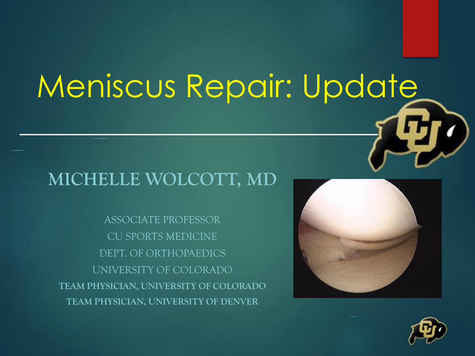
Meniscus Repair: Update
MICHELLE WOLCOTT, MD
ASSOCIATE PROFESSOR
CU SPORTS MEDICINE
DEPT. OF ORTHOPAEDICS
UNIVERSITY OF COLORADO
TEAM PHYSICIAN, UNIVERSITY OF COLORADO
TEAM PHYSICIAN, UNIVERSITY OF DENVER
Meniscus Repair: Update
No conflicts of interest
No disclosures
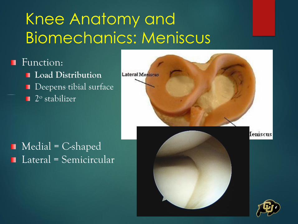
Knee Anatomy and
Biomechanics: Meniscus
Function:Load DistributionDeepens tibial surface2o stabilizer
Medial = C-shapedLateral = Semicircular
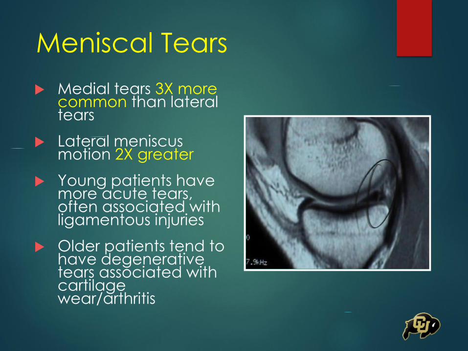
Meniscal Tears
Medial tears 3X more common than lateral tears
Lateral meniscus motion 2X greater
Young patients have more acute tears, often associated with ligamentous injuries
Older patients tend to have degenerative tears associated with cartilage wear/arthritis

Types of Meniscus Tears
Severity of Articular
Cartilage Wear=

Medial Meniscus Tear Morphology
and Chondral Degeneration of
the Knee: Is There a Relationship? Henry S, Mascarenhas R, Kowalchuk D,
Forsythe B, Irrgang JJ, Harner C
103 patients prospectively evaluated at the time of meniscal surgery
Chondral degeneration, tear morphology/location
Root/radial flap tears more strongly associated with medial compartment degeneration
Meniscus tears with increasing disruption of circumferential hoop fibers were significantly associated with cartilage lesions of increasing severity in both medial and lateral compartments
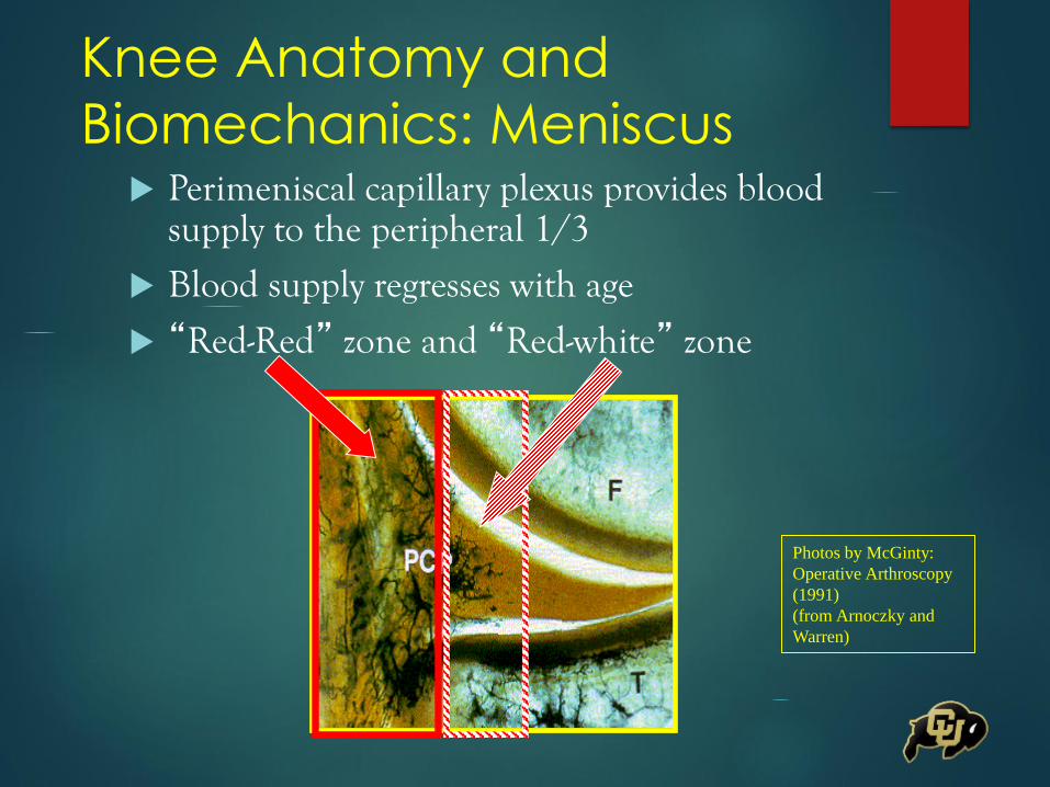
Knee Anatomy and
Biomechanics: Meniscus Perimeniscal capillary plexus provides blood
supply to the peripheral 1/3
Blood supply regresses with age
“Red-Red” zone and “Red-white” zone
Photos by McGinty:
Operative Arthroscopy
(1991)
(from Arnoczky and
Warren)
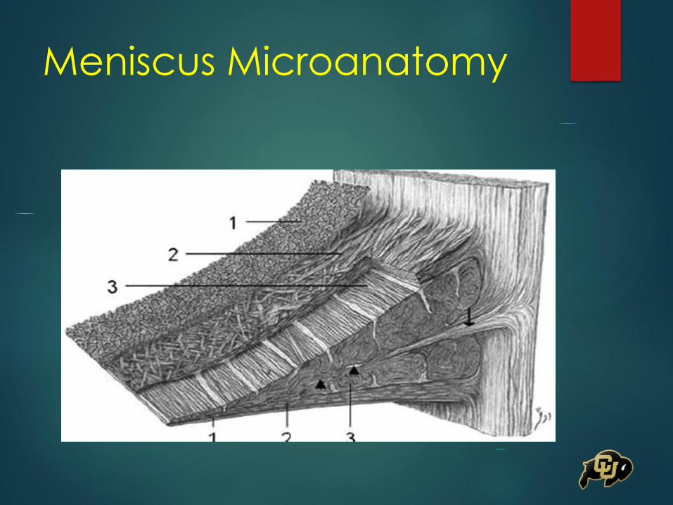
Meniscus Microanatomy
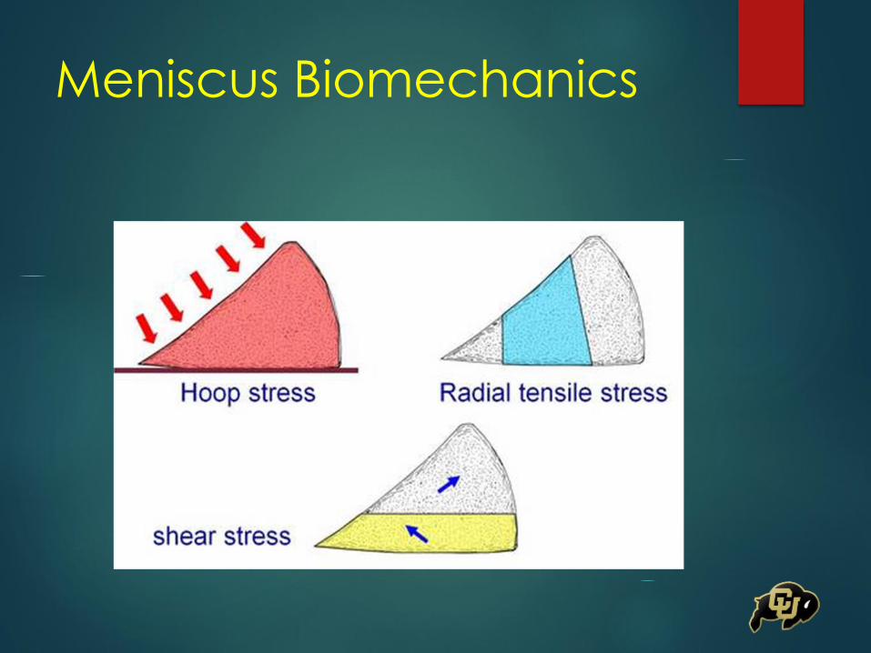
Meniscus Biomechanics
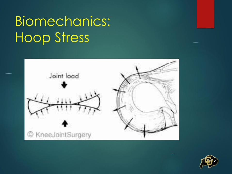
Biomechanics:
Hoop Stress

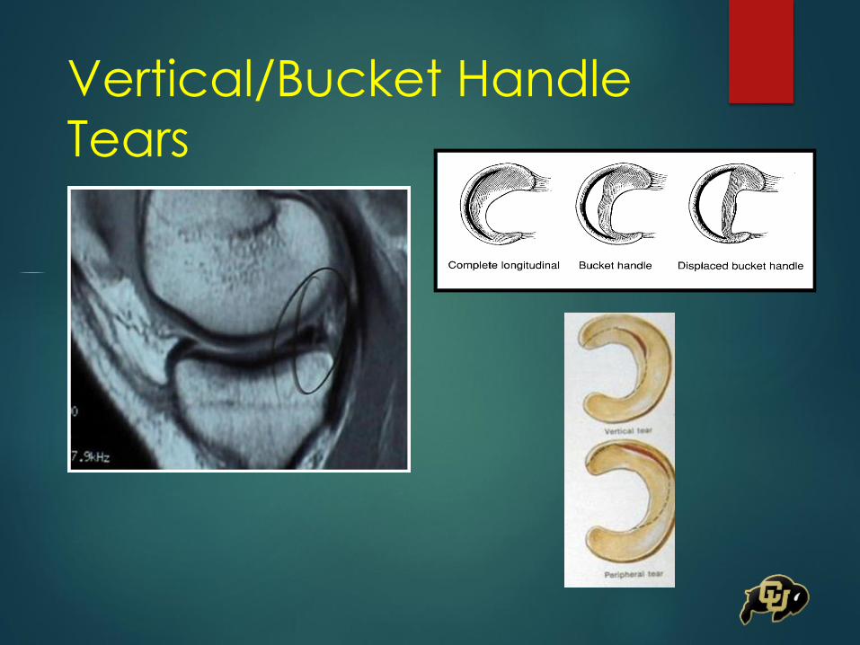
Vertical/Bucket Handle
Tears
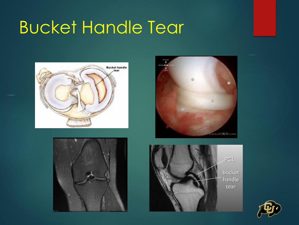
Bucket Handle Tear
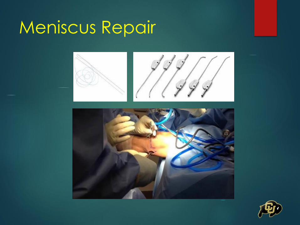
Meniscus Repair

Meniscus Repair
Inside-Out RepairGold Standard
Strongest Repair
Vertical Mattress
Meniscal Repair
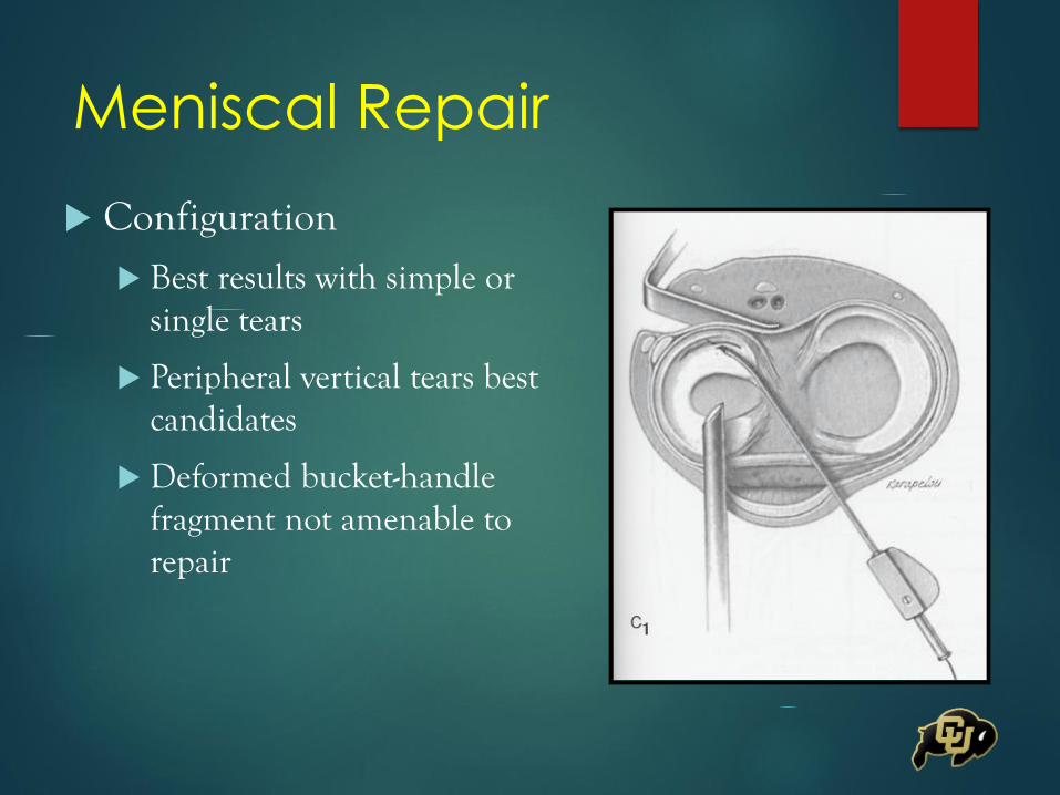
Configuration
Best results with simple or single tears
Peripheral vertical tears best candidates
Deformed bucket-handle fragment not amenable to repair
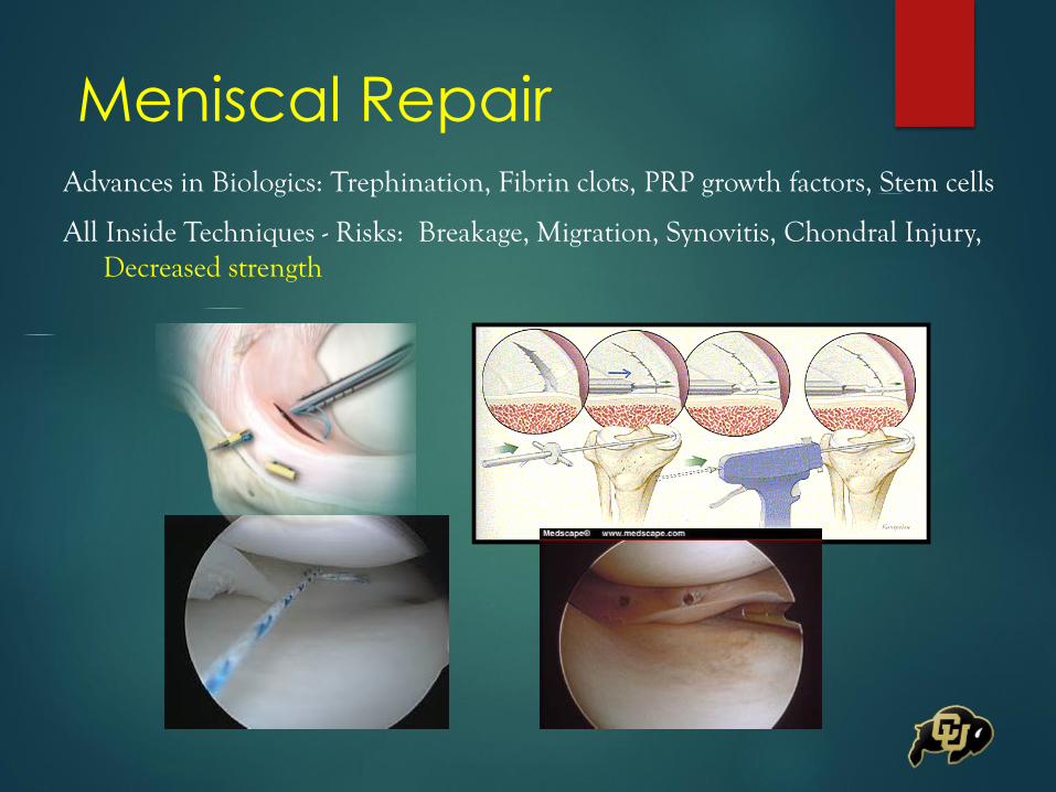
Meniscal RepairAdvances in Biologics: Trephination, Fibrin clots, PRP growth factors, Stem cells
All Inside Techniques - Risks: Breakage, Migration, Synovitis, Chondral Injury, Decreased strength

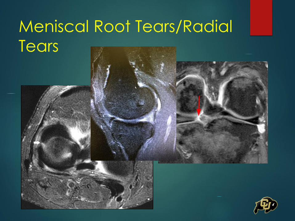
Meniscal Root Tears/Radial
Tears
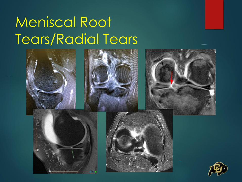
Meniscal Root
Tears/Radial Tears
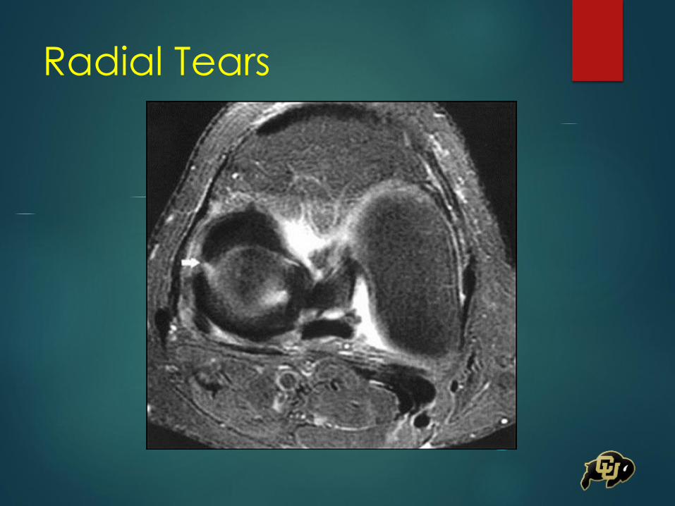
Radial Tears
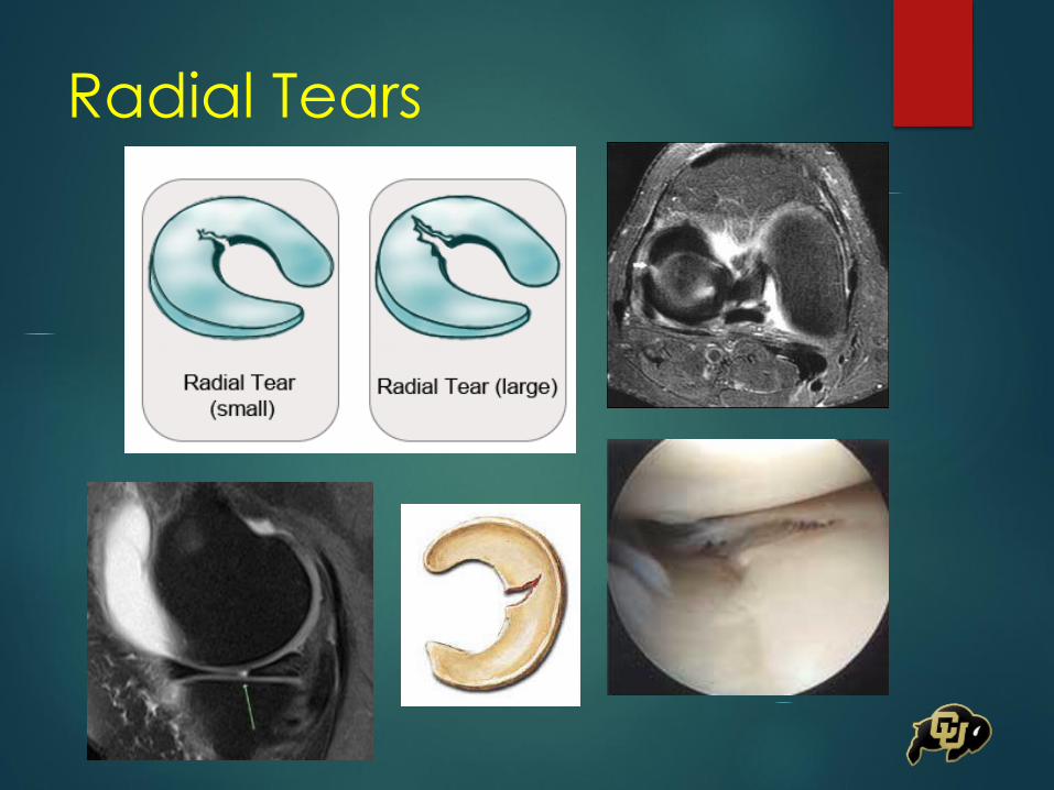
Radial Tears
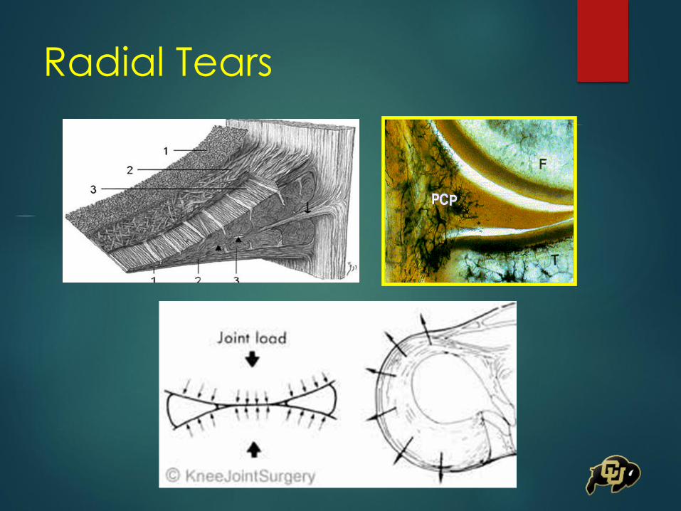
Radial Tears

Meniscus Root Tears
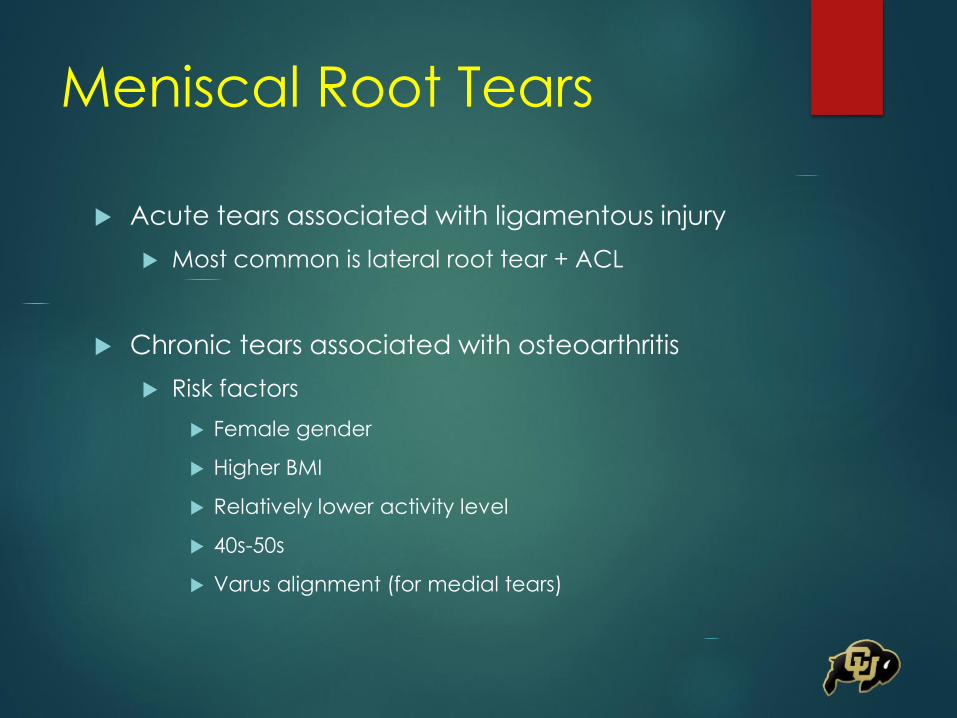
Meniscal Root Tears
Acute tears associated with ligamentous injury
Most common is lateral root tear + ACL
Chronic tears associated with osteoarthritis
Risk factors
Female gender
Higher BMI
Relatively lower activity level
40s-50s
Varus alignment (for medial tears)
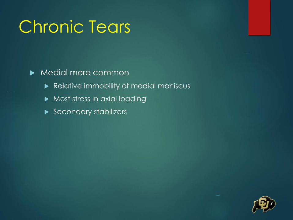
Chronic Tears
Medial more common
Relative immobility of medial meniscus
Most stress in axial loading
Secondary stabilizers
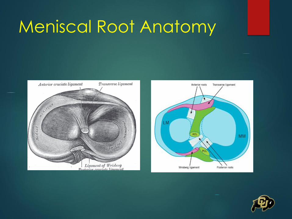
Meniscal Root Anatomy
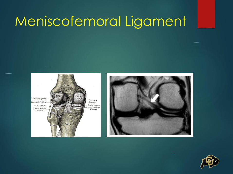
Meniscofemoral Ligament
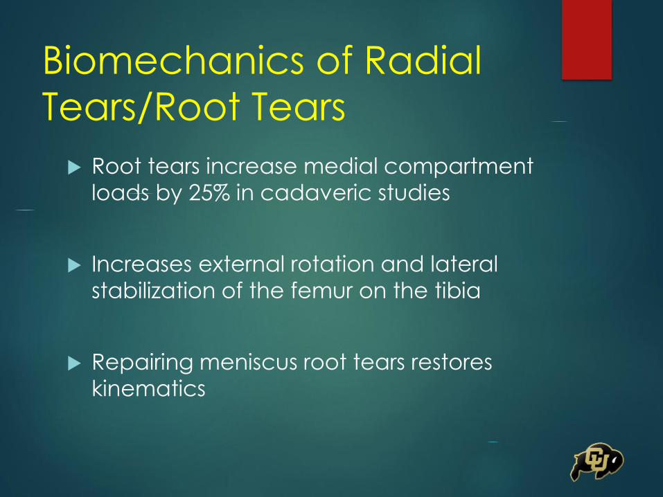
Biomechanics of Radial
Tears/Root Tears
Root tears increase medial compartment
loads by 25% in cadaveric studies
Increases external rotation and lateral
stabilization of the femur on the tibia
Repairing meniscus root tears restores
kinematics
Diagnosis
Symptoms may be similar to other types of meniscus
tears
Joint line tenderness
Pain with McMurrays
Swelling
Can also be nonspecific
Vague pain symptoms (posterior)
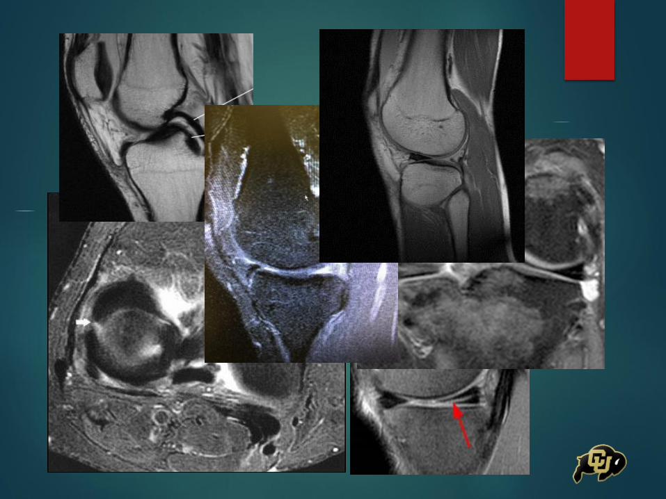
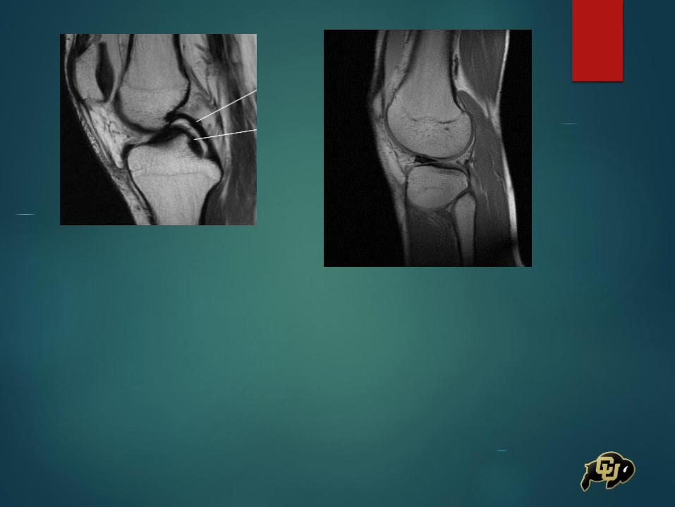
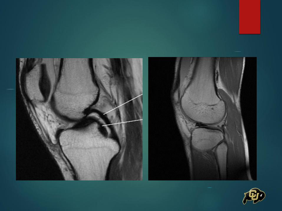
MRI
Arthroscopic Assessment

MRI
truncated triangle, cleft, marching cleft
and ghost meniscus signs
the use of all four signs increased the
detection rate for radial tears to 89%
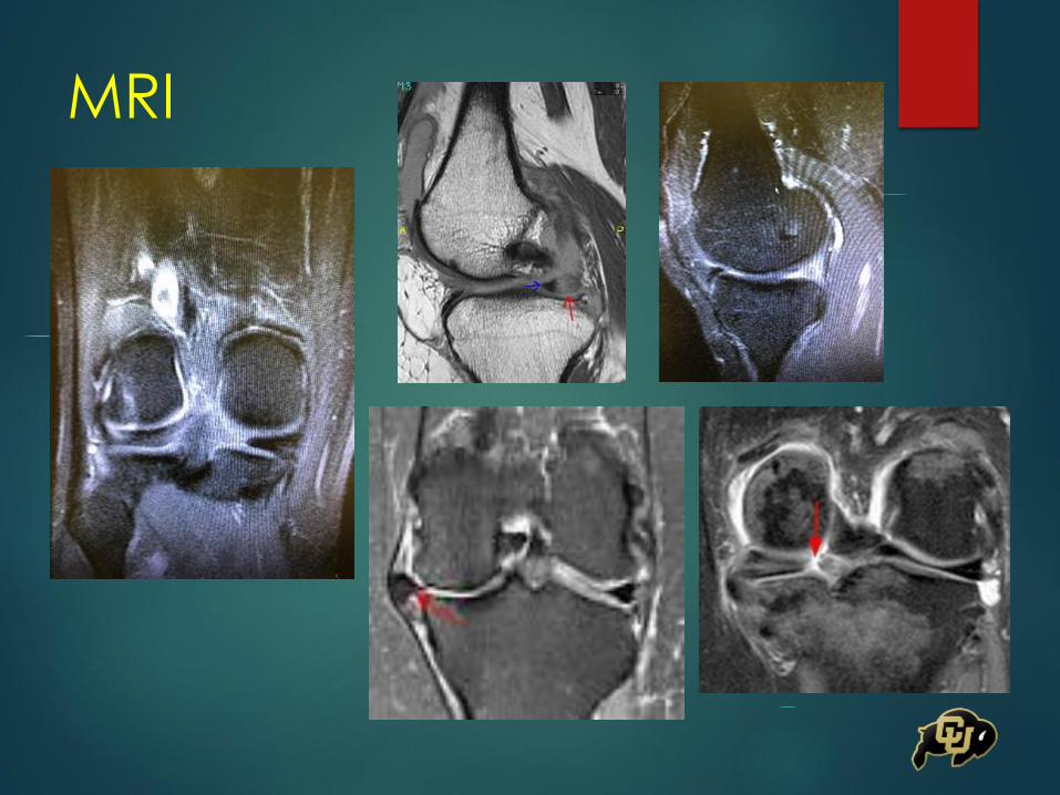
MRI
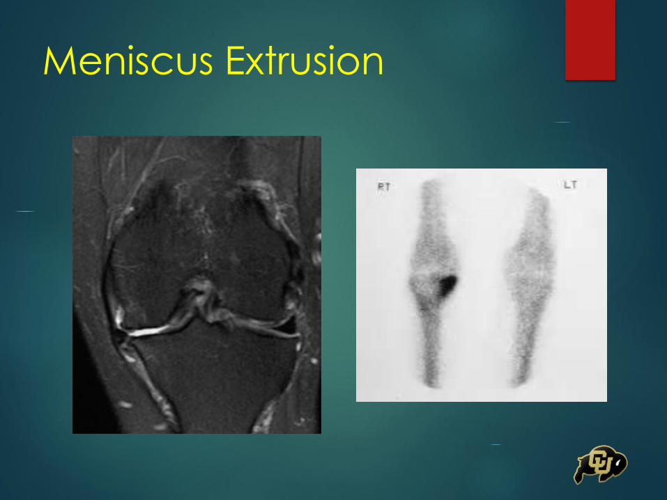
Meniscus Extrusion

Meniscal Repair
Radial Repair
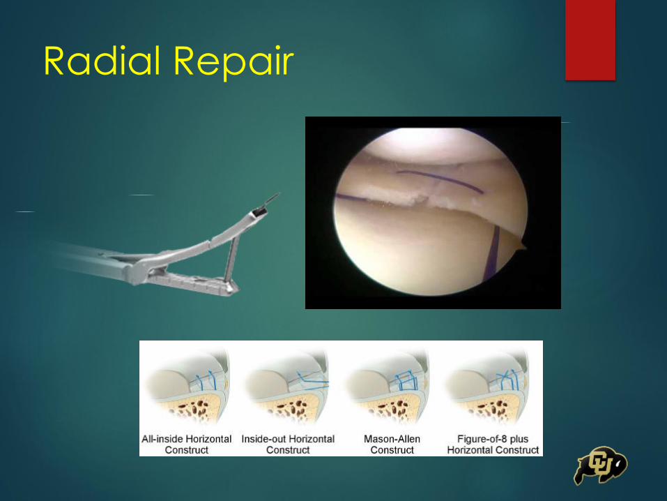
Radial Repair
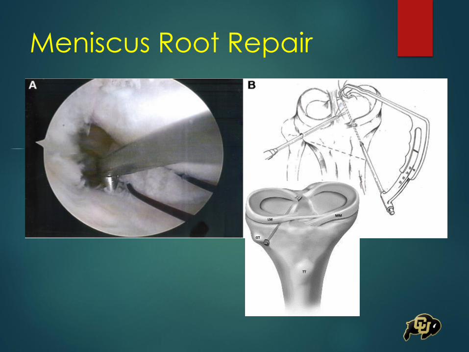
Meniscus Root Repair
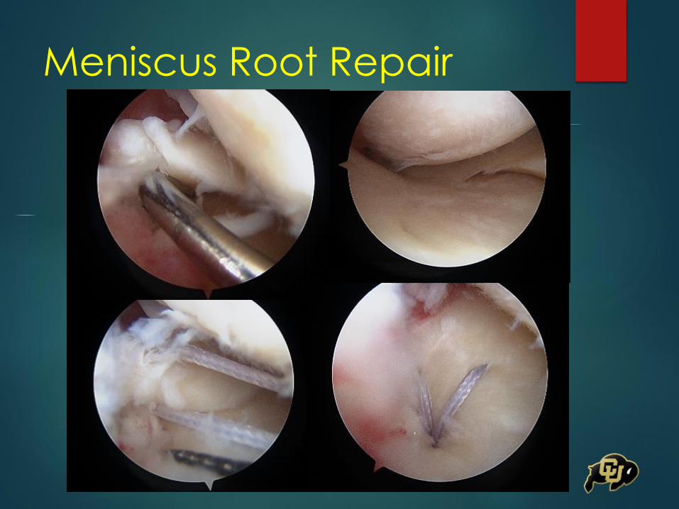
Meniscus Root Repair
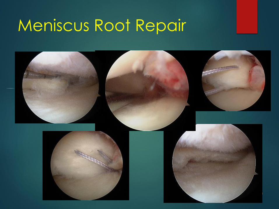
Meniscus Root Repair

Meniscus Root Repair
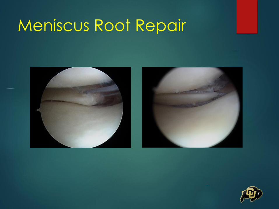
Meniscus Root Repair
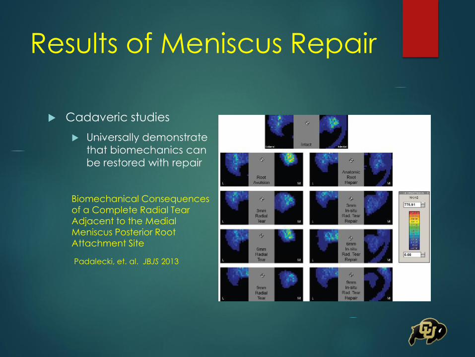
Results of Meniscus Repair
Cadaveric studies
Universally demonstrate
that biomechanics can
be restored with repair
Biomechanical Consequences of a Complete Radial Tear Adjacent to the Medial Meniscus Posterior Root Attachment Site
Padalecki, et. al. JBJS 2013
Results of Meniscus Repair
Meniscus Suture Repair: Minimum 10 yr Outcomes
in Patients Younger than 40 yrs Compared With Patients 40 and Older
Steadman, et al. AJSM 2015
No significant difference based on age (5.5% vs
5.3%)
82% vs 93% followup
No significant difference based on which meniscus
(med vs lat), concomitant ACL injury, microfracture
Results of Meniscus Repair
Arthroscopic Transtibial Pullout Repair for Posterior Medial Meniscal Root Tears: A Systematic Review of Clinical, Radiographic, and Second-Look Arthroscopic Results
Feucht, et al, Arthroscopy 2015
30 mos followup
Lysholm scores improved from 52.4 to 85.9
Radiographs – 84% showed no progression
MRI – 82% showed no progression of cartilage degeneration, 56% showed reduced meniscal extrusion
2nd look arthroscopy – healing status complete in 62%, partial in 34%, failed in 3%
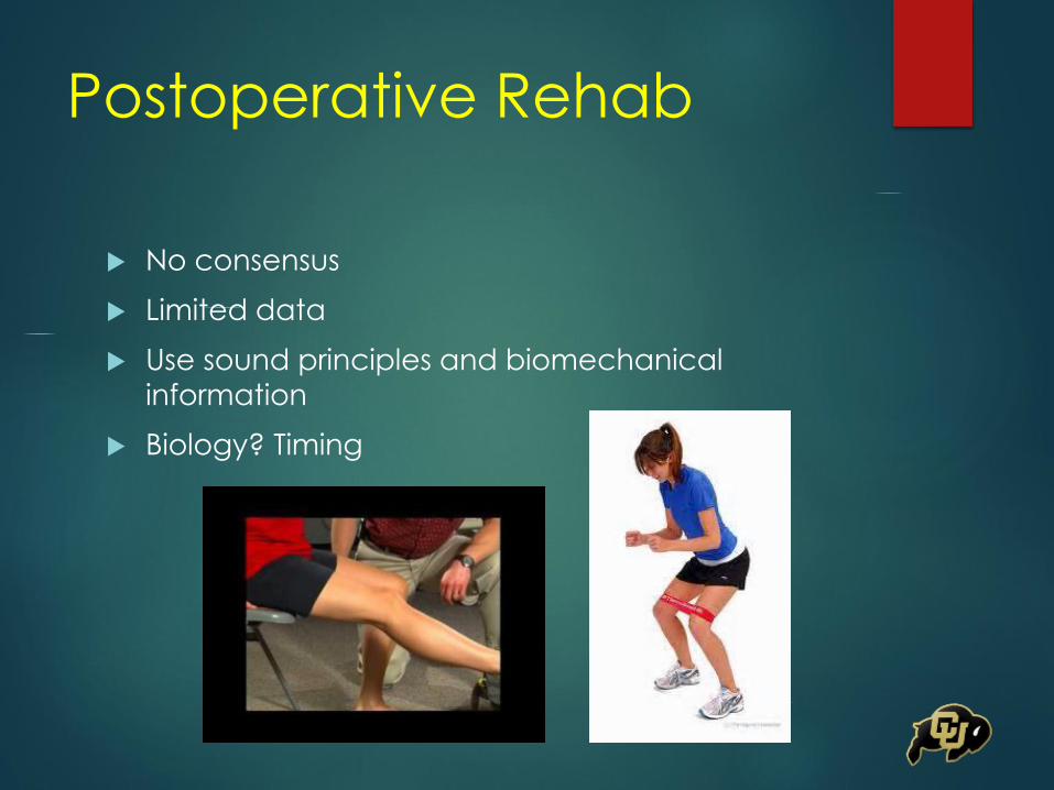
Postoperative Rehab
No consensus
Limited data
Use sound principles and biomechanical
information
Biology? Timing
Postop Rehab – Meniscal
Repair
Protect the repair in first 4-6 weeks
Restricted WB in first 4-6 weeks
WBAT in full ext
NWB for root and radial tears
Restrict ROM 0-90 deg x 6 wks
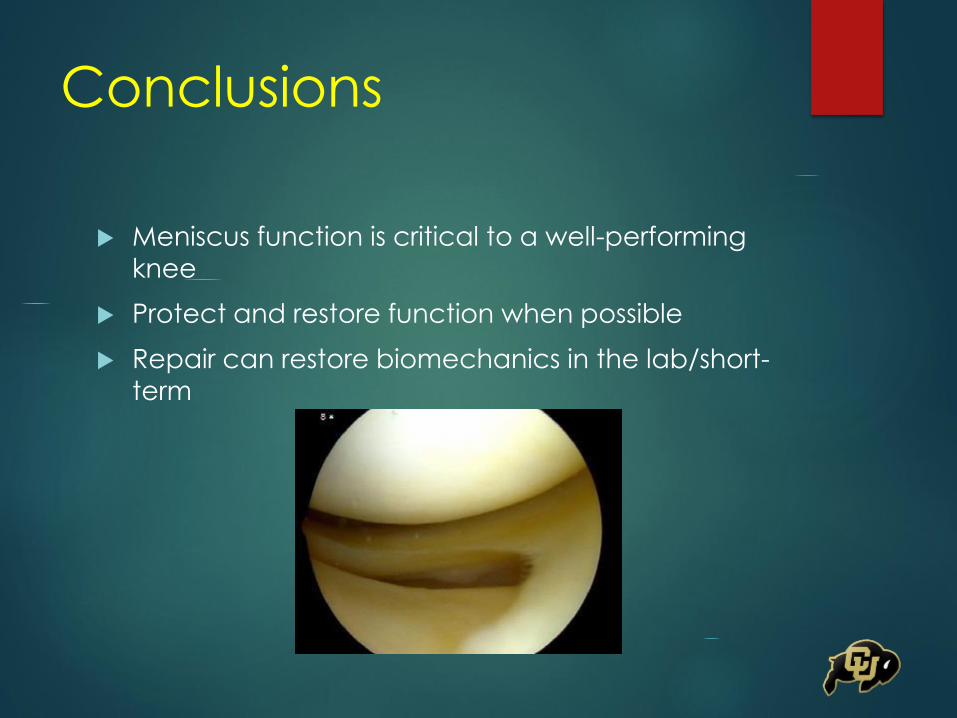
Conclusions
Meniscus function is critical to a well-performing
knee
Protect and restore function when possible
Repair can restore biomechanics in the lab/short-
term
Conclusions
Future directions
Biologics
Improved instrumentation/technique
Long-term outcome
Acute vs Chronic tears
Diagnosis
Can we identify tears earlier
References
Han SB, Shetty GM, Lee DH et.al. Unfavorable Results of Partial Meniscectomy for Complete Posterior Medial Meniscal Root Tear With Early Osteoarthritis: A 5- To 8- Year Followup Study. Arthroscopy 2010; 26:1326-1332.
Lee JH, Young JL, Kim KB et.al. Arthroscopic Pullout Suture Repair of Posterior Root Tear of the Medial Meniscus: Radiographic and Clinical Results With a 2-Year Follow-up. Arthroscopy 2009; 25: 951-958.
Feucht MJ, Kuhle J, Bode G et.al. Arthroscopic TranstibialPullout Repair for Posterior Medial Meniscus Root Tears: A Systematic Review of Clinical, Radiographic, and Second-Look Arthroscopic Results. Arthroscopy 2015; 31: 1808-1816.
Choi CJ, Choi YJ, Lee JJ et.al. MRI Evidence of Meniscal Extrusionin Medial Meniscus Posterior Root Tear. Arthroscopy2010; 26: 1602-1606.
Bhatia S, LaPrade CM, Ellman MB et.al. Meniscal Root Tears: Significance, Diagnosis, and Treatment. AJSM 42: 3016-3030
References
Steadman JR, Matheny LM, Singleton SB et.al. Meniscus Suture Repair: Minimum 10-Year Outcomes in Patients Younger Than 40 Years Compared With Patients 40 and Older. AJSM 2015; 43: 2222 – 2227.
Branch EA, Milchteim C, Aspey BA et.al. Biomechanical Comparison of Arthroscopic Repair Constructs for Radial Tears of the Meniscus. AJSM2015; 43: 2270-2276.
Allaire R, Muriuki M, Gilbertson L et.al. Biomechanical Consequences of a Tear of the Posterior Root of the Medial Meniscus: Similar to Total Meniscectomy. JBJS 2008; 90: 1922-1931.
Hein CN, Deperio JG, Ehrensberger MT et.al. Effects of Medial Meniscal Posterior Horn Avulsion and Repair on Meniscal Displacement. The Knee2011; 18: 1Biomechanical Consequences of the a Complete Radial Tear Adjacent to the Medial Meniscus Posterior Root Attachment Site: In Situ Pull-Out Repair Restores Derangement of Joint Mechanics. AJSM 2013; 42: 699-707.
Henry S, Mascarenhas R, Kowalchuk D et.al. Medial Meniscus Tear Morphology and Chondral Degeneration of the Knee: Is There a Relationship? Arthroscopy 2012; 28: 1124-1134

Thank You