metabolic engineering of kluyveromyces lactis for l-ascorbic
TRANSCRIPT

Rosa et al. Microbial Cell Factories 2013, 12:59http://www.microbialcellfactories.com/content/12/1/59
RESEARCH Open Access
Metabolic engineering of Kluyveromyces lactis forL-ascorbic acid (vitamin C) biosynthesisJúlio César Câmara Rosa1,2, Lívia Tavares Colombo1,2, Mariana Caroline Tocantins Alvim1,2, Nelson Avonce3,4,Patrick Van Dijck3,4 and Flávia Maria Lopes Passos1,2,5*
Abstract
Background: L-ascorbic acid (L-AA) is naturally synthesized in plants from D-glucose by 10 steps pathway. Thepathway branch to synthesize L-galactose, the key intermediate for L-ascorbic acid biosynthesis, has been recentlyelucidated. Budding yeast produces an 5-carbon ascorbic acid analogue Dehydro-D-arabinono 1,4-lactone (D-DAL),which is synthesized from D-arabinose. Yeast is able to synthesize L-ascorbic acid only if it is cultivated in thepresence of one of its precursors: L-galactose, L-galactono 1,4-lactone, or L-gulono 1,4-lactone extracted from plantsor animals. To avoid feeding the yeast culture with this “L” enantiomer, we engineered Kluyveromyces lactis withL-galactose biosynthesis pathway genes: GDP-mannose 3,5-epimerase (GME), GDP-L-galactose phosphorylase (VTC2)and L-galactose-1-phosphate phosphatase (VTC4) isolated from Arabidopsis thaliana.
Results: Plasmids were constructed and modified such that the cloned plant genes were targeted to the K. lactisLAC4 Locus by homologous recombination and that the expression was associated to the growth on D-galactoseor lactose. Upon K. lactis transformation, GME was under the control of the native LAC4 promoter whereas VTC2and VTC4 were expressed from the S. cerevisiae promoters GPD1 and ADH1 respectively. The expression in K. lactis,of the L-galactose biosynthesis genes was determined by Reverse Transcriptase-PCR and western blotting. Therecombinant yeasts were capable to produce about 30 mg.L-1 of L-ascorbic acid in 48 hours of cultivation whencultured on rich medium with 2% (w/v) D-galactose. We also evaluated the L-AA production culturing recombinantrecombinant strains in cheese whey, a waste product during cheese production, as an alternative source of lactose.
Conclusions: This work is the first attempt to engineer K. lactis cells for L-ascorbic acid biosynthesis by a fermentationprocess without any trace of “L” isomers precursors in the culture medium. We have engineered K. lactis strains capableof converting lactose and D-galactose into L-galactose, by the integration of the genes from the A. thaliana L-galactosepathway. L-galactose is a rare sugar, which is one of the main precursors for L-AA production.
Keywords: Kluyveromyces lactis, L-ascorbic acid, L-galactose, Metabolic engineering
BackgroundThe enediol ascorbate or L-ascorbic acid (L-AA), knownas Vitamin C, is an important metabolite in many or-ganisms. In eukaryotes, L-AA is essential for a variety ofcellular functions [1], acting as I) a scavenger of freeradicals [2]; ii) a reducing agent [3], iii) a cofactor forenzyme activity [4,5] iv) an intermediate for catechol-amines biosynthesis, and v) a limiting growth factor in
* Correspondence: [email protected]ório de Fisiologia de Microrganismos, Instituto de BiotecnologiaAplicada à Agropecuária (BIOAGRO), Universidade Federal de Viçosa, Brazil2Departamento de Microbiologia, Universidade Federal de Viçosa, campusViçosa, Minas Gerais, BrasilFull list of author information is available at the end of the article
© 2013 Rosa et al.; licensee BioMed Central LtCommons Attribution License (http://creativecreproduction in any medium, provided the or
plant development [6]. Most of the commercially availablevitamin C is synthetically synthesized by the Reichsteinprocess, using D-glucose as start material [7].L-AA is naturally produced in plants where its bio-
synthetic pathway has been completely elucidated [8,9]. Inmost cases, GDP-D-mannose is converted into L-galactose,which is further converted into L-AA [10]. Although theremay exist alternative routes [11,12] this pathway is recog-nized as the main route for L-AA biosynthesis [13,14].There are three enzymes required for the conversion ofGDP-D-mannose into L-galactose. The GDP-mannose 3,5-epimerase (GME) catalyzes the conversion of GDP-D-man-nose to GDP-L-gulose or to GDP-L-galactose, dependingwhether the epimerization occurs on 5’- carbon or on both
d. This is an Open Access article distributed under the terms of the Creativeommons.org/licenses/by/2.0), which permits unrestricted use, distribution, andiginal work is properly cited.

Rosa et al. Microbial Cell Factories 2013, 12:59 Page 2 of 13http://www.microbialcellfactories.com/content/12/1/59
3’- and 5’- carbon of GDP-D-mannose respectively [15].GDP-L-gulose seems to represent the minor part of theproducts (around 25% under equilibrium) and can also beconverted to L-AA [16]. The epimerization of D to L-substrates, which is rare in nature, is a crucial step to gener-ate the galactose enantiomer in the L-AA pathway. GDP-L-galactose is then converted to L-galactose 1-phosphate byGDP-L-galactose phosphorylase, encoded by the VTC2gene [17]. This gene encodes a member of the GalT/Apa1branch of the histidine triad protein superfamily that cata-lyzes the conversion of GDP-L-galactose to L-galactose1-phosphate in a reaction that consumes inorganicphosphate and produces GDP [9]. Müller-Moulé [18]constructed the VTC2:YFP fusion protein and unexpect-edly this protein was found not only in the cytosol, butalso in the nucleus, which suggests that GDP-L-Galactosephosphorylase/L-Galactose guanylyltransferase might be adual-function protein, which has both enzymatic andregulatory function in the L-AA biosynthesis pathway inA. thaliana. The third enzyme is L-galactose 1-phosphatephosphatase, encoded by the VTC4 gene [19], which is abifunctional enzyme that plays a role in both ascorbate aswell as myoinositol biosynthetic pathways, although itshows selective preference for L-galactose 1-phosphate[20]. The resulting L-galactose is then the main precursorfor L-AA biosynthesis.Yeasts are known to produce the 5-carbon ascorbic
acid analogue, Dehydro-D-arabinono 1,4-lactone (D-DAL),which is synthesized from D-arabinose. Although D-DALdoes not show any anti-scurvy activity, its physiochemicalproperties and biological activities are quite similar to thoseof L-AA. For this reason D-DAL can replace L-AA in someindustrial applications [21,22]. The structural motifs of theenzymes involved in the D-DAL biosynthetic pathway inyeast resemble those of the pathway in plants that convertsL-galactose into L-AA. D-DAL pathway enzymes fromCandida albicans and Saccharomyces cerevisiae haveshown to be able to convert a broad range of substratesbesides D-arabinose including L-galactose into theirrespective galactonic acids in vitro [23,24]. Furthermore,L-AA production in yeasts was achieved when appropriateprecursors such as L-galactose, L-galactono 1,4-Lactone,L-gulono 1,4-lactone were exogenously supplied in thegrowth medium [25]. Thus, isolation of genes involved inL-galactose production in plants provides biochemicalsupport to guide the metabolic capacity of industrial mi-croorganisms to produce L-AA by fermentation [7].Attempts have been made to synthesize L-AA in genet-
ically modified microorganisms. Sauer et al. [25] observeda high production of vitamin C in the culture supernatantof S. cerevisiae cells expressing the L-galactose dehy-drogenase (LDGH) and D-arabinose 1,4-lactone oxidase(ALO1) from yeast or the L-galactono-1, 4-lactone de-hydrogenase (AGD) from Arabidopsis thaliana, when
cultivated in a medium containing 250 mg.L-1 L-galactose.Further, Branduardi et al. [26] have engineered this strainwith GME and VTC4 from A. thaliana and also with L-fucose guanylyltransferase from Rattus norvegicus FGT inorder to convert D-glucose to L-AA completing the L-AApathway in S. cerevisiae. The L-AA production conferred anincreased stress tolerance under oxidative stress conditions.Kluyveromyces lactis is one of the most important
non-Saccharomyces yeast species used as an eukaryoticmodel and tool for biotechnological applications includ-ing an alternative host for heterologous gene expression.K. lactis has the ability of growing, by respiration, on awide range of substrates, including lactose with low glu-cose repression [27]. The genome has been completelysequenced and the Lac-Gal regulon, with the inducedgenes for lactose transport and hydrolysis, has been ex-tensively studied [28]. Many heterologous expressionsystems have been developed, based on the LAC4promoter with the production of lysozyme [29], serumalbumin [30], thermostable bacterial xylanase [31] andheparin sulfate sulphotransferase [32] as examples. Thepotential use of K. lactis as a host for protein expressionassociated to its physiological properties suggests thatthis yeast could also be used for large-scale proteinproduction in the food and pharmaceutical industry.Furthermore, its ability to express and process heterol-ogous proteins makes this yeast well suited for multipleproteins expression such as the enzymes involved in L-galactose metabolism from plants.Considering the high costs of using non-physiological
substrates in the L enantiomer form for industrial appli-cations, herein, we report the construction of K. lactisstrains capable to convert D-galactose or lactose into L-galactose, the main intermediate metabolite of the L-AApathway in plants, and its subsequent conversion intoL-ascorbic acid.
ResultsIsolation and cloning of the L-ascorbic acid pathwaygenes from Arabidopsis thalianaA cDNA library from Arabidopsis thaliana leaves wasused as template to amplify the three genes of the L-ascorbic acid (L-AA) pathway required for L-galactosesynthesis in K. lactis (see Materials and Methods). Theamino acid sequences encoded by the correspondingamplified genes GME, VTC2, VTC4 were determinedand verified to be the same as those in the Arabidopsisgenome database. The three genes were cloned in K.lactis expression vectors (Figure 1). The codons of theplant genes were not optimized for expressing in K.lactis. Carbone et al. (2003) [33] reported that Saccharo-myces sp. and plants shared the same preferred codons,supporting K. lactis as a host for unmodified plant genesexpression.
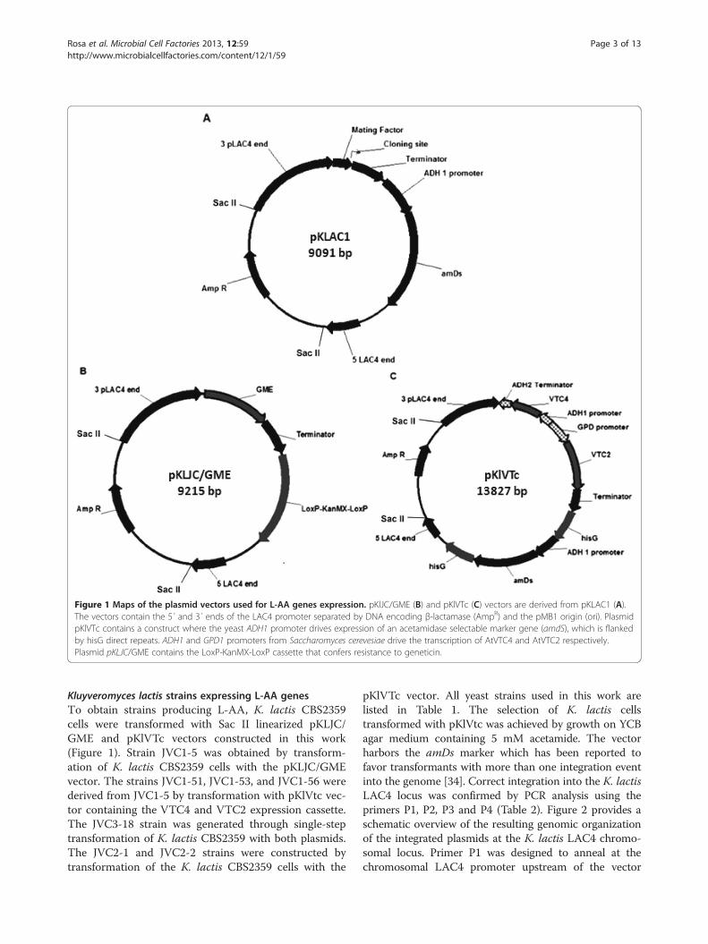
Figure 1 Maps of the plasmid vectors used for L-AA genes expression. pKlJC/GME (B) and pKlVTc (C) vectors are derived from pKLAC1 (A).The vectors contain the 5´ and 3´ ends of the LAC4 promoter separated by DNA encoding β-lactamase (AmpR) and the pMB1 origin (ori). PlasmidpKlVTc contains a construct where the yeast ADH1 promoter drives expression of an acetamidase selectable marker gene (amdS), which is flankedby hisG direct repeats. ADH1 and GPD1 promoters from Saccharomyces cerevesiae drive the transcription of AtVTC4 and AtVTC2 respectively.Plasmid pKLJC/GME contains the LoxP-KanMX-LoxP cassette that confers resistance to geneticin.
Rosa et al. Microbial Cell Factories 2013, 12:59 Page 3 of 13http://www.microbialcellfactories.com/content/12/1/59
Kluyveromyces lactis strains expressing L-AA genesTo obtain strains producing L-AA, K. lactis CBS2359cells were transformed with Sac II linearized pKLJC/GME and pKlVTc vectors constructed in this work(Figure 1). Strain JVC1-5 was obtained by transform-ation of K. lactis CBS2359 cells with the pKLJC/GMEvector. The strains JVC1-51, JVC1-53, and JVC1-56 werederived from JVC1-5 by transformation with pKlVtc vec-tor containing the VTC4 and VTC2 expression cassette.The JVC3-18 strain was generated through single-steptransformation of K. lactis CBS2359 with both plasmids.The JVC2-1 and JVC2-2 strains were constructed bytransformation of the K. lactis CBS2359 cells with the
pKlVTc vector. All yeast strains used in this work arelisted in Table 1. The selection of K. lactis cellstransformed with pKlVtc was achieved by growth on YCBagar medium containing 5 mM acetamide. The vectorharbors the amDs marker which has been reported tofavor transformants with more than one integration eventinto the genome [34]. Correct integration into the K. lactisLAC4 locus was confirmed by PCR analysis using theprimers P1, P2, P3 and P4 (Table 2). Figure 2 provides aschematic overview of the resulting genomic organizationof the integrated plasmids at the K. lactis LAC4 chromo-somal locus. Primer P1 was designed to anneal at thechromosomal LAC4 promoter upstream of the vector

Table 1 Yeasts strains used in this study
Strains Markers Cassette expression Plasmids Reference
CBS 2359 Wild type - - Genolevures consortium*
JVC1-5 KanR AtGME pKLJC/GME This study
JVC1-51 KanR, amDs AtGME, AtVTC2, AtVTC4 pKLJC/GME, pKlVTc This study
JVC1-53 KanR, amDs AtGME, AtVTC2, AtVTC4 pKLJC/GME, pKlVTc This study
JVC1-56 KanR, amDs AtGME, AtVTC2, AtVTC4 pKLJC/GME, pKlVTc This study
JVC2-1 amDs AtVTC2, AtVTC4 pKlVTc This study
JVC2-2 amDs AtVTC2, AtVTC4 pKlVTc This study
JVC3-18 KanR, amDs AtGME, AtVTC2, AtVTC4 pKLJC/GME, pKlVTc This study
KanR cassette conferring resistance to Geneticin.amDs acetamidase marker.*Kluyveromyces lactis strain used for Genome sequencing by the Génolures consortium (www.genolevures.org).
Rosa et al. Microbial Cell Factories 2013, 12:59 Page 4 of 13http://www.microbialcellfactories.com/content/12/1/59
integration site and the reverse primers P2 and P4 annealto pKlVTc and pKLJC/GME expression cassettes se-quence respectively. When multiple copies of the cassettewere integrated in tandem at the same locus, a 2.3 kb frag-ment was then amplified by using the forward primer P3in combination with either reverse primers P2 or P4 foreach vector. Single and multiple insertions from each
Table 2 List of primers used on this study
Name Sequence
GME-F 5’CTCGAGATGGGAACTACCAATGGAACAG3’
GME-RFlag 5’CCCGGCGGCCGTCACTTGTCATCGTCATCCT
VTC2-F 5’GCGGCCGCATGTTGAAAATCAAAAGAGTTC
VTC2-RFlag 5’AGGCCTTCACTTGTCATCGTCATCCTTGTAAT
VTC4-F 5’CTCGAGATGGCGGACAATGATTCTCTAG3’
VTC4-RFlag 5’AGGCCTTCACTTGTCATCGTCATCCTTGTAAT
VT4-F 5’CGACTCGGTACCATGGCGGACAATGATTCTC
VT4-R 5’CGACTCGAATTCTCACTTGTCATCGTCATCC
hisG I – F 5’TGTACACCAGTGGTGCATGAACGC3’
hisG I – R 5’ACATGTCTAGGGATAACAGGGTAATATAGA
hisG II – F 5’CGACTCCCCGGGCCAGTGGTGCATGAACGC
hisG II – R 5’CGACTCCTGCAGCTAGGGATAACAGGGTAA
KanMX-F 5’CGACTCTGTACACTGAAGCTTCGTACGCTGC
KanMX-R 5’CGACTCCCCGGGATCACCTAATAACTTCGTA
GPDADH1-F 5’CGACTCCATATG GCGGCCGCGTCGAAACT
GPDADH1-R 5’CGACTCGACGTC AAGCTTGGCATGCGAAG
KlACT1-F 5’ATGGATTCTGAGGTCGCTGC3’
KlACT1-R 5’TTAGAAACACTTCAAGTGAACGATGG3’
P1 5’ACACACGTAAACGCGCTCGGT3’
P2 5’ATCATCCTTGTCAGCGAAAGC3’
P3 5’ACCTGAAGATAGAGCTTCTAA3’
P4 5’GGTACCCCTAGGAGATCTAGCTC3’
Underlined are shown the Flag Tag sequence.In bold are represented the restriction site.In grey, the stop codon.
cassette were detected by the presence of 2.4 kb and2.3 kb amplicons respectively. The insertion of the cassetteinto the LAC4 locus by homologous recombination dupli-cates the LAC4 promoter region so that it can be targetedby another cassette resulting in multiple copies inte-gration. However, this analysis does not indicate the num-ber of integrated copies; we determined the exact copy
Restriction site
XhoI
TGTAATCCTCTTTTCCATCAGCCGCG3’ NotI
CGACC3’ NotI
CCTGAAGGACAAGGCACTCGGCGGC3’ StuI
XhoI
CTGCCCCTGTAAGCCGC3’ StuI
TAG3’ KpnI
TTG3’ EcoRI
BsrGI
CATGG3’ BsrGI
3’ XmaI/SmaI
TATAGACATGG3’ PstI
A3’ BsrGI
TAGCATACATTATAC3’ SmaI
AAGTTCTTGGTGTTTTAAAACT3’ NdeI /NotI
GAAAATGAGA3’ AatII / HindIII

Figure 2 Genomic organization of SacII linearized vectors into the K. lactis LAC4 locus upon transformation. (A) Scheme of single andmultiple copies of integrated GME cassettes detected by PCR using primers P1 and P3 (2.4 kb) or P3 and P4 (2.3 kb) respectively; (B) Scheme ofsingle and multiple copies of AtVTC2 and AtVTC4 cassettes detected by PCR using primers P1 and P2 (2.4 kb) or P3 and P2 (2.3 kb) respectively.
Rosa et al. Microbial Cell Factories 2013, 12:59 Page 5 of 13http://www.microbialcellfactories.com/content/12/1/59
number of each cassette integrated into K. lactis genomeby absolute quantification. The results are shown inTable 3. Most of the recombinant strains harbor at leastmore than one copy of the cassette except the strainJVC1-51. The JVC1-56 strain carries four copies of theGME gene integrated in tandem at the LAC4 locus.The GME gene is under the control of the inducible
LAC4 promoter upon integration by homologous re-combination. The strong constitutive S. cerevisiae pro-moters GPD1 and ADH1 drive the transcription of theVTC2 and VTC4 respectively. The expression analysisof L-AA pathway plant genes in K. lactis recombinant
cells was achieved by Reverse Transcriptase-PCR andthe flag-tagged proteins from total protein extract wereimmunoprecipitated before SDS-PAGE, blotted anddetected using monoclonal anti-Flag antibodies (Figure 3).All JVC1-5 derived strains, JVC1-51, JVC1-53 and JVC1-56, are expressing the L-galactose pathway genes, GME,VTC2, VTC4. The JVC1-5 only expresses GME and theJVC2-1 and JVC2-2 strains are the control strains forVTC2 and VTC4 expression.Simultaneous expression of the proteins GME (43.8 kDa),
VTC2 (49 kDa), and VTC4 (30 kDa) in the engineeredJVC3-18 and JVC1-5 derived strains should result in the

Table 3 Estimated copy number (ENC) of AtGME and AtVTC genes by absolute quantification
Strains CTa Copies (Copies.μL-1) ENC
ACT1 GME VTC ACT1 GME VTC GME VTC
JVC1-51 20.43 ± 0.37 21.52 ± 0.43 20.19 ± 1.23 2.95E + 04 4.35E + 03 1.61E + 04 1 1
JVC1-53 18.92 ± 0.61 19.52 ± 1.69 20.01 ± 0.08 1.34E + 04 4.26E + 04 1.14E + 04 3 1
JVC1-56 15.88 ± 2.28 15.43 ± 198 16.72 ± 0.32 1.06E + 05 4.02E + 05 1.70E + 05 4 2
JVC3-18 18.68 ± 0.35 20.41 ± 0.17 14.88 ± 0.95 8.21E + 04 1.58E + 04 2.98E + 05 1 4aaverage ± SD.
Rosa et al. Microbial Cell Factories 2013, 12:59 Page 6 of 13http://www.microbialcellfactories.com/content/12/1/59
production of L-galactose, when lactose or D-galactose areused as the carbon source in the growth medium. To ad-dress whether the plant genes integrated into the K. lactisgenome would allow the cells to produce L-galactose fromGDP- mannose, we analyzed the L-galactose content in therecombinant strains grown in YP medium supplementedwith 2% (w/v) D-galactose and in YPmediumwith 2% (w/v)lactose for 24 hours at 30°C, 200 rpm. Since we could notdetect intracellular L/D-Galactose production throughHPLC analysis, we believe that the expression, as shown bywestern blot analysis, of the GME, VTC2, and VTC4 in K.lactis cells did not result in any measurable L/D-galactosebiosynthesis (data not shown). Perhaps, the L-galactosesynthesized was immediately converted into L-AA by theD-DAL enzymes thereby preventing its intracellular accu-mulation. Hence, the recombinant strains were screenedfor L-AA production. They were grown for 48 hours inYP or in YNB medium supplemented with 2% (w/v)D-galactose or lactose before the level of L-AA was deter-mined. Figure 4 shows the intracellular L-AA levels pro-duced by the K. lactis strains that we engineered inthis study. The L-AA assay depends on the ability ofascorbate-like compounds to reduce Fe3+. The accumula-tion of intracellular ascorbate-like compounds in JVC1-5derived strains or in the JVC3-18 strain was 2 to 3 timeshigher, but only when cultivated in YP medium and not inminimal medium with D-galactose as carbon source
Figure 3 Expression analyses of L-AA pathway plant genes by recomtransformed with three early L-AA pathway plant genes from Arabidopsis tharboring the corresponding genes were used as controls for RT-PCR analyquality. (B) – Western blotting of flag-tagged immunoprecipitated proteinsK. lactis CBS2359 strain was used as negative control. The RNA extraction amedium with 2% (w/v) D-Galactose after 24 hours incubation at 30°C, 200
(Figure 4A). When cells were cultivated in both YP andYNB medium with lactose as the sole carbon source, theaccumulation was lower, but a two-fold increase was stillpresent in the JVC1-56 and JVC3-18 strains. We also eval-uated the ascorbate-like compounds production by cultur-ing recombinant strains in cheese whey, which is thewaste product during cheese production, as an alternativelactose source. However, when cheese whey was used assubstrate, all recombinant strains showed low intracellularascorbate-like compounds accumulation. In the untrans-formed strain, low levels of L-AA/D-DAL could be mea-sured in either minimal or rich medium supplementedwith D-galactose. Since this method cannot distinguishbetween introduced L-AA and the endogenous D-DAL,we identified and measured the L-AA in the recombinantstrains by HPLC analysis. Considering their quite similarphysical and chemical properties, the L-AA and D-DALpresented a different retention time with about 11.175 and12.003 min respectively. The strains JVC1-56 and JVC3-18resulted in higher L-AA production 14.4 and 7.73 mg.L-1
respectively when cultivated on YP medium supplementedwith 2% (w/v) galactose (Figure 4B).
DiscussionIn Figure 5 we present an overview of the L-AA pathwayas we have engineered it in K. lactis. The insertion of theL-AA pathway plant genes into the K. lactis genome
binant K. lactis cells. (A) RT-PCR using cDNA from K. lactis cellshaliana. C + − the cDNA from A. thaliana leaves and plasmidssis. KlACT1 gene from K. lactis CBS2359 was used as a control for RNAfrom K. lactis recombinant cells using monoclonal anti-flag antibody.nd total protein extraction were carried out from cells grown in YPrpm.

Figure 4 Intracellular ascorbate-like compounds and L-AA produced by K. lactis strains harboring plant genes. (A) Transformed yeastcells were grown on cheese whey, mineral medium (0.67% [w/v] YNB) and on rich (YP) medium (20 g.L-1peptone, 10 g.L-1 Yeast extract)supplemented with 2% (w/v) D-galactose or lactose, for 48 h (initial OD600 0.05). (B) Measurement of L-AA by HPLC Analysis. The retention timefor L-AA and D-DAL were 11.175 and 12.003 min respectively when applying a C18 column with 99:1 H2O/acetic acid as mobile phase (upper B).The strains were grown in YP medium supplemented with 2% (w/v) galactose for 48 hours. CBS2359 parental strain was taken as control.
Rosa et al. Microbial Cell Factories 2013, 12:59 Page 7 of 13http://www.microbialcellfactories.com/content/12/1/59
creates an alternative route to metabolize GDP-mannose,which is naturally produced in yeasts for cell wall con-struction [35]. Three other pathways for L-AA productionin plants have been described [36]: the L-Gulose pathway[15], the D-Galacturonic acid pathway [11], and theMyoinositol pathway [12], but these seem to be of minorimportance. GDP-mannose undergoes epimerization toGDP-L-galactose by GME activity. VTC2 and VTC4convert GDP-L-galactose in L-galactose that can be
Figure 5 L-AA pathway engineered in K. lactis cells using D-galactoseβ-galactosidase enzyme into D-Glucose and D-galactose which are promptC – mannose-6-phosphate isomerase, D – phosphomannomutase, E – man1-phosphate urydyltransferase, H – phosphoglucomutase, I – UDP-galactosephosphorylase, VTC4- L-Galactose 1-phosphate phosphatase, M – D-arabinose
used as substrate for L-AA biosynthesis by D-DAL path-way enzymes. The D-DAL pathway is the only knownroute which contains enzymes able to metabolizenon-physiological substrates such as L-galactose [7].Considering cofactor enzymes requirements, the newGDP-mannose branched pathway apparently does notaffect the cell redox balance. The overall GME reaction isredox neutral and uses bound NADP to aid the internalredox reactions needed for the epimerization reactions.
or lactose as substrate. Lactose is first hydrolyzed by thely metabolized. A- hexokinase, B - glucose-6-phosphate isomerase,nose-1-phosphate guanyltransferase, F – Galactokinase, G – galactose--1-epimerase, GME- GDP-mannose 3,5 epimerase, VTC2 – GDP-L-galactosedehydrogenase, N – D-arabinono 1,4 lactone oxydase.

Rosa et al. Microbial Cell Factories 2013, 12:59 Page 8 of 13http://www.microbialcellfactories.com/content/12/1/59
Besides the Glutathione/Thioredoxin reductase system,two alternative dehydrogenases in the external mitochon-drial membrane (NDE1 and NDE2) are the main source ofcytosolic NADPH reoxidation in K. lactis cells [37].NADPH reoxidation is extremely important to maintainthe pentose phosphate pathway which has been reportedmore active in K. lactis compared to S. cerevisiae [38].Lactose and galactose metabolism seem to have differ-
ent effects on this branched pathway. Probably, glucosereleased from lactose hydrolysis by β-galactosidase activ-ity, may somehow affect the activity of the L-galactosepathway enzymes. When lactose was utilized as carbonsource there was a higher ascorbate-like background inK. lactis CBS2359 cells (Figure 4A). Although there is noevidence of enzyme activity and metabolites detection,we believe that this background in K. lactis CBS2359 isdue to D-DAL naturally occurring in this yeast, which ismuch higher than in S. cerevisiae. This idea is also sup-ported by Porro & Sauer, 2003 [39].Cheese whey represents 85-95% of the milk volume
retaining about 85% of milk nutrients such as lactose,soluble proteins, lipids and minerals. It also contains ap-preciable quantities of lactic and citric acids, non-protein nitrogen compounds (urea and uric acid) andB-group vitamins [40]. The first reaction catalyzed byGMEp for GDP-L-galactose biosynthesis competes forthe GDP-mannose with the cell wall glycoproteins path-way enzymes. We suggest that rich medium can provideintermediate metabolites that could be promptly assimi-lated reducing metabolic flux towards biosyntheticpathways such as cell wall biosynthesis. Likely, the YPmedium might enhance the flux of GDP-mannose to-wards L-galactose formation and its subsequent conver-sion into L-AA. The HPLC analysis confirmed the L-AAproduction by the recombinant strains. JVC1-56 resultedin higher L-AA accumulation (14.40 mg.L-1) followed byJVC3-18 (7.73 mg.L-1). The total L-AA production fromall strains obtained in this study was about 30 mg.mL-1.Porro et al., 2004 [25] have reported the productionof 100 mg.L-1 L-AA by recombinant S. cerevisiae cells.However, this 3-times higher production was achievedby overexpressing the endogenous D-arabinono-1.4-lactone oxidase gene as well as L-galactose dehydrogen-ase in the presence of 250 mg.L-1 of L-galactose, themain L-AA precursor, in the growth medium. Herein,we report for the first time the production of L-AA inthe absence of any L-AA precursors such as L-galactose,L-galactono-1,4-lactone, or L-gulono-1,4-lactone interme-diates in the growth medium. The K. lactis recombinantstrains were able to intracellularly produce L-galactoseand convert it into a significant L-AA content without anyoverexpression of endogenous gene.The absolute quantification of each cassette into LAC4
promoter locus revealed that the strain JVC1-56 harbors
four copies of the GME gene. GME encodes the GDP-mannose 3,5- epimerase that catalyzes the conversion ofGDP-mannose into GDP-L-Galactose, the first reactionwhich competes for GDP-mannose with cell wall glyco-protein biosynthesis. Thus, the high expression level ofGMEp ensures the metabolic flow throughout the L-AAbiosynthetic engineered pathway. Cheese whey repre-sents an environmental problem due to its high volumesproduced. In addition, the high organic matter content,mainly lactose, exhibits a biochemical oxygen demand(BOD) of 30–50 g.L-1 and a chemical oxygen demand(COD) of 60 – 80 g.L-1 [40,41]. As cheese whey perme-ate is not as rich as supposed once it loses most of thewhey protein at ultrafiltration process, the ascorbate-likecompounds accumulation was lower when the cells weregrown in this medium. Nevertheless, the recombinant K.lactis strains can still convert lactose from whey to valu-able compounds such as L-AA based on their fermenta-tion capacity. Moreover, since L-AA acid-producingyeast strains have an improved stress resistance and ro-bustness [26], these strains may also be used as host forproducing heterologous proteins with industrial interestin biotechnological processes, in case it is shown thatour recombinant K. lactis strain is also more tolerant tothese conditions.The downstream L-galactose metabolism could be
the bottleneck for L-AA biosynthesis throughout thisengineered pathway since D-DAL enzymes regulation inyeast has not extensively been elucidated. The D-DALproduction is observed when yeasts are grown on somesources of D-aldoses such as D-glucose, D-galactose, D-mannose or D-arabinose [42]. The kinetic parameters ofD-arabinose dehydrogenase (Ara2) and D-arabinono-1,4 lactone oxydase (Alo1) have been determined in vitroand the results have demonstrated low substrate specifi-city [43]. The Alo1 enzyme has a putative domain forthe covalent FAD molecule similar to the domain foundin oxygen-dependent oxidoreductases. Spickett et al.(2000) [44] found that the production of L-AA ana-logues is strongly influenced by the aeration of the cul-ture. Probably the key regulatory enzyme, Alo1p, maybe dependent on the dissolved oxygen levels. Besides,this enzyme seems to play a role in oxidative stress re-sponse. When the S. cerevisiae alo1Δ strain was grownin the presence of H2O2, cells were more sensitive whilethe overexpression leads to resistance. However nochanges in the transcription levels of the ALO1 genewere observed under the same conditions. Thus, tran-scriptional and post translation regulation of the genesfrom D-DAL pathway in yeast must be considered inthis process. Thus, a better understanding about theregulation and functionality of the D-DAL biosyntheticgenes in K. lactis, might be the main target in order toimprove L-AA biosynthesis.

Rosa et al. Microbial Cell Factories 2013, 12:59 Page 9 of 13http://www.microbialcellfactories.com/content/12/1/59
ConclusionsThis work is the first attempt of engineering K. lactis cellsfor L-ascorbic acid biosynthesis by fermentation takingadvantage of its natural ability to grow on lactose andwithout any exogenously addition of its precursors in thegrowth medium. By the insertion of the L-galactosepathway genes from A. thaliana, we engineered K. lactisstrains capable of converting lactose and D-galactose intoL-galactose, a rare sugar which is one of the main precur-sors for L-AA production.
MethodsStrains and growth conditionsEscherichia coli TOP10 cells [F- mcrA Δ(mrr-hsdRMS-mcrBC) φ80lacZΔM15 ΔlacX74 nupG recA1 araD139Δ(ara-leu)7697 galE15 galK16 rpsL(StrR) endA1 λ-] wereused to amplify the plasmids. E. coli cells were grown onLuria Bertani (LB) medium (10 g.L-1 tryptone, 5 g.L-1
yeast extract, 10 g.L-1 NaCl, pH 7.5) with or without100 μg.mL-1 ampicillin at 37°C. E. coli TOP10 cellsharboring the vector pGEM T easy were grown on solidLB medium supplemented with 1 mM isopropyl β-D-thiogalactopyranoside (IPTG) and 40 μg.mL-1 5-bromo-4-chloro-3-indolyl- beta-D-galactopyranoside (X-Gal).Kluyveromyces lactis CBS2359 strain was used as host forprotein expression on this work. YPD medium (20 g.L-1pep-tone, 10 g.L-1 Yeast extract, 20 g.L-1Dextrose) or YPGal(20 g.L-1peptone, 10 g.L-1 Yeast extract, 20 g.L-1 Galactose)were routinely used for obtaining biomass of the recombin-ant and parental yeast strains at 30°C. For solid medium20 g.L-1 agar was added. YCB (Yeast Carbon Base - Sigma)medium supplemented with 5 mM acetamide and YPDcontaining 200 μg.mL-1 geneticin were used to select K.lactis cells transformed with the vectors constructed on thiswork. Cheese whey, YNB (Yeast Nitrogen Base - Sigma) orYP medium supplemented with 20 g.L-1 galactose or lactosewas used to grow the cells for ascorbate-like compoundsand L-AA measurements.
L-ascorbic acid pathway genes amplificationL-AA pathway genes from Arabidopsis thaliana, GDP-D-Mannose 3',5'-Epimerase [GME (E.C. 5.1.3.18)], GDP-L-Galactose Phosphorylase [VTC2(E.C.2.7.7.220], L-Galactose-1-Phosphate Phosphatase [VTC4 ( E.C. 3.1.3.23)were amplified using A. thaliana cDNA, kindly provided byDr. Filip Rolland (K.U. Leuven, Belgium), as a template.Phusion High Fidelity DNA polymerase was used for PCRamplification and primers are listed in Table 2. Ampli-fication cycles comprised 5 minutes 95°C, 1 minute 95°C,30 seconds Tmx, 90 seconds 72°C, 5 minutes 72°C. Tmx
was 58°C for GME, 66°C for VTC2 and 60°C for VTC4amplification. L-AA pathway genes were tagged with theFlag Tag (Asp-Tyr-Lys-Asp-Asp-Asp-Asp-Lys) by addingthe corresponding DNA sequence in each primer (Table 2).
Construction of expression cassettesMaps of the plasmids used in this study are shown inFigure 1. pKLAC1 plasmid [30] was used as startingpoint. pMB7-A [45] was used as template for hisG frag-ments amplification, 1 minute 94°C, 1 minute 63°C, 1 mi-nute 68°C (34 cycles), with the primers hisGI-F andhisGI-R, hisGII-F and hisGII-R. HisG fragments weresubcloned into pGEM T easy Vector and further trans-ferred to pKLAC1 generating the plasmid pKLhisG2.The repeat hisG sequences flank the amdS (acetamidase)marker for its removal by homologous recombination inthe counterselection procedure. Bidirectional promoterin the pBEVY-L vector [46], ScGPD1 and ScADH1, andthe ADH2 terminator sequence were amplified using theprimers GPDADH1-F and GPDADH1-R in the followingamplification cycles: 20 sec 98°C, 20 sec 63°C, 45 sec72°C (34 cycles). The resulting 1405 bp fragment wassubcloned into the pGEM vector linearized by AatII andNdeI, generating the vector pGDPADH1. The AtVTC4gene was inserted into pGPDADH1 linearized by EcoRIand KpnI. Finally, the AtVTC4 expression cassette,under the control of the ADH1 promoter, was cut outfrom the pGPDADH1 vector and cloned into pKLhisG2,linearized with HindIII and NotI. Afterwards, theAtVTC2 gene was released from the pGEM Vector withNotI and StuI digestion and transferred to pKLhisG2,linearized with the same restriction sites resulting in thevector pKlVTc. pKLAC1 was digested with HindIII andXhoI, followed by treatment with Klenow enzyme andalso with T4 DNA ligase to destroy the signal secretionsequence of the alpha mating factor. The AtGME genewas released from the pGEM vector by cutting withXhoI and StuI and inserted into the SalI and StuI sitesfrom pKLAC1 α-mating factor free vector generatingthe vector pKLJC/GME. The LoxP-KanMX-LoxP cassettewas amplified by PCR using pYX012 (Novagen) as a tem-plate and the primers KanMX-F and KanMX-R in thefollowing amplification cycles: 3 minutes 98°C, 20 sec98°C, 20 sec 63°C, 45 sec 72°C (34 cycles). The cassettewas further inserted into BsrGI and XmaI site frompKLJC/GME vector. All ligation reactions were performedwith Rapid DNA Ligation Kit from RocheW.
Yeast transformationKluyveromyces lactis transformation was carried outaccording to Kooistra et al. 2004 [47], with some modi-fications. Fresh CBS2359 cells were plated on YPD agarmedium and incubated overnight at 30°C. An isolatedcolony was grown in 2 mL YPD culture at 30°C, 200 rpmovernight. 50 mL YPD were inoculated with these 2 mLpre-cultured cells to start O.D600 0.0025 per mL (0.1 OD).When O.D600 reached approximately 1, the cells wereharvested at 3000 rpm for 5 minutes at 4°C and washedwith 25 mL sterile ice-cold electroporation buffer EB

Rosa et al. Microbial Cell Factories 2013, 12:59 Page 10 of 13http://www.microbialcellfactories.com/content/12/1/59
(10 mM Tris–HCl, pH 7.5, 270 mM sucrose and 1 mMMgCl2). 25 mL YPD medium containing 25 mM DTT and20 mM HEPES pH 8.0 were added and further incubatedat 30°C for 30 minutes without shaking. Cells were col-lected at 3000 rpm for 5 minutes at 4°C and washed with10 mL sterile ice-cold EB buffer. Cells were resuspendedin 0.2 mL ice-cold EB and added to 60 μL aliquots ofcompetent cells. To each aliquot 50 μg Salmon SpermDNA (SS-DNA) plus 2 μg transforming DNA was addedand kept on ice for 15 minutes. The mixture was trans-ferred to a chilled electroporation cuvette (2 mm) andelectroporated at 1 KV, 25 μF, and 400 Ohm. Immediately,1 mL YPD was added and the mixture was incubated at30°C for 3 hours, 200 rpm. The cells were harvested at3000 rpm for 5 minutes at 4°C and washed with sterilewater. Cells were plated on selective agar plates and keptat 30°C for 2 days.
Total DNA extraction and yeast transformants screeningCells were grown in 2 mL YPD at 30°C to saturation. Bio-mass was collected by centrifugation, resuspended in0.2 mL lysis buffer (2% Triton X-100, 1% SDS, 100 mMNaCl, 10 mM Tris pH8, 1 mM EDTA) and transferred toa 2 mL screwcap tube. Afterwards, 0.2 mL PCI [phenolpH 6.7- chloroform-isoamylalcohol (25:24:1)] and 0.3 gglass beads were added. The cells were broken using thefastprep machine, speed 6 for 20 sec followed by centrifu-gation at 14,000 rpm for 10 minutes. The supernatant wastransferred to a new tube; 0.5 mL ethanol was added andkept at −20°C for at least 20 minutes. The total DNA waspelleted by centrifugation at 14,000 rpm for 10 minutes,washed with 70% ethanol and dried at room temperature.The DNA samples were dissolved in 30 μL nuclease-freeH20 and kept at −20°C. The correct cassette integrationinto the LAC4 locus was confirmed by colony PCR or byusing their total DNA as template. For colony PCR, iso-lated colonies obtained on selective media were trans-ferred to fresh selective agar media for the isolation ofsingle colonies. Single colonies were picked up with a ster-ile toothpick and dissolved in 100 μL 0.01 M NaOH andkept at room temperature for 45 minutes. A 1.5 μL aliquotof this sample or 1 μL from total purified DNA was usedas a template for a 50 μL PCR reaction. The specificprimers used to detect the single or multiple cassetteinsertions into the LAC4 promoter locus are indicated inTable 1. The amplification cycles comprised 5 minutes98°C, 45 seconds 98°C, 30 seconds 58°C, 1 minute 72°C(35 cycles), and 5 minutes 72°C.
Integrated cassette absolute quantificationThe ACT1 gene, which is a single-copy gene in K. lactischromosomal DNA, was amplified from the K. lactisCBS2359 strain and used as reference to normalize thedata. The PCR product was purified using the GenElute™
PCR Clean-Up Kit (Sigma-Aldrich™) and cloned intopGEM T Easy vector (Promega, Madison, WI, USA). Thevectors pKlJC/GME (9215 bp) and pKlVTc (13827 bp)harboring the AtGME and AtVTC2/AtVTC4 genes re-spectively, plus pGEM/Act1 were used to construct thestandard curves for DNA absolute quantification of theyeast transformants. Genomic DNA from each strain andthe vectors constructed in this study were quantified usingNanoDrop 2000 (Thermo Fisher Scientific Inc, USA) anddiluted to 10 ng.μl-1. The real-time PCR analysis wasperformed in 96-well optical plates in technical triplicateswith primers designed using Primer3 software [48]. 2 μl ofthe diluted DNA or plasmid DNA dilutions, 0.2 μM offorward and reverse primer, and PlatinumW SYBRW GreenqPCR Super Mix-UDG (Invitrogen) in a 1 X final concen-tration, were added for a 25 μl final volume reaction. TheCFX96™ Real-Time PCR Detection System (BioRad) wasused as follows; 2 min at 50°C, then 2 min at 95°Cfollowed by 40 cycles of 15 s at 95°C and 30 s at 60°C. Theconversion of mass concentration of the vector to copyconcentration was done following the equation [49]:
DNA copyð Þ ¼ 6:02 � 1023 copies mol−1� � � DNA amount
DNA length bpð Þ � 660 g mol−1bp−1� �
A tenfold serial dilution was used for all plasmids toconstruct the standard curves, with pGEM/Act1 rangingfrom 6 × 10² to 6 × 10⁸ copies.μl-1, pKlJC/GME rangingfrom 4 × 10² to 4 × 10⁸ copies.μl-1, and pKlVTc rangingfrom 3 × 10² to 3 × 10⁸ copies.μl-1. With these calcu-lations, the precise number of molecules added to subse-quent real-time PCR runs was calculated, providing astandard for copy number quantification of AtGME andAtVTC2/VTC4 genes. The CT values were plotted againstthe log of the number of molecules and each standardcurve was generated by a linear regression. By relating theCT value to a standard curve it was possible to determinethe exact copy concentration of the target gene. Afterdetermining the standard curve, the standard plasmid di-lutions were performed simultaneously in a run with thetotal DNA samples from the yeasts transformants. TheAtGME and AtVTC2/VTC4 copy number was calculatedby dividing the copy concentration of these genes by thatof ACT1 gene. The experiments were performed in bio-logical triplicate using three preparations of total DNAfrom independent biological samples.
Total RNA extraction from yeast and RT-PCRThe cells were grown overnight in 5 mL YPGal medium at30°C, 250 rpm. The cells were pelleted by centrifugationand the supernatant was discarded. The total RNA fromrecombinant K. lactis yeast cells was extracted using theTrizolW method (Invitrogen). The cDNA synthesis fromthe total RNA extracted was achieved using the Reverse

Rosa et al. Microbial Cell Factories 2013, 12:59 Page 11 of 13http://www.microbialcellfactories.com/content/12/1/59
Transcription System from PromegaW. A 2 μL cDNA ali-quot from each sample was used in a 50 μL PCR reactionin order to qualitatively detect mRNA expression of the L-AA pathway plant genes inserted into K. lactis genome.The RT-PCR was performed using the same primers andamplification cycles used for plant genes amplification.
Protein extraction, immunoprecipitation and westernblottingThe recombinant cells were precultured overnight in3 mL YPGal, 20 rpm at 30°C and used to inoculate 50 mLYPGal. When the culture reached the OD600 of 5, the cellswere pelleted by centrifugation at 3,000 rpm, 4°C for 5 mi-nutes and washed with ice-cold Phosphate buffered saline(PBS, 140 mM NaCl, 2.7 mM KCl, 10 mM Na2HPO4,1.8 mM KH2PO4 at pH 7.3). Protein extraction was car-ried out with glass beads in lysis buffer containing 1× PBS,0.001% Triton X-100, 8.7% glycerol, 25 mM MgCl2,10 mM EDTA (pH 7), 10 mM dithiotreitol, 100 mM NaF,4 mM Na3VO4, 1 mM β-glycerophosphate and one tabletof Complete Protease Inhibitor Cocktail (Roche). Totalprotein content was measured according to Bradford,1975 using bovine serum albumin (BSA) as standard. Analiquot, comprising 400 to 500 μg total protein extract,was used for flag tagged protein immunoprecipitation withmonoclonal anti-FLAG antibodies (M2, Sigma-Aldrich) byincubation with Protein G agarose (Roche) for 3 hours at4°C. SDS sample buffer (5X: 250 mM Tris–HCl, 10% SDS,0.5% bromophenol blue, 1.4 M β-mercapto-ethanol) wasadded after three wash steps and stored at −20°C.Proteins were separated by SDS-polyacrylamide gel elec-
trophoresis on the NUPAGE Novex Bis-Tris mini Gelsystem (InvitrogenW). Separated proteins were transferredto nitrocellulose membrane (HybondC extra, Amersham)and detected by incubation with monoclonal anti-Flagantibodies and horseradish peroxidase-conjugated anti-mouse IgG secondary antibodies (Amersham) and detectedusing the Supersignal West Pico Luminol solution (ThermoScientific). Immunoblots' chemiluminescence was imagedusing Fujifilm LAS-4000 mini, and the accompanyingsoftware Image Reader LAS-4000 (Life Science FujiPhotofilm Co., Ltd).
Measurement of intracellular L-galactose formationRecombinant cells precultured in 3 ml YPGal were usedto inoculate 50 mL YPGal, 30°C, 200 rpm for 24 hours.The cells were harvested by filtration on nitrocellulosefilters 0.45 μm, transferred to 8 mL methanol/chloro-form (5 mL MeOH/3 mL Chloroform) and kept at −20°Covernight. Aliquots from the supernatant were taken,transferred to 2 mL tubes and cleared by centrifugation at12,000 rpm at 4°C for 10 minutes. Fractions of the super-natant were dried by speedvac and resuspended in 1 mLmilliQ H2O. Charged compounds were removed from
the sample using Dowex ion-exchange resins (1:1 v/v)50WX8-200 (Sigma-Aldrich) and 1×8 200 (Acros Or-ganics) The samples were used immediately for HPLC ana-lysis (CarboPac PA1 anion-exchange column, 10 μm, 4 ×250 mm, DIONEX, eluent: 100 mM and 16 mM NaOH,flow rate: 1 mL.min-1, detection: pulse amperometry ED40gold electrode) using pure D-galactose (Sigma-Aldrich,G0750) and L-galactose (Sigma, G7134) as standards.
Determination of ascorbate-like compounds and L-Ascorbic acidFor intracellular L-ascorbic acid determination, yeastcells were pregrown in 3 mL YP or YNB medium supp-lemented with 2% (w/v) galactose or lactose. These cellswere used to inoculate 50 mL of either medium at aninitial optical density of 0.1. The cells were grown for24 hours, harvested by centrifugation at 5000 rpm for5 minutes at 4°C and washed once with ice cold distilledH2O. The cell pellet was resuspended in about twice thevolume with ice cold 10% (w/v) trichloroacetic acid,vortexed vigorously for 2 min and kept on ice for 20 mi-nutes. The supernatant was cleared from cell debris bycentrifugation. Ascorbate-like compounds were deter-mined spectrophotometrically according the methodadapted from Sullivan et Clarke (1955) [50]: 135 μL ofsample was mixed with 40 μL 85% (v/v) H3PO4, 675 μL0,5% (w/v) α’α’ dipyridyl and 135 μL 1% (w/v) FeCl3.After incubation at room temperature for 10 minutesthe absorbance at 525 nm was measured. The identityand L-AA measurements were achieved by high perform-ance liquid chromatography with Luna 5u C18 column(250 × 4.6 mm, Phenomenex) with 99:1 H2O/acetic acidas eluent, a flow rate of 0.5 mL.min-1, and UV detectionset at 254 nm and the L-AA content was calculated usingthe L-AA standard curve. The L-AA (cat. nº A5960) andD-DAL (cat. nº 58320) standard curve was made usingreagents from sigma Aldrich.
Statistical analysisThe ascorbate-like compounds and L-AA measurementexperiments were carried out at least three times. Herein,we reported mean values as well as for L-AA standardcurve. Student’s t-test was performed with p < 0.05.
AbbreviationsL-AA: L-ascorbic acid; AtGME: Arabidopsis thaliana GDP-mannose-3,5-epimerase; AtVTC2: Arabidopsis thaliana GDP-L-galactose phosphorylase;AtVTC4: Arabidopsis thaliana L-galactose-1-phosphate phosphatase;D-DAL: Dehydro-D-arabinono-1,4-lactone; ScGPD1: Saccharomycescerevisiae Glycerol-3-phosphate dehydrogenase; ScADH1: Saccharomycescerevisiae alcohol dehydrogenase; LDGH: L-galactose dehydrogenase;ALO1: D-arabinose-1,4-lactone oxidase; AGD: L-galactona-1,4-lactonedehydrogenase; FGT: L-fucose guanylyltransferase; LAC4: β-galactosidase;BOD: Biochemical oxygen demand; COD: Chemical oxygen demand;EB: Electroporation buffer; SS-DNA: Salmon sperm DNA; RT-PCR: Reversetranscriptase PCR.

Rosa et al. Microbial Cell Factories 2013, 12:59 Page 12 of 13http://www.microbialcellfactories.com/content/12/1/59
Competing interestsThe authors declare that they have no competing interests.
Authors’ contributionsFMLP conceived the idea. JCCR and PVD have designed all experimentalstrategy. JCCR performed most of the experiments. JCCR, PVD and FMLPhave equally contributed to the interpretation of data and to the preparationof the current version of this submission. NA contributed with the vectorconstruction, LTC performed the absolute quantification measurements,MCTA performed the L-AA HPLC analysis. All authors read and approved thefinal manuscript.
AcknowledgementsThis work was supported by the Brazilian agencies CNPq, CAPES and theFund for Scientific Research Flanders (FWO). The authors thank Jason F.Siegel for the English revision.
Author details1Laboratório de Fisiologia de Microrganismos, Instituto de BiotecnologiaAplicada à Agropecuária (BIOAGRO), Universidade Federal de Viçosa, Brazil.2Departamento de Microbiologia, Universidade Federal de Viçosa, campusViçosa, Minas Gerais, Brasil. 3Laboratory of Molecular Cell Biology, Institute ofBotany and Microbiology, KU, Leuven. 4Department of MolecularMicrobiology, VIB, Kasteelpark Arenberg 31, B-3001 Leuven-Heverlee,Flanders, Belgium. 5Av. P. H. Rolfs s/nº, 36571-000, Laboratório de Fisiologiade Microrganismos, BIOAGRO, Universidade Federal de Viçosa, Viçosa–MG,Brazil.
Received: 11 March 2013 Accepted: 20 May 2013Published: 22 June 2013
References1. Kojo S: Vitamin C: basic metabolism and its function as an index of
oxidative stress. Curr Med Chem 2004, 11:1041–1064.2. Wenzel U, Nickel A, Kuntz S, Daniel H: Ascorbic acid suppresses
drug-induced apoptosis in human colon cancer cells by scavengingmitochondrial superoxide anions. Carcinogenesis 2004, 25:703–712.
3. Asard H, Horemans N, Caubergs RJ: Transmembrane electron transport inascorbate-loaded plasma membrane vesicles from higher plants involvesa b-type cytochrome. FEBS Lett 1992, 306:143–146.
4. Peterkofsky B: Ascorbate requirement for hydroxylation and secretion ofprocollagen: relationship to inhibition of collagen synthesis in scurvy.Am J Clin Nutr 1991, 54:1135S–1140S.
5. Rebouche CJ: Ascorbic acid and carnitine biosynthesis. Am J Clin Nutr1991, 54:1147S–1152S.
6. Arrigoni O: Ascorbate system in plant development. J Bioenerg Biomembr1994, 26:407–419.
7. Hancock RD, Galpin JR, Viola R: Biotechnological approaches for L-ascorbicacid production. Trends Biotechnol 2002, 20(7):299–305.
8. Smirnoff N, Wheeler GL: Ascorbic Acid in Plants: Biosynthesis andFunction. Crit Rev Biochem Mol Biol 2000, 35(4):291–314.
9. Linster CL, Gomez TA, Christensen KC, Adler LN, Young BD, Brenner C,Clarke SG: Arabidopsis VTC2 encodes a GDP-L-Galactose Phosphorylase,the last unknown enzyme in the Smirnoff-Wheeler pathway to ascorbicacid in plants. J Biol Chem 2007, 282(26):18879–18885.
10. Wheeler GL, Jones MA, Smirnoff N: The biosynthetic pathway of vitamin Cin higher plants. Nature 1998, 393(6683):365–369.
11. Agius F, Gonzalez-Lamothe R, Caballero JL, Munoz-Blanco J, Botella MA,Valpuesta V: Engineering increased vitamin C levels in plants byoverexpression of a D-galacturonic acid reductase. Nat Biotechnol 2003,21:177–181.
12. Lorence A, Chevone BI, Mendes P, Nessler CL: Myo-inositol oxygenaseoffers a possible entry point into plant ascorbate biosynthesis.Plant Physiol 2004, 134:1200–1205.
13. Valpuesta V, Botella MA: Biosynthesis of L-ascorbic acid in plants: newpathways for an old antioxidant. Trends Plant Sci 2004, 9:573–577.
14. Ishikawa T, Dowdle J, Smirnoff N: Progress in manipulating ascorbic acidbiosynthesis and accumulation in plants. Physiol Plant 2006, 126:343–355.
15. Wolucka BA, Montagu MV: GDP-mannose 3,5 epimerase forms GDP-L-gulose, a putative intermediate for the de Novo biosynthesis of vitaminC in plants. J Biol Chem 2003, 278(48):47483–47490.
16. Major LL, Wolucka BA, Naismith JH: Structure and function of GDP-mannose-3’,5’-epimerase: an enzyme which performs three chemicalreactions at the same active site. J Am Chem Soc 2005, 127:18309–18320.
17. Dowdle J, Ishikawa T, Gatzek S, Rolinski S, Smirnoff N: Two genes inArabidopsis encoding GDP-L-galactose phosphorylase are required forascorbate biosynthesis and seedling viability. Plant J 2007, 52:673–689.
18. Müller-Moulé P: An expression analysis of the ascorbate biosynthesisenzyme VTC2. Plant Mol Biol 2008, 68:31–41.
19. Laing WA, Bulley S, Wright M, Cooney J, Jensen D, Barraclough D, MacraeEA: Highly specific L-galactose-1-phosphate phosphatase on the path toascorbate biosynthesis. PNAS 2004, 101(48):16976–16981.
20. Torabinejad J, Donahue JL, Gunesekera BN, Allen-Daniels MJ, Gillaspy GE:VTC4 is a bifunctional enzyme that affects myoinositol and ascorbatebiosynthesis in plants. Plant Physiol 2009, 150:951–961.
21. Shao YY, Seib PA, Kramer KJ, van Galen DA: Synthesis and properties ofD-erythroascorbic acid and its vitamin C activity in the tobaccohornworm (Manduca sexta). J Agric Food Chem 1993, 41:1391–1396.
22. Huh WK, Lee BH, Kim ST, Kim YR, Rhie GE, Baek YW, Hwang CS, Lee JS, KangSO: D-Erythroascorbic acid is an important antioxidant molecule inSaccharomyces cerevisiae. Mol Microbiol 1998, 30:895–903.
23. Kim ST, Huh WK, Kim JY, Hwang SW, Kang SO: D-arabinose dehydrogenaseand biosynthesis of erythroascorbic acid in Candida albicans. BiochimBiophys Acta 1996, 1297:1–8.
24. Kim ST, Huh WK, Lee BH, Kang SO: D-Arabinose dehydrogenase and its genefrom Saccharomyces cerevisiae. Biochim Biophys Acta 1998, 1429:29–39.
25. Sauer M, Branduardi P, Valli M, Porro D: Production of L-ascorbic acid bymetabolically engineered Saccharomyces cerevisiae andZygosaccharomyces bailii. Appl Environ Microbiol 2004, 70(10):6086–6091.
26. Branduardi P, Fossati T, Sauer M, Pagani R, Mattanovich D, Porro D:Biosynthesis of vitamin c by yeast leads to increased stress resistance.PLoS One 2007, 2(10):e1092.
27. Baruffini E, Goffrini P, Donnini C, Lodi T: Galactose transport inKluyveromyces lactis: major role of the glucose permease Hgt1. FEMSYeast Res 2006, 6:1235–1242.
28. Schaffrath R, Breunig KD: Genetics and molecular physiology of the yeastKluyveromyces lactis. Fungal Genet Biol 2000, 30:173–190.
29. Iwata T, Tanaka R, Suetsugu M, Ishibashi M, Tokunaga H, Kikuchi M,Tokunaga M: Efficient secretion of human lysozyme from the yeast,Kluyveromyces lactis. Biotechnol Lett 2004, 26:1803–1808.
30. Colussi P, Taron CH: Kluyveromyces lactis LAC4 promoter variants that lackfunction in bacteria but retain full function in yeast. Appl EnvironMicrobiol 2005, 71:7092–7098.
31. Yin T, Miao LL, Guan FF, Wang GL, Peng Q, Li BX, Guan GH, Li Y: Optimizedmedium improves expression and secretion of extremely thermostablebacterial xylanase, XynB, in Kluyveromyces lactis. J Microbiol Biotechnol2010, 20(11):1471–1480.
32. Zhou X, Chandarajoti K, Pham TQ, Liu R, Liu J: Expression of heparansulfate sulfotransferases in Kluyveromyces lactis and preparation of PAPS.Glycobiology 2011, 21(6):771–780.
33. Carbone A, Zinovyev A, Képès F: Codon adaptation index as a measure ofdominating codon bias. Bioinformatics 2003, 19:2005–2015.
34. Read JD, Colussi PA, Ganatra MB, Taron CH: Acetamide selection ofKluyveromyces lactis cells transformed with an integrative vector leads tohigh-frequency formation of multicopy strains. Appl Environ Microbiol2007, 73(16):5088–5096.
35. Klis FM: Review: cell wall assembly in yeast. Yeast 1994, 10:851–869.36. Hancock R, Viola R: Biosynthesis and catabolism of L-ascorbic acid in
plants. Crit Rev Plant Sci 2005, 24:167–188.37. González-Siso MI, García-Leiro A, Tarrío N, Cerdán ME: Sugar metabolism,
redox balance and oxidative stress response in the respiratory yeastKluyveromyces lactis. Microb Cell Fact 2009, 8:46.
38. González–Siso MI, Ramil E, Cerdán ME, Freire–Picos MA: Respirofermentativemetabolism in Kluyveromyces lactis: ethanol production and the Crabtreeeffect. Enzyme Microb Technol 1996, 18:585–591.
39. Porro D, Sauer M: Ascorbic acid production from yeast; 2003. U.S. Patent6,630,330.
40. Siso MI: The biotechnological utilization of cheese whey: a review.Biores Technol 1996, 57:1–11.
41. Domingues L, Dantas MM, Lima N, Teixeira JA: Continuous ethanolfermentation of lactose by a recombinant flocculating Saccharomycescerevisiae strain. Biotechnol Bioeng 1999, 64:692–697.

Rosa et al. Microbial Cell Factories 2013, 12:59 Page 13 of 13http://www.microbialcellfactories.com/content/12/1/59
42. Murakawa S, Sano S, Yamashita H, Takahashi T: Biosynthesis ofD-erytrhoascorbic acid by Candida. Agric Biol Chem 1977, 41:1799–1800.
43. Lee BH, Huh WK, Kim ST, Lee JS, Kang SO: Bacterial production ofD-erythroascorbic acid and L-ascorbic acid through functionalexpression of Saccharomyces cerevisiae D-arabinono-1,4-lactone oxidasein Escherichia coli. Appl Environ Microbiol 1999, 65:4685–4687.
44. Spickett CM, Smirnoff N, Pitt AR: The biosynthesis of erythroascorbate inSaccharomyces cerevisiae and its role as an antioxidant. Free Rad Biol Med2000, 28:183–192.
45. Fonzi WA, Irwin MY: Isogenic strain construction and gene mapping inCandida albicans. Genetics 1993, 134:717–728.
46. Miller CA, Martinat MA, Hyman LE: Assessment of aryl hydrocarbonreceptor complex interactions using pBEVY plasmids: expression vectorswith bi-directionalpromo ters for use in Saccharomyces cerevisiae.Nucleic Acids Res 1998, 26:3577–3583.
47. Kooistra R, Hooykaas PJ, Steensma HY: Efficient gene targeting inKluyveromyces lactis. Yeast 2004, 21:781–792.
48. Rozen S, Skaletsky H: Primer3 on the WWW for general users and forbiologist programmers. Methods Mol Biol 2000, 132:365–386.
49. Whelan JA, Russel NB, Whelan MA: A method for the absolutequantification of DNA using real time PCR. J Immunol Methods 2003,278:261–269.
50. Sullivan MX, Clarke CN: A highly specific procedure for ascorbic acid.J Assoc Of Agr Chem 1955, 38:514–518.
doi:10.1186/1475-2859-12-59Cite this article as: Rosa et al.: Metabolic engineering of Kluyveromyceslactis for L-ascorbic acid (vitamin C) biosynthesis. Microbial Cell Factories2013 12:59.
Submit your next manuscript to BioMed Centraland take full advantage of:
• Convenient online submission
• Thorough peer review
• No space constraints or color figure charges
• Immediate publication on acceptance
• Inclusion in PubMed, CAS, Scopus and Google Scholar
• Research which is freely available for redistribution
Submit your manuscript at www.biomedcentral.com/submit