methodologyforanti-geneanti-igf-itherapyof malignanttumours
TRANSCRIPT
Hindawi Publishing CorporationChemotherapy Research and PracticeVolume 2012, Article ID 721873, 12 pagesdoi:10.1155/2012/721873
Research Article
Methodology for Anti-Gene Anti-IGF-I Therapy ofMalignant Tumours
Jerzy Trojan,1, 2, 3 Yuexin X. Pan,4 Ming X. Wei,5 Adama Ly,1 Alexander Shevelev,6
Maciej Bierwagen,7 Marie-Yvonne Ardourel,8 Ladislas A. Trojan,1, 2 Alvaro Alvarez,2
Christian Andres,9 Maria C. Noguera,3 Ignacio Briceno,3 Beatriz H. Aristizabal,10
Heliodor Kasprzak,7 Huynh T. Duc,1 and Donald D. Anthony4
1INSERM U542 and U602, Paul Brousse Hospital, Paris XI University, 16 Avenue. PV Couturier, 94807 Villejuif, France2Laboratory of Gene Therapy, Faculty of Medicine, Cartagena’s University, Cartagena de Indias, Colombia3Faculty of Medicine, La Sabana University, Chia, Autopista Norte de Bogota, Colombia4Department of General Medical Sciences, Case Western Reserve University, 2085 Adelbert Road, Cleveland, OH 44106 USA5Cellvax, Veterinary National School, 7 Avenue General De Gaule, 94704 Maisons Alfort, France6Laboratory of Cell Engineering, Cardiology Institute, Moscow University, Cherepkowskaya Street, Moscow 12 1552, Russia7Department of Gene Therapy and Department of Neurosurgery, Collegium Medicum, Nicolas Copernic University,M. Curie Sklodowska Street, 85067 Bydgoszcz, Poland
8Laboratory of Neurobiology, Faculty of Science, Orleans’ University, 45067 Orleans, France9INSERM U930, Bretonneau Hospital, Tours’ University, 2 Bd Tonnelle, 37044 Tours, France
10Laboratory of Molecular Diagnostic, Pablo Tobon Uribe Hospital, UPB University, Medellın, Colombia
Correspondence should be addressed to Jerzy Trojan, [email protected]
Received 1 May 2011; Revised 25 October 2011; Accepted 31 October 2011
Academic Editor: Haruaki Tomioka
Copyright © 2012 Jerzy Trojan et al. This is an open access article distributed under the Creative Commons Attribution License,which permits unrestricted use, distribution, and reproduction in any medium, provided the original work is properly cited.
The aim of this study was to establish the criteria for methodology of cellular “anti-IGF-I” therapy of malignant tumoursand particularly for glioblastoma multiforme. The treatment of primary glioblastoma patients using surgery, radiotherapy, andchemotherapy was followed by subcutaneous injection of autologous cancer cells transfected by IGF-I antisense/triple helixexpression vectors. The prepared cell “vaccines” should it be in the case of glioblastomas or other tumours, have shown a change ofphenotype, the absence of IGF-I protein, and expression of MHC-I and B7. The peripheral blood lymphocytes, PBL cells, removedafter each of two successive vaccinations, have demonstrated for all the types of tumour tested an increasing level of CD8+ andCD8+28+ molecules and a switch from CD8+11b+ to CD8+11. All cancer patients were supervised for up to 19 months, the periodcorresponding to minimum survival of glioblastoma patients. The obtained results have permitted to specify the common criteriafor “anti-IGF-I” strategy: characteristics sine qua non of injected “vaccines” (cloned cells IGF-I(−) and MHC-I(+)) and of PBLcells (CD8+ increased level).
1. Introduction
Current treatment options for patients with advancedmalignant tumours, including brain tumour glioblastoma(mortality approaching 100%), such as surgery, radiation,or hormone therapy are limited in efficacy; therefore thesearch for new strategies: innovative chemotherapy [1],use of inhibitors, including antibodies, antisense oligonu-cleotides, short peptides, and other small molecules [2–4],or cellular immune therapy [5] constitutes a permanentchallenge.
We have previously described the immune cellular/anti-gene anti-IGF-I approach [4], targeting IGF-I, the growthfactor playing a principal role in the tumour growthprocesses [6]. Such strategy of anti-gene, of antisense ortriple helix approach [7–9], has permitted to stop the devel-opment of the following animal tumours: glioma, hepatoma,melanoma and teratocarcinoma (containing three tissuederivatives) as well as to treat human gliomas mediatedby immune antitumour CD8+ T cells induced in vivo byinjection of cellular “vaccines” presenting immunogeniccharacteristics (expression of MHC-I) (Figure 1) [4, 10–12].
2 Chemotherapy Research and Practice
The principal goal of this work—Phase I gene therapy ofglioblastoma—was to establish the criteria of methodologyfor clinical trial based on principal results of previouslydescribed studies—the immune antitumour phenomenonobserved in the antisense anti-IGF-I treatment of rat andhuman gliomas, and signaled by the increase of CTLCD8+ in the tumour tissue as well as in peripheral bloodlymphocytes [4]. In the present work, we have used thestrategy of combined antisense/triple helix technologies toprepare the anti-gene anti-IGF-I “vaccines” and investigatean immune response in treated patients with malignanttumours expressing IGF-I. Comparatively, the tumours rep-resenting three tissue derivatives were considered: principallyneuroectodermal-glioblastoma, entodermal-liver and coloncancers, and mesodermal-cancers of ovary, uterus, andprostate [4, 23, 24].
2. Materials and Methods
2.1. Ethical Consideration. Human experiments were con-ducted in accordance with the Declaration of Helsinki(1964). The experiment was conducted with the understand-ing and the consent of the human subject. The responsibleEthical Committees have approved the experiments.
The approval for the gene therapy clinical trial (basedon NIH clinical protocolno. 1602, Bethesda, Maryland, 24.11. 1993), containing scientific basis of methodology, celltherapy product standardization of preparation, detailedclinical protocol including inclusion criteria and exclusioncriteria (i.e., HIV and EBV active infection), and the letterof agreement, was administrated by the Bioethical Com-missions of the L. Rydygier Medical University, Bromberg(Bydgoszcz), Jagiellonian University, Cracow, Poland no.KB/176/2001, 28. 06. 2002, and (no. KBET/184/L/2000, 21.09. 2000), La Sabana University, Chia, Colombia, no. P004-10, 15. 12. 2010, Cartagena’s University, Colombia, no.3-19.10.2011, and registered by international Wiley GeneTherapy Clinical Trial database, Stockholm, no. 635 and 636(J Gene Med, updated 2002). The protocol was verified byMinistry of Health, AFSSAPS Committee, Paris, France, 03.06. 2005, and by NATO Science program 2003–2007 (no. LST980517).
2.2. Preparation of Cell “Vaccines”
2.2.1. Plasmids. The IGF-I antisense and triple helix tech-nologies, both suppressing IGF-I expression, were used toconstruct episome-based plasmids either pMT-Anti-IGF-Iexpressing IGF-I RNA antisense, or pMT-AG inducing theIGF-I RNA-DNA triple helix, coming from pMT-EP “empty”vector [10, 25]. The cassette contains the Epstein-BarrVirusorigin of replication and the gene encoding nuclear antigenI, which together drive extrachromosomal replication. In thepMT-AG triple helix, the cassette consists of a 23 bp DNAfragment cloned into the pMT-EP vector, which transcribesa third RNA strand forming a triple helix structure within thetarget region of the human IGF-I gene (Figure 1). The triplehelix structure forming IGF-I RNA- DNA structure, giving
rise to used IGF-I triple helix gene therapy approach, waslargely described in previously published papers; moreover,the experimental data in vitro accompanied by controlexperiments constituted either by use of antisense techniqueor by use of control “empty” vectors were also performed[19, 25].
The vector and the cells transfected with these vectorswere tested for the presence of DNA sequence of EBV virus—in the vector, the 4.4 Kb sequence of EBV is inserted. Thetests of PCR EBV have given the negative results (Texcell-Institut Pasteur, ref. 114/01/1054D-02/07 and -01/03; report27.03.1996). Although the testes were done in 1996, theseresults are still valuable because the total sequence of usedvectors was never changed.
2.2.2. Establishment of Primary Cell Cultures. The cancer cellswere originated from surgically removed biopsies of pri-mary malignant tumours as follows: glioblastoma (astrocy-toma grade IV, glioblastoma multiforme), hepatocarcinoma(differentiated adenocarcinoma), colon carcinoma (differ-entiated adenocarcinoma), ovary carcinoma (cystadeno-carcinoma), uterus carcinoma (endometrial adenocarci-noma), and prostate carcinoma (adenocarcinoma, cyto-logic malignancy, grade III). Two cases of each malignanttumour were investigated. Surgical resections [10] weredone in the University Hospital of Bromberg (Bydgoszcz),Poland. Primary cell lines originated from every biopsy wereestablished during 3-4 weeks [19], simultaneously in threecountries (Bromberg and Cracow, Poland, Paris, France, andCartagena, Colombia).
The removed cancer tissue material was vial to establishthe cell culture if the biopsy was used before 24 hours fol-lowing surgery. Cells were cultured in DMEM (GIBCO-BRL)supplemented with 10% FCS, 2 mM glutamine, 100 U/mLpenicillin, and 100 ug/mL streptomycin, at 37◦C and 5%CO2. In the case of glioblastoma and colon cancer, primaryhuman cell lines established previously (CWRU, Cleveland,and Paul Brousse Hospital, Villejuif) have played a roleof “cell line controls” for verifications of IGF-I presence(immunocytochemical reaction for IGF-I, using antibodiesanti-IGF-I, and confirmed by RT-PCR), and MHC-I and B7antigens absence (immunocytochemical or flow cytometryanalysis using antibodies anti-MHC-I and anti-B7) [19, 26,27].
RT-PCR (reverse transcriptase-polymerase chain reac-tion) technique was applied as described earlier [27]. RNAfrom cells was isolated using High Pure RNA IsolationKit (Roche Diagnostics GmbH no.1828665). The appliedcomponents of RT PCR were used according Reverse Tran-scription System Promega Corporation (no. A3500). Thefollowing primers were used for RT PCR study of humanIGF-I:
forward primer IGF-I: GCATCTCTTCTACCTGGCG-CTG, and reverse primer IGF-I: CAGGCTTGAGGGGTG-CGCAATA (sequence according to “rgd” Human GenomeDatabase).
We notice that the efficiency rate in the in vitro establish-ment of tumour cell lines was 100% [19, 27] and that this

Chemotherapy Research and Practice 3
Transfection
∗
∗AS IGF-I
∗TH IGF-Ior
Cloning
Transfected cells Transfected cells
Transfected cells
50% 50%
Apoptotic cells
Injection
In vivo immune response
T-CD8 and APC
Tumor
MT-1
EBVori-P
EBNA-1
Amp
Vector
ori
Hyg
PolyA
SV 40
Hind III
Nhe I
Not I
Xho I
Sfi I
Nae I
Bam HI
pMT/EP
In vitro tumo cells
IGF-I− and −MHC-I+ and −B7+
IGF-I−MHC-I+
B7+
IGF-I−MHC-I+
B7+
Figure 1: Mechanism of anti-gene anti-IGF-I (antisense/triple helix) therapy of malignant tumours. The case of glioblastoma therapy:hypothetically, the same mechanism should exist in the treatment of other tumours expressing IGF-I. The mechanism of antisense therapyis a combination of an augmentation of the immune antitumour response and of an inhibition of signal transduction pathway that isinvolved in the transformed phenotype of the tumour. Tumour cells are transfected in vitro with a vector encoding IGF-I cDNA in antisenseorientation, or with a vector inducing a formation of triple helix IGF-I structure. The transfected tumor glial cells, in absence of IGF-I,become immunogenic-expressing MHC-I and B7 molecules, and apoptotic as follows. The expression of MHC-I is due to the presenceof TAP1; the expression of B7 is related directly with signal transduction pathway: TK/IRS/PI3K/PKC; the phenomenon of apoptosisis also related with signal transduction pathway: TK/IRS/PI3K/AKT/Bcl2 [4, 13–18]. After in vivo injection, together with the antigenpresenting cells, APC, they activate the T CD8 (CD8CD28) lymphocytes inducing immune antitumour response against the malignantglioma (expressing MHC-I) [4, 15, 19–22].
issue has not represented a limit in the number of patientsthat could be enrolled in this study. In addition, the qualitycontrol of the tumour cultured cells has concerned thetest for mycoplasma, endotoxin, and aerobic and anaerobicbacteria.
2.2.3. Transfection of Cell Lines. Using both antisense andtriple helix anti-IGF-I expressing vectors, transfection wasdone during 2-3 weeks, by either Ca++/Ph technique orFuGENE 6 Transfection Reagent (Boehringer Mannheim)[19]. 48 hours after transfection, the selection of transfected
4 Chemotherapy Research and Practice
700
600
500
400
300
200
100
M 1 2 3
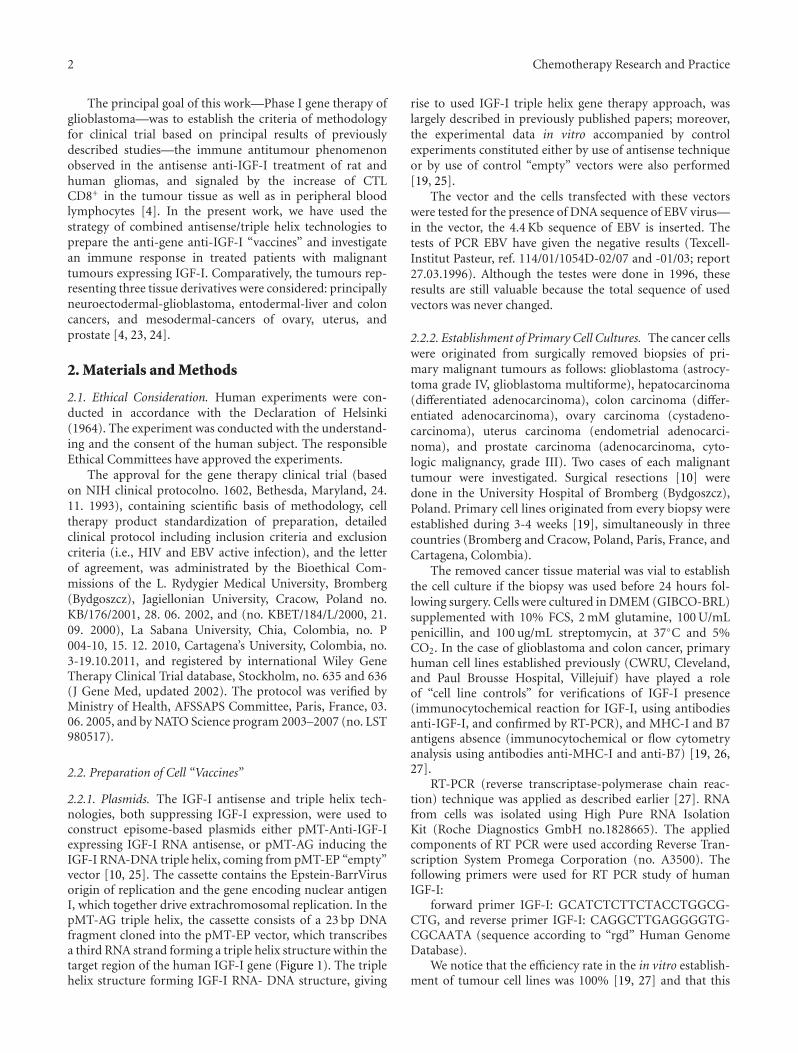
Figure 2: Expression of IGF-I in primary human glioma cell line.RT PCR technique. M—marker. 1—presence of IGF-I in parentalnon transfected cells (200 bp band of amplified DNA using IGF-I primer; see Methods). 2—absence of IGF-I expression in cellstransfected with antisense anti-IGF-I vector. 3—absence of IGF-Iexpression in cells transfected with triple helix anti-IGF-I vector.
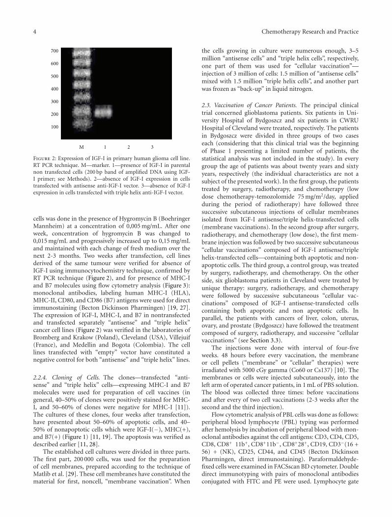
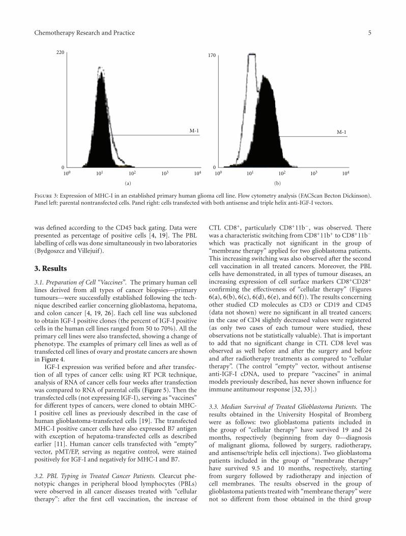
cells was done in the presence of Hygromycin B (BoehringerMannheim) at a concentration of 0,005 mg/mL. After oneweek, concentration of hygromycin B was changed to0,015 mg/mL and progressively increased up to 0,15 mg/mLand maintained with each change of fresh medium over thenext 2-3 months. Two weeks after transfection, cell linesderived of the same tumour were verified for absence ofIGF-I using immunocytochemistry technique, confirmed byRT PCR technique (Figure 2), and for presence of MHC-Iand B7 molecules using flow cytometry analysis (Figure 3):monoclonal antibodies, labeling human MHC-I (HLA),MHC-II, CD80, and CD86 (B7) antigens were used for directimmunostaining (Becton Dickinson Pharmingen) [19, 27].The expression of IGF-I, MHC-I, and B7 in nontransfectedand transfected separately “antisense” and “triple helix”cancer cell lines (Figure 2) was verified in the laboratories ofBromberg and Krakow (Poland), Cleveland (USA), Villejuif(France), and Medellin and Bogota (Colombia). The celllines transfected with “empty” vector have constituted anegative control for both “antisense” and “triple helix” lines.
2.2.4. Cloning of Cells. The clones—transfected “anti-sense” and “triple helix” cells—expressing MHC-I and B7molecules were used for preparation of cell vaccines (ingeneral, 40–50% of clones were positively stained for MHC-I, and 50–60% of clones were negative for MHC-I [11]).The cultures of these clones, four weeks after transfection,have presented about 50–60% of apoptotic cells, and 40–50% of nonapoptotic cells which were IGF-I(−), MHC(+),and B7(+) (Figure 1) [11, 19]. The apoptosis was verified asdescribed earlier [11, 28].
The established cell cultures were divided in three parts.The first part, 200 000 cells, was used for the preparationof cell membranes, prepared according to the technique ofMatlib et al. [29]. These cell membranes have constituted thematerial for first, noncell, “membrane vaccination”. When
the cells growing in culture were numerous enough, 3–5million “antisense cells” and “triple helix cells”, respectively,one part of them was used for “cellular vaccination”—injection of 3 million of cells: 1.5 million of “antisense cells”mixed with 1.5 million “triple helix cells”, and another partwas frozen as “back-up” in liquid nitrogen.
2.3. Vaccination of Cancer Patients. The principal clinicaltrial concerned glioblastoma patients. Six patients in Uni-versity Hospital of Bydgoszcz and six patients in CWRUHospital of Cleveland were treated, respectively. The patientsin Bydgoszcz were divided in three groups of two caseseach (considering that this clinical trial was the beginningof Phase 1 presenting a limited number of patients, thestatistical analysis was not included in the study). In everygroup the age of patients was about twenty years and sixtyyears, respectively (the individual characteristics are not asubject of the presented work). In the first group, the patientstreated by surgery, radiotherapy, and chemotherapy (lowdose chemotherapy-temozolomide 75 mg/m2/day, appliedduring the period of radiotherapy) have followed threesuccessive subcutaneous injections of cellular membranesisolated from IGF-I antisense/triple helix-transfected cells(membrane vaccinations). In the second group after surgery,radiotherapy, and chemotherapy (low dose), the first mem-brane injection was followed by two successive subcutaneous“cellular vaccinations” composed of IGF-I antisense/triplehelix-transfected cells—containing both apoptotic and non-apoptotic cells. The third group, a control group, was treatedby surgery, radiotherapy, and chemotherapy. On the otherside, six glioblastoma patients in Cleveland were treated byunique therapy: surgery, radiotherapy, and chemotherapywere followed by successive subcutaneous “cellular vac-cinations” composed of IGF-I antisense-transfected cellscontaining both apoptotic and non apoptotic cells. Inparallel, the patients with cancers of liver, colon, uterus,ovary, and prostate (Bydgoszcz) have followed the treatmentcomposed of surgery, radiotherapy, and successive “cellularvaccinations” (see Section 3.3).
The injections were done with interval of four-fiveweeks. 48 hours before every vaccination, the membraneor cell pellets (“membrane” or “cellular” therapies) wereirradiated with 5000 cGy gamma (Co60 or Cs137) [10]. Themembranes or cells were injected subcutaneously, into theleft arm of operated cancer patients, in 1 mL of PBS solution.The blood was collected three times: before vaccinationsand after every of two cell vaccinations (2-3 weeks after thesecond and the third injection).
Flow cytometric analysis of PBL cells was done as follows:peripheral blood lymphocyte (PBL) typing was performedafter hemolysis by incubation of peripheral blood with mon-oclonal antibodies against the cell antigens: CD3, CD4, CD5,CD8, CD8+ 11b+, CD8+11b−, CD8+28+, CD19, CD3−(16 +56) + (NK), CD25, CD44, and CD45 (Becton DickinsonPharmingen, direct immunostaining). Paraformaldehyde-fixed cells were examined in FACSscan BD cytometer. Doubledirect immunotyping with pairs of monoclonal antibodiesconjugated with FITC and PE were used. Lymphocyte gate
Chemotherapy Research and Practice 5
220
0
M-1
100 101 102 103 104
(a)
170
0100 101 102 103 104
M-1
(b)
Figure 3: Expression of MHC-I in an established primary human glioma cell line. Flow cytometry analysis (FACScan Becton Dickinson).Panel left: parental nontransfected cells. Panel right: cells transfected with both antisense and triple helix anti-IGF-I vectors.
was defined according to the CD45 back gating. Data werepresented as percentage of positive cells [4, 19]. The PBLlabelling of cells was done simultaneously in two laboratories(Bydgoszcz and Villejuif).
3. Results
3.1. Preparation of Cell “Vaccines”. The primary human celllines derived from all types of cancer biopsies—primarytumours—were successfully established following the tech-nique described earlier concerning glioblastoma, hepatoma,and colon cancer [4, 19, 26]. Each cell line was subclonedto obtain IGF-I positive clones (the percent of IGF-I positivecells in the human cell lines ranged from 50 to 70%). All theprimary cell lines were also transfected, showing a change ofphenotype. The examples of primary cell lines as well as oftransfected cell lines of ovary and prostate cancers are shownin Figure 4.
IGF-I expression was verified before and after transfec-tion of all types of cancer cells: using RT PCR technique,analysis of RNA of cancer cells four weeks after transfectionwas compared to RNA of parental cells (Figure 5). Then thetransfected cells (not expressing IGF-I), serving as “vaccines”for different types of cancers, were cloned to obtain MHC-I positive cell lines as previously described in the case ofhuman glioblastoma-transfected cells [19]. The transfectedMHC-I positive cancer cells have also expressed B7 antigenwith exception of hepatoma-transfected cells as describedearlier [11]. Human cancer cells transfected with “empty”vector, pMT/EP, serving as negative control, were stainedpositively for IGF-I and negatively for MHC-I and B7.
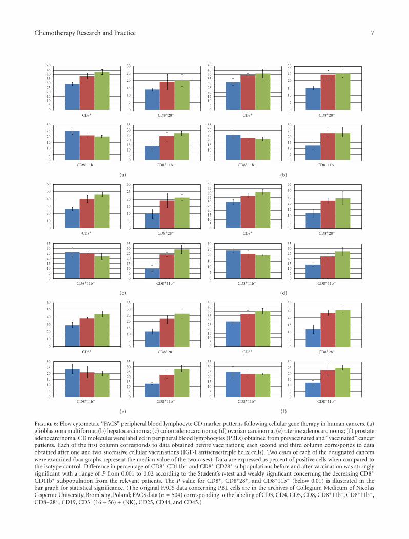
3.2. PBL Typing in Treated Cancer Patients. Clearcut phe-notypic changes in peripheral blood lymphocytes (PBLs)were observed in all cancer diseases treated with “cellulartherapy”: after the first cell vaccination, the increase of
CTL CD8+, particularly CD8+11b−, was observed. Therewas a characteristic switching from CD8+11b+ to CD8+11b−
which was practically not significant in the group of“membrane therapy” applied for two glioblastoma patients.This increasing switching was also observed after the secondcell vaccination in all treated cancers. Moreover, the PBLcells have demonstrated, in all types of tumour diseases, anincreasing expression of cell surface markers CD8+CD28+
confirming the effectiveness of “cellular therapy” (Figures6(a), 6(b), 6(c), 6(d), 6(e), and 6(f)). The results concerningother studied CD molecules as CD3 or CD19 and CD45(data not shown) were no significant in all treated cancers;in the case of CD4 slightly decreased values were registered(as only two cases of each tumour were studied, theseobservations not be statistically valuable). That is importantto add that no significant change in CTL CD8 level wasobserved as well before and after the surgery and beforeand after radiotherapy treatments as compared to “cellulartherapy”. (The control “empty” vector, without antisenseanti-IGF-I cDNA, used to prepare “vaccines” in animalmodels previously described, has never shown influence forimmune antitumour response [32, 33].)
3.3. Median Survival of Treated Glioblastoma Patients. Theresults obtained in the University Hospital of Brombergwere as follows: two glioblastoma patients included inthe group of “cellular therapy” have survived 19 and 24months, respectively (beginning from day 0—diagnosisof malignant glioma, followed by surgery, radiotherapy,and antisense/triple helix cell injections). Two glioblastomapatients included in the group of “membrane therapy”have survived 9.5 and 10 months, respectively, startingfrom surgery followed by radiotherapy and injection ofcell membranes. The results observed in the group ofglioblastoma patients treated with “membrane therapy” werenot so different from those obtained in the third group

6 Chemotherapy Research and Practice
(a) (c)
(d)(b)
Figure 4: Examples of in vitro culture of primary human cancer cells derived from surgical biopsies. (a) and (c) Primary ovarycystadenocarcinoma-derived cells. (b) and (d) Primary prostate adenocarcinoma-derived cells. Parental ovary cancer cells (a) and prostatecancer cells (b) are attached efficiently in the fourth day of culture (arrows). “Antisense/triple helix” anti-IGF-I ovary cancer cells (c) andprostate cancer cells (d), both twenty days after transfection, form the established lines characterized often by the clusters of round apoptoticcells, becoming progressively small (c, arrow; d, arrow up). They are accompanied by nonapoptotic and more voluminous cells (d, arrowdown) presenting generally elongated shape (c and d). The transfected cells are always different from nontransfected parental cells (a, b), asit was demonstrated previously in cases of human glioma and hepatoma cell lines established from primary tumours of glioblastoma andhepatocarcinoma [30, 31] X400.
700600500400300200100
M 1 2 3 4 5 6 7 8 9 10 11 12
Figure 5: Expression of IGF-I in human cancer cell lines. RT PCRtechnique. M—marker. Lines 1 to 6—presence of IGF-I in parentalnontransfected cancer cell lines derived from 1—glioblastoma,2—hepatocarcinoma, 3—colon adenocarcinoma, 4—ovary cystadenocarcinoma, 5—uterus endometrial adenocarcinoma, and 6—prostate adenocarcinoma (200 bp band of amplified DNA usingIGF-I primer; see Section 2. Lines 7 to 12—absence of IGF-Iexpression in cancer cells transfected with both antisense and triplehelix anti-IGF-I vectors: 7—glioblastoma, 8—hepatocarcinoma,9—colon adenocarcinoma, 10—ovary cyst adenocarcinoma, 11—uterus endometrial adenocarcinoma, and 12—prostate adenocarci-noma.
treated by classical therapy; in the last group median survivalwas as 10 and 11 months. For this reason, admitting that thegroup of glioblastoma patients treated with antisense/triplehelix cell injection has given the significant results, all othercancer patients (age 20–65 years: two cases of liver, colon,
ovary, uterus, and prostate cancer diseases) were treated,after surgery and radiotherapy with this type of “cellulartherapy”. Moreover the period of 19 months was chosen asthe end of clinical observations in all treated cancer patients.At 19 months, all these cancer patients were alive and thetreatments were well tolerated (we do not include the detailsof clinical observations concerning different types of treatedcancers, because it is not the subject of this work). The onlysecondary observed effect including glioblastoma patientswas that of increased temperature up to 38-39◦C persistingduring two-three days after every of cell vaccination.
Among the patients treated in USA (University Hospitalsof Cleveland) with anti-gene anti-IGF-I “cellular therapy”(two cell injections), two of the treated patients forminga group of maximum median OS have both survived19 months. The other group of three patients have notresponded so positively to the therapy, showing the mediansurvival compared with that of “membrane” therapy. Thetherapy done in USA has shown that the number of cellvaccinations (between one and four) was not related to themedian OS. Concerning serial MRI/CT performed in USApatients: 1-2-month intervals before vaccine showed contin-uous growth of the intracerebral tumour. MRI one monthafter vaccination showed first evidence of an unequivocal
Chemotherapy Research and Practice 7
0
5
10
15
20
25
30
05
101520253035404550
0
5
10
15
20
25
30
05
101520253035
CD8+ CD8+28+
CD8+11b+ CD8+11b−
(a)
0
5
10
15
20
25
30
05
101520253035404550
05
1015202530
0
5
10
15
20
25
3035
CD8+ CD8+28+
CD8+11b+ CD8+11b−
(b)
0
5
10
15
20
25
30
0
10
20
30
40
50
60
05
1015202530
05
10152025303535
CD8+ CD8+28+
CD8+11b+ CD8+11b−
(c)
0
5
10
15
20
25
30
35
05
101520253035404550
0
5
10
15
20
25
05
10152025303530
CD8+ CD8+28+
CD8+11b+ CD8+11b−
(d)
0
5
10
15
20
25
30
35
0
10
20
30
40
50
60
0
5
10
15
20
25
05
10152025303530
CD8+ CD8+28+
CD8+11b+ CD8+11b−
(e)
0
5
10
15
20
25
30
05
101520253035404550
05
1015202530
0
5
10
15
20
25
3035
CD8+ CD8+28+
CD8+11b+ CD8+11b−
(f)
Figure 6: Flow cytometric “FACS” peripheral blood lymphocyte CD marker patterns following cellular gene therapy in human cancers. (a)glioblastoma multiforme; (b) hepatocarcinoma; (c) colon adenocarcinoma; (d) ovarian carcinoma; (e) uterine adenocarcinoma; (f) prostateadenocarcinoma. CD molecules were labelled in peripheral blood lymphocytes (PBLs) obtained from prevaccinated and “vaccinated” cancerpatients. Each of the first column corresponds to data obtained before vaccinations; each second and third column corresponds to dataobtained after one and two successive cellular vaccinations (IGF-I antisense/triple helix cells). Two cases of each of the designated cancerswere examined (bar graphs represent the median value of the two cases). Data are expressed as percent of positive cells when compared tothe isotype control. Difference in percentage of CD8+ CD11b− and CD8+ CD28+ subpopulations before and after vaccination was stronglysignificant with a range of P from 0.001 to 0.02 according to the Student’s t-test and weakly significant concerning the decreasing CD8+
CD11b+ subpopulation from the relevant patients. The P value for CD8+, CD8+28+, and CD8+11b− (below 0.01) is illustrated in thebar graph for statistical significance. (The original FACS data concerning PBL cells are in the archives of Collegium Medicum of NicolasCopernic University, Bromberg, Poland; FACS data (n = 504) corresponding to the labeling of CD3, CD4, CD5, CD8, CD8+11b+, CD8+11b−,CD8+28+, CD19, CD3−(16 + 56) + (NK), CD25, CD44, and CD45.)

8 Chemotherapy Research and Practice
decrease in size of tumours viewed by radiology in UniversityHospitals of Cleveland. Moreover, all patients treated in USAhad advanced disease with cerebral edema at the time of firsttreatment with vaccine and also were receiving treatmentwith high dose of decadron or related steroids to reducethe effect of CNS edema. This of course has caused furtherjeopardy to the immune system and can explain the relativelynegative results in three last treated cases.
4. Discussion
New ways of treating malignant tumours, and very efficientchemotherapies in particular, are constantly being investi-gated; the best example is that of temozolomide introducedin glioblastoma treatment by Stupp et al. [1]. This type ofchemotherapy has permitted to increase the median survivalup to 15–18 months of glioblastoma patients, showing areal success comparing to current 8–11 months of mediansurvival using surgery and radiotherapy only [1, 2, 4].
The gene therapy may represent a novel approach forcancer therapy. The elucidation of the molecular biologyof cancer cells in recent years has progressively identifiedthe different molecular pathways altered in various cancers.Activation of the PI3K/AKT/GWK3/GS pathway is mediatedby some tyrosine kinase receptors, under the control ofseveral growth factors and cytokines as EGF, PDGF, VEGF,TGFbeta, CSF, and especially IGF-I, whose receptor, IGF-I-R,plays a principal role in the tumour growth process [4, 6, 34].For most of the pathways that have been disclosed, it has beena problem to develop selective molecules having a relevantclinical impact in malignant diseases, including uncuredglioblastoma [35]. To target specific genetic defects, theantisense oligonucleotides have become one of the importantanticancer approaches used in clinical trials [36].
The glioblastoma, as well as other malignant tumours,was recently successfully treated by antisense therapy target-ing TGF beta, using either antisense anti-TGF beta expressingvector [37] or particularly applying the oligodeoxynu-cleotides [38, 39]. Using phosphorothioate TFG beta 2antisense oligonucleotides (AP-12009), an internationalphase II/III study was initiated in patients with TGF beta-overexpressing tumours such as high-grade gliomas, and by2005-2006, the trial was ongoing in over 140 patients withanaplastic astrocytoma (AA) or glioblastoma; the treatmentwas very well tolerated. In 2007, at that time overallsurvival was 24 months, and in the control group, survivalwas 20 months [38, 40]. Results from the clinical trialsconcerning other tumours overexpressing TGF beta werealso recently published (pancreatic carcinoma, metastaticmelanoma, or advanced colorectal carcinoma); the treatmentwas well tolerated in all types of tumour diseases [38].Other antisense approaches of malignant tumour treatmenthave been developed recently, since 2001, especially those ofantisense anti-IGF-I-Receptor [6, 41]. AS IGF-I-R strategy oftreatment of glioblastoma [41] was not continued. It seemsthat this therapy could be more efficient if the cell “vaccines”were prepared after cloning of IGF-I-R antisense cells forMHC-I expression.
In anti-gene anti-IGF-I approach, we have applied bothantisense and triple helix technologies, permitting to stopsimultaneously the expression of IGF-I on translation andtranscription levels [4]. Moreover, in vivo AS IGF-I approachwas also developed [42]; 45 patients with PHC werecotransfected in vivo with antisense IGF-I expression vectorand sense B7.1 expression vector. At two years followingtreatment of PHC stage II, there was marked reduction intumour recurrence—from 62 to 20%.
In antisense anti TGF-beta technique as well as inanti-IGF-I and IGF-I-R approaches, the immune antitu-mour response was signalled as a principal mechanism ofantisense technology inhibiting growth factors and theirsignalling pathway [19, 37]. The mechanism of antisensetherapy targeting growth factors and their receptors is acombination of an augmentation of the immune antitumourresponse and of an inhibition of the signal transductionpathway—PI3K/AKT/GWK3/GS—that is involved in thetransformed phenotype of the tumour [4, 13]. As far asPI3K/AKT/GWK3/GS pathway (in relation with glioma) isconsidered, it was recently demonstrated that in experimen-tal antisense antiglycogen synthetase, GS, tumour therapy,the transfected AS GS cells were also immunogenic (MHC-Iexpression) [13, 14]. Anyway in AS GS strategy an immuneantitumour response was not as striking as when usingAS IGF-I approach. This shows that AS IGF-I appearsas a dominant tool for the arrest of tumour progression.Moreover, targeting IGF-I instead of IGF-I receptor seemsmore efficient: because of downstream elements involvedin the IGF-I-R transduction pathway, signals from IGF-I-R can be inappropriate or exaggerated [34]. Nevertheless, ifcrosstalk of IGF-I’s related different pathways is considered,IGF-I, through its binding to IGF-I-R, which activatesPI3K/AKT transduction cascade, has been reported to blockthe apoptosis pathway (IRS/PI3K/AKT/Bcl or AKT/GSK3 orCa2+ or caspases) [4, 6]. The final result of AS IGF-I approachincluding the TK/PI3K/AKT pathway elements inhibition isan immune response mediated in vivo by lymphocytes T CD8and APC cells [4]. As to PI3K/AKT/GWK3/GS pathways ofIGF-I AS or TGFbeta AS therapies, we cannot avoid therelation with PI3K/PKC/RAF/MAPK chain, and we cannotexclude that the inhibition of TK/PI3K/AKT pathway usingAS IGF-I approach can be reinforced by “side” effect ofMAPK inhibition [4]. The inhibitors of RAF targeting theATP binding site, as well as the inhibitors of MAPK at anon-ATP site, were also introduced in cancer clinical trials[32, 43].
Using described here IGF-I antisense/triple helix strategy,all treated patients have well tolerated the three injectionsof transfected cancer cells. The PBL cells have shownan increase in CD8+CD28+ molecules with a character-istic switching from CD8+11b+ to CD8+11b− phenotype,observed after two cell vaccinations, reflecting the enhancedactivation of cytotoxic T-cells in blood. These results con-cerning the switching CD8+11b+ to CD8+11b− in differenttreated tumours have confirmed previously obtained data inglioblastoma and hepatoma treatment using antisens anti-IGF-I approach [4, 28]. The work in progress has also shown,in different treated tumours described here, an increased

Chemotherapy Research and Practice 9
percentage of T CD25 (interleukin-2 receptor), in thecontext of CD4, which has confirmed the results obtainedin glioblastoma treatment [4]. (Currently we are engagedin Phase II with 60 patients treated for different typesof cancers mentioned earlier, including lung and stomachcancers.) The only secondary observed effect was increasedtemperature, 38-39◦C, confirming the immune responseinduced by antisense/triple helix “vaccines”. The specificityof immune response was every time confirmed as follows. Asdemanded by approved clinical trials (see higher mentionedBioethical Committees from 1993 to 2010), the removedPBLs of patients were tested in vitro for immune responseafter contact with autologous tumor cells to demonstrateantitumor activation throught high percentage of specificT cells observed after vaccination compared to controls—before vaccination (the tumoral cells were labeled with Cr51before the test of lyses in the presence of CTL cells [33, 44];data not shown).
Regarding injection of cell membranes, the switchingmentioned earlier was not significant. The challenge ofinjection of membranes, isolated from IGF-I antisense/triplehelix-transfected cells expressing MHC-I, has proved that thewhole transfected cell population is necessary to producean in vivo antitumour effect. At first, the cytoplasm of thetransfected cells contains the IGF-I antisense RNA and IGF-Itriple helix RNA-DNA structures constituting the principleof anti-gene cellular therapy [25, 45]. Next, the cellular ther-apy described here has shown that both cell populations, aswell MHC-I and B7 expressing transfected cells as apoptoticcells, are necessary to induce in vivo an immune antitumourresponse involving APC activating CD8+ T cells [4, 11, 30, 45,46]. (It was previously demonstrated that doubly transfectedcells, using antisense anti-MHC-I and anti-B7 vectors, losetheir apoptotic and immune antitumour characters [19]. Itwas, this way, shown that both processes, immunogenicity(MHC-I and B7 expression) and apoptosis, “work” together[11, 19, 31].) On the other side we have previously comparedthe efficiency of gene therapy—using the injection of IGF-IAS nucleotides, and that of described here “cellular therapy”much more promising. In gene therapy approach, after theinjection of IGF-I AS nucleotides directly to the tumor, thecancer cells internalizing AS nucleotides could not becomeimmediately immunogenic to induce the rapid immuneresponse, and for the same reason to develop the efficientapoptosis. Moreover, in the gene therapy, the cotransfectionwith B7 expression vector was necessary to reinforce theimmune response [28, 42].
As far as the relationship between anti-gene anti-IGF-I technology and immunogenicity is considered, theabsence of IGF-I synthesis in “antisense-” and “triple-helix”-transfected cells could lead to a compensative increase inIGF-I receptor (tyrosine kinase); IGF-I and IGF-II present infoetal calf serum of culture medium, as well as intracellularIGF-II can interact with the type I receptor [47]. Indeed, theincrease of IGF-I receptor level could explain the expressionof B7. There is a known relation between the signaltransduction pathway of tyrosine kinase and the inductionof B7 molecules: enhancement in B-7 costimulation througha cAMP mechanism linked to tyrosine kinase of the CD 28
receptor has been previously reported [15]. (The costimula-tory B7 molecule in antigen presenting cells (APCs) is boundto the counter-receptor CD28 and/or CTLA4 expressedon the T-cells [20, 48, 49].) B7 was present in differentantisense and triple helix anti-IGF-I transfected cancer cellsbut absent in transfected human hepatoma and in previouslydescribed murine hepatoma [47]. Those results have alsodemonstrated that the expression of MHC-I in human-transfected hepatoma cells was much higher than that intransfected human glioma cells. This strong expression ofMHC-I in human-transfected hepatoma lines (5 times,compared to human-transfected glioma lines) could explainthat the presence of MHC-I was sufficient to induce T CD8lymphocytes response in the absence of B7 antigen [28]. Tosummarize the immune antitumour mechanism of anti-geneanti-IGF-I strategy, this aspect was published previously (i.e.,[4, 28]). As far as largely studied glioma treatment is con-sidered, and similarly other concerned tumours, the mech-anism concerns the reaction between activated lympho-cytes expressing CD8CD28 and immune molecules MHC-I and B7. The following chain reaction could occur: cul-tured cloned glioma cells (IGF-I(+), MHC-I(−), B7(−)) ⇒cultured transfected anti-gene IGF-I cells (IGF-I(−), MHC-I(+), B7(+)) ⇒ injection (glioblastoma patients) ⇒ induc-tion of CTL CD8(+) CD28(+) ⇒ destruction of injectedtransfected anti-gene IGF-I cells (IGF-I(−), MHC-I(+),B7(+)), and arrest of a solid glioma tumour (IGF-I(+/−),MHC-I(+), B7(+)) (see also caption of Figure 1).
The immune criteria of used vaccines are strongly relatedto the preparation of cancer cells to be used as vaccine.Cancer cells cultivated under stem cell-permissive conditionsmore closely reflect the tumor of origin, including the geneticprofile, than the parental tumor adherently growing cellsunder conventional cultivation conditions [50, 51]. In ourexperimental clinical trial, to avoid this effect of “contamina-tion” increased by numerous passages, the primary cancerscells and transfected cancer cells were systematically clonedafter every passage to obtain in vitro, in the first case 100%IGF-I(+), MHC-I(−), and in the second case 100% IGF-I(−), MHC-I(+) expression. The vaccines prepared with nocloned cells (both cancer cells and transfected cancer cells)did not induce the immune response in vivo in animals andin patients.
Moreover, to produce their immune antitumour effect,the vaccines were composed of—criteria sine qua non— themixture of higher characterized cancer-transfected cells andof cancer cell-derived apoptotic cells.
We need to add that if the all clinical results concerningthis work could be cited, we would be obliged to give thesummary of clinical data concerning 42 cancer patientsand their laboratory data analyzed in this paper (Brombergand Cleveland), including corresponding original FACS ofPBL cells labeling of every patient. The article concerningonly clinical results of every patient (clinical data of treatedcancer patients are in archives of four hospitals, in Bromberg,Cracow, Cleveland, New York, Shanghai, and Bangkok; 70patients only in Bromberg) including detailed inclusion andexclusion criteria, clinical and laboratory data— PCR and RTPCR of IGF-I; immunocytochemistry of cytokines, growth

10 Chemotherapy Research and Practice
factors, and MHC-I and B7 molecules; and blood test ofevery patient—, will be published as separate article withobligatory statistics, not treated in the presented manuscript.
5. Conclusions
Our presented work concerns the criteria established formethodology of anti-IGF-I gene therapy analyzing our dif-ferent previous basic and clinical results obtained in Europe,USA, and Asia, following our NATO science program (seeAcknowledgment), and published recently [4, 27, 28], per-mitting to start Phase I and II in South America (Colombia)and Africa (Senegal). This way we have established thecommon criteria for selection of vaccines (expression ofIGF-I, MHC-I, B7) and of PBL cells markers (CD8+-relatedmolecules) in patients presenting the arrest of growingtumours. The various therapies in the treatment of cancer arestill experimental [52]. A number of strategies for inhibitinggene expression have been developed including the triplehelix approach, antisense cDNA, and oligodeoxynucleotides.Among the new strategies in the efforts of treating malignanttumours expressing different growth factors, and morespecifically IGF-I, TGF beta, VEGF, or EGF [3, 6, 35, 37],the anti-gene therapy approach, either antisense or triplehelix, appears as a promising solution [39]. Although inthe presented work only limited numbers of glioblastomapatients were treated, the clinical results obtained are positive(minimum survival has reached 19 months). The anti-gene anti-IGF-I therapy, giving a strong immune antitu-mour response in different comparatively studied tumourdiseases, presents all characteristics of cell immunotherapy(CD8+and CD28+ expression in T lymphocytes, and MHC-I and B7 expression in “vaccine” cells) including apoptoticphenomenon [4, 15, 45, 53]. We suggest that anti-genecell therapy, giving comparable results to those of currentlyapplied chemotherapy, inhibitors, or antibodies [1, 2, 4, 54],could be used either alone [39, 55] or as combined therapies,that is, antisenses targeting simultaneously different elementsof growth factors signalling pathway [13, 14, 32, 43], or asantisense/chemotherapy. The combined anticancer strategiesconsidering the role of immune antitumour response [35,56–62], including study of control CD8(+) T-cell effectorfunctions [63], new tools of cell transfection [64] andespecially the search for new oncoproteins [65], and growthfactor targets [6, 14, 35, 66, 67], appear as the near futurechallenge. Among growth factors, targeting IGF-I system inrelation with cancer therapy constitutes a permanent basicand clinical research [68–70].
Conflict of Interests
The authors have declared that no conflict of interests exist(research, clinical trial, financial gain).
Acknowledgments
The authors would like to thank Dr. R. Stupp (CHU,Lausanne), Professor G. Hilldebrand (Erasmus Hospital,
Bruxelles), and Dr. M. Molano (National Institut of Can-cerology, Bogota) for helpful discussion of results, andProfessors W. Szymanski and M. Wolski (Medical Universityof Bydgoszcz) and Professor T. Popiela (Collegium Medicum,Krakow) for providing malignant tumour biopsies, and forsuggestions concerning clinical trial phase I. They thank Dr.P. Kopinski (Medical University of Bydgoszcz) for analyzingPBL blood samples, Dr. L. C. Upegui-Gonzalez (INSERM,Villejuif) for suggestions concerning the establishment ofprimary tissue cultures, T. W. Trojan (Cartagena’s University)for photographic in vitro results and for graphic presentationof PBL labelling data, and Y. Celis (Secretariat of Health,Bogota) for final corrections. This work was supported byGrant of NATO Science Program, no. CLG LST 980517,Cellvax SA (Lille, France), Grant of American CancerSociety (Cleveland, USA), Grants of the State Committee forScientific Research, Poland, nos. 6 P05C 016 20 and 3 P05B089 23, Grant of La Sabana University (Colombia), and byfinancial support of INSERM, France.
References
[1] R. Stupp, M. E. Hegi, B. Neyns et al., “Phase I/IIa study ofcilengitide and temozolomide with concomitant radiotherapyfollowed by cilengitide and temozolomide maintenance ther-apy in patients with newly diagnosed glioblastoma,” Journal ofClinical Oncology, vol. 28, no. 16, pp. 2712–2718, 2010.
[2] P. Y. Wen, W. K. A. Yung, K. R. Lamborn et al., “Phase I/IIstudy of imatinib mesylate for recurrent malignant gliomas:North American Brain Tumor Consortium Study 99-08,”Clinical Cancer Research, vol. 12, no. 16, pp. 4899–4907, 2006.
[3] Q. Pan, Y. Chanthery, W. C. Liang et al., “Blocking neuropilin-1 function has an additive effect with anti-VEGF to inhibittumor growth,” Cancer Cell, vol. 11, no. 1, pp. 53–67, 2007.
[4] J. Trojan, J. F. Cloix, M. Y. Ardourel, M. Chatel, and D.D. Anthony, “Insulin-like growth factor type I biology andtargeting in malignant gliomas,” Neuroscience, vol. 145, no. 3,pp. 795–811, 2007.
[5] J. Kjaergaard, L. X. Wang, H. Kuriyama, S. Shu, and G.E. Plautz, “Active immunotherapy for advanced intracranialmurine tumors by using dendritic cell-tumor cell fusionvaccines,” Journal of Neurosurgery, vol. 103, no. 1, pp. 156–164,2005.
[6] R. Baserga, “The insulin-like growth factor-I receptor as atarget for cancer therapy,” Expert Opinion on TherapeuticTargets, vol. 9, no. 4, pp. 753–768, 2005.
[7] J. L. R. Rubenstein, J. F. Nicolas, and F. Jacob, “Constructionof a retrovirus capable of transducing and expressing genesin multipotential embryonic cells,” Proceedings of the NationalAcademy of Sciences of the United States of America, vol. 81, no.22, pp. 7137–7140, 1984.
[8] P. B. Dervan, “Reagents for the site-specific cleavage ofmegabase DNA,” Nature, vol. 359, no. 6390, pp. 87–88, 1992.
[9] C. Helene, “Control of oncogene expression by antisensenucleic acids,” European Journal of Cancer Part A, vol. 30, no.11, pp. 1721–1726, 1994.
[10] J. Trojan, T. R. Johnson, S. D. Rudin et al., “Gene therapy ofmurine teratocarcinoma: separate functions for insulin-likegrowth factors I and II in immunogenicity and differentia-tion,” Proceedings of the National Academy of Sciences of theUnited States of America, vol. 91, no. 13, pp. 6088–6092, 1994.

Chemotherapy Research and Practice 11
[11] S. Ellouk-Achard, S. Djenabi, G. A. De Oliveira et al., “In-duction of apoptosis in rat hepatocarcinoma cells by expres-sion of IGF-I antisense c-DNA,” Journal of Hepatology, vol. 29,no. 5, pp. 807–818, 1998.
[12] S. Trabado, P. N. Van Binh, C. Martin et al., “Stimulation ofanti-melanoma immune effectors via modified tumour cellsexhibiting inhibited IGF-I and low CD9,” Biomedicine andPharmacotherapy, vol. 61, no. 8, pp. 494–498, 2007.
[13] D. R. Premkumar, B. Arnold, E. P. Jane, and I. F. Pollack,“Synergistic interaction between 17-AAG and phosphatidyli-nositol 3-kinase inhibition in human malignant glioma cells,”Molecular Carcinogenesis, vol. 45, no. 1, pp. 47–59, 2006.
[14] M. Ardourel, M. Blin, J. L. Moret et al., “A new putative targetfor antisense gene therapy of glioma: glycogen synthase,”Cancer Biology and Therapy, vol. 6, no. 5, pp. 719–723, 2007.
[15] R. H. Schwartz, “Costimulation of T lymphocytes: the role ofCD28, CTLA-4, and B7/BB1 in interleukin-2 production andimmunotherapy,” Cell, vol. 71, no. 7, pp. 1065–1068, 1992.
[16] S. Patel, B. Doble, and J. R. Woodgett, “Glycogen synthasekinase-3 in insulin and Wnt signalling: a double-edgedsword?” Biochemical Society Transactions, vol. 32, no. 5, pp.803–808, 2004.
[17] J. Trojan, A. Ly, M. X. Wei et al., “Antisense anti IGF-I cellulartherapy of malignant tumours: immune response in cancerpatients,” Biomedicine and Pharmacotherapy, vol. 64, no. 8, pp.576–578, 2010.
[18] M. E. Beckner, G. T. Gobbel, R. Abounader et al., “Glycolyticglioma cells with active glycogen synthase are sensitive toPTEN and inhibitors of PI3K and gluconeogenesis,” Labora-tory Investigation, vol. 85, no. 12, pp. 1457–1470, 2005.
[19] A. Ly, H. T. Duc, M. Kalamarides et al., “Human glioma cellstransformed by IGF-I triple helix technology show immuneand apoptotic characteristics determining cell selection forgene therapy of glioblastoma,” Journal of Clinical Pathology,vol. 54, no. 4, pp. 230–239, 2001.
[20] F. A. Harding, J. G. McArthur, J. A. Gross, D. H. Raulet, andJ. P. Allison, “CD28-mediated signalling co-stimulates murineT cells and prevents induction of anergy in T-cell clones,”Nature, vol. 356, no. 6370, pp. 607–609, 1992.
[21] A. Wu, S. Wiesner, J. Xiao et al., “Expression of MHC I andNK ligands on human CD133+ glioma cells: possible targetsof immunotherapy.,” Journal of neuro-oncology, vol. 83, no. 2,pp. 121–131, 2007.
[22] T. Di Tomaso, S. Mazzoleni, E. Wang et al., “Immunobi-ological characterization of cancer stem cells isolated fromglioblastoma patients,” Clinical Cancer Research, vol. 16, no.3, pp. 800–813, 2010.
[23] B. Djavan, M. Waldert, C. Seitz, and M. Marberger, “Insulin-like growth factors and prostate cancer,” World Journal ofUrology, vol. 19, no. 4, pp. 225–233, 2001.
[24] Y. Wu, S. Yakar, L. Zhao, L. Hennighausen, and D. LeRoith,“Circulating insulin-like growth factor-I levels regulate coloncancer growth and metastasis,” Cancer Research, vol. 62, no. 4,pp. 1030–1035, 2002.
[25] A. Shevelev, P. Burfeind, E. Schulze et al., “Potential triplehelix-mediated inhibition of IGF-I gene expression signifi-cantly reduces tumorigenicity of glioblastoma in an animalmodel,” Cancer Gene Therapy, vol. 4, no. 2, pp. 105–112, 1997.
[26] T. Popiela, M. Sierzega, T. Gach, P. Jarocki, and J. Trojan,“Phase I trial of colorectal cancer immunotherapy usingautologous cancer cells transfected with an IGF-I antisenseplasmid [abstract],” Acta Chirurgica Belgica, vol. 5, no. 103,pp. s2–s3, 2003.
[27] L. A. Trojan, P. Kopinski, A. Mazurek et al., “IGF-I triple helixgene therapy of rat and human gliomas,” Roczniki AkademiiMedycznej w Bialymstoku, vol. 48, pp. 18–27, 2003.
[28] L. A. Trojan, A. Ly, L. C. Upegui-Gonzalez et al., “Antisenseanti IGF-I therapy of primary hepatic cancer,” African Journalof Cancer, vol. 1, pp. 1–10, 2009.
[29] M. A. Matlib, M. Kihara, C. Farrell, and R. C. Dage, “TheNa+-Ca2+ exchange system in vascular smooth muscle cellmembrane vesicles isolated from cultured cells and from tissueis similar,” Biochimica Biophysica Acta, vol. 939, no. 1, pp. 173–177, 1988.
[30] L. C. Upegui-Gonzalez, A. Ly, M. Sierzega et al., “IGF-I triple helix strategy in hepatoma treatment,” Hepato-Gastroenterology, vol. 48, no. 39, pp. 660–666, 2001.
[31] J. Trojan, H. T. Duc, L. C. Upegui-Gonzalez et al., “Presenceof MHC-I and B-7 molecules in rat and human glioma cellsexpressing antisense IGF-I mRNA,” Neuroscience Letters, vol.212, no. 1, pp. 9–12, 1996.
[32] H. W. Lo, “Targeting Ras-RAF-ERK and its interactive path-ways as a novel therapy for malignant gliomas,” Current CancerDrug Targets, vol. 10, no. 8, pp. 840–848, 2010.
[33] B. Frankenberger, H. Pohla, E. Noessner et al., “Influence ofCD80, interleukin-2, and interleukin-7 expression in humanrenal cell carcinoma on the expansion, function, and survivalof tumor-specific CTLs,” Clinical Cancer Research, vol. 11, no.5, pp. 1733–1742, 2005.
[34] M. N. Pollak, E. S. Schernhammer, and S. E. Hankinson,“Insulin-like growth factors and neoplasia,” Nature ReviewsCancer, vol. 4, no. 7, pp. 505–518, 2004.
[35] R. K. Goudar, Q. Shi, M. D. Hjelmeland et al., “Combinationtherapy of inhibitors of epidermal growth factor recep-tor/vascular endothelial growth factor receptor 2 (AEE788)and the mammalian target of rapamycin (RAD001) offersimproved glioblastoma tumor growth inhibition,” MolecularCancer Therapeutics, vol. 4, no. 1, pp. 101–112, 2005.
[36] N. Dias and C. A. Stein, “Antisense oligonucleotides: basicconcepts and mechanisms,” Molecular cancer therapeutics, vol.1, no. 5, pp. 347–355, 2002.
[37] H. Fakhrai, J. C. Mantil, L. Liu et al., “Phase I clinical trial of aTGF-β antisense-modified tumor cell vaccine in patients withadvanced glioma,” Cancer Gene Therapy, vol. 13, no. 12, pp.1052–1060, 2006.
[38] K. H. Schlingensiepen, R. Schlingensiepen, A. Steinbrecheret al., “Targeted tumor therapy with the TGF-β2 antisensecompound AP 12009,” Cytokine and Growth Factor Reviews,vol. 17, no. 1-2, pp. 129–139, 2006.
[39] K. H. Schlingensiepen, B. Fischer-Blass, S. Schmaus, and S.Ludwig, “Antisense therapeutics for tumor treatment: theTGF-beta2 inhibitor AP 12009 in clinical development againstmalignant tumors,” Recent Results in Cancer Research, vol. 177,pp. 137–150, 2008.
[40] P. Hau, P. Jachimczak, R. Schlingensiepen et al., “Inhibition ofTGF-β2 with ap 12009 in recurrent malignant gliomas: frompreclinical to phase I/II studies,” Oligonucleotides, vol. 17, no.2, pp. 201–212, 2007.
[41] D. W. Andrews, M. Resnicoff, A. E. Flanders et al., “Results ofa pilot study involving the use of an antisense oligodeoxynu-cleotide directed against the insulin-like growth factor typeI receptor in malignant astrocytomas,” Journal of ClinicalOncology, vol. 19, no. 8, pp. 2189–2200, 2001.
[42] D. D. Anthony, “Ex vivo and in vivo IGF-1 antisense RNAstrategies for treatment of cancers in humans [abstract],”Cancer Gene Therapy, vol. 2, p. s322, 1997.

12 Chemotherapy Research and Practice
[43] J. Downward, “Targeting RAS signalling pathways in cancertherapy,” Nature Reviews Cancer, vol. 3, no. 1, pp. 11–22, 2003.
[44] N. Tokunaga, T. Murakami, Y. Endo et al., “Human monocyte-derived dendritic cells pulsed with wild-type p53 proteinefficiently induce CTLs against p53 overexpressing humancancer cells,” Clinical Cancer Research, vol. 11, no. 3, pp. 1312–1318, 2005.
[45] M. L. Albert, B. Sauter, and N. Bhardwaj, “Dendritic cellsacquire antigen from apoptotic cells and induce class I-restricted CTLS,” Nature, vol. 392, no. 6671, pp. 86–89, 1998.
[46] J. Trojan, T. R. Johnson, S. D. Rudin, J. Ilan, M. L. Tykocinski,and J. Ilan, “Treatment and prevention of rat glioblastomaby immunogenic C6 cells expressing antisense insulin-likegrowth factor I RNA,” Science, vol. 259, no. 5091, pp. 94–97,1993.
[47] C. Lafarge-Frayssinet, H. T. Duc, C. Frayssinet et al., “Anti-sense insulin-like growth factor I transferred into a rathepatoma cell line inhibits tumorigenesis by modulatingmajor histocompatibility complex I cell surface expression,”Cancer Gene Therapy, vol. 4, no. 5, pp. 276–285, 1997.
[48] G. J. Freeman, G. S. Gray, C. D. Gimmi et al., “Structure,expression, and T cell costimulatory activity of the murinehomologue of the human B lymphocyte activation antigenB7,” Journal of Experimental Medicine, vol. 174, no. 3, pp. 625–631, 1991.
[49] T. J. Kindt, R. A. Goldsby, and B. A. Osborne, Kuby Immunol-ogy, W. H. Freeman and Co, New York, NY, USA, 2007.
[50] P. Tunici, L. Bissola, E. Lualdi et al., “Genetic alterations andin vivo tumorigenicity of neurospheres derived from an adultglioblastoma,” Molecular Cancer, vol. 3, article no. 25, 2004.
[51] R. Galli, E. Binda, U. Orfanelli et al., “Isolation and charac-terization of tumorigenic, stem-like neural precursors fromhuman glioblastoma,” Cancer Research, vol. 64, no. 19, pp.7011–7021, 2004.
[52] R. Stupp and A. F. Hottinger, “Management of malignantglioma—quo vadis?” Onkologie, vol. 31, no. 6, pp. 300–302,2008.
[53] S. E. Townsend and J. P. Allison, “Tumor rejection after directcostimulation of CD8+ T cells by B7-transfected melanomacells,” Science, vol. 259, no. 5093, pp. 368–370, 1993.
[54] S. V. Labropoulos and E. D. Razis, “Imatinib in the treatmentof dermatofibrosarcoma protuberans,” Biologics, vol. 1, no. 4,pp. 347–353, 2007.
[55] L. Benimetskaya and C. A. Stein, “Antisense therapy: recentadvances and relevance to prostate cancer,” Clinical ProstateCancer, vol. 1, no. 1, pp. 20–30, 2002.
[56] B. Jansen, V. Wacheck, E. Heere-Ress et al., “Chemosensiti-sation of malignant melanoma by BCL2 antisense therapy,”Lancet, vol. 356, no. 9243, pp. 1728–1733, 2000.
[57] U. Zangemeister-Wittke, “Antisense to apoptosis inhibitorsfacilitates chemotherapy and TRAIL-induced death signaling,”Annals of the New York Academy of Sciences, vol. 1002, pp. 90–94, 2003.
[58] O. L. Rincon, L. R. Pareja, S. Jaramillo, and B. H. Aristiza-bal, “Human papillomavirus, immune response and cervicalcancer: a complex relationship,” Review Colombian of Obstetricand Ginecology, vol. 58, no. 3, pp. 58–68, 2007.
[59] D. A. Reardon, J. A. Quinn, J. J. Vredenburgh et al., “Phase1 trial of gefitinib plus sirolimus in adults with recurrentmalignant glioma,” Clinical Cancer Research, vol. 12, no. 3 I,pp. 860–868, 2006.
[60] F. M. Lemoine, M. Cherai, C. Giverne et al., “Massive expan-sion of regulatory T-cells following interleukin 2 treatmentduring a phase I-II dendritic cell-based immunotherapy of
metastatic renal cancer,” International Journal of Oncology, vol.35, no. 3, pp. 569–581, 2009.
[61] M. Cavazzana-Calvo, S. Hacein-Bey-abina, and A. Fischer,“Ten years of gene therapy: thoughts and perspectives,”Medecine/Sciences, vol. 26, no. 2, pp. 115–118, 2010.
[62] J. Tang, P. Flomenberg, L. Harshyne, L. Kenyon, and D. W.Andrews, “Glioblastoma patients exhibit circulating tumor-specific CD8+ T cells,” Clinical Cancer Research, vol. 11, no.14, pp. 5292–5299, 2005.
[63] P. Chappert, M. Leboeuf, P. Rameau et al., “Antigen-specificTreg impair CD8+ T-cell priming by blocking early T-cellexpansion,” European Journal of Immunology, vol. 40, no. 2,pp. 339–350, 2010.
[64] T. Le Gall, D. Loizeau, E. Picquet et al., “A novel cationiclipophosphoramide with diunsaturated lipid chains: synthesis,physicochemical properties, and transfection activities,” Jour-nal of Medicinal Chemistry, vol. 53, no. 4, pp. 1496–1508, 2010.
[65] S. Engelen, L. A. Trojan, S. Sacquin-Mora, R. Lavery, and A.Carbone, “Joint evolutionary trees: a large-scale method topredict protein interfaces based on sequence sampling,” PLoSComputational Biology, vol. 5, no. 1, Article ID e1000267,2009.
[66] L. A. Trojan, P. Kopinski, M. X. Wei et al., “IGF-I: fromdiagnostic to triple-helix gene therapy of solid tumors,” ActaBiochimica Polonica, vol. 49, no. 4, pp. 979–990, 2002.
[67] J. T. Mack, C. B. Brown, and K. D. Tew, “ABCA2 as atherapeutic target in cancer and nervous system disorders,”Expert Opinion on Therapeutic Targets, vol. 12, no. 4, pp. 491–504, 2008.
[68] Y. Adachi, H. Yamamoto, H. Ohashi et al., “A candidate tar-geting molecule of insulin-like growth factor-I receptor forgastrointestinal cancers,” World Journal of Gastroenterology,vol. 16, no. 46, pp. 5779–5789, 2010.
[69] D. Moro-Sibilot, M. Coudurier, and S. Lantuejoul, “Targetinginsulin-like growth factors in the treatment of cancer,” Revuedes Maladies Respiratoires, vol. 27, no. 8, pp. 959–963, 2010.
[70] D. Sachdev, “Targeting the Type I insulin-like growth factorsystem for breast cancer therapy,” Current Drug Targets, vol.11, no. 9, pp. 1121–1132, 2010.

Submit your manuscripts athttp://www.hindawi.com
Stem CellsInternational
Hindawi Publishing Corporationhttp://www.hindawi.com Volume 2014
Hindawi Publishing Corporationhttp://www.hindawi.com Volume 2014
MEDIATORSINFLAMMATION
of
Hindawi Publishing Corporationhttp://www.hindawi.com Volume 2014
Behavioural Neurology
EndocrinologyInternational Journal of
Hindawi Publishing Corporationhttp://www.hindawi.com Volume 2014
Hindawi Publishing Corporationhttp://www.hindawi.com Volume 2014
Disease Markers
Hindawi Publishing Corporationhttp://www.hindawi.com Volume 2014
BioMed Research International
OncologyJournal of
Hindawi Publishing Corporationhttp://www.hindawi.com Volume 2014
Hindawi Publishing Corporationhttp://www.hindawi.com Volume 2014
Oxidative Medicine and Cellular Longevity
Hindawi Publishing Corporationhttp://www.hindawi.com Volume 2014
PPAR Research
The Scientific World JournalHindawi Publishing Corporation http://www.hindawi.com Volume 2014
Immunology ResearchHindawi Publishing Corporationhttp://www.hindawi.com Volume 2014
Journal of
ObesityJournal of
Hindawi Publishing Corporationhttp://www.hindawi.com Volume 2014
Hindawi Publishing Corporationhttp://www.hindawi.com Volume 2014
Computational and Mathematical Methods in Medicine
OphthalmologyJournal of
Hindawi Publishing Corporationhttp://www.hindawi.com Volume 2014
Diabetes ResearchJournal of
Hindawi Publishing Corporationhttp://www.hindawi.com Volume 2014
Hindawi Publishing Corporationhttp://www.hindawi.com Volume 2014
Research and TreatmentAIDS
Hindawi Publishing Corporationhttp://www.hindawi.com Volume 2014
Gastroenterology Research and Practice
Hindawi Publishing Corporationhttp://www.hindawi.com Volume 2014
Parkinson’s Disease
Evidence-Based Complementary and Alternative Medicine
Volume 2014Hindawi Publishing Corporationhttp://www.hindawi.com