methods for cell genotyping
TRANSCRIPT
US 20110033862A1
(12) Patent Application Publication (10) Pub. No.: US 2011/0033862 A1 (19) United States
Rabinowitz et al. (43) Pub. Date: Feb. 10, 2011
(54) METHODS FOR CELL GENOTYPING
Matthew RabinoWitz, Portola Valley, CA (US); David S. Johnson, San Francisco, CA (US); Johan Baner, Palo Alto, CA (US); Zachary Demko, Somerville, MA (US); Cengiz Cinnioglu, Sunnyvale, CA (US)
(75) Inventors:
Correspondence Address: GREENBERG TRAURIG, LLP ONE INTERNATIONAL PLACE, 20th FL, ATTN: PATENT ADMINISTRATOR BOSTON, MA 02110 (US)
(73) Assignee: Gene Security Network, Inc.
(21) Appl. No.: 12/918,445
(22) PCT Filed: Feb. 19, 2009
(86) PCT No.: PCT/US09/34506
§ 371 (0X1)’ (2), (4) Date: Oct. 7, 2010
Publication Classi?cation
(51) Int. Cl. C12Q 1/68 (2006.01) C12P 19/34 (2006.01)
(52) U.S. Cl. .......................................... .. 435/6; 435/915
(57) ABSTRACT
Methods for cell genotyping are disclosed herein. A method for determining the genomic data of one or a small number of cells, or from fragmentary DNA, Where a limited quantity of genetic data is available may include adding one or more targeted primers to a Whole genome ampli?cation of a cell, increasing the accuracy With Which key alleles are measured in the context of a Whole genome ampli?cation. The genetic material from a single cell may be divided into fractions, each of Which may be separately genotyped, alloWing the recon struction of the cells haplotype. The genetic material from a single cell may be divided into fractions, each of Which may be separately genotyped, and the distribution of the various alleles in the different fractions may be used to determine the ploidy state of one or a plurality of chromosomes in the cell.
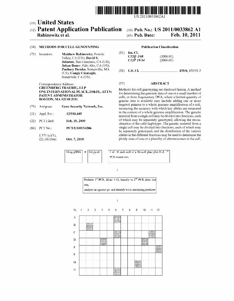
[0 ng gDNA 9 0.6 pg’ul. | 111. 9 each well of a 96-well plate plus 9 11L 1st
PCR master mix
run,
Perform 1Sl PCR, dilute l/15, transfer to 2"“ PCR plate and
analyze on agarose gel and identify Wells containing products

Patent Application Publication Feb. 10, 2011 Sheet 1 0f 3 US 2011/0033862 A1
10 ng gDNA -> 0.6 pg/uL l uL 9 each Well ofa 96-well plate plus 9 uL 1“
PCR master mix
Perform 1s1 PCR, dilute l/15, transfer to 2mi PCR plate and
run,
analyze on agarose gel and identify wells containing products
FIG. 1

Patent Application Publication Feb. 10, 2011 Sheet 2 0f 3 US 2011/0033862 A1
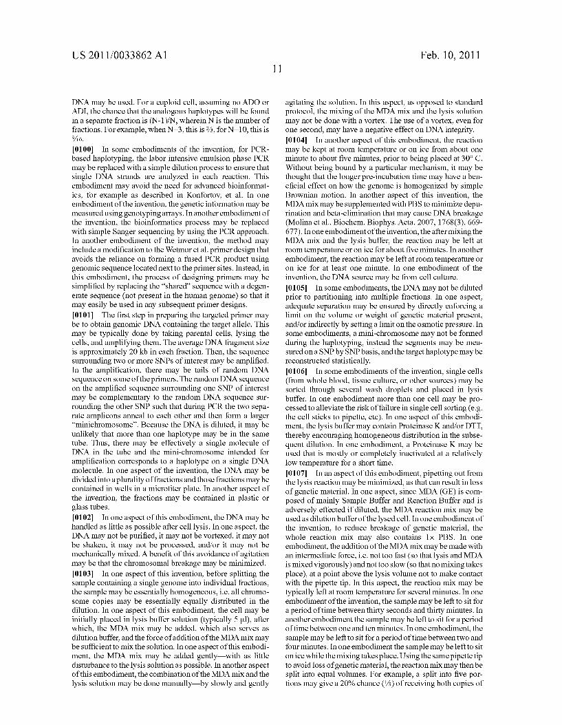
‘\j-RAGTTCAACATTAAATAATGGAAATAACTTTCTACAACTTTAT
ATCCACAAAGATTATACAACATTCTTAAACATTAGATTACATAATTGAAAGGCAT
TTTTTTGTTTAGGATAAATACTGACACCAAAAATAAGTAATTTTATAAAAATATA
ATCAAATGAGQTTGCCAAATGCATTTCCTGAATATATGTATATATAACTTTCTATA TGGAGATTAATATTAGTAATAACATTTATGATGAACAAATCAGTTCATGAAGGTT
TTAAAAATGATGATTCTCATATTAATTGATGTTATATAAAzX-Yiii‘TA;
. A. . I V .. . . I.‘ . . 1km
AGCAATTCTCCCACZGTGGGCCTCCCAAAGTGTTGGGATTACGGATGTGAGCCACT
ATGCCCAGCCACTTACACTCTTCCTATTCTAAATTTATTTAAAAATATTTTATTGA
TATQGTTTTTAAATAACACACTGTTTTATTTCCATTTTCACTTGGGTCTCCTGTCAC TTTATATTTTAAAAAAGAACACATGGTAAATTCAATACACGGATCAGTTTAATTT
TTGGTCTGACTCATTTTCTTTTTCTTGAAGGCAAGAATACAAAGTTTTAAAATCA
GTATGTAAAGGA‘HRK:
1_OUT AGTTCAACATTAAATAATGGAAATAACTTTCTAC
SEQ ID NO: 21
1_OLAP_2 actagatctacgtgtaagtcatggacttcA'i‘Arl‘AACA'FCAA'ITAA’I‘A'I‘GAGAATCAT SEQ ID NO: 22
2_OLAP_1 gaagtccatgax:ttacazgtagatctagtTCAAGCAATTCTCCCACGT
SEQ ID NO: 23
ZiOUT CCTTTACATACTGATTTTAAAACTTTGT
SEQ ID NO: 24
FIG. 2

Patent Application Publication Feb. 10, 2011 Sheet 3 0f 3 US 2011/0033862 A1
FIG. 3

US 2011/0033862 A1
METHODS FOR CELL GENOTYPING
FIELD
[0001] The embodiments disclosed herein relate generally to the ?eld of acquiring and/or manipulating high ?delity genetic data for medically predictive purposes, and more particularly to a system Which alloWs genetic data from a single or small number of cells to be measured With high accuracy, and for the genetic haplotypes and ploidy states to be determined.
BACKGROUND
Preimplantation Genetic Diagnosis [0002] In 2006, across the globe, roughly 800,000 in vitro fertilization (IVF) cycles Were run. Of the nearly 140,000 cycles run in the US, about 10,000 involved pre-implantation genetic diagnosis (PGD). Current PGD techniques are unregulated, expensive and can be unreliable: error rates for screening disease-linked loci or aneuploidy are on the order of 10%, each screening test costs roughly $5,000, and a couple is forced to choose betWeen testing aneuploidy, Which af?icts roughly 50% of IVF embryos, or screening for dis ease-linked loci on the single cell. There is a great need for an affordable technology that can reliably determine genetic data from a single cell in order to screen in parallel for aneu ploidy, monogenic diseases such as Cystic Fibrosis, and sus ceptibility to complex disease phenotypes Which have mul tiple genetic markers. [0003] The process of PGD during IVF currently involves biopsy of embryos generated using assisted conception tech niques. There are tWo potential sources of embryonic genetic material for PGD aneuploidy screening: a single blastomere from cleavage stage embryos (typically day 3 post-fertiliza tion) and 4-10 tropechtoderm cells from blastocyst stage embryos (typically day 5 post-fertilization). Using cleavage stage single cell biopsy is the most common approach to PGD. Isolation of single cells from human embryos, While highly technical, is noW routine in IVF clinics. Polar bodies, blastomeres, and tropechtoderm cells have been isolated With success. HoWever, there is only a limited amount of time available for preimplantation testingimost clinics aim to transfer the embryos to the mother Within 32 hours of biop sy. Consequently, diagnostic methods must be rapid as Well as accurate. It is possible, though not preferred by most IVF clinics, to cryopreserve embryos after biopsy, in Which case they can be transferred months or even years after they have been isolated. [0004] To biopsy an embryo, it is usually transferred to a special cell culture medium and a hole is introduced into the Zona pellucida using an acidic solution, laser, or mechanical technique. The technician then uses a biopsy pipette to remove a single blastomere With a visible nucleus in the case of day 3 embryos, or a small number of tropechtoderm cells in the case of day 5 embryos. Features of the DNA of the biop sied cell(s) can be measured using a variety of techniques. Since only a single copy of the DNA is available from one cell, direct measurements of the DNA of one or a small number of cells are error-prone, or noisy. There is a great need for a technique that can improve the accuracy of these genetic measurements using a small number of cells. [0005] In cases Where the goal is to diagnose a particular genetic mutation, the ampli?cation of the DNA is typically carried out using a single primer, or set of primers, speci?
Feb. 10, 2011
cally designed to amplify a particular locus or small number of loci of interest. When only one primer or a small set of primers is used, the allele dropout (ADO) rate tends to be quite loW. HoWever, in the case Where the goal is to measure the genotype of the cell at hundreds or thousands of loci, the ampli?cation is typically done With a method such as Whole genome ampli?cation (WGA). In these cases, one or a set of generic primers (sometimes called universal primers or ran dom primers) are typically used. In these methods, ADO rate at a given allele tends to be signi?cantly higher than When a targeted primer is used. There is a need for a technique that can combine the ability of WGA to genotype a large number of alleles With a method that can provide genotyping of key alleles With the high level of accuracy possible When perform ing targeted ampli?cation.
MDA and WGA
[0006] Whole genome ampli?cation is Widely used When analyZing the genome of a single cell, and in theory, results in the ampli?cation of all loci on the DNA. One major limiting factor in this process are ADOs, i.e. missing information about one orboth alleles of a locus. The mechanisms ofADOs are largely unknown and may occur during lysis, ampli?ca tion or analysis of single cells. Typical ADO rates from Whole genome ampli?cations typically range from 20 to 60%. In addition, the identity of alleles that drop out are effectively random. [0007] Prior to the development of Whole genome ampli? cation, the most Widely used method for analyzing speci?c loci in single cells Was polymerase chain reaction (PCR), Which requires many cells per locus in order to compensate for uninformative DNA fragments. HoWever, PCR is not use ful for analyZing more than one loci per cell because multiple PCR primers cannot be used simultaneously. Additionally, non-random ampli?cations, e.g. multiplex PCRs, can suffer from sequence speci?c ADO due to high GC containing loci or non-optimal primer pairs. [0008] An alternative method of analyZing loci in single cells is ?uorescent in situ hybridiZation (FISH), but the method is basically limited to a feW loci per cell. Whole genome ampli?cation has evolved as a means to analyZe several targets post-ampli?cation. HoWever, random ampli? cations, eg using multiple displacement ampli?cation (MDA) kit or a WGA kit act on every target sequence, and thus still suffer from random ADO. [0009] In some contexts, such as PGD, it may be desirable to measure a large number of alleles, Wherein a subset of those alleles is of particular importance. It is desirable to measure a large number of alleles, and simultaneously maximiZe the accuracy of the genotyping of those targeted alleles. Unfor tunately, no method has yet been shoWn to decrease the like lihood of a desired subset of alleles to drop out upon ampli ?cation and genotyping, as compared to the overall set of alleles, in the context of WGA. [0010] For example, if a clinician Wishes to make predic tions regarding the genetic susceptibility of an embryo to one or more potential diseases, it is necessary to be able to deter mine the identity of those disease-linked alleles in the embryo in question. Simply measuring the disease-linked allele car ries a risk of an allele miss-call, or ADO. There is an as yet unmet need for such a technology Which combines the ability to measure a large number of alleles and also the ability to maximiZe the accuracy With Which a speci?c subset of those alleles is measured.

US 2011/0033862 A1
[0011] One method used to avoid the problem of ADO at alleles of interest is to measure multiple polymorphic alleles that lie Within a disease gene and/or closely ?anking it. This can enable the deduction of the high-risk (i.e. mutation car rying) haplotypes that have been inherited by embryo, and can overcome some of the di?iculties associated With particu lar markers being uninformative for a given family, or the problem of ADO measure of disease-linked alleles. The knowledge of su?icient ?anking polymorphic alleles, mea surable using a Whole genome ampli?cation technique, along With the knoWledge of the haplotype can increase the ability to correctly deduce the identity of the target allele(s) if it Was not measured correctly. While the use of ?anking alleles, measurable When using Whole genome ampli?cation tech niques, can be used to increase the accuracy With Which key alleles are determined, there is an as yet unmet need for a technology that can combine the ability to measure a large number of ?anking alleles and at the same time decrease the likelihood of an uninformative measurement at the target allele itself.
Single Cell Haplotyping
[0012] Many methods exist for genotypingirevealing Which alleles an individual carries at different genetic loci. A harder problem is haplotypingidetermining Which alleles lie on each of the tWo homologous chromosomes in a diploid individual. For instance, an individual may have the genotype AB/ab (heterozygous at each of loci A and B), but could carry haplotypes AB and ab or, conversely, Ab and aB. Conven tional approaches to haplotyping require the use of several generations of cells to reconstruct haplotypes Within a pedi gree, or use statistical methods to estimate the prevalence of different haplotypes in a population. Several molecular hap lotyping methods have been proposed, but have been limited to small numbers of loci, usually over short distances. [0013] Determining the phase or haplotype of tWo or more genetic variants (single nucleotide polymorphisms, SNPs) has long been a challenging task due to technical issues When Working With a feW or single DNA strands. Among the prob lems are loss of material (eg attachment to equipment), sensitivity (e.g. not enough material), or integrity (DNA is fragile and breaks easily). Integrity plays an important part When setting up any haplotyping assay, since fragmentation may affect the distance betWeen SNPs that can be analyZed, or generate false haplotypes if diploid fragments are sWitched. [0014] Haplotype-based methods offer a poWerful approach to disease gene mapping, based on the association betWeen the causal mutations and the ancestral haplotypes on Which they arose. The genome, human or other species, can be parsed objectively into haplotype blocks: siZable regions over Which there is little evidence for historical recombina tion and Within Which only a feW common haplotypes are observed. The boundaries of such blocks and speci?c haplo types they contain are highly correlated across populations; these haplotype frameWorks provide substantial statistical poWer in association studies of common genetic variation across each region, and facilitate comprehensive genetic association studies of human disease. [0015] Quantitative traits such as drug responsiveness or disease susceptibility may be more strongly correlated With certain haplotypes than With certain genotypes, particularly Where several polymorphic loci fall Within a single gene. Hence, both the discovery of an association betWeen a trait and a polymorphism, and the implications of this association
Feb. 10, 2011
for an individual, depend on knoWledge of haplotypes. Hap lotype structure is also important in understanding the evolu tion of a species and of populations Within it, as haplotype blocks are shuf?ed in successive generations. The persistence of ancestral haplotypes can also be used to simplify genotyp ing experiments: the genotype at one locus may serve as a proxy for the genotypes of neighboring loci if they lie Within the same conserved haplotype block. [0016] Current methods for haplotyping are quite diverse and involve for example PCR, FISH, and rolling circle ampli ?cation (RCA). The common theme is to isolate individual DNA strands, for example in emulsion phases, on glass slides, or circulariZe them, folloWed by ampli?cation and readout, or in the case of FISH, hybridization of ?uorescently labeled oligonucleotides. Most methods Work With very loW concen trations of input DNA to avoid scoring or mix up of both alleles of a SNP, making them relatively inef?cient and hard to optimiZe. [0017] Cloning in hybridomas or in bacterial or yeast cells, or the natural occurrence of hydatidiform moles arising from a single haploid gamete, can also be used to isolate a single haplotype Which can then be revealed by simple genotyping. Such approaches, hoWever, are limited to the analysis of small numbers of loci over short distances (typically a feW hundred base pairs). Other methods have been based on the analysis by PCR of single DNA molecules Which, of course, represent single haplotypes. The most direct implementation of this strategy is the genotyping of single sperm, in Which meiosis has done the job of isolating a single copy of each chromo some.
[0018] Other approaches rely upon the division of genetic matter into fractions to isolate (statistically) single DNA mol ecules, folloWed by the speci?c ampli?cation and genotyping of tWo or more loci. HoWever, this method is unable to mea sure haplotypes of more than about 20-30 kb in length, that do not involve more than a feW loci, and the methods are often ine?icient since only a feW of the highly dilute samples Which are genotyped Will prove to contain informative molecules. A method, described by Wetmur (Wetmur et al., Nucl. Acids Res., 2005, 33(8), 2615-2619), uses linking emulsion PCR (LE-PCR), Which enables formation of mini-chromosomes preserving phase information of tWo polymorphic loci, hence the haplotype. A draWback to all of these methods is that they require a large number of DNA copies (thousands to millions of cells) to produce accurate results. There is a great need for a technique that can accomplish the effective haplotyping of a cell by isolating individual haplotypes from a single cell, or a small number of cells.
[0019] The main problem in haplotyping is knoWing Whether one is looking at information from a single continu ous DNA strand (implied chromosome), or fragmented DNA strands belonging to the same chromosome copy. For example, if tWo consecutive SNPs (located on the same chro mosome) are measured in a given reaction, there is a risk that there Was chromosomal breakage betWeen them, and one of the measured SNPs is actually from a homologous chromo some as the other measured SNP, thus resulting in a false haplotype deduction. Any method that involves conditions that induce DNA strand breakage, either through chemical or mechanical means, Will have some likelihood of such false haplotyping. [0020] Examples in the prior art dealing With this problem include dilution of DNA obtained from bulk tissue or blood samples doWn to levels Where it is statistically unlikely (hoW

US 2011/0033862 A1
ever still possible) that tWo copies of the same sequence ended up in the same reaction (Konfortov et al., Nucl. Acids Res., 2007, 35(1), e6). Alternatively one can break up the entire reaction into several fractions With the hope that each fraction contains a unique chromosome (Wetmur). Note that these techniques do not solve the problem, they just make it less probable that the problem Will have an impact on a given haplotype determination. A signi?cant drawback to these methods is that they are done With bulk genetic material derived from many cells, Well more than a thousand, and typically in the millions. These methods have not been appli cable to DNA samples derived from one or a small number of cells. Another draWback to these methods is that they typi cally start With prepared genomic DNA from many cells, and the methods of preparation typically result in a greater degree of DNA fragmentation, especially puri?cation steps, thereby limiting hoW far apart along the genome different SNPs can be phased. There exists a need for a technique that can deter mine the haplotypes by isolating different haplotypes present in the genetic material found in one or a small number of cells. There exists a need for a technique that can determine the haplotypes by isolating different haplotypes present in the cells of an individual that minimiZes the possibility of false haplotyping due to strand breakage.
Aneuploidy [0021] Normal humans have tWo sets of 23 chromosomes in every diploid cell, With one set from each parent. Aneup loidy, the state of a cell With extra or missing chromosome(s), and uniparental disomy, the state of a cell With tWo of a given chromosome both of Which originate from one parent, is believed to be responsible for a large percentage of failed implantations and miscarriages, and some genetic diseases. When only certain cells in an individual are aneuploid, the individual is said to exhibit mosaicism. Detection of chromo somal abnormalities can identify individuals or embryos With conditions such as DoWn syndrome, Klinefelter’s syndrome, and Turner syndrome, among others, and potentially increase the chances of a successful pregnancy. Testing for chromo somal abnormalities is especially important as the age of a potential mother increases: betWeen the ages of 35 and 40 it is estimated that betWeen 40% and 50% of the embryos are abnormal, and above the age of 40, more than half of the embryos are like to be abnormal. The main cause of aneup loidy is nondisjunction during meiosis. Maternal nondisjunc tion constitutes 88% of all nondisjunction, of Which 65% occurs in meiosis I and 23% in meiosis II. Common types of human aneuploidy include trisomy from meiosis I nondis junction, monosomy, and uniparental disomy. In a particular type of trisomy that arises in meiosis II nondisjunction, or M2 trisomy, an extra chromosome is identical to one of the tWo normal chromosomes. M2 trisomy (also called mitotic tri somy) is particularly dif?cult to detect. There is a great need for a better method that can detect for many or all types of aneuploidy at most or all of the chromosomes e?iciently and With high accuracy, especially a method that can determine aneuploidy states involving multiple identical chromosomes, such as With mitotic trisomy, or some cases of uniparental disomy. [0022] Karyotyping, the traditional method used for the prediction of aneuploidy and mosaicism is giving Way to other more high-throughput, more cost effective methods such as FloW Cytometry (FC) and FISH. Karyotyping involves the isolation of a single cell, the staining of the
Feb. 10, 2011
chromosomes in that cell, and the visualiZation and identi? cation of the chromosomes. A major draWback to karyotyping is the high cost. Currently, the vast majority of prenatal diag noses use FISH, Which can determine large chromosomal aberrations and PCR/electrophoresis, and Which can deter mine the identity of a small number of SNPs or other alleles. FISH involves the chromosome-speci?c hybridiZation of ?uorescently tagged probes to cellular DNA, and subsequent visualiZation and quanti?cation of the amount of ?uorescent probes present. One advantage of FISH is that it is less expen sive than karyotyping, but the technique is complex and expensive enough that generally only a small selection of chromosomes are tested (usually chromosomes 13, 18, 21, X, Y; also sometimes 8, 9, 15, 16, 17, 22). In addition, FISH has a loW level of speci?city. Roughly seventy-?ve percent of PGD today measures high-level chromosomal abnormalities such as aneuploidy, using FISH, With error rates on the order of 10-15%. There is a great demand for an aneuploidy screen ing method that has a higher throughput, loWer cost, Wider scope, and greater accuracy. [0023] There are a number of methods described in the literature for determining the ploidy state (or chromosome count) of one or a number of cells that make use of genotyp ing. For example, Handyside (PCT ?ling WO/2007/ 0057647) discusses the concept of using measured SNP data to assemble a notional haplotype of an embryo, looking for aberrations from the expected measured SNP data given the notional haplotype, and ?agging any aberrations as likely cases of aneuploidy. One draWback to these methods is that they are unable to detect ploidy states such as uniparental disomy (UPD) and mitotic trisomy, Where there are tWo iden tical chromosomes in the embryo. Another draWback is that they require the haplotypes of one or both of the parents, a non-trivial issue. There exists a need for a neW method that utiliZes genetic information that can be gathered in a highly e?icient, cost effective manner that also alleviates the inabil ity to determine ploidy states due to multiple copies of iden tical chromosomes such as UPD and mitotic trisomy. Further more, there exists a need for a neW method that utiliZes genetic information that can be gathered in a highly e?icient, cost effective manner that does not require the knoWledge of parental haplotypes. [0024] Using the current state of the art methods to measure genetic data may have draWbacks, such as the lack of cur rently knoWn Ways to amplify and measure the genetic data of one or a small number of cells, at a large number of alleles, While being able to reduce the ADO rate and/or increase the accuracy of the measurements at a subset of key alleles as compared to the entire set of measured alleles. Also, When measuring the genetic data of a diploid cell(s), it is often important to be able to determine the haplotypes of the cell. HoWever, this may be dif?cult to do in an effective manner on one or a small number of cells. It may also be dif?cult to determine the ploidy state of a cell With high accuracy, at all of the chromosomes, and in an ef?cient manner With respect to costs and time.
SUMMARY
[0025] Methods of cell genotyping are disclosed herein. In the context of Whole genome ampli?cation of small quantities of genetic material, Whether through ligation-mediated PCR (LM-PCR), multiple displacement ampli?cation (MDA), or other methods, dropouts of loci occur randomly and unavoid ably. It is often desirable to amplify and genotype the Whole

US 2011/0033862 A1
genome nonspeci?cally, and at the same time increase the chance that a particular locus, or set of loci, are ampli?ed and measured accurately. When determining the identity of a key allele (typically a disease-linked allele), there are multiple Ways to increase the likelihood that the key allele is identi?ed correctly. One Way may be to use targeted primers in the ampli?cation to raise the likelihood that the target allele is measured correctly. Another may be to use Whole genome ampli?cation techniques to measure multiple polymorphic alleles that lie Within a disease gene and/or closely ?anking it, enabling the deduction of the high-risk (i.e. mutation carry ing) haplotypes that have been inherited by embryo. Embodi ments of the present invention include methods for simulta neous loci targeting and Whole genome ampli?cation.
[0026] According to aspects illustrated herein, there is pro vided a method that enables one to amplify the Whole genome of a single cell, or small number of cells, While biasing the ampli?cation to amplify a set of desired loci preferentially. The addition of “spike-in” primers (locus-speci?c primers) may loWer the likelihood that the loci of special importance are subject to ADO. In one embodiment of the invention, a method minimiZes the chances that one or more single nucle otide polymorphisms (SNPs) of interest drop out during WGA using a single or small number of cells. This method may be advantageous When the number of alleles of interest may be suf?ciently large that techniques such as FISH may be incapable of making allele calls at all of the alleles of interest, and array based genotyping may be necessary.
[0027] According to aspects illustrated herein, there is pro vided a method for determining genetic haplotypes using the genetic material from only one (1) cell, or a small number of cells, Which by de?nition may have a knoWn number of chromosome copies. In one aspect of this embodiment, the method avoids purifying the DNA, Which may minimiZe DNA damage. This method may minimiZe DNA strand breakage, and thus loWer the chance of false haplotyping. When a small number of cells are used in one reaction, this approach may further loWer the risk of ADO, though it may require dividing the genetic material into a larger number of fractions. This risk may also be mitigated by using informat ics based approaches, such as Parental SupportTM, that utiliZe the knoWledge of genetic data measured on related individu als, and/or publicly available haplotype databases such as those supported by the Hapmap Project and from the Perlegen Human Haplotype Project, to infer genetic data not measured or measured incorrectly. The Parental SupportTM method is described in US. application Ser. No. 11/603,406 and US. application Ser. No. 12/076,348 and the entirety of both of these applications are hereby incorporated herein by refer ence for the respective teachings therein.
[0028] According to aspects illustrated herein, there is pro vided a method for detecting aneuploidy by dividing the genetic matter from a single cell, or a small number of cells, into a plurality of fractions before ampli?cation and genotyp ing, Which may be performed for individual fractions. Since only a single copy of the genome may be present in a single cell, each fraction in Which a given allele may be found implies a different homologous chromosome. From the num ber of fractions in Which the various alleles from a given chromosome are found, it may be possible to determine the ploidy state of that chromosome in the cell. This method may alloW the detection of types of aneuploidy due to multiple identical chromosomes in a cell. Additionally, this method may not require the knoWledge of parental haplotypes.
Feb. 10, 2011
[0029] The systems, methods, and techniques of embodi ments of the present invention may be used to in conjunction With embryo screening in the context of IVF, or prenatal testing procedures, in the context of non-invasive prenatal diagnosis. The systems, methods, and techniques of embodi ments of the present invention may be employed in methods of increasing the probability that the embryos and fetuses obtain by in vitro fertilization are successfully implanted and carried through the full gestation period, and result in healthy babies. Further, the systems, methods, and techniques of embodiments of the present invention may be employed in methods to decrease the probability that the embryos and fetuses, Which are obtained by in vitro fertilization, implanted and gestated, are at risk for chromosomal, congenital or other genetic disorders. [0030] Various embodiments provide certain advantages. Not all embodiments of the invention share the same advan tages and those that do may not share them under all circum stances. Further features and advantages of the embodiments, as Well as the structure of various embodiments are described in detail beloW With reference to the accompanying draWings.
BRIEF DESCRIPTION OF THE DRAWINGS
[0031] The presently disclosed embodiments Will be fur ther explained With reference to the attached draWings, Wherein like structures are referred to by like numerals throughout the several vieWs. The draWings shoWn are not necessarily to scale, With emphasis instead generally being placed upon illustrating the principles of the presently dis closed embodiments. [0032] FIG. 1 is a schematic illustration of an illustrative embodiment of an experimental setup for WGA using spike in primers; [0033] FIG. 2 is an illustration of an illustrative embodi ment of a spike-in primer design; [0034] and [0035] FIG. 3 is a schematic illustration of an illustrative embodiment of a PCR approach. [0036] While the above-identi?ed draWings set forth pres ently disclosed embodiments, other embodiments are also contemplated, as noted in the discussion. This disclosure presents illustrative embodiments by Way of representation and not limitation. Numerous other modi?cations and embodiments can be devised by those skilled in the art Which fall Within the scope and spirit of the principles of the pres ently disclosed embodiments.
DETAILED DESCRIPTION
[0037] The inventions are not limited in their application to the details of construction and the arrangement of compo nents set forth in the folloWing description or illustrated in the draWings. The inventions are capable of being arranged in other embodiments and of being practiced or of being carried out in various Ways. Also, the phraseology and terminology used herein is for the purpose of description and should not be regarded as limiting. The use of “including,” “comprising,” or “having,” “containing,” “involving,” and variations thereof herein, is meant to encompass the items listed thereafter and equivalents thereof as Well as additional items. It Will be readily apparent to one of skill in the art that the primers (SEQ ID NOs: 1-24) disclosed in the present application may also include the reverse complement thereof.

US 2011/0033862 A1
[0038] Aspects of the inventions are described below With reference to illustrative embodiments. It should be under stood that reference to these illustrative embodiments is not made to limit aspects of the inventions in any Way. Instead, illustrative embodiments are used to aid in the description and understanding of various aspects of the inventions. Therefore, the following description is intended to be illustrative, not limiting. [0039] The headings of or in the various sections in this application are inserted for convenience only and are not intended to affect the meaning or interpretation of this appli cation. Any disclosures in any part of this application may be employed With any disclosures in another part of the applica tion. For example, techniques described in “Using the GR MDA Spike-In” section may be used in combination With or in lieu of techniques described in “Using the Sigma WGA Kit” section.
[0040] Some embodiments of the present invention are designed to determine the genetic data from one or a small number of cells With high accuracy. Some embodiments of the present invention include a method for Whole genome ampli?cation With targeted ampli?cation of certain alleles of interest using “spike-in” primers. Since WGA involves generic primers, ADO rates tend to be higher than in targeted ampli?cation, Where the primers may be speci?cally designed for loci of interest. In some embodiments, primers are used that have been speci?cally designed for loci of inter est in conjunction With generic primers in the context of WGA, such that it may be possible to realize the bene?t of both methods: ampli?cation of the entire genome as Well as high accuracy ampli?cation of loci of interest. [0041] Some embodiments may include a method for deter mining the haplotype of a single or small number of cells by division of the DNA before ampli?cation. In some aspects of this invention, the free genetic material (e.g., blastomeric, etc.) from one or a small number of cells may be divided into a suf?ciently large number of fractions such that it may be unlikely to ?nd more than one haplotype per sample. Since the majority of the haplotypes may be in different fractions, and each fraction may be ampli?ed and genotyped individu ally, so that the individual haplotypes may be measured sepa rately using standard genotyping methodology. In some embodiments, techniques may be used to minimiZe the DNA strand breakage, thus increasing the chance that large unbro ken haplotypic sections may be measured in individual Wells and enhancing the ability to reconstruct the entire haplotype of the target from the genotypes measured from the individual fractions.
[0042] Some embodiments may include a method for deter mining the ploidy state of a cell, at some or all of the chro mosomes, by dividing the genetic material from a single cell into a plurality of fractions, and then separately amplifying and genotyping each fraction. By determining the number of fractions in Which a given allele may be detected, for many alleles on a given chromosome, it may be possible to deter mine the number of chromosomes present in the cell. The distribution of the number of fractions in Which each allele is found, for a set of alleles on a given chromosome, may be indicative of the ploidy state of a given chromosome. In some aspects, the ploidy state can be determined either by observ ing the maximum number of fractions that alleles from a given chromosome are found, and taking that to be the ploidy state. In some aspects, the ploidy state may be determined by comparing the observed distribution of alleles to the expected
Feb. 10, 2011
distributions for different ploidy states, given the conditions of the experiment, and taking the actual ploidy state to be the one Whose expected distribution most closely matches the observed distribution. [0043] The present methods may be relevant in the context of Gene Security NetWork’s proprietary Parental SupportTM (PS) method. The Parental SupportTM method uses the mea sured genetic data from a single or small number of cells, along With parental genetic data and the knoWledge of a mechanism of meiosis, as inputs to determine the genotype at a plurality of alleles, and the ploidy state of an embryo, of a fetus, or of any target cell or group of cells. The spike-in method described herein may generate data that may be opti miZed in the context of being used as input for the Parental SupportTM method. The haplotyping method described herein may determine haplotypes that can be used in the context of determining parental or target haplotypes for use as input for the Parental SupportTM method. The method for determining ploidy states may be used to augment or replace aspects of the Parental SupportTM method. [0044] In some embodiments, the subject, such as a target individual, may be an embryo, and the purpose of applying the disclosed method to the genetic data of the embryo may be to alloW a doctor or other agent to make an informed choice of Which embryo(s) should be implanted during IVF. In some embodiments, the target individual may be a fetus, and the purpose of applying the disclosed method to genetic data of the fetus may be to alloW a doctor or other agent to make an informed choice about possible clinical decisions or other actions to be taken With respect to the fetus. In some embodi ments, the target individual may be a fetus, and the nucleated fetal cell(s) may be isolated in a non-invasive manner from maternal blood.
De?nitions
[0045] Genotyping: may be the genetic constitution of a cell or a subject (i.e. the speci?c allele makeup of the subject), usually With reference to a speci?c character under consider ation. The term may also refer to a subj ect’s speci?c genomic sequence, or a representative genomic sequence of a species or group; a genotype may be a measurement of hoW an indi vidual differs or may be specialiZed Within a group of indi viduals or a species. A subject’s genotype can be measured With regard to a particular gene or genes of interest, including the location, such as the number chromosome on Which the gene may be located, Which of the tWo homologous chromo somes the gene may be located on, and Where along the chromosome the gene may be located, and the identity, such as the sequence of base pairs that may make up the gene. The genotype may also refer to the number and origin of chromo somes in the subject’s genome. The term genotype may also include non-hereditary DNA mutations that are not classi cally understood as representing a subject’s genotype. For example, the term may be meant to apply to the genotype of a particular cancer, Wherein the genotype of the disease may be distinct from the genotype of the subject that has the cancer.
[0046] SNP (Single Nucleotide Polymorphism): may be a single nucleotide that may differ betWeen the genomes of tWo members of the same species. As used herein, there may not be any limit on the frequency With Which each variant occurs. [0047] Locus: also referred to as an “allele”; may be a particular region of interest on the DNA of an individual, Which may refer to a SNP, the site of a possible insertion or

US 2011/0033862 A1
deletion, or the site of some other relevant genetic variation. Disease-linked SNPs may also refer to disease-linked loci or disease-linked alleles.
[0048] To call an allele: may mean to determine the state of a particular locus of DNA. This may involve calling a SNP, determining Whether an insertion or deletion is present at that locus, determining the number of insertions that may be present at that locus, or determining Whether some other genetic variant is present at that locus. [0049] To clean genetic data: may mean to take imperfect genetic data and correct some or all of the errors or ?ll in missing data at one or more loci.
[0050] Segment of a Chromosome: may be a section of a chromosome that can range in size from one base pair to the entire chromosome; also called a “minichromosome.” Chro mosome: may refer to either a full chromosome, or also a segment of a chromosome.
[0051] Haplotypic Data: also called “phased data” or “ordered genetic data”; may be data from a single chromo some in a diploid orpolyploid genome, such as the segregated maternal or paternal copy of a chromosome in a diploid genome.
[0052] Unordered Genetic Data: may be pooled genetic data derived from measurements on tWo or more chromo
somes in a diploid or polyploid genome, such as both the maternal and paternal copies of a chromosome in a diploid genome.
[0053] Genetic data ‘in’, ‘of’, ‘at’ or ‘on’ an individual: may mean the data describing aspects of the genome of an indi vidual and may refer to one or a set of loci, partial or entire sequences, partial or entire chromosomes, or the entire genome.
[0054] Target Individual: also called “subject”; may be a subject or individual Whose genetic data is being determined or otherWise analyzed. “Target individual” may refer to an adult, a juvenile, a fetus, an embryo, a blastocyst, a blas tomere, a cell or set of cells from an individual, from a cell line, or any set of genetic material. The target individual may be alive, dead, frozen, or in stasis. In some embodiments, the genetic data may be from humans, While in some embodi ments, the target individual may be any other DNA containing organism. In some embodiments, the target individual may be non-human vertebrates (e.g., dogs, cats, horses, coWs, pigs, etc.), companion animals (e.g., dogs, cats, hamsters, etc.), livestock (coWs, horses, sheep, etc.), the production of “cul tivated” animals (eg race horses, “pure-bred” varieties of dogs or cats, etc.), or any other nucleic acid containing organ ism.
[0055] Related Individual: may be any individual Who is genetically related, and thus may share haplotype blocks With the target individual. Some examples of related individuals include biological father, biological mother, son, daughter, brother, sister, half-brother, half-sister, grandfather, grand mother, uncle, aunt, nepheW, niece, grandson, granddaughter, cousin, clone, the target individual himself/herself/ itself, and/ or other individuals With knoWn genetic relationship to the target. The term “related individual” may also encompasses any embryo, zygote, fetus, sperm, egg, blastomere, blasto cyst, or polar body derived from a related individual.
[0056] Whole Genome Ampli?cation (WGA): may be a technique designed to amplify all or some of the DNA in a sample. Some methods to perform WGA may include the commercially available GE MDA kit and the Sigma WGA kit.
Feb. 10, 2011
Some other methods may include degenerated oligonucle otide primed PCR (DOP-PCR) and ligation mediated PCR (LM-PCR). [0057] WGA kit (Sigma): may be a commercially available kit for conducting Whole genome ampli?cation. [0058] Free DNA: may mean DNA Which is not contained by a cell Wall. [0059] Allele Drop Out (ADO): may mean a situation that may occur during genotyping Where an allele fails to amplify and the expected allele may not be measured correctly. An ADO may be a false negative When measuring genotypic data. In the case of a homozygous allele, it may not be pos sible to recognize anADO that is not also a LDO; in the case of a heterozygous allele that is not knoWn to be heterozygous, it may not be possible to differentiate betWeen an ADO and the case that that allele is homozygous. [0060] Allele Drop In (ADI): may mean a situation that may occur during genotyping Where an allele is measured to have a certain identity, but Where that measurement may be incorrect and the actual genetic material may support that determination. AnADI may be a false positive When measur ing genotypic data. [0061] Locus Drop Out (LDO): may mean a situation that may occur during genotyping Where both of tWo homologous alleles fail to amplify. [0062] Spike-in: may mean inclusion of locus-speci?c oli gonucleotide primers to the usual reagents used during Whole genome ampli?cation. [0063] Phasing: may mean an act of determining the hap lotypic genetic data of a target individual given unordered, diploid genetic data, or other genetic data. [0064] One or a small number of cells: may mean one cell, tWo cells, up to ?ve cells, as many as tWenty cells, more than tWenty cells, any number or range in betWeen, or any combi nation thereof, as not all embodiments are intended to be limited in this manner. Note this also may refer to an amount of free DNA (such as in the case of non-invasive prenatal diagnosis) that may approximately correspond to the amount of DNA found in one or a small number of cells.
[0065] Ploidy calling: also chromosome copy number call ing, may be an act of determining the number and identity of chromosomes present in a cell. [0066] Ploidy State: may be the number and identity of one or more chromosomes in a cell.
[0067] Base Pair (bp): may be an elementary unit of DNA; 1 kb equals 1,000 base pairs; 1 Mb equals 1,000,000 base pairs. [0068] Nucleic Acid: may be any macromolecule com posed of chains of monomeric nucleotides and may carry genetic information or form structures Within cells. Examples of nucleic acids include deoxyribonucleic acid (DNA) and ribonucleic acid (RNA). Nucleic acids may also include arti ?cial nucleic acids, such as peptide nucleic acid (PNA), Mor pholino and locked nucleic acid (LNA), as Well as glycol nucleic acid (GNA) and threose nucleic acid (TNA). Nucleic acids may also include any nucleobases, nucleosides, nucle otides and deoxynucleotides. [0069] Some embodiments of the invention can be used in conjunction With the informatics-based approaches, such as Parental Suppor‘tTM. The Parental SupportTM method may be used to determine the genetic data, With high accuracy, of one or a small number of cells, speci?cally to determine disease related alleles, other alleles of interest, and/or the ploidy state of the cell(s).

US 2011/0033862 A1
[0070] The Parental SupportTM technology may use genetic measurements of the parents to improve the reliability of allele calls and ploidy calls on the child cells. To improve the accuracy in calling an allele derived from the mother, in the absence of maternal haplotype information, the Parental Sup portTM may use non-phase data of multiple blastomeres (pref erably three or more) in order to determine Which segments of Which maternal chromosomes contributed to the embryos and to indirectly phase the genetic data of the mother. Paternal haplotype data can be obtained by measuring sperm cells, as they each contain only one copy of each chromosome. Mater nal haplotypes may be more dif?cult to determine. Embodi ments of the invention described herein may enable direct measurement of the maternal haplotype. In this Way, the measurements of multiple different children or embryos may not be necessary to infer the maternal haplotype and the ploidy status and allele calls from a sample of a single child cell, such as a single blastomere or a single fetal sample, may be reliably determined. [0071] The Parental SupportTM method may enable the cleaning of incomplete or noisy genetic data using the genetic data of one or more related individuals as a source of infor
mation. It may also enable the determination of chromosome copy number using said genetic data. The Parental SupportTM method may be particularly useful in the context of facilitat ing diagnoses focusing on inheritable diseases, chromosome copy number predictions, increased likelihoods of defects or abnormalities, as Well as making predictions of susceptibility to various disease-and non-disease phenotypes for individu als to enhance clinical and lifestyle decisions.
[0072] The Parental SupportTM method may make the use of knoWn parental genetic data, such as haplotypic and/or diploid genetic data of the mother and/or the father, together With the knoWledge of the mechanism of meiosis and the imperfect measurement of the target DNA, in order to recon struct, in silico, the target DNA at the location of key loci With a high degree of con?dence. The Parental SupportTM method can reconstruct not only SNPs that Were measured poorly, but also insertions and deletions, and SNPs or Whole regions of DNA that Were not measured at all. Furthermore, the Parental SupportTM method can both measure multiple disease-linked loci as Well as screen for aneuploidy, from a single cell.
[0073] The haplotypic genetic data and ploidy data that can be generated by the methods of measuring the ampli?ed DNA from one cell using the methods described herein can be used for multiple purposes. For example, in the context of preim plantation genetic diagnosis or prenatal diagnosis, they canbe used for detecting aneuploidy, uniparental disomy, sexing the individual, as Well as for making a plurality of phenotypic predictions based on phenotype-associated alleles. Currently, in IVF laboratories, due to the techniques used, it may be the case that one blastomere can only provide enough genetic material to test for one disorder, such as aneuploidy, or a particular monogenic disease. Since some embodiments of the methods disclosed herein and Parental SupportTM may have the common step of measuring a large set of SNPs from a single or small number of cells, regardless of the type of prediction to be made, a physician, parent, or other agent may not be limited to a single or small number of disorders for Which to screen. Instead, the option may exist to screen for as many genes and/ or phenotypes as the state of medical knoWl edge Will alloW. With the disclosed method, one advantage to identifying particular conditions to screen for prior to geno typing the blastomere is that if it is decided that certain loci
Feb. 10, 2011
are especially relevant, then a more appropriate set of SNPs Which are more likely to co-segregate With the locus of inter est can be selected. Additionally, one or a set of targeted locus-speci?c primers can be included, as described else Where in this disclosure. Both of these actions can increase the likelihood that alleles of interest Will be measured accu rately.
WGA Spike-In
[0074] In some embodiments, a method of performing Whole genome ampli?cation on a DNA sample from a target individual may include: adding one or more spike-in primers to the DNA that target one or more loci of interest; and amplifying the DNA using a method for Whole genome ampli?cation; Wherein the addition of the spike-in primers decreases the likelihood of allele drop out at the one or more loci of interest. [0075] In one aspect of this embodiment, the DNA sample may be a single cell, tWo cells, 3-5 cells, more than ?ve cells, or fetal DNA isolated from maternal blood. In an aspect, the DNA sample may be from more than 10, more than 20, more than 50 cells. In another aspect of this embodiment, the ampli ?cation may be done using a WGA kit from Sigma or a GE MDA kit. In an aspect, the ampli?cation may include using a commercial Whole genome ampli?cation kit. In an aspect, the Whole genome ampli?cation kit can be a non-commercial preparation, or a combination of commercially-available and non-commercially available reagents for Whole genome ampli?cation. [0076] In another aspect of this embodiment, there may be a single locus of interest, 2-5 loci of interest, 5-10 loci of interest, 10-15 loci of interest, 15 -20 loci of interest, or more than 20 loci of interest. In another aspect of this embodiment, the spike-in primer may be designed to amplify a product betWeen about 200 bp and about 1000 bp, or about 600 bp. In another aspect, the spike-in primer may be designed to amplify a product of about 300 bp, of about 400 bp, of about 500, of about 700 bp, of about 800 bp, of about 900 bp, of about 1000 bp, of about 1100 bp. In another aspect of this embodiment, the likelihood of allele drop out may be decreased by up to about 20%, up to about 25%, up to about 30%, up to about 35%, up to about 40%, up to about 45%, up to about 50%, up to about 55%, up to about 60%, up to about 65%, up to about 70%, up to about 75%, up to about 80%, up to about 85%, up to about 90%, up to about 95%, or over 95%. [0077] In another aspect of this embodiment, the method further includes synthesizing the spike-in primers. In another aspect of this embodiment, the method further includes mea suring the genotype of the ampli?ed DNA. In another aspect of this embodiment, the method is used in combination With an informatics method such as the Parental SupportTM method. [0078] When DNA from a single or small number of cells may be ampli?ed in the context of Whole genome ampli?ca tion, for example using the WGA kit from Sigma or the GE MDA kit, a problem may be allele drop out (ADO). ADO rates may be betWeen 20% and 60%, and can range anyWhere from more than 0% to 100%. The reasons for ADOs may not be entirely knoWn, ADOs can occur both in a random manner (unpredictable) and systematically (e. g., high GC containing sequences are more dif?cult to amplify and may drop out more frequently). Events in both lysis and ampli?cation may contribute to ADO, such that chromatin may not be released from histones during lysis, part of genome may be trapped on

US 2011/0033862 A1
equipment, or the ampli?cation if biased against certain regions. In one aspect of this embodiment, even When ADO occurs at a locus of interest, the locus can be inferred by calling the alleles on either side of the locus of interest. In some aspects of the invention, the spike-in primers may be designed to amplify not only the locus of interest, but also a number of alleles on either side of said locus. In some aspects of the invention, the spike-in primer may be designed to amplify about 100 bp to about 500 bp on either side of each locus of interest. In some aspects of the invention, the spike-in primer may be designed to amplify about 250 bp to about 300 bp on either side of each locus of interest. In some aspects of the invention, the spike-in primer may be designed to amplify about 300 bp on either side of each locus of interest. In some aspects of the invention, the spike-in primer may be designed to amplify from about 50 bp, about 100 bp, about 150 bp, about 200 bp, about 250 bp, about 300 bp, about 350 bp, about 400 bp, about 450 bp, about 500 bp, about 550 bp or more than about 550 bp on either side of each locus of interest.
[0079] The addition of “spike-in” primers during WGA may signi?cantly loWer the likelihood that the loci of interest are subject to ADO. In some embodiments of the present invention, primers targeting regions surrounding the SNPs of interest may be included in the WGA assay. In some embodi ments, the WGA may be done using a WGA kit from Sigma. In some embodiments, the WGA may be done using the GE MDA kit. In some embodiments of the invention, the WGA can be done using, but not be limited to: preampli?cation of particular loci before generalized ampli?cation by MDA or LM-PCR, the addition of targeted PCR primers to universal primers in the generaliZed PCR step of LM-PCR, and the addition of targeted PCR primers to degenerate primers in MDA. In some embodiments, the MDA-type kit may be REPLI-g Kit (Qiagen). In some embodiments, the WGA assay may be done using PEP-PCR (primer extension pre ampli?cation PCR) or DOP-PCR (degenerate oligonucle otide primer PCR). In some embodiments, the targeted prim ers may be added at the same time as the generic primers, or before the generic primers so that they can preferentially hybridize to the target regions of DNA. [0080] Different WGA methods may require different tim ing of primer introduction. In some embodiments of the invention, the Sigma WGA kit may be used and the spike-in primers may be added in the ampli?cation. In one experiment using an embodiment, spike-in of a single primer pair reduced LDO rates from 79% to 7%. In a multiplex spike-in, the average LDO decreased from 29% to 9%. In another embodi ment of the invention, the GE MDA kit may be used, and the spike-in primers may be added in the lysis. In one experiment using an embodiment, spike-in of speci?c primers to the lysis reaction reduced LDO rates from 26% to 7% for a single spiked-in primer, and to 8% for multiplex spiked-in primers. Data relating to the above-described embodiments may be found in Tables 2, 3 and 4.
[0081] Some embodiments of the present invention may involve measuring the polymorphic alleles ?anking the alle les of interest and using the identity of the ?anking alleles to determine Which parental haplotype is present in a cell. Using this method, identity of any incorrectly measured alleles of interest can be inferred. In some embodiments, the alleles of interest may be disease-linked alleles. While these embodi ments can mitigate the problems associated With incorrectly measured alleles of interest, it may still be desirable to maxi miZe the accuracy With Which the desired disease-linked, or
Feb. 10, 2011
otherWise targeted alleles may be measured. In some embodi ments of the invention, the method of measuring the one or more alleles of interest With maximal accuracy may be com bined With the method of measuring the ?anking polymor phic alleles to make accurate genetic determinations. [0082] In some embodiments of the invention, the method for determining the genetic data from the Whole genome, While biasing the ampli?cation to reduce the ADO rate at certain alleles of interest, may be used in conjunction With an informatics based approach, such as the Parental SupportTM method. This combination can be used to determine the iden tity of certain disease-related alleles or other alleles of inter est, and/or to determine the ploidy state of the individual, With maximal accuracy. In some embodiments of the invention, the determination of the identity of the alleles of interested and/ or ploidy state may be done for the purpose of choosing Which embryo(s) to implant or not implant, or to making clinical decisions regarding a fetus. The higher the accuracy of the genetic data, the more accurate the predictions of the infor matics based approach may be able to make. Embodiments of the invention may provide optimiZed genetic data for the informatics based method, such that the accuracy of the sub sequent predictions may be maximiZed.
Using the Sigma WGA Kit
[0083] In some embodiments of the invention, the WGA may be performed using a WGA kit from Sigma in combina tion With speci?c spike-in primers. Performing WGA using the Sigma WGA kit may entail the following general steps: (1) cell lysis and DNA preparation, (2) addition of Library Preparation mix, and (3) ampli?cation (includes addition of Ampli?cation mix). The DNA preparation may involve neu traliZation or puri?cation, and the ampli?cation step may be repeated. In one aspect of this embodiment, the spike-in prim ers may be included in the “ampli?cation” step alongside the regular WGA primers. The inclusion of the spike-in primers into the “Library Preparation” step (most likely Where adap tors are attached to DNA fragments) may have no effect on the target’s ADO rate, possibly because they too Were tagged by adaptors (they Were included here initially due to this steps’ temperature pro?le). [0084] In some embodiments of the invention, the primers may be added into the PCR ampli?cation mix. In these embodiments, the inclusion of locus speci?c oligonucle otides (primers) may result in the bias of ampli?cation toWards the regions of interest. The effect may be loWerADO rates of targeted loci compared to ADO rates of the untargeted loci. In one aspect of this embodiment, the primers may be located so that the PCR product can be ampli?ed along With the method’s PCRiWhICh may span 200-1500 bp. In one aspect of this invention, the spike-in primer pairs may be betWeen about 200 bp and about 1500 bp. In another aspect of this invention, the spike-in primer pairs may be from about 400 bp to about 1200 bp. In another aspect of this invention, the spike-in primer pairs may be from about 500 bp to about 1000 bp. In another aspect of this invention, the spike-in primer pairs may be from about 600 bp to about 750 bp. In another aspect of this invention, the spike-in primer pairs may be about 600 bp. [0085] The ef?ciency of the ampli?cation Was evaluated by performing a 2'” targeted PCR With only the spiked-in prim ers, Where the DNA produced in the initial ampli?cation Was used as the source DNA for the second PCR. Thus, if the targeted alleles ampli?ed as intended during the initial WGA

US 2011/0033862 A1
With spiked-in primers, then the second PCR may be expected to produce a product corresponding to the size of the designed initial primer. A combination of TAQMAN, Which can deter mine the exact identity of a given SNP, and information obtained from agarose gels, Which can detect LDO, Was used to differentiate betWeen a correctly called homozygous allele and a heterozygous allele measured With ADO. [0086] In one aspect of this invention, primers may be designed and synthesized for each locus of interest. In this aspect, the primers may be designed toWards regions of approximately 300 bp on each side of the SNP, or toWards regions immediately up stream and doWnstream, of their SNP to the sense (+) and non-sense (—) genomic strands, respec tively (See Table 1). In one aspect of the invention, the con centration of the spike-in primers may be about equal to the primer concentration in regular PCR. In one aspect of this embodiment, the concentration of the spike-in primers may be approximately 250 nM. In another aspect of this embodi ment, the concentration of the spike-in primers may be betWeen about 100 nM and about 350 nM. In some aspects of the invention, the concentration of spike-in primers may be about 10 nM, about 25 nM, about 50 nM, about 100 nM, about 150 nM, about 200 nM, about 250 nM, about 300 nM, about 350 nM, about 400 nM, less than 10 nM, more than 400 nM or any range therebetWeen.
TABLE 1
Feb. 10, 2011
ments, the step of multiple displacement ampli?cation may include using a commercial multiple displacement ampli? cation kit. In some embodiments, the multiple displacement kit can be a non-commercial preparation, or a combination of commercially-available and non-commercially available reagents for multiple displacement ampli?cation. [0089] In some embodiments, the targeted primers may be added during the cell lysis step. In one aspect of this embodi ment, the targeted primers may be added to the lysis solution. Addition of spike-in primers during MDA may serve to decrease the ADO rate at alleles of interest. Inclusion of the targeted primers into the MDA kit may have no effect. In one aspect of this embodiment, the lysis buffer solution may not be alkaline. If the lysis solution is an alkali solution, in another aspect of this embodiment, a neutralization buffer may be added prior to MDA mix to alloW primer hybridiza tion along With some time for proper hybridization. Either the alkali buffer or the neutralization solution may contain the spike-in primers. In another aspect of this embodiment, the composition of the lysis buffer may be unknoWn. In this aspect, the method may involve the addition of Tris-HCl, KCl and/or MgCl2 to the lysis solution. Both the neutralization of the lysis buffer and the addition of Tris-HCl, KCl and MgCl2 Were found to increase scoring rates of the targeted SNPs. In
PCR primers used in the spike-in experiments Numbers represent loci usinq ABI TAQMAN probe annotations .
Primer sequences, 600 by spike-ins
656642 Fw
SEQ ID NO: 1 ttctctgtgcctctgttttcttg 656763 Fw
SEQ ID NO: 7
656642 Rev
SEQ ID NO: 2
656763 Rev
SEQ ID NO: 8 tagctcatgtccctgcctctc
2540897 Fw
SEQ ID NO: 3
13096 Fw
SEQ ID NO: 9 tacttgtcctgctgcgatgt
2540897 Rev
SEQ ID NO: 4
13096 Rev
SEQ ID NO: 10
ttccccaggacattaagagcg
2668640 Fw
SEQ ID NO: 5
2602208 Fw
SEQ ID NO: 11 tttgtttgttttgctgcttgc
2668640 Rev
SEQ ID NO: 6
2602208 Rev
SEQ ID NO: 12
cctctccctccaactccaatt
aggaatttggttgtggagagc
acctgtcttccctcccagtt
atgtggccacttgaaaaagaa
acccacccctagaggttctga
tgttccaggcactgaacaaca
aacgctgtgccagattctctt
Using the GE MDA Spike-In
[0087] As described above, the idea of including (spike-in) locus speci?c oligonucleotides (primers) may be to bias ampli?cation toWards the regions of interest, With the effect of loWering ADO rates of the targeted loci compared to non spiked ampli?cations, While leaving non-targeted loci unal tered. In one embodiment of the invention, the WGA may be conducted using a GE MDA kit and spike in primers. Per forming WGA using the GE MDA kit may entail the folloW ing general steps: (1) cell lysis and DNA preparation, (2) ampli?cation (includes addition of MDA mix), and (3) heat inactivation of enzyme. The DNA preparation may involve neutralization, puri?cation, putting the DNA into sample buffer, and/ or denaturation. [0088] Not all embodiments of the multiple displacement ampli?cation may be limited in this manner. In some embodi
another embodiment of the invention, dithiothreitol (DTT) may be added to the lysis step.
[0090] In another embodiment of this invention, primers may also be designed to target betWeen 1 kb and 5 kb on each side of the target SNP. In another embodiment of this inven tion, primers may be designed to target betWeen 2 kb and 3 kb on each side of the target SNP. In another embodiment of this invention, primers may be designed to targets approximately 2.5 kb on each side of the target SNP. In another embodiment of this invention, primers of a length betWeen 300 bp and 900 bp may be added into the GE MDA kit during the lysis step. In another embodiment of this invention, primers of a length betWeen 500 bp and 700 bp may be added into the GE MDA kit during the lysis step. In another embodiment of this inven tion, primers of approximately 600 bp length may be added into the GE MDA kit during the lysis step.

US 2011/0033862 A1
[0091] Thus, the primers may need to be targeted to regions at proper distances from the SNPs. A spike-in primer pair (forward and reverse, i.e. toWards sense and non-sense strand, Which Was superior to using only one primer per locus) designed to amplify a product of approximately 600 bp may Work better than a primer pair designed for a 70 bp product, or 5 kb product. This can most likely be explained by hoW the MDA is functioning; MDA requires certain lengths of DNA strands to amplify el?ciently, but strands of too great a length may suffer breaks or polymerase fall-off. In one embodiment of the present invention, the spike-in primer pair may be designed to amplify a product of betWeen about 50 bp and about 1000bp. In another aspect, the primer pair may be designed to amplify a product betWeen about 100 bp and about 750 bp. In another aspect, the spike-in primer pair may be designed to amplify a product betWeen about 200 bp and 600 bp. In another aspect, the spike-in primer pair may be designed to amplify a product about 500 bp. In another aspect, the spike-in primer pair may be designed to amplify a product about 600 bp. In another aspect, the spike-in primer pair may be designed to amplify a product about 150 bp, about 200 bp, about 250 bp, about 300 bp, about 350 bp, about 400 bp, about 450 bp, about 500 bp, about 550 bp, about 600 bp, about 650 bp, about 700 bp, about 750 bp, about 800 bp, less than about 150 bp or more than about 800 bp, as not all embodiments of the present invention are intended to be limited in this manner. In one embodiment, primers may be designed toWards regions approximately 300 bp upstream and 300 bp doWn stream of their SNP to the sense (+) and non-sense (—) genomic strands, respectively (see Table 1).
Single Cell Haplotyping by Division
[0092] A challenge to preimplantation genetic diagnosis may be to determine the haplotype of the embryo from the non-phased genetic data measured from the single cell blas tomere. Paternal haplotype determination may be relatively straightforward to obtain by measuring sperm cells, as they each contain only one haplotype. HoWever, maternal haplo types may be more dif?cult to determine. In this context, there may be a great need to determine haplotypes in an ef?cient manner. Some embodiments of the present invention provide solutions to these issues. [0093] Some embodiments of the invention include a method for determining the haplotype of a DNA sample, Which may include: dividing the DNA into a plurality of fractions; genotyping the DNA in each fraction individually; and reconstructing the haplotype of the one or more cells using the genotypes determined in the genotyping step. [0094] In one aspect of this embodiment, the DNA sample may contain DNA from a single cell, from tWo cells, or from three to ?ve cells. In another aspect of this embodiment, the DNA may be divided into from tWo to ?ve fractions, from six to ten fractions, from eleven to tWenty fractions, from tWenty one to ?fty fractions, or over ?fty fractions. In another aspect of this invention, the DNA may not be puri?ed before being divided in the dividing step. In another aspect of this inven tion, the DNA may be handled With minimal or no mechanical agitation. In another aspect of this invention, the DNA may originate from one or more recently lysed cells. [0095] In another aspect of this invention, the method may further include the step of amplifying the DNA in each of the fractions before it is genotyped in the genotyping step. In another aspect of this invention, the method may further include the step of diluting the DNA before dividing it into
Feb. 10, 2011
fractions in the dividing step. In one aspect of this embodi ment, the DNA may be diluted doWn to from about 0.01 to about 0.2, from about 0.2 to about 0.4, or from about 0.4 to about 0.8 copies of the genome per Well. In another aspect of this embodiment, the DNA may be diluted doWn to about equal to or less than one chromosome per Well. In some embodiments, the DNA may be diluted doWn to less than about 0.01 copies of the genome per Well. In some embodi ments, the DNA may be diluted to more than about 0.8 copies of the genome per Well. [0096] Some embodiments of the invention may involve a method that alloWs the haplotype of a single or small number of cells to be determined. Typically, using prior art method ology, When a single or a small number of cells are ampli?ed, efforts are made to keep the DNA concentrated so as not to lose any information. In contrast, in some embodiments, the methods may take the same minimal amount of genetic mate rial, dilute it and then divide it into a plurality of individual fractions, Which then are each ampli?ed and genotyped. In some embodiments, the genetic material may be diluted doWn to approximately 0.1 copy of the genome per reaction Well, folloWed by amplifying the DNA present in the sample(s). In some embodiments, individual copies of alleles may be iso lated in separate Wells of a multi Well plate. In some embodi ments of the invention, the DNA may be divided into betWeen ?ve and tWenty Wells. In another aspect, the measured genetic data from a plurality of DNA division, ampli?cation, and genotyping assays may be combined to give overlapping haplotype data so that the entire haplotype of the target can be reconstructed.
[0097] In some embodiments, a bioinforrnatics approach, such as the Parental SupportTM method, may be used after ampli?cation and genotyping to reassemble the haplotype as needed. In some embodiments, the starting genetic material may be from one cell. In another embodiment, the genetic material may be from ?ve or feWer cells. In some embodi ments of the invention, a plurality of single cells may be run in parallel, and the haplotypic information obtained from the parallel genotyping steps may be combined. In some embodi ments, the genetic material may be from a blastomere. In some embodiments of the invention, the genetic material may be from a fetal cell.
[0098] In some embodiments, the method may be used to link genetic markers to a disease. In some embodiments, only one cell may be available for analysis. In another aspect, one to ?ve cells may be available for analysis. In another aspect, the analysis may not be limited by cell number constraints. In some embodiments, the method may be used to analyZe indi vidual cells from a tumor or other tissue biopsy Wherein the tissue sample may be heterogeneous. Genotyping heteroge neous samples using methods described in the prior art may involve using multiple cells as the source of genetic material. In the case of genetically heterogeneous tissue, this Would give ambiguous and/or inaccurate data, as cells With different genotypes could not be differentiated. In some embodiments, the methods disclosed herein may alloW the determination of the various haplotypes present in the heterogeneous sample. In this aspect, more than one haplotype may be determined from a single tissue sample that contains at least tWo cells. [0099] Dividing the cellular DNA into multiple fractions before ampli?cation and genotyping may be a method designed to mechanically separate the haplotypes so that they can be read separately. This method may be used When one or a small number of cells are used, or a very dilute solution of
US 2011/0033862 A1
DNA may be used. For a euploid cell, assuming no ADO or ADI, the chance that the analogous haplotypes Will be found in a separate fraction is (N -1)/N , Wherein N is the number of fractions. For example, When N:3, this is 2/3, for NIl 0, this is 9/10.
[0100] In some embodiments of the invention, for PCR based haplotyping, the labor intensive emulsion phase PCR may be replaced With a simple dilution process to ensure that single DNA strands are analyzed in each reaction. This embodiment may avoid the need for advanced bioinformat ics, for example as described in Konfortov, et al. In one embodiment of the invention, the genetic information may be measured using genotyping arrays. In another embodiment of the invention, the bioinformatics process may be replaced With simple Sanger sequencing by using the PCR approach. In another embodiment of the invention, the method may include a modi?cation to the Wetmur et al. primer design that avoids the reliance on forming a fused PCR product using genomic sequence located next to the primer sites. Instead, in this embodiment, the process of designing primers may be simpli?ed by replacing the “shared” sequence With a degen erate sequence (not present in the human genome) so that it may easily be used in any subsequent primer designs. [0101] The ?rst step in preparing the targeted primer may be to obtain genomic DNA containing the target allele. This may be typically done by taking parental cells, lysing the cells, and amplifying them. The average DNA fragment size is approximately 20 kb in each fraction. Then, the sequence surrounding tWo or more SNPs of interest may be ampli?ed. In the ampli?cation, there may be tails of random DNA sequence on some of the primers. The random DNA sequence on the ampli?ed sequence surrounding one SNP of interest may be complementary to the random DNA sequence sur rounding the other SNP such that during PCR the tWo sepa rate amplicons anneal to each other and then form a larger “minichromosome”. Because the DNA is diluted, it may be unlikely that more than one haplotype may be in the same tube. Thus, there may be effectively a single molecule of DNA in the tube and the mini-chromosome intended for ampli?cation corresponds to a haplotype on a single DNA molecule. In one aspect of the invention, the DNA may be divided into a plurality of fractions and those fractions may be contained in Wells in a microtiter plate. In another aspect of the invention, the fractions may be contained in plastic or glass tubes. [0102] In one aspect of this embodiment, the DNA may be handled as little as possible after cell lysis. In one aspect, the DNA may not be puri?ed, it may not be vortexed, it may not be shaken, it may not be processed, and/or it may not be mechanically mixed. A bene?t of this avoidance of agitation may be that the chromosomal breakage may be minimized. [0103] In one aspect of this invention, before splitting the sample containing a single genome into individual fractions, the sample may be essentially homogeneous, i.e. all chromo some copies may be essentially equally distributed in the dilution. In one aspect of this embodiment, the cell may be initially placed in lysis buffer solution (typically 5 [1.1), after Which, the MDA mix may be added, Which also serves as dilution buffer, and the force of addition of the MDA mix may be suf?cient to mix the solution. In one aspect of this embodi ment, the MDA mix may be added gentlyiWith as little disturbance to the lysis solution as possible. In another aspect of this embodiment, the combination of the MDA mix and the lysis solution may be done manuallyiby sloWly and gently
Feb. 10, 2011
agitating the solution. In this aspect, as opposed to standard protocol, the mixing of the MDA mix and the lysis solution may not be done With a vortex. The use of a vortex, even for one second, may have a negative effect on DNA integrity.
[0104] In another aspect of this embodiment, the reaction may be kept at room temperature or on ice from about one minute to about ?ve minutes, prior to being placed at 30° C. Without being bound by a particular mechanism, it may be thought that the longer pre-incubation time may have a ben e?cial effect on hoW the genome is homogenized by simple BroWnian motion. In another aspect of this invention, the MDA mix may be supplemented With PBS to minimize depu rination and beta-elimination that may cause DNA breakage (Molina et al., Biochem. Biophys. Acta, 2007, 1768(3), 669 677). In one embodiment of the invention, the after mixing the MDA mix and the lysis buffer, the reaction may be left at room temperature or on ice for about ?ve minutes. In another embodiment, the reaction may be left at room temperature or on ice for at least one minute. In one embodiment of the invention, the DNA source may be from cell culture.
[0105] In some embodiments, the DNA may not be diluted prior to partitioning into multiple fractions. In one aspect, adequate separation may be ensured by directly enforcing a limit on the volume or Weight of genetic material present, and/or indirectly by setting a limit on the osmotic pressure. In some embodiments, a mini-chromosome may not be formed during the haplotyping, instead the segments may be mea sured on a SNP by SNP basis, and the target haplotype may be reconstructed statistically. [0106] In some embodiments of the invention, single cells (from Whole blood, tissue culture, or other sources) may be sorted through several Wash droplets and placed in lysis buffer. In one embodiment more than one cell may be pro cessed to alleviate the risk of failure in single cell sorting (eg the cell sticks to pipette, etc). In one aspect of this embodi ment, the lysis buffer may contain Proteinase K and/or DTT, thereby encouraging homogeneous distribution in the subse quent dilution. In one embodiment, a Proteinase K may be used that is mostly or completely inactivated at a relatively loW temperature for a short time.
[0107] In an aspect of this embodiment, pipetting out from the lysis reaction may be minimized, as that can result in loss of genetic material. In one aspect, since MDA (GE) is com posed of mainly Sample Buffer and Reaction Buffer and is adversely effected if diluted, the MDA reaction mix may be used as dilution buffer of the lysed cell. In one embodiment of the invention, to reduce breakage of genetic material, the Whole reaction mix may also contains 1>< PBS. In one embodiment, the addition of the MDA mix may be made With an intermediate force, i.e. not too fast (so that lysis and MDA is mixed vigorously) and not too sloW (so that no mixing takes place), at a point above the lysis volume not to make contact With the pipette tip. In this aspect, the reaction mix may be typically left at room temperature for several minutes. In one embodiment of the invention, the sample may be left to sit for a period of time betWeen thirty seconds and thirty minutes. In another embodiment the sample may be left to sit for a period of time betWeen one and ten minutes. In one embodiment, the sample may be left to sit for a period of time betWeen tWo and four minutes. In one embodiment the sample may be left to sit on ice While the mixing takes place. Using the same pipette tip to avoid loss of genetic material, the reaction mix may then be split into equal volumes. For example, a split into ?ve por tions may give a 20% chance (1/5) of receiving both copies of

US 2011/0033862 A1
a chromosome in the same portion. Each portion may be treated as a separate MDA reaction and analyzed separately, for example using TAQMAN, PCR, real -time PCR With SYBR detection, or ILLUMINA arrays, or by other means. In the case Where other Whole genome ampli?cation methods are used, the same approach may be taken if possible: the lysis may be diluted With the ampli?cation reagents and then split, in order to reduce the risk of losing genetic material due to pipetting. [0108] In some embodiments of the invention, in order to increase the possibility of complete coverage of all chromo somes, this method may be used on a plurality of cells in parallel. In this aspect, the information from different reac tions may be used if stretches of separate but neighboring haplotype blocks With overlapping sections can be found. In one embodiment of the invention, tWo to tWenty cells may be run in parallel; in another embodiment, four to eight cells may be run in parallel. In another embodiment the plurality of cells may be processed in the same reaction volume and the num ber of fractions into Which the cellular DNA may be divided is adjusted accordingly.
Ploidy Calling [0109] Some embodiments of the invention may include a method for determining the ploidy state of one or more chro mosomes of a single cell containing nuclear DNA, Wherein a given chromosome is associated With a knoWn set of alleles. The method may include: dividing the DNA into a plurality of fractions; genotyping the DNA in each of the fractions; deter mining the number of fractions in Which each allele that is associated With a given chromosome is detected; and using the data from the determining step to determine the ploidy state of that chromosome. [0110] In one aspect of this embodiment, DNA may be divided into from tWo to ?ve fractions, from six to ten frac tions, from ten to tWenty fractions, or over tWenty fractions. In another aspect the DNA may originate from a recently lysed cell. [0111] In another aspect of this embodiment, the method may further include the step of amplifying the DNA in each of the fractions before it is genotyped in the genotyping step. In another aspect of this embodiment, the method may further include the step of diluting the DNA before dividing it into fractions. In one aspect of this embodiment, the DNA may be diluted doWn to from about 0.01 to about 0.2, from about 0.2 to about 0.4, or from about 0.4 to about 0.8 copies of the genome per Well. In another aspect of this embodiment, the DNA may be diluted doWn to about equal to or less than one chromosome per Well. [0112] In another aspect of this invention, a Bayesian method may be used to determine the most likely ploidy state of the cell in the using step. In another aspect of this embodi ment, the Well Frequency Distribution (WFD) or the Highest Well Frequency (HWF) method may be used to determine the ploidy state of the cell in the using step. [0113] In some embodiments of the invention, the genetic matter from a single cell may be divided into a plurality of fractions, and the DNA in each of those fractions may be individually ampli?ed and genotyped at a plurality of alleles. In this embodiment, the measured genotypes may be used to determine the ploidy state of the cell. Since the initial division of the DNA may have occurred When there Was only one copy of DNA, the number of fractions in Which a given allele is found may imply the minimum number of homologous chro
Feb. 10, 2011
mosomes, from Which that allele originated, that may have been present in the cell. The “Well frequency,” or WF, is the number of Wells (fractions) in Which a given allele is detected. The highest Well frequency (HWF) is the highest number of fractions that any alleles from a given chromosome are found. In this embodiment, the ploidy state at that chromosome may be taken to be the HWF of the alleles that are found on that chromosome. This method is referred to herein as the HWF method.
[0114] For the HWF method to give accurate results, the number of alleles that are measured on each chromosome may be suf?ciently large that at least some alleles With the HWF corresponding to the ploidy state of that chromosome may be very likely to be measured. In one embodiment, at least 20 alleles may be measured on each chromosome. In one
embodiment, at least 50 alleles may be measured on each chromosome. In another embodiment, at least 100 alleles may be measured on each chromosome. In another embodi ment, at least 200 alleles may be measured on each chromo some.
[0115] In an example of the application of this method, the genetic material from one cell may be split into three fractions (NI3), and then the DNA in each of the fractions may be ampli?ed and genotyped. If the cell is trisomic at one (or more) of those chromosomes, then each allele on the trisomic chromosome(s) may be present three times, and for each allele (assuming a 0% ADO rate), there may be a (N-1)(N 2)/N2:2/9 chance that each of the three copies of the allele Will end up in different fractions. By measuring a number of alleles, n, such that n may be much greater than ?ve (for example N:25, 50 or 100) on each chromosome, it may be highly likely that at least some of the alleles on trisomic chromosomes may be detected in three different Wells, and thus it may be possible to detect the trisomy With high accu racy. For disomic chromosomes, there may be a (l/N) possi bility that tWo diploid copies of a given allele are in the same fraction, and a (N-1)/N:2/3 chance that the tWo copies of a given allele may be in different fractions. Provided that the number of alleles measured per chromosome may be adequate to determine trisomy With high accuracy, this method may also be able to determine disomy With high accuracy. Another Way to increase the chances that the HWF is detected may be to increase the number of fractions (Wells) into Which the DNA is divided.
[0116] A high ADO rate may also decrease the accuracy of the method, as the chance of measuring at least one allele With the HWF for each chromosome Will drop. One Way to com pensate for this problem may be to measure more alleles. Consider a non-Zero ADO rate Where the drop outs may be randomly distributed. The chance of both alleles of a homolo gous chromosome being detected in different fractions may be [(N-1)/N]*(1-ADO)2, and in the case of a trisomic chro mosome, the chance that all three alleles are detected in different fractions is [(N-1)(N-2)/N2]*(1-ADO)3. For the case Where ADO is 50%, then the chance of a given allele having WFI2, in the case of a disomy may be 1/6 (2/3><1/4), and the chance of a given allele having WFI3, in the case of a trisomy may be 1/36 (2/9X1/s). For example, in the case of a blastomere With trisomy 21, Where 360 alleles from chromo some 21 are measured, and a 50% ADO rate is observed, one Would expect that, on average, 45 of the alleles Would have WFIO, 185 ofthe alleles Would have WFII, 120 ofthe alleles

US 2011/0033862 A1
Would have WFI2, and 10 of the alleles Would have WFI3. The HWF of three may be detected, on average, for ten alleles.
[0117] The accuracy of the HWF method can also be com promised by a high ADI rate. One expects that for euploid cells, a given allele may be found in no more than tWo of the fractions. If genetic material from one or more alleles is found in three of the fractions, this implies that three homologous chromosomes containing that allele may have been present in the original cell, and thus it may be trisomic for the chromo some that corresponds to that allele. HoWever, a non-Zero ADI rate can result in a given allele being detected in more fractions than there Were corresponding chromosomes in the original cell. For example, an ADI could result in a given allele being detected in three fractions When, in reality, the cell Was euploid. In one embodiment, the ploidy state of a given chromosome may only be taken to be the HWF for the set of alleles found on that chromosome if the number of alleles With the HWF exceeds a threshold percentage of alle les.
[0118] Given a suf?ciently large number of alleles on a given chromosome, combined With a suf?ciently large num ber of fractions, it may be possible to determine the ploidy state, using this approach, With a high level of con?dence. In one aspect of this embodiment, the number of alleles called may be more than about 20. In another aspect of this embodi ment, the number of alleles called may be more than about 40. In one aspect of this invention, the number of fractions may be betWeen tWo and ?ve. In another aspect of this invention, the number of fractions may be more than about ?ve. In another aspect of this invention, the number of fractions may be more than about ten. In another aspect of this invention, the number of fractions may be more than ?fteen, more than tWenty and/ or more than tWenty-?ve. In another embodiment of the invention, a Bayesian approach to computing may be used to compute the probability of each hypothesis given the data, either as a function of ADO rate and ADI rate, or integrated over all possible levels of ADO and ADI, and Where each hypothesis corresponds to a given possible ploidy state. In one aspect of the invention, the ploidy state can be determined With greater than 90% con?dence. In another embodiment of the invention, the ploidy state can be determined With greater than 99% con?dence.
[0119] In another embodiment of the invention, the ploidy state of a cell at a given chromosome can be determined by looking at the distribution of the WFs of the set of alleles found on that chromosome, and comparing that distribution to expected distributions for different possible ploidy states. The distribution of the WP Within the set of alleles on a given chromosome is called the Well frequency distribution, or WFD. For example, if We measure ten alleles on chromosome 21, and if tWo different alleles are detected in tWo Wells, ?ve alleles are detected in one Well only, and the remaining three alleles are not measured in any Wells, We Would say that the WFD (01112):(31512). Different ploidy states give rise to dif ferent WFD for a given set of experimental conditions. The observed WFD for the allele in that set may be characteristic of a speci?c ploidy state, and can be used to determine the ploidy state of the cell. In one embodiment, the WFD for a set of alleles found on the same chromosome can be used to determine the ploidy state of that chromosome. This method is referred to herein at the WFD method.
[0120] The likelihood that a given allele may be detected in Zero, one, tWo, three or more fractions depends on a number
Feb. 10, 2011
of factors, including the ploidy state of the chromosome on Which the allele may be located, the number of fractions into Which the genetic material may be divided, the ADO rate, the ADI rate, and the homogeneity of the mixing. By analyZing the measured genetic data from each individual Well, it may be possible to reconstruct the factors, such as ADO and ADI that affect the WFD for a set of alleles, and calculate the expected WFD for different ploidy states. By comparing the measured distribution to the expected distributions calculated for different ploidy states, it may be possible to determine the most likely actual ploidy state of one or a plurality of chro mosomes in the cell.
[0121] An advantage of the WFD method over the HWF method is that feWer allele calls per chromosome may be necessary to reach a given level of con?dence. In some embodiments, the ploidy determination may be made by looking at the distribution of WP, and comparing it to an expected distribution of WFs, as calculated for the possible conditions (ADO, ADI, etc.). This may be because the WFD method uses the WP data from all of the alleles, While the HWF method only utiliZes data from those alleles With the HWF; therefore When using the HWF method, more alleles must be measured to get a similar number of informational alleles.
[0122] A brief example is given here for the purposes of illustration. Take the above example, With a cell that is tri somic at chromosome 21, Where the DNA Was divided into three fractions, and ampli?ed With anADO rate of 50%. One Would expect the WFD (0111213) for a trisomic cell to be, on average, in a ratio of(913712412), in the case ofa euploid cell, one Would expect the WFD to be (1814211210), and for a monosomic cell, the WFD Would be expected to be (361361 010). The observed WFD is then compared to the set of expected WFD for the different possible ploidy states, and the ploidy state that is most likely to be statistically true is taken to be the correct ploidy state for that chromosome. In another embodiment of the invention, a Bayesian approach to com puting is used to compute the probability of each hypothesis given the data, either as a function of ADO rate and allele drop in (ADI) rate, or integrated over all possible levels of ADO and ADI, and Where each hypothesis corresponds to a given possible ploidy state. Other factors may have an impact on the distribution of the chromosomal fragments, and thus the Wells in Which alleles are measured; these can be taken into account Without changing the essence of the invention.
[0123] In another embodiment of the invention, other forms of aneuploidy, such as nullsomy, monosomy, disomy, tri somy, and tetrasomy can be detected. In another embodiment, this method can detect uniparental disomy and mitotic triso mies Where tWo (or more) chromosomes are genetically iden tical. This method may be in contrast to mo st methods that use genotyping to determine ploidy states. Additionally, this method can differentiate mitotic trisomies from disomies, and uniparental disomy from monosomies, Which most methods are not able to do. In one aspect of the invention, this method is combined With other informatics based approaches, such as the Parental SupportTM method, to identify the number and origin of all the chromosomes in a cell. In some embodiments of the invention, the methods disclosed herein may be used in the context of preimplantation genetic diagnosis during IVF. In some embodiments of the invention, the methods disclosed herein may be used in the context of prenatal diagnosis. [0124] As opposed to the embodiments concerning haplo typing, the embodiments involving ploidy calling by dividing

US 2011/0033862 Al
the single cell genetic material into a plurality of fractions may be more accurate When the chromosomal segments are fragmented such that each of the alleles on a chromosome are be divided into each fraction in a roughly statistical manner. Therefore, in the embodiments concerning ploidy calling, no special care may be taken to handle the DNA in a manner that Will minimize strand breakage. [0125] The accuracy of the method for ploidy determina tion, described herein, may increase as the number of frac tions increases. With increasing fraction number, the chances that the individual alleles from homologous chromosomes may be detected in different fractions increases. Moreover, the distributions of WF for different ploidy states may become more distinctive as the number of fractions increases.
[0126] The speci?c methods described herein for dividing genetic material from a single cell into a plurality of fractions, amplifying and genotyping those fractions, in the context of determining the haplotype of that cell, may be applied in the context of determining the ploidy state. There are many meth ods that can be used to amplify and genotype genetic material originating from a single cell, some described herein, and any of those methods could be used in conjunction With, or as part of the method described here for the purpose of ploidy deter mination. [0127] A person of ordinary skill in the art Would recognize that given the bene?t of this disclosure, various aspects and embodiments of this disclosure may implemented in combi nation or separately. For example, the invention includes the embodiment Wherein the ploidy state, haplotype or Whole genome ampli?cation information may be used for the pur pose of embryo selection during in-vitro fertilization or pre natal genetic diagnosis. Another embodiment of the present invention includes the combination of the methods and Wherein the ploidy state, haplotype and/or Whole genome ampli?cation information may be generated from the same DNA sample. In an aspect of this embodiment, the DNA sample can also be used to generate genetic information on a target individual using the Parental SupportTM method. In one aspect of this embodiment, the DNA sample may be a single cell. [0128] In one aspect of any of the above embodiments, one or a plurality of parameters can be altered Without changing the essence of the invention. For example, the genetic data may be obtained using any high throughput genotyping plat form, it may be obtained from any genotyping method, or it may be simulated, inferred or otherWise knoWn. [0129] In one aspect of any of the above embodiments, it may be possible to use the methods disclosed herein in the context of cancer genotyping and/or ploidy determination, Where one or more cancer cells may be considered the target individual, and the non-cancerous tissue of the individual afflicted with cancer may be considered to be the related individual. The non-cancerous tissue of the individual afflicted with the target could provide the set of genotype calls of the related individual that Would alloW chromosome copy number determination of the cancerous cell or cells using the methods disclosed herein. [0130] In an aspect of any of the above embodiments of the invention, the techniques described for measuring genetic data are applied to the process of pre-implantation diagnosis during in vitro fertilization. In the above embodiments, it may be envisioned that the use of this method may facilitate diag noses focusing on inheritable diseases, chromosome copy number predictions, increased likelihoods of defects or
Feb. 10, 2011
abnormalities, as Well as making predictions of susceptibility to various disease-and non-disease phenotypes for individu als. According to some embodiments, the systems, methods, and techniques of the invention are used in methods to decrease the probability for the implantation of an embryo speci?cally at risk for a congenital disorder and/ or a chromo some abnormality by testing at least one cell removed from early embryos conceived by in vitro fertilization and trans ferring to the mother’s uterus those embryos determined not to have inherited the congenital disorder.
[0131] In an aspect of any of the above embodiments of the invention, the techniques described for measuring genetic data may be applied to the process of prenatal diagnosis in conjunction With amniocentesis, chorion villus biopsy (CVB), fetal tissue sampling, or other non-invasive prenatal diagnosis. In the above embodiments, it may be envisioned that the use of these methods may facilitate diagnoses focus ing on inheritable diseases, chromosome copy number pre dictions, increased likelihoods of defects or abnormalities, as Well as making predictions of susceptibility to various dis ease-and non-disease phenotypes for individuals. Any of the embodiments detailed above can be used for prenatal diag nosis at an early stage of pregnancy. In one aspect of this embodiment, the prenatal diagnosis using the above methods can be done before about 14 Weeks of gestation. In another aspect of the invention, the prenatal diagnosis can be per formed betWeen about ten Weeks of gestation to about four teen Weeks of gestation. In another aspect of the invention, the prenatal diagnosis can be performed before about ten Weeks of gestation. In aspect of this embodiment, DNA from the fetus can be collected from the material blood for analysis in the above methods.
[0132] In one aspect of any of the above embodiments, the congenital disorder may be a malformation, neural tube defect, chromosome abnormality, DoWn’s syndrome (or tri somy 21), Trisomy 18, spina bi?da, cleft palate, Tay Sachs disease, sickle cell anemia, thalassemia, cystic ?brosis, Hun tington’s disease, and/or fragile x syndrome. Chromosome abnormalities include, but are not limited to, DoWn syndrome (extra chromosome 21), Turner Syndrome (45X0) and Klinefelter’s syndrome (a male With 2 X chromosomes). In one embodiment, the malformation may be a limb malforma tion. Limb malformations include, but are not limited to, amelia, ectrodactyly, phocomelia, polymelia, polydactyly, syndactyly, polysyndactyly, oligodactyly, brachydactyly, achondroplasia, congenital aplasia or hypoplasia, amniotic band syndrome, and cleidocranial dysostosis. In one aspect of this embodiment, the malformation may be a congenital mal formation of the heart. Congenital malformations of the heart include, but are not limited to, patent ductus arteriosus, atrial septal defect, ventricular septal defect, and tetralogy of fallot. In another aspect of this embodiment, the malformation may be a congenital malformation of the nervous system. Con genital malformations of the nervous system include, but are not limited to, neural tube defects (e.g., spina bi?da, menin gocele, meningomyelocele, encephalocele and anenceph aly), Amold-Chiari malformation, the Dandy-Walker malfor mation, hydrocephalus, microencephaly, megencephaly, lissencephaly, polymicrogyria, holoprosencephaly, and agen esis of the corpus callosum. In another aspect of this embodi ment, the malformation may be a congenital malformation of the gastrointestinal system. Congenital malformations of the gastrointestinal system include, but are not limited to, steno sis, atresia, and imperforate anus.

US 2011/0033862 A1
[0133] According to some embodiments, the systems, methods, and techniques of the invention may be used in methods to increase the probability of implanting an embryo obtained by in vitro fertilization that is at a reduced risk of carrying a predisposition for a genetic disease. In another embodiment, the methods and techniques of the invention may be used to determine the probability of a fetus having a predisposition for a genetic disease. In one aspect of this embodiment, the genetic disease may be either mono genic or multigenic. Genetic diseases include, but are not limited to, Bloom Syndrome, Canavan Disease, Cystic ?brosis, Familial Dysautonomia, Riley-Day syndrome, Fanconi Anemia (Group C), Gaucher Disease, Glycogen storage disease 1a, Maple syrup urine disease, Mucolipidosis IV, Niemann-Pick Disease, Tay-Sachs disease, Beta thalessemia, Sickle cell anemia, Alpha thalessemia, Beta thalessemia, Factor XI De? ciency, Friedreich’s Ataxia, MCAD, Parkinson disease-juve nile, Connexin26, SMA, Rett syndrome, Phenylketonuria, Becker Muscular Dystrophy, Duchennes Muscular Dystro phy, Fragile X syndrome, Hemophilia A, Alzheimer demen tia-early onset, Breast/ Ovarian cancer, Colon cancer, Diabe tes/MODY, Huntington disease, Myotonic Muscular Dystrophy, Parkinson Disease-early onset, Peutz-Jeghers syndrome, Polycystic Kidney Disease, Torsion Dystonia, and other genetic diseases or disorders.
[0134] In one embodiment of the invention, one or more of the disclosed methods may be employed in conjunction With other methods, such as the Parental SupportTM method, to determine the genetic state of one or more embryos for the purpose of embryo selection in the context of IVF, or for prenatal diagnosis. This may include the harvesting of eggs from the prospective mother and fertilizing those eggs With sperm from the prospective father to create one or more
embryos. It may involve performing embryo biopsy to isolate a blastomere from each of the embryos. It may involve ampli fying and genotyping the genetic data from each of the blas tomeres, or analyzing fetal genetic material isolated from the maternal blood, CVB, amniocentesis or other method. It may involve amplifying and genotyping the genetic data of a blas tomere or fetal genetic material using the spike-in method described herein. It may include obtaining, amplifying and genotyping a sample of diploid genetic material from each of the parents, as Well as one or more individual sperm from the
father. It may involve determining the genetic haplotypes of the blastomere, or of the genetic material of related individu als using the methods described herein. It may involve incor porating the measured diploid and haploid data of both the mother and the father, along With the measured genetic data of the embryo of interest into a dataset. It may involve using one or more of the statistical methods disclosed in this patent to determine the most likely state of the genetic material in the embryo given the measured or determined genetic data. It may involve the determination of the ploidy state of the embryo of interest using the measured diploid genotype, and an informatics based approach such as PS. It may involve the determination of the ploidy state of the embryo of interest using the distribution of alleles that are detected in a plurality of fractions, each fraction having been created by dividing the genetic material from a single cell prior to ampli?cation and genotyping. It may involve the determination of the presence of a plurality of knoWn disease-linked alleles in the genome of
Feb. 10, 2011
the embryo. It may involve making phenotypic predictions about the embryo. It may involve generating a report that is sent to the physician of the couple so that they may make an informed decision about Which embryo(s) to transfer to the prospective mother.
Example 1
Sigma WGA Kit Spike-In Protocol
[0135] Single tissue culture cells (AG16777, Coriell) Were sorted through ?ve 1>< PBS droplets by mouth pipette and placed in 5 [1L Lysis and Fragmentation buffer (according to Sigma’s instructions), and the reactions incubated at 50° C. for 1 hour, 99° C. for 4 min, and placed on ice. A 2 [1L Library Preparation mix Was added composed of 1 [1L 1>< Single Cell Library Preparation Buffer (Sigma), 0.5 [1L Library Stabili zation Solution (Sigma), and 0.5 [1L Library Preparation Enzyme (Sigma). Reactions Were incubated according to Sigma. A 30 [LL Ampli?cation mix Was then added composed of 3.75 pL 10>< Ampli?cation Master Mix (Sigma), 2.5 [1L WGA DNA Polymerase (Sigma), 1 [1L 10 [1M each of the tWo spike-in primers (in the multiplex spike-ins; 1 pL of a 0.3 nM primer pool Was added), and 23 [LL H2O. Reactions Were incubated according to Sigma. Five microliters of the solution Was loaded on a 1.5% agarose gel (to verify WGA ef?ciency) While another 5 [1L products Were diluted 1/ 15 and 1/50 by addition of 70 and 245 pL H2O, respectively.
[0136] For the PCR based analysis, 1 pL of 1/50 diluted WGA products Were added to 9 [1L Advantage 2 (Clontech) PCR mix containing 2 pmol each of the spiked-in primers. Reactions Were placed in a thermocycler and incubated at 95° C. for 3 min, folloWed by 34 cycles of 95° C. for 30 sec and 65° C. for 40 sec. 8 pL of the solution Was then loaded on
1.5% agarose gel (0.7><SYBR Green I, Invitrogen). Absence of ampli?cation product Was categorized as locus dropout (LDO). [0137] For TAQMAN analysis, 2.5 pL of 1/15 diluted MDA products Were combined With 2.5 [1L TAQMAN® Universal PCR Master Mix (ABI) and TAQMAN primer/probe mix (20x, ABI), according to manufacturer, and analyzed in a ABI 7900 instrument. Multiplex spike-ins (With 8 primer pairs imme diately spanning their SNP, respectively) Were analyzed using 1><BioRad MasterMix, 1 82 l 1/ 50 dilutedAmpli?cation prod ucts, and 2 [1L 2.5 uM (each) primer mix in total 6 [1.1, and results veri?ed With Dissociation Curve analysis.
Results
[0138] The results are summarized beloW from single cell spike-in of six primer pairs either individually (Table 2) or in multiplex (Table 3). The data in Table 2 shoWs that the use of a single spike-in primer loWers the LDO rate from 79% to 7%. The data in Table 3 shoWs that the use of multiple spike-in primers (in this case 7 or 8) loWered the LDO rate from 29% to 9%. Note that TAQMAN Was used to measure the LDO rate of the non-targeted alleles to ensure that the addition of the spike-in primers did not adversely affect the ADO rate of the non-targeted alleles.

US 2011/0033862 A1
TABLE 2
Summary of results from single cell spike-in of single locus speci?c primers based on agarose gel analysis and TAQMAN. Data is presented in the format (a/b), Where ‘a’ is the number
of alleles displaying LDO and ‘b’ is the number ofalleles tested. Data for non-target alleles were derived from TAQMAN
analysis ofnon-related SNPs. *Two samples had smeared PCR products spanning the correct size.
LDO (#/total cells)
No spike-in Single spike-in
Target Nontarget Target Nontarget Locus alleles alleles alleles alleles
13096 6/7 16/28 0/7 5/14 2668640 7/7 16/28 0/7 3/14 2602208 2/7 4/14 0/7 0/7 656642 5/7 1/14 0/7 0/7 65 6763 6/7 6/14 3/7 0/7
2540897 7/7 4/14 0/7 0/7
Total 33/42 47/112 3/42 8/56 79% 42% 7% 14%
TABLE 3
Summary of results from single cell WGA with multiplex spike-ins based on SYBR analysis on an ABI 7900
instrument. Data is presented in the format (a/b), Where ‘a’ is the number ofalleles displaying LDO and ‘b’ is the number of alleles tested. Data for non-target alleles were derived from TAQMAN analysis of non-related SNPs.
LDO in Multiplex spike-in, Invitrogen primer pools
Pool A Pool B Pool C No spike-in (8-plex) (8-plex) (7-plex)
Target 1 5/19 0/7 0/6 0/7 Target 2 5/19 0/7 3/6 0/7 Target 3 8/19 1/7 2/6 1/7 Target 4 5/19 1/7 0/6 4/7 Target 5 8/19 0/7 0/6 1/7 Target 6 4/19 0/7 0/ 6 0/7 Target 7 4/19 1/7 0/6 0/7 Target 8 3/13 0/7 0/6 N/A
Total 42/146; 3/56; 5/48; 6/49; 29% 5% 10% 12%
(A+B+C) 14/153; 9% Nontarget 55/270; 23/91; 8/78; 41/112, loci 20% 25% 10% 37%
Example 2
GE MDA Kit Spike-In Protocol
[0139] Single tissue culture cells (GM1 1392, Coriell) were sorted through ?ve 1>< PBS droplets by mouth pipette and placed in 4 [1L lysis buffer (Proteinase K in Reconstitution buffer (Arcturus)) supplemented with 0.5 [1L 0.1 M DTT, 0.3 uL 1M KCl, 0.24 uL 25 mM MgC12, 0.06 uL Tris-HCl (pH 7.5), and 1 [1L 5 uM primers (each, of forward and reverse). Reactions were incubated at 56° C. for 1 hour, 95° C. for 10 min, 25° C. for 15 min, and then placed on ice. A 26.1 [1L MDA mix was added composed of 12 [LL Sample buffer (GE), 12 [LL Reaction buffer (GE), 1.2 [1L Enzyme Mix (GE), 0.9 [1L
16 Feb. 10, 2011
BSA (10 [Lg/[1.1). Reactions were incubated at 30° C. for 2 hours, followed by 95° C. for 5 min, and ?nally diluted 1/ 15 by adding 450 pL H20. [0140] For the PCR based analysis, 1 pL of 1/ 15 diluted MDA products were added to 9 [1L Advantage 2 (Clontech) PCR mix containing 2 pmol each of the spiked-in primer pair (note; all six primer pairs was used in the multiplex spike-in, but each pair was then used separately in this 2'” PCR). Reactions were placed in a thermocycler and incubated at 95° C. for 3 min, followed by 34 cycles of 95° C. for 30 sec and 65° C. for 40 sec. 8 pL of the solution was then loaded on
1.5% agarose gel (0.7><SYBR Green I, Invitrogen). Absence of ampli?cation product was categorized as locus dropout (LDO). [0141] For TAQMAN analysis, 2.5 pL of 1/15 diluted MDA products were combined with 2.5 [1L TAQMAN® Universal PCR Master Mix (ABI) and TAQMAN primer/probe mix (20x, ABI), according to manufacturer, and analyzed in a ABI 7900 instrument.
[0142] Alternative Lysis Methods that Can be Used: [0143] In the Alkaline lysis, 2.5 pL of Alkaline lysis buffer (200 mM KOH, 50 mM DTT) were used, 0.5 pL of 5 [1M each spike-in primer (Fw+Rev) for a total volume of 3.0 uL. The lysis solution was neutralized with a solution made up of 2.5 [1L Neutralization buffer (900 mM Tris, 300 mM KCl, 200 mM HCl). After adding, the total volume is 5.5 [1.1. The samples were kept at 25° C. for 15 min, then on ice, and proceed to MDA.
[0144] In the ‘New PK’ lysis, it was found to be bene?cial to perform a two-step addition of spike-in/MDA reagents, in order for the primers to hybridize before the MDA hexamers are introduced. Therefore, 4.00 pL M-PER (Piercenet, #78503), 0.20 uL 2.5M KOH, 0.05 uL 1M DTT, 0.75 uL Proteinase K (Sigma, #P4850), and 1.00 pL of a 5 [1M solu tion was mixed in each spike-in primer (Fw+Rev) for a total volume of 6.1 uL. The cells were left to lyse at 37° C. for 30 min (or in some cases longer), and then heated to 85° C. for 5 min, before returning to room temperature. Then 0.35 pL 1M KCl, 0.28 [L 50 mM EDTA, and 0.62 [L 1M Tris-HCl pH 7.6 (Sigma, #T2788) were added, for a total volume of 7.35 pL. This mixture was allowed to sit at 25° C. (rt) for 15 min, then on ice. In the ?nal step, 40 [LL MDA mix was added.
Results
[0145] Table 4 summarizes the results from single cell spike-in of six primerpairs either individually or in multiplex. The data show that the use of a single spike-in primers lowers the LDO rate from 26% to 7%, and that the use of multiplex spike-in primers (in this case 6) lowered the ADO rate from 26% to 8%. Note that TAQMAN was used to measure the ADO rate of the non-targeted alleles to ensure that the addition of the spike-in primers did not adversely affect the ADO rate of the non-targeted alleles. [0146] Note that in all of the spike-in methods disclosed here, the maximum number of targeted primers that can be run without further optimization is typically from ?ve to ten. In some cases, as many as 20 or even 100 could be run with
additional optimization of conditions, or as technological advances allow.


