microevolution and the genetic basis of vertebrate...
TRANSCRIPT
333
19
Microevolution and the Genetic Basis of
Vertebrate Diversity:
Examples from Teleost FishesSydney A. Stringham* and Michael D. Shapiro†
If it could be demonstrated that any complex organ existed, which could not possibly have been formed by
numerous, successive, slight modifications, my theory would absolutely break down.
—charles darwin ( 1859)
Nature does make jumps now and then, and a recognition of the fact is of no small importance in disposing of
many minor objections to the doctrine of transmutation.
—thomas huxley ( 1860)
Great transformations among the vertebrates can only be appreciated and under-stood by elucidating the micro- transformational mechanisms responsible for form and function. However, when studying major transformations that occurred many millions of years ago, we have limited access to the molecular mechanisms un-derlying these changes. For example, evolutionary biologists can only dream of using controlled genetic crosses between birds and non- avian theropod dinosaurs to map the key genetic changes in the evolution of flight, or crossing a fish and a tetrapod to identify the genes that matter in fin versus limb development and func-tion. Even among extant vertebrates, anatomically divergent species are typically too distantly related to allow traditional genetic approaches, which require the pro-duction of fertile offspring. Moreover, although the complete sequences of many vertebrate genomes are now available, determining which of the millions of DNA
* Department of Biology, University of Utah† Department of Biology, University of Utah
You are reading copyrighted material published by University of Chicago Press. Unauthorized posting, copying, or distributing of this work except as permitted under U.S. copyright law is illegal and injures the author and publisher.
334 sydney a . stringham and michael d . shapiro
sequence and structural differences among species are actually responsible for particular trait differences re-mains a major challenge.
Organismal diversity, and morphological diversity in particular, is rooted in changes to developmental programs. That is, major anatomical changes among adults of different populations and species must mani-fest sometime between fertilization of an egg and sexual maturity. Developmental differences, in turn, are regulated largely (but by no means exclusively) by changes in genetic programs. Much of what we know about the molecular genetic basis of vertebrate devel-opment comes from mechanistic studies of traditional laboratory models such as the mouse, chicken, African claw- toed frog, and zebrafish. Despite major advances in our understanding of organismal construction from normal and mutant inbred laboratory populations, we know considerably less about the genetic and develop-mental basis of natural variation among vertebrates. Evolutionary developmental genetics (often referred to as “evo- devo”) takes advantage of variation in the wild to directly address the link between genotype and phenotype among species, which will lead to a better understanding of the molecular origins of diversity.
In contrast to most other chapters in this volume, we focus on variation and transformations among pop-ulations and closely related species. This scale of inves-tigation has the advantage of using traditional genetic approaches to understand vertebrate diversity, a strat-egy that typically is not available when studying major transformations among lineages with distant common ancestors. Fortunately, in a limited number of extant species, different populations have evolved anatomi-cal, physiological, or behavioral changes of a magnitude that typically characterizes different species. Not many species meet this criterion, but the ones that do are emerging as important models in evolutionary genetics and developmental biology.
By understanding the genetic changes that underlie phenotypic changes in these special cases, we can be-gin to address central questions about the mechanisms underlying morphological transformations within and among species. For example, how many genetic changes underlie substantial morphological changes? Where do these changes occur, in the coding or regulatory regions of genes? Finally, do the same genetic changes underlie
the repeated evolution of similar traits in different pop-ulations and species?
We focus here on examples of particularly strik-ing variation in teleost fishes. With nearly 29,000 ex-tant species (Santini et al. 2009), teleosts are among the most successful radiations of vertebrates. In some cases, changes among populations within a species are so pronounced that they resemble in magnitude the differences among species. These cases of intraspecific variation in extant taxa are especially important to our understanding of the mechanisms that give rise to phe-notypic transformations, and perhaps ultimately to new species and adaptive radiations. Within teleosts, we discuss examples of genetic mechanisms of diversifi-cation in sticklebacks, Mexican cavefish, and African cichlids. Each of these groups evolved dramatic— and repeated— phenotypic transformations in response to novel habitats, and each provides an ideal framework to examine the genetic basis of organismal diversity. These are not the only teleost groups in which the ge-netic basis of variation has been studied; however, the traits and transformations we highlight below intro-duce important themes and trends in the evolution of teleosts and other vertebrates.
Each of these groups of teleosts also offers impor-tant advantages as a model system in evolutionary ge-netics. First, different populations or closely related species within each group can be interbred to produce fertile offspring. This important characteristic facilitates traditional genetic mapping of traits of interest. Second, all three groups have been studied for many decades from the perspectives of ecology, natural history, and to a lesser extent, classical genetics and developmen-tal biology. This foundation provides an important entry point to dissect the molecular genetic changes that control organismal diversity. Below, we consider micro- evolutionary transformations in each group, then discuss their impact on our understanding of broader trends of the genetic basis of vertebrate diversity.
Sticklebacks (Family Gasterosteidae)
Sticklebacks comprise seven species of small teleost fish that are widespread and often locally abundant across the Northern Hemisphere. A subset of these species exhibits tremendous intraspecific variation in
You are reading copyrighted material published by University of Chicago Press. Unauthorized posting, copying, or distributing of this work except as permitted under U.S. copyright law is illegal and injures the author and publisher.

335microevolution and the genetic basis of vertebrate diversity
skeletal morphology, body shape, color, behavior, and physiological adaptations. The most recent adaptive radiation of the threespine stickleback began with the retreat of glacial ice less than 20,000 years ago (Ber-natchez and Wilson 1998; Hewitt 2000). This retreat created new inland freshwater habitats, which were subsequently colonized by marine stickleback popula-tions. The transition to resident freshwater environ-ments presented novel trophic, predatory, and physi-ological challenges. For example, freshwater habitats vary dramatically from marine habitats in temperature, topological complexity, water chemistry, and predator
loads (Heuts 1947; Hagen and Gilbertson 1973b; Moodie et al. 1973; Hagen and Moodie 1982; Coad 1983; Giles 1983; Reimchen 1992, 1995; Kitano et al. 2008).
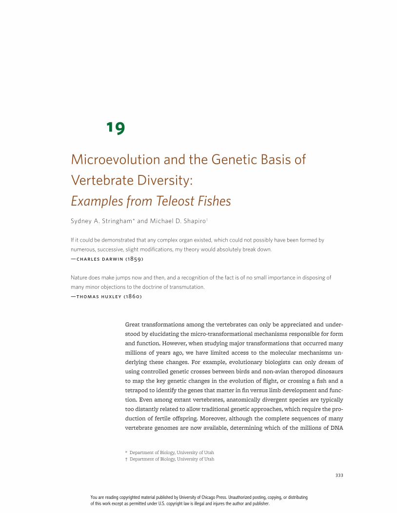
Geographically and phylogenetically distant popu-lations of threespine sticklebacks have evolved strik-ingly similar suites of characteristics in response to the shift to freshwater habitats. For example, many populations have lost major components of their bony armor, including the lateral plates and pelvic girdle, in response to new predator loads and other factors (Bell and Foster 1994) (fig. 19.1). Furthermore, parallel phe-notypic changes occur not only among populations of
fig. 19.1 (a) Variation in lateral plate number in marine threespine sticklebacks: complete morph (top), partial morph (center), and low morph
(bottom). Bony structures in all panels were visualized by staining with alizarin red. Fish found in marine habitats nearly always possess 30
or more plates per side (a phenotype referred to as the “complete morph”). In freshwater, fish typically have less than 10 plates per side (“low
morph”), or, less frequently, have an intermediate number of plates (“partial morph”) (Hagen and Gilbertson 1972). Partial morphs exhibit a
stereotypical pattern of plate loss, with plates at the most anterior and most posterior regions of the body and a mid- body gap in between.
Images courtesy of Jun Kitano, modified after Kitano et al. (2008). (b) Ventral (top) and lateral (bottom) illustrations of the stickleback pelvis
and ectocoracoid. The ectocoracoid is located anterior to the pelvis. (c) Pelvic loss has evolved in multiple populations of freshwater threespine
sticklebacks (G. aculeatus) (top) and ninespine sticklebacks (P. pungitius) (bottom). In both species, the ancestral marine populations possess
a complete pelvis; therefore, this trait has evolved independently in each species. (d) Ventral view of ninespine sticklebacks with a complete
pelvis (left) and a missing pelvis (right).
You are reading copyrighted material published by University of Chicago Press. Unauthorized posting, copying, or distributing of this work except as permitted under U.S. copyright law is illegal and injures the author and publisher.

336 sydney a . stringham and michael d . shapiro
three spine sticklebacks (Gasterosteus aculeatus), the focus of most recent genetic and genomic studies, but also across species that diverged millions of years ago (e.g., the ninespine stickleback Pungitius pungitius, and the brook stickleback Culaea inconstans) (Nelson and Atton 1971; Wootton 1976; Blouw and Boyd 1992; Bell and Foster 1994; Ziuganov and Zotin 1995). Thus, this multispecies system provides an excellent model to ex-amine the genetics of adaptive traits on both micro- and macro evolutionary levels.
Armor Plate Variation
Armor plates are composed of thin dermal bone and al-most completely cover the lateral sides of marine three-spine sticklebacks (“complete morph”; fig. 19.1a, top). In contrast, the number and size of these plates is reduced in most freshwater populations (“low morph”; fig. 19.1a, bottom) in response to strong selection in freshwater habitats (discussed below), and the genetic basis of this variation has been the subject of classical genetic studies for decades (Hagen and Gilbertson 1973a; Avise 1976; Ziuganov 1983; Banbura 1994). Laboratory crosses between different morphs showed that probably only a few genes control most of the variation in plate number (Hagen and Gilbertson 1973a; Avise 1976; Ziuganov 1983; Banbura 1994).
More recently, Colosimo et al. (2004) used a mo-lecular genetic approach to identify the major locus controlling plate reduction. To do this, they crossed a complete-morph marine fish (Hokkaido Island, Japan) to a low-morph freshwater fish (Paxton Lake, British Columbia); the grandchildren (F2 progeny) of this cross showed a wide range of plate morphologies, including fish that had high or low numbers of plates like their grandparents. By looking for associations between plate phenotypes and segments of chromosomes inher-ited from either the complete- or low- morph grandpar-ent, Colosimo et al. (2004) found a single position in the genome (a quantitative trait locus, or QTL) on linkage group (LG) 4 that largely determined whether fish had the complete, partial, or low- plate morph (see fig. 19.2). Other studies suggested that LG4 controls plate pheno-types in multiple populations of threespine sticklebacks (Cresko et al. 2004; Schluter et al. 2004). However, key questions remained: which gene(s) in the major QTL region controlled armor variation, and were the muta-
tions the same or different among the many popula-tions with low plates?
Further genetic mapping studies showed that vari-ation in the gene Ectodysplasin (Eda) was the most likely cause of armor diversity (Colosimo et al. 2005). In vertebrates, Eda plays a key role in the development of several tissues derived from the ectoderm, includ-ing hair, teeth, sweat glands, and scales (Thesleff and Mikkola 2002; Kangas et al. 2004; Harris et al. 2008). The external armor of sticklebacks is also derived from ectoderm. Importantly, Colosimo et al. (2005) showed that, by injecting low- plated embryos with an engi-neered DNA construct containing a functional version of Eda, they could partially restore plate formation in low- plated fish. This provided functional evidence that Eda plays a critical role in plate development.
Strikingly, nearly every low- plated population throughout the range of the species appears to have the same chromosome segment containing the Eda gene (Colosimo et al. 2005). This indicates that the repeated evolution of low plates probably resulted from selec-tion on the same mutant version of Eda, rather than by independent mutations in Eda in each population. The key to the spread of the low- plate allele resides in the marine populations that colonize new freshwater habitats: the low- plate version of Eda typically found in freshwater populations is also found in a small propor-tion of marine fish, suggesting that high- plated ocean populations are a “genetic reservoir” for the low- plate allele (Colosimo et al. 2005). Once the allele enters a freshwater habitat with the arrival of new marine colo-nists, selection drives it to high frequency. Transition from high to low plates can happen very quickly. In one Alaskan lake population, for example, Bell et al. (2004) observed a dramatic shift from predominantly high- plated to low- plated in less than 12 years (also see Kitano et al. 2008). Paradoxically, while the genetic ba-sis for this trait is well understood and there is strong evidence for selection on plate phenotypes and the Eda locus, the ecological mechanism driving selection is less clear (reviewed in Barrett 2010).
Reduction and Loss of the Pelvic Fin Complex
In addition to variation in lateral armor, at least 20 freshwater populations of threespine stickleback also exhibit reduction or loss of the pelvis (Bell 1974; Moodie
You are reading copyrighted material published by University of Chicago Press. Unauthorized posting, copying, or distributing of this work except as permitted under U.S. copyright law is illegal and injures the author and publisher.

fig. 19.2 Schematic of quantitative trait locus (QTL) mapping in a laboratory cross. (a) Individuals or populations that show variation in a
trait of interest (in this case stripes) are crossed to produce F1 offspring (b), which exhibit a phenotype intermediate to the phenotypes of the
parental generation. F1 individuals are crossed to produce an F
2 generation (c), which will now show segregation of the trait of interest if the
number of genes controlling the trait is small. In this case, only the phenotypic extremes (dark stripes or no stripes, but not intermediate
stripes) are shown. The genomes of the F2 individuals are then analyzed with a set of genomic markers to detect statistical associations be-
tween genotypes and phenotypes. These associations define QTL, which are chromosome regions that are linked to phenotypes of interest. The
identity of the specific genes that underlie phenotypic variation might not immediately be known because QTL associations often span many
genes. Chromosome segments inherited from the striped and unstriped founders of the cross are indicated by black and white, respectively,
and only the chromosome containing the causative mutation is depicted here. If individuals that inherit one version of the chromosome seg-
ment (black) nearly always exhibit one phenotype (dark stripes) and individuals inheriting the alternative version (white) nearly always exhibit
the alternative phenotype (no stripes), then that segment is probably linked (physically close on a chromosome) to the causative mutation. In
this example, a dashed box indicates the chromosome region associated with the stripe trait. The different versions of the chromosomes can
be detected using markers such as polymorphic microsatellite markers (short repeat sequences that often differ in length among individuals)
and single nucleotide polymorphisms (SNPs). These markers are assembled into linkage groups, and relative marker positions are determined
based on recombination rates. Ideally, each chromosome in the genome will be represented by a single linkage group, and together the groups
comprise a linkage map. Likelihood of odds (LOD) scores provide a statistical test of associations between genotypes and traits. The LOD plot
at bottom right shows a region of a chromosome that exceeds a significance threshold (dashed line) and is therefore associated with variation
in the trait of interest.
You are reading copyrighted material published by University of Chicago Press. Unauthorized posting, copying, or distributing of this work except as permitted under U.S. copyright law is illegal and injures the author and publisher.

338 sydney a . stringham and michael d . shapiro
and Reimchen 1976; Campbell and Williamson 1979; Edge and Coad 1983; Bell 1987). The stickleback pelvis is homologous to the pelvic fin skeleton of other teleosts as well as to the tetrapod hind limb. It is composed of a pelvic girdle and serrated pelvic spines that provide protection from gape- limited predators such as large piscivorous fish (Hoogland et al. 1957; Hagen and Gil-bertson 1972; Moodie 1972; Gross 1978; Lescak and von Hippel 2011) (fig. 19.1b– d). However, reduction of pelvic structures is advantageous in some populations where grasping predators such as aquatic invertebrates are a greater threat, especially to juvenile fish (Hoogland et al. 1957; Reimchen 1980, 1983; Bell et al. 1993; Bell and Orti 1994; Bourgeois et al. 1994). Large pelvic skeletons could be disadvantageous in these habitats because spines provide an additional surface for insects to cap-ture and hold their prey (Reimchen 1980; Reist 1980; Ziuganov and Zotin 1995; Marchinko 2009).
Using a QTL mapping approach similar to the ar-mor plate study, Shapiro et al. (2004) identified the gene Pitx1 as a major influence on pelvic morphology. Pitx1 contributes to hind limb identity and development in vertebrates, and mice with an inactive form (knock-out) of the gene exhibit reduced and malformed hind limbs but normal forelimbs (Lanctôt et al. 1999; Marcil et al. 2003). Furthermore, sticklebacks from the genetic mapping cross that retained pelvic spines showed a marked asymmetry with larger spines on the left side, a feature also seen in the limbs of mice with an inac-tive version of Pitx1 and humans with a Pitx1 mutation (Lanctôt et al. 1999; Gurnett et al. 2008).
Unlike in the mouse Pitx1 knockout, mutations were not found in the coding region of Pitx1 in pelvisless freshwater stickleback populations compared to ma-rine fish (Shapiro et al. 2004). Consequently, the Pitx1 proteins encoded by the marine and freshwater popula-tions were the same. However, the location of the gene’s expression was drastically different between popula-tions. As in other vertebrates, Pitx1 was expressed in the developing pelvis of marine larvae. In contrast, expression was greatly reduced or absent in the pel-vic region of freshwater stickleback larvae, yet other regions of normal expression, such as the jaws, were not affected (Shapiro et al. 2004; Shapiro, Marks, et al. 2006). Therefore, the change in Pitx1 was predicted to affect a DNA sequence that regulates when and where the gene is expressed. Chan et al. (2010) confirmed this
hypothesis by finding DNA deletions near the Pitx1 gene in several pelvic- reduced populations. When attached to the protein- coding sequence of Pitx1 and injected into embryos from pelvisless sticklebacks, this regula-tory region (also known as an enhancer) was capable of restoring pelvic development, thus verifying that the deletion was critical in the evolution and development of pelvic reduction. In contrast to repeated selection on the same low- plate version of Eda, Chan et al. detected different deletions near Pitx1 in different populations, suggesting that pelvic reduction in threespine stickle-backs arose repeatedly by independent mutations in different populations.
A likely factor in the repeated involvement of the Pitx1 regulatory element, as opposed to mutations in the coding sequence of the gene, is pleiotropy; that is, selection on one trait, such as pelvic reduction, has the potential to affect development of other traits con-trolled by the same gene. In mice, the pleiotropic effects of Pitx1 mutations are especially pronounced: complete inactivation of the gene leads not only to hind limb anomalies, but also jaw and brain deformities (Lanctôt et al. 1999). In contrast, the pelvis- specific regulatory mutation in sticklebacks yields an adaptive phenotype that is specific to one trait, while leaving other develop-mental roles of Pitx1 intact (Shapiro et al. 2004; Chan et al. 2010).
Pelvic reduction is not limited to a single species of stickleback. The ninespine stickleback (Pungitius pun-gitius) diverged from the threespine stickleback at least 10 million years ago, yet these two species have a simi-lar history of postglacial freshwater colonization and re-peated evolution of pelvic reduction (Aldenhoven et al. 2010). Based on studies of the ninespine stickleback from two localities (Canada and Finland), Pitx1 appears to play a role in pelvic reduction in this species as well (Shapiro, Bell, and Kingsley 2006; Shikano et al. 2013). These results in extant, genetically tractable stickleback species might hold clues about mechanisms of pelvic reduction in other species as well. For example, the ex-tensive fossil record of Gasterosteus doryssus, an ex-tinct relative of the threespine stickleback, documents the repeated evolution of pelvic reduction in a Miocene population (Bell 1974b; Bell et al. 1985; Bell 1988). As in modern threespine sticklebacks, pelvic reduction in G. doryssus shows a pronounced left- side bias, a mor-phological signature of Pitx1- mediated changes (Sha piro
You are reading copyrighted material published by University of Chicago Press. Unauthorized posting, copying, or distributing of this work except as permitted under U.S. copyright law is illegal and injures the author and publisher.

339microevolution and the genetic basis of vertebrate diversity
et al. 2004; Shapiro, Bell, and Kingsley 2006). This mor-phological trend extends beyond sticklebacks, as pel-vic remnants in manatees also show a left- side bias (Shapiro, Bell, and Kingsley 2006). The genetic basis of hind limb reduction in manatees is not known, but this shared morphological signature of Pitx1- mediated reduction provides clues about the molecular mecha-nisms involved. Together, these examples show that genetics in one species can potentially generate hy-potheses for study in other, less genetically tractable species.
Pitx1 probably does not universally play a major role in pelvic reduction, however. In another population of ninespine sticklebacks (Point MacKenzie, Alaska), the major QTL for pelvic reduction is clearly not Pitx1 (Sha-piro et al. 2009). This result suggests that ninespine stickleback populations use both the same and differ-ent genetic mechanisms as threespine sticklebacks to converge on the same pelvic phenotype.
Body Shape Variation
Sticklebacks from a variety of habitats exhibit enor-mous variation in overall body shape. The ancestral marine form is generally large and streamlined with a deep body and head, long fins, and a narrow caudal re-gion. These adaptations are thought to be optimal for navigating open water (Walker 1997; Walker and Bell 2000; Spoljaric and Reimchen 2007; Albert et al. 2008). Freshwater populations, particularly those that inhabit littoral regions and feed on macroinvertebrates, gener-ally have bodies that are short and deep, with shorter fins and a wider caudal region, resulting in a more ma-neuverable body that is better suited to foraging and evading predators in a complex habitat (Webb 1982; Walker 1997; Walker and Bell 2000; Spoljaric and Reim-chen 2007).
While many studies have highlighted recurring trends in body shape and their link to particular habi-tats, less is known about the genetic architecture of these changes (reviewed in Reid and Peichel 2010). To address this shortcoming, Albert et al. (2008) used a cross between marine and freshwater fish to conduct QTL mapping for body and head shape. Perhaps not surprisingly, they found that the genetic architecture of body shape is more complex than discrete traits such as plate variation and pelvic reduction. However, similar
to discrete traits, the same genomic regions underlie similar body shape traits in different populations. For example, some of the same chromosome regions influ-ence differences not only between marine and freshwa-ter populations, but also between semi- isolated benthic and limnetic populations that occur within several lakes (Gow et al. 2006; Reid and Peichel 2010).
Collectively, these studies suggest that similar suites of shape changes are key transformations in ad-aptation to new freshwater habitats, and similar suites of genes might govern these repeated changes species- wide (also see Hohenlohe et al. 2010; Jones, Chan, et al. 2012; Jones, Grabherr, et al. 2012).
Summary
Molecular genetic studies of microevolutionary trans-formations in sticklebacks provide important insights into general trends underlying the molecular basis of a classic adaptive radiation. First, dramatic pheno-typic changes such as pelvis and armor reduction can result largely from changes at a few genetic loci (e.g., Pitx1 and Eda, respectively, plus a modest number of loci of small effect). Furthermore, repeated evolution of the same trait can result from repeated selection on a common ancestral chromosome segment (lateral armor evolution and Eda) or independent mutations in the same gene ( pelvic evolution and Pitx1). However, com-parisons across stickleback species suggest that these mechanisms are not necessarily universal. Other adap-tive changes, such as body shape modifications that characterize populations in different habitats, have a more complex genetic architecture, yet still repeatedly involve a similar suite of genomic regions.
Mexican Cavefish (Family Characidae, Astyanax mexicanus)
Introduction
As with freshwater habitat specialization in stickle-backs, cave specialization has resulted in the repeated evolution of similar traits across diverse lineages of metazoans, including teleost fishes. Constructive traits that are common in cave- dwelling animals include in-creased numbers of taste buds, increased fat storage, larger egg size, and more sensitive nonvisual sensory
You are reading copyrighted material published by University of Chicago Press. Unauthorized posting, copying, or distributing of this work except as permitted under U.S. copyright law is illegal and injures the author and publisher.

340 sydney a . stringham and michael d . shapiro
systems (Culver 1982); regressive traits, such as loss of eyes and pigmentation, have evolved repeatedly across phyla as well.
The Mexican cavefish (Astyanax mexicanus) is an ideal model to study the genetic basis of cave pheno-types in vertebrates. Multiple populations within this species have converged on similar phenotypes, pro-viding another opportunity to test whether the same or different genetic mechanisms underlie repeated morphological changes. At least 30 populations of A. mexicanus are distributed across northeastern Mexico (Hubbs and Innis 1936; Wilkens and Burns 1972; Mitch-ell et al. 1977; Espinasa et al. 2001), and phylogenetic analyses suggest that the cave form does not have a sin-gle evolutionary origin (Espinasa and Borowsky 2001; Dowling et al. 2002; Strecker et al. 2003; Strecker et al. 2004).
Pigmentation Variation
In the darkness of a cave environment, the usual roles of pigmentation (camouflage, mate selection, etc.) are no longer relevant and the loss of pigmentation has oc-curred in cave- dwelling species across phyla. However, the adaptive significance (if any) of this phenotype in cavefish and other cave animals is still unclear. Pigmen-tation variation in cavefish encompasses a number of distinct phenotypes, including complete albinism, pig-mentation reduction, and decreased melanophore num-ber, each with a distinct genetic architecture.
Albinism was long known to be controlled by a sin-gle major locus, and possibly the same gene in multiple populations (Sadoglu 1957; Sadoglu and McKee 1969; Wilkens 1988). More recently, QTL mapping in cave-fish led to the discovery of a deletion in the Oca2 gene that underlies albinism in the Pachón population (Pro-tas et al. 2006) (fig. 19.3a– c). Oca2 encodes a key pro-tein in melanin synthesis, and mutations in this gene also cause albinism in both humans and mice (Rinchik et al. 1993; Yi et al. 2003). Albinism in a second cavefish population, Molino, is also due to a deletion in Oca2, but this deletion is distinct from the Pachón version and therefore must have arisen independently (Protas et al. 2006). Albinism in a third population, Japonés, probably results from a regulatory mutation in the same gene as no coding changes were identified (Protas et al. 2006). Hence, as with Pitx1 and pelvic reduction in
sticklebacks, different mutations in the same gene led to similar phenotypes in different populations.
Another pigment- reduction phenotype, brown (characterized by brown instead of black eyes and re-duced melanophore number), results from mutations in the Melanocortin- 1 receptor (Mc1r) gene (fig. 19.3d– f ). Mc1r encodes a receptor protein expressed in pigment- producing cells, and its activity can regulate melanin content and melanocyte dispersal in fish (Richardson et al. 2008; Tezuka et al. 2011). Like Oca2 and albinism, the brown phenotype results from more than one muta-tion in different cavefish populations, although at least one of these mutations has probably spread to several populations (Gross et al. 2009).
Together, these examples of pigment variation il-lustrate that convergent phenotypes can occur by in-dependent mutations in the same genes (similar to the repeated evolution of pelvic reduction in sticklebacks), and by selection on standing genetic variants (similar to repeated evolution of armor phenotypes in stickle-backs). In cavefish, independent deletions in the coding region of Oca2, as well as a possible regulatory muta-tion, have both been implicated in albinism. Likewise, independent mutations in Mc1r led to repeated evolu-tion of the brown phenotype, perhaps by a combina-tion of selection on mutant alleles that originated in the surface population, and new mutations in different cave populations (Gross et al. 2009).
Eye Loss
One of the most dramatic changes in cavefish compared to their surface- dwelling relatives is severe eye reduc-tion (fig. 19.3a– c). During embryonic development in cavefish, eyes begin to form but eventually stall and de-generate, beginning with the lens (Cahn 1958; Yamamoto et al. 2004). However, transplanting a surface fish lens into a developing cavefish eye can halt degeneration, demonstrating that this structure is a critical signaling center in eye development (Jeffery and Martasian 1998; Yamamoto and Jeffery 2000; Strickler, Yamamoto, and Jeffery 2007).
Genetic and developmental experiments suggest that between 6 and 12 genes contribute to eye regres-sion in cavefish (Wilkens 1988; Protas et al. 2007), and that the same genetic mechanisms do not underlie re-gression in all cave populations (Wilkens 1971; Wilkens
You are reading copyrighted material published by University of Chicago Press. Unauthorized posting, copying, or distributing of this work except as permitted under U.S. copyright law is illegal and injures the author and publisher.

341microevolution and the genetic basis of vertebrate diversity
and Strecker 2003; Borowsky 2008). This complex trait probably entails genetic pathways that control cell death and proliferation (Protas et al. 2007; Strickler, Byerly, and Jeffery 2007; Gross et al. 2008), response to environmental stress (Hooven et al. 2004), photo-receptor development (Kozmik 2008; Strickler and Jef-fery 2009), and morphogenesis (Jeffery and Martasian 1998; Yamamoto et al. 2004; Strickler and Jeffery 2009). In summary, eye degeneration in cavefish is probably not under simple genetic control. Although several spe-cific genes have been shown to affect eye development in this species, no specific mutations have yet been identified that correlate with the eyeless phenotype in any cave population.
Selection, Neutral Mutation, and Pleiotropy
While it is intuitive to envision natural selection driv-ing the acquisition of heightened sensory traits such as increased taste bud number and increased sensitivity to vibrations in a cave environment, the adaptive conse-quences of eye and pigment loss are less clear. Perhaps unnecessary structures in a dark environment, such as the eye, are a liability; for example, eyes could be tar-gets for predators, injury, or infection (Poulson 1963; Poulson and White 1969; Culver 1982; Jeffery 2005).
Alternatively, neutral mutation could explain eye and pigment loss (Kimura and Ohta 1971; Culver 1982; Wil-kens 1988). In a dark environment, otherwise deleterious mutations in pigment and eye developmental pathways might not be selected against, as long as they do not result in other disadvantageous phenotypes. Therefore, given sufficient time, pathways involved in eye and pig-ment development could accumulate enough mutations for the associated structures to be lost. Interestingly, in genetic crossing experiments, cave alleles tend only to contribute to decreases in eye size, consistent with selection on eye regression, while cave alleles contrib-ute to both increases and decreases in number of me-lanophores, suggesting drift might play a central role in pigmentation traits (Protas et al. 2007).
The loss of eyes and pigmentation in cavefish might also result from pleiotropy. Genetic and experimental evidence suggest that eye reduction might be a sec-ondary effect of selection on alleles that are advanta-geous in the cave environment for increased gustatory or mechanical sensitivity (Yamamoto et al. 2004, 2009; Yoshizawa et al. 2010, 2013; Borowsky 2013). For exam-ple, in hybrid crosses between cave and surface fish, the number of taste buds is inversely correlated with eye size (Yamamoto et al. 2009). A compelling example of this effect on the developmental level comes from
fig. 19.3 Surface morph of Astyanax mexicanus (a) compared to cavefish populations from the Molino (b) and Pachón (c) populations. Each
of these cave populations exhibits pigment loss mediated by Oca2 and eye reduction (white boxes). In some populations, these changes have
probably evolved independently. (d– f’) The partially pigmented “brown” phenotype results from a decrease in melanin content and number of
melanophores (pigment- containing cells). The severity of the phenotype depends on the number of cave alleles of Mc1r in an individual. In
this example, two copies of the Pachón allele yield the most severe phenotype. Boxed area in (d) indicates area of magnification in (d’– f’).
(a– c) Images courtesy of Richard Borowsky; (d– f’) images courtesy of Josh Gross, modified after Gross et al. (2009).
You are reading copyrighted material published by University of Chicago Press. Unauthorized posting, copying, or distributing of this work except as permitted under U.S. copyright law is illegal and injures the author and publisher.

342 sydney a . stringham and michael d . shapiro
the gene Sonic hedgehog (Shh), which is expressed in the oral- pharyngeal region and the developing taste buds of both cave and surface forms. When this gene is experimentally overexpressed in both forms, embryos develop wider jaws and more taste buds, as well as smaller eyes (Yamamoto et al. 2004, 2009).
Summary
As in sticklebacks, genetic dissection of derived traits in cavefish demonstrates that dramatic phenotypes can potentially fall under the control of a modest number of genomic regions of large effect. Furthermore, these studies also show that similar phenotypes can arise through independent mutations in the same genes: Oca2 and Mc1r underlie pigmentation variation in sev-eral populations, but different populations carry dif-ferent mutations. Derived pigmentation traits in cave-fish can also result from either coding or regulatory mutations: at least one population of albino cavefish probably harbors a regulatory mutation in Oca2, while most other albino populations have coding changes that lead to a decrease or loss of function. Other phe-notypes, such as eye loss, are genetically more compli-cated and are probably the result of changes in multi-ple genes.
Although great strides are being made to identify the genetic basis of derived traits, these data do not necessarily lead directly to an understanding of the adaptive significance of phenotypes. Both pigment and eye reduction might result from positive selection for these traits, neutral mutation, or pleiotropy as the re-sult of selection on other, as yet unknown, adaptive phenotypes.
Cichlids (Family Cichlidae)
Background
Cichlids, a third example of a morphologically di-verse and species- rich group of teleosts, inhabit lakes throughout Central and South America, Madagascar, India, and Africa. Several lakes throughout this range include classic examples of rapid adaptive radiations. Two especially notable cases occur in the African rift lakes, where more than 500 species in Lake Victoria and over 700 species in Lake Malawi arose within the last
1 million years after multiple colonization events and hy-bridization (Banister and Clarke 1980; Meyer et al. 1990; Owen et al. 1990; Meyer 1993; Kocher et al. 1995; Turner et al. 2001; Joyce et al. 2011). Within a single lake, these species occupy habitats from shallow water to depths of over 100 meters. Different species also have diverse feeding strategies from generalist fish, zooplankton, and algae feeders to specialized crab, snail, and scale eaters (reviewed in Turner 2007). Furthermore, similar feeding strategies have arisen multiple times, provid-ing another opportunity to examine the genetic basis of convergence in adaptively relevant phenotypes (Kocher et al. 1993) (fig. 19.4a). Like sticklebacks and cavefish,
fig. 19.4 (a) A sample of the cichlid diversity in Lake Tanganyika
(left) and Lake Malawi (right), highlighting the convergent phe-
notypes that have evolved independently in these two lakes.
(b) Labeotropheus fuelleborni (top left) feeds by biting algae from
rock surfaces. This species has a shorter lower jaw and tricuspid
teeth (top right). In contrast, Metriaclima zebra (bottom left) is a
suction feeder with a long lower jaw and bicuspid dentition (bottom
right). Images courtesy of Craig Albertson, modified after Albertson
and Kocher (2006).
You are reading copyrighted material published by University of Chicago Press. Unauthorized posting, copying, or distributing of this work except as permitted under U.S. copyright law is illegal and injures the author and publisher.

343microevolution and the genetic basis of vertebrate diversity
genetic mapping of derived traits in cichlids is greatly facilitated by the ability of many distinct forms to in-terbreed and produce fertile offspring in a laboratory setting.
Feeding Morphology
Some of the best- studied adaptive traits in cichlids in-volve craniofacial structures. Different cichlid species have evolved to feed on an enormous variety of food types, and this diversification has produced a wide range of specialized head, jaw, and tooth morpholo-gies (Albertson and Kocher 2006) (fig. 19.4). Genetic control of jaw and head morphology is highly complex and involves at least 40 chromosome regions, many of them affecting multiple elements of the feeding appara-tus (Albertson and Kocher 2001; Albertson et al. 2003a, 2003b).
To reduce this complexity, Albertson et al. (2005) specifically examined functionally relevant aspects of jaw morphology in two divergent species. The first spe-cies, Metriaclima zebra, feeds on algae, diatoms, and plankton from the water column, and has a narrow, forward- directed mouth optimized for suction feeding (Ribbink et al. 1983). In contrast, the jaw of Labeotro-pheus fuelleborni is short and square with a downward orientation that allows it to bite algae from rocks while remaining horizontal (Ribbink et al. 1983). One QTL identified in the Albertson et al. study included Bone Morphogenetic Protein 4 (Bmp4), a member of a large gene family that also regulates growth and differen-tiation during craniofacial development in other verte-brates (Abzhanov et al. 2004; Wu et al. 2004). At early developmental stages, the jaws of the suction- feeder M. zebra had much lower Bmp4 expression than the biting- feeder L. fuelleborni (Albertson et al. 2005). In-terestingly, when Albertson et al. overexpressed Bmp4 in the embryos of zebrafish (suction- feeders, like M. ze-bra), the lower jaw shape shifted to a shape more suited for biting (like L. fuelleborni ). Therefore, the results of experimental developmental studies in the zebrafish model system were consistent with genetic findings in wild cichlid species.
In another study using the same two species, Rob-erts et al. (2011) implicated the gene Patched 1 (Ptch1)— a receptor in the hedgehog pathway that contributes to dermal bone development (Abzhanov et al. 2007)— in
morphological differences in the lower jaw. Beyond M. zebra and L. fuelleborni, additional species- specific al-leles of Ptch1 were found in other cichlids with diver-gent feeding strategies, suggesting that this gene might affect jaw morphology in multiple lineages (Roberts et al. 2011).
Summary
The search for molecular changes that contribute to adaptive changes among cichlid species has thus far identified a small number of genes that contribute to diversity in feeding morphology, a key feature of this group’s radiation. However, the genetic basis of varia-tion in feeding structures is complex, with numerous chromosome regions contributing to differences in morphology. As with body shape variation in stickle-backs and eye reduction in cavefish, feeding morphol-ogy in cichlids involves several genomic regions that contribute to variation in multiple structures.
Discussion
Genetic Architecture of Derived Traits
The examples outlined above show that the genetic ar-chitecture of some major morphological changes can be relatively simple, with large effects produced by changes in only a few genes or genomic regions. Plate and pelvic reduction in sticklebacks, as well as albinism in cavefish, are largely controlled by single major genes. However, some derived traits have a more complex genetic architecture, including changes in stickleback body shape, variation in cichlid jaw morphology, and reduction of the cavefish eye. These contrasting de-grees of complexity might represent different temporal stages of morphological transformations. Theoretical models of adaptation by new mutations (as opposed to selection on standing genetic variation) suggest that a small number of initial mutations lead to large fitness effects, so early adaptive stages can have a simple ge-netic architecture; subsequently, “modifier” mutations of smaller effect accumulate over time (Orr 1998; Orr 2002). By this model, several examples of genetically simple changes discussed above might reflect very re-cent transformations, while a more complex architec-ture could potentially reflect a longer period of trait
You are reading copyrighted material published by University of Chicago Press. Unauthorized posting, copying, or distributing of this work except as permitted under U.S. copyright law is illegal and injures the author and publisher.

344 sydney a . stringham and michael d . shapiro
evolution or selection on a large number of preexisting genetic variants.
We also note that, in all three teleost examples, sev-eral QTL regions control more than one trait. For in-stance, in sticklebacks, LG4 appears to be a “hotspot” of variation in body shape, lateral plates, and pelvic phenotypes (Colosimo et al. 2004; Shapiro et al. 2004; Albert et al. 2008; Shapiro et al. 2009). In cavefish, 13 genomic regions are known to influence multiple traits (Protas et al. 2008); these regions could contain multiple genes that affect a suite of traits beneficial to cave- dwellers, or single genes that have pleiotropic ef-fects. Finally, in cichlids, LG5 influences tooth morphol-ogy, female sex determination, pigmentation, and also contains genes important for color perception (Carleton and Kocher 2001; Albertson et al. 2003a; Streelman et al. 2003; Kocher 2004; Streelman and Albertson 2006). This trend is by no means limited to loci that underlie diver-sity in fishes; the genetic clustering of QTL that control ecologically relevant traits could allow rapid evolutionary change through linkage of advantageous alleles in many different organisms (e.g., Garber and Quisenberry 1927; Mather 1950; Sheppard 1953; Murray and Clarke 1973; Joron et al. 2006; Joron et al. 2011).
Coding versus Regulatory Mutations
Among the teleost examples we discuss above, some of the genetic changes are (or are predicted to be) in noncoding regulatory regions of genes, while others di-rectly affect protein- coding sequences, which in turn can affect protein function. This dichotomy, and relative contributions of each type of mutation to evolutionary change in general, has sparked considerable interest in the recent evolutionary genetics literature (e.g., Hoek-stra and Coyne 2007; Wray 2007; Carroll 2008; Stern and Orgogozo 2008). While it is clear that not all evolution-ary change results from cis- regulatory mutations, a num-ber of hypotheses have been put forth to explain why these noncoding mutations might be a primary driver of evolutionary change, especially morphological change. One compelling argument centers on the modularity of regulatory regions (reviewed in Carroll 2008). Modular-ity refers to the semi- independent function of each cis- regulatory element with respect to other cis- regulatory elements. Therefore, a mutation in one of several regu-latory regions of a gene can affect gene expression in
only a subset of tissues or developmental time points, thereby avoiding potentially detrimental side effects on other developmental processes ( pleiotropy). The poten-tial importance of regulatory changes has been appreci-ated since the description of bacterial operons by Jacob and Monod (1961), and cis- regulatory changes are clearly important in morphological, physiological, and behav-ioral evolution (reviewed in Wray 2007).
An argument against the dominance of cis- regulatory changes in evolutionary change is that there are cur-rently more confirmed examples of coding changes, but this could simply be because coding mutations are much easier to identify than regulatory mutations (reviewed in Stern and Orgogozo 2008). However, the pace of discovery (or implication) of cis- regulatory changes has recently begun to closely track the discov-ery of coding changes (Stern and Orgogozo 2008). In summary, both coding and regulatory mutations have the potential to contribute to significant evolutionary transformations, and ongoing work in fishes and other organisms will further elucidate general trends, if any exist.
Convergent Evolution
Teleosts exhibit repeated evolution of similar pheno-types among different populations within a species, and in some cases, between species. In many popula-tions of threespine sticklebacks, lateral armor reduc-tion evolved by repeated selection on a standing variant of the Eda locus. In contrast, other convergent evolu-tionary changes are the products of different mutations in the same genes. For example, different mutations in Pitx1 underlie pelvic reduction in several populations of threespine sticklebacks, and Oca2 and Mc1r mutations differ among cavefish populations with similar pigmen-tation phenotypes.
Comparisons between stickleback species also yield novel insights about convergent phenotypes. For ex-ample, pelvic reduction in at least two populations of ninespine sticklebacks probably results from changes to Pitx1, just as in threespine sticklebacks (Shapiro, Bell, and Kingsley 2006; Shikano et al. 2013). However, in an-other population of ninespine sticklebacks, pelvic re-duction is controlled by a genomic region distinct from Pitx1; QTL for other skeletal traits (including lateral ar-mor) and sex determination also differ between the two
You are reading copyrighted material published by University of Chicago Press. Unauthorized posting, copying, or distributing of this work except as permitted under U.S. copyright law is illegal and injures the author and publisher.

345microevolution and the genetic basis of vertebrate diversity
species (Shapiro et al. 2009). Therefore, a multispecies approach can be particularly informative in dissecting a broad range of genetic mechanisms underlying similar phenotypes.
Future Directions
Biologists are intensely interested in how vertebrates undergo transformations both great and small, yet we know remarkably little about the genetic basis of phe-notypic change. In several examples above, QTL results were leveraged to fine- map and functionally test spe-cific candidate genes for the evolution of derived traits. While these cases are exciting, it is important to note that they are also currently the exceptions— mapping traits to the gene level and demonstrating functional consequences of mutations is still uncommon.
Traits with a simple genetic architecture are easier to analyze than those with more genetic complexity, and many traits that have been examined in natural populations of teleosts and other organisms are ones
that are relatively easy to see and quantify. Therefore, observable and relatively simple traits are preferentially studied, and we have a poorer understanding of com-plex anatomical, physiological, and behavioral traits that are undoubtedly important for evolutionary trans-formations (Rockman 2012).
New genomic tools, and the ability to compare doz-ens of genomes simultaneously, can help identify signa-tures of selection in suites of genes that affect traits that are not easily visualized. Recent studies, perhaps most notably in sticklebacks (Hohenlohe et al. 2010; Jones, Chan, et al. 2012; Jones, Grabherr, et al. 2012), have taken this “bottom- up” approach to identify genomic regions under selection in marine versus freshwater environ-ments, as well as in benthic versus limnetic freshwater habitats. With precipitous drops in the cost of DNA se-quencing and generation of new genetic resources, we expect that techniques pioneered for a limited number of species will become widely available to investigate important evolutionary transformations in other verte-brates as well.
* * *
Acknowledgments
We thank Eric Domyan, Josh Gross, Craig Miller, and the editors for comments on earlier drafts of this chap-ter. M. D. S. is forever grateful for mentorship by Farish Jenkins in the lab, field, and classroom. This work was supported by NIH training grant T32- GM007464 (S.A.S.), NSF grants IOS- 0744974 and DEB- 1149160 (M.D.S.), and the Burroughs Wellcome Fund (M.D.S.).
Glossary
Allele: Variant of a given gene or marker.
Coding mutation: A change in DNA sequence that
occurs in a part of a gene that codes for a protein.
Genetic architecture: A general description of how
traits are controlled by genotypes. For example,
genetic architecture includes the number and
location of genes that underlie a trait, as well
as the number of alleles at these loci and the
interactions among them.
Genetic marker: A DNA sequence that shows varia-
bility among individuals, and thus the inheritance
of different alleles can be traced from one gener-
ation to the next. Examples include single nucle-
otide polymorphisms (SNPs) and microsatellites
(simple DNA sequence repeats).
Genotype: The genetic makeup of an organism.
Linkage group: A group of genes or genetic markers
that reside on the same chromosome. Genes or
markers that are physically close to one another
tend to be inherited together; as a result, markers
can be ordered by tracking transmission from
one generation to the next (also called genetic
mapping). The sum of linkage groups comprises a
linkage map.
Locus ( plural: loci): The location of a gene or DNA
sequence on a chromosome or linkage group.
You are reading copyrighted material published by University of Chicago Press. Unauthorized posting, copying, or distributing of this work except as permitted under U.S. copyright law is illegal and injures the author and publisher.

346 sydney a . stringham and michael d . shapiro
Phenotype: The observable characteristics of an
organism.
Pleiotropy: When one gene affects more than one
trait or developmental process.
QTL (quantitative trait locus): A genomic region
that contributes to variation in a trait. Quantita-
tive traits are typically controlled by multiple loci.
QTL mapping: An experimental approach that often
begins by crossing strains of organisms that
diff er in a trait or traits of interest. Molecular
markers across the genome are used to track the
co- inheritance of genotypes and phenotypes of
offspring. Correlations between the trait(s) of
interest and molecular markers are assessed (see
fig. 19.2, and Miles and Wayne 2008).
Regulatory (cis- ) mutation: A change in DNA se-
quence that affects a region controlling the level
or location of expression of a gene, but (typically)
does not affect the protein encoded by the gene
(see also Wray 2007; Carroll 2008).
References
Abzhanov, A., M. Protas, B. R. Grant, P. R. Grant, and C. J. Tabin. 2004. Bmp4 and morphological variation of beaks in Darwin’s finches. Science 305:1462– 1465.
Abzhanov, A., S. J. Rodda, A. P. McMahon, and C. J. Tabin. 2007. Regulation of skeletogenic differentiation in cranial dermal bone. Development 134:3133– 3144.
Albert, A. Y., S. Sawaya, T. H. Vines, A. K. Knecht, C. T. Miller, B. R. Summers, S. Balabhadra, D. M. Kingsley, and D. Schluter. 2008. The genetics of adaptive shape shift in stickleback: pleiotropy and effect size. Evolution 62:76– 85.
Albertson, R. C., and T. D. Kocher. 2001. Assessing morphological differences in an adaptive trait: a landmark- based morphometric approach. J. Exp. Zool. 289:385– 403.
Albertson, R. C., and T. D. Kocher. 2006. Genetic and developmental basis of cichlid trophic diversity. Heredity (Edinb.) 97:211– 221.
Albertson, R. C., J. T. Streelman, and T. D. Kocher. 2003a. Directional selection has shaped the oral jaws of Lake Malawi cichlid fishes. Proc. Natl. Acad. Sci. USA 100:5252– 5257.
Albertson, R. C., J. T. Streelman, and T. D. Kocher. 2003b. Genetic basis of adaptive shape differences in the cichlid head. J. Hered. 94:291– 301.
Albertson, R. C., J. T. Streelman, T. D. Kocher, and P. C. Yelick. 2005. Integration and evolution of the cichlid mandible: the molecular basis of alternate feeding strategies. Proc. Natl. Acad. Sci. USA 102:16287– 16292.
Aldenhoven, J. T., M. A. Miller, P. S. Corneli, and M. D. Shapiro. 2010. Phylogeography of ninespine sticklebacks (Pungitius pungitius) in North America: glacial refugia and the origins of adaptive traits. Mol. Eco.l 19:4061– 4076.
Avise, J. C. 1976. Genetics of plate morphology in an unusual population of threespine sticklebacks (Gasterostus aculeatus). Genet. Res. 27:33– 46.
Banbura, J. 1994. A new model of lateral plate morph inheritance in the threespine stickleback, Gasterosteus aculeatus. Theor. Appl. Genet. 88:871– 876.
Banister, K. E., and M. A. Clarke. 1980. A revision of the large Barbus (Pisces, Cyprinidae) of Lake Malawi with a reconstruction of the history of the southern African Rift Valley lakes. J. Nat. Hist. 14:483.
Barrett, R. D. 2010. Adaptive evolution of lateral plates in three- spined stickleback Gasterosteus aculeatus: a case study in functional analysis of natural variation. J. Fish. Biol. 77:311– 328.
Bell, A. M., G. Orti, J. A. Walker, and J. P. Koenings. 1993. Evolution of pelvic reduction in threespine stickleback fish— a test of competing hypotheses. Evolution 47:906–914.
Bell, M. A. 1974. Reduction and loss of the pelvic girdle in Gasterosteus (Pisces): a case of parallel evolution. Nat. Hist. Mus. L.A. Contrib. Sci. 257:1– 36.
Bell, M. A. 1987. Interacting evolutionary constrains in pelvic reduction of threespine sticklebacks, Gasterosteus aculeatus (Pisces, Gasterosteidae). Biol. J. Linn. Soc. 31:347– 382.
Bell, M. A. 1988. Stickleback fishes: bridging the gap between population biology and paleobiology. Trends Ecol. Evol. 3:320– 325.
Bell, M. A., W. E. Aguirre, and N. J. Buck. 2004. Twelve years of contemporary armor evolution in a threespine stickleback population. Evolution 58:814– 824.
Bell, M. A., J. V. Baumgartner, and E. C. Olson. 1985. Patterns of temporal change in single morphological characters of a Miocene stickleback fish. Paleobiology 11:258– 271.
Bell, M. A., and S. A. Foster. 1994. The Evolutionary Biology of the Threespine Stickleback. Oxford: Oxford University Press.
Bell, M. A., and G. Orti. 1994. Pelvic reduction in threespine stickleback from Cook Inlet lakes: geographic distribution and intrapopulation variation. Copeia 1994:314– 325.
Bernatchez, L., and C. C. Wilson. 1998. Comparative phylogeo graphy of Nearctic and Palearctic fishes. Mol. Ecol. 7:431–452.
Blouw, D. M., and G. J. Boyd. 1992. Inheritance of reduction, loss, and asymmetry of the pelvis of Pungitius pungitius (ninespine stickleback). Heredity 68:33– 42.
Borowsky, R. 2008. Restoring sight in blind cavefish. Curr. Biol. 18:R23– 24.
Borowsky, R. 2013. Eye regression in blind Astyanax cavefish may facilitate the evolution of an adaptive behavior and its sensory receptors. BMC Biol. 11:81.
Bourgeois, J. F., D. M. Blouw, and M. A. Bell. 1994. Multivariate analysis of geographic covariance between phenotypes and environments in the threespine stickleback, Gasterosteus aculeatus. Can. J. Zool. 72:1497– 1509.
Cahn, P. H. 1958. Comparative optic development in Astyanax mexicanus and two of its blind cave derivatives. Bull. Am. Mus. Nat. Hist. 115:75– 112.
Campbell, R. N., and R. B. Williamson. 1979. The fishes of inland waters in the Outer Hebrides. Proc. R. Soc. Edinb. 77B:377– 393.
Carleton, K. L., and T. D. Kocher. 2001. Cone opsin genes of african cichlid fishes: tuning spectral sensitivity by differential gene expression. Mol. Biol. Evol. 18:1540– 1550.
You are reading copyrighted material published by University of Chicago Press. Unauthorized posting, copying, or distributing of this work except as permitted under U.S. copyright law is illegal and injures the author and publisher.

347microevolution and the genetic basis of vertebrate diversity
Carroll, S. B. 2008. Evo- devo and an expanding evolutionary synthesis: a genetic theory of morphological evolution. Cell 134:25– 36.
Chan, Y. F., M. E. Marks, F. C. Jones, G. Villarreal Jr., M. D. Shapiro, S. D. Brady, A. M. Southwick, D. M. Absher, J. Grimwood, J. Schmutz, R. M. Myers, D. Petrov, B. Jonsson, D. Schluter, M. A. Bell, and D. M. Kingsley. 2010. Adaptive evolution of pelvic reduction in sticklebacks by recurrent deletion of a Pitx1 enhancer. Science 327:302– 305.
Coad, B. W. 1983. Plate morphs in freshwater samples of Gasterosteus aculeatus from Arctic and Atlantic Canada: complementary comments on a recent contribution. Can. J. Zool. 61:1174– 1177.
Colosimo, P. F., K. E. Hosemann, S. Balabhadra, G. Villarreal Jr., M. Dickson, J. Grimwood, J. Schmutz, R. M. Myers, D. Schluter, and D. M. Kingsley. 2005. Widespread parallel evolution in sticklebacks by repeated fixation of Ectodysplasin alleles. Science 307:1928– 1933.
Colosimo, P. F., C. L. Peichel, K. Nereng, B. K. Blackman, M. D. Shapiro, D. Schluter, and D. M. Kingsley. 2004. The genetic architecture of parallel armor plate reduction in threespine sticklebacks. PLoS Biol. 2:E109.
Cresko, W. A., A. Amores, C. Wilson, J. Murphy, M. Currey, P. Phillips, M. A. Bell, C. B. Kimmel, and J. H. Postlethwait. 2004. Parallel genetic basis for repeated evolution of armor loss in Alaskan threespine stickleback populations. Proc. Natl. Acad. Sci. USA 101:6050– 6055.
Culver, D. C. 1982. Cave Life : Evolution and Ecology. Cambridge, MA: Harvard University Press.
Darwin, C. 1859. On the Origin of Species by Means of Natural Selection. London: John Murray.
Dowling, T. E., D. P. Martasian, and W. R. Jeffery. 2002. Evidence for multiple genetic forms with similar eyeless phenotypes in the blind cavefish, Astyanax mexicanus. Mol. Biol. Evol. 19:446– 455.
Edge, T. A., and B. W. Coad. 1983. Reduction of the pelvic skeleton in the three- spined stickleback Gasterosteus aculeatus in 2 lakes of Quebec Canada. Can. Field- Nat. 97:334– 336.
Espinasa, L., and R. B. Borowsky. 2001. Origin and relationships of cave populations of the blind Mexican tetra Astyanax fasciatus, in the Sierra de El Abra. Environ. Biol. Fishes 62:233– 237.
Espinasa, L., P. Rivas- Manzano, and H. Espinosa Pérez. 2001. A new blind cave fish population of the genus Astyanax: geography, morphology and behavior. Environ. Biol. Fishes 62:329– 344.
Garber, R. J., and K. S. Quisenberry. 1927. The inheritance of length of syle in buckwheat. J. Agric. Res. 34:181– 183.
Giles, N. 1983. The possible role of environmental calcium levels during the evolution of phenotypic diversity in Outer- Hebridean populations of the three- spined stickleback, Gasterosteus aculeatus. J. Zool. 199:535.
Gow, J. L., C. L. Peichel, and E. B. Taylor. 2006. Contrasting hybridization rates between sympatric three- spined sticklebacks highlight the fragility of reproductive barriers between evolutionarily young species. Mol. Ecol. 15:739– 752.
Gross, H. P. 1978. Natural selection by predators on the defensive apparatus of the three- spined stickleback, Gasterosteus aculeatus L. Can. J. Zool. 56:398– 413.
Gross, J. B., R. Borowsky, and C. J. Tabin. 2009. A novel role for Mc1r in the parallel evolution of depigmentation
in independent populations of the cavefish Astyanax mexicanus. PLoS Genet. 5:e1000326.
Gross, J. B., M. Protas, M. Conrad, P. E. Scheid, O. Vidal, W. R. Jeffery, R. Borowsky, and C. J. Tabin. 2008. Synteny and candidate gene prediction using an anchored linkage map of Astyanax mexicanus. Proc. Natl. Acad. Sci. USA 105:20106– 20111.
Gurnett, C. A., F. Alaee, L. M. Kruse, D. M. Desruisseau, J. T. Hecht, C. A. Wise, A. M. Bowcock, and M. B. Dobbs. 2008. Asymmetric lower- limb malformations in individuals with homeobox PITX1 gene mutation. Am. J. Hum. Genet. 83:616– 622.
Hagen, D. W., and L. G. Gilbertson. 1972. Geographic variation and environmental selection in Gasterosteus aculeaus L. in the Pacific northwest, America. Evolution 26:32– 51.
Hagen, D. W., and L. G. Gilbertson. 1973a. The genetics of plate morphs in freshwater threespine sticklebacks. Heredity 31:75– 84.
Hagen, D. W., and L. G. Gilbertson. 1973b. Selective predation and the intensity of selection acting upon the lateral plates of threespine stickleacks. Heredity 30:75– 84.
Hagen, D. W., and G. E. E. Moodie. 1982. Polymorphism for plate morphs in Gasterosteus aculeatus on the east coast of Canada and an hypothesis for their global distribution. Can. J. Zool. 60:1032– 1042.
Harris, M. P., N. Rohner, H. Schwarz, S. Perathoner, P. Konstantinidis, and C. Nusslein- Volhard. 2008. Zebrafish eda and edar mutants reveal conserved and ancestral roles of ectodysplasin signaling in vertebrates. PLoS Genet. 4:e1000206.
Heuts, M. C. 1947. The phenotypical variability of Gasterosteus aculeatus (L.) populations in Belgium; its bearing on the general geographical variability of the species. Antwerpen: Standarrd- boekhandel.
Hewitt, G. 2000. The genetic legacy of the Quaternary ice ages. Nature 405:907– 913.
Hoekstra, H. E., and J. A. Coyne. 2007. The locus of evolution: evo devo and the genetics of adaptation. Evolution Int. J. Org. Evolution 61:995– 1016.
Hohenlohe, P. A., S. Bassham, P. D. Etter, N. Stiffler, E. A. Johnson, and W. A. Cresko. 2010. Population genomics of parallel adaptation in threespine stickleback using sequenced RAD tags. PLoS Genet. 6:e1000862.
Hoogland, R. D., D. Morris, and N. Tinbergen. 1957. The spines of sticklebacks (Gasterosteus and Pygosteus) as means of defense against predators (Perca and Esox). Behaviour 10:205– 230.
Hooven, T. A., Y. Yamamoto, and W. R. Jeffery. 2004. Blind cavefish and heat shock protein chaperones: a novel role for hsp90alpha in lens apoptosis. Int. J. Dev. Biol. 48:731–738.
Hubbs, C. L., and W. T. Innis. 1936. The first known blind fish of the family Characidae: a new genus from Mexico. Occas. Papers Mus. Zool. Univ. Michigan 342:1– 7.
Huxley, T. H. H. 1860. The origin of species. Westminster Review 17:541– 570.
Jacob, F., and J. Monod. 1961. Genetic regulatory mechanisms in the synthesis of proteins. J. Mol. Biol. 3:318– 356.
Jeffery, W. R. 2005. Adaptive evolution of eye degeneration in the Mexican blind cavefish. J. Hered. 96:185– 196.
Jeffery, W. R., and D. P. Martasian. 1998. Evolution of eye regres sion in the cavefish Astyanax: apoptosis and the Pax- 6 gene. Amer. Zool. 38:685– 696.
You are reading copyrighted material published by University of Chicago Press. Unauthorized posting, copying, or distributing of this work except as permitted under U.S. copyright law is illegal and injures the author and publisher.

348 sydney a . stringham and michael d . shapiro
Jones, F. C., Y. F. Chan, J. Schmutz, J. Grimwood, S. D. Brady, A. M. Southwick, D. M. Absher, R. M. Myers, T. E. Reimchen, B. E. Deagle, D. Schluter, and D. M. Kingsley. 2012. A genome- wide SNP genotyping array reveals patterns of global and repeated species- pair divergence in sticklebacks. Curr. Biol. 22:83– 90.
Jones, F. C., M. G. Grabherr, Y. F. Chan, P. Russell, E. Mauceli, J. Johnson, R. Swofford, M. Pirun, M. C. Zody, S. White, et al. 2012. The genomic basis of adaptive evolution in threespine sticklebacks. Nature 484:55– 61.
Joron, M., L. Frezal, R. T. Jones, N. L. Chamberlain, S. F. Lee, C. R. Haag, A. Whibley, M. Becuwe, S. W. Baxter, L. Ferguson, et al. 2011. Chromosomal rearrangements maintain a polymorphic supergene controlling butterfly mimicry. Nature 477:203–206.
Joron, M., R. Papa, M. Beltran, N. Chamberlain, J. Mavarez, S. Baxter, M. Abanto, E. Bermingham, S. J. Humphray, J. Rogers, et al. 2006. A conserved supergene locus controls colour pattern diversity in Heliconius butterflies. PLoS Biol. 4:e303.
Joyce, D. A., D. H. Lunt, M. J. Genner, G. F. Turner, R. Bills, and O. Seehausen. 2011. Repeated colonization and hybridization in Lake Malawi cichlids. Curr. Biol. 21:R108– 109.
Kangas, A. T., A. R. Evans, I. Thesleff, and J. Jernvall. 2004. Nonindependence of mammalian dental characters. Nature 432:211– 214.
Kimura, M., and T. Ohta. 1971. Theoretical Aspects of Population Genetics. Princeton, NJ: Princeton University Press.
Kitano, J., D. I. Bolnick, D. A. Beauchamp, M. M. Mazur, S. Mori, T. Nakano, and C. L. Peichel. 2008. Reverse evolution of armor plates in the threespine stickleback. Curr. Biol. 18:769– 774.
Kocher, T. D. 2004. Adaptive evolution and explosive speciation: the cichlid fish model. Nat. Rev. Genet. 5:288– 298.
Kocher, T. D., J. A. Conroy, K. R. McKaye, and J. R. Stauffer. 1993. Similar morphologies of cichlid fish in Lakes Tanganyika and Malawi are due to convergence. Mol. Phylogenet. Evol. 2:158– 165.
Kocher, T. D., J. A. Conroy, K. R. McKaye, J. R. Stauffer, and S. F. Lockwood. 1995. Evolution of NADH dehydrogenase subunit 2 in east African cichlid fish. Mol. Phylogenet. Evol. 4:420– 432.
Kozmik, Z. 2008. The role of Pax genes in eye evolution. Brain Res. Bull. 75:335– 339.
Lanctôt, C., A. Moreau, M. Chamberland, M. L. Tremblay, and J. Drouin. 1999. Hindlimb patterning and mandible development require the Ptx1 gene. Development 126:1805– 1810.
Lescak, E. A., and F. A. von Hippel. 2011. Selective predation of threespine stickleback by rainbow trout. Ecol. Freshwat. Fish. 20:308– 314.
Marchinko, K. B. 2009. Predation’s role in repeated phenotypic and genetic divergence of armor in threespine stickleback. Evolution 63:127– 138.
Marcil, A., E. Dumontier, M. Chamberland, S. A. Camper, and J. Drouin. 2003. Pitx1 and Pitx2 are required for development of hindlimb buds. Development 130:45– 55.
Mather, K. 1950. The genetical architecture of heterostyly in Primula sinensis. Evolution 4:340– 352.
Meyer, A. 1993. Phylogenetic relationships and evolutionary processes in East African cichlid fishes. Trends Ecol. Evol. 8:279– 284.
Meyer, A., T. D. Kocher, P. Basasibwaki, and A. C. Wilson. 1990. Monophyletic origin of Lake Victoria cichlid fishes suggested by mitochondrial DNA sequences. Nature 347:550– 553.
Miles, C. M., and M. Wayne. 2008. Quantitative Trait Locus (QTL) Analysis. Nat. Educ. 1:1– 8.
Mitchell, R. W., W. H. Russell, and W. R. Elliot. 1977. Mexican eyeless characin fishes, genus Astyanax: environment, distribution and evolution. Spec. Publ. Mus. Texas Tech Univ. 12:1– 89.
Moodie, G. E. E. 1972. Predation, natural selection and adaptation in an unusual threespine stickleback. Heredity 28:155– 167.
Moodie, G. E. E., J. D. McPhail, and D. W. Hagen. 1973. Experimental demonstration of selective predation on Gasterosteus aculeatus. Behaviour 47:95– 105.
Moodie, G. E. E., and T. Reimchen. 1976. Phenetic variation and habitat differences in Gasterosteus populations of the Queen Charlotte Islands. Syst. Zool. 25:49– 61.
Murray, J., and B. Clarke. 1973. Supergenes in polymorphic land snails— examples from genus Partula. Genetics 74:S188– S189.
Nelson, J. S., and F. M. Atton. 1971. Geographic and morphological variation in the presence and absence of the pelvic skeleton in the brook stickleback, Culaea inconstans (Kirtland), in Alberta and Saskatchewan. Can. J. Zool. 49:343– 352.
Orr, H. A. 1998. The population genetics of adaptation: the distribution of factors fixed during adaptive evolution. Evolution 52:935– 949.
Orr, H. A. 2002. The population genetics of adaptation: the adaptation of DNA sequences. Evolution 56:1317– 1330.
Owen, R. B., R. Crossley, T. C. Johnson, D. Tweddle, I. Kornfield, S. Davison, D. H. Eccles, and D. E. Engstrom. 1990. Major low levels of Lake Malawi and their implications for speciation rates in cichlid fishes. Proc. R. Soc. London B 240:519– 553.
Poulson, T. L. 1963. Cave adaptation in amblyopsid fishes. Am. Mid. Nat. 70:257– 290.
Poulson, T. L., and W. B. White. 1969. The cave environment. Science 165:971– 981.
Protas, M., M. Conrad, J. B. Gross, C. Tabin, and R. Borowsky. 2007. Regressive evolution in the Mexican cave tetra, Astyanax mexicanus. Curr. Biol. 17:452– 454.
Protas, M., I. Tabansky, M. Conrad, J. B. Gross, O. Vidal, C. J. Tabin, and R. Borowsky. 2008. Multi- trait evolution in a cave fish, Astyanax mexicanus. Evol. Dev. 10:196– 209.
Protas, M. E., C. Hersey, D. Kochanek, Y. Zhou, H. Wilkens, W. R. Jeffery, L. I. Zon, R. Borowsky, and C. J. Tabin. 2006. Genetic analysis of cavefish reveals molecular convergence in the evolution of albinism. Nat. Genet. 38:107– 111.
Reid, D. T., and C. L. Peichel. 2010. Perspectives on the genetic architecture of divergence in body shape in sticklebacks. Integr. Comp. Biol. 50:1057– 1066.
Reimchen, T. E. 1980. Spine deficiency and polymorphism in a population of Gasterosteus aculeatus— an adaptation to predators. Can. J. Zool. 58:1232– 1244.
Reimchen, T. E. 1983. Structural relationships between spines and lateral plates in threespine stickleback (Gasterosteus aculeatus). Evolution 37:931– 946.
Reimchen, T. E. 1992. Injuries on stickleback from attacks by a toothed predator (Oncorhynchus) and implications for the evolution of lateral plates. Evolution 46:1224.
Reimchen, T. E. 1995. Predator- induced cyclical changes in lateral plate frequencies of Gasterosteus. Behaviour 132:1079.
You are reading copyrighted material published by University of Chicago Press. Unauthorized posting, copying, or distributing of this work except as permitted under U.S. copyright law is illegal and injures the author and publisher.

349microevolution and the genetic basis of vertebrate diversity
Reist, J. D. 1980. Predation upon pelvic phenotypes of brook stickleback, Culaea inconstans, by selected invertebrates. Can. J. Zool. 58:1253– 1258.
Ribbink, A. J., A. C. Marsh, C. C. Ribbink, and B. J. Sharp. 1983. A preliminary survey of the cichlid fishes of rocky habitats in Lake Malawi. S. Afr. J. Zool. 18:149– 310.
Richardson, J., P. R. Lundegaard, N. L. Reynolds, J. R. Dorin, D. J. Porteous, I. J. Jackson, and E. E. Patton. 2008. mc1r Pathway regulation of zebrafish melanosome dispersion. Zebrafish 5:289– 295.
Rinchik, E. M., S. J. Bultman, B. Horsthemke, S. T. Lee, K. M. Strunk, R. A. Spritz, K. M. Avidano, M. T. Jong, and R. D. Nicholls. 1993. A gene for the mouse pink- eyed dilution locus and for human type II oculocutaneous albinism. Nature 361:72– 76.
Roberts, R. B., Y. Hu, R. C. Albertson, and T. D. Kocher. 2011. Craniofacial divergence and ongoing adaptation via the hedgehog pathway. Proc. Natl. Acad. Sci. USA 108:13194– 13199.
Rockman, M. V. 2012. The QTN program and the alleles that matter for evolution: all that’s gold does not glitter. Evolution 66:1– 17.
Sadoglu, P. 1957. A Mendelian gene for albinism in natural cave fish. Experientia 13:394.
Sadoglu, P., and A. McKee. 1969. A second gene that affects eye and body color in Mexican blind cavefish. J. Hered. 60:10– 14.
Santini, F., L. J. Harmon, G. Carnevale, and M. E. Alfaro. 2009. Did genome duplication drive the origin of teleosts? A comparative study of diversification in ray- finned fishes. BMC Evol. Biol. 9:194.
Schluter, D., E. A. Clifford, M. Nemethy, and J. S. McKinnon. 2004. Parallel evolution and inheritance of quantitative traits. Am. Nat. 163:809– 822.
Shapiro, M. D., M. A. Bell, and D. M. Kingsley. 2006. Parallel genetic origins of pelvic reduction in vertebrates. Proc. Natl. Acad. Sci. USA 103:13753– 13758.
Shapiro, M. D., M. E. Marks, C. L. Peichel, B. K. Blackman, K. S. Nereng, B. Jonsson, D. Schluter, and D. M. Kingsley. 2004. Genetic and developmental basis of evolutionary pelvic reduction in threespine sticklebacks. Nature 428:717– 723.
Shapiro, M. D., M. E. Marks, C. L. Peichel, B. K. Blackman, K. S. Nereng, B. Jónsson, D. Schluter, and D. M. Kingsley. 2006. Corrigendum: genetic and developmental basis of evolutionary pelvic reduction in threespine sticklebacks. Nature 439.
Shapiro, M. D., B. R. Summers, S. Balabhadra, J. T. Aldenhoven, A. L. Miller, C. B. Cunningham, M. A. Bell, and D. M. Kingsley. 2009. The genetic architecture of skeletal convergence and sex determination in ninespine sticklebacks. Curr. Biol. 19:1140– 1145.
Sheppard, P. M. 1953. Polymorphism, linkage and the blood groups. Am. Nat. 87:283– 294.
Shikano, T., V. N. Laine, G. Herczeg, J. Vilkki, and J. Merila. 2013. Genetic architecture of parallel pelvic reduction in ninespine sticklebacks. G3 Genes Genom. Genet. 3:1833– 1842.
Spoljaric, M. A., and T. E. Reimchen. 2007. 10,000 years later: evolution of body shape in Haida Gwaii three- spined stickleback. J. Fish Biol. 70:1484.
Stern, D. L., and V. Orgogozo. 2008. The loci of evolution: how predictable is genetic evolution? Evolution 62:2155– 2177.
Strecker, U., L. Bernatchez, and H. Wilkens. 2003. Genetic divergence between cave and surface populations of Astyanax in Mexico (Characidae, Teleostei). Mol. Ecol. 12:699– 710.
Strecker, U., V. H. Faundez, and H. Wilkens. 2004. Phylogeography of surface and cave Astyanax (Teleostei) from Central and North America based on cytochrome b sequence data. Mol. Phylogenet. Evol. 33:469– 481.
Streelman, J. T., and R. C. Albertson. 2006. Evolution of novelty in the cichlid dentition. J. Exp. Zool. B Mol. Dev. Evol. 306:216– 226.
Streelman, J. T., R. C. Albertson, and T. D. Kocher. 2003. Genome mapping of the orange blotch colour pattern in cichlid fishes. Mol. Ecol. 12:2465– 2471.
Strickler, A. G., M. S. Byerly, and W. R. Jeffery. 2007. Lens gene expression analysis reveals downregulation of the anti- apoptotic chaperone αA- crystallin during cavefish eye degeneration. Dev. Genes Evol. 217:771– 782.
Strickler, A. G., and W. R. Jeffery. 2009. Differentially expressed genes identified by cross- species microarray in the blind cavefish Astyanax. Int. Zool. 4:31.
Strickler, A. G., Y. Yamamoto, and W. R. Jeffery. 2007. The lens controls cell survival in the retina: evidence from the blind cavefish Astyanax. Dev. Biol. 311:512– 523.
Tezuka, A., H. Yamamoto, J. Yokoyama, C. van Oosterhout, and M. Kawata. 2011. The MC1R gene in the guppy (Poecilia reticulata): genotypic and phenotypic polymorphisms. BMC Res. Notes 4:31.
Thesleff, I., and M. L. Mikkola. 2002. Death receptor signaling giving life to ectodermal organs. Sci. STKE 2002:pe22.
Turner, G. F. 2007. Adaptive radiation of cichlid fish. Curr. Biol. 17:R827– 831.
Turner, G. F., O. Seehausen, M. E. Knight, C. J. Allender, and R. L. Robinson. 2001. How many species of cichlid fishes are there in African lakes? Mol. Ecol. 10:793– 806.
Walker, J. A. 1997. Ecological morphology of lacustrine three- spine stickleback Gasterosteus aculeatus L. (Gasterosteidae) body shape. Biol. J. Linn. Soc. 61:3– 50.
Walker, J. A., and M. A. Bell. 2000. Net evolutionary trajectories of body shape evolution within a microgeographic radiation of threespine sticklebacks. J. Zool. Lond. 252:293– 302.
Webb, P. W. 1982. Locomotor patterns in the evolution of actinopterygian fishes. Am. Zool. 22:329– 342.
Wilkens, H. 1971. Genetic interpretation of regressive evolu tionary processes: studies of hybrid eyes of two Astyanax cave populations (Characidae, Pisces). Evolution 25:530– 544.
Wilkens, H. 1988. Evolution and genetics of epigean and cave Astyanax fasciatus (Characidae, Pisces). Evol. Biol. 23:271– 367.
Wilkens, H., and R. L. Burns. 1972. A new Anoptichthys cave population (Characidae, Pisces). Ann. Spéléol. 27:263– 270.
Wilkens, H., and U. Strecker. 2003. Convergent evolution of the cavefish Astyanax (Charcidae, Telesotei): genetic evidence from reduced eye- size and pigmentation. Biol. J. Linn. Soc. 80:545– 554.
Wootton, R. J. 1976. The Biology of the Sticklebacks. London: Academic.
Wray, G. A. 2007. The evolutionary significance of cis- regulatory mutations. Nat. Rev. Genet. 8:206– 216.
Wu, P., T. X. Jiang, S. Suksaweang, R. B. Widelitz, and C. M. Chuong. 2004. Molecular shaping of the beak. Science 305:1465– 1466.
You are reading copyrighted material published by University of Chicago Press. Unauthorized posting, copying, or distributing of this work except as permitted under U.S. copyright law is illegal and injures the author and publisher.

350 sydney a . stringham and michael d . shapiro
Yamamoto, Y., M. S. Byerly, W. R. Jackman, and W. R. Jeffery. 2009. Pleiotropic functions of embryonic sonic hedgehog expression link jaw and taste bud amplification with eye loss during cavefish evolution. Dev. Biol. 330:200– 211.
Yamamoto, Y., and W. R. Jeffery. 2000. Central role for the lens in cavefish eye degeneration. Science 289:631– 633.
Yamamoto, Y., D. W. Stock, and W. R. Jeffery. 2004. Hedgehog signalling controls eye degeneration in blind cavefish. Nature 431:844– 847.
Yi, Z., N. Garrison, O. Cohen- Barak, T. M. Karafet, R. A. King, R. P. Erickson, M. F. Hammer, and M. H. Brilliant. 2003. A 122.5- kilobase deletion of the P gene underlies the high prevalence of oculocutaneous albinism type 2 in the Navajo population. Am. J. Hum. Genet. 72:62– 72.
Yoshizawa, M., S. Goricki, D. Soares, and W. R. Jeffery. 2010. Evolution of a behavioral shift mediated by superficial neuromasts helps cavefish find food in darkness. Curr. Biol. 20:1631– 1636.
Yoshizawa, M., K. E. O’Quin, and W. R. Jeffery. 2013. Evolution of an adaptive behavior and its sensory receptors promotes eye regression in blind cavefish: response to Borowsky (2013). BMC Biol. 11:82.
Ziuganov, V. V. 1983. Genetics of osteal plate polymorphism and microevolution of threespine stickleback (Gasterosteus aculeatus L). Theor. Appl. Genet. 65.
Ziuganov, V. V., and A. A. Zotin. 1995. Pelvic girdle polymorphism and reproductive barriers in the ninespine stickleback Pungitius pungitius (L.) from northwest Russia. Behaviour 132:1095– 1105.
You are reading copyrighted material published by University of Chicago Press. Unauthorized posting, copying, or distributing of this work except as permitted under U.S. copyright law is illegal and injures the author and publisher.