microrna-15b participates in the development of diabetic
TRANSCRIPT

6018
Abstract. – OBJECTIVE: To investigate the role of microRNA-15b in diabetic retinopathy and its underlying mechanism.
MATERIALS AND METHODS: Diabetes rat model was established by streptozotocin injec-tion. The mRNA expression of microRNA-15b in retinal capillary endothelial cells and peri-cytes of diabetic rats was detected by quan-titative Real Time-Polymerase Chain Reaction (qRT-PCR). The mRNA and protein expressions of vascular endothelial growth factor A (VEG-FA) were detected by qRT-PCR and Western blot, respectively. MicroRNA-15b mimics or in-hibitor were transfected into retinal capillary en-dothelial cells and pericytes of diabetic rats, re-spectively. The mRNA expressions of microR-NA-15b and VEGFA were detected by qRT-PCR. Cell counting kit-8 (CCK-8) assay was used to detect the proliferation of capillary endotheli-al cells and pericytes. Dual-Luciferase reporter gene assay was conducted to verify the binding condition of microRNA-15b and VEGFA. RNA im-munoprecipitation (RIP) assay was performed to determine whether microRNA-15b could bind to AGO2. Rescue experiments were finally carried out by detecting the proliferation of retinal cap-illary endothelial cells and pericytes after down-regulation or overexpression of microRNA-15b and VEGFA.
RESULTS: The expression of microRNA-15b decreased, whereas VEGFA expression in-creased in retinal capillary endothelial cells and pericytes of diabetic rats. High expression of mi-croRNA-15b in retinal capillary endothelial cells and pericytes resulted in VEGFA down-regula-tion and decreased proliferation. RIP assay re-sults indicated that microRNA-15b could interact with AGO2. Additionally, Dual-Luciferase report-er gene assay showed that VEGFA is a direct tar-get gene of microRNA-15b. VEGFA overexpres-sion could reverse the inhibited proliferation of
retinal capillary endothelial cells and pericytes induced by microRNA-15b overexpression. Sim-ilarly, VEGFA knockdown could reverse the ef-fect of the low expression of microRNA-15b on the proliferation of retinal capillary endothelial cells and pericytes.
CONCLUSIONS: Low expression of microR-NA-15b in retinal capillary endothelial cells and pericytes of diabetic rats promotes the devel-opment of diabetic retinopathy by up-regulat-ing VEGFA.
Key Words:Retinal capillary endothelial cell, Pericyte, Diabetic
retinopathy, MicroRNA-15b, VEGFA.
Introduction
Diabetes mellitus (DM) is a metabolic disease characterized by hyperglycemia. Long-term hy-perglycemia could cause dysfunction of many organizations and tissues. Diabetic retinopathy (DR) is one of the most common complications of DM1. The incidence of DR increases with age and disease duration of DM. The prevalence rate of DR is over 50% in DM patients with a histo-ry of more than 10 years2. The pathogenesis of DR is very complicated, including inflammation, cell proliferation, cell differentiation, neuronal apoptosis, etc. However, effective prevention and treatment for DR are still lacking3,4.
VEGFA is a member of the vascular endo-thelial growth factor (VEGF) family. Biological functions of VEGFA are mediated by its specif-ic receptor. VEGFA plays an important role in embryogenesis and pathological angiogenesis5.
European Review for Medical and Pharmacological Sciences 2019; 23: 6018-6025
Y. XU1, S.-C. XIE2, Y.-C. MA3
1Department of Ophthalmology, Binzhou City Center Hospital, Binzhou, China2Department of Ophthalmology, Shan County Central Hospital, Heze, China3Department of Ophthalmology, Gaomi People’s Hospital, Weifang, China
Yi Xu and Shengchao Xie contributed equally to this work
Corresponding Author: Yanchun Ma, MM; e-mail: [email protected]
Low expression of microRNA-15b promotes the proliferation of retinal capillary endothelial cells and pericytes by up-regulating VEGFA in diabetic rats

MicroRNA-15b participates in the development of diabetic retinopathy
6019
The expression of cytokine is up-regulated in the retina of diabetic patients. In the non-proliferative phase of DR, VEGFA expression is upregulated in the local capillary non-perfusion region, ulti-mately leading to leakage of retinal capillaries6. In the terminal stage of DR, VEGFA can promote endothelial proliferation and participate in the pathological growth of neovascularization. Sin-gle nucleotide polymorphism (SNPs) in VEGFA is associated with the development of DR, and anti-VEGFA therapy is effective7. Yang et al8 have shown that miRNA can regulate VEGFA expres-sion in retinal endothelial cells. High expression of miR-126 can inhibit the migration of chorioret-inal endothelial cells in glucose-induced rhesus monkeys by blocking VEGFA and PIK3R2.
MicroRNAs (miRNAs) are a class of endoge-nous and highly conserved, small molecular, non-coding RNAs that directly inhibit or degrade the translation of target mRNA, thus regulating cell proliferation and apoptosis9. It has been found10 that many miRNAs are specifically expressed in the retina and play an important role in regu-lating angiogenesis in DR. MiR-200b, miR-146a and miR-126 regulate angiogenesis, inflammato-ry pathway and oxidation pathway in DR. MiR-146a inhibits inflammation and apoptosis in DR progression by inhibiting IL-6-induced STAT3/VEGF11. In addition, it has been found that miR-NA-1273g-3p participates in the development of DR by regulating autophagy and lysosome12. MiR-200b/c plays a protective role in DR by inhibiting dysfunction of high glucose-induced HRMEC13. MiR-195 is up-regulated in DR, which can accel-erate oxidative stress-induced retinal endothelial injury via targeting mitochondrial protein 214. These results suggested that miRNA may have multiple targeted regulatory mechanisms in DR. Gottmann et al15 have shown that microRNA-15b expression is associated with baseline blood glu-cose level and may be an indicator of DM. How-ever, the significance of microRNA-15b in DR has not been reported. The aim of this study is to investigate the role of microRNA-15b in diabetic retinopathy and its underlying mechanism.
Materials and Methods
Animal Model Construction and Primary Cells Extraction
This study was approved by the Animal Eth-ics Committee of Binzhou City Center Hospital Animal Center. The diabetic rat model was estab-
lished by feeding rats with high-sugar and high-fat diet for 8 weeks. Then, rats were in a fasting state for 10 hours before injection, followed by in-traperitoneal administration of 50 mg/kg with 2% streptozotocin (STZ) solution (STZ was dissolved in 0.1 mmol/L, pH 4.6 sterilized citrate-sodium citrate buffer solution). One week later, blood samples of tail vein were taken to measure the fasting blood glucose level. Rats with fasting blood glucose level higher than 16.7 mmol/L for more than 5 days were selected for the following experiments. Diabetic rats were then fed with high-sugar and high-fat diet for 12 weeks. The extraction of primary EC (retinal endothelial cells) and RP (retinal pericytes) were based on the previous description16,17. Cells were cultured in low-glucose Dulbecco’s Modified Eagle’s Me-dium (DMEM; Gibco, Grand Island, NY, USA) containing 20% fetal bovine serum (FBS; Gibco, Grand Island, NY, USA), 100 U/mL penicillin and 100 µg/mL streptomycin at 37° C in a 5% CO2 incubator. The culture medium was changed 4 times a week. Cell passage was performed at a ratio of 1:2 until the confluence degree of 90%.
Cell TransfectionThe primary cells were inoculated at a density
of 1×10⁷/mL and cell transfection was performed when the confluence was over 60%. 250 µL of low-glucose DMEM without antibiotics was added to the 1.5 mL centrifuge tube. An appro-priate amount of microRNA-15b mimics, mi-croRNA-15b inhibitor or corresponding controls was transferred into a centrifuge tube. 5 µL of Lipofectamine 2000 (Invitrogen, Carlsbad, CA, USA) was mixed evenly with 250 µL of the me-dium, and stored at room temperature for 5 min. MiRNA and Lipofectamine 2000 were mixed, maintained for 15 minutes and then added to cul-ture cells. After transfection for 4 to 6 hours, 20% FBS and low-glucose DMEM without antibiotics were replaced.
RNA Extraction and Quantitative Real Time-Polymerase Chain Reaction (qRT-PCR)
The total RNAs were extracted by TRIzol method (Invitrogen, Carlsbad, CA, USA), and reverse transcription was used to obtain cDNAs. Reverse transcriptional cDNA products were used as a template, and the relative expression of VEGFA was detected by quantitative Real Time-Polymerase Chain Reaction (qRT-PCR). Each sample was set up for two replicates. Ac-

Y. Xu, S.-C. Xie, Y.-C. Ma
6020
cording to the Ct value at the end of the reaction (Ct value was the number of amplification cy-cles corresponding to the threshold value of the Real-Time fluorescence signal in each reaction tube), the relative expression of each sample gene was calculated by the method of 2-ΔΔCt. Primers used in the study were as follows: VEG-FA (F: 5’-CTGTACCTCCACCATGCCAA-3’, 5’-GCTGCGCTGATAGACATCCA-3’); GAP-DH (F: 5’-CACCCACTCCTCCACCTTTG-3’, R: 5’-CCACCACCCTGTTGCTGTAG-3’); MicroR-NA-15b (F: 5′-CTTCTGTCTATCACATAAGT-GG-3′, R: 5′-GGTCCAAGTCAATTCCATG-3′); U6 (F: 5′-AACGCTTCACGAATTTGCGT-3′, R: 5′- CCAAGCTTATGACAGCCATCATC-3′).
Western BlotCells were washed twice with Phosphate-Buff-
ered Saline (PBS) and lysed in cell lysis solu-tion on ice. Cell supernatant was collected after centrifugation. The protein concentration was detected by bicinchoninic acid (BCA) protein quantitative kit (Pierce, Waltham, MA, USA). The protein sample was prepared by 5 × sodium dodecyl sulphate-polyacrylamide gel electropho-resis (SDS-PAGE) and boiled at 100°C. 50 µg of protein was electrophoresed and transferred to the membrane. Membranes were blocked in 5% skimmed milk at room temperature. The primary antibody with a dilution of 1: 1000 was used to incubate overnight at 4°C, followed by the incubation of the second antibody coupled with goat anti-rabbit horseradish peroxidase (di-lution 1:2000). The chemiluminescence system (Bio-Rad Laboratories, Hercules, CA, USA) was adopted for membrane exposure.
Cell Counting Kit-8 (CCK-8) AssayCells were prepared into single cell suspen-
sions with DMEM containing 20% FBS. 200 µL of cell suspension was added in each well of the 96-well plate. The cells were cultured for 96 hours under normal culture conditions. 10 µL of CCK-8 solution (Dojindo Laboratories, Kumamo-to, Japan) was added to each well and incubated for 2 hours at 37°C in the dark. Cell proliferation was detected at 6 h, 24 h, 48 h, 72 h, and 96 h, respectively. The absorbance at the wavelength of 450 nm was measured using a microplate reader.
Dual-Luciferase Reporter Gene AssayTarget genes were predicted by TargetScan Re-
lease 3.4 (http://www.targetscan.org/). The plas-mids of Luciferase reporter gene were transfected
until 70% of cell confluence, and three duplicated wells were set in each sample. The wild-type se-quences VEGFA WT 3’UTR and the mutant-type sequences VEGFA MUT 3’UTR were construct-ed. After co-transfection for 24 hours, 50 μL of 1×PLB was added in each well. 100 μL of mixed Luciferase assay reagent II was added in 10 μL of suspension per well. The Luciferase reaction intensity was detected 2 s later.
RNA Immunoprecipitation (RIP) Experiment
Cells were collected and centrifuged with 900 μL of RIP buffer at 4°C for 10 minutes. 10 μL of RIP cell lysate was absorbed and heated at 95°C for 10 minutes with 10 μL 2×SDS buffer solution addition at 95°C for 10 minutes. The superna-tant was used as protein input for SDS-PAGE electrophoresis. Eppendorf (EP) tube containing supernatant was incubated overnight at 4°C and placed on the magnetic frame the next day. The supernatant was discarded and 0.5 ml of RIP wash buffer was added and mixed evenly. EP tube was placed on the magnetic frame, and the supernatant was discarded. Cells were washed five times. Before the last washing, 500 μL of magnetic bead suspension solution was absorbed, and 1 × SDS buffer solution was added for 10 min heating at 95°C. The supernatant was removed for SDS-PAGE electrophoresis after centrifuging magnetic bead.
Statistical AnalysisStatistical Product and Service Solutions
(SPSS) 19.0 software (IBM, Armonk, NY, USA) was adopted for statistical analysis. The experi-mental results were expressed as mean ± standard deviation. The t-test was used to compare the differences between the two groups. p<0.05 was considered to be statistically significant.
Results
Expressions of MicroRNA-15b and VEGFA in Retinal Capillary Endothelial Cells and Pericytes of Diabetic Rats
To study the role of microRNA-15b in DR, the expression of microRNA-15b in retinal capillary endothelial cells and pericytes of diabetic rats was detected by qRT-PCR. The qRT-PCR results showed that the expression of microRNA-15b was lower in EC and RP cells than that in the normal control group (Figure 1A). Meanwhile, we detect-

MicroRNA-15b participates in the development of diabetic retinopathy
6021
ed the expression of VEGFA in retinal capillary endothelial cells and pericytes of diabetic rats. Compared with the control group, the expression of VEGFA in retinal capillary endothelial cells and pericytes of diabetic rats was significantly in-creased (Figure 1B, 1C). These results suggested that microRNA-15b and VEGFA may be involved in the pathogenesis of DR.
MicroRNA-15b Inhibited the Proliferation of EC and RP Cells
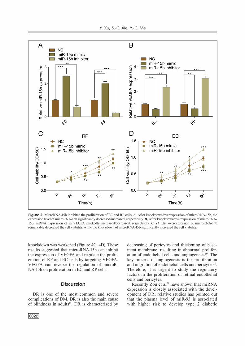
The expression of microRNA-15b in EC and RP cells was down-regulated after transfection of microRNA-15b inhibitor, whereas it was up-regu-lated after transfection of microRNA-15b mimics (Figure 2A). Meanwhile, we found that VEGFA expression was negatively related to the expres-sion of microRNA-15b (Figure 2B). To further study the effects of microRNA-15b on EC and RP cells, we examined the effects of overexpression or knockdown of microRNA-15b on the prolifer-ation of EC and RP cells. After overexpression of microRNA-15b in RP, cell viability decreased significantly, whereas the knockdown of microR-NA-15b remarkably increased the viability of RP cells (Figure 2C). MicroRNA-15b also inhibited the proliferation of EC cells (Figure 2D). These results suggested that microRNA-15b might play a role in DR by regulating the proliferation of EC and RP cells.
MicroRNA-15b Targeted Binding VEGFAShibuya et al7 have shown that VEGFA can
promote endothelial cell proliferation and par-ticipate in the pathological growth of neovas-
cularization. TargetScan predicted the potential binding site in 3’-UTR between microRNA-15b and VEGFA, suggesting that microRNA-15b may regulate the expression of VEGFA (Figure 3A). The binding condition of microRNA-15b to AGO2 protein was detected by RIP assay. In RP and EC cells, microRNA-15b was remarkably en-riched in the anti-AGO2 antibody compared with that in the negative control IgG antibody. These results indicated that microRNA-15b can be com-bined with AGO2 (Figure 3B). To further verify the regulatory effect of microRNA-15b on VEG-FA in RP and EC cells, Dual-Luciferase reporter gene assay was performed. Luciferase activity in VEGFA-WT 3’UTR group decreases; however, there was no significant difference in Luciferase activity of cells transfected with VEGFA-MUT 3’UTR (Figure 3C, 3D). These results suggested that VEGFA is the target gene of microRNA-15b and microRNA-15b could inhibit the expression of VEGFA.
VEGFA Can Reverse the Effects of MicroRNA-15b on Cell Proliferation in EC and RP Cells
To further verify the role of VEGFA in the proliferation of RP and EC cells regulated by microRNA-15b, we overexpressed microRNA-15b and VEGFA in RP and EC at the same time. The proliferative activity of EC and RP cells de-creased after overexpression of microRNA-15b, while the knockdown of VEGFA attenuated this inhibitory effect (Figure 4A, 4B). In addition, after knockdown of microRNA-15b and VEGFA, the promoted proliferation induced by microRNA-15b
Figure 1. Expressions of microRNA-15b and VEGFA in EC and pericytes RP cells of diabetic rats. A, Compared with the normal control group, the expression level of microRNA-15b in EC and RP cells was markedly decreased. B, Compared with the normal control group, mRNA expression of VEGFA in EC cells and RP cells was remarkably increased. C, Compared with the normal control group, the expression level of VEGFA protein in EC cells and RP cells was significantly increased.
Y. Xu, S.-C. Xie, Y.-C. Ma
6022
knockdown was weakened (Figure 4C, 4D). These results suggested that microRNA-15b can inhibit the expression of VEGFA and regulate the prolif-eration of RP and EC cells by targeting VEGFA. VEGFA can reverse the regulation of microR-NA-15b on proliferation in EC and RP cells.
Discussion
DR is one of the most common and severe complications of DM. DR is also the main cause of blindness in adults18. DR is characterized by
decreasing of pericytes and thickening of base-ment membrane, resulting in abnormal prolifer-ation of endothelial cells and angiogenesis19. The key process of angiogenesis is the proliferation and migration of endothelial cells and pericytes20. Therefore, it is urgent to study the regulatory factors in the proliferation of retinal endothelial cells and pericytes.
Recently Zou et al21 have shown that miRNA expression is closely associated with the devel-opment of DR; relative studies has pointed out that the plasma level of miR-93 is associated with higher risk to develop type 2 diabetic
Figure 2. MicroRNA-15b inhibited the proliferation of EC and RP cells. A, After knockdown/overexpression of microRNA-15b, the expression level of microRNA-15b significantly decreased/increased, respectively. B, After knockdown/overexpression of microRNA-15b, mRNA expression of in VEGFA markedly increased/decreased, respectively. C, D, The overexpression of microRNA-15b remarkably decreased the cell viability, while the knockdown of microRNA-15b significantly increased the cell viability.

MicroRNA-15b participates in the development of diabetic retinopathy
6023
retinopathy. Yang et al22 have shown that miR-181a can inhibit ocular angiogenesis by interfer-ing with the expression of vascular endothelial growth factor. The overexpression of miR-29a can down-regulate angiotensin expression and delay the development of DR23. In this work, we found that the expression of microRNA-15b decreased in retinal capillary endothelial cells and pericytes in diabetic rats. Those results suggested that microRNA-15b may be involved in the pathogenesis of DR. After overexpression of microRNA-15b, cell viability significantly de-creased and knockdown of microRNA-15b ob-tained the opposite result. MicroRNA-15b could play a role in DR by regulating the proliferation of EC cells and RP cells.
VEGF can promote the proliferation of ret-inal capillary endothelial cells, induce the formation of neovascularization and increase
vascular permeability24. In addition, VEGF is the key factor in the development of DR and the initiator of diabetic fundus change. Fur-thermore, VEGF can also up-regulate the ex-pressions of intercellular adhesion molecules (including ICAM-l, VCAM-1, sVAP-1, etc.), resulting in adhesion between leukocytes and vascular endothelial cells, blood stasis, fur-ther aggravating damage of blood-retina bar-rier function25. VEGFA is found to be most relevant to the DR in the VEGF family. Recent studies8 have shown that miRNA can regulate VEGFA expression in retinal endothelial cells. In this work, we verified that VEGFA is the tar-get gene of microRNA-15b by Dual-Luciferase reporter gene assay. Further research showed that VEGFA can reverse the regulation of mi-croRNA-15b on the proliferation of EC and RP cells. The above results suggested that microR-
Figure 3. MicroRNA-15b target binding with VEGFA. A, Construction of VEGFA-WT and VEGFA-MUT. B, In RIP experiments, microRNA-15b could bind to AGO2 in EC and RP cells. C, D, The overexpression of microRNA-15b markedly decreased the cell viability, while microRNA-15b knockdown increased the cell viability.

Y. Xu, S.-C. Xie, Y.-C. Ma
6024
NA-15b can directly inhibit the expression of VEGF and cell proliferation, which may play a crucial role in the regulation of DR.
Conclusions
We found that microRNA-15b is lowly ex-pressed in retinal capillary endothelial cells and pericytes in diabetic rats. The low expression
of microRNA-15b promotes the proliferation of retinal capillary endothelial cells and pericytes by up-regulating VEGFA, thereby participating in the development of DR.
Conflict of InterestThe Authors declare that they have no conflict of interests.
Figure 4. VEGFA can reverse the role of microRNA-15b in EC and RP cells. A, B, The overexpression of microRNA-15b reduced the viability of EC and RP cells, while the overexpression of VEGFA weakened the down-regulation of microRNA-15b mimic on the cell viability. C, D, The proliferative activity of EC and RP cells was reversed by VEGFA overexpression.

MicroRNA-15b participates in the development of diabetic retinopathy
6025
References
1) Solomon SD, Chew e, Duh eJ, Sobrin l, Sun JK, Van-DerbeeK bl, wyKoff CC, GarDner Tw. Erratum. Di-abetic retinopathy: a position statement by the American Diabetes Association. Diabetes Care 2017; 40: 412-418.
2) ola mS, nawaz mi, SiDDiquei mm, al-amro S, abu el-aSrar am. Recent advances in understanding the biochemical and molecular mechanism of dia-betic retinopathy. J Diabetes Complications 2012; 26: 56-64.
3) Chen y, men K, li Xf, li J, liu m, fan zq. Efficacy and safety of dipeptidyl peptidase-4 inhibitors in the treatment of type 2 diabetes mellitus patients with moderate to severe renal impairment: a me-ta-analysis. Eur Rev Med Pharmacol Sci 2018; 22: 3502-3514.
4) naThan Dm, bebu i, laChin Jm. Frequency of evi-dence-based screening for diabetic retinopathy. N Engl J Med 2017; 377: 195.
5) meSquiTa J, CaSTro-De-SouSa JP, Vaz-Pereira S, neVeS a, PaSSarinha la, Tomaz CT. Evaluation of the growth factors VEGF-a and VEGF-B in the vitre-ous and serum of patients with macular and reti-nal vascular diseases. Growth Factors 2018; 36: 48-57.
6) nGuyen qD, brown Dm, marCuS Dm, boyer DS, Pa-Tel S, feiner l, GibSon a, Sy J, runDle aC, hoPKinS JJ, rubio rG, ehrliCh JS. Ranibizumab for diabetic macular edema: results from 2 phase III random-ized trials: RISE and RIDE. Ophthalmology 2012; 119: 789-801.
7) Shibuya m, ClaeSSon-welSh l. Signal transduction by VEGF receptors in regulation of angiogenesis and lymphangiogenesis. Exp Cell Res 2006; 312: 549-560.
8) yanG wz, yanG J, Xue lP, Xiao lb, li y. MiR-126 overexpression inhibits high glucose-induced mi-gration and tube formation of rhesus macaque choroid-retinal endothelial cells by obstructing VEGFA and PIK3R2. J Diabetes Complications 2017; 31: 653-663.
9) maSTroPaSqua r, ToTo l, CiPollone f, SanToViTo D, CarPineTo P, maSTroPaSqua l. Role of microRNAs in the modulation of diabetic retinopathy. Prog Retin Eye Res 2014; 43: 92-107.
10) GonG q, Su G. Roles of miRNAs and long non-coding RNAs in the progression of diabetic reti-nopathy. Biosci Rep 2017; 37: BSR20171157.
11) ye ea, STeinle JJ. miR-146a suppresses STAT3/VEGF pathways and reduces apoptosis through IL-6 signaling in primary human retinal microvas-cular endothelial cells in high glucose conditions. Vision Res 2017; 139: 15-22.
12) ye z, li zh, he Sz. miRNA-1273g-3p involvement in development of diabetic retinopathy by modu-
lating the autophagy-lysosome pathway. Med Sci Monit 2017; 23: 5744-5751.
13) DinG y, hu z, luan J, lV X, yuan D, Xie P, yuan S, liu q. Protective effect of miR-200b/c by inhib-iting vasohibin-2 in human retinal microvascular endothelial cells. Life Sci 2017; 191: 245-252.
14) zhanG r, GarreTT q, zhou h, wu X, mao y, Cui X, Xie b, liu z, Cui D, JianG l, zhanG q, Xu S. Upregu-lation of miR-195 accelerates oxidative stress-in-duced retinal endothelial cell injury by targeting mitofusin 2 in diabetic rats. Mol Cell Endocrinol 2017; 452: 33-43.
15) GoTTmann P, ouni m, SauSSenThaler S, rooS J, STirm l, JahnerT m, KamiTz a, hallahan n, JonaS w, friT-SChe a, harinG hu, STaiGer h, bluher m, fiSCh-er-PoSoVSzKy P, VoGel h, SChurmann a. A computa-tional biology approach of a genome-wide screen connected miRNAs to obesity and type 2 diabe-tes. Mol Metab 2018; 11: 145-159.
16) Su X, SorenSon Cm, Sheibani n. Isolation and char-acterization of murine retinal endothelial cells. Mol Vis 2003; 9: 171-178.
17) buzney Sm, maSSiCoTTe SJ, heTu n, zeTTer br. Ret-inal vascular endothelial cells and pericytes. Dif-ferential growth characteristics in vitro. Invest Ophthalmol Vis Sci 1983; 24: 470-480.
18) brownlee m. Biochemistry and molecular cell bi-ology of diabetic complications. Nature 2001; 414: 813-820.
19) Chen m, CurTiS Tm, STiTT aw. Advanced glycation end products and diabetic retinopathy. Curr Med Chem 2013; 20: 3234-3240.
20) momeni a, Dyani ma, ebrahimi e, SeDehi m, naDe-ri a. Association of retinopathy and intima media thickness of common carotid artery in type 2 dia-betic patients. J Res Med Sci 2015; 20: 393-396.
21) zou hl, wanG y, GanG q, zhanG y, Sun y. Plasma level of miR-93 is associated with higher risk to develop type 2 diabetic retinopathy. Graefes Arch Clin Exp Ophthalmol 2017; 255: 1159-1166.
22) yanG C, Tahiri h, Cai C, Gu m, GaGnon C, harDy P. microRNA-181a inhibits ocular neovascularization by interfering with vascular endothelial growth fac-tor expression. Cardiovasc Ther 2018; 36: e12329.
23) zhanG lq, Cui h, wanG l, fanG X, Su S. Role of mi-croRNA-29a in the development of diabetic ret-inopathy by targeting AGT gene in a rat model. Exp Mol Pathol 2017; 102: 296-302.
24) aDamiS aP, miller Jw, bernal mT, D’amiCo DJ, folK-man J, yeo TK, yeo KT. Increased vascular endo-thelial growth factor levels in the vitreous of eyes with proliferative diabetic retinopathy. Am J Oph-thalmol 1994; 118: 445-450.
25) Simo-SerVaT o, hernanDez C, Simo r. Usefulness of the vitreous fluid analysis in the translational re-search of diabetic retinopathy. Mediators Inflamm 2012; 2012: 872978.