microscopic identification of vine black pigment in a ... · characteristic of vegetable cells,...
TRANSCRIPT

SM Analytical and Bioanalytical Techniques
Gr upSM
How to cite this article Rodríguez-Simón LR, López VDS and León-Coloma MÁ. Microscopic Identification of Vine Black Pigment In A Tempera Painting By Francisco
De Goya. SM Anal Bioanal Technique. 2017; 2(2): 1014.
OPEN ACCESS
ISSN: 2573-3729
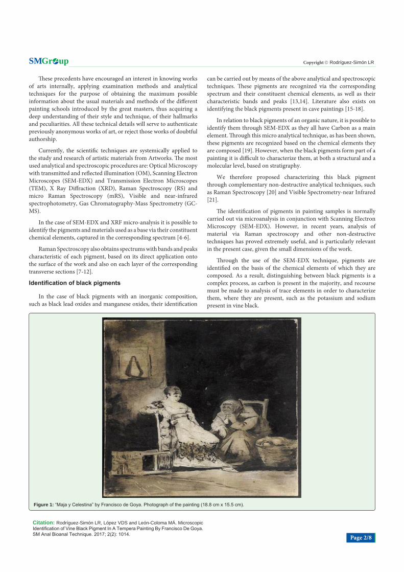
IntroductionThis work by Francisco de Goya [1], entitled Maja y Celestina (Figure 1) is a painting rendered
on a translucent alabaster board, 16 mm thick, employed to create a monochrome painting, wherein only different intensities of black are observed, brought into being via a series of different brushstrokes applied to the surface of the painting to create a transparent or opaque effect depending on the luminosity and chiaroscuro within the composition. Furthermore, Goya makes use of burin incisions to outline forms and accentuate areas of greatest luminosity.
Identifying the components present in works of art constitutes a research activity that has been being developed for several decades [2,3], because of interest in finding out about the artistic materials and painting techniques used by artists in the creation of their works.
Research Article
Microscopic Identification of Vine Black Pigment In A Tempera Painting By Francisco De GoyaLuis Rodrigo Rodríguez-Simón1*, Vicente Del Sol López2 and Miguel Ángel León-Coloma3
1Department of Paint and Restoration, University of Granada, Spain 2Department of Computer Languages and Systems, University of Granada, Spain3Department of Historical Heritage, University of Jaén, Spain
Article Information
Received date: Jul 31, 2017 Accepted date: Dec 04, 2017 Published date: Dec 07, 2017
*Corresponding author
Luis Rodrigo Rodríguez-Simón, Department of Paint and Restoration, University of Granada, Spain, Tel: +34 958 24 97 82; Email: [email protected]
Distributed under Creative Commons CC-BY 4.0
Keywords Goya; 18th Century; Painting; Alabaster Medium; Vine Black; Scanning Electron Microscopy; Co focal Micro-Raman Spectroscopy; Visible and Near-Infrared Spectrophotometry; Optical Microscope; Gas Chromatography-Mass Spectrometry
Abstract
This paper aims to advance our understanding of the morphological differentiation of vine black pigment encountered in a monochrome painting on alabaster produced by Francisco de Goya: Maja y Celestina.
To this end, three instrumental analytical techniques have been combined:
1) Micro-Raman Spectroscopy (mRS) applied “in situ”, focusing the laser directly on to the surface of the work and also on the stratigraphy prepared via a micro sample of the painting.
2) Scanning Electron Microscopy connected to an Energy-Dispersive X-Ray spectroscopy system (SEM-EDX), applied to the stratigraphy and also to a small portion of black-paint powder, scrapped from the edge of the work.
3) Visible and near-infrared spectrophotometry, applied “in situ” on to the surface of the picture.
The results obtained via the mRS are inconclusive, as is the case with the results obtained via visible and near-infrared spectrophotometry.
However, observation of the high magnifications enabled by the scanning electron microscopy and EDX microanalysis have allowed us to identify morphologically vine black pigment, which are combined with one another black pigment (lamp black) to form the single color present in the painting.
Thus, in the case of vine black pigment, polygonal forms were observed that may indicate the rigid walls characteristic of vegetable cells, given its botanical origin.
Gold leaf particles were also found, added to the mix of blacks, and we believe that they may be indicative of Goya’s aesthetic decisions, adding brightness and warmth to the dark color. Furthermore, a range of other pigments were also identified: lead white, bone black and black iron oxides, which, in principle, we attribute to contamination in the workshop.
The black pigments that were identified are bound together by a mixture of linseed oil and egg yolk, denoting a tempera grass painting technique.
Highlights
• This study focuses on the first work executed on an alabaster board that is classified as an original by Francisco de Goya [1].
• The painting involves a pictorial composition rendered exclusively in black.
• Via SEM-EDX it has been possible to morphologically differentiate vine black pigment based on the cellular structure.
• Polygonal shapes were observed with SEM and may indicate the rigid walls characteristic of vegetable cells.
• This innovative study proposes the morphological characterization of vine black pigment.
Citation: Rodríguez-Simón LR, López VDS and León-Coloma MÁ. Microscopic Identification of Vine Black Pigment In A Tempera Painting By Francisco De Goya. SM Anal Bioanal Technique. 2017; 2(2): 1014. Page 2/8
Gr upSM Copyright Rodríguez-Simón LR
These precedents have encouraged an interest in knowing works of arts internally, applying examination methods and analytical techniques for the purpose of obtaining the maximum possible information about the usual materials and methods of the different painting schools introduced by the great masters, thus acquiring a deep understanding of their style and technique, of their hallmarks and peculiarities. All these technical details will serve to authenticate previously anonymous works of art, or reject those works of doubtful authorship.
Currently, the scientific techniques are systemically applied to the study and research of artistic materials from Artworks. The most used analytical and spectroscopic procedures are: Optical Microscopy with transmitted and reflected illumination (OM), Scanning Electron Microscopes (SEM-EDX) and Transmission Electron Microscopes (TEM), X Ray Diffraction (XRD), Raman Spectroscopy (RS) and micro Raman Spectroscopy (mRS), Visible and near-infrared spectrophotometry, Gas Chromatography-Mass Spectrometry (GC-MS).
In the case of SEM-EDX and XRF micro-analysis it is possible to identify the pigments and materials used as a base via their constituent chemical elements, captured in the corresponding spectrum [4-6].
Raman Spectroscopy also obtains spectrums with bands and peaks characteristic of each pigment, based on its direct application onto the surface of the work and also on each layer of the corresponding transverse sections [7-12].
Identification of black pigments
In the case of black pigments with an inorganic composition, such as black lead oxides and manganese oxides, their identification
can be carried out by means of the above analytical and spectroscopic techniques. These pigments are recognized via the corresponding spectrum and their constituent chemical elements, as well as their characteristic bands and peaks [13,14]. Literature also exists on identifying the black pigments present in cave paintings [15-18].
In relation to black pigments of an organic nature, it is possible to identify them through SEM-EDX as they all have Carbon as a main element. Through this micro analytical technique, as has been shown, these pigments are recognized based on the chemical elements they are composed [19]. However, when the black pigments form part of a painting it is difficult to characterize them, at both a structural and a molecular level, based on stratigraphy.
We therefore proposed characterizing this black pigment through complementary non-destructive analytical techniques, such as Raman Spectroscopy [20] and Visible Spectrometry-near Infrared [21].
The identification of pigments in painting samples is normally carried out via microanalysis in conjunction with Scanning Electron Microscopy (SEM-EDX). However, in recent years, analysis of material via Raman spectroscopy and other non-destructive techniques has proved extremely useful, and is particularly relevant in the present case, given the small dimensions of the work.
Through the use of the SEM-EDX technique, pigments are identified on the basis of the chemical elements of which they are composed. As a result, distinguishing between black pigments is a complex process, as carbon is present in the majority, and recourse must be made to analysis of trace elements in order to characterize them, where they are present, such as the potassium and sodium present in vine black.
Figure 1: “Maja y Celestina” by Francisco de Goya. Photograph of the painting (18.8 cm x 15.5 cm).

Citation: Rodríguez-Simón LR, López VDS and León-Coloma MÁ. Microscopic Identification of Vine Black Pigment In A Tempera Painting By Francisco De Goya. SM Anal Bioanal Technique. 2017; 2(2): 1014. Page 3/8
Gr upSM Copyright Rodríguez-Simón LR
Initial identification of the pigments [19] was carried out via SEM-EDX, focusing on a stratigraphy prepared from a micro sample that was extracted from the painting. The results of the microanalysis of this dark hue suggest that it is a blend of carbon black, vine black and small particles of gold leaf, bound together via a mixture of linseed oil and egg yolk [19].
As well some experiments have been carried out using Raman Spectroscopy to identify black pigments based on carbon, taking actual pigments as a reference [22-24]. The representative peaks have then been compared to artistic samples in order to identify the black pigments present, taking into account the similarity between their characteristic Raman bands [25].
In the present case, analysis to identify black pigments via Raman spectroscopy [20] whether applied to the stratigraphy or directly to the work in situ has failed to produce conclusive results. Analysis of the surface of the painting employing visible and near-infrared spectrophotometry [21] also failed to differentiate between the different types of black present, which had previously been detected via SEM-EDX.
This paper intends to make advances in the identification of vine black pigment. Therefore, we propose an analysis of possible differentiation taking into consideration morphological factors relating to their internal structure, using the large increases allowed by the scanning electron microscopy technique.
Similarly, the detection of gold particles a very high purity, via SEM-EDX enables us to suggest that their presence is the result of an aesthetic decision taken by the painter, rather than the product of possible contamination.
Materials and MethodsMaterials
The materials employed within this study are as follows:
A stratigraphy of the painting, prepared from a micro sample encased in methacrylate resin.
A small amount of powder taken from the painting, by scrapping a brushstroke running along the edge of the alabaster.
Analytical techniques
These materials have been observed and analyzed via the following techniques:
Scanning Electron Microscopy (SEM Leo 1430 VP) with microanalysis of elements (EDX): Scanning electron microscopy, SEM Leo 1430VP, linked to a system for the microanalysis of elements via energy-dispersive X-ray spectroscopy, Inca 350, version 17 (hereinafter, SEM-EDX). Scanning electron microscopy enables the identification of elements with low atomic numbers, including carbon. Obtaining images of Back-Scattered Electrons (BSE) shows the average atomic number of the pigments, and the images of Secondary Electrons (SE) provide information on texture and structure, whilst analysis of the elements making up the pigments is undertaken via Energy-Dispersive X-Ray spectroscopy (EDX). The SE and BSE images and analysis via energy-dispersive X-ray spectroscopy were acquired at an acceleration voltage of 20 Kv. The
spectra were acquired over 50 seconds, with a resolution of 20 ev/Ch and an acquisition rate of 3000cps.
Confocal micro-Raman spectroscopy [mRS]: Raman spectra were obtained employing a an InVia Raman microscope (Renishaw) with a Raman signal acquisition geometry of 180º (backscattering), a 50x lens (numerical aperture of 0.75) and attenuating filters between 0.1% and 0.05% of maximum laser strength, the output power being 20 mW. The equipment was fitted with a Leica DM LM microscope, connected to a video camera, with a holographic notch filter to eliminate Rayleigh dispersion, a 1200 L/mm diffraction grating and a 400 x 575 pixels CCD-type detector. Excited lines of both 514.5 and 785 nm in a diode laser were employed to analyse these types of pigment.
Raman analysis was carried out on the stratigraphy and also directly on the work. The confocal properties of the equipment enable us to place the painting directly under the objective lens of the microscope connected to the spectrometer, allowing us to carry out non-destructive, in situ analysis.
Visible and near-infrared spectrometry: The spectrophotometry data was collected using an Ocean Optics USB2000+ spectrometer with a Lab sphere integrating sphere, 8º/h geometry (specular component included) with 5/16 inch sample port diameter. A voltage stabilized 7Watt tungsten-halogen lamp (mod. HL-2000-FHSA) optimized from 360-2000 nm range was used together with Spectral on diffuse white (99% reflectance) as reflectance standard.
Other instrumental techniques
Optical microscope: Preliminary examination of the painting samples was carried out, on the one hand, via a polarized light optical microscope, with parallel and crossed Nichols (Carl Zeiss, Jenapol U model), which enables observation of the internal microstructure deriving from the development process of the work and the microscope image with the actual colors of the layers, determined by the pigments present and their distribution, particle size and texture.
On the other hand, an ultraviolet microscope was employed (Olympus, CX41RF model), affording information on the fluorescence given off by the pigments and binding agents.
Gas chromatography/mass spectrometry: This technology enables identification of the binding agents that provide cohesion to the pigments, allowing us to determine the painting technique. The samples were treated with Meth-Prep II to detect oils, resins and waxes. In the case of carbohydrates and proteins, hydrolysis and subsequent derivatisation via silylation with TBDMSTFA in pyridine was employed.
Results and DiscussionAnalysis with optical microscope
Analysis of this monochrome painting by Goya was carried out, firstly, via a single stratigraphy that, under the optical microscope, provided us with information on the internal structure and the sequence of layers of which it is composed: an initial layer, corresponding to the alabaster medium, and two further superficial layers that make up the black paint layer (Figure 2) .

Citation: Rodríguez-Simón LR, López VDS and León-Coloma MÁ. Microscopic Identification of Vine Black Pigment In A Tempera Painting By Francisco De Goya. SM Anal Bioanal Technique. 2017; 2(2): 1014. Page 4/8
Gr upSM Copyright Rodríguez-Simón LR
Analysis of the stratigraphy via SEM-EDX
EDX microanalysis enabled us to identify calcium sulphate as the main component (Figure 3), calcium carbonate and a mix of calcium and magnesium carbonate as secondary components of the gypsum, given its alabaster form, and also silicates originating from clays, which are natural contaminants of gypsum (Figure 4). These components confirm the gypsies nature of the material employed as a medium.
The painting layers of this cross-section also revealed the presence of vine black, carbon black, gold, quartz and earth, as pigments employed by Goya to create the work. Identification [19] was carried out on the basis of the chemical elements present in the SEM-EDX spectra (Figures 5-7).
SEM-EDX observation under high magnification
However, in this study our aim is to make advances in the characterization of vine black color, which are mixed with carbon black within the painting, proving difficult to distinguish via SEM-EDX, as carbon is the main component in each case. Therefore, to identify them via microanalysis when employing this instrumental technique, we must make recourse to the identification and detection of trace elements within the existing pigments, such as potassium, which is characteristic of vine black (Figure 5) .
As a result, we intend to study the vine black pigment taking their morphological differences as a reference point, to which end, the high
Figure 3: Spectrum obtained via SEM-EDX microanalysis, with the characteristic peaks of gypsum that makes up the alabaster medium.
Figure 5: Spectrum corresponding to the vine black pigment, with the characteristic peaks of carbon and potassium as a trace element.
Figure 4: Spectrum obtained via SEM-EDX microanalysis, with the characteristic peaks of calcium carbonate and the mix of calcium and magnesium carbonate, secondary components of the gypsum, in its alabaster form, and also silicates originating from clays, which are normally natural contaminants of gypsum.
Figure 6: Spectrum corresponding to the carbon black pigment, with its characteristic peak.
Figure 2: a) Photograph obtained by Optical microscopy. b, c) Photograph obtained via scanning electron microscopy of the stratigraphy extracted from the painting, shown Pictorial layer and under strong magnification, wherein it is possible to distinguish (figure. c) the gypseous structure of the alabaster that serves as a medium.

Citation: Rodríguez-Simón LR, López VDS and León-Coloma MÁ. Microscopic Identification of Vine Black Pigment In A Tempera Painting By Francisco De Goya. SM Anal Bioanal Technique. 2017; 2(2): 1014. Page 5/8
Gr upSM Copyright Rodríguez-Simón LR
magnification enabled through the technique of electron microscopy will serve as a tool, enabling us to observe the materials selected in our investigation that are outlined in the Materials and Methods section.
Therefore, the two samples were studied in detail under high magnifications, whilst simultaneously undertaking the microanalysis associated with this technique, with the aim of verifying the characterization of the internal particles. In each case, the characteristic elements of the pigments were identified, revealing themselves to be vine black, carbon black, and gold. Moreover, both the stratigraphy and powder from brushstroke on the edge of the alabaster contained fragments of other black pigments, such as bone black, iron oxides and lead white. We believe that the presence of traces of these pigments could be the result of contamination of the brush or the palette, rather than a decision on the part of the painter to create a mix of four black pigments. However, based on this premise, we justify the mix of vine black and carbon black with a view to obtaining a more stable color: carbon black provides a strong color, but it has an amorphous and inconsistent structure, acquiring a greater degree of solidity when mixed with vine black, a pigment that does not offer the same capacity to blacken.
When observing the stratigraphy under high magnification, the two pigments in question could not be distinguished at a morphological level, given the cohesion between the pictorial strata brought about by the binding agent, compacting the mixture in such a manner that we were unable to visualize the existence of structures at an internal level. Nevertheless, as indicated above, analysis of the samples via SEM-EDX enabled us to identify the characteristic chemical elements associated with each pigment, as shown in the corresponding spectra.
However, the sample entailing powder scrapped from the painting enabled us to identify of vine black on the basis of their morphology, as high magnification has uncovered structures that has made differentiation viable.
Thus, in the case of the vine black pigment, polygonal forms were observed that may indicate the rigid walls characteristic of vegetable cells, given its botanical origin. SEM-EDX confirmed the presence of potassium, a trace element that characterizes vegetable or vine black (Figures 8- 10).
Furthermore, in the two samples (stratigraphy and powdered paint), observation under high magnification enabled us to morphologically verify the presence of minute gold leaf flakes [26]
that are included within the two pigments in question: golden particles that have been identified via SEM-EDX as gold of a very high purity (Figures 7 and 11).
Raman analysis
Analysis of the stratigraphy was also undertaken employing a complementary technique for pigment identification: Raman spectroscopy, which failed to provide conclusive results in terms of the differentiation of these colors.
Raman spectroscopy analysis was undertaken in situ (Figure 12) focusing the laser directly on to the surface of the painting, selecting
Figures 8, 9 and 10: Polygonal forms that, in our view, indicate the rigid walls characteristic of vegetable cells under high magnification.
Figure 7: Spectrum corresponding to the small flakes of gold leaf included within the mix black pigments, with its characteristic peaks, which illustrate the purity of the gold.
Citation: Rodríguez-Simón LR, López VDS and León-Coloma MÁ. Microscopic Identification of Vine Black Pigment In A Tempera Painting By Francisco De Goya. SM Anal Bioanal Technique. 2017; 2(2): 1014. Page 6/8
Gr upSM Copyright Rodríguez-Simón LR
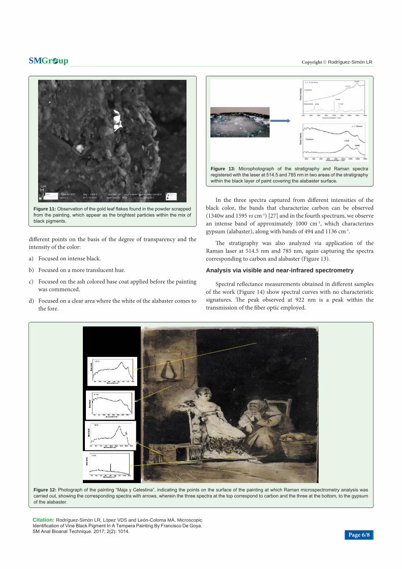
different points on the basis of the degree of transparency and the intensity of the color:
a) Focused on intense black.
b) Focused on a more translucent hue.
c) Focused on the ash colored base coat applied before the painting was commenced.
d) Focused on a clear area where the white of the alabaster comes to the fore.
In the three spectra captured from different intensities of the black color, the bands that characterize carbon can be observed (1340w and 1595 vs cm-1) [27] and in the fourth spectrum, we observe an intense band of approximately 1000 cm-1, which characterizes gypsum (alabaster), along with bands of 494 and 1136 cm-1.
The stratigraphy was also analyzed via application of the Raman laser at 514.5 nm and 785 nm, again capturing the spectra corresponding to carbon and alabaster (Figure 13).
Analysis via visible and near-infrared spectrometry
Spectral reflectance measurements obtained in different samples of the work (Figure 14) show spectral curves with no characteristic signatures. The peak observed at 922 nm is a peak within the transmission of the fiber optic employed.
Figure 13: Microphotograph of the stratigraphy and Raman spectra registered with the laser at 514.5 and 785 nm in two areas of the stratigraphy within the black layer of paint covering the alabaster surface.
Figure 11: Observation of the gold leaf flakes found in the powder scrapped from the painting, which appear as the brightest particles within the mix of black pigments.
Figure 12: Photograph of the painting “Maja y Celestina”, indicating the points on the surface of the painting at which Raman microspectrometry analysis was carried out, showing the corresponding spectra with arrows, wherein the three spectra at the top correspond to carbon and the three at the bottom, to the gypsum of the alabaster.

Citation: Rodríguez-Simón LR, López VDS and León-Coloma MÁ. Microscopic Identification of Vine Black Pigment In A Tempera Painting By Francisco De Goya. SM Anal Bioanal Technique. 2017; 2(2): 1014. Page 7/8
Gr upSM Copyright Rodríguez-Simón LR
Conclusion
We confirm that the combination of the different analytical techniques used has not facilitated the definitive characterization of the black pigments of an organic nature, as they are bound together forming a consistent pictorial mass.
Because of this difficulty, we propose distinguishing black pigments in the painting under study via the morphological differences found in the highly magnified images captured via scanning electron microscopy, bearing in mind the cellular aspects related to their internal structure.
Thus, in the case of the vine black pigment, polygonal forms were observed that may indicate the rigid walls characteristic of vegetable cells.
The carbon black pigment can be associated with the indefinite formations made up by small particles with an amorphous structure, which appear mixed with the polygonal vegetable-black forms.
In this painting, these two pigments are closely intertwined, as they have been mixed together via the binding agent (mixture of linseed oil and egg yolk).
Gold leaf particles were also found within the mix of blacks, indicating the aesthetic intent of the painter, who incorporated these particles with the aim of affording luminosity and light to this dark hue.
Moreover, other pigments were identified: lead white, bone black and iron oxide blacks. The presence of these hues cannot easily be ascribed to decisions on the part of the painter, as the mixture appears overly complex for a single color: black. Therefore, we explain the presence of these elements on the basis of contamination in the workshop resulting from old brushes or a dirty palette.
The identified binders are linseed oil and egg yolk which characterize the painting technique as fat tempera.
References
1. Rodríguez-Simón LR. Maja y Celestina. Una nueva pintura sobre alabastro en el catálogo de Francisco de Goya. Cuadernos de Arte de la Universidad de Granada. 2013; 145-170.
2. Gettens RJ, FitzHugh EW. Azurite and blue verditer. Studies in Conservation. 1966; 11: 54-61.
3. Plester J. Ultramarine, natural and artificial. Studies in Conservation. 1966; 11: 62-91.
4. Spring M. Perugino’s Painting Materials: Analysis and context within sixteenth-century easel painting. In Brunetti BG, Seccaroni C, Sgamellotti A. (eds). The Painting Technique of Pietro Vannucci, called Il Perugino. Proceediings of the Labs TECH Workshop. Firenze: Nardini Editore, Kermesquaderni. 2004: 21-28.
5. Spring M. Raphael’s Materials: Some new discoveries and their context within early sixteenth-century painting. In Roy, A. and Spring, M. (eds). Proceedings of the Eu-ARTECH workshop. Firenze: Nardini Editore, Kermesquaderni. 2007; 77-86.
6. Favaro M, Guastoni A, Marini F, Bianchin S, Gambirasi A. Characterization of lapis lazuli and corresponding purified pigments for a provenance study of ultramarine pigments used in Works of art. Anal Bioanal Chemistry. 2012; 402: 2195-2208.
7. Civici N, Demko O, Clark RJH. Identification of pigments used on late 17th century Albanian icons by total reflection X-ray fluorescence and Raman microscopy. Journal of Cultural Heritage. 2005; 6: 157-164.
8. Nevin A, Loring Melia J, Osticioli I, Gautier G, Colombini MP. The identificacion of copper oxalates in a 16th century Cypriot exterior wall painting using micro FTIR, micro Raman spectroscopy and Gas Chromatography-Mass Spectrometry. Journal of Cultural Heritage. 2008; 9: 154-161.
9. Muralha VSF, Burgio L, Clark RJH. Raman spectroscopy analysis of pigments on 16-17th c. Persian manuscripts. Spectrochimica Acta Part A. 2012; 92: 21-28.
10. Gutiérrez-Neira PC, Agulló-Rueda F, Climent-Font A, Garrido C. Raman spectroscopy analysis of pigments on Diego Velázquez paintings. Vibrational Spectroscopy. 2013; 69: 13-20.
11. Pozzi F, Jan van den Berg K, Fiedler I, Casadio F. A systematic analysis of red lake pigments in French Impressionist and Post-Impressionist paintings by surface-enhanced Raman spectroscopy (SERS). Journal of Raman Spectroscopy. 2014; 45: 1119-1126.
12. Frano Ka, Mayhew HE, Svoboda SA, Wustholz KL. Combined SERS and Raman analysis for the identification of red pigments in cross-sections from historic oil paintings. Analyst. 2014; 139: 6450-6455.
13. Lahlil S, Lebon M, Beck L, Rousselière H, Vignaud C, Reiche I, et al. The first in situ micro-Raman spectroscopic analysis of prehistoric cave art of Rouffignac St-Cernin, France. Journal Raman Spectroscopy. 2012; 43: 1637-1643.
14. Rampazzi L, Campo L, Cariati F, Tanda G, Colombini MP. Prehistoric wall paintings: The case of the Domus de Janas Necropolis (Sardinia, Italy). Archaeometry. 2007; 49: 559-569.
15. Chalmin E, Vignaud C, Salomon H, Farges F, Susini J, Menu M. Mineral discovered in Paleolithic black pigments by transmission electron microscopy and micro-X-ray absorption near-edge structure. Applied Physics A. 2006; 83: 213-218.
16. Vázquez C, Maier MS, Parera SD, Yacobaccio H, Solá P. Combining TXRF, FT-IR and GC-MS information for identification of inorganic and organic components in black pigments of rock art from Alero Hornillos 2 (Jujuy, Argentina). Anal Bioanal Chem. 2008; 391: 1381-1387.
17. Jezequel P, Wille G, Bény C, Delotme F, Dean-Prost V, Cottier R., et al. Characterization and origin of black and red Magdalenian pigments from Grottes de la Garenne (Vallée moyenne de la Creuse-France): a mineralogical and geochemical approach of the study of pehistorical paintings. Journal or Archaeological Science. 2011; 38: 1165-1172.
18. Eastaugh N, Walsh V, Chaplin T, Siddall R. Pigment Compendium: A Dictionary and Optical Microscopy of Historic Pigments. Oxford: Elsevier Butterworth-Heinemann. 2004.
Figure 14: Spectral reflectance measurements in different areas of the work.

Citation: Rodríguez-Simón LR, López VDS and León-Coloma MÁ. Microscopic Identification of Vine Black Pigment In A Tempera Painting By Francisco De Goya. SM Anal Bioanal Technique. 2017; 2(2): 1014. Page 8/8
Gr upSM Copyright Rodríguez-Simón LR
19. Rodríguez-Simón, LR., Parra Crego E. Caracterización de pigmentos y aglutinantes mediante VPSEM y GC-MS en una pintura firmada por Francisco de Goya: Maja y Celestina. En: Heritage and Design of Geometrical Forms. Granada: Universidad de Granada. 2011; 385-408.
20. Rodríguez-Simón LR, López-Ramírez MR, Navas N, Manzano E. Black pigments identification on alabaster painting: Maja y Celestina by Francisco de Goya, in situ mRS and SEM-EDX analysis. In: IRUG 10, book of abstracts; Barcelona: Universidad de Barcelona. 2012; 192-193.
21. Rodríguez-Simón LR., Del Sol López V, Manzano Moreno E, Navas Iglesias N, López-Ramírez MR. Advances in the study of black pigments via spectroscopic techniques in a painting by Francisco de Goya. In: TECHNART 2013. Analytical Spectroscopy in Art and Archaeology; Abstract book; Amsterdam: Rijksmuseum. 2013; 86.
22. Tomasini E, Siracusano G, Maier MS. Spectroscopic, morphological and chemical characterization of historic pigments based on carbon. Paths for the identification of an artistic pigment. Microchemical Journal. 2012; 102: 28-37.
23. Tomasini EP, Halac EB, Reinoso M, Di Liscia EJ, Maier MS. Micro-Raman spectroscopy of carbón-based black pigments. Journal of Raman Spectroscopy. 2012; 43: 1671-1675.
24. Coccato A, Jehlicka J, Moens L, Vandenabeele P. Raman spectroscopy for the investigation of carbon-based black pigments. Journal Raman Spectroscopy. 2015; 46: 1003-1015.
25. Tomasini EP, Gómez B, Halac EB, Reinoso M, Di Liscia EJ, Siracusano G, et al. Identificatiion of carbón-based black pigments in four South American polychrome wooden sculptures by Raman microscopy. Heritage Science. 2015; 3: 19.
26. Rodríguez-Simón LR. Maja y Celestina. Una pintura sobre alabastro firmada por Francisco de Goya. El proceso creativo y la técnica de ejecución pictórica. Kermes. 2011; 84: 37- 44.
27. Pagés-Camagna S, Duval A, Guicharnaud H. “Study of Gustave Moreau’s black drawings: identification of the graphic materials by Raman microspectrometry and PIXE, J. Raman Spectroscopy. 2004; 35: 628-632.