minor field study: molecular epidemiology of rabies in são paulo … · 2013-01-29 · 3...
TRANSCRIPT
Sveriges lantbruksuniversitet Fakulteten för veterinärmedicin och husdjursvetenskap Institutionen för biomedicin och veterinär folkhälsovetenskap
Minor Field Study: Molecular epidemiology of rabies in São Paulo State and Minas Gerais
State, Brazil
Emeli Torsson
Uppsala
2013
Examensarbete inom veterinärprogrammet
ISSN 1652-8697 Examensarbete 2013:21

SLU Sveriges lantbruksuniversitet
Minor Field Study: Molecular epidemiology of rabies in São Paulo State and Minas Gerais State, Brazil
Minor Field Study: Molekylär epidemiologisk studie för
rabies i São Paulo State och Minas Gerais State, Brasilien
Emeli Torsson
Handledare: Prof. Mikael Berg,
Institutionen för biomedicin och veterinär folkhälsovetenskap, SLU
Biträdande handledare: Prof. Paulo Eduardo Brandão,
Department of Preventive Veterinary Medicine and Animal Health, School of Veterinary Medicine
University of São Paulo
Examinator: Susanna Sternberg-Lewerin, Institutionen för biomedicin och veterinär folkhälsovetenskap, SLU
Examensarbete inom veterinärprogrammet, Uppsala 2013 Fakulteten för veterinärmedicin och husdjursvetenskap
Institutionen för biomedicin och veterinär folkhälsovetenskap, SLU Kurskod: EX0751, Nivå A2E, 30hp
Key words: rabies, epidemiology, Brazil
Nyckelord: rabies, epidemiologi, Brasilien
Online publication of this work: http://epsilon.slu.se ISSN 1652-8697
Examensarbete 2013:21

ABSTRACT ............................................................................................................................... 1
SAMMANFATTNING .............................................................................................................. 1
INTRODUCTION ...................................................................................................................... 2
Aim ......................................................................................................................................... 2
LITERATURE STUDY ............................................................................................................. 3
Background ............................................................................................................................ 3
Rabies virus ............................................................................................................................ 5
The disease ............................................................................................................................. 9
Methods for prevention of rabies ......................................................................................... 13
MATERIALS AND METHODS ............................................................................................. 15
Background to methods ........................................................................................................ 15
Samples ................................................................................................................................ 17
Execution of methods ........................................................................................................... 18
RESULTS ................................................................................................................................. 22
DISCUSSION .......................................................................................................................... 25
CONCLUSION ........................................................................................................................ 26
ACKNOWLEDGMENTS ........................................................................................................ 26
REFERENCES ......................................................................................................................... 26
APPENDIX I
1
ABSTRACT
Rabies is a fatal zoonotic disease spread worldwide. The most common sources of infection for all animals and humans are bites from dogs or bats. The aim of this degree project was to diagnose and determine the source of infection for 11 rabies samples from São Paulo State and Minas Gerais State, Brazil. Diagnosis was made through direct immunofluorescence assay, mouse inoculation and RT-PCR. The sources of infection were determined by sequencing 234 nucleotides of the 5’ end of the N-gene and align these with homologous sequences retrieved from GenBank. All seven samples for which satisfactory sequences could be achieved clustered with the sequences related to vampire bat with a bootstrap value of 98%.
SAMMANFATTNING
Rabies är en dödlig zoonos som är spridd över hela världen. De vanligaste smittkällorna för djur och människor är bett från hundar eller fladdermöss. Målet med detta examensarbete var att diagnostisera och fastställa smittkällan för 11 rabiesprover från São Paulo State och Minas Gerais State, Brasilien. Diagnos ställdes via direkt immunofluorescens, musinokulering och RT-PCR. Smittkällan fastställdes via sekvensering av 234 nukleotider av 5´-delen på N-genen vilka jämfördes med homologa sekvenser hämtade från GenBank. Alla sju prover för vilka tillfredställande sekvenser gick att uppnå anhopades, med en sannolikhet på 98 %, inom de sekvenser som relateras till vampyrfladdermusen.
2
INTRODUCTION
Rabies is a zoonosis that affects the central nervous system and produces an acute and incurable encephalitis (King et al., 2011; OIE, 2009). Rabies and rabies-like encephalitis are spread all over the world and can induce disease in all mammals, including humans (Oliveira et al., 2010). Rabies virus is a negative stranded RNA-virus that belongs to the genus Lyssavirus (Greek: lyssa = rage) in the family Rhabdoviridae (King et al., 2011). Classic rabies (genotype 1) is the most important genotype epidemiologically due to the fact that it causes the greatest number of encephalitis compared to the other genotypes (Oliveira et al., 2010). Symptoms of disease are the same for all genotypes and include a rapid disease consisting mostly of behavioural changes such as aggression and/or apathy (Hanlon et al., 2007). Rabies virus have been isolated from several animal species in Brazil, including dogs, foxes, cats, cattle, and bats (Mochizuki et al., 2012). Classic rabies virus in bats have been found only in bats resident in the American continents (Rupprecht et al., 2002). Desmodus rotundus, the common vampire bat, is the most common haematophagous bat (Schaefer et al., 2005) and is the main rabies source of infection for cattle and bat-transmitted human rabies is now an increasing problem in Brazil (Mayen, 2003; Schaefer et al., 2005).
Epidemiological studies on rabies virus are performed to uncover the source of infection and with help of the results focus control measures against rabies. This study contains samples from one cat, two horses and eight cattle from São Paulo State and Minas Gerais State, Brazil.
Aim
The aim of this study was three-fold. 1) Examine samples received at USPs official rabies diagnosis laboratory from rabies-suspected animals for presence of rabies virus. 2) Determine the most probable original host for rabies virus strains isolated from central nervous system of animals suspected of rabies previously stored at the laboratory. Results will be visualized as a phylogenetic tree. And 3) learn the techniques and approaches used to diagnose, analyse and decide most probable host in a case of rabies as an example disease, but also other infectious diseases as the protocol is similar in most cases of molecular epidemiology studies.
3
LITERATURE STUDY
Background History
The first written record of rabies is from the Eshnunna code, 23rd century BC: “If a dog is mad and the authorities have brought the fact to the knowledge of its owner; if he does not keep it in, and it bites a man and cause his death, then the owner shall pay two thirds of a mine of silver.” Rabies is a disease that has existed for centuries and this record shows that its transmissible nature between animal and man was known early in the rabies history (Baer, 2007). Rabies has affected people and cultures all around the world, including likely being one of the sources giving rise to the myths of werewolves and vampires. There have also been many myths about the treatment of rabies, including eating a chicken’s brain, having the tongue of a rabies infected dog in your shoe and dipping rabies infected persons in water until they nearly drowned over and over again, and in that way treating the dehydration associated with rabies and the clinical sign of hydrophobia. One treatment used with some success on both gladiators and by South American Indians was burning the wound inflicted by a rabies infected animal with hot coal or hot iron over and over again for several days and thereby killing the virus (Baer, 2007).
Outbreaks of rabies appear to have been rare in Europe until the Middle Ages, when epizootics of rabies in dogs were reported from many European countries. Most of the early history around rabies has its source in Europe (Baer, 2007). There were reports of rabies in bats in South America before the colonization by Spain and Portugal, but rabies in dogs were first reported when the big wave of immigrates from Europe came in the 19th century, as people probably brought their dogs with them to the American continents (Baer, 2007; Kobayashi et al., 2011).
When talking about the history of rabies you can not do it without mentioning Louis Pasteur. Pasteur discovered that the brain and spinal cord “housed” the virus and in 1885 produced the first vaccine against rabies out of dried spinal cords from rabies-infected rabbits. His first human patient who received this vaccine was young Joseph Meister. A rabid dog had repeatedly bitten Joseph, and 12 hours after the attack Pasteur started his treatment. The treatment lasted for 21 days, with injections of rabbit spinal cord solution once or twice a day. Joseph survived and never showed any symptoms of rabies and so was the first person successfully vaccinated against rabies (Baer, 2007).
Since this many humans have received vaccine against rabies, even though there have been a great deal of changes to Pasteur’s vaccine. The difficult production and the amount of dried rabbit spinal cords made his treatment regime problematic. A big change in Pasteur’s method for attenuation of the virus was when the inactivation by phenol was initiated early in the 20th century; this method of vaccine production is still used in some developing countries. Only 35 years after Pasteur’s first rabies vaccine Japan started the first vaccination programme against rabies in dogs in the cities of Nagasaki and Tokyo (Baer, 2007).
4
Rabies today
Rabies is often seen as a disease from the past, but is in fact still an increasing problem in many countries. The virus is spread almost completely worldwide, with some islands as exceptions. In several countries including United Kingdom, Ireland, Sweden, Norway, Iceland, Japan, Australia, New Zeeland, Singapore, most of Malaysia, Papua New Guinea, the Pacific Islands and some islands in Indonesia, classic rabies haven’t been seen for many years. Some of these countries may have viruses closely related to the classic rabies virus, but are still seen as rabies free. Rabies is well controlled among domestic animals in developed nations, but rabies in dogs is still a serious issue in some areas of Africa, the Middle East, Asia and Latin America. In those countries where canine rabies is under control the reservoirs in wildlife has become more interesting and more important (OIE, 2009).
40 % of humans bitten by suspected rabies infected animals are of the age 15 years or younger and as rabies is a disease that mostly affects people living under poor conditions in remote rural areas there is a big problem with underreporting (“WHO | Rabies,” n.d.). World Health Organization (WHO) states in their fact sheet about rabies that more than 55,000 people die of rabies every year, mostly in Africa and Asia (“WHO | Rabies,” n.d.). One study from Tanzania estimating human rabies mortality from dog bite injuries found that the predicted incidence of human rabies was up to 100 times greater than the incidence officially recorded in the studied area (Cleaveland et al., 2002). If this number is extrapolated on all rabies cases in the world it could be as many as 5 million people falling victim to rabies every year.
Rabies in Brazil
Rabies was first described in South America in the 16th century when Spanish soldiers were attacked by vampire bats and attained a disease with symptoms consistent with rabies (Baer, 2007). Canine rabies was first reported from Peru in 1803 and three years later an outbreak of rabies was seen in imported English dogs in Argentina (Baer, 2007; Kobayashi et al., 2011). Rabies is today endemic in Brazil and a range of rabies viruses have been isolated from different animal species, including dogs, foxes, cats, cattle and bats (hematophagous, insectivorous and frugivorous) (Kobayashi et al., 2011; Mochizuki et al., 2012). Due to mass vaccinations of dogs, promotion of responsible pet ownership, timely pre- and post-exposure prophylaxis, epidemiological surveillance and laboratory diagnosis, and community health education, human rabies cases have declined 95% since 1980 in the Americas (“WHO | Rabies,” n.d.). In urban areas in Brazil canine rabies is as good as extinct, but transmission of rabies from bats to humans is an increasing public health problem (Da Rosa et al., 2006; Queiroz et al., 2012).
Rabies in Europe and Sweden
Many European countries are today free of rabies in terrestrial animals, these countries include: Ireland, the United Kingdom, Sweden, Norway, Finland, Denmark, the Netherlands, Luxembourg, Belgium, France, Switzerland, the Czech Republic, Italy, Spain and Portugal (Bourhy et al., 2005). In Europe the main transmitter of classic rabies is the red fox and a new epidemiological cycle, maintained by the racoon dog seems to be developing in the Baltic countries and Poland (Bourhy et al., 2005). Canine rabies has disappeared from the countries
5
in the European Union, but there is a risk that illegal transportation of animals from the south and east borders will import rabies in to these countries again. Many travellers are not aware of the risk they take when travelling with their non-vaccinated animals to endemic regions or the risk of adopting animals in another country and take them home to a non-endemic area such as Europe (Bourhy et al., 2005).
To control classic rabies in Europe several countries have distributed anti-rabies vaccine baits in the wild. This started in Switzerland in 1978, followed by Germany, Italy, Belgium, France and Luxembourg during the 1980s. After a peak in wildlife rabies in 1989 many other countries followed their example and undertook oral vaccination campaigns of different sizes (Bourhy et al., 2005). In Sweden classic rabies hasn’t occurred in wild animals since 1886 (“Rabies - SVA,” n.d.).
European bats have been found to carry the European Bat Lyssavirus type 1 and 2 (EBLV1 and EBLV2), and Swedish bats in the county of Skåne have tested positive for antibodies against EBLV. None of the bats that tested positive for antibodies tested positive for virus in their saliva, this means that the bats have encountered the virus, but did not at that time function as a source of infection. Active surveillance of bats in Sweden is done yearly since the year of 2008 by the Swedish National Veterinary Institute (“Rabiesövervakning av fladdermus - SVA,” n.d.).
The last two human cases of rabies in Sweden were individuals who were infected abroad, one man in India in 1974 and one young woman in Thailand in 2000. Both patients contracted the virus from rabies infected dogs (Höjer et al., 2001).
Rabies virus Etiology
Rabies virus belongs to the genus Lyssavirus, in the family of Rhabdoviridae, in the order Mononegavirales. The genus Lyssavirus includes 12 different viruses grouped into two phylogroups that include seven genotypes (“ICTV Virus Taxonomy: 2011 release (current),” n.d.; Oliveira et al., 2010). These two phylogroups are genetically and immunopathologically different (Meslin et al., 1996; Oliveira et al., 2010). Rabies virus (genotype 1) belongs to phylogroup I together with EBLV1 (genotype 5), EBLV2 (genotype 6), Australian bat lyssavirus (genotype 7) and Duvenhage virus (genotype 4) (Oliveira et al., 2010). Rabies virus, and all other lyssaviruses, can cause acute progressive encephalitis in all mammals, including humans; even though for example EBLV1 and 2 have only been described as the cause of infection in four humans worldwide to date (Bourhy et al., 2005; King et al., 2011).
Morphology
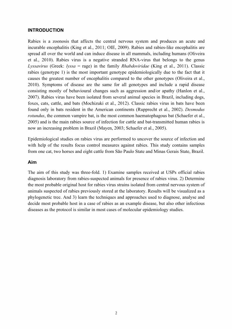
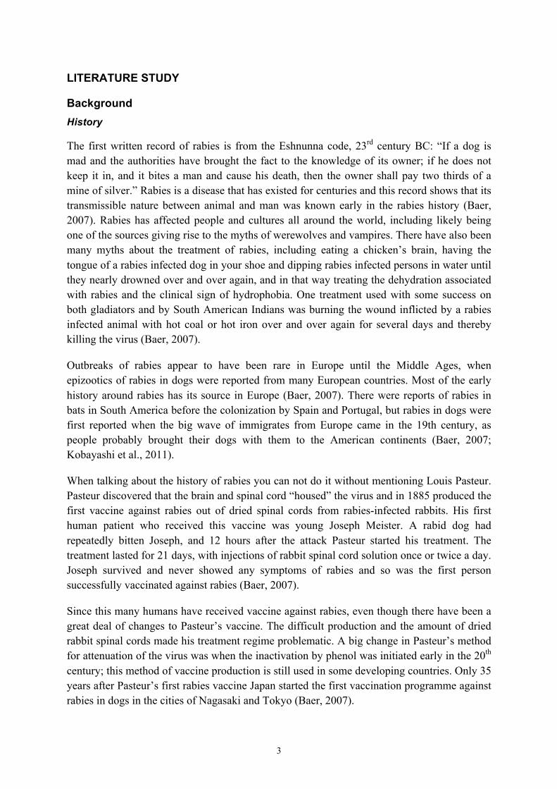
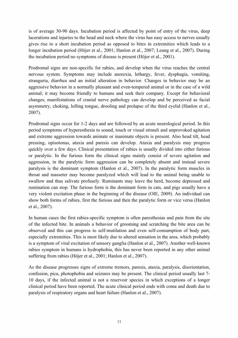
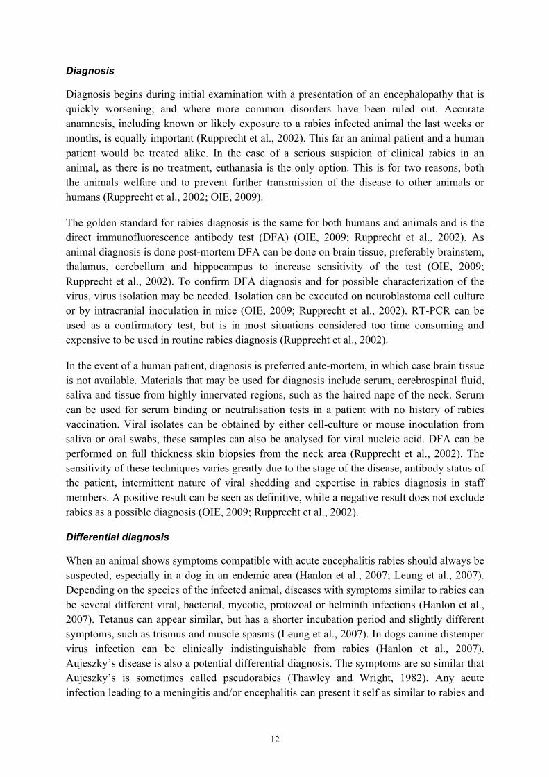
The rabies virions are bullet-shaped, 60-110 x 130-250 nm in size (figure 1), and are built up by two structural units, one internal nucleocapsid and a lipid envelope derived from the host cytoplasmic membrane during budding (figure 2). The nucleocapsid consists of L-, P- and N-protein together with RNA. Protruding through the lipid membrane are knobbed spikes of

6
approximately 10 nm in length consisting of trimers of the viral glycoprotein (G) (King et al., 2011).
Figure 1. Electron microscopy pictures of rabies virus. Note the bullet shape. (http://www.virology.net/Big_Virology/Special/Rabies1/Rabies.html)
Figure 2. General structure and major components of rabies virus. (http://www.cdc.gov/rabies/transmission/virus.html)
Gene function and structure
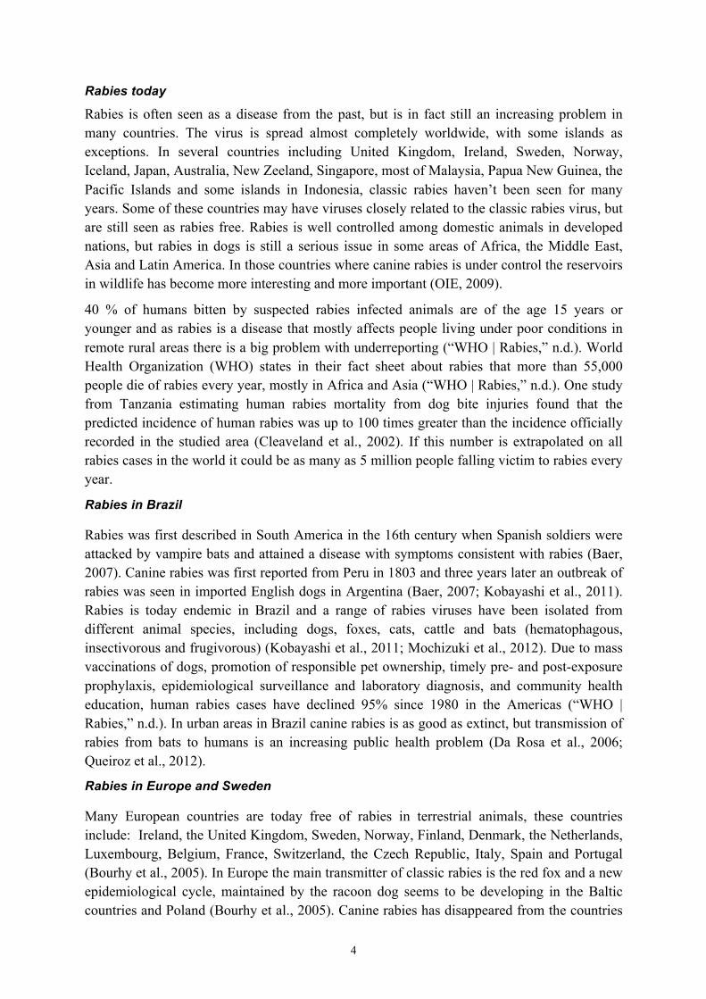
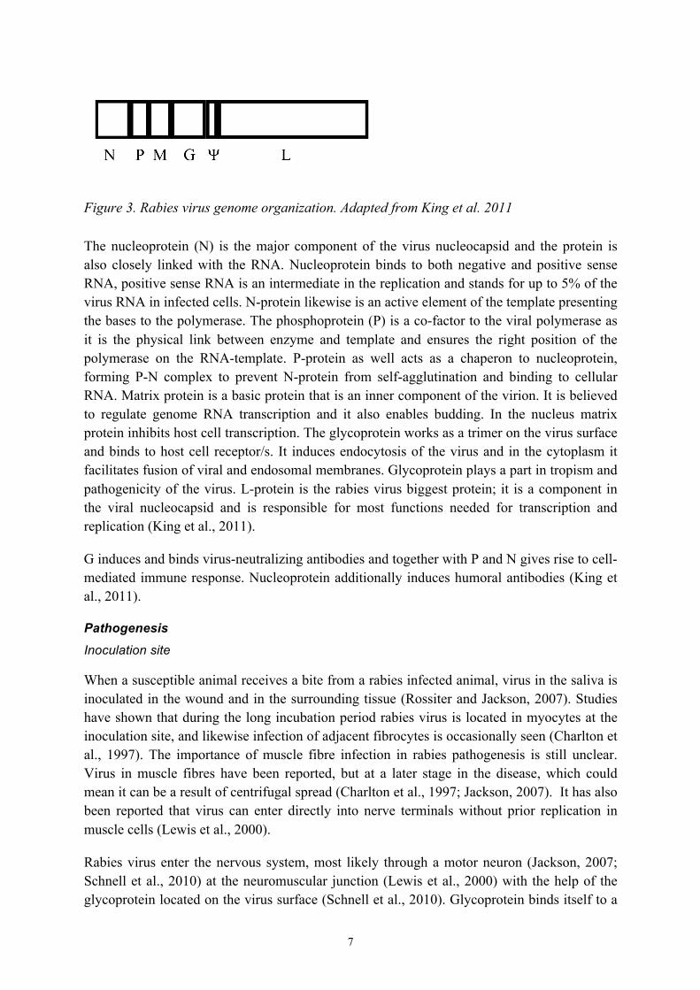
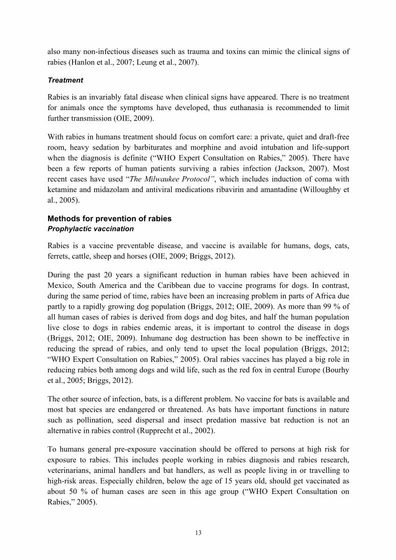
The rabies virus genome consists of a single molecule of linear, negative sense, single stranded ribonucleic acid (RNA) of about 12 kilobases in size. The genome encodes for five proteins in the order 3’-N-P-M-G-L-5’ (nucleoprotein, phosphoprotein, matrix protein, glycoprotein and large polymerase protein) separated by non-transcribing regions, 2 - 423 nucleotides in length (figure 3). Intergentic regions increase in length in the 3’-5’ direction and may in this way induce progressive slowing and decreasing efficiency of transcription (King et al., 2011; Tordo et al., 1986). The 423-nucleotide long intergenic region is located between the G and L gene and is called a remnant gene or pseudogene (Ψ). This region starts and stops with sequences resembling rabies start and stop signals, but would only encode for an 18 amino acids long peptide. This in combination with comparison to other Rhabdoviridae that has a functional gene in this position makes it plausible that the long intergenic region is a remnant gene and a step in the virus evolution (Tordo et al., 1986; Wunner, 2007).
7
Figure 3. Rabies virus genome organization. Adapted from King et al. 2011
The nucleoprotein (N) is the major component of the virus nucleocapsid and the protein is also closely linked with the RNA. Nucleoprotein binds to both negative and positive sense RNA, positive sense RNA is an intermediate in the replication and stands for up to 5% of the virus RNA in infected cells. N-protein likewise is an active element of the template presenting the bases to the polymerase. The phosphoprotein (P) is a co-factor to the viral polymerase as it is the physical link between enzyme and template and ensures the right position of the polymerase on the RNA-template. P-protein as well acts as a chaperon to nucleoprotein, forming P-N complex to prevent N-protein from self-agglutination and binding to cellular RNA. Matrix protein is a basic protein that is an inner component of the virion. It is believed to regulate genome RNA transcription and it also enables budding. In the nucleus matrix protein inhibits host cell transcription. The glycoprotein works as a trimer on the virus surface and binds to host cell receptor/s. It induces endocytosis of the virus and in the cytoplasm it facilitates fusion of viral and endosomal membranes. Glycoprotein plays a part in tropism and pathogenicity of the virus. L-protein is the rabies virus biggest protein; it is a component in the viral nucleocapsid and is responsible for most functions needed for transcription and replication (King et al., 2011).
G induces and binds virus-neutralizing antibodies and together with P and N gives rise to cell-mediated immune response. Nucleoprotein additionally induces humoral antibodies (King et al., 2011).
Pathogenesis Inoculation site
When a susceptible animal receives a bite from a rabies infected animal, virus in the saliva is inoculated in the wound and in the surrounding tissue (Rossiter and Jackson, 2007). Studies have shown that during the long incubation period rabies virus is located in myocytes at the inoculation site, and likewise infection of adjacent fibrocytes is occasionally seen (Charlton et al., 1997). The importance of muscle fibre infection in rabies pathogenesis is still unclear. Virus in muscle fibres have been reported, but at a later stage in the disease, which could mean it can be a result of centrifugal spread (Charlton et al., 1997; Jackson, 2007). It has also been reported that virus can enter directly into nerve terminals without prior replication in muscle cells (Lewis et al., 2000).
Rabies virus enter the nervous system, most likely through a motor neuron (Jackson, 2007; Schnell et al., 2010) at the neuromuscular junction (Lewis et al., 2000) with the help of the glycoprotein located on the virus surface (Schnell et al., 2010). Glycoprotein binds itself to a
8
receptor or receptors still unknown, several different receptors have been suggested, including nicotine acetylcholine receptor (nAchR), neuronal cell adhesion molecule (NCAM1) and low-affinity nerve growth factor receptor (p75NTR) (Lewis et al., 2000; Schnell et al., 2010). Rabies virus use one or more of these receptors to enter the host cell by the endosomal pathway and can then be found in either clathrin-coated pits, in uncoated vesicles or, later in lysosomes (Lewis et al., 2000; Schnell et al., 2010). Whether or not the uncoating from these vesicles occurs at the nerve terminal or after axonal transport is unclear, but studies have shown that the glycoprotein is responsible for the required pH-decrease that induce fusion between endosomal and viral membranes and releases the nucleocapsid into the cytoplasm (King et al., 2011).
Neuron and replication
Replication can not take place in the nerve terminal, consequently the virus need to be transported to the neuronal cell body (Lewis et al., 2000; Schnell et al., 2010). How this is done is still to be proven, but two different mechanisms have been proposed. Either the rabies virus capsid alone is transported to the cell body using viral phosphoprotein and the cytoplasmic dynein motor complex or the entire virion is transported in a vesicle and then instead is dependent on the viral glycoprotein (Schnell et al., 2010). There are flaws in both of these theories that makes the mechanism behind the retrograde axoplasmic transport unclear, but it is a certainty that it occurs (Lewis et al., 2000; Schnell et al., 2010). Studies have shown that the virus moves approximately 50-100 mm/day in retrograde direction along the neuron axon (Jackson, 2007).
As the virion, or a part of it, reach the cell body, transcription and replication can ensue. Since rabies virus genome is negative sense direct translation is not possible, so on reaching the neuronal cell body the virus start transcribing positive sense RNA corresponding to the various viral genes. Transcription of the five viral genes is mediated by the RNA-dependent RNA polymerase (L-protein) and its co factor, the phosphoprotein (Schnell et al., 2010; King et al., 2011). A switch from transcription to replication is made when enough nucleoprotein has been transcribed. Full-length complementary, positive sense, copies of the viral genome is produced, and these are then used as template for negative sense RNA. Regulating transcription and replication is the matrix protein (King et al., 2011; Schnell et al., 2010). After assembling into new virions, matrix protein and glycoprotein mediates budding in which the virus get its lipid envelope from the host cell (Schnell et al., 2010).
Central nervous system
Rabies virus spreads in the central nervous system the same way as in the peripheral nervous system, by fast axonal transport. When the virus reaches the neurons in the central nervous system, usually in the spinal cord, there is a quick spread of virus within the CNS along the neuroanatomical pathways (Jackson, 2007). This involves first the brainstem and deep cerebellar nuclei, and further spread to cerebellar Purkinje cells, diencephalon, basal ganglia and cerebral cortex. Finally the virus reaches the hippocampus (Jackson, 2007).
Neuron apoptosis is not part of rabies pathogenesis. Studies have shown that the more pathogenic the rabies virus is, the less apoptosis is induced. While the virus is inside a neuron
9
cell, it will not induce an immune response and will thereby avoid the hosts innate immune and antiviral response (Schnell et al., 2010; Wang et al., 2005).
Centrifugal spread
As the virus has taken over the central nervous system and replicated, a centrifugal spread of new virions is commenced. This spread takes place in both somatic and autonomic neurons and results in an infection of a wide range of organs. An important organ involved is the major salivary glands, resulting in production of saliva high in virus concentration. This saliva is then the means of further transmission of virus to other hosts. Rabies virus also reaches lacrimal glands, cornea, skin, heart, gastrointestinal tract and adrenal glands. As rabies virus is transported in neurons, viremia will never occur and antigen cannot be isolated from blood samples (Jackson, 2007).
Gross lesions and histopathology
Macroscopic examination of the brain in rabies patients normally shows only mild and non-specific changes; a mild cerebral oedema can be seen in most cases. Microscopic examination of the central nervous system also presents relatively mild changes, even absent in some cases, especially when compared to the severe and fatal clinical signs. Changes include varying degrees of mononuclear inflammatory cell infiltration of the leptomeninges, perivascular cuffing, microglial activation and neurophagia. This picture is not unique for rabies, and can be seen in several other viral encephalitises. However, rabies has one unique trait and that is the eosinophlic cytoplasmic viral inclusion bodies, Negri bodies, found in neuron cells (Rossiter and Jackson, 2007). Negri bodies can be seen on hematoxylin and eosin stained sections as dense, well-defined, oval or round, eosinophilic cytoplasmic inclusions (Rossiter and Jackson, 2007). Research indicate that replication and transcription of the virus occur in these Negri bodies (Schnell et al., 2010).
The mononuclear inflammatory infiltration is typically composed of mostly lymphocytes and monocytes, with a small number of plasma cells. Perivascular cuffs are also dominated by lymphocytes and monocytes; their density and spread can vary greatly among individuals, but are mostly seen in the grey matter of the brainstem and spinal cord. In many cases of rabies neurophagia of degenerated and dying neurons by microglia/macrophages is reported, also this in varying degree (Rossiter and Jackson, 2007).
Histopathological changes in the peripheral nervous system include inflammatory, reactive and degenerative changes. These changes affect neural cell bodies in both sensory and autonomic ganglia and their satellite cells as well as the sensory and motor neurons and their Schwann cells (Rossiter and Jackson, 2007).
The disease Hosts and reservoirs
All mammals are susceptible for rabies, including humans and marine mammals, but to varying degree (Meslin et al., 1996; Hanlon et al., 2007; OIE, 2009). There are many existing strains of rabies virus and each strain is maintained by a particular reservoir host or hosts
10
(Hanlon et al., 2007; Meslin et al., 1996; OIE, 2009). Infection of a competent reservoir animal with a rabies variant specifically adapted to that species is particularly good at inducing behavior that will ensure virus transmission and maximize viral shedding in saliva (Hanlon et al., 2007). Reservoir species are those mammals that are capable of a sustained intraspecies maintenance of a virus variant in a specific geographic area (Hanlon et al., 2007). Important maintenance species include members of the order Canidae (dogs, jackals, coyotes, wolves, foxes, arctic foxes and raccoon dogs), Mustelidae (skunks, martens, weasels and stoats), Procyonidae (raccoons) and Chiroptera (bats) (OIE, 2009). Bats are seen as the primary reservoir in all continents, but classic rabies virus (genotype 1) has only been recovered from bats indigenous on the American continents. Rabies in free-ranging healthy bats may not be common, but the bats that humans come in contact with is usually ill or in other ways incapacitated, and among these bats rabies is not uncommon (Rupprecht et al., 2002). Feline adapted rabies virus have not been reported, even tough cats are susceptible, often infected from other animals and can transmit the virus (Rupprecht et al., 2002; OIE, 2009). Each virus variant seem adapted to its reservoir species and this strongly affect host receptiveness to infection. Certain virus variants may have characteristics that can influence tropism, host behavior or clinical outcome (Hanlon et al., 2007). If a virus infect a host which is not its natural reservoir the susceptibility will vary, it can either be more susceptible and induce hyper-acute encephalitis, with a quick death and little virus shed in the saliva, or the new host may survive a longer period of time and spread the virus more efficiently than the former host (Hanlon et al., 2007).
Incubation period in rabies may be long, but this is not a question of true latency such as for retroviruses and herpes viruses that integrate their genome with the host genome. Also the idea of a carrier state or persistently infected animal, where the infected animal remain healthy but shed virus for a long period of time seem of little epidemiological significance (Rupprecht et al., 2002).
Transmission
The most reliable and most important route of infection is the bite route. Other transmission routes such as direct contact of saliva from rabies infected animal to mucous membranes can, but rarely does, lead to clinical disease (Rupprecht et al., 2002; Hanlon et al., 2007; King et al., 2011). Rabies virus is not transmittable through intact skin (OIE, 2009). The dog is the major reservoir and vector in most endemic areas, such as Africa, Asia, Central America and parts of South America (OIE, 2009; Rupprecht et al., 2002). In parts of the world where canine rabies under control, bites from bats are the most important route of transmission (Rupprecht et al., 2002). These two different routes of transmission can be seen as two different epidemiological cycles: terrestrial and aerial (Queiroz et al., 2012).
Incubation period and clinical signs
There are no specific symptoms for rabies, except sudden changes in behavior, and one can say that with rabies the abnormal becomes the typical. This variation in symptoms may be associated to the site of primary CNS lesion, viral strain, dose and/or route of infection (Hanlon et al., 2007). The incubation period can vary between a few days to several years, but
11
is of average 30-90 days. Incubation period is affected by point of entry of the virus, deep lacerations and injuries to the head and neck where the virus has easy access to nerves usually gives rise to a short incubation period as opposed to bites in extremities which leads to a longer incubation period (Höjer et al., 2001; Hanlon et al., 2007; Leung et al., 2007). During the incubation period no symptoms of disease is present (Höjer et al., 2001).
Prodromal signs are non-specific for rabies, and develop when the virus reaches the central nervous system. Symptoms may include anorexia, lethargy, fever, dysphagia, vomiting, stranguria, diarrhea and an initial alteration in behavior. Changes in behavior may be an aggressive behavior in a normally pleasant and even-tempered animal or in the case of a wild animal; it may become friendly to humans and seek their company. Except for behavioral changes, manifestations of cranial nerve pathology can develop and be perceived as facial asymmetry, choking, lolling tongue, drooling and prolapse of the third eyelid (Hanlon et al., 2007).
Prodromal signs occur for 1-2 days and are followed by an acute neurological period. In this period symptoms of hyperesthesia to sound, touch or visual stimuli and unprovoked agitation and extreme aggression towards animate or inanimate objects is present. Also head tilt, head pressing, opisotonus, ataxia and paresis can develop. Ataxia and paralysis may progress quickly over a few days. Clinical presentation of rabies is usually divided into either furious or paralytic. In the furious form the clinical signs mainly consist of severe agitation and aggression, in the paralytic form aggression can be completely absent and instead severe paralysis is the dominant symptom (Hanlon et al., 2007). In the paralytic form muscles in throat and masseter may become paralyzed which will lead to the animal being unable to swallow and thus salivate profusely. Ruminants may leave the herd, become depressed and rumination can stop. The furious form is the dominant form in cats, and pigs usually have a very violent excitation phase in the beginning of the disease (OIE, 2009). An individual can show both forms of rabies, first the furious and then the paralytic form or vice versa (Hanlon et al., 2007).
In human cases the first rabies-specific symptom is often paresthesias and pain from the site of the infected bite. In animals a behavior of grooming and scratching the bite area can be observed and this can progress to self-mutilation and even self-consumption of body part, especially extremities. This is most likely due to altered sensation in the area, which probably is a symptom of viral excitation of sensory ganglia (Hanlon et al., 2007). Another well-known rabies symptom in humans is hydrophobia, this has never been reported in any other animal suffering from rabies (Höjer et al., 2001; Hanlon et al., 2007).
As the disease progresses signs of extreme tremors, paresis, ataxia, paralysis, disorientation, confusion, pica, photophobia and seizures may be present. The clinical period usually last 7-10 days, if the infected animal is not a reservoir species in which exceptions of a longer clinical period have been reported. The acute clinical period ends with coma and death due to paralysis of respiratory organs and heart failure (Hanlon et al., 2007).
12
Diagnosis
Diagnosis begins during initial examination with a presentation of an encephalopathy that is quickly worsening, and where more common disorders have been ruled out. Accurate anamnesis, including known or likely exposure to a rabies infected animal the last weeks or months, is equally important (Rupprecht et al., 2002). This far an animal patient and a human patient would be treated alike. In the case of a serious suspicion of clinical rabies in an animal, as there is no treatment, euthanasia is the only option. This is for two reasons, both the animals welfare and to prevent further transmission of the disease to other animals or humans (Rupprecht et al., 2002; OIE, 2009).
The golden standard for rabies diagnosis is the same for both humans and animals and is the direct immunofluorescence antibody test (DFA) (OIE, 2009; Rupprecht et al., 2002). As animal diagnosis is done post-mortem DFA can be done on brain tissue, preferably brainstem, thalamus, cerebellum and hippocampus to increase sensitivity of the test (OIE, 2009; Rupprecht et al., 2002). To confirm DFA diagnosis and for possible characterization of the virus, virus isolation may be needed. Isolation can be executed on neuroblastoma cell culture or by intracranial inoculation in mice (OIE, 2009; Rupprecht et al., 2002). RT-PCR can be used as a confirmatory test, but is in most situations considered too time consuming and expensive to be used in routine rabies diagnosis (Rupprecht et al., 2002).
In the event of a human patient, diagnosis is preferred ante-mortem, in which case brain tissue is not available. Materials that may be used for diagnosis include serum, cerebrospinal fluid, saliva and tissue from highly innervated regions, such as the haired nape of the neck. Serum can be used for serum binding or neutralisation tests in a patient with no history of rabies vaccination. Viral isolates can be obtained by either cell-culture or mouse inoculation from saliva or oral swabs, these samples can also be analysed for viral nucleic acid. DFA can be performed on full thickness skin biopsies from the neck area (Rupprecht et al., 2002). The sensitivity of these techniques varies greatly due to the stage of the disease, antibody status of the patient, intermittent nature of viral shedding and expertise in rabies diagnosis in staff members. A positive result can be seen as definitive, while a negative result does not exclude rabies as a possible diagnosis (OIE, 2009; Rupprecht et al., 2002).
Differential diagnosis
When an animal shows symptoms compatible with acute encephalitis rabies should always be suspected, especially in a dog in an endemic area (Hanlon et al., 2007; Leung et al., 2007). Depending on the species of the infected animal, diseases with symptoms similar to rabies can be several different viral, bacterial, mycotic, protozoal or helminth infections (Hanlon et al., 2007). Tetanus can appear similar, but has a shorter incubation period and slightly different symptoms, such as trismus and muscle spasms (Leung et al., 2007). In dogs canine distemper virus infection can be clinically indistinguishable from rabies (Hanlon et al., 2007). Aujeszky’s disease is also a potential differential diagnosis. The symptoms are so similar that Aujeszky’s is sometimes called pseudorabies (Thawley and Wright, 1982). Any acute infection leading to a meningitis and/or encephalitis can present it self as similar to rabies and
13
also many non-infectious diseases such as trauma and toxins can mimic the clinical signs of rabies (Hanlon et al., 2007; Leung et al., 2007).
Treatment
Rabies is an invariably fatal disease when clinical signs have appeared. There is no treatment for animals once the symptoms have developed, thus euthanasia is recommended to limit further transmission (OIE, 2009).
With rabies in humans treatment should focus on comfort care: a private, quiet and draft-free room, heavy sedation by barbiturates and morphine and avoid intubation and life-support when the diagnosis is definite (“WHO Expert Consultation on Rabies,” 2005). There have been a few reports of human patients surviving a rabies infection (Jackson, 2007). Most recent cases have used “The Milwaukee Protocol”, which includes induction of coma with ketamine and midazolam and antiviral medications ribavirin and amantadine (Willoughby et al., 2005).
Methods for prevention of rabies Prophylactic vaccination
Rabies is a vaccine preventable disease, and vaccine is available for humans, dogs, cats, ferrets, cattle, sheep and horses (OIE, 2009; Briggs, 2012).
During the past 20 years a significant reduction in human rabies have been achieved in Mexico, South America and the Caribbean due to vaccine programs for dogs. In contrast, during the same period of time, rabies have been an increasing problem in parts of Africa due partly to a rapidly growing dog population (Briggs, 2012; OIE, 2009). As more than 99 % of all human cases of rabies is derived from dogs and dog bites, and half the human population live close to dogs in rabies endemic areas, it is important to control the disease in dogs (Briggs, 2012; OIE, 2009). Inhumane dog destruction has been shown to be ineffective in reducing the spread of rabies, and only tend to upset the local population (Briggs, 2012; “WHO Expert Consultation on Rabies,” 2005). Oral rabies vaccines has played a big role in reducing rabies both among dogs and wild life, such as the red fox in central Europe (Bourhy et al., 2005; Briggs, 2012).
The other source of infection, bats, is a different problem. No vaccine for bats is available and most bat species are endangered or threatened. As bats have important functions in nature such as pollination, seed dispersal and insect predation massive bat reduction is not an alternative in rabies control (Rupprecht et al., 2002).
To humans general pre-exposure vaccination should be offered to persons at high risk for exposure to rabies. This includes people working in rabies diagnosis and rabies research, veterinarians, animal handlers and bat handlers, as well as people living in or travelling to high-risk areas. Especially children, below the age of 15 years old, should get vaccinated as about 50 % of human cases are seen in this age group (“WHO Expert Consultation on Rabies,” 2005).
14
Post-exposure prophylaxis
As rabies virus usually do not enter the nervous system, where the immune system cannot reach it, directly at exposure, there is a window for post-exposure prophylaxis (PEP). If the WHO recommended guidelines for PEP is strictly followed it is a virtual guarantee that clinical rabies will not evolve in the exposed patient (Rupprecht et al., 2002; “WHO Expert Consultation on Rabies,” 2005). Treatment for prevention of clinical rabies in an exposed person ought to begin as soon as possible after exposure, beginning with a thorough wound cleaning for a minimum of fifteen minutes using water, soap and an antiseptic (for example iodine or ethanol). This is then followed by administration of rabies vaccine and, in some cases, depending on earlier vaccination status and the severity of the wound, human rabies immunoglobulin (OIE, 2009; “WHO Expert Consultation on Rabies,” 2005).
Post-exposure prophylaxis in animals is not considered advisable as it might increase human exposure and further transmission, and is only practiced in a few Asian countries (OIE, 2009).
15
MATERIALS AND METHODS
Background to methods Direct immunofluorescence antibody test
Direct immunofluorescence antibody test (DFA) was developed in the late 1950s and is still the golden standard when it comes to rabies diagnosis (Meslin et al., 1996; Rupprecht et al., 2002). It is a quick and reliable method as long as the examiner is sufficiently experienced and a good quality conjugate and fluorescence microscope is supplied (Meslin et al., 1996). Essential to a quick and accurate diagnosis is likewise fresh and unfixed samples. Because of the pattern of viral spread in the brain, examination of the brainstem and medulla is essential to make the correct diagnosis (Rupprecht et al., 2002).
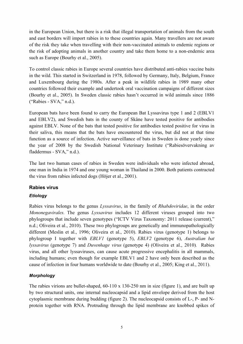
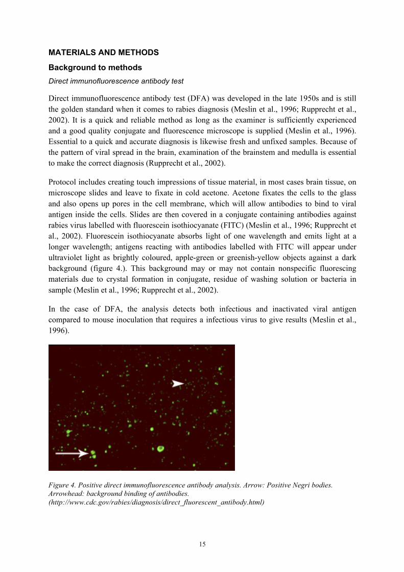
Protocol includes creating touch impressions of tissue material, in most cases brain tissue, on microscope slides and leave to fixate in cold acetone. Acetone fixates the cells to the glass and also opens up pores in the cell membrane, which will allow antibodies to bind to viral antigen inside the cells. Slides are then covered in a conjugate containing antibodies against rabies virus labelled with fluorescein isothiocyanate (FITC) (Meslin et al., 1996; Rupprecht et al., 2002). Fluorescein isothiocyanate absorbs light of one wavelength and emits light at a longer wavelength; antigens reacting with antibodies labelled with FITC will appear under ultraviolet light as brightly coloured, apple-green or greenish-yellow objects against a dark background (figure 4.). This background may or may not contain nonspecific fluorescing materials due to crystal formation in conjugate, residue of washing solution or bacteria in sample (Meslin et al., 1996; Rupprecht et al., 2002).
In the case of DFA, the analysis detects both infectious and inactivated viral antigen compared to mouse inoculation that requires a infectious virus to give results (Meslin et al., 1996).
Figure 4. Positive direct immunofluorescence antibody analysis. Arrow: Positive Negri bodies. Arrowhead: background binding of antibodies. (http://www.cdc.gov/rabies/diagnosis/direct_fluorescent_antibody.html)

16
Mouse inoculation
As a single negative DFA test cannot rule out infection, a simultaneous virus isolation in either cell culture or a mouse inoculation is done (OIE, 2009). With mouse inoculation even a single infectious virion can give a positive result, which it would not do in DFA (personal communication, P. Eduardo Brandão, 2012).
Mice under 21 days of ages are inoculated intracranial with a solution containing tissue from the rabies suspected animal and are then supervised daily for signs of rabies for at least 21 days post-inoculation (Meslin et al., 1996).
Reverse transcriptase polymerase chain reaction
Reverse transcription (RT) is a method used to convert RNA into complementary DNA (cDNA). One way to achieve this is with the help of an enzyme from a retrovirus, Moloney Murine Leukemia Virus (M-MLV). The RT is not an amplifying reaction; one RNA template will give one cDNA. This cDNA can then be used in the polymerase chain reaction (PCR). PCR is a method for amplification of fragments of DNA chosen by the help of oligonucleotide primers. It is an iterative process consisting of three steps, denaturation of the double stranded template DNA by heat, annealing of the oligonucleotide primers to the now single stranded target sequence, and then extension of the annealed primers by a thermostable DNA polymerase. In most cases the Taq polymerase is used, derived from the bacterium Thermophilus aquaticus (Sambrook and Russell, 2001).
The temperature used in the cycle varies with different reactions. Temperature at denaturing depends on the G and C content in the template, as G and C bind to each other by three hydrogen bonds and A and T only by two, more G and C demands a higher temperature. At the annealing step the temperatures depends on the primers melting temperature, as a temperature 2-10°C lower then this point is used. Extension of primers is carried out at the optimal temperature for the DNA polymerase to work, usually around 72-78°C (Sambrook and Russell, 2001).
Ingredients needed in a PCR reaction include thermostable DNA polymerase, oligonucleotide primers that targets the wanted DNA sequence, deoxynucleoside triphosphates (dNTPs), dATP, dTTP, dCTP and dGTP in equal amounts, magnesiumchloride as a co-factor to the DNA polymerase, buffer to maintain a correct pH (around pH 7,2 at 72°C) and template DNA (Sambrook and Russell, 2001).
Primers used in present study (table 2) aim for the rabies virus N-gene that encodes for the nucleoprotein. The rabies virus nucleoprotein is involved in the intracellular viral replication and interacts with the host’s proteins. This leads to a high functional restriction and thus low mutation rate and stability in the gene. As a result, the N-gene is a good target for primers and phylogenetic studies (Eduardo Brandão, 2012; Oliveira et al., 2010).
Results of a PCR are visualized with a gel electrophoresis. The gel electrophoresis is used to separate, identify and purify DNA segments with the help of an electric field in which the
17
shorter fragments of DNA will travel farthest in the gel. Received bands are compared to a stepladder of for example 100 base pairs per step (Sambrook and Russell, 2001).
In those cases where an ante mortem diagnosis is preferred, of a human case or animal case, where religious objectives oppose euthanasia, real-time PCR of saliva has proven to be the most trustworthy analysis method (Nagaraj et al., 2006; Saengseesom et al., 2007). Real-time PCR differs from conventional RT-PCR described above in that it is a quantifying reaction that can determine virus concentration in sample and results are shown directly during the amplification on a computer display, thereby shortening time to diagnosis as no gel electrophoresis is needed (Sambrook and Russell, 2001).
DNA sequencing
DNA sequencing protocol used in this study is based on the use of dideoxynucleoside triphosphates (ddNTPs). These ddNTPs can be substituted for deoxynucleosides (dNTPs) at random position in the template during a polymerase chain reaction. ddNTPs and dNTPs are added to the reaction in equal amounts and the probability of binding one or the other to the template is thereby equal. The difference between a ddNTP and a dNTP is that the ddNTP is missing the 3’ OH-group; this prevents formation of a phosphodiester bond with the following dNTP. The consequence is that binding a ddNTP to the chain makes further extension impossible. Instead of several amplifications of identical size at the end of the PCR reaction amplifications of every length possible with the difference of one nucleotide between them is present (Sambrook and Russell, 2001).
To each of the four ddNTPs (ddATP, ddTTP, ddGTP, ddCTP) a different dye is linked to the nitrogenous base via a linker. When samples are run through a capillary electrophoresis, passing through a laser light, computer software identifies each nucleotide based on the distinctive colour and wavelength that the ddNTPs emits. In this way the software can identify each base according to the fluorescent peak and the distance between succeeding peaks and display it in the form of a chromatogram (Sambrook and Russell, 2001).
Samples
One of the aims was to diagnose samples received at University of São Paulo’s official rabies laboratory. Unfortunately only one such sample arrived during the stay at USP, sample 62 that arrived 17th September. This sample was from a horse that had been hospitalized at the University Animal Hospital due to neurological symptoms and died there, unclear if it was euthanized or died as a consequence of its disease.
My Brazilian supervisor, Professor Paulo Eduardo Brandão, selected 10 additional samples for me to work with. These 10 samples are from other studies performed at University of São Paulo.
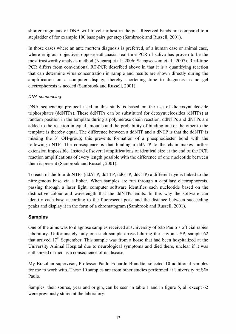
Samples, their source, year and origin, can be seen in table 1 and in figure 5, all except 62 were previously stored at the laboratory.
18
Figure 5. Map of southeast Brazil, sample collection sites marked with stars. (Map: www.cairn.info)
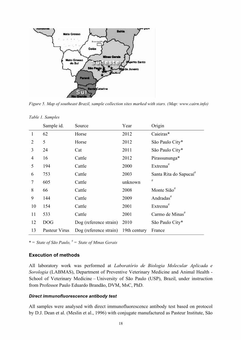
Table 1. Samples
Sample id. Source Year Origin
1 62 Horse 2012 Caieiras*
2 5 Horse 2012 São Paulo City* 3 24 Cat 2011 São Paulo City*
4 16 Cattle 2012 Pirassununga*
5 194 Cattle 2000 Extrema#
6 753 Cattle 2003 Santa Rita do Sapucaí#
7 605 Cattle unknown #
8 66 Cattle 2008 Monte Sião#
9 144 Cattle 2009 Andradas#
10 154 Cattle 2001 Extrema#
11 533 Cattle 2001 Carmo de Minas#
12 DOG Dog (reference strain) 2010 São Paulo City* 13 Pasteur Virus Dog (reference strain) 19th century France
* = State of São Paulo, # = State of Minas Gerais
Execution of methods
All laboratory work was performed at Laboratório de Biologia Molecular Aplicada e Sorologia (LABMAS), Department of Preventive Veterinary Medicine and Animal Health - School of Veterinary Medicine – University of São Paulo (USP), Brazil, under instruction from Professor Paulo Eduardo Brandão, DVM, MsC, PhD.
Direct immunofluorescence antibody test
All samples were analysed with direct immunofluorescence antibody test based on protocol by D.J. Dean et al. (Meslin et al., 1996) with conjugate manufactured as Pasteur Institute, São
19
Paulo. Impressions of samples, including positive and negative control, were made on glass slides. From sample 62 four slides was made, one each from cortex, cerebellum, medulla oblongata and hippocampus. As a positive control, brain from newly euthanized mice inoculated with fixed Pasteur virus was used. Samples were fixated in acetone at -20°C for 60 minutes. After fixation, slides were taken out of the acetone and dried with a hairdryer. An area with good tissue were chosen on every slide and circled with nail polish. The circles were then evenly filled with conjugate, no specific amount. Slides were incubated in +37°C for 30 minutes alongside wet cotton to create humidity preventing the test material from drying. After incubation the samples were washed by being soaked in phosphate buffered saline (PBS) for 2x5 minutes and then briefly soaked in distillate water. Slides were again dried with a hairdryer. A drop of glycerol was applied to the slide before a cover glass was placed over the chosen tissue of interest. All slides were then observed in a fluorescence microscope and samples with fluorescence compatible in size, localization, colour and intensity with rabies virus inclusion bodies, Negri bodies, was classified as positive.
Mouse inoculation
Three samples were inoculated into mice. Two of the three samples were diagnosed as positive for rabies virus before, though had not been isolated. A 20 % dilution of brain tissue was prepared with a diluent, containing PBS, rabbit sera, penicillin and gentamycin. Tubes were mixed by vortex and then centrifuged for 2 minutes, 8500 rcf; the received supernatant was used for inoculation. Approximately 0.03 ml of supernatant was inoculated intracranial in five mice, 27 days old, for sample 5 and 16, and 10 mice, < 21 days old, for sample 62. Mice were evaluated for clinical signs of rabies, including weakness, loss of appetite, ataxia, paralysis and death, twice a day for 30 days or until death. Mice dying within 5 days of inoculation were not defined as rabies positive since death most likely was caused by the trauma of inoculation.
RNA extraction
Approximately 100 µl of each sample that had a positive DFA result, one positive control consisting of brain tissue from a mouse inoculated with fixed Pasteur virus and one negative control, were transferred to 1.5 ml Eppendorf tubes and 1 ml of TRIzol reagent (Invitrogen®) was added. Samples were mixed by vortex and incubated in room temperature for at least 10 minutes. After incubating 200 µl of chloroform were added to samples, mixed by vortex and incubated in room temperature for 10 minutes. Samples were then centrifuged for 15 minutes, at 4°C and 12.000 rcf. 500 µl of the received supernatant were mixed with 500 µl of propanol, vortexed and incubated for 10 minutes in room temperature. Samples were centrifuged for 15 minutes, 4°C, 12.000 rcf, and the supernatant were discarded. To each tube 950 µl of ethanol (75%) were added and samples centrifuged for 10 minutes, 4°C, 12.000 rcf. The supernatant was again discarded and samples dried in 56°C until completely dry. 40 µl of ultrapure water (Milli-Q) was added to all samples and samples were heated in 56°C for 10 minutes. Samples that were not used for further analysis directly were kept at -20°C.

20
Reverse Transcription
Reverse Transcription (RT) was performed on all samples above. A master mix containing, for one reaction, 2 µl 5x1st strand buffer, 1 µl dNTPs [10mM], DTT 1 µl [100mM], sense and antisense primers (see table 2) 1 µl [10 µM] each and M-MLV 0.5 µl [200 U/ µl] were mixed and added to 3.5 µl extracted RNA. These primers aim for a 234 nucleotide long sequence at the end of the N-gene, first intergenic region and beginning of the P-gene. RNA and M-MLV were kept on ice or in freezer at all times. The samples were incubated in 42°C for 1 hour. Samples that were not used for further analysis directly were kept at -20°C.
Polymerase chain reaction
After RT polymerase chain reaction (PCR) were performed on all samples. A master mix containing, for one reaction, 5 µl 10x PCR buffer, 1.25 µl MgCl2 [50mM], dNTPs 8 µl [1.25mM], 2.5 µl sense and antisense primers (table 2) [10 µM], Taq polymerase [5 U/ µl] and 15.125 µl ultrapure water (Milli-Q) were mixed and added to 2.5 µl of cDNA received from the RT reaction. PCR was performed with 5 minutes of 94°C and then 35 cycles of 94°C for 45 seconds, 55°C for 45 seconds and 72°C for 2 minutes. After 35 cycles samples were kept at 10°C until taken out of the thermocycler. Samples that were not used for further analysis directly were kept at -20°C.
Table 2. Primers used in RT and PCR
Primer Orientation Sequence Position in SAD strain
504 Sense 5′TATACTCGAATCATGATGAATGGAGGTCGACT 3′ 1290 - 1317
304 Antisense 5’ TTGACGAAGATCTTGCTCAT 3’ 1514 - 1523
SAD = Street Alabama Dufferin rabies virus reference strain
Gel electrophoresis
To visualize results from the PCR a gel electrophoresis was executed. A gel of 1.5% agarose and 0.5 x TBE buffer (Tris base, boric acid, EDTA and ultrapure water), was prepared in a tray of proper size. Samples were dyed with Gel Loading Buffer (30% glycerol, bromophenol blue and water) and a 100-kilobase/step ladder was used as a size reference. Gel electrophoresis were done at 125-130 V, in a bath of 0.5 x TBE buffer and allowed to run for ≈ 60 minutes. The gel was then let to soak in a tub of diluted Gelred for approximately 20-30 minutes before reading the results in UV illumination using GeneSnap program. Samples with bands at the right length, 234 nucleotides, were regarded as positive and deemed fit for DNA sequencing.
21
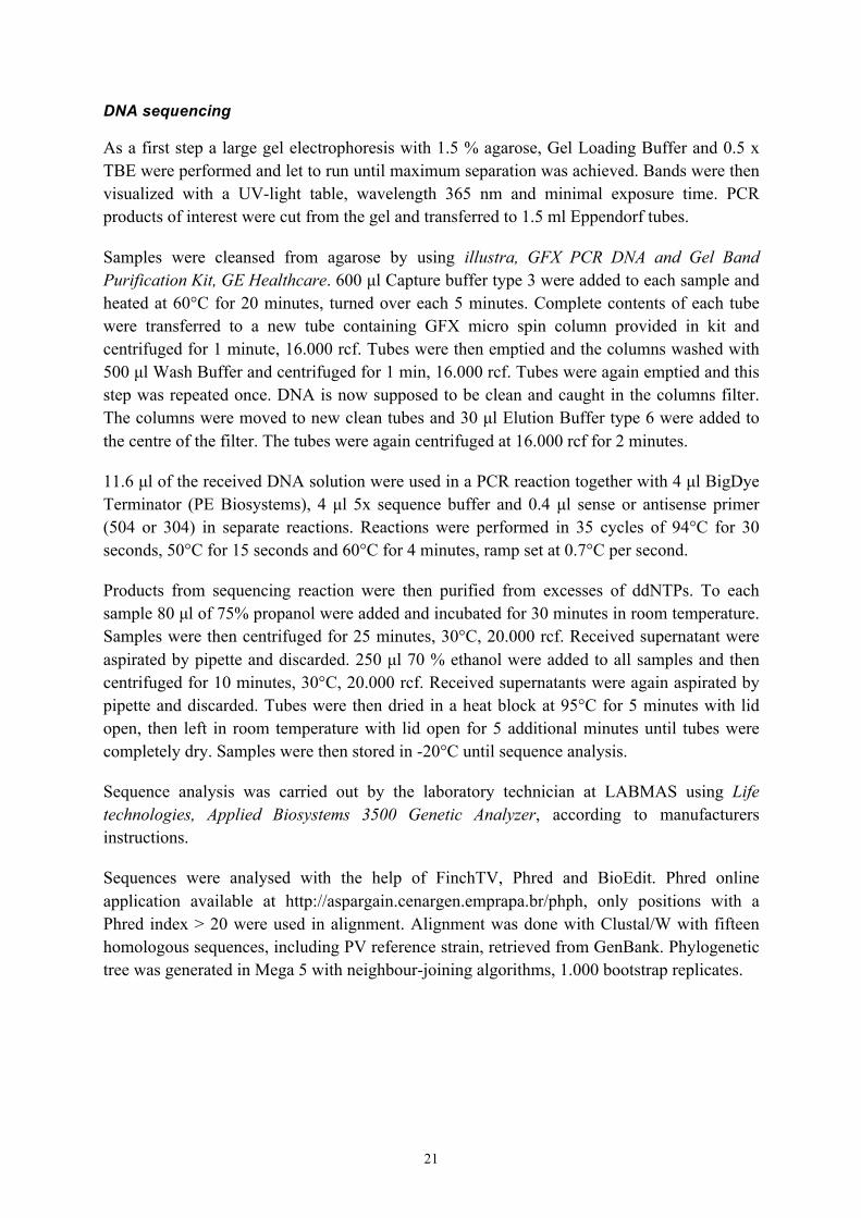
DNA sequencing
As a first step a large gel electrophoresis with 1.5 % agarose, Gel Loading Buffer and 0.5 x TBE were performed and let to run until maximum separation was achieved. Bands were then visualized with a UV-light table, wavelength 365 nm and minimal exposure time. PCR products of interest were cut from the gel and transferred to 1.5 ml Eppendorf tubes.
Samples were cleansed from agarose by using illustra, GFX PCR DNA and Gel Band Purification Kit, GE Healthcare. 600 µl Capture buffer type 3 were added to each sample and heated at 60°C for 20 minutes, turned over each 5 minutes. Complete contents of each tube were transferred to a new tube containing GFX micro spin column provided in kit and centrifuged for 1 minute, 16.000 rcf. Tubes were then emptied and the columns washed with 500 µl Wash Buffer and centrifuged for 1 min, 16.000 rcf. Tubes were again emptied and this step was repeated once. DNA is now supposed to be clean and caught in the columns filter. The columns were moved to new clean tubes and 30 µl Elution Buffer type 6 were added to the centre of the filter. The tubes were again centrifuged at 16.000 rcf for 2 minutes.
11.6 µl of the received DNA solution were used in a PCR reaction together with 4 µl BigDye Terminator (PE Biosystems), 4 µl 5x sequence buffer and 0.4 µl sense or antisense primer (504 or 304) in separate reactions. Reactions were performed in 35 cycles of 94°C for 30 seconds, 50°C for 15 seconds and 60°C for 4 minutes, ramp set at 0.7°C per second.
Products from sequencing reaction were then purified from excesses of ddNTPs. To each sample 80 µl of 75% propanol were added and incubated for 30 minutes in room temperature. Samples were then centrifuged for 25 minutes, 30°C, 20.000 rcf. Received supernatant were aspirated by pipette and discarded. 250 µl 70 % ethanol were added to all samples and then centrifuged for 10 minutes, 30°C, 20.000 rcf. Received supernatants were again aspirated by pipette and discarded. Tubes were then dried in a heat block at 95°C for 5 minutes with lid open, then left in room temperature with lid open for 5 additional minutes until tubes were completely dry. Samples were then stored in -20°C until sequence analysis.
Sequence analysis was carried out by the laboratory technician at LABMAS using Life technologies, Applied Biosystems 3500 Genetic Analyzer, according to manufacturers instructions.
Sequences were analysed with the help of FinchTV, Phred and BioEdit. Phred online application available at http://aspargain.cenargen.emprapa.br/phph, only positions with a Phred index > 20 were used in alignment. Alignment was done with Clustal/W with fifteen homologous sequences, including PV reference strain, retrieved from GenBank. Phylogenetic tree was generated in Mega 5 with neighbour-joining algorithms, 1.000 bootstrap replicates.

22
RESULTS
Direct immunofluorescence antibody test
The sample received on 17th September, sample 62, tested negative on DFA on all four slides. Remaining samples all tested positive on DFA, which was the expected outcome for these samples.
Mouse inoculation
Samples 5, 16 and 62 were inoculated intracranial in 5-10 mice per sample. Mice from sample 62 did not show any signs related to rabies virus during 30 days of observation. Sample 62 was diagnosed as rabies-negative based on inoculation and DFA.
In mice from sample 16 two out of five mice began showing symptoms associated with rabies on day 14. Symptoms included anorexia, visible weight loss, ruffled fur, ataxia, hyperventilation, paresis in hind legs and paralysis. All 5 mice in that group were euthanized on day 15 and tested with DFA. All five mice tested positive for rabies virus. In mice inoculated with sample 5 only one mouse developed symptoms of rabies and also tested positive on DFA. Remaining four mice did not show any signs during the observation period and were not tested with DFA.
Table 3. Results of DFA and mouse inoculation
Sample id. Source DFA Inoculation
1 62 Horse Negative Negative
2 5 Horse Positive Positive, day 16, 1/5 mice
3 24 Cat Positive * 4 16 Cattle Positive Positive, day 14, 2/5 mice
5 194 Cattle Positive *
6 753 Cattle Positive *
7 605 Cattle Positive *
8 66 Cattle Positive * 9 144 Cattle Positive *
10 154 Cattle Positive *
11 533 Cattle Positive *
12 DOG Dog Positive *
13 Pasteur Virus Dog Positive Positive, day 5, 5/5 mice
*Not performed in this study, positive in earlier inoculations
RT-PCR
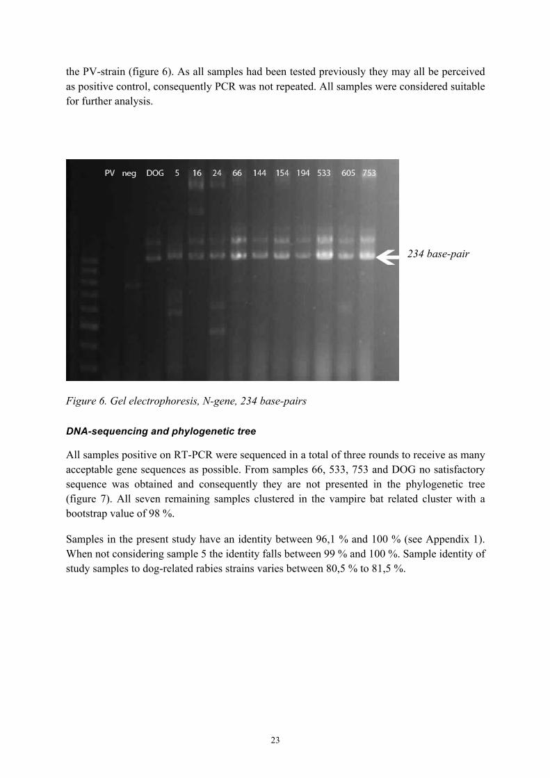
On samples positive on DFA RT-PCR were performed. Sense and antisense primers used resulted in a 234-base-pair fragment. All samples tested positive except the positive control,
23
the PV-strain (figure 6). As all samples had been tested previously they may all be perceived as positive control, consequently PCR was not repeated. All samples were considered suitable for further analysis.
234 base-pair
Figure 6. Gel electrophoresis, N-gene, 234 base-pairs DNA-sequencing and phylogenetic tree
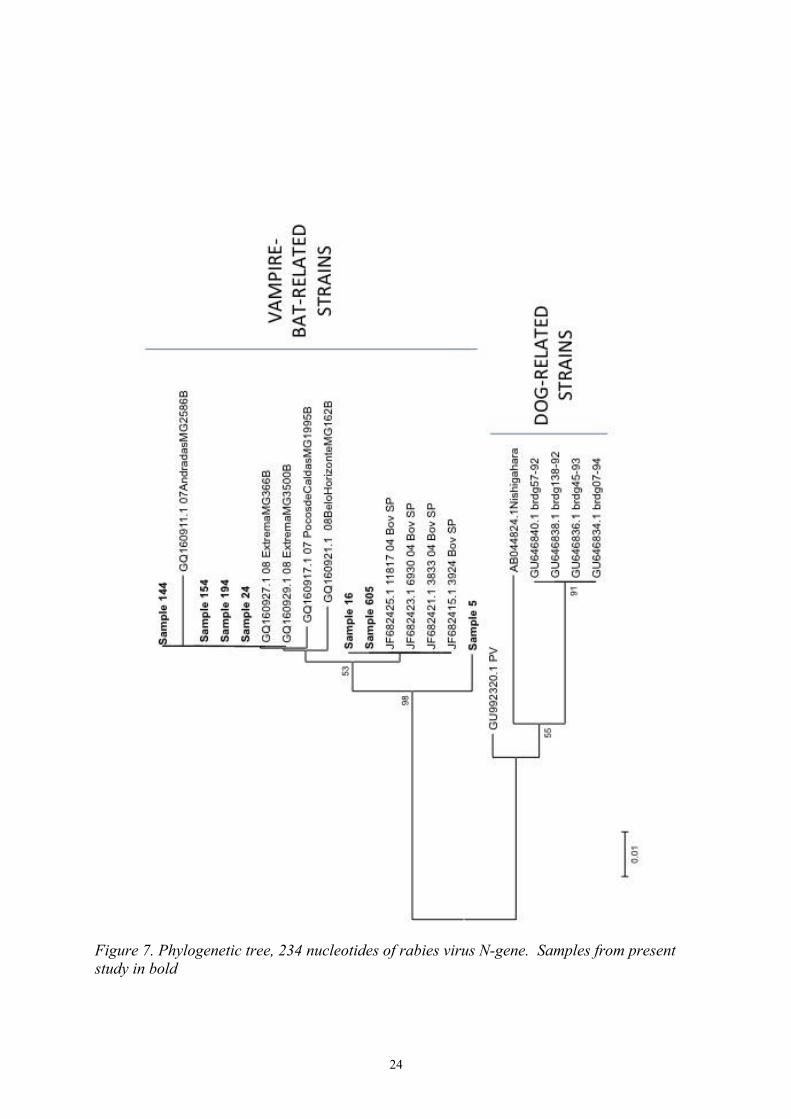
All samples positive on RT-PCR were sequenced in a total of three rounds to receive as many acceptable gene sequences as possible. From samples 66, 533, 753 and DOG no satisfactory sequence was obtained and consequently they are not presented in the phylogenetic tree (figure 7). All seven remaining samples clustered in the vampire bat related cluster with a bootstrap value of 98 %.
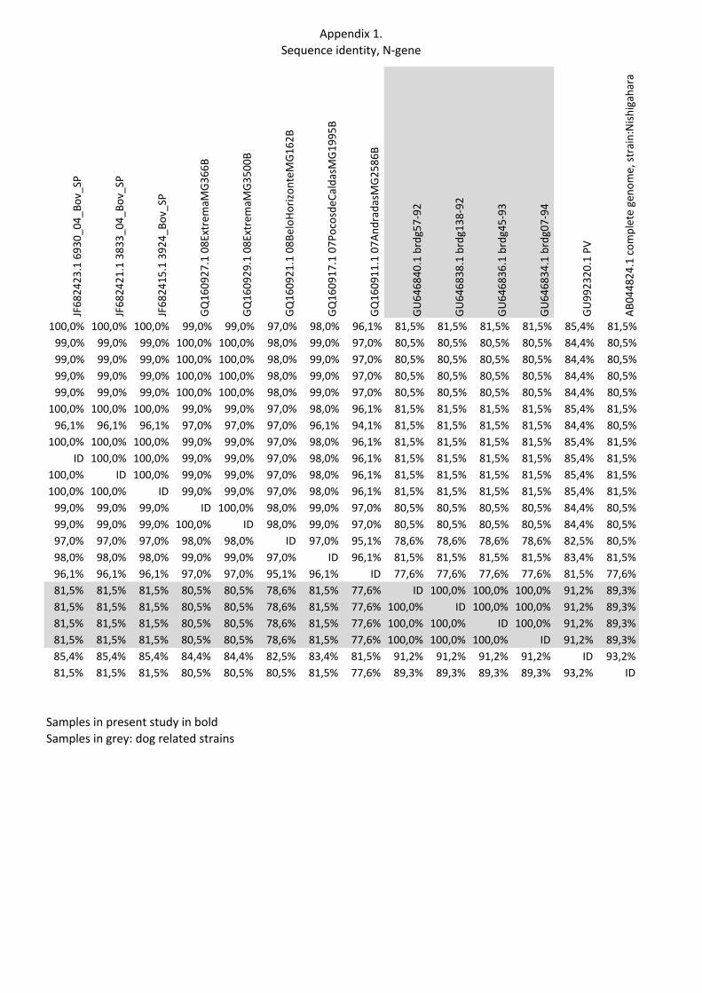
Samples in the present study have an identity between 96,1 % and 100 % (see Appendix 1). When not considering sample 5 the identity falls between 99 % and 100 %. Sample identity of study samples to dog-related rabies strains varies between 80,5 % to 81,5 %.
24
Figure 7. Phylogenetic tree, 234 nucleotides of rabies virus N-gene. Samples from present study in bold

25
DISCUSSION
234 nucleotides of the 5´end of the rabies virus nucleoprotein gene and following intergenic region were sequenced for 7 of the 10 positive samples. All seven samples cluster together with the vampire bat related sequences retrieved from GenBank. There are three different species of haematophagous bats: Desmodus rotundus, Diphylla ecaudata and Diaemus youngii. The most common of these is the common vampire bat, D. rotundus, and it is likewise the most important rabies vector and reservoir, especially for bovine rabies (Mayen, 2003). Studies have shown that rabies infection range from 0 – 3 % for D. rotundus (Da Rosa et al., 2006). This is the expected outcome considering that these samples originate from Brazilian cattle.
No difference in virus strain clustering could be seen on the basis of time or space of sample collecting. This is in line with what have been reported before in this region (Carnieli et al., 2009; Schaefer et al., 2005) and demonstrates rabies virus stability and mechanisms to adapt to the respective host species, rather than a geographic area (Schaefer et al., 2005).
All samples in this study have a nucleotide identity ≥ 96,1%, if not considering sample 5 ≥ 99%. Sample 5 is from a horse located in São Paulo City from the year of 2012. By analysis made in the present study it was not possible to deduce why this sample was different from the others. Possibilities include rabies virus transmission from frugivorous or insectivorous bat rather than from a haematophagous bat (Oliveira et al., 2010) or chance mutation in the virus itself. Even though sample 5 clusters on a branch of its own in the phylogenetic tree all samples have a high identity between them. The genetic relationship of these viruses to canine rabies strains retrieved from GenBank were between 80,5 – 81,5 %.
Interesting was that the sample collected from a cat, sample 24, also clustered with the vampire bat related viruses. Felines, that do not have a specific rabies virus strain of their own, is commonly infected with a canine rabies virus strain (Eduardo Brandão, 2012; Frymus et al., 2009). The cat in question was living in São Paulo City where canine rabies is under control by means of vaccination programmes (Da Rosa et al., 2006; Queiroz et al., 2012) and the cat furthermore had a history of catching and playing with a bat a few weeks before showing neurological symptoms. Alterations in the bats natural environment has forced bats to migrate to urban areas in search of new food resources, this is mostly seen and most important in the Amazon region where humans live in more primitive buildings and closer to the bats habitats (Barbosa et al., 2008; da Rosa et al., 2006). As canine rabies develops to a more controlled disease, bat-related rabies becomes more important as a public health concern (Da Rosa et al., 2006; Mayen, 2003). It is also important to remember that even though D. rotundus is the one species that most commonly transmit rabies virus, frugivorous and insectivorous bats can also spread the disease to other animals or humans (Carnieli et al., 2009; Queiroz et al., 2012; Schaefer et al., 2005).
26
CONCLUSION
To conclude, all samples in this study clustered within the vampire bat related rabies virus strains, which were expected as vaccination programmes have as good as eradicated canine rabies in the study area.
ACKNOWLEDGMENTS
This study is a degree project as a part of my Master of Veterinary Science at the Swedish University of Agriculture Science. The study was funded by SIDA (Swedish International Development Cooperation Agency) as a Minor Field Study Scholarship and Gulli Strålfeldts fund and Veterinärmedicinska fakultetens stipendiesamfond trough the Swedish University of Agriculture Science. Laboratory equipment and reagents were provided and financed by LABMAS, Department of Preventive Veterinary Medicine and Animal Health, at the University of São Paulo. I would like to thank SIDA, SLU and USP for funding and aiding me in my degree project.
I would like to thank my Brazilian supervisor Professor Paulo Eduardo Brandão at the University of São Paulo for excellent guidance theoretically and practically in the laboratory as well as my Swedish supervisor Professor Mikael Berg for help and support during the full length of my project. I would like to recognize Professor Paulo Eduardo Brandão once again for taking exceptionally good care of me throughout my stay in São Paulo.
REFERENCES
Baer, G.M., 2007. 1 - The History of Rabies, in: Alan C. Jackson, William H. Wunner (Eds.), Rabies (Second Edition). Academic Press, Oxford, p. 1–I.
Barbosa, T.F.S., Medeiros, D.B. de A., Travassos da Rosa, E.S., Casseb, L.M.N., Medeiros, R., Pereira, A. de S., Vallinoto, A.C.R., Vallinoto, M., Begot, A.L., Lima, R.J. da S., Vasconcelos, P.F. da C., Nunes, M.R., 2008. Molecular epidemiology of rabies virus isolated from different sources during a bat-transmitted human outbreak occurring in Augusto Correa municipality, Brazilian Amazon. Virology 370, 228–236.
Bourhy, H., Dacheux, L., Strady, C., Mailles, A., 2005. Rabies in Europe in 2005. Euro Surveill. 10, 213–216.Baer, G.M., 2007. 1 - The History of Rabies, in: Alan C. Jackson, William H. Wunner (Eds.), Rabies (Second Edition). Academic Press, Oxford, pp. 1–I.
Bourhy, H., Dacheux, L., Strady, C., Mailles, A., 2005. Rabies in Europe in 2005. Euro Surveill. 10, 213–216.
Briggs, D.J., 2012. The role of vaccination in rabies prevention. Curr Opin Virol 2, 309–314.
Carnieli, P., Jr, Castilho, J.G., Fahl, W. de O., Véras, N.M.C., Timenetsky, M. do C.S.T., 2009. Genetic characterization of Rabies virus isolated from cattle between 1997 and 2002 in an epizootic area in the state of São Paulo, Brazil. Virus Res. 144, 215–224.
27
Charlton, K.M., Nadin-Davis, S., Casey, G.A., Wandeler, A.I., 1997. The long incubation period in rabies: delayed progression of infection in muscle at the site of exposure. Acta Neuropathol. 94, 73–77.
Cleaveland, S., Fèvre, E.M., Kaare, M., Coleman, P.G., 2002. Estimating human rabies mortality in the United Republic of Tanzania from dog bite injuries. Bull. World Health Organ. 80, 304–310.
Da Rosa, E.S.T., Kotait, I., Barbosa, T.F.S., Carrieri, M.L., Brandão, P.E., Pinheiro, A.S., Begot, A.L., Wada, M.Y., De Oliveira, R.C., Grisard, E.C., Ferreira, M., Lima, R.J. da S., Montebello, L., Medeiros, D.B.A., Sousa, R.C.M., Bensabath, G., Carmo, E.H., Vasconcelos, P.F.C., 2006. Bat-transmitted human rabies outbreaks, Brazilian Amazon. Emerging Infect. Dis. 12, 1197–1202.
Eduardo Brandão, P., 2012. Personal communication 2012-09-17, 2013-01-11.
Frymus, T., Addie, D., Belák, S., Boucraut-Baralon, C., Egberink, H., Gruffydd-Jones, T., Hartmann, K., Hosie, M.J., Lloret, A., Lutz, H., Marsilio, F., Pennisi, M.G., Radford, A.D., Thiry, E., Truyen, U., Horzinek, M.C., 2009. Feline rabies. ABCD guidelines on prevention and management. J. Feline Med. Surg. 11, 585–593.
Hanlon, C.A., Niezgoda, M., Rupprecht, C.E., 2007. 5 - Rabies in Terrestrial Animals, in: Alan C. Jackson, William H. Wunner (Eds.), Rabies (Second Edition). Academic Press, Oxford, pp. 201–VIII.
Höjer, J., Sjöblom, E., Berglund, O., Hammarin, A.L., Grandien, M., 2001. [The first case of rabies in Sweden in 26 years. Inform travellers abroad about risks and treatment following suspected infection]. Lakartidningen 98, 1216–1220.
ICTV Virus Taxonomy: 2011 release (current) [WWW Document], n.d. ICTV. URL http://ictvonline.org/virusTaxonomy.asp?version=2011 (accessed 10.14.12).
Jackson, A.C., 2007. 8 - Pathogenesis, in: Alan C. Jackson, William H. Wunner (Eds.), Rabies (Second Edition). Academic Press, Oxford, pp. 341–XI.
King, A.M., Lefkowitz, E., Adams, M.J., Carstens, E.B., 2011. Virus Taxonomy: Ninth Report of the International Committee on Taxonomy of Viruses. Elsevier.
Kobayashi, Y., Suzuki, Y., Itou, T., Ito, F.H., Sakai, T., Gojobori, T., 2011. Evolutionary history of dog rabies in Brazil. J. Gen. Virol. 92, 85–90.
Leung, A.K.C., Davies, H.D., Hon, K.-L.E., 2007. Rabies: epidemiology, pathogenesis, and prophylaxis. Adv Ther 24, 1340–1347.
Lewis, P., Fu, Y., Lentz, T.L., 2000. Rabies virus entry at the neuromuscular junction in nerve-muscle cocultures. Muscle Nerve 23, 720–730.
Mayen, F., 2003. Haematophagous bats in Brazil, their role in rabies transmission, impact on public health, livestock industry and alternatives to an indiscriminate reduction of bat population. J. Vet. Med. B Infect. Dis. Vet. Public Health 50, 469–472.
Meslin, F.-X., Kaplan, M.M., Koprowski, H., 1996. Laboratory techniques in rabies, Fourth edition. ed. World Health Organization.
28
Mochizuki, N., Kawasaki, H., Silva, M.L., Afonso, J.A., Itou, T., Ito, F.H., Sakai, T., 2012. Molecular epidemiology of livestock rabies viruses isolated in the northeastern Brazilian states of Paraíba and Pernambuco from 2003 - 2009. BMC Res Notes 5, 32.
Nagaraj, T., Vasanth, J.P., Desai, A., Kamat, A., Madhusudana, S.N., Ravi, V., 2006. Ante mortem diagnosis of human rabies using saliva samples: comparison of real time and conventional RT-PCR techniques. J. Clin. Virol. 36, 17–23.
OIE, 2009. Rabies [WWW Document]. URL http://www.oie.int/fileadmin/Home/eng/Publications_%26_Documentation/docs/pdf/rabies.pdf (accessed 4.10.12).
Oliveira, R. de N., De Souza, S.P., Lobo, R.S.V., Castilho, J.G., Macedo, C.I., Carnieli, P., Jr, Fahl, W.O., Achkar, S.M., Scheffer, K.C., Kotait, I., Carrieri, M.L., Brandão, P.E., 2010. Rabies virus in insectivorous bats: implications of the diversity of the nucleoprotein and glycoprotein genes for molecular epidemiology. Virology 405, 352–360.
Queiroz, L.H., Favoretto, S.R., Cunha, E.M.S., Campos, A.C.A., Lopes, M.C., De Carvalho, C., Iamamoto, K., Araújo, D.B., Venditti, L.L.R., Ribeiro, E.S., Pedro, W.A., Durigon, E.L., 2012. Rabies in southeast Brazil: a change in the epidemiological pattern. Arch. Virol. 157, 93–105.
Rabies - SVA [WWW Document], n.d. . URL http://sva.se/sv/Djurhalsa1/Epizootier/Rabies/ (accessed 10.14.12).
Rabiesövervakning av fladdermus - SVA [WWW Document], n.d. . URL http://sva.se/sv/Djurhalsa1/Epizootier/Rabies/Rabiesovervakning-av-fladdermus/ (accessed 10.14.12).
Rossiter, J.P., Jackson, A.C., 2007. 9 - Pathology, in: Alan C. Jackson, William H. Wunner (Eds.), Rabies (Second Edition). Academic Press, Oxford, pp. 383–XIII.
Rupprecht, C.E., Hanlon, C.A., Hemachudha, T., 2002. Rabies re-examined. The Lancet Infectious Diseases 2, 327–343.
Saengseesom, W., Mitmoonpitak, C., Kasempimolporn, S., Sitprija, V., 2007. Real-time PCR analysis of dog cerebrospinal fluid and saliva samples for ante-mortem diagnosis of rabies. Southeast Asian J. Trop. Med. Public Health 38, 53–57.
Sambrook, J., Russell, D.W., 2001. Molecular Cloning: A Laboratory Manual. CSHL Press.
Schaefer, R., Batista, H.B.R., Franco, A.C., Rijsewijk, F.A.M., Roehe, P.M., 2005. Studies on antigenic and genomic properties of Brazilian rabies virus isolates. Vet. Microbiol. 107, 161–170.
Schnell, M.J., McGettigan, J.P., Wirblich, C., Papaneri, A., 2010. The cell biology of rabies virus: using stealth to reach the brain. Nat. Rev. Microbiol. 8, 51–61.
Thawley, D.G., Wright, J.C., 1982. Pseudorabies virus infection in raccoons: a review. J. Wildl. Dis. 18, 113–116.
29
Tordo, N., Poch, O., Ermine, A., Keith, G., Rougeon, F., 1986. Walking along the rabies genome: is the large G-L intergenic region a remnant gene? Proc Natl Acad Sci U S A 83, 3914–3918.
Wang, Z.W., Sarmento, L., Wang, Y., Li, X., Dhingra, V., Tseggai, T., Jiang, B., Fu, Z.F., 2005. Attenuated rabies virus activates, while pathogenic rabies virus evades, the host innate immune responses in the central nervous system. J. Virol. 79, 12554–12565.
WHO | Rabies [WWW Document], n.d. WHO. URL http://www.who.int/mediacentre/factsheets/fs099/en/index.html (accessed 10.4.12).
WHO Expert Consultation on Rabies : first report [WWW Document], 2005. . URL http://apps.who.int/iris/handle/10665/43262 (accessed 10.30.12).
Willoughby, R.E., Jr, Tieves, K.S., Hoffman, G.M., Ghanayem, N.S., Amlie-Lefond, C.M., Schwabe, M.J., Chusid, M.J., Rupprecht, C.E., 2005. Survival after treatment of rabies with induction of coma. N. Engl. J. Med. 352, 2508–2514.
Wunner, W.H., 2007. 2 - Rabies Virus, in: Alan C. Jackson, William H. Wunner (Eds.), Rabies (Second Edition). Academic Press, Oxford, pp. 23–II.

30

Appendix 1.Sequence identity, N-‐gene
Seq-‐> Sample 16
Sample 154
Sample 144
Sample 194
Sample 24
Sample 605
Sample 5
JF68
2425
.1 118
17_04_Bo
v_SP
Sample 16 ID 99,0% 99,0% 99,0% 99,0% 100,0% 96,1% 100,0%Sample 154 99,0% ID 100,0% 100,0% 100,0% 99,0% 97,0% 99,0%Sample 144 99,0% 100,0% ID 100,0% 100,0% 99,0% 97,0% 99,0%Sample 194 99,0% 100,0% 100,0% ID 100,0% 99,0% 97,0% 99,0%Sample 24 99,0% 100,0% 100,0% 100,0% ID 99,0% 97,0% 99,0%Sample 605 100,0% 99,0% 99,0% 99,0% 99,0% ID 96,1% 100,0%Sample 5 96,1% 97,0% 97,0% 97,0% 97,0% 96,1% ID 96,1%JF682425.1 11817_04_Bov_SP 100,0% 99,0% 99,0% 99,0% 99,0% 100,0% 96,1% IDJF682423.1 6930_04_Bov_SP 100,0% 99,0% 99,0% 99,0% 99,0% 100,0% 96,1% 100,0%JF682421.1 3833_04_Bov_SP 100,0% 99,0% 99,0% 99,0% 99,0% 100,0% 96,1% 100,0%JF682415.1 3924_Bov_SP 100,0% 99,0% 99,0% 99,0% 99,0% 100,0% 96,1% 100,0%GQ160927.1 08ExtremaMG366B 99,0% 100,0% 100,0% 100,0% 100,0% 99,0% 97,0% 99,0%GQ160929.1 08ExtremaMG3500B 99,0% 100,0% 100,0% 100,0% 100,0% 99,0% 97,0% 99,0%GQ160921.1 08BeloHorizonteMG162B 97,0% 98,0% 98,0% 98,0% 98,0% 97,0% 97,0% 97,0%GQ160917.1 07PocosdeCaldasMG1995B 98,0% 99,0% 99,0% 99,0% 99,0% 98,0% 96,1% 98,0%GQ160911.1 07AndradasMG2586B 96,1% 97,0% 97,0% 97,0% 97,0% 96,1% 94,1% 96,1%GU646840.1 brdg57-‐92 81,5% 80,5% 80,5% 80,5% 80,5% 81,5% 81,5% 81,5%GU646838.1 brdg138-‐92 81,5% 80,5% 80,5% 80,5% 80,5% 81,5% 81,5% 81,5%GU646836.1 brdg45-‐93 81,5% 80,5% 80,5% 80,5% 80,5% 81,5% 81,5% 81,5%GU646834.1 brdg07-‐94 81,5% 80,5% 80,5% 80,5% 80,5% 81,5% 81,5% 81,5%GU992320.1 PV 85,4% 84,4% 84,4% 84,4% 84,4% 85,4% 84,4% 85,4%AB044824.1 complete genome, strain:Nishigahara 81,5% 80,5% 80,5% 80,5% 80,5% 81,5% 80,5% 81,5%
Samples in present study in boldSamples in grey: dog related strains
Appendix 1.Sequence identity, N-‐gene
JF68
2423
.1 693
0_04
_Bov_SP
JF68
2421
.1 383
3_04
_Bov_SP
JF68
2415
.1 392
4_Bo
v_SP
GQ16
0927
.1 08ExtremaM
G366
B
GQ16
0929
.1 08ExtremaM
G350
0B
GQ16
0921
.1 08B
eloH
orizo
nteM
G162
B
GQ16
0917
.1 07P
ocosde
CaldasMG1
995B
GQ16
0911
.1 07A
ndradasM
G258
6B
GU64
6840
.1 brdg57-‐92
GU64
6838
.1 brdg138
-‐92
GU64
6836
.1 brdg45-‐93
GU64
6834
.1 brdg07-‐94
GU99
2320
.1 PV
AB04
4824
.1 com
plete geno
me, strain:Nish
igahara
100,0% 100,0% 100,0% 99,0% 99,0% 97,0% 98,0% 96,1% 81,5% 81,5% 81,5% 81,5% 85,4% 81,5%99,0% 99,0% 99,0% 100,0% 100,0% 98,0% 99,0% 97,0% 80,5% 80,5% 80,5% 80,5% 84,4% 80,5%99,0% 99,0% 99,0% 100,0% 100,0% 98,0% 99,0% 97,0% 80,5% 80,5% 80,5% 80,5% 84,4% 80,5%99,0% 99,0% 99,0% 100,0% 100,0% 98,0% 99,0% 97,0% 80,5% 80,5% 80,5% 80,5% 84,4% 80,5%99,0% 99,0% 99,0% 100,0% 100,0% 98,0% 99,0% 97,0% 80,5% 80,5% 80,5% 80,5% 84,4% 80,5%
100,0% 100,0% 100,0% 99,0% 99,0% 97,0% 98,0% 96,1% 81,5% 81,5% 81,5% 81,5% 85,4% 81,5%96,1% 96,1% 96,1% 97,0% 97,0% 97,0% 96,1% 94,1% 81,5% 81,5% 81,5% 81,5% 84,4% 80,5%
100,0% 100,0% 100,0% 99,0% 99,0% 97,0% 98,0% 96,1% 81,5% 81,5% 81,5% 81,5% 85,4% 81,5%ID 100,0% 100,0% 99,0% 99,0% 97,0% 98,0% 96,1% 81,5% 81,5% 81,5% 81,5% 85,4% 81,5%
100,0% ID 100,0% 99,0% 99,0% 97,0% 98,0% 96,1% 81,5% 81,5% 81,5% 81,5% 85,4% 81,5%100,0% 100,0% ID 99,0% 99,0% 97,0% 98,0% 96,1% 81,5% 81,5% 81,5% 81,5% 85,4% 81,5%99,0% 99,0% 99,0% ID 100,0% 98,0% 99,0% 97,0% 80,5% 80,5% 80,5% 80,5% 84,4% 80,5%99,0% 99,0% 99,0% 100,0% ID 98,0% 99,0% 97,0% 80,5% 80,5% 80,5% 80,5% 84,4% 80,5%97,0% 97,0% 97,0% 98,0% 98,0% ID 97,0% 95,1% 78,6% 78,6% 78,6% 78,6% 82,5% 80,5%98,0% 98,0% 98,0% 99,0% 99,0% 97,0% ID 96,1% 81,5% 81,5% 81,5% 81,5% 83,4% 81,5%96,1% 96,1% 96,1% 97,0% 97,0% 95,1% 96,1% ID 77,6% 77,6% 77,6% 77,6% 81,5% 77,6%81,5% 81,5% 81,5% 80,5% 80,5% 78,6% 81,5% 77,6% ID 100,0% 100,0% 100,0% 91,2% 89,3%81,5% 81,5% 81,5% 80,5% 80,5% 78,6% 81,5% 77,6% 100,0% ID 100,0% 100,0% 91,2% 89,3%81,5% 81,5% 81,5% 80,5% 80,5% 78,6% 81,5% 77,6% 100,0% 100,0% ID 100,0% 91,2% 89,3%81,5% 81,5% 81,5% 80,5% 80,5% 78,6% 81,5% 77,6% 100,0% 100,0% 100,0% ID 91,2% 89,3%85,4% 85,4% 85,4% 84,4% 84,4% 82,5% 83,4% 81,5% 91,2% 91,2% 91,2% 91,2% ID 93,2%81,5% 81,5% 81,5% 80,5% 80,5% 80,5% 81,5% 77,6% 89,3% 89,3% 89,3% 89,3% 93,2% ID
Samples in present study in boldSamples in grey: dog related strains