modality of treatment for the distal end radius · pdf fileinternational journal of scientific...
TRANSCRIPT

International Journal of Scientific and Research Publications, Volume 4, Issue 11, November 2014 1 ISSN 2250-3153
www.ijsrp.org
Modality of Treatment for the Distal End Radius
Fracture
Dr Bhuvnesh Chaturvedi, Dr Sk Irfan Ali, Dr Sumit Krishna
Abstract- Fracture of the distal end of radius is one of the most
common fractures. It occurs in middle aged and elderly women
commonly. It also occurs in young men with high velocity,
injury. With increase in longevity and activity in middle aged to
elderly population, there is increase in number of this fractures
various surgical interventions are available presently, like
percutaneous pinning, intra focal pinning, external fixator and
plate fixation.
Methods and Materials:
30 Patients who have sustained fractures of the distal radius
admitted in the department of orthopaedics at M.V.J. Medical
College & Research Hospital from July 2011 to July 2013
Inclusion criteria 1) Adults between age group of 20 years to 60
years with fracture lower end of radius 2) All patients having
isolated Distal end radius fracture Exclusion criteria 1) Distal
radius fracture associated with other injuries around the wrist
joint .2) Open Fracture.3) Pathological Fracture .4) Distal
radius Fracture associated with neurovascular complications
Results:
The assessment of functional outcome was made according
to modified clinical system of Green and O’Brien 1978 and Brad
way et al 1989 Overall results were graded as acceptable
(excellent and good), fair or poor. In our study, 6 patients had
full range of movements, no pain, returned to previous job and
had 100% strength to that of normal side and the results were
considered as excellent. Another 3 patients had full range of
movements as compared to that of opposite side with mild pain
not affecting the function of wrist. They also scored more than
90% and the results were considered as excellent. There was 9
case of Excellent, 14 cases of Good, 5 cases of Fair and 2 cases
showed poor Result.14 patients had limitation of movements of
wrist and forearm by 20% and decreased hand strength by 15-
20% as compared to that of normal side, but they did not have
pain. The function of hand was not affected and they continued
their previous profession. They scored 80% and the result was
considered as good.
Five patients had limitation of movements of wrist and
forearms by 25% as compared to that of normal side, with mild
pain, unable to lift heavy weights and their hand grip strength
was also decreased by 20 to 25 % as compared to that of normal
side. They scored between 65 - 79% and the results were
considered as fair. Two patients had moderate pain, which
subsiding with analgesics. They were
unable to do heavy manual work, but could carry out daily
activities. They had Restriction of wrist movements by 50% as
compared to that of normal side. They scored less than 65% and
the result was considered as poor.
Conclusion:
From the present study on 30 patients with distal end radius
fractures after analyzing the observation and looking at the
results we conclude the following. Fracture of distal end of
radius is having a bimodal age distribution in our study we find
young individual between 21-30yrs mostly male sustain road
traffic accident as a common mode of injury. Right side was
affected more than the left side. Restoration of anatomy of distal
end of radius, early mobilization and less complication were
achieved using Platting .Platting is a better method of treatment
for fracture of the distal end of radius
I. INTRODUCTION
racture of the distal end of radius is one of the most common
fractures. It occurs in middle aged and elderly women
commonly. It also occurs in young men with high velocity injury
.With increase in longevity and activity in middle aged to elderly
population, there is increase in number of this fractures.
Patients with fracture distal end of radius have many
complications more frequently are generally appreciated
and
Failure in management may cause Permanent disability. Distal
radius fractures crush the mechanical foundation of the man’s
most elegant tool, the hand and the grip.
Pain and disability have resulted due to subsequent malunion
of unstable fracture of distal radius which were managed by
conservative method like plaster cast alone.
Recently surgical management has been widely
recommended and performed to prevent disability. Several
studies has shown convincingly that functional outcome
is good when the anatomy is restored by obtaining good
reduction of fracture fragments, maintaining the angulations of
the articular surface of radius and radial Length.
Various surgical interventions are available presently, like
percutaneous pinning, intra focal pinning, external fixator and
plate fixation. External fixator may be performed in a bridging
technique and a non bridging technique. Bridging external fixator
allows distraction across the radio carpal joint.
The present study “A Comparative study of management of
the fracture of the distal end radius by external fixator Vs
plating” was undertaken in department of Orthopaedics at
M.V.J.M.C & R.H to study fracture healing &
functional outcome in distal radius fracture following
external fixation & platting.
II. ANATOMY OF THE WRIST
SURFACE ANATOMY
The surface anatomy of the wrist offers many clues to the
features of the underlying structures and their inter relationships.
F
International Journal of Scientific and Research Publications, Volume 4, Issue 11, November 2014 2
ISSN 2250-3153
www.ijsrp.org
Posterior aspect
The skin of the posterior aspect of the wrist is thicker and
more mobile than the anterior skin and is covered by short hairs,
which are more numerous on the ulnar side than the radial side.
The dorso radial flares of the radial metaphysis and the ulnar
head forms prominent features that are visible and palpated over
proximal part of dorsal aspect of wrist. Just distal to the course of
the out-cropping tendons Extensor pollicis brevis (EPB),
abductor pollicis longus, Extensor carpi radialis longus (ECRL)
and Extensor carpi radialis brevis (ECRB) muscles can be
palpated. Between the second and third extensor compartment is
a bony prominence called Listers’ tubercle, which behaves as a
pulley for the tendon of extensor pollicis longus (EPL) and also
serves as a critical landmark because it marks one’s orientation to
the scapholunate joint and the interfacet prominence of the distal
radius.
Anterior Aspect
This area is covered by thin, practically hairless skin, which
is rather adherent to the underlying fascia and has limited
mobility, especially distally. The fascia about
the wrist and the distal radius begins in the forearm as ante
brachial fascia and becomes thick distally as it coalesces with
deeper fascia to form the flexor retinaculum and transverse
carpal ligament. These tendinous longitudinal protrusions
are the limits of grooves on the lateral and the medial sides where
the radial and the ulnar artery can be palpated. The central
protrusion corresponds to the flexor tendons parallel to the
median nerve, lateral to the Flexor Carpi Radialis (FCR) tendon
and the medial to the Flexor Carpi Ulnaris (FCU). Three
transverse skin creases proximal, middle, and distal correspond
respectively to the ulnar head, the radio carpal joint and
the mid carpal joint.
Radial aspect
The hallmark of the radial surface of the wrist is the
“anatomical snuff box”. It is the region between the EPL and
EPB tendons and it is made most pronounced by extending and
abducting the thumb. Radial styloid process can be palpated at
the proximal part and the waist and distal pole of the scaphoid at
the distal part of this region. The radial artery, if patent, can be
traced through the anatomical snuff box as it courses dorsally.
The branches of superficial radial nerve can be palpated through
this region coursing in a distal direction.
Ulnar Aspect
Analogous to the anatomical snuff box an “ulnar snuff box”
can be imagined in the interval between the tendons of ECU and
FCU muscles on the ulnar surface of the wrist. The ulnar styloid
process is palpable within the ulnar snuff box when forearm is in
neutral position.
The Bones of forearm
The distal end of radius
The widest part, it is four sided in section. Its lateral surface
is slightly rough projecting distally as a styloid process palpable
when tendons around it are slack. Distal is the smooth carpal
articular surface, divided by a ridge into medial and lateral
areas. The medial is quadrangular and the lateral is triangular
and curving on the styloid process. The anterior surface is a
thick prominent ridge palpable even though over lying tendons,
2cms proximal to the thenar eminence. The medial surface is the
ulnar notch, smooth, anteroposteriorly concave for the
articulation with the ulna’s head. The posterior surface displays a
palpable dorsal tubercle limited medially by an
oblique grove and in line with the cleft between the index and the
middle fingers. A wide shallow grove lateral to it is divided by a
faint vertical ridge.
III. CLASSIFICATION OF DISTAL RADIUS
FRACTURES
The presentation of a classification of fractures of the distal
radius must begin with an initial recognition of the different
common types of fractures. Colle’s’ fracture is the most
common.
(1) Colle’s fracture: It is a distal metaphyseal fracture of
distal radius, which occurs within 2 cm of the articular surface
and may extend into the distal radio-carpal or radio-ulnar joints.
Dorsal angulation (silver fork deformity), dorsal displacement,
radial angulation and radial shortening are present. There is often
an accompanying fracture of the ulnar styloid, which may signify
avulsion of the TFC insertion.
(2) Smith’s fracture or reverse colle’s’ fracture: It is a
palmer angulated fracture of the distal radius with a “Garden
spade” deformity. The hand and wrist are displaced forward or
palmarly with respect to the forearm. The fracture may be extra-
articular or intra-articular, or part of a fracture-dislocation of the
wrist. Smith fractures are classified as modified Thomas
classification into three types as follows.
(3) Barton’s fracture is actually a fracture-dislocation or
subluxations in which the rim of the distal radius, dorsally or
palmarly, is displaced with the hand and carpus
(4) Pouteau fracture: In 1783 Pouteau of France first
described the fractures of distal radius.
I. Radiological appearance or fracture displacement
direction
1. AO classification
2. Sarmiento classification
3. Linstrom classification
II. The mechanism of injury
1. Castainign classification
2. Fernandez classification
3. Linscheid classification
III. Articular joint surface involvement
1. Myo classification
2. McMurtry and Jupiter classification
3. Melone classification
IV. Degree of comminution
1. Gartland and Werley classification
2. Jenkins classification
3. The Older classification
V. Bone calcification and resistance
1. Sennwald and Segmuller classification

International Journal of Scientific and Research Publications, Volume 4, Issue 11, November 2014 3
ISSN 2250-3153
www.ijsrp.org
VI. More recent classifications tend to provide therapeutic
options and prognosis to prevent redisplacement and
malunions.
1. Cooney’s classification
2. Mathoulin, Letrosue and Saffar classification
The Frykman’s Classification
It is now used to recognize the intra-articular distal radius
fracture that we identify as the Frykman fracture to separate it
from the colles’ and smith fractures.
AO classification
There are three AO categories:
1) ‘Extra-articular’ fractures which do not involve the
radiocarpal joint surface at all.
2) ‘Partial articular’ fractures which involve this joint, but a
portion of the articular surface remains in continuity with the
diaphysis.
3) ‘Complete articular’ fractures which are distinguished by
complete separation of the involved articular surface and the
diaphysis
Fig 11: AO Classification
IV. TREATMENT OF THE DISTAL RADIUS
FRACTURES
Percutaneous Direct Pinning

International Journal of Scientific and Research Publications, Volume 4, Issue 11, November 2014 4
ISSN 2250-3153
www.ijsrp.org
An early technique, in fact, one of the first used for fixation
of distal radius fractures, was percutaneous pinning, usually
entering at the level of the radial styloid Process. Some variations
in the point of penetration and the direction of the pins were
presented, but the aim was always to fix the mobile fragment to
the opposite cortex proximal to the fracture. This type of pinning
cannot prevent re displacement of certain fragments, and this is
particularly true of intra-articular and osteoporotic fractures.
Direct pinning of the fragments, especially the posterior medial
fragment (which can involve DRUJ), through the distal ulna,
adds stability to the structure.
External Fixation
This technique was proposed for comminuted fractures, and
it has improved the reduction of comminuted intra-articular
fractures. Ligamentotaxis can exert influence on fragments in
which capsule ligamentous attachments are still intact. The
traction is exerted mainly by the strong volar ligament plane on
the anterior rim of the distal radius. The dorsal tilt may not be
completely reduced because dorsal ligaments are thinner and in a
transverse plane.
Plate Fixation
This is indicated in fractures with volar displacement, such
as Barton’s and Smith’s fractures and in selected cases of dorsal
displacement where rigid fixation can provide for early wrist
motion. Plate fixation can provide either a buttress effect or hold
the distal epiphysis by cortico cancellous screws. The pre molded
plates reproducing the distal curvature of the radius are best, as
they give an anatomic reduction. The disadvantage of this
technique is difficulty in screw placement if the fracture is
severely comminuted and number of soft tissue complications
have been noted.
COMPLICATIONS OFTHE DISTAL RADIUS
FRACTURES
1. Distal radioulnar subluxations, dislocation.
2. Depressed major articular components
3. Difficult reduction; unstable reduction maintained only by
extreme position.
4. Median or ulnar nerve stretch, contusion or compression.
5. Acute carpal tunnel syndrome.
6. Tendon damage.
7. Peripheral nerve injury in external fixation errors.
8. Post-reduction swelling, compartment syndrome.
9. Carpal tunnel syndrome.
10. Associated carpal injury.
11. Radial nerve Dysaesthesias.
12. Pain dysfunction syndrome
V. MATERIALS AND METHODS
Source of Data
30 Patients who have sustained fractures of the distal radius
admitted in the department of orthopaedics at M.V.J. Medical
College & Research Hospital from July 2011 to July 2013.
Method of Collection of Data
Inclusion criteria
1) Adults between age group of 20 years to 60 years with
fracture lower end of radius
2) All patients having isolated Distal end radius fracture
Exclusion criteria
1) Distal radius fracture associated with other injuries around
the wrist joint
2) Open Fracture
3) Pathological Fracture
4) Distal radius Fracture associated with neurovascular
complications
Sample size : 30 cases (15External fixator and 15 Plating)
Sample Procedure : a prospective study
Methods
Patients with distal end radius fractures admitted in MVJ
hospital after meeting the inclusion and exclusion criteria were
taken up. All patients were evaluated preoperatively by clinical
and roentgen graphic examination. Systemic, hematological
investigations, chest X-ray and assessment of cardiac status using
ECG were done as a routine and pre anesthesia evaluation done.
Preoperatively patients were immobilized with POP or splints.
Patients were informed about the operative procedure and
consent taken.
Selection of patients:
1. Patients were randomly selected for external fixator or
platting
External Fixator
The Joshi type of External fixator was used in our study.
This fixator consists of, distractor bar attachment with the schanz
pins (screws). In our series, the joshi type of External fixator was
applied in all the cases. We have used two 3mm schanz screws
for radius and two 2.5 mm schanz screws for the second
metacarpal, and 4 mm connecting rods.
Instruments Used for the Procedure
1. External fixator set
2. Spanner No 7&8
3. Drill-bits
4. Electric drill & hand drill
5. T handle
6. Scalpel blade
7. Image intensifier
Surgical Technique (EXTERNAL FIXATOR)
Under regional block Anesthesia (Brachial block) or GA
depending upon anesthesiologist preference, patient was placed
supine on the operating table. The forearm and hand were
scrubbed with Betadine and saline. The tourniquet was applied
over the arm. The forearm and hand were painted with Betadine
and draped. The operating forearm was placed on a radiolucent
arm-board. Closed reduction was done under C-arm.
In this technique , 5mm incision for 4 shanz pins, 2 in the
middle third of the radius on the dorso lateral aspect about 10-

International Journal of Scientific and Research Publications, Volume 4, Issue 11, November 2014 5
ISSN 2250-3153
www.ijsrp.org
12cm from distal end and 2-3cm apart. We have done soft tissue
dissection using a hemostat, care taken to avoid injury to radial
nerve. Another 2 incision over the base of the second
metacarpal on dorso lateral aspect about 1-2cm apart were done,
3mm shanz pin were inserted in the radius, and 2.5mm shanz
pins was introduced in second metacarpal, then with fixator pins
securely in place, clamps and external fixator rod were mounted
to shanz pin. The clamps were loosened and longitudinal
traction was given with manual molding of the fracture
fragments back into a more normal alignment and gentle flexion
and ulnar deviation was maintained. The reduction was
confirmed through image intensifier and then external fixation
device was locked into place. The tension across the wrist
generated by the external fixator device which provides enough
ligamentotaxis was confirmed by image intensifier wherein,
radiocarpal articulation was seen to be 1 mm wider than the mid-
carpal joint in A-P projection.
SURGICAL TECHNIQUE (PLATTING)
INSTRUMENTS USED:
Ellis Buttress plate or locking compression plate of varying
length
2.5mm drill bit and 3.5mm drill sleeve system.
3.5mm LCP drill bit and sleeve system.
Hand drill and power drill.
Tap for 3.5mm cortical screws and 3.5mm depth gauge.
Hexagonal screw driver for 3.5mm cortical screws and
locking screw driver.
Other instrument like retractors, periosteal elevator,
reduction clamps, bone lever.
Pneumatic tourniquet.
Postoperative Care and Rehabilitation
The check X-rays were taken in both A-P and Lateral views.
The reduction of the fracture was confirmed and amount of
distraction was also studied by radio carpal joint space in A-P
view, which should be 1 mm wider than the midcarpal joint
space.
Active exercises of fingers, thumb, elbow, forearm and
shoulder were commenced from the day 1 of operation. On the
3rd post operative day the dressing was removed. The pins were
cleaned in external fixator and small dressing done applied in
platting case. Patient was discharged after the 3rd day with an
advice to clean the pins alternate days and was reviewed after 1
week followed by fortnightly. The patient was followed up after
2 weeks, 4 wks, 6wks, 8 wks and 12 wks. On demonstration of
the radiological union, the external fixator was removed after 5-7
weeks (average 6 weeks) and physiotherapy of the wrist was
commenced. A removable splint for forearm was applied during
night time and was removed during day time for physiotherapy
for another 2 weeks and wrist and finger exercises were taught to
continue at home.
The follow up period was ranging from minimum 3 months
to a maximum of 24 month (Average 9 months). During the
follow up, all the patients were observed for any possible
complication. Each patient was evaluated for functional recovery
at the end of three months and also at the latest follow up visit up
by clinical and radiological examination.
International Journal of Scientific and Research Publications, Volume 4, Issue 11, November 2014 6
ISSN 2250-3153
www.ijsrp.org
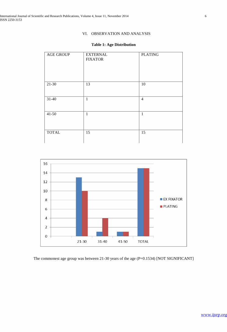
VI. OBSERVATION AND ANALYSIS
Table 1: Age Distribution
The commonest age group was between 21-30 years of the age (P=0.1534) [NOT SIGNIFICANT]
AGE GROUP EXTERNAL
FIXATOR
PLATING
21-30 13 10
31-40 1 4
41-50 1 1
TOTAL 15 15

International Journal of Scientific and Research Publications, Volume 4, Issue 11, November 2014 7
ISSN 2250-3153
www.ijsrp.org
Table 2: Sex distribution
SEX EX FIXATOR PLATTING
MALE 9 8
FEMALE 6 7
TOTAL 15 15
Male patient are affected more as compare to the women’s patients (P=0.7125) [NOT SIGNIFICANT]
Table 3: Mode of injury
MODE OF INJURY EXTERNAL FIXATOR PLATING
RTA 11 7
FALL 2 5
FALL FROM HEIGHT 2 3
TOTAL 15 15

International Journal of Scientific and Research Publications, Volume 4, Issue 11, November 2014 8
ISSN 2250-3153
www.ijsrp.org
Road traffic accident was the most common mode of injury in both the type (P=0.3050) [NOT SIGNIFICANT]
Table 4: Side Affected
Right side is more common site of the injury in both the series (P=0.0986) [NOT SIGNIFICANT]
SIDE AFFECTED EX FIXATOR PLATTING
RIGHT 9 13
LEFT 6 2
TOTAL 15 15

International Journal of Scientific and Research Publications, Volume 4, Issue 11, November 2014 9
ISSN 2250-3153
www.ijsrp.org
Table 5: TYPE OF FRACTURE
TYPE OF
FRACTURE
EX FIXATOR PLATING
CLOSED 5 11
OPEN 10 4
TOTAL 15 15
More number of the open type fractures was noted in case of the external fixator (P=0.0281) [SIGNIFICANT]
Table 6: AO CLASSIFICATION
CLASSIFICATION EX FIXATOR PLATING
A2 1 4
A3 5 3
B2 3 2
B3 0 0
C2 3 4
C3 3 2
TOTAL 15 15

International Journal of Scientific and Research Publications, Volume 4, Issue 11, November 2014 10
ISSN 2250-3153
www.ijsrp.org
A3 was the commonest type of the fracture noted in the series (P=0.5845) [NOT SIGNIFICANT]
Table 7: RESULTS
Platting shows better results as compared to the external fixator (P=0.9579) [NOT SIGNIFICANT]
RESULTS EX FIXATOR PLATTING
EXCELLENT 4 5
GOOD 7 7
FAIR 3 2
POOR 1 1
TOTAL 15 15
International Journal of Scientific and Research Publications, Volume 4, Issue 11, November 2014 11
ISSN 2250-3153
www.ijsrp.org

International Journal of Scientific and Research Publications, Volume 4, Issue 11, November 2014 12
ISSN 2250-3153
www.ijsrp.org

International Journal of Scientific and Research Publications, Volume 4, Issue 11, November 2014 13
ISSN 2250-3153
www.ijsrp.org

International Journal of Scientific and Research Publications, Volume 4, Issue 11, November 2014 14
ISSN 2250-3153
www.ijsrp.org
International Journal of Scientific and Research Publications, Volume 4, Issue 11, November 2014 15
ISSN 2250-3153
www.ijsrp.org
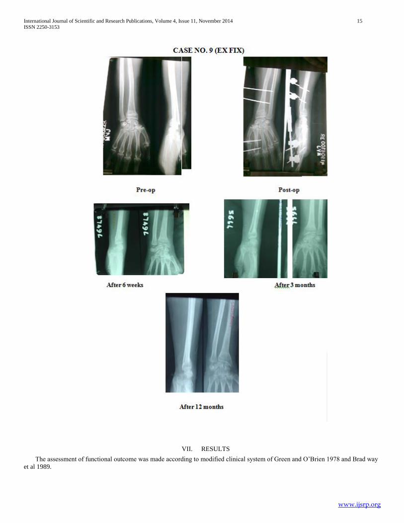
VII. RESULTS
The assessment of functional outcome was made according to modified clinical system of Green and O’Brien 1978 and Brad way
et al 1989.

International Journal of Scientific and Research Publications, Volume 4, Issue 11, November 2014 16
ISSN 2250-3153
www.ijsrp.org
This modified score includes independent scores for motion, strength, pain, and activity level, which can be objectively graded as
per the table below. To achieve an excellent result full range motion of wrist and forearm, strength, function of hand and comfort must
be present.
Table 8: The Modified Green- O’Brein clinical scoring system.
Category Score Findings
Pain 25 None
20 Mild
15 Moderate (medication required)
00 Severe (requires narcotics)
Function 25 same job
20 Different job
15 Able, no job
00 Unable
Motion 25 100%
15 75-99%
10 50-74% of normal side
5 25-49%
00 0-24%
Strength 25 100%
15 75-99%
10 50-74% of normal side
5 25-49%
00 0-24%

International Journal of Scientific and Research Publications, Volume 4, Issue 11, November 2014 17
ISSN 2250-3153
www.ijsrp.org
Scoring : Excellent : 90-100%
Good : 80-89%
Fair : 65-79%
Poor : <65%
Overall results were graded as acceptable (excellent and good), fair or poor. In our study, six patients had full range of
movements, no pain, returned to previous job and had 100% strength to that of normal side and the results were considered as
excellent. Another three patients had full range of movements as compared to that of opposite side with mild pain not affecting the
function of wrist. They also scored more than 90% and the results were considered as excellent. There was 9 case of Excellent, 14
cases of Good, 5 cases of Fair and 2 cases showed poor Result.
Excellent : 9 [5- Plating & 4- Ext Fixator] cases (30.2%)
Good : 14 [7- Plating & 7- Ext Fixator] cases (46.66%)
Fair : 5 [2- Plating & 3- Ext Fixator] cases (16.66%)
Poor : 2 [1- Plating & 1- Ext Fixator] cases ( 6.66%)
VIII. CONCLUSION
From the present study on 30 patients with distal end radius
fractures after analyzing the observation and looking at the
results we conclude the following.
Fracture of distal end of radius is having a bimodal age
distribution in our study we find young individual between 21-
30yrs mostly male sustain road traffic accident as a common
mode of injury. Right side was affected more than the left side.
Restoration of anatomy of distal end of radius, early mobilization
and less complication were achieved using Platting
Platting is a better method of treatment for fracture of the
distal end of radius.
REFERENCES
[1] K.S. Leung, W. Y. Shen. Ligamentotaxis and bone grafting for communitted fracture of distal radius. JBJS (Br), 1989; 71-B: 838-842.
[2] Vidal, J.; Buscacyret, C.; Paran, M. and Mulka, J. Ligamentotaxis. In Mears, D. C., ed. External skeletal Fixation. Baltimore: Williams & Wilkins, 493-96, 1983.
[3] WP Cooney, RL Linscheid. External pin fixation for colle’s fracture. JBJS
(Am), 1979; 61: 840-845.
[4] Vaughan PA., Lui SM., Harringtom JJ., Maistrelli GL. “Treatment of unstable fractures of distal radius by external fixation”. JBJS, 1985; 67-B: 385.
[5] Foster DE. Kopta JA. “Update on the external fixators in the treatment of
wrist fractures”. Clin. Orthop. 1986; 204:177.
[6] Szabo RM.,Weber. SC. “Comminuted Intra-articular fractures of the distal
radius”. Clin. Orthop. May 1988; 230: 39-47.
[7] Edward GS. Intra articular fractures of the distal radius treated with the small AO external fixator. J Bone Joint Surg [Am] 1991; 73 (A) : 1241-50.
[8] Jakim I., Pieterse., Sweet MBE. “External fixation for intra-articular fractures of the distal radius”. JBJS. 73-B: March 1991; 302-306.
[9] David S. Ruch, Anastasios Papadonikolakis. Compared the functional and
radiographic outcomes of volar and dorsal plating of intra-articular distal radius fractures. J Hand Surg 2006; 31A:9–16.
AUTHORS
First Author – Dr Bhuvnesh Chaturvedi
Second Author – Dr Sk Irfan Ali
Third Author – Dr Sumit Krishna