molecular and phenotypic characterization of endophytic
TRANSCRIPT
Molecular and phenotypic
characterization of endophytic Sebacinoid
strains
Dissertation zur Erlangung des Doktorgrades (Dr. rer. nat.)
der Naturwissenschaftlichen Fachbereiche der Justus–Liebig–Universität Gießen
durchgeführt am Institut für Phytopathologie und Angewandte Zoologie
vorgelegt von M.Sc. Magdalena Basiewicz
aus Polen
Gießen 2010
Dekan: Prof. Dr Volkmar Wolters 1. Gutachter: Prof. Dr. Karl–Heinz Kogel 2. Gutachter: Prof. Dr. Gabriele Klug


For my Parents and Sister
Dla moich Rodzicow i Siostry

I
Parts of this work have already been published:
Zuccaro, A.*, Basiewicz, M.*, Zurawska, M., Biedenkopf, D., and Kogel K.–H. 2009
Karyotype analysis, genome organization, and stable genetic transformation of the root
colonizing fungus Piriformospora indica Fungal Genetics and Biology 46, 8, 543–550
* These authors contributed equally to this work.
Papers in preparation
Basiewicz, M., Weiss, M., Kogel K.–H., Zuccaro, A. Molecular and phenotypic
characterization of Sebacina vermifera strains associated with orchids and the description
of Piriformospora glomeralium sp. nov.

II
III
Index
1. Introduction 1
1.1 Rhizosphere 1
1.2 Endophyte 1
1.3 Sebacinales 2
1.4 Piriformospora indica 3
1.5 Genome size estimation and sequencing 4
1.6 Translation elongation factor 1 alpha (TEF) and glycerol–3–
phosphate dehydrogenase (GAPDH) 6
1.7 Extracellular enzymes secreted by fungi 8
1.7.1 Cellulase 9
1.7.2 Pectinolitic enzymes 9
1.7.3 Laccase 9
1.7.4 Peroxidase 10
1.7.5 Esterase 10
1.7.6 Lipase 11
1.7.7 Proteinase 11
1.8 Objectives 11
2. Materials and methods 13
2.1 Fungal and plant material 13
2.2 Microscope analysis 16
2.3 Translation elongation factor1–α gene analysis for Sebacinales
isolates and environmental samples 17
2.4 DNA extraction 17
2.5 Southern blot analysis 18
2.6 Genome estimation 20
2.6.1 Real–time PCR 20
2.6.2 Pulsed Field Gel Electrophoresis 23
2.7 Plate enzymatic assays 24
IV
2.7.1 Cellulase activity 24
2.7.2 Pectinase activity 24
2.7.3 Laccase activity 25
2.7.4 Peroxidase activity 25
2.7.5 Protease activity 25
2.8 Spectrophotometric enzymatic assay 26
2.8.1 Laccase activity 26
2.8.2 Peroxidase activity 27
2.8.3 Esterase activity 27
2.8.4 Lipase activity 28
2.8.5 Determination of total protein content 29
2.9 P. indica protoplasts regeneration 29
3. Results 30
3.1 Analysis of translation elongation factor 1 alpha gene 30
3.2 Southern blot analysis 31
3.3 Genome estimation 33
3.4 Enzyme activity–plate’s tests 37
3.5 Spectrophotemetric test of Piriformospora indica 40
3.6 Piriformospora glomeralium sp. nov. Zuccaro Weiss
ex multinucleate rhizoctonia 45
3.7 Protoplast regeneration 46
4. Discussion 47
4.1 Sebacinales genome sizes estimation 47
4.2 P. indica protoplast regeneration 52
4.3 Biochemical analysis of Sebacinales 53
5. Summary / Zusammenfassung 59
6. References 62
Introduction
1
1. Introduction
1.1 Rhizosphere
Rhizosphere is the zone around plant’s root where the most intensive interactions between
plant host and bacterial or fungal partners take place. Many fungal interaction are parasitic
and can lead to diseases, the other ones are mutualistic symbioses which are beneficial to
host plants. The results of microbial activity in the rhizosphere are changes in root patterns
and nutrients availability to plants. Direct reactions between members of different
microbial types often affect promotion of key processes assisting host’s growth and health.
All interactions occurring around plant roots are, at least indirectly, mediated by plant.
Many naturally occurring rhizospheric bacteria and fungi are antagonistic toward
pathogens (Kiely et al. 2006). They compete for colonization or infection sites as well as
carbon and nitrogen sources. Moreover, pathogens can be inhibited by antimicrobial
substances, such as antibiotics, secreted by rhizospheric organism. Additional, indirect
mechanisms improve plant nutrition, modify root anatomy, and lead to changes in
microbial community in the rhizosphere, and activation of plant defence mechanisms
(Whipps 2001, Barea et al. 2005).
1.2 Endophyte
The fungi associated with plants are highly diverse, some of them are endophytes. The
term fungal endophyte defines a fungus of which at least a significant part of its life cycle
resides in a plant, and which colonizes tissues without causing symptoms of disease.
Endophytes from rhizosphere can be easily distinguished from mycorrhizae by lacking
external hyphal networks and mantels. Fungal endophytes can grow inter– and intra–
cellulary as well as endo– and epi–phytically (Schulz and Boyle 2005). They are not
restricted to one environment but were detected in various surroundings including those
with extreme characteristic (Zhang et al. 2001).
Endophytic fungal communities adapt to different physiological conditions, in
consequence they were detected in the wide spectrum of plant tissue types. Many neutral
fungal endophytes are asleep pathogens which may be activated and cause infectious
symptoms when the host plant is aged and/or stressed. In addition, plant’s endophitic
association with fungus can influence environment by determination of plant and microbial
biodiversity (Clay and Holah 1999).
Introduction
2
The endophytic microbial communities play an essential role in the physiology of host
plants. Host, colonized by endophyte, often have more vigour due to secretion of plant
growth–promoting substances such as indole–3–acetic acid (Ek et al. 1983, Robinson et al.
1998) or cytokines (Crafts and Miller 1974), and improvement of the hosts’ absorption of
nutritional nitrogen (Lyons et al. 1990) and phosphorus (Gasoni and Stegman de Gurfinkel
1997; Malinowski et al. 1999). Additionally, the endophyte partner can extensively
enhance plants resistance to biotic and abiotic challenges (Latch 1993). These beneficial
features have been observed in infected plants exposed to several abiotic stress such as
drought (Cheplick et al. 2000), heavy metals (Monneta et al. 2001), culture medium pH
lower than optimal (Lewis 2004), high salinity (Waller et al. 2005) as well as a biotic one
including microbial infections (Lewis 2003, Rodriguez et al. 2004, Waller et al. 2005),
insect pests (Breen 1994, Vázquez de Aldana et al. 2004) and herbivores attack (Schardl
and Phillips 1997, Mandyam and Jumpponen 2005).
1.3 Sebacinales
Sebacinales belong to a taxonomically, ecologically, and physiologically diverse group of
fungi in the Basidiomycota. They have been identified worldwide and form a broad
spectrum of mycorrhizal types. This unique phenomenon significantly influence natural
ecosystems (Weiss et al. 2004, Selosse et al. 2007). Ectomycorrhiza, orchid, ericoid,
jungermannioid and cavendishoid mycorrhiza are formed by Sebacinales. Ectomycorrhiza
(ECM) is an association where the fungus forms a hyphal mantle or layer around and
enters into roots and grows only between cortical cells forming a Hartig net (Agrios 2005,
Glen et al. 2002, Selosse et al. 2002). Fungi that colonize members of the Orchid family
belong to the orchid mycorrhiza type. Orchid’s protocorm cells are penetrated by fungal
hyphae during the saprotrophic stage. In consequence, seedlings can continue their
development (ed. Trigiano 2003). Ericoid mycorrhiza is formed between fungi, and species
of the Ericaceae and Epacridaceae. Plants from these families have very fine root systems.
Fungal hyphae pass through the cortical cells. In the later stadium plant cells are packed
with intracellular hyphal coils (Schmid et al. 1995). Recently Kottke et al. (2003) proved
that Sebacinales create symbiotic association with leafy liverworts of the subclass
Jungermanniidae. Although the liverworts do not form roots, they proposed the name
‘jungermannioid mycorrhiza’. During mycorrhiza growth, fungal hyphae formed coils in
the stem cells. In contrast to jungermannioid mycorrhiza build by Ascomycetes no or very
few ingrowths pegs were found. Cavendishoid mycorrhiza seems to be similar to ericoid
Introduction
3
mycorrhizas because of the presence of coils in roots, an irregular mantle and weak hyphal
growth between epidermal cells (Setaro et al. 2006).
Ultrastructural and microscopical characteristic placed Sebacinales within the wood–decay
fungi from the order Auriculariales (Bandoni 1984). However, molecular phylogenetic
analysis change Sebacinales taxonimic position (Weiss et al. 2001). Exidioid basidia
without clamp connections throughout the fructifications and thickened walls of tramal
hyphae were detected for both Sbacinales and Auriculariales (Wells and Oberwinkler
1982). Moreover, phytlogenetic analyses based on nuclear sequence of the large ribosomal
subunit distinguish two subgroups A and B within that order which differ in their ecology
(Weiss et al. 2004). Orchid mycorrhizas and ectomycorrhizas belong to subgroup A. The
second subgroup is more diverse and contains ericoid, cavendishoid and jungermannioid
mycorrhiza, Sebacina vermifera isolates from autotrophic mycorrhiza, endophytic
Piriformospora indica and multinucleate rhizoctonia in the sense of Warcup (Weiss et al.
2004). S. vermifera complex is very absorbing group. They have been characterized as
growth promoters. Positive influence of those isolates on barley (Hordeum vulgare) was
demonstrated by Deshmukh et al. 2006. S. vermifera MAFF305830 were characterized as
the best growth promoter and confered the higher reduction of powdery mildew infection.
On the other hand, in similar experiments with switchgrass (Panicum virgatum L) the
longest shoots were produced by the plants inoculated with strain MAFF305828, and the
longest roots had plants colonized by the strain MAFF305830 (Ghimire et al. 2009). Those
two Sebacina vermifera isolates were also examined in order to verify fungal development
in the barley tissue. Tissue penetration patterns as well as hyphal structures observed
during the expansion of these isolates were similar to those created by P. indica. The only
differences were detected for the speed of fungal development in planta (Waller et al.
2008).
1.4 Piriformospora indica
Piriformospora indica belongs to the order Sebacinales and colonize roots of a broad
spectrum of mono– and dicotyledonous plants including Arabidopsis thaliana, barley,
wheat and tobacco (Sahay and Varma 1999, Varma et al. 1999, Waller et al. 2005, Serfling
et al. 2007). The fungus was discovered in the rhizosphere of the woody shrubs Prosopsis
juliflora and Zizyphus nummularia in the Indian Thar desert in 1997 (Varma et al. 1998).
Since then, P. indica scientific interest increased exponentially (38 papers published to
date, NCBI). Wide range of colonized species, including agronomically important plants,
Introduction
4
makes it a very promising organism in agriculture. In contrast to AMF, the ability of
creating symbiosis with Arabidopsis thaliana gives the opportunity for fast and effective
study of the molecular basis of fungal–plant interaction.
P. indica enhances growth and yield of plant hosts, protect them against biotic (resistance
to diseases) or abiotic stress (salt stress) (Rai et al. 2001, Barazani et al. 2005, Waller et al.
2006). The influences of P. indica on colonized plants mimic to a certain extent
physiological effects of arbuscular mycorrhizal fungi. Although P. indica is a root
endophyte, it confers resistance against leaf pathogens (Deshmukh et al. 2006). Similar to
AMF, the fungus is strictly limited to the cortex, where it develops intracellular coils that
are different from the arbuscules of AM fungi (Varma et al. 1999). However, by
comparison to AM fungi, P. indica does not induce plant marker genes known to be
involved in the arbuscular mycorrhiza formation as for example PT11 phosphate
transporter or a gene containing peptidoglycan binding LysM domain 1 (Gutjahr et al.
2008).
Microscopic investigation of barley plants inoculated by P. indica chlamydospores showed
fungus enters via root hairs. Germinating chlamydospores, closely attached to the
rhizodermal cell walls, penetrate the subepidermal cells through intercellular spaces in
within 12 to 24 hours, where they branch and continue to grow. Fungal hyphae extend their
growth in rhizodermal and cortical cells at later colonization stages. The fungus also
penetrates through the basal parts of root hair cells, in which bifurcated hyphae form
chlamydospores (Deshmukh et al. 2006).
Further analyses were performed in order to comprehend the response of barley roots to P.
indica colonization by transcriptional and metabolic profiling. The largest group of
differentially regulated genes revealed in that study was those involved in plant
defence/stress responses (Schäfer et al. 2009).
1.5 Genome estimation and sequencing
The genome comprises the total genetic information of the organism. The rapid
development of sequencing technologies within last few years makes these tools
commonly available and allows getting genetic information of whole organism very fast.
2487 genome sequencing projects are running (state October 2010), 827 of them being
completed (http://www.ncbi.nlm.nih.gov/genomes/static/gpstat.html). The genomic
information is essential for better understanding the biochemistry and molecular biology of
the analyzed organisms.
Introduction
5
The recognition of mechanisms of genetic variation in the pathogen, for instance, is
essential for developing effective control measures for the disease. Identification of factors
responsible for regulation of symbiotic processes (like host recognition and infection,
control of host defence reaction) will help to understand fungal role in plant development
and physiology. It allows also to study the ecological significance of symbioses and to
comprehend the responses of organisms to their natural environments. In addition, genes
involved in ecological adaptation can be clearly defined.
The genome size of ectomycorrhizal basidiomycete Laccaria bicolor is aprox. 65 Mb and
was the largest sequenced Basidiomycete genome (Martin et al. 2008). The availability of
this genome strongly contribute in deeper understanding the interaction between symbiont
and plants within their ecosystem, clarify also mechanisms which are used to obtained
carbon and nitrogen that are essential in plant production. L. bicolour genome analysis
revealed a large number of small secreted proteins of unknown function. Some of them
may play a role in initiating symbiosis because they are only expressed in symbiotic
tissues. Lack of plant cell walls degrading enzymes was observed in L. bicolour genome,
however, it possess enzymes which can degrade other polysaccharides, suggesting the
mechanisms used to grow both in soil and in association with plants (Martin and Selosse
2008). The Perigord black truffle Tuber melanosporum Vittad. (Ascomycota) is the largest
sequenced fungal genome (aprox. 125 Mb) published so far (Martin et al. 2010). The
investigations of T. melanosporum genome allow better understanding of the biology and
evolution of the ectomycorrhizal symbiosis as well as support identification of processes
that trigger fruit body formation. Beside L. bicolour, Coprinopsis cinerea (Basidiomycota),
a model organism for mushroom–forming, has also been sequenced (37 Mb) to examine
multicellular development in fungi. Studies on this fungus based on DNA–mediated
transformation and RNAi silencing have provided important knowledge on the regulation
of mushroom fruiting, mating pheromone, and receptor signalling pathways (Stajich et al.
2010). The genome of arbuscular mycorrhizal fungus (AMF) is also analyzed. The first
information about global organization of the Glomus intraradices genome was in 2004.
Hijri and Sanders (2004) predicted G. intraradices genome size 14.07 ± 3.52 Mb. Since
that time complete annotation and assembling is not finished. Only annotation of the
mitochondrial genome (70 608 bp) is completed (Martin et al. 2008, Glomus Genome
Consortium (GGC) Symposium). AMF are unique obligate symbionts. Their hyphae are
coenocytic and multinucleate therefore organelles and nutrients can be transported over
Introduction
6
long distances. Moreover, it has been shown that AMF harbour genetically different nuclei
(Kuhn et al. 2001), making further analysis more complicated.
The information about genome size can provide clues to evolutionary relationship. The
new genomic data can give more insights in the genetic background of analyzed fungi and
allow investigating in details closely related organisms. Genus Filobasidiella for example,
contains approximately 38 Cryptococcus species. Two of them: Cryptococcus neoformans
and Cryptococcus bacillisporus are the casual agents of the majority of human and animal
disease. The Cryptococcus bacillisporus genome is approximately 20 Mb, and it is
organized in 14 chromosomes. The same number of chromosomes but smaller genome
approx. 19 Mb has the C. neoformans (Loftus et al. 2005). The haploid genome of the
other Basidiomycetes pathogenic fungus Puccinia graminis, which causes stem rust in
small cereal crops such as wheat, oat, rye, and barley is estimated at 80 Mb, organized in
18 chromosomes. The genome of Puccinia triticina, the causal agent of leaf rust in wheat
is estimated to range from 100–124 Mb.
Fungal genomes vary a lot in sizes. Puccinia triticina has the biggest genome size between
Basidiomycetes described till now (NCBI ENTREZ genome project). On the other hand,
Malassezia globosa, lipid–dependent yeast belonging to normal human microflora, has the
smallest genome, approximately 9 Mb (Xu et al. 2007). Some pneumonia agents
Pneumocystis carinii, Pneumocystis carinii f. sp. hominis, and Pneumocystis carinii f. sp.
muris, members of Ascomycetes, have even smaller genomes 6.5–8.4 Mb (Sesterhenn et
al. 2009).
Before a sequencing project of whole genome will start, its size should be estimated in
order to deliver important information for proper preparation and costs prediction. There
are few techniques available which can be used for fungal genome estimation such as: flow
cytometry, reassociation kinetics, genomic reconstruction, pulsed field gel electrophoresis
(PFGE), real–time PCR, and confocal microscope. Usually results from at least two of
them are combined to ensure that prediction is accurate.
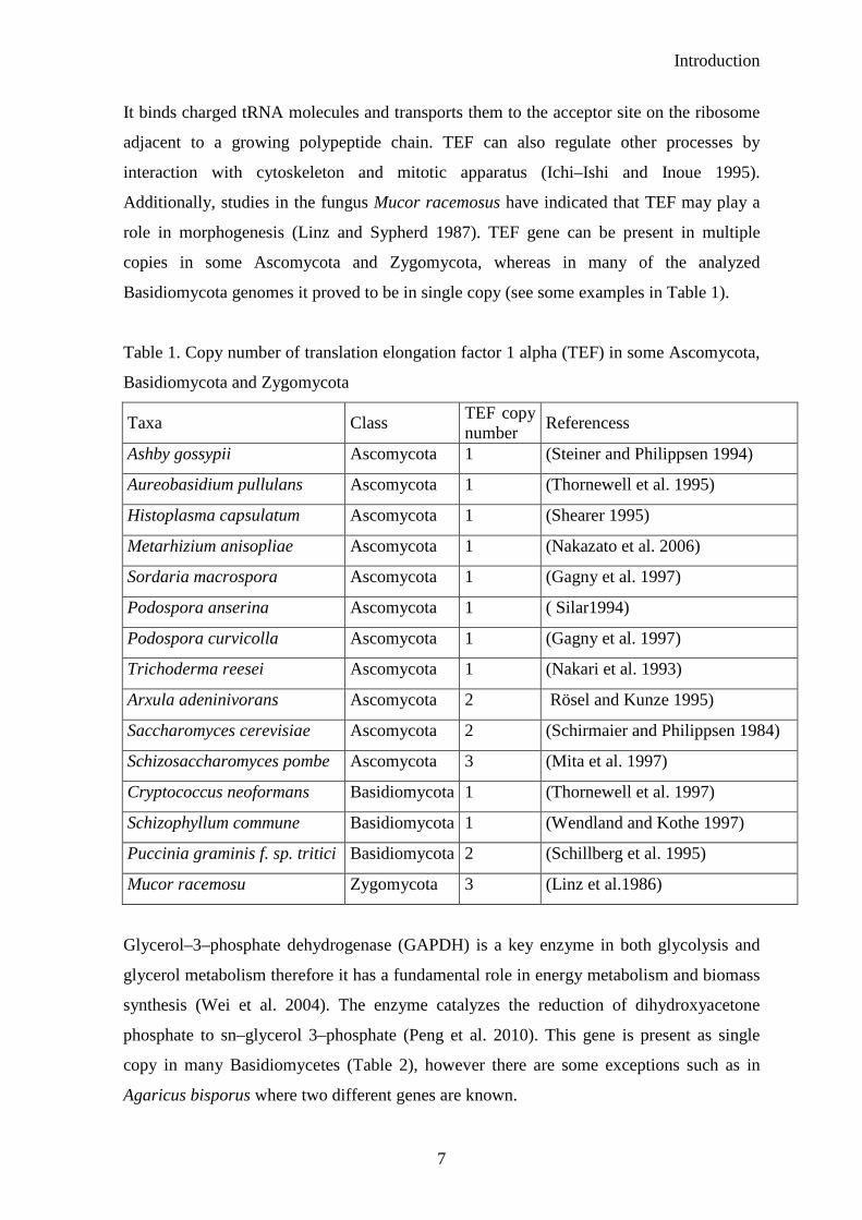
1.6 Translation elongation factor 1 alpha (TEF) and glycerol–3–phosphate dehydrogenase
(GAPDH)
Translation elongation factor 1 alpha (TEF) gene encode an abundant and highly conserved
protein which plays an important role in the elongation cycle of protein synthesis in
eukaryotic cells (Merrick 1992). In eukaryotes, TEF is the second most profuse protein
after actin, combining 1–2 % of the total protein in normal growing cells (Condeelis 1995).
Introduction
7
It binds charged tRNA molecules and transports them to the acceptor site on the ribosome
adjacent to a growing polypeptide chain. TEF can also regulate other processes by
interaction with cytoskeleton and mitotic apparatus (Ichi–Ishi and Inoue 1995).
Additionally, studies in the fungus Mucor racemosus have indicated that TEF may play a
role in morphogenesis (Linz and Sypherd 1987). TEF gene can be present in multiple
copies in some Ascomycota and Zygomycota, whereas in many of the analyzed
Basidiomycota genomes it proved to be in single copy (see some examples in Table 1).
Table 1. Copy number of translation elongation factor 1 alpha (TEF) in some Ascomycota,
Basidiomycota and Zygomycota
Taxa Class TEF copy number
Referencess
Ashby gossypii Ascomycota 1 (Steiner and Philippsen 1994)
Aureobasidium pullulans Ascomycota 1 (Thornewell et al. 1995)
Histoplasma capsulatum Ascomycota 1 (Shearer 1995)
Metarhizium anisopliae Ascomycota 1 (Nakazato et al. 2006)
Sordaria macrospora Ascomycota 1 (Gagny et al. 1997)
Podospora anserina Ascomycota 1 ( Silar1994)
Podospora curvicolla Ascomycota 1 (Gagny et al. 1997)
Trichoderma reesei Ascomycota 1 (Nakari et al. 1993)
Arxula adeninivorans Ascomycota 2 Rösel and Kunze 1995)
Saccharomyces cerevisiae Ascomycota 2 (Schirmaier and Philippsen 1984)
Schizosaccharomyces pombe Ascomycota 3 (Mita et al. 1997)
Cryptococcus neoformans Basidiomycota 1 (Thornewell et al. 1997)
Schizophyllum commune Basidiomycota 1 (Wendland and Kothe 1997)
Puccinia graminis f. sp. tritici Basidiomycota 2 (Schillberg et al. 1995)
Mucor racemosu Zygomycota 3 (Linz et al.1986)
Glycerol–3–phosphate dehydrogenase (GAPDH) is a key enzyme in both glycolysis and
glycerol metabolism therefore it has a fundamental role in energy metabolism and biomass
synthesis (Wei et al. 2004). The enzyme catalyzes the reduction of dihydroxyacetone
phosphate to sn–glycerol 3–phosphate (Peng et al. 2010). This gene is present as single
copy in many Basidiomycetes (Table 2), however there are some exceptions such as in
Agaricus bisporus where two different genes are known.

Introduction
8
Table 2. Copy number of glycerol–3–phosphate dehydrogenase (GAPDH)) in some
Ascomycota, Basidiomycota and Zygomycota
Taxa Class GAPDH copy number
Referencess
Aspergillus nidulans Ascomycota 1 (Punt et al. 1988)
Beauveria bassiana Ascomycota 1 (Liao et al. 2008)
Saccharomyces cerevisiae Ascomycota 1 (Sprague and Cronan 1977)
Flammulina velutipes Basidiomycota 1 (Kuo et al. 2004)
Lentinus edodes Basidiomycota 1 (Hirano et al. 1999)
Phanerochaete chrysosporium
Basidiomycota 1 (Harmsen et al. 1992)
Schizophyllum commune Basidiomycota 1 (Harmsen et al. 1992)
Pseudozyma flocculosa Basidiomycota 1 (Neveu et al. 2007)
Agaricus bisporus Basidiomycota 2 (Harmsen et al. 1992)
Mucor racemosu Zygomycota 3 (Wolff and Arnau 2001)
1.7 Extracellular enzymes secreted by fungi
The penetration of the external plant layers is an essential task for successful colonization
of the host tissues by endophytic fungi. This effect can be obtained by either mechanical
fracture of the protective tissues or by enzymatic digestion. In plant pathogens both
mechanical and enzymatic components of the penetration mechanism have been at least
partly demonstrated (Kolattukudy 1985, Howard et al. 1991). Based on the lifestyle and
genome size of the fungus Idnurm and Howlett (2001) estimated that plant pathogenic
fungi genomes consist 60–360 virulence or pathogenicity genes. Some of them are
involved in the infection structure formation, synthesis of toxins or cell wall-degrading
enzymes (Madrid et al. 2003, Möbius and Hertweck 2009, Werner et al. 2007). Other
genes are important during establishment of a compatible pathogenic interaction.
Endophytes occupy the same ecological niche as most pathogens, therefore, it can be
assumed that they utilize the same strategy employed by pathogens for the penetration of
the host tissues (Petrini et al. 1992). At the beginning of colonization process, endophytic
fungi have to achieve at least partial degradation of cell wall. Extracellular enzymes,
proteins that catalyze different types of chemical reactions, might be one of the main tools
in that process. Those proteins can be divided into six main groups: oxidoreductases,
lyases, hydrolases, transferases, ligases and isomerases (http://www.brenda–enzymes.org/,
Introduction
9
Chang et al. 2009). Fungal cellulases and pectinases can be very active while plant cell
wall degradation. As a response to intracellular plant protection mechanisms fungal
endophytes secrete supplementary enzymes such as esterase, laccase, peroxidase and
proteinase (Burke and Cairney 2002, Ramstedt and Soderhall 1983).
1.7.1 Cellulase
Cellulase belongs to hydrolases and plays important role in digestion of two major
components of plant cell walls–cellulose and hemicellulose. Sequence analysis and
biochemical characterization of cellulase genes have shown that many of them are
multifunctional proteins. They are composed of distinct domains arranged in several
combinations. Many cellulase–degrading organisms secrete several enzymes that act
synergistically (Sandgren et al. 2001). Furthermore, they have evolved a battery of
enzymes having different specificities with respect to endo/exo mode of action (Beguin
and Aubert 1994).
1.7.2 Pectinolitic enzymes
Pectin is a complex of polysaccharides present in most primary cell walls which bind cells
together by forming gel–like matrix (Wozny 2000). Fungi secrete a various number of
enzymes to digest pectin which operates through different degradation pathways such as
deesterification, hydrolysation or depolymerization. This huge range of activities suggests
the great fungal adaptation to host tissues. Pectinases can also play a role during the
establishment of ectomycorrhizal symbiosis. However, the level of enzyme production is
not very high (Garcia–Romera et al. 1991, Ramstedt and Soderhall 1983). Despite that,
plants produce polygalacturonase–inhibiting proteins (PGIPs) which reduce aggressive
potential of pectinases and limit fungal invasion. Additionally, the host plant can influence
fungal enzyme production by pectin content in cell wall. It has been demonstrated that
pectin content level is higher in Dicots than in Monocots (Jarvis et al. 1988).
1.7.3 Laccase
Laccase is a blue copper protein which catalyses the reduction of O2 to H2O using a
number of phenolic compounds as hydrogen donors (Thurston 1994). Laccase contributes
to lignin degradation by oxidising free phenolic groups to phenoxy cation radicals as well
as non–phenolic lignin model compounds. This enzyme is associated with morphogenesis
in some Basidiomycota and Ascomycota strains (Das et al. 1997, Worrell et al. 1986,

Introduction
10
Rehman and Thurston 1992). Additionally, it is involved in physiological processes related
to pathogenesis like melanin synthesis essential for survival and longevity of fungal
propagules (Bell and Wheeler 1986, Edens et al. 1999). The enzyme has been also detected
in zones of mycelial contact between competing basidiomycetes (White and Boddy 1992,
Iakovlev and Stenlid 2000). Subsequently, it has been suggested that laccase is involved in
detoxification of phenols (Haars and Huttermann 1981) and protection against host
oxidative responses (Edens et al. 1999). Many fungi secrete multiple laccase isozymes,
encoded by differentially expressed genes that may fulfil different functions. Coprinopsis
cinerea has two subfamilies of laccases with 15 and 2 nonallelic members, respectively
(Kilaru et al. 2006). Five laccase genes have been identified in Trametes villosa (Yaver et
al. 1996). P. indica enzyme activity in axenic culture was demonstrated using laccase
specific antibody LccCbr2 (Kellner et al. 2007)
1.7.4 Peroxidase
Peroxidases are enzymes extremely widespread and diversified, present in almost all living
organisms. They play crucial role in lignin degradation. Fungi secrete two main
peroxidases: lignin peroxidase (LiP) and manganese peroxidase (MnP). They are heme–
containing glycoproteins which require hydrogen peroxide as an oxidant and they can be
secreted in several isoenzymes form into the cultivation medium (Hatakka 1994). On the
other hand plants are also able to exude peroxidases. Class III of plant peroxidases is
described as group of enzymes involved in a broad range of physiological processes,
including plant defence (Passardi et al. 2005, Almagro et al. 2009, Gonzalez et al. 2010).
1.7.5 Esterase
Esterases are enzymes which hydrolyze esters present in biological material of all kinds of
organisms. A wide spectrum of esterases exists with different substrate specificity, protein
structure, and biological function, therefore it can be assumed that they have evolved to
enable access to carbon sources or to be involved in catabolic pathways (Machado and
Castro–Prado 2001, Bornscheuer et al. 2002). Those enzymes do not hydrolyze long–chain
fatty acid esters and prefer water–soluble substrates (Bornscheuer et al. 2002). Esterase
isozyme patterns can be used for taxonomic purposes in plant–fungal interactions, and,
because of their common expression in varius mycorrhizal fungi, they are also good
indicators of changes in fungal activity (Sen 1990, Timonen and Sen 1998). Additionally,
Introduction
11
esterase indicates catabolic activity in soil, which directly correlates with microbial activity
(Vazquez et al. 2000).
1.7.6 Lipase
Lipases are esterases which can hydrolyse long–chain tri–aclyglycerides. Lipases can be
distinguished from esterases by the phenomenon of interfacial activation–high catalytic
activity which is observed only in the presence of a hydrophobic phase, a lipid droplet
dispersed in water or an organic solvent. This situation is associated to the presence of a
hydrophobic oligopeptide protecting the entrance to the active site. In a hydrophobic
environment, the lid moves aside and the substrate can enter the binding pocket
(Bornscheuer et al. 2002). The enzyme can be secreted by filamentous fungi, however the
production depend on the strain, the composition of the growth medium (carbon and
nitrogen sources, pH) and cultivation conditions (temperature, agitation and dissolved
oxygen concentration). The enzyme is heat resistant, and plays an important role in the
breakdown and mobilization of lipids within the cells of an individual as well as transfer of
lipids from one organism to another (Shukla and Gupta 2007).
1.7.7 Proteinase
Proteinase belongs to a big family of proteolytic enzymes important in the metabolism of
all organisms. The main plant cell component such as cellulose and other carbohydrate
polymers are held together by protein linkages therefore proteolytic enzymes may also
have a role in fungal invasion of the plant host (Sreedhar et al. 1999). Extracellular
proteinase from ericoid mycorrhizal endophytes can degrade complex organic substrates
and provide its host plants nitrogen normally unavailable to them (Leake and Read 1989).
External pH regulates both activity and production of fungal proteinases (Leake and Read
1990).
1.8 Objectives
The main aim of my thesis was molecular and phenotypic characterization of seven strains
belonging to the order Sebacinales. Generally, Sebacinales have been worldwide identified
and comprehend a wide spectrum of lifestyles. Nonetheless, only few isolates are cultured
by now. The study encompass root endophyte Piriformospora indica, Australian orchid
mycorrhizae Sebacina vermifera strains and orchidaceous rhizoctonia isolate from pot
cultures (multinucleate rhizoctonia DAR29830) which were described as plant growth
Introduction
12
promoters and resistance inducer for abiotic and biotic stress. In order to better understand
the relationship between Sebacinales isolates and to provide a novel genetic marker for
molecular environmental analysis we investigated phylogenetic connection among
Sebacina vermifera isolates, multinucleate rhizoctonia DAR29830, Piriformospora indica
and three environmental samples from south Germany. Moreover, the closest related
fungus to P. indica isolated by Williams in the 1984 from a spore of Glomus fasiculatum
but never classified taxonomically known as multinucleate rhizoctonia was described as a
new species and named as Piriformospora glomeralium.
In order to elucidate the molecular processes and identify the fungal factors that lead to a
successful symbiosis of P. indica and other Sebacinales with its plant partners as well as
for better understanding the mechanism of the symbiosis, the genome size of mentioned
fungi was estimated. First, the techniques such as Pulsed Field Gel Electrophoresis (PFGE)
and real–time PCR was establish for Piriformospora indica genome size estimation and
further applied for the genome size determination for other fungi belonging to the order
Sebacinales. Real–time PCR method relies on absolute quantification a one copy gene in
genomic DNA sample. Therefore TEF gene (translation elongation factor 1 alpha) was
confirmed to fulfil those conditions in all Sebacinales isolates. Furthermore, to affirm the
accuracy of this approach the second gene–GAPDH (glycerol–3–phosphate
dehydrogenase) was used as well. In addition, Saccharomyces cerevisiae was used for
validation of the method. Southern blot analysis was performed to prove the copy number
of GAPDH in P. indica genome. Moreover, a procedure for fungi protoplast preparation
was developed and the best conditions for its regeneration were evaluated.
Sebacinales are successful in plant root colonization, therefore, they must secrete
substances which allow them to enter into the plant organ. Extracellular enzyme can play
an important role in that process, consequently, the profile of enzymes excreted by
Sebacinoid strains was characterised. The special emphasis was put on P. indica.

Materials and Methods
13
2 Materials and Methods
2.1 Fungal and plant material
Piriformospora indica DSM11827 isolates were obtained from Deutsche Sammlung von
Mikroorganismen und Zellkulturen, Braunschweig, Germany. Six Sebacina vermifera
strains (Table 3.) were obtained from the National Institute of Agrobiological Sciences
(Tsukuba, Japan), multinucleate rhizoctonia DAR29830 was kindly provided by Karl–
Heinz Rexer (University of Marburg, Marburg, Germany). Rhizoctonia solani AG8 was
supplied by Timothy Paulitz from Washington State University, USA. The haploid
Saccharomyces cerevisiae genotype BY4741, MATa (ACC. No. Y02321) and the diploid
S. cerevisiae genotype FY1679, MATa/MATa (ACC. No. 10000D) were received from
Euroscarf, Frankfurt, Germany. S. vermifera MAFF305837 and S. vermifera MAFF305835
were propagated on solid or liquid Malt–Yeast–Extract–Pepton medium (MYP) and all
other Sebacinales isolates as well as R. solani on Complete Medium (CM, Pham et al.,
2004), whereas both S. cerevisiae strains were grown on Yeast–Extract–Peptone–
Dextrose–Adenine medium (YPAD) (Guthrie and Fink 2002). All fungi strains were
grown at 24 °C in liquid cultures by shaking t 120 rpm speed.
Table 3. Sebacinales isolates
Fungus isolate Host name
P. indica DSM11827 Prosopis juliflora and Zizyphus nummularia (woody shrubs)
S. vermifera MAFF305830 Crytostylis reniformis (Orchid)
S. vermifera MAFF305842 Microtis uniflora (Orchid)
Piriformospora glomeralium ( ex multinucleate rhizoctonia DAR29830)
Trifolium subterraneum
S. vermifera MAFF305828 Eriochilus cucullatus (Orchid)
S. vermifera MAFF305837 Caladenia dilatata (Orchid)
S. vermifera MAFF305835 Caladenia catenata (Orchid)
S. vermifera MAFF305838 Caladenia tesselata (Orchid)
Materials and Methods
14
CM medium MYP Medium
20x salt solution 50 ml Malt–extract 7.0 g
Glucose 20 g Peptone (Soya) 1.0 g
Peptone 2 g Yeast extract 0.5 g
Yeast extract 1 g dest. water 1000 ml
Casamino acid 1 g autoclaved
Microelements 1 ml
Agar–agar 15 g
dest. water 950 ml
autoclaved
20x salt solution Microelements
NaNO3 120 g MnCl2 x 4H2O 6.00 g
KCl 10.4 g H3BO3 1.50 g
MgSO4 x 7H2O 10.4 g ZnSO4 x 7H2O 2.65 g
KH2PO4 430.4 g KI 0.75 g
dest. water 1000 ml Na2MoO4 x 2H2O 2.40 mg
CuSO4 x 5H2O 130 mg
dest. water 1000 ml
YPAD
Yeast extract 10 g
Peptone 20 g
Glucose 20 g
Adenine hemisulphate 100 mg
Agar–agar 15 g
dest. water 1000 ml
autoclaved
Environmental samples
Four independent environmental samples (Table 4) collected from two different areas in
Germany were analyzed. DNA samples were kindly provided by Michael Weiss from
Tübingen University and they belong to a poll of environmental collection encompassing

Materials and Methods
15
DNA isolated from root material. They were used in ITS – 28S rDNA phylogeny in Weiß
et al. 2010.
Table 4. Environmental isolates
DNA sample number
host plant
15 Lolium perenne 65 Medicago lupulina 80 Anthyllis vulneraria 41 Rumex acetosa
Barley (Hordeum vulgare L.) cultivar Golden Promise was obtained from the Leibniz
Institute of Plant Genetics and Crop Plant Research (IPK) in Gatersleben, Germany. Barley
seeds were surface–sterilized with 6 % sodium hypochloride, rinsed in water and
germinated for 2 days on sterile filter paper. Afterwards, seedlings were transferred into
the jars (5 seedlings/jar) and grown on liquid or solid modified plant nutrient medium (1/10
PNM) under 16h light (47 µmol m–2 s–1) at 24 °C. In order to check enzyme production
barley plants were inoculated with P. indica or Piriformospora glomeralium. Four–week
old fungal mycelia were crashed using a fine blender and applied as inoculum.
1/10 PNM Fe–EDTA
1M KNO3 0.5 ml FeSO4 x 7H2O 2.5 g
0.36M KH2PO4 1 ml Na2EDTA 3.36 g
0.14M K2HPO4 1 ml water 400 ml
1M MgSO4 x 7H2O 2 ml bring to boil
1M Ca(NO3)2 0.2 ml stir 30 min while cooling
Fe–EDTA 2.5 ml bring to final volume 450 ml
NaCl 1 ml
Gelrite 4 g
bring to final volume 1 l with water
pH 5.6; autoclaved For spectrophotometric enzymatic tests P. indica was grown on liquid 1/10 PNM with
shaking 120 rpm.
Materials and Methods
16
2.2 Microscope analysis
Microscopic analyses were performed in order to estimate P. indica genome size and to
measure multinucleate rhizoctonia structures. Syto 9 and propidium iodide (PI)
(LIVE/DEAD® Bac Light™ Bacterial Viability Kit Invitrogen) were applied in that study
for staining nuclei.
To determine the nuclear ploidy level of P. indica, chlamydospores were collected from 4–
week–old CM–agar plates with 0.002 % Tween water. Chlamydospores were washed 3
times with 0.002 % Tween water and resuspend in 0.9 % NaCl to the final concentration of
109 –1010 spores/ml. S. cerevisiae (1n and 2n) cells were collected by centrifugation from 4
to 5 days–old liquid culture. In order to remove the medium, they were washed three times
in 0.9 % NaCl and resuspended in the same buffer to the final concentration of 109 –1010
cells/ml. The same volume (approx. 250 µl) of P. indica spores and 1n or 2n S. cerevisiae
cells suspensions were mixed together and stained with 0.5 µl of Syto 9 and PI followed by
15 minutes incubation in darkness on ice. Afterwards, excess stain was removed by
washing 3 times with 0.9 % NaCl and resuspended in that buffer. The fungal material was
spread onto glass slides, covered with cover glass and analyzed under confocal laser
scanning microscope Leica TCS SP2 (Leica, Bensheim, Germany). Serial optical
sectioning images were taken (set manually, 0.10 µm steps) for both P. indica and S.
cerevisiae. Fluorescence of each section of the nucleus was measured using software
provided with microscope as follow: first the area of each analyzed nucleus was marked
and its fluorescence was automatically measured by software. This procedure was repeated
for each section image of analyzed nucleus. Further, the histogram values of fluorescence
intensity were summed up and used for genome estimation (Cano et al. 1998). S. cerevisiae
(1n and 2n) was used as standard organism. The histogram fluorescence value of S.
cerevisiae 2n is higher than the intencity of the haploidnucleus since fluorescence is
directly proportional to the amount of DNA present. Based on that assumption the genome
size of P. indica was estimated.
The diameter of spores as well as hyphal width, number of nuclei per cell and spore of
Piriformospora glomeralium (ex multinucleate rhizoctonia) were analyzed under
fluorescent microscope Axioplan 2 (Zeiss SMT, Oberkochen, Germany). P. glomeralium
spores were collected as described above for P. indica. The P. glomeralium hyphal
material was collected from 4–week–old liquid culture, washed few times with 0.9 % NaCl
and stained as described for P. indica spores.
Materials and Methods
17
2.3 Translation elongation factor1–α gene analysis for Sebacinales isolates and
environmental samples
DNA from environmental samples was amplified using the primer pair tef420f/tef420r
(Table 6.) with the AccuPrime™ Taq DNA Polymerase (Invitrogen) according to the
manufacturer’s instructions. PCR for Sebacinales isolates were performed using the primer
pairs EF1–983f/EF1–2212r, EF1–983f/EF1–1953r, EF1–983f/EF1–2218r (Table 6.). The
obtained PCR products were cloned using pGEM®–T Easy Vector Systems (Promega
GmbH, Mannheim, Germany) and sequenced in both directions with the M13f/r primers.
Two clones from each PCR were sequenced and further analyzed.
2.4 DNA extraction
DNA was extracted from four week old liquid Sebacinales cultures and two week old S.
cerevisiae culture using two different approaches.
Doyle & Doyle modified method followed by a CsCl centrifugation
200–300 mg frozen fungal mycelium were grinded in liquid nitrogen, and incubated in 700
µl pre–warmed to 65 ºC extraction buffer with β–mercaptoethanol for 20–30 minutes.
Next, material was washed using 700 µl chloroform/isoamylalkohol (24:1) and centrifuged
13000 rpm in 4 °C for 15 min. The washing step was repeated one more time. Afterwards
DNA was precipitated by adding 50 µl 10 M NH4OAc, 60 µl 3 M NaOAc (pH 5.5) and
500 µl isopropanol. To receive high concentration of DNA, precipitation took place over
night in 4 ºC. Subsequently, DNA was washed by 500 µl 70 % EtOH/10 mM NH4OAc.
After ethanol evaporatoin DNA was dissolved in TE buffer. Later CsCl– centrifugation
cleaning step was performed. 10 g of CsCl was mixed with 500 µl ethidium bromide
(EtBr) and 5 ml of DNA samples, further 5ml ultracentrifuge tube was fulfill with the
mixture and centrifuged at 56000 rpm, 20 ºC for 24 h in Beckman XL 70 centrifuge rotor
VTI 90. After centrifugation the red band DNA stained by EtBr was obtained. Genomic
DNA band was collected using the needle attached to the syringe. EtBr was removed from
DNA by repeated extraction using CsCl saturated 2–butanol. Later, DNA was precipitated
by 1/10 volume of 3 M NaOAc and 2 volume of 100 % EtOH and incubated –20 ºC at least
1 h. DNA pellet was washed by cold 70 % EtOH. When EtOH evaporated, DNA was
dissolved in TE or water.
Materials and Methods
18
Extraction buffer
1 M Tris–HCl 100 ml
0.5 M EDTA 40 ml
NaCl 81.82 g
CTAB 20 g
Na2S2O5 10 g
bring to final volume 1 l with water
autoclaved
before use add
ß–mercaptoethanol 2 ml
FastDNA® Spin Kit for soil (MP Biomedicals, LLC., Illkirch, France) according to the
manufacturer’s protocol.
2.5 Southern blot analysis
10 µg of genomic DNA was digested with 30 Units of restriction proper enzyme (Table 5.)
over night (or at least 10 h) *. Digested DNA was separated on 0.8 % TAE gel. The gel run
at 35 V in 4 ºC over night. After electrophoresis gel was stained with EtBr and
photographed. Later the gel was washed twice in 0.25 N HCl for 15 min, rinsed with
deionised water, and incubated for 15 min in solution T. Then, transferring apparatus was
assembled. After over night transfer, the membrane was left for drying for 2 h in RT and
later crosslink (2 x 50 s, 250 mJoule). Next membrane was washed 2 min in 2xSSC buffer
and prehybridized in prehybridization buffer containing carrier DNA over night in 65 ºC.
Following, the prehybridization buffer was replaced with hybridization buffer
encompassing specific, radioactive–labeled probe. Hybridization process took place at
least 12 h at 65 ºC. Subsequently, the membrane was washed twice with buffer I and buffer
II. After washing, membrane was saran wrapped, put to the Phosphor Imager box and
exposed for at least 3–4 h.
Table 5. Restriction enzymes applied for fungal, genomic DNA digestion.
organism restriction enzymes
P. indica DSM11827 Bam HI, Hind III, SacI
P. glomeralium Bam HI, Hind III
S. vermifera MAFF305842 Bam HI, Hind III
Materials and Methods
19
Solution T 10 x TAE
0.4 M NaOH 16 g/l Tris 48.4 g
0.6 M NaCl 35.06 g/l acetic acid (glacial) 11.4 ml
EDTA 2.92 g
20xSSC dest H2O 1 l
3 M NaCl pH 8.5
0.3 M Na–citrate; pH 7.0 Autoclaved
Prehybridization buffer 5xHSB
H2O 15 ml PIPES 30.3 g
5 x HSB 6 ml disolve in 300 ml dest H2O pH 6.8
Denhardts III 3 ml
10 % SDS 3 ml add
5 M NaCl 600 ml
mixed together and heat to 65 ºC 0.5 M EDTA 40 ml
rechecked pH
add 3 ml of freshly boiled carrier DNA adjust to 1 l with water
Autoclaved
carrier DNA Denhardts III
BSA (fraction V) 4 g
DNA sodium salt from Salmon Testes 125 mg SDS 20 g
dest. H2O 25 ml Ficoll–400 4 g
PVP–360 4 g
heat to boiling Na4P2O7 x10 H2O 10 g
store at –20 °C dissolve in 200 ml H2O
washing buffer I washing buffer II
(2x SSC / 1 % SDS) (1xSSC / 0.5 % SDS)
dest H2O 800 ml dest H2O 900 ml
20xSSC 100 ml 20xSSC 50 ml
10 % SDS 100 ml 10 % SDS 50 ml
Materials and Methods
20
Southern probe preparation
As probe was used 100 ng of DNA (PCR product specific for each analyzed fungus) in
final volume 25 µl (if it was necessary 1x TE was used as dissolvent). DNA was
denaturated in 95 ºC for 5 min, subsequently, cooled on ice for 5 min. Labelling beads
(Amersham Ready– To–Go DNA Labelling Beads [–32P] dCTP) was dissolved in 20 µl 1x
TE and mixed with denaturated DNA and 5 µl α–dCTP–32P and incubated 30–60 min in 37
ºC. Afterwards, the α–dCTP–32P which did not incorporate to the probe was cleaned by
Illustra microspin G–25 columns (Amersham). The column was vortexed very good, its tip
was broken and it was centrifuged for 1min in 735 rpm in 4 ºC. Supernatant was thrown
away and 50 µl of sample was loaded on the column and it was centrifuged for 2 min in
735 rpm in 4 ºC. Labelled probe was denaturated in 95 ºC for 5 min before use, nest kept 3
min on ice and mixed with pre–warmed hybridization buffer.
Hybridization buffer
H2O 7 ml
5 x HSB 3 ml
Denhardts III 1.5 ml
10 % SDS 1.5 ml
mixed together and heat to 65 ºC
*
After digestion DNA from S. vermifera MAFF305830, S. vermifera MAFF305828 and S.
vermifera MAFF305842 was precipitated. 1/10 volume of 3 M NaOAc pH 4.8 and 3
volume of ethanol were added to digested DNA and incubated in –70 ºC for 20 min.
Following incubation DNA was spun down for 10 min, the pellet was washed with 70 %
ethanol, and centrifuged one more time. DNA was air–dried and resuspend in water.
Further, DNA was loaded on agarose gel and further preceded.
2.6 Genome estimation
2.6.1 Real–time PCR
Genome size was estimated using real–time PCR. This technique based on absolute
quantification of one copy gene and needed standard DNA preparation. Therefore, specific
PCR products were generated for the ribosomal protein S3 gene–RPS3 of the haploid and

Materials and Methods
21
the diploid S. cerevisiae as well as for translation elongation factor 1 alpha–TEF of
Sebacinales and additionally glycerol–3–phosphate dehydrogenase–GAPDH for P. indica
using the respective outer primer pairs RPS3–F1/R1 (Wilhelm et al. 2003),
tef420S6f/tef420S6r for S. vermifera MAFF305828 as well as S. vermifera MAFF305842,
tef420f/tef420r for S. vermifera MAFF305830, and P. glomeralium (Table 6.). Primers
tef420f/tef420r and gpd383f/gpd383r were applied for P. indica (Table 6.). These PCR
products contain the binding sites for the nested primers used in real–time PCR analysis.
Standards were obtained in PCR performed in a Gene Amp® PCR System 9700 PE
Applied Biosystem thermo cycler in a total volume of 25 µl containing 1x reaction buffer
(DNA Cloning Service), 2.5 mM MgCl2 (DNA Cloning Service, Hamburg, Germany), 0.5
U Taq DNA polymerase (DNA Cloning Service), 0.3 µM each forward and reverse primer,
200 µM each deoxynucleotide (dATP, dCTP, dGTP, and dTTP), and 50 ng genomic
template DNA. After an initial denaturation step at 95 °C for 5 min, 35 cycles were
performed as follow: denaturation at 95 °C for 30 s, primer annealing at temperature
characteristic for each primers (Table 6.) for 30 s, elongation at 72 °C for 1min, and a final
extension at 72 °C for 10 min. The PCR products were run on the agarose gel, purified
using the NucleoSpin Extract II (Macherey–Nagel GmbH, Düren, Germany) and eluted in
water. Quality and quantity of all purified standard DNA samples were determined by
NanoDrop.
Quantitative PCR amplifications with the primer pairs PRS3–F2/R2 for both S. cerevisiae
strains; tef150f/tef150r and gpd–f/gpd–r for P. indica, tef150S1r/tef150S6f for S. vermifera
MAFF305828, tef150S1f/tef150S1r for S. vermifera MAFF305830 and S. vermifera
MAFF305842, tef150f/tef150MRr multinucleate rhizoctonia were performed in 20 µl
SYBR green JumpStart Taq ReadyMix (Sigma–Aldrich, München, Germany) with 350 nM
oligonucleotides, using an Mx3000P thermal cycler (Stratagene, La Jolla, USA). Each run
consists of series fresh made five standards (10–fold serial dilutions) and 1 µl of 2–3
different dilutions of the genomic DNA samples in 2–3 technical repetitions. PCR
condition for the GAPDH gene were slightly different than for all other genes and primer’s
pairs: 35 cycles with 30 s at 95 °C, 1 min at 57 °C, 30 s at 72 °C and 58 °C, 1 min at 72 °C
and a final extension at 72°C for 10 min. Real–time PCR performed for PRS3–F2/R2 and
all tef primers were conducted: initial denaturation for 10 min at 95 °C, followed by 35
cycles with 30 s at 95 °C, 1 min at temperature characteristic for each primers (Table 6.),
30 s at 72 °C and a final extension at 72 °C for 10 min. The melting curve was examined

Materials and Methods
22
every run at the end of cycling to ensure amplification of only a single PCR product. Ct
values were assigned by the Mx3000P V2 software (Stratagene, Heidelberg) provided with
the instrument. The estimation of the genome size based on the C values was determined as
described before by Wilhelm et al. (2003). In short, the size of one haploid genome (C
value) was calculated from the ratio of the mass of template DNA (m–determined by UV
absorbance) and the copy number of the target sequence (N–determined by real time PCR),
C = m/N. The genome size was calculated by Γ = (C x NA)/MBp where NA is Avogadro’s
number (6.022 x 10²³ mol–1) and MBp is the mean molar mass of a base pair (660 g mol–1).
Table 6. Sequences of primers used in that study
primer
name sequence 5'–3' Tm
tef420f gctgattgcgctatcctcat 55 °C
tef420r cttgacctccttcgaccatc 55 °C
tef420S6f gctgattgcgccattctcat 57 °C
tef420S6r cttgttttccttggtccatc 57 °C
tef150f tcgtcgctgtcaacaagatg 58 °C
tef150r accgtcttggggttgtatcc 58 °C
tef150MRr accgtcttggggttgtagcc 58 °C
tef150S1f tcgtcgccgtcaacaagatg 58 °C
tef150S1r acagtcttggggttgtatcc 58 °C
tef150S6f tcgtcgcgtcaacaagatg 58 °C
EF1–983f gcyccygghcaycgtgayttyat 62 °C
EF1–2212r ccracrgcracrgtytgtctctcat 62 °C
EF1–1953r ccrgcracrgtrtgtctcat 62 °C
EF1–2218r atgacaccracrgcracrgtytg 62 °C
gpd383f ctcgacaagtacgacccaca 55 °C
gpd383r gcattcctgaagacgatacg 55 °C
gpd–f gattgaaatcttggccgtca 58 °C
gpd–r ttgccgtcctttacttcgac 58 °C
RPS3– F1 cgctgacggtgtcttctac 55 °C
RPS3– R1 cggaaacaacttcacaa 55 °C
Materials and Methods
23
RPS3– F2 ccaaccaagaccgaagttat 57 °C
RPS3– R2 gacagcggacaaacca 57 °C
M13f gttttcccagtcacgac 55 °C
M13r aacagctatgaccatga 55 °C
2.6.2 Pulsed Field Gel Electrophoresis
In order to separate fungal chromosomes on the PF agarose gel protoplasts were produced.
Four–week–old fungal cultures were crashed using a fine blender. 200 ml of liquid CM
were inoculated with 1 ml of homogenate and incubated for 2 days at 24 ºC with shaking.
Then the mycelium was collected by filtration through sterile miracloth (Merck, Eurolab,
Darmstadt, Germany), washed few times using 0.9 % NaCl and incubated 1 h at 37 ºC in a
protoplasting solution. Later, protoplasts were filtered through a miracloth and washed
three times with cold STC buffer. To prepare chromosomal DNA the pre–wormed
protoplast suspension was mixed with equal volume of 1.8 % BioRad pulsed field certified
agarose gel at 55 ºC. The solidified plugs were incubated in proteinase K buffer for 12 h
and washed three times with washing buffer. This step was repeated two times. Plugs were
stored in washing buffer at 4 ºC. Experiments were performed on a Bio–Rad CHEF DR III
apparatus. The run conditions are detailed in Table 7. After electrophoresis gels were
stained with 0.5 µg/ml of ethidium bromide and photographed. Chromosomal DNA from
S. cerevisiae (Bio–Rad) and Schizosaccharomyces pombe (Bio–Rad) were used as size
standards.
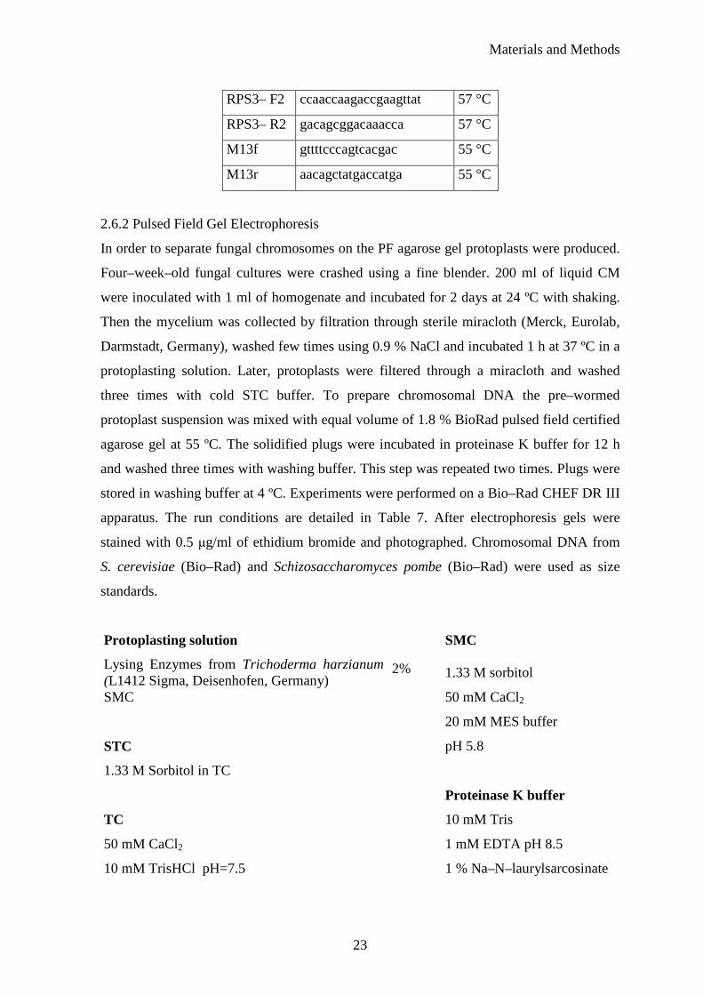
Protoplasting solution SMC
Lysing Enzymes from Trichoderma harzianum (L1412 Sigma, Deisenhofen, Germany)
2% 1.33 M sorbitol
SMC 50 mM CaCl2
20 mM MES buffer
STC pH 5.8
1.33 M Sorbitol in TC
Proteinase K buffer
TC 10 mM Tris
50 mM CaCl2 1 mM EDTA pH 8.5
10 mM TrisHCl pH=7.5 1 % Na–N–laurylsarcosinate

Materials and Methods
24
Table 7. PFGE running condition for each analyzed fungus. (T–temperature)
organism condition agarose concentration in the gel
running buffer
T
block 1 48 h 2 V 1–1800 s angel 100°
block 2 48 h 2 V 1–2000 s angel 106° S.vermifera MAFF291366
block 3 24 h 6 V 1–120 s angel 120°
0.8 % gel TBE 0.8xTBE 14 °C
block 1 48 h 2 V 1–1800 s angel 100° P. glomeralium
block 2 48 h 2 V 1–2000 s angel 106° 0.8 % gel TAE 0.8xTAE 4 °C
block 1 69 h 2 V 1–1800 s angle 100º P. indica
block 2 48 h 2 V 1–2000 s angel 106° 0.8 % gel TAE 1xTAE 14 °C
block 1 48 h 2 V 1–1800 s angel 100°
block 2 48 h 2 V 1–2000 s angel 106°
S.vermifera MAFF305842 S.vermifera MAFF305828 block 3 24 h 6 V 1–120 s angel 120°
0.8 % gel TAE 0.8xTAE 4 °C
2.7 Plate enzymatic assays
Tests for extracellular enzymes activity were performed in triplicates following the
methods describe in Kreisel and Schauer (1987). Mycelial plugs were cut from the edges
of colonies on 7 days old culture and were used as inoculum for all plate tests. The
extracellular enzymes activities were analyzed after two weeks.
2.7.1 Cellulase activity
Fungi were cultivated on medium enclosed 2.5 % malt extract, 1 % cellulose (SERVA,
FEINBIOCHEMICA, Heidelberg, Germany) and 2 % agar. The enzyme activity was
checked by spreading Lugol’s solution (2 % iodine and 4 % potassium iodide in water,
both Sigma, Deisenhofen, Germany). The clear area in the medium around the colony
indicated cellulose degradation.
2.7.2 Pectinase activity
To investigate pectinase activity fungi were propagated on the plates where 0.1 % yeast
extract with 1.5 % agar was enriched by 0.5 % pectin (Roth, Karlsruhe, Germany). Plates
were evaluated by flooding them with 1 % solution of hexadecyltrimetylammonium
Materials and Methods
25
bromide (Sigma, Deisenhofen, Germany) around the growing mycelium. The clear zone
around colonies suggested that fungus digested the substrate.
2.7.3 Laccase activity
To check laccase activity, medium contained 2.5 % malt extract and 2 % agar (MAE) was
used. A dark blue coloration after 3, 24 or 72 h after spreading of 0.1 M α–naphthol
(Sigma, Deisenhofen, Germany) in 96 % ethanol on the surface of the growing mycelium
indicated extracellular laccase activity. Along, the laccase production was tested during
interspecific interactions. For this purpose cocultures of the Sebacinoid strains with the
root pathogen R. solani were examinated. Sebacinoid isolates grew slower than R. solani
therefore they were precultured on MAE medium for one week before inoculation. The
enzyme activity was inspected after one week co–culture as described above. Additionally
laccase activity of P. indica and P. glomeralium was verified in coculture with barley
roots. Barley plants were inoculated with 105 chlamydospores. Furthermore, barley mock–
treated, autoclaved barley roots inoculated with chlamydospores and barley inoculated
with autoclaved fungal mycelium were analyzed. Presence of an enzymatic activity was
proved five and seven days after chlamydospores inoculation by spreading of 0.1 M α–
naphthol.
2.7.4 Peroxidase activity
Fungi grew as described by the laccase test. After 2 weeks, attendance of peroxidase was
evaluated by flooding plates with a fresh– prepared mixture of 0.4 % H2O2 (Roth,
Karlsruhe, Germany) and 1 % pyrogallol (Sigma, Deisenhofen, Germany) dissolved in
water. Plates were checked after 3, 24 or 72 h after substrate applying. A dark yellow /
brown color around the mycelium indicated peroxidase activity.
2.7.5 Protease activity
Analyzed fungi grown on medium containing 8 % gelatine (VWR PROLABO, Darmstadt,
Germany) dissolved in water at pH 6. The fungal ability to liquefy the solid media
indicates proteases production. The test was read after 5, 7, 10, 12 and 14 days growth. For
excluding any additional not enzymatic gelatine degradation plates were kept for 24 h at
4 °C.
Materials and Methods
26
2.8 Spectrophotometric enzymatic assay
For spectrophotometric assay barley plants as well as P. indica were grown in liquid 1/10
PNM. In order to obtain autoclaved barley roots, two weeks old barley plants were
harvesting and roots were autoclaved 20 min at 120 °C. Plant material was inoculated with
crashed P. indica mycelium. The samples were collected 1, 1.5, 2, 3, 5, 7, 10 and 15 days
after inoculation. For each enzyme activity measurement, medium from a culture were
assembled and filtered through miracloth. To remove the small particles like
chlamydospores, it was purified once more using membrane filter with pore diameter 0.45
µm (Whatman, Dassel, Germany) as well. Subsequently, the collected material was
concentrated with centrifugal devices for biomolecular separation (MACROSEP 10K
OMEGA PALL Life Sciences, Mexico) according to the manufacturer’s protocol. The
collected supernatant was utilized for further analysis.
All tests were carried out in BioTek Synergy 2 Multi–Mode Microplate Reader.
2.8.1 Laccase activity (Harkin and Obst 1973)
Laccase activity was detected using 2, 2’azino–bis–3–ethylbenzthiazoline–6–sulphonic
acid (ABTS) (Sigma, Deisenhofen, Germany) as a substrate in sodium tartrate buffer pH 3.
The enzyme activity was measure immediately after preparing reaction mixture. The
absorbance was read at 420 nm in 30 °C for 15 min. One unit of enzyme activity was
defined as the amount of enzyme required for oxidation of 1 µmol ABTS in 1 min.
Reaction mixture
0.05 M Sodium Tartrate buffer pH 3 50 µl
5 mM ABTS 100 µl
culture filtrate 100 µl
The enzyme activity was calculated using the formula below:
d ε V
F V ∆E L U
ABTSEn
totalnm 420 1-
⋅⋅⋅⋅
=
∆E420nm – absorbance per minute
Vtotal – the total volume of reaction mixture (0.25 ml)
F – dilution factor
VEn – the volume of culture (0.1 ml)
Materials and Methods
27
ε ABTS – extension of coefficient 0.0432 L µmol–1 cm–1
d – the distance the light travels through the material – layer thickness (0.7)
2.8.2 Peroxidase activity (Childs and Bardsley 1975)
Peroxidase activity was measured using a modified procedure describe for laccase activity
above. The enzyme activity was checked using ABTS in sodium tartrate buffer pH 3 with
hydrogen peroxide H2O2 (Sigma, Deisenhofen, Germany) as an additional substrate.The
enzyme activity was measured immediately after preparing reaction mixture. The one unit
of enzyme activity was defined as above.
Reaction mixture
0.05 M Sodium Tartrat buffer pH 3 50 µl
5 mM ABTS 100 µl
2 mM H2O2 100 µl
culture filtrate 100 µl
The enzyme activity was calculated using formula below:
d ε V
F V ∆E L U
ABTSEn
totalnm 420 1-
⋅⋅⋅⋅
=
∆E420nm – absorbance per minute
Vtotal – the total volume of reaction mixture (0.35ml)
F – dilution factor
Ven – the volume of enzyme (0.1ml)
ε ABTS – extension of coefficient 0.0432 L µmol–1 cm–1
d – the distance the light travels through the material – layer thickness (0.7)
2.8.3 Esterase activity
Para– nitrophenylacetat (pNPA) (Sigma, Deisenhofen, Germany) was used as a substrate
for esterase activity determination. The enzyme activity was measured immediately after
preparing the reaction mixture. The absorbance was read at 405 nm in 30 °C for 15 min.
One unit of enzyme activity was defined as the amount of enzyme required to hydrolyze 1
µmol para– nitrophenylacetat per 1 min at pH 6.5.

Materials and Methods
28
Reaction mixture
80 mM potassium phosphate buffer pH 6.5 50 µl
10 mM pNPA 100 µl
culture filtrate 100 µl
The enzyme activity was calculated using formula below:
d ε V
F V ∆E L U
pNPAEn
totalnm 405 1-
⋅⋅⋅⋅
=
∆E405nm – absorbance per minute
Vtotal – the total volume of reaction mixture (0.25ml)
F – dilution factor
VEn – the volume of enzyme (0.1ml)
ε pNPA – extension of coefficient 0.0183 L µmol–1 cm–1
d – the distance the light travels through the material – layer thickness (0.7)
2.8.4 Lipase activity (Winkler and Stuckmann 1979)
Lipase activity was determined using 4–nitrophenyl–palmitate (4NPP) (Sigma,
Deisenhofen, Germany) as a substrate in the potassium phosphate buffer pH 8.8. The
enzyme activity was measured immediately after preparing the reaction mixture. The
absorbance was read at 410 nm in 37 °C for 15 min. One unit of enzyme activity was
defined as the amount of enzyme required to hydrolyze of 1 µmol 4–nitrophenyl–palmitate
per 1 min in pH 8.8.
Substrate preparation (4NPP – buffer)
4NPP 15 mg
isopropanol 5 ml
sonification for 5–10 s
Deoxycholic acid Na salt (Roth, Karlsruhe, Germany) 110 mg
Gum Arabic (Roth, Karlsruhe, Germany 50 mg
potassium phosphate buffer pH 8.8 45 ml
10 min sonification
Materials and Methods
29
Reaction mixture
4NPP – buffer 100 µl
culture filtrate 50 µl
The enzyme activity was calculated using formula below:
d 15min V
60min ∆E L U
En
366nm 1-
⋅⋅⋅
=
∆E366nm – absorbance per minute
Vtotal – the total volume of reaction mixture (0.25 ml)
VEn – the volume of enzyme (0.1 ml)
d – the distance the light travels through the material – layer thickness (0.7)
2.8.5 Determination of total protein content
The protein content of all analyzed samples was determinate using Bradford assay. The
protein amount in each sample was estimated by reference to standard curve for bovine
serum albumin (BSA) (Sigma, Deisenhofen, Germany) in the range 5–120 µg/ml. All
samples were analyzed in triplicate.
Reaction mixture
Bradford solution (Roth, Karlsruhe, Germany) 200 µl
culturefiltrate / standard (BSA) 50 µl
2.9 P. indica protoplasts regeneration
P. indica protoplasts were prepared as described in the PFGE part. In order to examine the
best condition for their regeneration few osmotic stabilizers were tested. The complex
medium as well as top agar was supplemented by 0.3 M sucrose, 0.6 M sorbitol or 0.6 M
mannitol. The same concentration of protoplasts was mixed with liquid top agar and spread
on the bottom agar containing the same stabilizers. Regenerations took place at 28 °C and
every 24 h protoplasts regeneration was checked.
As controls water and STC were included in the regeneration tests. After 7 days
regeneration efficiency was compared by counting the growing colonies.
Results
30
3 Results
3.1 Analysis of translation elongation factor 1 alpha gene
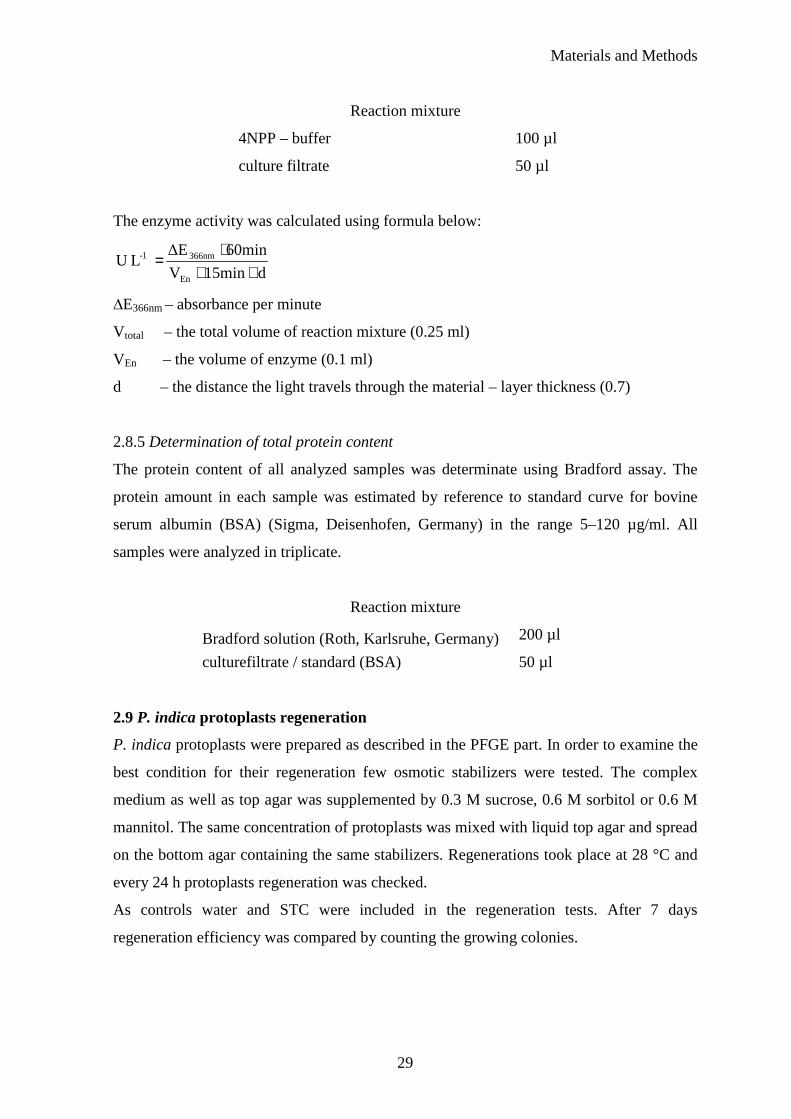
The translation elongation factor 1 alpha (TEF) gene was chosen for the phylogenetic study of
Sebacinales. Additionally to Sebacinales isolates, three independent environmental samples,
collected from two different areas in Germany, were analyzed with Sebacinales specific
primers. The sequences of the two TEF gene introns were the same for all environmental
clones sequenced but different from the Sebacinales isolates (Fig. 1.). The phylogenetic
analysis placed them close to P. indica showing that closely related fungi are present in
Germany (Fig. 1.). TEF phylogenetic analysis demonstrates that P. glomeralium (ex
multinucleate rhizoctonia) is the closest related strain to P. indica from all the Sebacinales
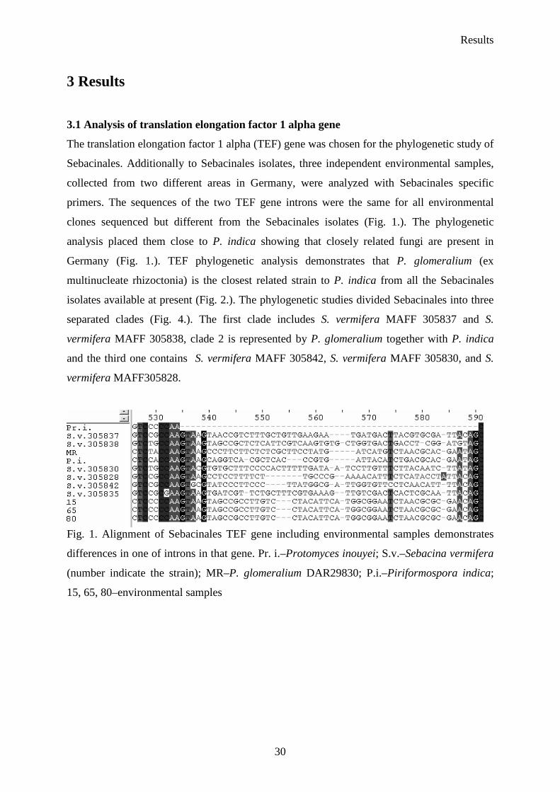
isolates available at present (Fig. 2.). The phylogenetic studies divided Sebacinales into three
separated clades (Fig. 4.). The first clade includes S. vermifera MAFF 305837 and S.
vermifera MAFF 305838, clade 2 is represented by P. glomeralium together with P. indica
and the third one contains S. vermifera MAFF 305842, S. vermifera MAFF 305830, and S.
vermifera MAFF305828.
Fig. 1. Alignment of Sebacinales TEF gene including environmental samples demonstrates
differences in one of introns in that gene. Pr. i.–Protomyces inouyei; S.v.–Sebacina vermifera
(number indicate the strain); MR–P. glomeralium DAR29830; P.i.–Piriformospora indica;
15, 65, 80–environmental samples
Results
31
Fig. 2. TEF gene based phylogenetic analysis of P. indica related fungi
3.2 Southern blot analysis
Southern blot analyses were performed to verify copy number of the TEF gene in Sebacinales
genomes. Additionally, the P. indica GAPDH gene was investigated. Genomic DNA digested
with restriction enzymes was separated on agarose gel, transferred on nylon membrane and
further hybridized with specific radioactive labelled probe. The results obtained for P. indica
showed only one band for both analyzed genes proving that they are single copy (Fig. 3). The
same enzymes combination (Bam HI, Hind III and Sac I) was implement for examination of
TEF gene copy number in the other Sebacinales strains: P. glomeralium and S. vermifera
MAFF305828 (Fig. 4.). S. vermifera MAFF 305830 have also only one copy of that gene
(Zuccaro unpublished data). After genomic DNA digestion of S. vermifera MAFF305842
with Hind III and hybridization with specific probe multiple bands were detected. However
after DNA digestion with Bam HI only one band was observed (Zuccaro unpublished data).

Results
32
Fig. 3. Study of TEF (A) and GAPDH (B) genes copy number using southern blot approach.
Genomic DNA was digested by three different enzymes and hybridized with specific
radioactive labelled probe. Bam HI, Hind III and XbaI were used for TEF gene and Bam HI,
Hind III and Sac I were applied for GAPDH.
Fig. 4. Study of TEF gene copy number in P. glomeralium (A) and S. vermifera
MAFF305828 (B) using southern blot approach. Genomic DNA was digested by two
different enzymes–Bam HI (1), and Hind III (2) and hybridized with specific radioactive
labelled probes.
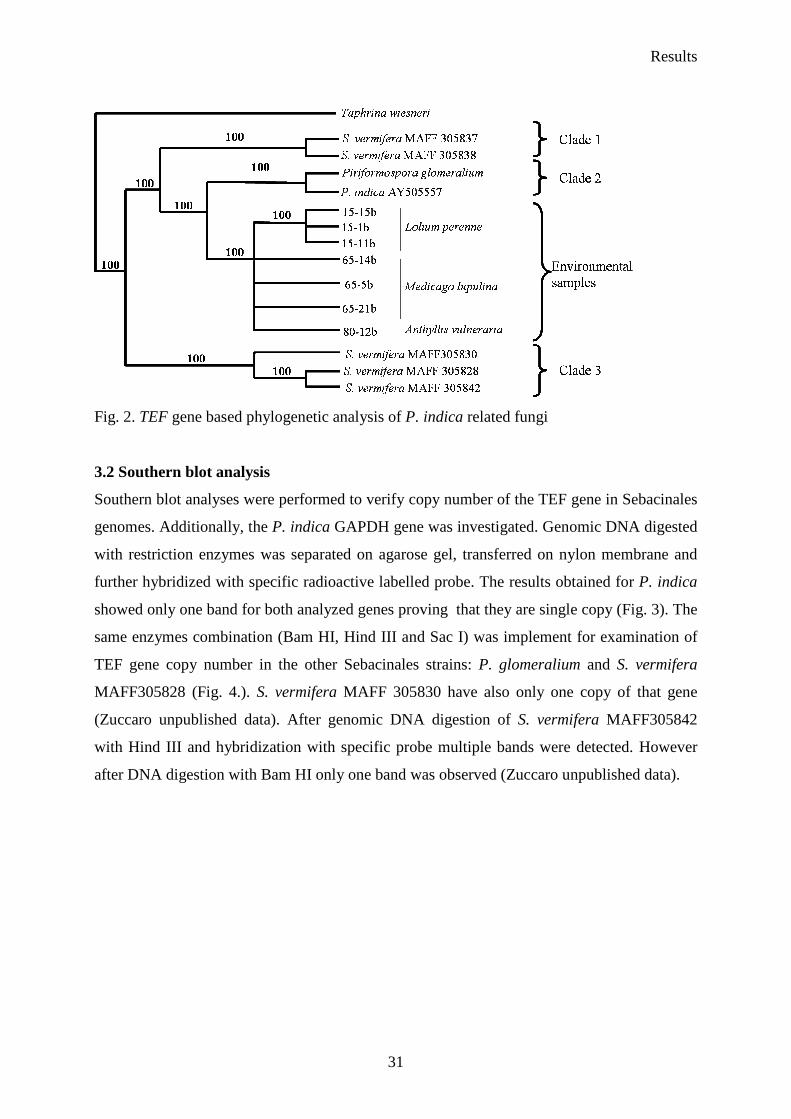
Furthermore, P. indica chromosomes separated by PFGE were transferred onto nylon
membrane and hybridized with a probe specific for GAPDH and TEF. GAPDH and TEF
produce one single band on the PFGE and were located on the third and on the first
chromosome respectively (Fig. 5.). The smallest band detected on the gel was verified as
mitochondrial DNA (Fig. 5.).
Results
33
PFGE GAPDH mit TEFPFGE GAPDH mit TEF
Fig. 5. Localization of GAPDH and TEF genes and identification of the mitochondrial DNA
on P. indica chromosomes using southern blot technique. PFGE–chromosomes separated
using PFGE, GAPDH, TEF–localization of analyzed genes, mit–mitochondrial DNA.
Southern blot analysis was performed using specific radioactive probes.
3.3 Genome estimation
Two different techniques were used to estimate the genome size of five Sebacinoid strains:
real–time PCR and Pulsed Field Gel Electrophoresis (PFGE). Additionally, confocal
microscopy technique were applied for P. indica.
The real time PCR method based on the absolute quantification of one copy gene in genomic
DNA sample. S. cerevisiae was chosen as control standard organism. The genome size
predicted using that approach and applying primers specific for the Saccharomyces cerevisiae
ribosomal protein S3 gene (ScRPS3) in four independent experiments was in the range of the
known genome size for this organism (12 Mb, Table 8.). The efficiency of real–time PCR for
S. cerevisiae was 94 ± 2 %. Relying on the analysis of other Basidiomycota genomes two
genes TEF and GAPDH were expected to be single copy in the Sebacinales genomes.
Southern blot analysis using specific probe for those two genes proved that they are single
copy therefore they were applied for P. indica genome size calculation. Using TEF gene in
eight independent real time PCR runs from CsCl purified DNA, the haploid genome size for
P. indica was 15.6 Mb ± 2.75 (Table 8.). Using the second gene GAPDH in four independent
runs the obtained genome size value of 15.3 Mb ± 3.5 (Table 8.). The real–time PCR
efficiency for the TEF and GAPDH genes was 100 ± 3 % and 94 ± 2 % respectively. The P.
Results
34
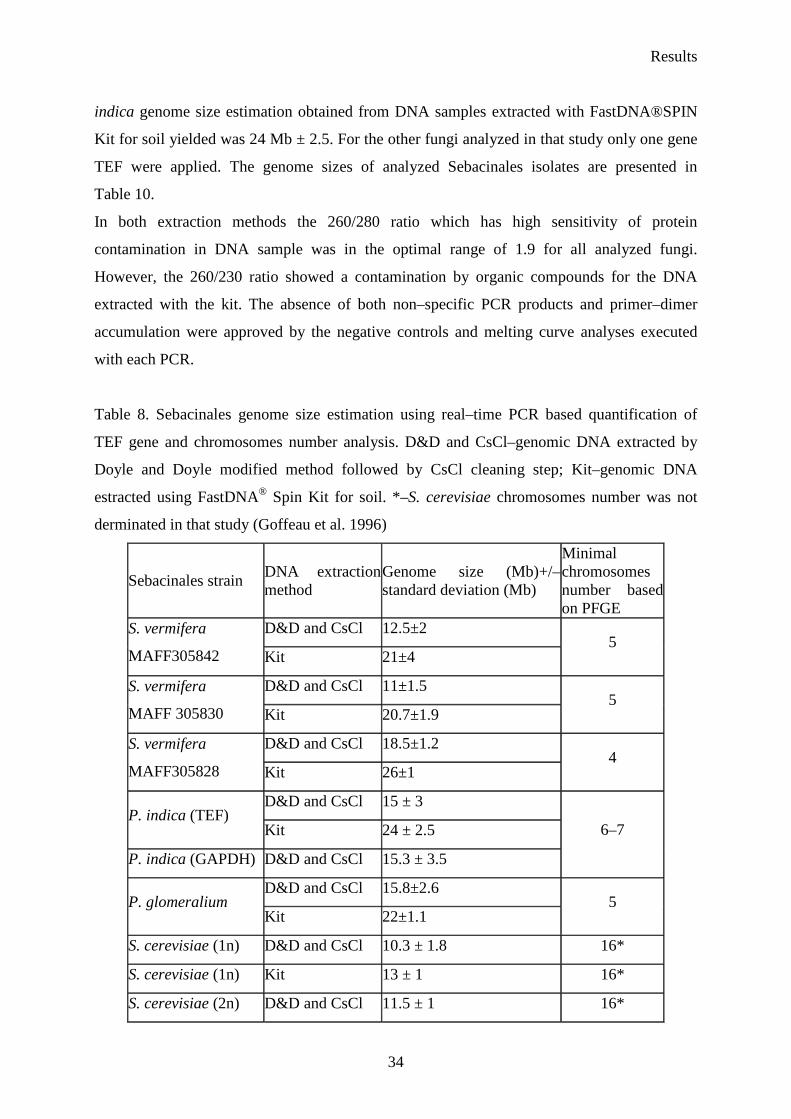
indica genome size estimation obtained from DNA samples extracted with FastDNA®SPIN
Kit for soil yielded was 24 Mb ± 2.5. For the other fungi analyzed in that study only one gene
TEF were applied. The genome sizes of analyzed Sebacinales isolates are presented in
Table 10.
In both extraction methods the 260/280 ratio which has high sensitivity of protein
contamination in DNA sample was in the optimal range of 1.9 for all analyzed fungi.
However, the 260/230 ratio showed a contamination by organic compounds for the DNA
extracted with the kit. The absence of both non–specific PCR products and primer–dimer
accumulation were approved by the negative controls and melting curve analyses executed
with each PCR.
Table 8. Sebacinales genome size estimation using real–time PCR based quantification of
TEF gene and chromosomes number analysis. D&D and CsCl–genomic DNA extracted by
Doyle and Doyle modified method followed by CsCl cleaning step; Kit–genomic DNA
estracted using FastDNA® Spin Kit for soil. *–S. cerevisiae chromosomes number was not
derminated in that study (Goffeau et al. 1996)
Sebacinales strain DNA extraction method
Genome size (Mb)+/– standard deviation (Mb)
Minimal chromosomes number based on PFGE
D&D and CsCl 12.5±2 S. vermifera
MAFF305842 Kit 21±4 5
D&D and CsCl 11±1.5 S. vermifera
MAFF 305830 Kit 20.7±1.9 5
D&D and CsCl 18.5±1.2 S. vermifera
MAFF305828 Kit 26±1 4
D&D and CsCl 15 ± 3 P. indica (TEF)
Kit 24 ± 2.5
P. indica (GAPDH) D&D and CsCl 15.3 ± 3.5
6–7
D&D and CsCl 15.8±2.6 P. glomeralium
Kit 22±1.1 5
S. cerevisiae (1n) D&D and CsCl 10.3 ± 1.8 16*
S. cerevisiae (1n) Kit 13 ± 1 16*
S. cerevisiae (2n) D&D and CsCl 11.5 ± 1 16*
Results
35
To separate fungal chromosomal DNA using PFGE different conditions were applied (see
Table 7.). In all runs chromosomes sizes were calculated over the standards S. cerevisiae and
Sch. pombe. The molecular karyotype of P. indica determined by that technique demonstrated
a pattern of six faint chromosomal bands ranging in size from 1.3 Mb to 5.4 Mb. The genome
size of the merged P. indica electrophoretic bands calculated from three different gels was
predicted to be about 15.8 Mb ± 0.3. The appearance of chromosomes larger than 5.4 Mb was
verified by extension of electrophoretic conditions (Fig. 6.). The zone, where big
chromosomes were expected, was fully resolved and no additional bands were detected. The
gel after PFGE indicated at least 5 chromosomal bands for S. vermifera MAFF305830,
MAFF305842 and P. glomeralium and at least 4 for S. vermifera MAFF305828. Similar to P.
indica, S. vermifera MAFF305830 and P. glomeralium have one big chromosome in the range
of 5.4 Mb. The gels indicate that S. vermifera MAFF305842 and S. vermifera MAFF305828
have at least one chromosome bigger than the biggest chromosome of size marker–Sch.
pombe (5.7 Mb) (Fig. 6.). Moreover, the smallest chromosome for S. vermifera MAFF305842
and S. vermifera MAFF305828 was still bigger than 2.2 Mb. P. indica and P. glomeralium
have an additional small chromosome in the range of 1 Mb. The estimation of genome size
relied on electrophoretic separation of chromosomes conferred a minimal size of 17 Mb for S.
vermifera MAFF305830, 14.4 Mb for P. glomeralium, 22.3 Mb for S. vermifera
MAFF305842 and 19.6 Mb for S. vermifera MAFF305828. The strength signal of the gel
staining with ethidium bromide for S. vermifera MAFF305842 and S. vermifera
MAFF305828 propose the presence of a higher number of chromosomes which were not
separated under the tested conditions (Fig. 6.). Although varied condition was applied the
separation was not improved.

Results
36
Fig. 6. Separation of P. indica (Pi), P. glomeralium (MR), S. vermifera MAFF305830 (S1), S.
vermifera MAFF305842 (S2), S. vermifera MAFF305828 (S6) chromosomes by Pulsed Field
Gel Electrophoresis (PFGE). M1–Saccharomyces cerevisiae (Bio–Rad) and M2–
Schizosaccharomyces pombe (Bio–Rad) size standards.
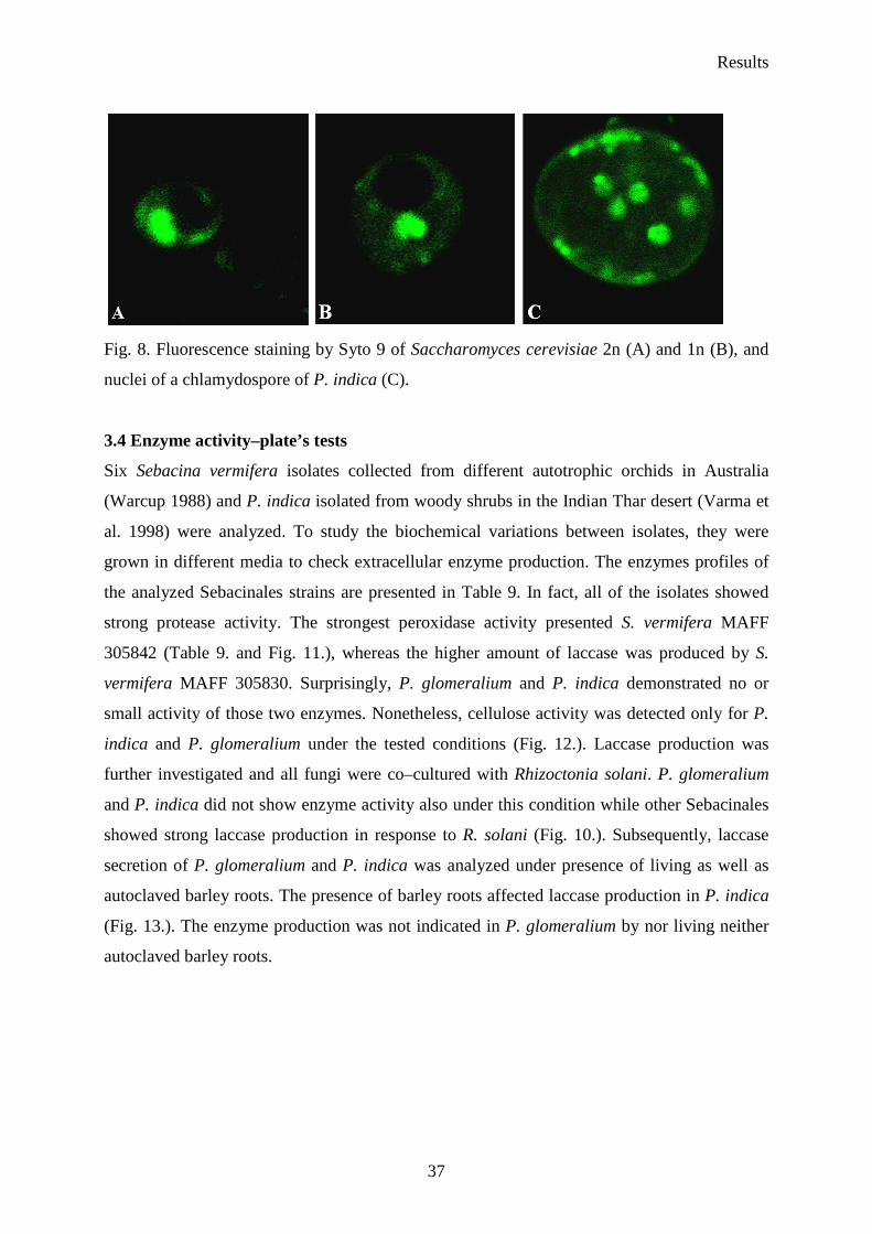
P. indica genome size was additionally estimated using confocal scanning microscope (Fig. 7.
and Fig. 8). Fluorescence histogram of 12 nuclei stained with syto 9 in chlamydospores was
measured. By comparison to the fluorescence of Saccharomyces cerevisiae (1n and 2n) which
nuclei were stained under the same condition, the genome of analyzed fungus was predicted.
The value of the mean histogram for P. indica was placed in between that of the two S.
cerevisiae strains suggesting that P. indica genome range 17–22 Mb what confirmed results
obtained by pulsed field gel electrophoresis and real–time PCR.
Fig. 7. Value of mean histogram fluorescence for P. indica and S. cerevisiae strains. P.i.–P.
indica, S.c. (2n)–S. cerevisiae (2n) and S.c. (1n)–S. cerevisiae (1n)
Results
37
Fig. 8. Fluorescence staining by Syto 9 of Saccharomyces cerevisiae 2n (A) and 1n (B), and
nuclei of a chlamydospore of P. indica (C).
3.4 Enzyme activity–plate’s tests
Six Sebacina vermifera isolates collected from different autotrophic orchids in Australia
(Warcup 1988) and P. indica isolated from woody shrubs in the Indian Thar desert (Varma et
al. 1998) were analyzed. To study the biochemical variations between isolates, they were
grown in different media to check extracellular enzyme production. The enzymes profiles of
the analyzed Sebacinales strains are presented in Table 9. In fact, all of the isolates showed
strong protease activity. The strongest peroxidase activity presented S. vermifera MAFF
305842 (Table 9. and Fig. 11.), whereas the higher amount of laccase was produced by S.
vermifera MAFF 305830. Surprisingly, P. glomeralium and P. indica demonstrated no or
small activity of those two enzymes. Nonetheless, cellulose activity was detected only for P.
indica and P. glomeralium under the tested conditions (Fig. 12.). Laccase production was
further investigated and all fungi were co–cultured with Rhizoctonia solani. P. glomeralium
and P. indica did not show enzyme activity also under this condition while other Sebacinales
showed strong laccase production in response to R. solani (Fig. 10.). Subsequently, laccase
secretion of P. glomeralium and P. indica was analyzed under presence of living as well as
autoclaved barley roots. The presence of barley roots affected laccase production in P. indica
(Fig. 13.). The enzyme production was not indicated in P. glomeralium by nor living neither
autoclaved barley roots.
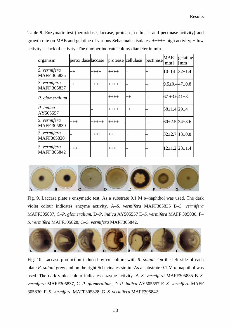
Results
38
Table 9. Enzymatic test (peroxidase, laccase, protease, cellulase and pectinase activity) and
growth rate on MAE and gelatine of various Sebacinales isolates. +++++ high activity; + low
activity; – lack of activity. The number indicate colony diameter in mm.
organism peroxidase laccase protease cellulase pectinase MAE [mm]
gelatine [mm]
S. vermifera MAFF 305835
++ ++++ ++++ – + 10–14 32±1.4
S. vermifera MAFF 305837
++ ++++ +++++ – – 9.5±0.4 47±0.8
P. glomeralium – – ++++ ++ – 67 ±3.6 41±3
P. indica AY505557
+ – ++++ ++ – 58±1.4 29±4
S. vermifera MAFF 305830
+++ +++++ ++++ – – 60±2.5 34±3.6
S. vermifera MAFF305828
– ++++ ++ + – 32±2.7 13±0.8
S. vermifera MAFF 305842
++++ + +++ – – 12±1.2 23±1.4
Fig. 9. Laccase plate’s enzymatic test. As a substrate 0.1 M α–naphthol was used. The dark
violet colour indicates enzyme activity. A–S. vermifera MAFF305835 B–S. vermifera
MAFF305837, C–P. glomeralium, D–P. indica AY505557 E–S. vermifera MAFF 305830, F–
S. vermifera MAFF305828, G–S. vermifera MAFF305842.
Fig. 10. Laccase production induced by co–culture with R. solani. On the left side of each
plate R. solani grew and on the right Sebacinales strain. As a substrate 0.1 M α–naphthol was
used. The dark violet colour indicates enzyme activity. A–S. vermifera MAFF305835 B–S.
vermifera MAFF305837, C–P. glomeralium, D–P. indica AY505557 E–S. vermifera MAFF
305830, F–S. vermifera MAFF305828, G–S. vermifera MAFF305842.
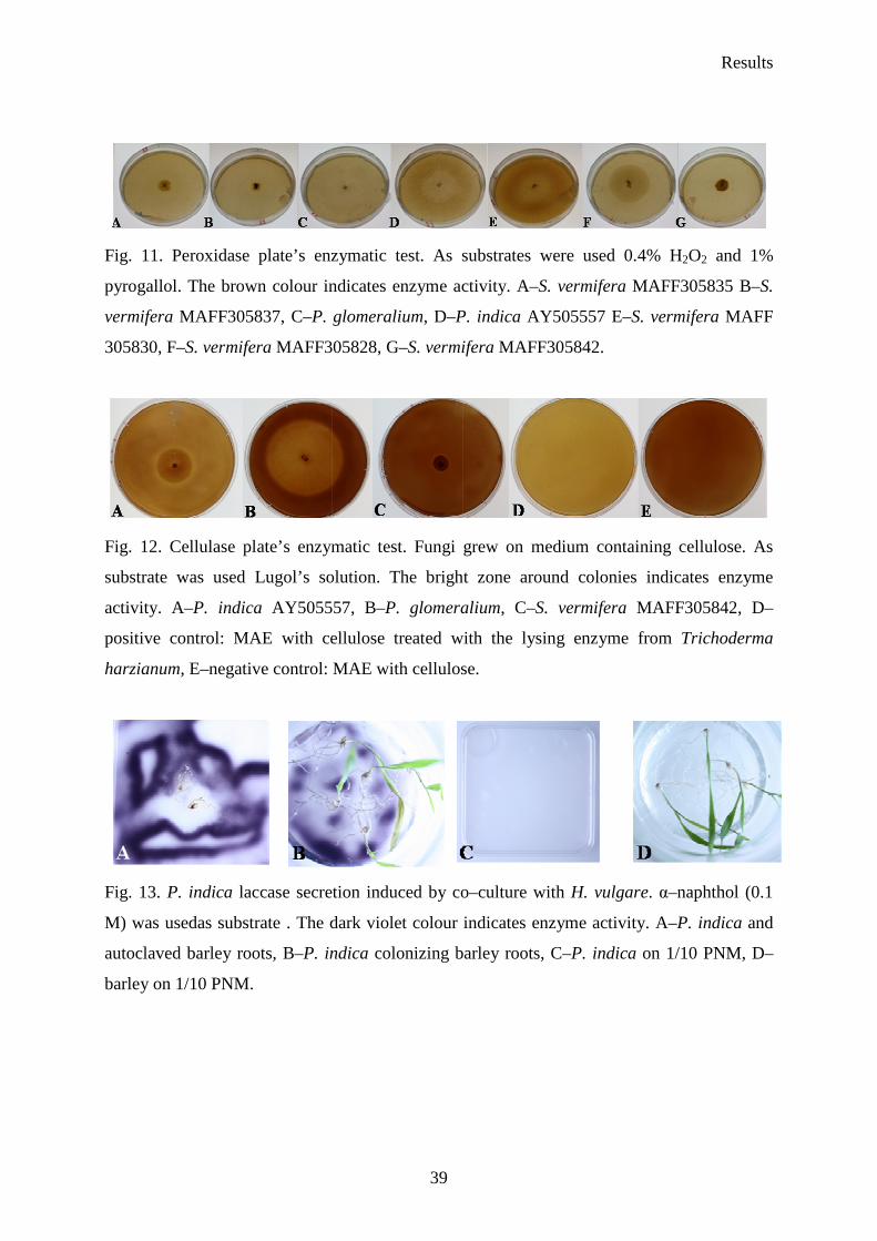
Results
39
Fig. 11. Peroxidase plate’s enzymatic test. As substrates were used 0.4% H2O2 and 1%
pyrogallol. The brown colour indicates enzyme activity. A–S. vermifera MAFF305835 B–S.
vermifera MAFF305837, C–P. glomeralium, D–P. indica AY505557 E–S. vermifera MAFF
305830, F–S. vermifera MAFF305828, G–S. vermifera MAFF305842.
Fig. 12. Cellulase plate’s enzymatic test. Fungi grew on medium containing cellulose. As
substrate was used Lugol’s solution. The bright zone around colonies indicates enzyme
activity. A–P. indica AY505557, B–P. glomeralium, C–S. vermifera MAFF305842, D–
positive control: MAE with cellulose treated with the lysing enzyme from Trichoderma
harzianum, E–negative control: MAE with cellulose.
Fig. 13. P. indica laccase secretion induced by co–culture with H. vulgare. α–naphthol (0.1
M) was usedas substrate . The dark violet colour indicates enzyme activity. A–P. indica and
autoclaved barley roots, B–P. indica colonizing barley roots, C–P. indica on 1/10 PNM, D–
barley on 1/10 PNM.
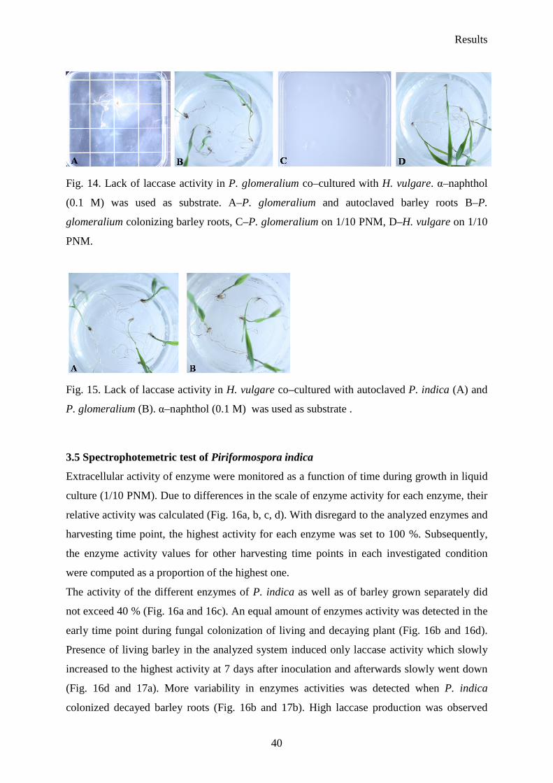
Results
40
Fig. 14. Lack of laccase activity in P. glomeralium co–cultured with H. vulgare. α–naphthol
(0.1 M) was used as substrate. A–P. glomeralium and autoclaved barley roots B–P.
glomeralium colonizing barley roots, C–P. glomeralium on 1/10 PNM, D–H. vulgare on 1/10
PNM.
Fig. 15. Lack of laccase activity in H. vulgare co–cultured with autoclaved P. indica (A) and
P. glomeralium (B). α–naphthol (0.1 M) was used as substrate .
3.5 Spectrophotemetric test of Piriformospora indica
Extracellular activity of enzyme were monitored as a function of time during growth in liquid
culture (1/10 PNM). Due to differences in the scale of enzyme activity for each enzyme, their
relative activity was calculated (Fig. 16a, b, c, d). With disregard to the analyzed enzymes and
harvesting time point, the highest activity for each enzyme was set to 100 %. Subsequently,
the enzyme activity values for other harvesting time points in each investigated condition
were computed as a proportion of the highest one.
The activity of the different enzymes of P. indica as well as of barley grown separately did
not exceed 40 % (Fig. 16a and 16c). An equal amount of enzymes activity was detected in the
early time point during fungal colonization of living and decaying plant (Fig. 16b and 16d).
Presence of living barley in the analyzed system induced only laccase activity which slowly
increased to the highest activity at 7 days after inoculation and afterwards slowly went down
(Fig. 16d and 17a). More variability in enzymes activities was detected when P. indica
colonized decayed barley roots (Fig. 16b and 17b). High laccase production was observed

Results
41
earlier (3 dai) in comparison to the case when fungus colonized living host roots. At 5th day
after inoculation enzyme secretion immediately decrease and rose again on 7th day. In
addition, by the second day after inoculation significant increase in activities of other
enzymes were noticed. The highest lipase secretion was detected at later times–10 dai (Fig.
16b and 18d). The highest esterase production was noted after 10 days of co–culture (Fig. 16b
and 18b). Peroxidase activity secreted in all inspected sets remained small and did not vary
dramatically within two weeks of experiment (Fig. 16). However, considerable increase was
detected at 15th day after inoculation in medium (Fig. 17c). No noticeable changes were
detected in esterase and lipase activity when P. indica and barley were propagated alone in
1/10 PNM (Fig. 18). These results indicate that the enzymes production was predominantly
associated with the presence of the symbiotic partner.
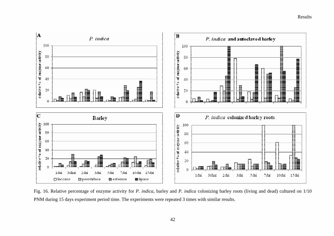
Results
42
Fig. 16. Relative percentage of enzyme activity for P. indica, barley and P. indica colonizing barley roots (living and dead) cultured on 1/10
PNM during 15 days experiment period time. The experiments were repeated 3 times with similar results.

Results
43
Fig 17. Variation in laccase and peroxidase activity for P. indica, barley and P. indica colonizing barley roots both (living and dead) cultured on
1/10 PNM during 15 days experiment period time. Standard deviation is calculated from 3 independent experiments.
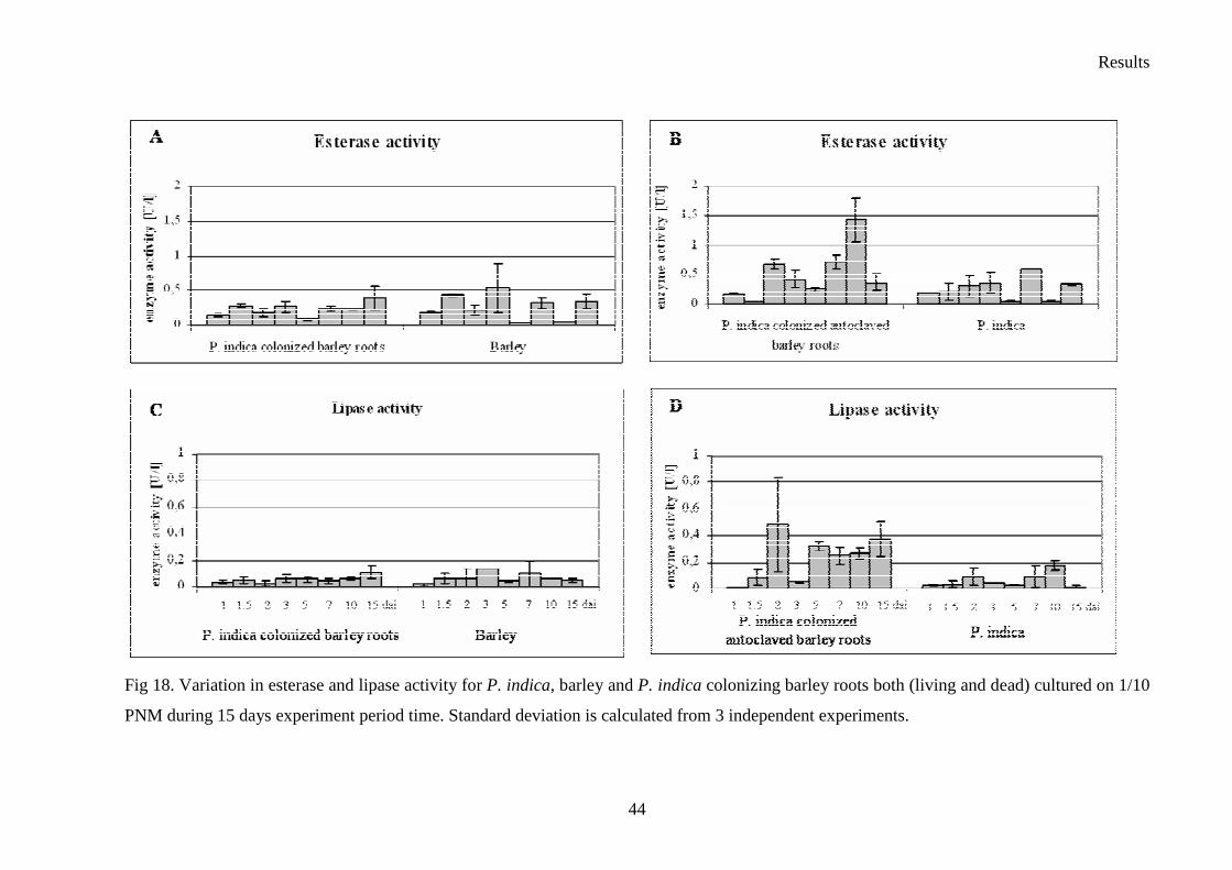
Results
44
Fig 18. Variation in esterase and lipase activity for P. indica, barley and P. indica colonizing barley roots both (living and dead) cultured on 1/10
PNM during 15 days experiment period time. Standard deviation is calculated from 3 independent experiments.
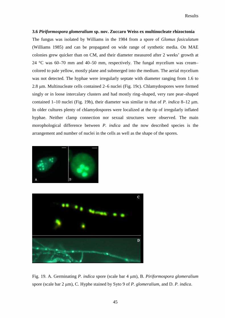
Results
45
3.6 Piriformospora glomeralium sp. nov. Zuccaro Weiss ex multinucleate rhizoctonia
The fungus was isolated by Williams in the 1984 from a spore of Glomus fasiculatum
(Williams 1985) and can be propagated on wide range of synthetic media. On MAE
colonies grew quicker than on CM, and their diameter measured after 2 weeks’ growth at
24 °C was 60–70 mm and 40–50 mm, respectively. The fungal mycelium was cream–
colored to pale yellow, mostly plane and submerged into the medium. The aerial mycelium
was not detected. The hyphae were irregularly septate with diameter ranging from 1.6 to
2.8 µm. Multinucleate cells contained 2–6 nuclei (Fig. 19c). Chlamydospores were formed
singly or in loose intercalary clusters and had mostly ring–shaped, very rare pear–shaped
contained 1–10 nuclei (Fig. 19b), their diameter was similar to that of P. indica 8–12 µm.
In older cultures plenty of chlamydospores were localized at the tip of irregularly inflated
hyphae. Neither clamp connection nor sexual structures were observed. The main
morophological difference between P. indica and the now described species is the
arrangement and number of nuclei in the cells as well as the shape of the spores.
Fig. 19. A. Germinating P. indica spore (scale bar 4 µm), B. Piriformospora glomeralium
spore (scale bar 2 µm), C. Hyphe stained by Syto 9 of P. glomeralium, and D. P. indica.
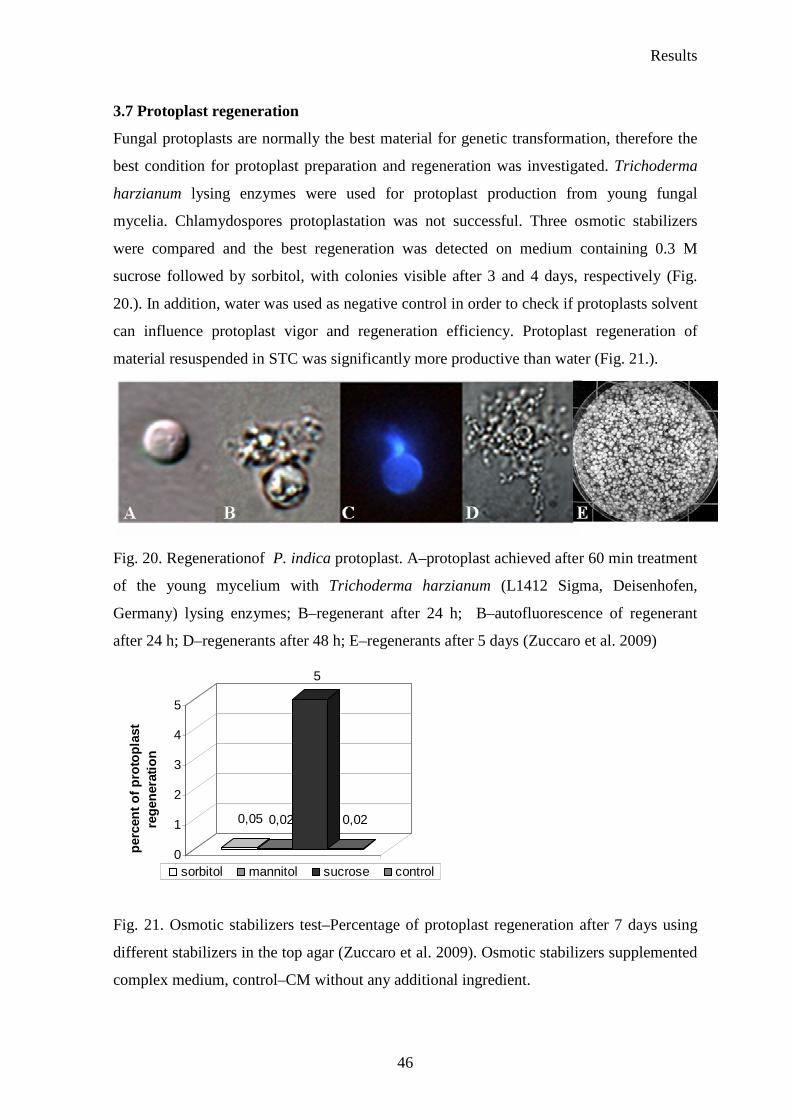
Results
46
3.7 Protoplast regeneration
Fungal protoplasts are normally the best material for genetic transformation, therefore the
best condition for protoplast preparation and regeneration was investigated. Trichoderma
harzianum lysing enzymes were used for protoplast production from young fungal
mycelia. Chlamydospores protoplastation was not successful. Three osmotic stabilizers
were compared and the best regeneration was detected on medium containing 0.3 M
sucrose followed by sorbitol, with colonies visible after 3 and 4 days, respectively (Fig.
20.). In addition, water was used as negative control in order to check if protoplasts solvent
can influence protoplast vigor and regeneration efficiency. Protoplast regeneration of
material resuspended in STC was significantly more productive than water (Fig. 21.).
Fig. 20. Regenerationof P. indica protoplast. A–protoplast achieved after 60 min treatment
of the young mycelium with Trichoderma harzianum (L1412 Sigma, Deisenhofen,
Germany) lysing enzymes; B–regenerant after 24 h; B–autofluorescence of regenerant
after 24 h; D–regenerants after 48 h; E–regenerants after 5 days (Zuccaro et al. 2009)
0,05 0,02
5
0,02
0
1
2
3
4
5
per
cen
t o
f p
roto
pla
st
reg
ener
atio
n
1sorbitol mannitol sucrose control
Fig. 21. Osmotic stabilizers test–Percentage of protoplast regeneration after 7 days using
different stabilizers in the top agar (Zuccaro et al. 2009). Osmotic stabilizers supplemented
complex medium, control–CM without any additional ingredient.
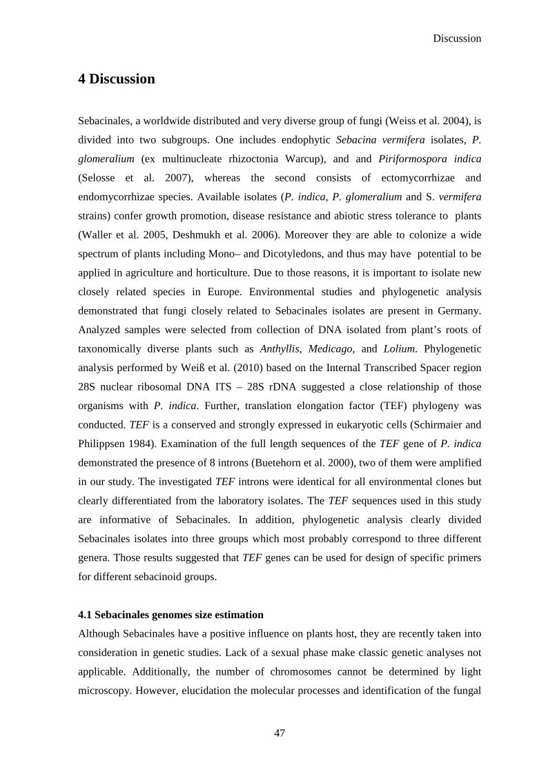
Discussion
47
4 Discussion
Sebacinales, a worldwide distributed and very diverse group of fungi (Weiss et al. 2004), is
divided into two subgroups. One includes endophytic Sebacina vermifera isolates, P.
glomeralium (ex multinucleate rhizoctonia Warcup), and and Piriformospora indica
(Selosse et al. 2007), whereas the second consists of ectomycorrhizae and
endomycorrhizae species. Available isolates (P. indica, P. glomeralium and S. vermifera
strains) confer growth promotion, disease resistance and abiotic stress tolerance to plants
(Waller et al. 2005, Deshmukh et al. 2006). Moreover they are able to colonize a wide
spectrum of plants including Mono– and Dicotyledons, and thus may have potential to be
applied in agriculture and horticulture. Due to those reasons, it is important to isolate new
closely related species in Europe. Environmental studies and phylogenetic analysis
demonstrated that fungi closely related to Sebacinales isolates are present in Germany.
Analyzed samples were selected from collection of DNA isolated from plant’s roots of
taxonomically diverse plants such as Anthyllis, Medicago, and Lolium. Phylogenetic
analysis performed by Weiß et al. (2010) based on the Internal Transcribed Spacer region
28S nuclear ribosomal DNA ITS – 28S rDNA suggested a close relationship of those
organisms with P. indica. Further, translation elongation factor (TEF) phylogeny was
conducted. TEF is a conserved and strongly expressed in eukaryotic cells (Schirmaier and
Philippsen 1984). Examination of the full length sequences of the TEF gene of P. indica
demonstrated the presence of 8 introns (Buetehorn et al. 2000), two of them were amplified
in our study. The investigated TEF introns were identical for all environmental clones but
clearly differentiated from the laboratory isolates. The TEF sequences used in this study
are informative of Sebacinales. In addition, phylogenetic analysis clearly divided
Sebacinales isolates into three groups which most probably correspond to three different
genera. Those results suggested that TEF genes can be used for design of specific primers
for different sebacinoid groups.
4.1 Sebacinales genomes size estimation
Although Sebacinales have a positive influence on plants host, they are recently taken into
consideration in genetic studies. Lack of a sexual phase make classic genetic analyses not
applicable. Additionally, the number of chromosomes cannot be determined by light
microscopy. However, elucidation the molecular processes and identification of the fungal
Discussion
48
factors that lead to a successful symbiosis of P. indica and other Sebacinales with its plant
partners is essential for better understanding the mechanism of that interaction. Analysis of
sequences of whole genome seems to be the most suitable method to provide a complete
story of biological networks. Although genome sequencing technologies developed very
fast over last few years, some basic studies are required before, to make sequencing
process fast and further analysis more efficient. Correct genome estimation is one of the
most important tasks which should be performed before applying genome sequencing
technologies. It is essential for sequencing costs valuation. We decided to predict genome
size of five Sebacinales strains using few available molecular methods. Techniques such
as: flow cytometry, reassociation kinetics, genomic reconstruction, PFGE, real–time PCR,
confocal microscope can be implemented in that purpose. However, each of them has some
limitation. Flow cytometry determines relative nuclear DNA content per spore and was
used for genome estimation for fungi such as the basidiomycete rust fungus Puccinia
recondita (Eilam et al. 1994), arbuscular mycorrhizal fungus Glomus intraradices (Hijri et
al. 2004), or the etiologic agent of histoplasmosis, ascomycete fungus–Histoplasma
capsulatum (Carr and Shearer Jr 1998). Sebacinales chlamydospores are multinucleate
therefore this method cannot be applied. Reassociation kinetics, reconstruction or real time
PCR based on one copy gene analysis. Genomes of a few fungi were investigated using
those approaches. Genomes of the obligate Oomycetes pathogen Bremia lactucae (Francis
et al. 1990) and the basidiomycete Paxillus involutus forming ectomycorrhizal symbiosis
(Le Quere et al. 2002) were analysed using reassociation kinetics (reassociation rate of
denatured DNA is measured under defined conditions). Reconstruction technique (the one
copy gene is used as a hybridization probe) was employed for genome analysis of such
organisms as Phytophthora megasperma f. sp. glycinea (Mao and Tyler 1991) and
Ascomycetes Colletotrichum graminicola (Randhir and Hanau 1997). A real–time PCR
based approach was established for strain 368 FY1679 of Saccharomyces cerevisiae, the
platyfish Xiphophorus maculatus and Homo sapiens sapiens (Wilhelm et al. 2003) and
applied also for such Ascomycetes fungi as: Cladonia grayi (Armaleo and May 2009) and
Zygosaccharomyces species (Solieri et al. 2008). False recognition of one copy gene can
be the reason of wrong genome size prediction. Additionally, real–time PCR requires very
good quality and quantity of DNA. For some organisms achievement of those terms might
be problematic. Too big size of chromosomes might be important barrier in PFGE
Discussion
49
approach. Hence, the best way for correct prediction of genome size is combining few (at
least two techniques), which rely on completely different assumption.
In that study, genome sizes of analyzed fungi were estimated using real–time PCR and
PFGE. First, both methods were established for P. indica and further applied for other
Sebacinales isolates. The real–time PCR approach relied on absolute quantification single
copy gene in genomic DNA. Based on the presumption that TEF and GAPDH are both
single copy in the P. indica genome, southern blot analysis of digested genomic DNA and
chromosomes separated by PFGE were performed. Although in some Ascomycetes and
Zygomycetes the TEF gene was detected in multiple copies, in almost all Basidiomycetes
genomes analyzed so far only one copy of this gene was detected. The GAPDH gene is
also present in single copy in many of Basidiomycetes. However some exceptions such as
Agaricus bisporus with two different GAPDH genes are known. According to southern
blot analysis, TEF and GAPDH are one copy genes. Similar assay was used for P.
glomeralium and Sebacina vermifera isolates. In the genome of analyzed fungi TEF gene
is most probably present only one time in the homokaryotic genome, therefore it was used
for genome sizes estimation. Southern blot analysis performed for S. vermifera
MAFF305842 did not give clear indication concerning the TEF gene copy number.
Genomic DNA digested with Bam HI and hybridized with specific probe showed one
band, however multiple bands were detected when Hind III enzyme was used (Zuccaro
unpublished data). Those findings proposed presence of SNPs (single–nucleotide
polymorphism) in TEF gene of S. vermifera MAFF305842. Further investigation of that
gene should be performed. Additionally, for P. indica TEF and GAPDH were localized on
chromosomes. TEF is located on first chromosome (5.4 Mb) and GAPDH on third
(2.5 Mb).
Fungal DNA was isolated using two different techniques: modified method from Doyle
and Doyle followed by a CsCl centrifugation and FastDNA® SPIN Kit for soil. Results are
displayed in Table 8. Genome size estimated using kit extracted DNA was 30–50 %
(depending on strain) bigger than with the second method for all Sebacinales isolates.
Protein contaminations in samples of genomic DNA were not detected. The ratio of
absorbance 260/280 was in the optimal range of 1.9 for both DNA extraction methods.
However, organic compounds were present in kit extracted DNA. The ratio of absorbance
260/230 was below the optimal value for each isolate. Those findings may explain the
differences in the genome size predicted using diverse methods of DNA isolation. S.
Discussion
50
cerevisiae (1n and 2n) was used to validate that method. DNA from S. cerevisiae 1n was
extracted using both method (Doyle and Doyle followed by a CsCl centrifugation and kit).
The genome size predicted using primers specific for the ScRPS3 gene was in the range of
the known genome size for this organism (12 Mb). Extraction method as well as the ploidy
of organisms used for DNA isolation did not influence genome size estimation. Those
results might suggest that Sebacinales cells contain some components which strongly
interfere both with buffers used for DNA extraction either directly with DNA and inhibit
extraction procedure. Without consideration of DNA extraction method, S. vermifera
MAFF305828 seems to have the biggest genome and, S. vermifera MAFF 305830 the
smallest one in between analyzed Sebacinales strains.
The second method implemented for genome determination was Pulse Field Gel
Electrophoresis. PFGE is an effective technique for separating big fragments of DNA such
as chromosomes, and is a meaningful tool for basic genetic studies, especially in lower
eukaryotes such as fungi. Chromosome–sized DNA molecules of Sebacinales isolates were
successfully obtained after young mycelium protoplastation and resolved by PFGE. The
electrophoretic conditions permitted the separation of 6–7 chromosomal bands in the range
of 1.3 to 5.4 Mb for P. indica. The total size of them agreed with the genome size
estimated by quantitative real–time PCR. The karyotypes achieved under these conditions
were reproducible. The staining intensity of the chromosome bands 3 and 5 was more
intensive than other bands. The separation in this part of the gel was not satisfying. Those
results displayed the possibility of attendance either heterologous chromosomes with
similar or identical size or multiple copies of a homologous chromosome. Among the
investigated isolates the karyotype analysis confirmed that P. glomeralium is the closest
related fungus to P. indica. Similar genome size and number of chromosomes separated
those two fungi from S. vermifera isolates. The large size of S. vermifera MAFF305842
and MAFF305828 chromosomes was the reason of not adequate separation. Nonetheless, it
is clear that the smallest chromosomes from P. indica (about 1.3 Mb) and P. glomeralium
(about 1.5 Mb) are not present in the other isolates. Specific differences in the chromosome
profiles within isolates from the same clade were also evident. The genome size
determinated by PFGE in an organism whose ploidy is unknown may lead to incorrect
conclusions due to incapacity during separating homologous chromosomes (Torres–
Guerrero 1999).
Discussion
51
Despite the clearly diversity in the chromosome profiles among isolates, genome sizes
estimated by those two techniques did not vary particularly within the clades. 8 %
dissimilarity was observed between P. indica and P. glomeralium and a maximum of 28 %
within the Sebacina vermifera strains from clade 3. Those differences might be present due
to gene duplication or loss, horizontal transfer events and transposable element. P. indica
genome size was additionally analysed using confocal scanning microscope. The staining
procedure applied for chlamydospores and hyphe worked very well. However microscopic
observation, in the same set conditions, such different structure like hyphae and S.
cerevisiae cells, used as a standard organism, was not possible. Genome size estimated
using that technique confirmed genome size to be in the average of 22 Mb. The dimensions
of genome sizes support the thesis that sebacinoid fungi from the subclade B (Weiss et al.
2004) hold a relatively small genome. Genome sizes among known Basidiomycota ranged
25–125 Mb, with high level of repetitive DNA. Such genome sizes are characteristic for
mushrooms like Coprinopsis cinerea (37 Mb, Stajich et al. 2010), Schizophyllum commune
(38 Mb http://www.ncbi.nlm.nih.gov/sites/entrez?Db=genomeprj&cmd=ShowDetailView
&TermToSearch=12852), Puccinia graminis (81 Mb, http://www.ncbi.nlm.nih.gov/
sites/entrez?Db= genomeprj&cmd=ShowDetailView&TermToSearch=12848). Pathogenic
Basidiomycetes possess smaller genomes. The plant pathogen Ustilago maydis genome is
20 Mb (Kämper et al. 2006) or that of Cryptococcus neoformans causing a human disease–
is 19 Mb (Loftus et al. 2005). Despite small genomes, Sebacinales are free–living and non–
pathogenic fungi. The TEF sequence analysis suggested already that P. indica introns
might be small. Moreover, investigation of genomic date achieved after pyro–sequencing
implying that P. indica has a very compact genome with very less repetition (Zuccaro et al.
in prep.). Sebacinales genome size predictions additionally proved that analyzed
Sebacinales strains are distinct and supported division those isolates into 3 clades.
An additional band smaller than 0.2 Mb was often observed for P. indica on the gel after
PFGE. This band was identified using southern blot approach as a mitochondrial DNA.
Normally, fungal mitochondrial genetic information size ranged from 19 kb (Sch. pombe
(Lang et al. 1983)) to 170 kb (Agaricus bitorquis (Hintz et al. 1985)) and generally is
located on circular, or rarely linear, double–stranded DNA molecules. It was not always
detected on the gel, its presence or absence has been linked to sample preparation. Genetic
information from mitochondrium was seen only for P. indica.

Discussion
52
4.2 P. indica protoplast regeneration
P. indica can be a model organism representing Sebacinales. Genome data together with
transformation system can provide a lot of information about the fungus as well as the
genetic nature of symbiosis. Efficient procedure for protoplast production and proper
regeneration condition for them are essential for good transformation system. In that study,
the effective protocol for protoplastation was developed together with the best condition
for P. indica protoplast regeneration. A lot of protoplastation protocols were published so
far which based on the biochemical removal of the cell wall using different enzymes.
Protoplast from filamentous Ascomycete Ashbya gossypii, for example, was obtained using
zymolase (Choi 2006), mycelium from dimorphic Zygomycete Benjaminiella poitrasii was
incubated first in protease solution, later treated with a mixture of dithiothreitol and β–
mercaptoethanol and then directly suspended in the cell wall lysing enzyme mixture
(Sigma lysing mixture, zymolyase and lyticase) (Chitnis and Deshpande 2002). Mixture of
lysing enzyme from Trichoderma harzianum supported by mazerozyme, driselase and
pectilyse was tested for Sebacinales, however protoplastation efficiency was not satisfied.
The best effect in protoplast quality and quantity was achieved using lysing enzyme from
Trichoderma harzianum. This commercially available substnance contains cocktail of cell
wall degrading enzymes including β–glucanase with some cellulase, protease and chitinase
activities. The enzymes composition allowed for very fast cell wall degradation. In
consequence, exposure time of destructive enzyme activity was reduced. Protoplasts
gathered in shorter exposure times to lytic enzymes have notable ability to regenerate than
those which were incubated for longer time (Zhou et al. 2008). In protocol established in
that study fungal mycelium was incubated with enzyme solution 1 h, whereas in instruction
mentioned above from 1 h to 5 h. Additionally, the right osmotic stabilizer applied in the
top agar and medium lead to the successful regeneration process. Those substances can
keep the balance of interior and exterior osmotic pressure of the protoplasts, and guard
them from being broken. The cell wall constitution diversifies among species, therefore
different concentrations of various osmotica will be optimal for each species. A lot of
chemicals including inorganic salts, sugars and sugar alcohols can be applied as osmotic
pressure stabilizers (Davis 1985). Inorganic salts are more effective with filamentous
fungi, and sugar and sugar alcohols with yeasts and higher plants (Lalithakumari 1996).
Mannitol, sorbitol and sucrose were tested here. From among of them the best effect was
observed on medium with sucrose. Furthermore, results obtained in that study showed that
Discussion
53
STC buffer is better protoplast solvent than water. The main ingredient of that buffer, sugar
alcohol–sorbitol protects protoplast against destruction.
4.3 Biochemical analysis of Sebacinales
In order to establish close interaction with the plant host, Sebacinales must overcome few
barriers or natural plant defence mechanisms. The first diffculty might be plant cell wall
composed of cellulose, hemicellulose, xylan and lignin. Cellulose, for example, can be
digested only by fungi and some bacteria. Later, in response to invader, host is able to
produce some substances with antifungal activity. We analyzed, therefore, some enzymes
essential in plant cell wall degradation and later during breaking the plant defence
apparatus. The diversity in enzyme activity provides clear identification that physiological
processes are taking place in the analyzed environment.
To screen enzymes secreted by Sebacinales some plate’s enzymatic tests were performed.
Cellulase, pectinase, laccase, peroxidase and proteinase were analyzed. Although all fungi
grew well in axenic culture, enzymes productions were not always detectable. Cellulose is
one of the most important and very difficult digested, by other organisms, polysaccharide
present in plant cell wall. Among analyzed Sebacinales strains only P. glomeralium and P.
indica presented higher cellulase activity. In culture of S. vermifera MAFF305828 small
cellulase activity was also observed. Cellobiose–the intermediate product of cellulose
hydrolysis is known as an effective inhibitor of that enzyme (Walker and Wilson 1991).
This phenomenon might explain the lack of enzyme activity in the test. S. vermifera strains
might secrete cellulase, however, the cellobiose accumulation could immediately stop the
further activity. Moreover, glucose was also described as enzyme inhibitor (Walker and
Wilson 1991). The complete cellulolysis could break down cellulose into glucose units
and, in consequence, block enzyme activity. Besides, the amount of secreted enzyme could
be so faint making them not possible to identify in the applied test. Only one species – S.
vermifera MAFF305835 secreted pectinases. Pectin–the substrate for that enzyme is
present in plant cell walls next to cellulose. S. vermifera MAFF305835 does not produce
cellulase in axenic culture but we can assume that it exudes pectinase which allows
entering into the plant cell. For mycorrhiza fungi such as: Suillus variegatus, Suillus
bovinus, Piloderma croceum, Pisolithus tinctorius, Paxillus involutus and Amanita
muscaria no significant enhancement of pectinase activity was observed after induction
with pectin, polygalacturonic acid or galacturonic acid (Ramstedt and Soderhall 1983). In
Discussion
54
addition, minor enzyme production can be undetectable by the test (Garcia–Romera et al.
1991). On the other hand, the low activities of cellulase and pectinase secreted by
investigated fungi could be an adaptation to a symbiotic life. Mycorrhizal fungi must
maintain a careful balance during plant roots colonizing process. The symbiont must avoid
eliciting substances which initiate host resistance reactions. Lack of cell degrading
enzymes has been already observed. In the Laccaria bicolor genome, genes responsible for
secretion of mentioned enzymes, were not detected. Probably L. bicolor as well as S.
vermifera strains use different modes of root colonization. One of the possible ways is the
‘crack entry’ where the hyphae colonizing the root surface overrun plant tissue through
breaks in the epidermis cells. Martin et al. (2008a) proposed that L. bicolor may use
ectomycorrhiza–induced expansins for releasing the host cell wall components and
promoting the growth of the hyphae in the symbiotic apoplastic space. The minimal set of
hydrolytic enzymes such as polysaccharide hydrolases, lyases and pectin esterases was
found also in ubiquitous pathogen U. maydis genome. Those findings agreed with its
biotrophic lifestyle, in which minimization of damage to the host are relevant in order to
avoid plant defence responses (Kämper et al. 2006). The similar mechanisms can be used
by S. vermifera isolates. Sebacinales strains investigated in my work presented quite high
proteinase activity during growth on gelatine medium. The plate enzymatic tests indicate
that high production of laccase for all Sebacina vermifera isolates and no activity for both
P. indica and P. glomeralium under experimental growth condition. Lack of laccase
activity was previously observed also for other Basidiomycetes such as Serpula lacrymans
(Score et al. 1997). Additionally, P. indica exudes very low amount of peroxidase, in case
of P. glomeralium the enzyme amount was not detectable. Laccase and peroxidase belong
to oxidoreductases involved in protection against host oxidative responses and are
commonly secreted by fungi, especially by Basidiomycetes (Edens et al. 1999). Besides,
laccase has several, very important physiological functions like detoxification of phenolic
compounds. In addition, Kellner et al. (2007) analyzed laccase production by some
Basidioimycetes fungi including P. indica. Presence of laccase activity in medium where
fungus was propagated was proved using laccase specific antibody LccCbr2. Their results
showed very weak (67 kDa), however, clear band indicating laccase activity. Moreover, P.
indica genome analysis determined that fungus has at least two laccase genes with signal
peptide (Zuccaro et al. in prep). All those findings suggested that P. indica has machinery
necessary for the secretion of this enzyme, therefore the best conditions for laccase
Discussion
55
production were further investigated. In addition, impact of cultural conditions on
repression of the laccase gene expression was already observed (Luisa et al. 2004). The
similar phenomenon could have taken place in investigated conditions for Piriformospora
strains. Those two strains are closely related to Sebacina vermifera isolates, however, they
are clearly divided from them. Piriformospora strains position on phylogenetic tree as well
as origin of Sebacina vermifera isolates (they belong to Orchid mycorrhizae group) might
explain differences in laccase secretion.
The distinctions in the enzyme profiles for Sebacinales corresponded with their
phylogenetic affinity proved by both, individual and combined analyses conducted using
ITS, 28S and TEF DNA sequences. Strong laccase activity demonstrated by all Sebacina
vermifera isolates as well as lack of cellulase secretion and opposite enzyme activity in
Piriformospora strains clearly divide those fungi in three clades.
Additionally to plate’s tests, variability in P. indica laccases, peroxiadases, esterases, and
lipases production were analyzed in order to examine fungus response to plants roots. The
activity values of all measured enzymes secreted by P. indica were very low. The highest
value (around 10 U/l) was detected for peroxidase, for other enzymes the activity did not
increased over 2 U/l. Those amounts of secreted enzymes in analyzed conditions are not
sufficient to use the fungus for commercial enzyme production under this growth
conditions.
P. indica cultivated on 1/10 PNM demonstrated very slow growth rate as well as low,
however detectable, activity of all analyzed enzymes. In fact, that medium, suitable for
plants without carbon source, can be considered as minimal medium for fungus.
Observation of P. indica implied reduction of its metabolism. Similarly to P. indica
growing without symbiotic partner, barley without symbiont did not secret the higher
amount of analyzed enzymes. All of them did not increase over 20–30 % of the highest
activity, what can be consider as normal ratio for plant growing in sterile condition.
Completely different results were observed when plants (either living or dead) were
present in the system. Almost all analyzed enzymes respond to decay plant material.
During first 36 h after inoculation the low enzyme activity was observed. Since 2nd day
post inoculation the increase of activity was identified. Only peroxidases secretion
measured in those circumstances was very weak. Significant changes in enzymes
production were also detected whereas the living plant partner appears in the environment.
Discussion
56
Laccase was one of the enzymes which response was evident in that growth conditions.
The enzymes variation was more visible when P. indica colonized decay plant roots.
On plate’s tests containing MAE without any supplementary ingredients as well as on solid
1/10 PNM P. indica laccase activity with α–naphtol as a substrate was not observed.
However, more sensitive spectrophotometric assay detected small enzyme production. In
consequence of not favourable growth conditions fungus was forced to gain carbon source
from its own. Hiscox et al. (2010) demonstrated higher production of this enzyme in
interaction zones between T. versicolor and different species of asco– and basidiomycota:
Stereum gausapatum, Daldinia concentrica, Bjerkandera adusta, Fomes fomentarius,
Hypholoma fasciculare whereas no enormous fluctuations were detected in other regions.
These findings suggested substantial function of the enzyme during competition between
two organisms and, therefore, can have significant impact on natural environment.
Coculture with R. solani was conducted in order to examine enzyme secretion by
Sebacinales isolates in presence of competitor. Increase of laccase exudation in that system
in contact zone between all scrutinized strains and R. solani was detecetd. Obtained results
were not clear for P. indica and P. glomeraliu. The dark violet colour indicating laccase
secretion was present only in contact zone, whereas on the plates with another Sbacinales
strains mycelium became violet in area where contact between two fungi was limited.
Those findings might suggest either enzyme activity came from R. solani or from both
interacting partners but the enzyme secretions was strickted only to contact zone. The
macro– and microscopical observation implied that P. indica was able to grow in presence
of R. solani without any problem. P. indica mycelium grew parallel to R. solani and did
not demonstrate any stress symptoms. Moreover, Ghahfarokhi and Goltapeh (2010)
mentioned that P. indica is able to colonize hyphae of another fungus. This phenomenon
was also observed in my study. The similar remarks were detected in coculture P.
glomeralium with R. solani. Furthermore, P. glomeralium established more aggressive
relationship whit competitor what was noticed on plates test as well as under microscope.
R. solani growth was clearly inhibited by P. glomeralium. R. solani did not grow as fast as
on plates with another Sebacinales isolates and the colour indicating laccase activity was
stronger. The further analysis of that phenomenon should be performed. In tests, conducted
with P. indica grown in presence of dead or living barley roots on solid 1/10 PNM, laccase
activity was detected 5 days after inoculation in the area around roots colonized by fungus.
The absence of laccase activity during axenical growth, and presence while living or dead
Discussion
57
barley roots were present, showed inducible nature of that enzyme. Interestingly, the
enzyme activity, examined with the same procedure, was not observed for P. glomeralium.
The previous studies demonstrate that P. indica and P. glomeralium have different
influence on barley. Both fungi promote plant growth and reduce disease symptoms,
however P. indica’s positive effect is stronger (Deshmukh et al. 2006). These results do
not exclude presence of laccase genes in fungus genome, they might just propose that P.
glomeralium require other hosts for enzyme induction. Study of laccase secretion by P.
indica colonizing living barley roots in liquid 1/10 PNM demonstrated that enzyme
production slowly increased to get maximum value at 7th day after inoculation. Laccase
plays important role during lignin degradation as well as in neutralization of toxic phenols
(Haars and Huttermann 1981) and defence against host oxidative responses (Edens et al.
1999). Microscopic investigation indicated that around third day after inoculation fungus
already penetrated the plant cortex cell and further begun inter– and intra–cellular growth
(Deshmukh et al. 2006). At that time laccase activity increased. When maximal activity of
enzyme was observed, chlamydospores were formed within the plant root. Schaefer at al.
(2009) checked plant response to P. indica. 459 (3 dai) and 509 (7 dai) genes were
differentially regulated in P. indica colonized roots compared to mock–treated. Genes
involved in plant defence/stress responses formed the largest group of variously regulated
genes. The plant immune system answered to invaders and got feedback from fungus in
higher laccase secretion. Laccase secretion was also very high when instead of living
barley roots, dead ones were colonized by the fungus. The higher enzyme secretion level
was observed three days after inoculation. Peroxidases activity measured in those
circumstances was very weak. Similarly, lack of peroxidase with laccases as the
predominant extracellular phenoloxidase was observed for white rot fungus Pycnoporus
cinnabarinus (Eggert et al. 1996).
In presence of decay barley roots production of lipases by P. indica were induced in the
highest degree at 2nd day after inoculation Lipases are essential for catalyze the hydrolysis
of ester bonds in lipids. Belcarz et al. (2005) demonstrated that, in the presence of humic
acids derived from brown coal, Basidiomycete white–rot fungus Bjerkandera adusta
synthesized significant amounts of laccase and lipase. Lipids occurred naturally in humic
substances. The process of medium preparation in my study (autoclaving roots in 1/10
PNM) supports the presence of degrading plant cell wall components including lipids and
can be compared to humic acids.
Discussion
58
Esterase, which belongs to the same enzymes group with lipase, presented the highest
activity while P. indica colonized decay too. General, those enzymes demonstrate higher
activity in the rhizosphere colonized by mycorrhiza fungi than non–mycorrhizal controls
(Vazquez et al. 2000). P. indica is not a arbuscular mycorrhiza fungus, though some
mycorrhiza characteristic can be detected. In addition, one of the hypothesis concerning
endophytes proposed that they become saprotrophs after the onset of senescence of host
tissue. This life style modification can be due to the alteration taking place in host tissue
during senescence, which allows mycelium to penetrate the epidermis and colonize host
surface (Promputtha et al. 2007).
Lipase and esterase activities stopped on the level not higher than 25 % of highest activity
in presence of living plant in investigated environment. Those results suggest that both
enzymes are not relevant in analyzed condition.
Summary
59
5 Summary/ Zusammenfassung
The order Sebacinales belongs to a taxonomically, ecologically, and physiologically
diverse group of fungi within the Phylum Basidiomycota. Using several molecular
techniques they were detected all over the world. Few isolates, classified into the clade B,
are available at the moment: the root–colonizing mutualistic fungus Piriformospora indica,
various Sebacina vermifera isolates from autotrophic orchids, as well as Piriformospora
glomeralium (ex multinucleate rhizoctonia DAR29830, Warcup). All of them were
described as growth promoting and resistance–inducing fungi. In the present work, seven
Sebacinales isolates of the clade B were characterized molecularly and phenotypically. In
addition, the presence in Germany of fungi closely related to P. indica was proven.
Phylogenetic analyses conducted using DNA sequences from the 28S and the translation
elongation factor 1–α gene (TEF) showed that analyzed Sebacinales isolates represent at
least 3 distinct groups of isolates. Further, three independent environmental samples,
collected from two different areas in Germany were examined. The analysis demonstrated
that the sample respresented organisms closely related to P. indica. The analyzed introns of
their TEF gene had the same sequence. This finding suggests that one genotype is present
in different area of Germany which is associated with roots of taxonomically diverse
plants, including Anthyllis, Medicago and Lolium.
Moreover, the fungal genome sizes of five sebacinoid isolates were estimated using Pulsed
Field Gel Electrophoresis (PFGE) and real–time PCR based on the absolute quantification
of a single copy gene (TEF). The fungi have at least 4 to 7 chromosomes and a genome
size ranging from 21 to 26 Mb.
Morphological, physiological and molecular studies of the multinucleate rhizoctonia
DAR29830 isolate proved that this strain is the closest related to P. indica, the most
investigated member of the Sebacinales. Therefore it is designed in this study as a
provisional new species named Piriformospora glomeralium sp. nov.
Seven Sebacinales strains were tested for extracellular enzyme production including
peroxidase, laccase, protease, pectinase, and cellulase. In general, the enzymatic test
demonstrated strong protease activity in cultures of all the analyzed fungi. The orchid
mycorrhiza isolates showed stark laccase activity and lack of cellulase production in axenic
culture whereas the Piriformospora strains were able to digest this plant cell wall
component. Only one isolate proved to secrete pectinase. All Sebacinales isolates except P.
Summary
60
indica and P. glomeralium that were co–cultured with a fungal competitor (Rhizoctonia
solani) generated significant amounts of laccase. The presence of a plant host, life or dead,
had noticeable impact on laccase secretion by P. indica but no influence on P. glomeralium
enzyme exudation. In addition, production of laccase, peroxidase, esterase, and lipase by P.
indica in response to plant host was spectrophotometrically investigated. Diversity in
laccase and lipase activity was observed mostly in presence of decay plant material.
Differences in the enzyme profile for the analyzed Sebacinales strains agreed mostly with
their phylogenetic position. In addition, genome estimation as well as karyotype analysis
clearly confirmed the phylogenetic study and proved that Sebacina vermifera should be
considered as complex of isolates. Moreover, laccase secretion by P. indica can be induced
only by the presence of the plant symbiotic partner.
Zusammenfassung
Die Ordnung der Sebacinales gehört zu einer taxonomisch, ökologisch und physiologisch
diversen Gruppe von Pilzen im Phylum der Basidiomyceten. Weltweit können sie durch
verschiedene molekulare Techniken detektiert werden. Wenige Isolate, klassifiziert im
Untergruppe B, sind zurzeit verfügbar: der Wurzel–kolonisierende, mutualistische Pilz
Piriformospora indica, verschiedene Sebacina vermifera Isolate von autotrophen
Orchideen, genauso wie Piriformospora glomeralium (ex multinucleate rhizoctonia
DAR29830, Warcup). Für alle wurde beschrieben, dass sie das Wachstum fördern und
Resistenz vermitteln gegen biotischen und abiotischen Stress. In der vorliegenden Arbeit
wurden sieben Sebacinales–Isolate der Untergruppe B molekularbiologisch und
phänotypisch charakterisiert. Zusätzlich wurde das Vorkommen eines zu P. indica nahe
verwandten Genotyps in Deutschland bewiesen.
Phylogenetische Analysen, unter Verwendung von DNA–Sequenzen aus 28S und dem
Translationselongationsfaktor 1–α Gen (TEF), zeigten, dass diese Organismen mindestens
drei verschiedene Isolat–Gruppen repräsentieren. Zudem wurden drei unabhängige Proben
aus zwei verschiedenen Orten Deutschlands untersucht. Die Analyse zeigte, dass sie eng
verwandt sind mit P. indica. Die analysierten Introns des TEF–Gens haben die gleiche
Sequenz. Dies impliziert, dass ein Genotyp, welcher in verschiedenen Gegenden
Deutschland vorkommt, mit den Wurzeln taxonomisch diverser Pflanzen, inklusive
Anthyllis, Medicago und Lolium, assoziiert ist.

Summary
61
Darüber hinaus wurde die Genomgröße von fünf Sebacinoid–Isolaten durch Pulsed Field
Gel Electrophorese (PFGE) und quantitative Real–time PCR bestimmt. Die Pilze besitzen
zwischen vier und sieben Chromosomen und die Genomgröße variiert von 21 bis 26 Mb.
Morphologische, physiologische und molekulare Untersuchungen des multinuklearen
Rhizoctonia Isolates zeigten, dass dieser Stamm am engsten mit P. indica verwandt ist und
daher in dieser Abhandlung als Piriformospora glomeralium sp. nov. benannt wird.
Sieben Sebacinales–Stämme wurden auf extrazelluläre Ezymproduktion von Peroxidase,
Laccase, Protease, Pectinase und Zellulase getestet. Generell zeigten diese Tests eine
starke Peroxidase–Aktivität der Kulturen bei allen analysierten Pilzen. Die Orchideen–
Mycorrhiza–Isolate zeigten eine starke Laccase–Aktivität und ein Fehlen von Zellulase–
Produktion in axenischen Kulturen während die Piriformospora–Stämme diese
Zellwandkomponente zersetzen konnten. Nur ein Isolat sekretierte Pectinase. Alle
Sebacinales Isolate außer P. indica und P. glomeralium co–kultivert mit einem pilzlichen
Konkurrenten (Rhizoctonia solani) bildeten signifikante Mengen an Laccase. Die
Gegenwart des Wirts, tot oder lebendig, hatte einen bemerkenswerten Einfluss auf die
Laccase–Produktion von P. indica und keinen Einfluss auf die Enzymexudation von P.
glomeralium. Zusätzlich wurde die Produktion von Laccase, Peroxidase, Esterase und
Lipase bei P. indica in Bezug auf den Wirt photospektrometrisch untersucht. Unterschiede
in Laccase– und Lipase–Aktivität wurden vor allem in Gegenwart von totem
Pflanzenmaterial beobachtet. Unterschiede in den Enzymprofilen der analysierten
Sebacinales–Stämme korrelieren mit ihrer phylogenetischen Position. Außerdem
bestätigen die Genomgrößenbestimmung und die Karyotypen–Analyse, dass Sebacina
vermifera als ein Komplex von Isolaten angesehen werden sollte. Zudem kann die
Laccase–Sekretion bei P. indica nur durch Anwesenheit eines symbiotischen Partners
induziert werden.
References
62
6 References:
Agrios, G.N. (2005) Plant Pathology Fifth Edition Elsevier Academic Press;
Almagro, L., Gomez Ros, L. V., Belchi–Navarro, S., Bru, R., Ros Barcelo, A., et al. (2009)
Class III peroxidases in plant defence reactions. J. Exp. Bot. (60) 377–390;
Armaleo, D., and May, S. (2009) Sizing the fungal and algal genomes of the lichen
Cladonia grayi through quantitative PCR Symbiosis 49 (1) 43–51;
Arnold, A.E, Maynard, Z., Gilbert, G.S., Coley, P.D., Kursar, T.A. (2000) Are tropical
fungal endophytes hyperdiverse? Ecology Letters 3 (4) 267–274;
Arumuganathan, K. and Earle, E. D. (1991) Estimation of nuclear DNA content of plants
by flow cytometry. Plant Mol Biol Rep 9(3) 229–233;
Bandoni, R. J. (1984) The Tremellales and Auriculariales: an alternative classification,
transactions of the Mycological Society of Japan (25 ) 489–530;
Barazani, O., von Dahl, C. C., and Baldwin, I. T. (2007) Sebacina vermifera promotes the
growth and fitness of Nicotiana attenuata by inhibiting ethylene signalling. Plant Physiol
(144) 1223–1232;
Barea, J.– M., Pozo, M.J., Azcon, R., and Azcon, C. (2005) Aguilar Microbial co–
operation in the rhizosphere J Exp Bot 56 (417) 1761–1778;
Basim (Hacioúlu), E. and Basim, H. (2001) Pulsed–Field Gel Electrophoresis (PFGE)
technique and its use in molecular biology Turk J Biol (25) 405–418;
Beguin, P., and Aubert, J.– P. (1994) The biological degradation of cellulose FEMS
Microbiol Rev (13) 25–58;
Belcarz, A., Ginalska, G., and Kornillowicz–Kowalska, T. (2005) Extracellular enzyme
activities of Bjerkandera adusta R59 soil strain, capable of daunomycin and humic acids
degradation. Appl Microbiol Biotechnol 68: 686–694;
Bell, A., and Wheeler, M. (1986) Biosynthesis and functions of fungal melanins. Annu.
Rev. Phytopathol. (24) 411–451;
Bent, A.F., and Mackey, D. (2007) Elicitors, Effectors and R Genes. Annu. Rev.
Phytopathol. (45) 399–436;
Bieley P., MacKenzie C.R., Puls J., and Schneider H. (1986) Cooperativity of esterases
and xylanases in the enzymatic degradation of acetyl xylan. Biotechnology (4) 731–733;
Bornscheuer, U.T., Bessler, C., Srinivas, R., and Krishna, S.H. (2002) Optimizing lipases
and related enzymes for efficient application. Trends Biotech 20 (10);
References
63
Bradford, M.M. (1976) A rapid and sensitive method for the quantitation of microgram
quantities of protein utilizing the principle of protein–dye binding. Anal. Biochem.
72:248–254;
BRaunschweig ENzyme DAtabase http://www.brenda–enzymes.org/;
Breen, J.P. (1994) Acremonium endophyte interactions with enhanced plant resistance to
insects Annu. Rev. Entomol.. (39) 401–423;
Buchanan, B.B., Gruissem, W., and Jones, R.L. (2000) Biochemistry & Molecular biology
of Plants, American Society of Plant Physiologists Rockville, Meryland, 1101 – 1157;
Bütehorn, B., Rhody, D., and Franken, P. (2000) Isolation and characterisation of Pitef1
encoding thetranslation elongation factor EF–1a of the root endophyte Piriformospora
indica Plant Biol. 2, 687 – 692;
Cano, M.I.N., Cisalpino, P.S., Galindo, I., Ramírez, J.L., Mortara, R.A., da Silveira, J.F.,
(1998) Electrophoretic karyotypes and genome sizing of the pathogenic fungus
Paracoccidioides brasiliensis. J. Clin. Microbiol. 36, 742–747;
Carr, J., and Shearer Jr, G. (1998) Genome size, complexity, and ploidy of the pathogenic
fungus Histoplasma capsulatum J. Bacteriol. 180 (24) 6697–6703;
Cervelatti, E.P., Ferreira–Nozawa, M.S., Aquino–Ferreira, R., Fachin, A.L and Martinez–
Rossi, N.M. (2004) Electrophoretic molecular karyotype of the dermatophyte Trichophyton
rubrum Genet. Mol. Biol. 27 (1) 99–102;
Chang, A., Scheer, M., Grote, A., Schomburg, I., Schomburg, D. (2009) BRENDA,
AMENDA and FRENDA the enzyme information system: new content and tools in 2009.
Nucleic Acids Res. Vol. 37;
Cheplick, G.P., Perera, A., and Koulouris, K. (2000) Effect of drought on the growth of
Lolium perenne genotypes with and without fungal endophytes Functional Ecology (14),
657–667;
Childs, R.R., and Bardsley, W.G., (1975) The steady–state kinetics of peroxidase with
2,2'–azino–di–(3–ethylbenzthiazoline–6–sulphonic acid) as chromogen Biochem. J. 145,
93–103;
Chitnis, M.V. and Deshpande, M.V. (2002) Isolation and regeneration of protoplasts from
the yeast and mycelial form of the dimorphic zygomycete Benjaminiella poitrasii: Role of
chitin metabolism for morphogenesis during regeneration Microbiol. Res. 157, 29–37;
Choi, S. (2006) Construction of chromosome–specific BAC libraries from the filamentous
Ascomycete Ashbya gossypii Genomics & Informatics 4(2) 80–86;

References
64
Christov, L.P., and Prior, B.A. (1993) Esterases of xylan–degrading microorganisms:
Production, properties, and significance. Enzyme Microb. Technol. (15);
Clay, K. and Holah, J. (1999) Fungal endophyte symbiosis and plant diversity in
successional fields Science (285) 1742–1744;
Cohen, S.D. (2004) Endophytic–host selectivity of Discula umbrinella on Quercus alba
and Quercus rubra characterized by infection, pathogenicity and mycelial compatibility.
Eur J Plant Pathol 110: 713–721;
Colen, G., Gonc, R., Junqueira, A., and Moraes–Santos, T. (2006) Isolation and screening
of alkaline lipase–producing fungi from Brazilian savanna soil World Journal of
Microbiology & Biotechnology (22) 881–885;
Condeelis, J. (1995) Elongation factor 1 alpha, translation and the cytoskeleton. Trends
Biochem Sci. 20 (5) 169–170;
Costa, M.A.F. and Peralta, R.M. (1999) Production of lipase by soil fungi and partial
characterization of lipase from a selected strain (Penicillium wortmanii) J. Basic
Microbiol. 39 (1) 11–15;
Crafts, C.B., and. Miller, C. (1974) Detection and identification of cytokinins produced by
mycorrhizal fungi. Plant Physiol. (54) 586–588;
Das, N., Sengupta, S., and Mukherjee, M. (1997) Importance of laccase in vegetative
growth of Pleurotus florida. Appl Environ Microbiol (63) 4120–4122;
de Fatima, M., Machado, P.S., and de Castro–Prado M.A.A. (2001) Differential esterase
expression in developmental mutants of Aspergillus nidulans Biochem Genet (39) 11/12;
Deising, H., Nicholson, R.L., Haug, M., Howard, R.J., and Mendgen, K. (1992) Adhesion
pad formation and the involvement of cutinase and esterases in the attachment of
uredospores to the host cuticle The Plant Cell (4) 1101–1111;
Derewenda, U., Swenson, L., Green, R., Wei, Y., Dodson, G.G., Yamaguchi, S. , Haas,
M.J., Derewenda, Z.S (1994) An unusual buried polar cluster in a family of fungal lipases
Nature Structural Biology (1) 36 – 47;
Deshmukh, S., Huckelhoven, R., Schafer, P., Imani, J., Sharma, M., Weiss, M., Waller, F.
and Kogel, K.– H. (2006) The root endophytic fungus Piriformospora indica requires host
cell death for proliferation during mutualistic symbiosis with barley PNAS 103 (49)
18450–18457;
Ducros, V., Brzozowski, A.M., Wilson, K.S., Brown, S.H., Østergaard, P., Schneider, P.,
Yaver, D.S., Pedersen, A.H., and Davies, G.J. (1998) Crystal structure of the type–2 Cu

References
65
depleted laccase from Coprinus cinereus at 2.2 angstrom resolution. Nat Struct Biol (5)
310–316;
Eggert, C., Temp, U. and Eriksson, K.–E.L. (1996) The ligninolytic system of the white rot
fungus Pycnoporus cinnabarinus: purification and characterization of the laccase. Appl
Environ Microbiol 1151–1158;
Eilam, T., Bushnell, W.R., and Anikster, Y. (1994) Relative nuclear DNA content of rust
fungi estimated by flow cytometry of propidium iodide–stained pycniospores
Phytopathology 84 (7) 728–735;
Ek, M., Ljungquist, P.O., and Stenstrom E. (1983) Indole–3–acetic acid production by
mycorrhizal fungi determinated by gas chromatography–mass spectrophotometry. New
Phytol. 94, 401–407;
Francis, D.M., Hulbert, S.H., and Michelmore, R.W. (1990) Genome size and complexity
of the obligate fungal pathogen, Bremia lactucae Experimental Mycology (14) 299–309;
Gagny, B., Rossignol, M., and Silar, P. (1997) Cloning, sequencing, and transgenic
expression of Podospora curvicolla and Sordaria macrospora eEF1A genes: Relationship
between cytosolic translation and longevity in filamentous fungi. Fungal Genet Biol. (22)
191–198;
Garcia–Romera, J. Garcia–Garrido, M., and Ocampo, J.A. (1991) Pectinase activity in
vesicular–arbuscular mycorrhiza during colonization of lettuce Symbiosis (12) 189–198;
Gasoni L., and Stegman de Gurfinkel, B. (1997) The endophyte Cladorrhinum
foecundissimum in cotton roots: phosphorus uptake and host growth. Mycol Res 101 (7)
867–870;
Ghahfarokhi, R.M. and Goltapeh, M.E. (2010) Potential of the root endophytic fungus
Piriformospora indica, Sebacina vermifera and Trichoderma species in biocontrol of take–
all disease of wheat Gaeumannomyces graminis var. tritici in vitro. J. Agric. Sci. Technol.
6(1): 11–18;
Ghimire, S.R., Charlton N.D. and Craven K.D. 2009 The mycorrhizal fungus, Sebacina
vermifera, enhances seed germination and biomass production in switchgrass (Panicum
virgatum L). Bioenerg. Res. 2:51–58;
Ghosh, P.K., Saxena, R.K., Gupta, R., Yadav, R.P. and Davidson, S., (1996) Microbial
lipases: production and applications. Science Progress (79) 119 –157;
Gianinazzi, V. (1996) Pearson Plant Cell Responses to Arbuscular Mycorrhizal Fungi:
Getting to the Roots of the Symbiosis The Plant Cell (8) 1871–1883;
References
66
Glen, M., Tommerup, I.C., Bougher, N.L., and O’Brien P.A. (2002) Are Sebacinaceae
common and widespread ectomycorrhizal associates of Eucalyptusspecies in Australian
forests? Mycorrhiza (12) 243–247;
Goffeau, A.Barrell, B., Bussey, H., Davis, R.W. Dujon, B., Feldmann, H., Galibert, F.,
Hoheisel, J.D., Jacq, C., Johnston, M., Louis, E.J., Mewes, H.W., Murakami, Y.P.
Philippsen, Tettelin, H., Oliver, S.G. et al. 1996. Life with 6000 genes. Science 274: 546–
567;
Gonzalez, A.M., Marcel, T.C., Kohutova, Z., Stam, P., van der Linden, C.G., et al. (2010)
Peroxidase profiling reveals genetic linkage between peroxidase gene clusters and basal
host and non–host resistance to rusts and mildew in barley. PLoS ONE 5(8): e10495.
doi:10.1371/journal.pone.0010495;
Guthrie, C., and Fink, G.R. (2002) Guide to yeast genetics and molecular and cell biology.
Amsterdam; Boston; London: Academic Press;
Gutjahr, C., Banba, M., Croset, V., Kyungsook A., Miyao, A., Gynheung, A., Hirochika,
H., Imaizumi–Anraku, H., and Paszkowski, U (2008) Arbuscular Mycorrhiza–Specific
Signaling in Rice Transcends the Common Symbiosis Signaling Pathway The Plant Cell,.
20: 2989–3005;
Haars, A., Chet, I., and Huttermann, A. (1981) Effect of phenolic compounds and tannin
on growth and laccase activity of Fomes annosus. Eur J For Pathol (11) 67–76;
Harkin, J.H., and Obst, J.R., (1973) Syringaldazine, an effective reagent for detecting
laccase and peroxidase in fungi. Experientia 37, 381–387;
Harmsen, M.C., Schuren, F.H., Moukha, S.M., van Zuilen, C.M., Punt, P.J., Wessels, J.G.
(1992) Sequence analysis of the glyceraldehyde–3–phosphate dehydrogenase genes from
the basidiomycetes Schizophyllum commune, Phanerochaete chrysosporium and Agaricus
bisporus. Curr Genet. 22 (6) 447–454;
Harrison, M. (2005) Signaling in the mycorrhizal symbiosis. Annu. Rev. Microbiol. (59)
19–42;
Hijri, M., and Sanders, I.R. (2004) The arbuscular mycorrhizal fungus Glomus intraradices
is haploid and has a small genome size in the lower limit of eukaryotes Fungal Genetics
Biol (41) 253–261;
Hirano, T., Sato, T., Okawa, K., Kanda, K., Yaegashi, K., and Enei, H. (1999) Isolation
and characterization of the glyceraldehyde– 3–phosphate dehydrogenase gene of Lentinus
edodes. Biosci Biotechnol Biochem (63)1223–1227;

References
67
Hiscox, J., Baldrian, P., Rogers, H.J., and Boddy, L. (2010) Changes in oxidative enzyme
activity during interspecific mycelial interactions involving the white–rot fungus Trametes
versicolor Fungal Genetics Biol 47 (6) 562–571;
Hofrichter, M. (2002) Review: lignin conversion by manganese peroxidase (MnP) Enzyme
and Microbial Technology (30) 454–466;
http://www.ncbi.nlm.nih.gov/genomes/static/gpstat.html;
Iakovlev, A., and Stenlid, J. (2000) Spatiotemporal patterns of laccase activity in
interacting mycelia of wood–decaying basidiomycete fungi. Microb Ecol (39) 236–245;
Ichi–Ishi, A., and Inoue, H. (1995) Cloning, nucleotide sequence, and expression of tef–1,
the gene encoding translation elongation factor 1α (EF–1α) of Neurospora crassa. Jpn. J.
Genet. 70 (2 ) 273–287;
Idnurm, A., and Howlett, B.J. (2001) Pathogenicity genes of phytopathogenic fungi. Mol
Plant Pathol 2 241–255;
Iversen, S.L., and Jörgensen, M.G. (1995) Azocasein assay for alkaline protease in
complex fermentation broth biotechnology techniques Biotechnology Techniques 9 (8)
573–576;
Jarvis, M.C., Forsyth, W., and Duncan, H.J. (1988) A survey of the pectic content of
nonlignified Monocot cell walls Plant Physiol. 88 309–314;
Jesenska, Z., Pieckova, E. and Bernat, D., (1992) Heat resistant of fungi from soil. Int. J.
Food Microbiol. (16) 209 –214;
Kämper, J., Kahmann R., Bölker M, Ma, L.J., Brefort, T., Saville, B.J., Banuett, F.,
Kronstad, J.W., Gold, S.E., Müller, O., Perlin, M.H., Wösten, H.A., de Vries, R., Ruiz–
Herrera, J., Reynaga–Peña, C.G., Snetselaar, K., McCann, M., Pérez–Martín, J.,
Feldbrügge, M., Basse, C.W., Steinberg, G., Ibeas, J.I., Holloman, W., Guzman, P.,
Farman, M., Stajich, J.E., Sentandreu, R., González–Prieto, J.M., Kennell, J.C., Molina, L.,
Schirawski. J., Mendoza–Mendoza, A., Greilinger, D., Münch, K., Rössel, N., Scherer, M.,
Vranes, M., Ladendorf, O., Vincon, V., Fuchs, U., Sandrock, B., Meng, S., Ho, E.C.,
Cahill, M.J., Boyce, K.J., Klose, J., Klosterman, S.J., Deelstra, H.J., Ortiz–Castellanos, L.,
Li, W., Sanchez–Alonso, P., Schreier, P.H., Häuser–Hahn, I., Vaupel, M., Koopmann, E.,
Friedrich, G., Voss, H., Schlüter, T., Margolis, J., Platt, D., Swimmer, C., Gnirke, A.,
Chen, F., Vysotskaia, V., Mannhaupt, G., Güldener, U., Münsterkötter, M., Haase, D.,
Oesterheld, M., Mewes, H.W., Mauceli, E.W., DeCaprio, D., Wade, C.M., Butler, J.,
Young, S., Jaffe, D.B., Calvo, S., Nusbaum, C., Galagan, J., Birren, B.W. (2006) Insights
References
68
from the genome of the biotrophic fungal plant pathogen Ustilago maydis. Nature 444
(7115): 97–101;
Kellner H., Jehmlich N., Benndorf D., Hoffmann R., Rühl M., Hoegger P.J., Majcherczyk
A., Kües U., von Bergen M., Buscot F. (2007): Detection, quantification and identification
of fungal extracellular laccases using polyclonal antibody and mass spectrometry.
Enzymes and Microbial Technology 41: 694–701;
Kiely, P.D., Haynes, J.M., Higgins, C.H., Franks, A., Mark, G.L., Morrissey, J.P. and
O’Gara, F. (2006) Exploiting new systems–based strategies to elucidate plant–bacterial
interactions in the rhizosphere. Microb Ecol (51) 257–266;
Kilaru, S., Hoegger, P.J., Kües, U. (2006) The laccase multi–gene family in Coprinopsis
cinerea has seventeen different members that divide into two distinct subfamilies. Current
Genetics (50) 45–60;
Kolattukudy, P.E. (1985) Enzymatic penetration of the plant cuticle byfungal pathogens.
Annu Rev Phytopathol 23, 223–250;
Kopcewicz, J., and Lewak, S. (Edit.) (2002) Fizjologia roślin Wydawnictwo naukowe
PWN;
Kottke, I., Beiter, A., Weiss, M., Haug, I., Oberwinkler, F., and Nebel, M. (2003)
Heterobasidiomycetes form symbiotic associations with hepatics: Jungermanniales have
sebacinoid mycobionts while Aneura pinguis (Metzgeriales) is associated with a Tulasnella
species Mycol. Res. 107 (8) 957–968;
Kudryavtseva, O.A., Dunaevsky, Y.E., Kamzolkina, O.V., and Belozersky, M.A. (2008)
Fungal proteolytic enzymes: features of the extracellular proteases of xylotrophic
Basidiomycetes Microbiology 77 (6) 643–653;
Kuhn, G., Hijri, M., Sanders, I.R., 2001. Evidence for the evolution of multiple genomes in
arbuscular mycorrhizal fungi. Nature 414, 745–748;
Kuo, C.Y., Chou, S.Y., and Huang, C.T. (2004) Cloning of glyceraldehyde– 3–phosphate
dehydrogenase gene and use of the gpd promoter for transformation in Flammulina
velutipes. Appl Microbiol Biotechnol (65) 593–599;
Larraya, L.M., Perez, G., Penas, M.M., Baars, J.J.P. Mikosch, T.S.P., Pisabarro, A.G. and
Ramirez, L. (1999) Molecular karyotype of the white rot fungus Pleurotus ostreatus AEM
65 (8) 3413–3417;
Latch, G.C.M. (1993) Physiological interactions of endophytic fungi and their hosts. Biotic
stress tolerance imparted to grasses by endophytes. Agric. Ecosys. Environ. (44) 143–156;

References
69
Le Quere, A., Johansson, T. and Tunlid, A. (2002) Size and complexity of the nuclear
genome of the ectomycorrhizal fungus Paxillus involutus Fungal Genetics Biol (36) 234–
241;
Leake, J.R., and Read, D.J. (1989) The biology of mycorrhiza in the Ericaceae XIII– Some
characteristics of the extracellular proteinase activity of the ericoid endophyte
Hymenoscyphus ericae New Phytol. (112) 69–76;
Leake, J.R., and Read, D.J. (1990) Proteinase activity in mycorrhizal fungi I. The effects of
mineral and organic nitrogen sources on nduction of extracellular proteinase in
Hymenoscyphus ericae (Read) Korf & Kernan New Phytol. (116) 123–128;
Leutwiler, L.S., Hough–Evans, B.R. and Meyerowitz, E.M. (1984) The DNA of
Arabidopsis thaliana Mol Gen Genet (194) 15–23;
Lewis, G.C. (2004) Effects of biotic and abiotic stress on the growth of three genotypes of
Lolium perenne with and without infection by the fungal endophyte Neotyphodium lolii
Ann. Appl. Biol. (144) 53–63;
Liao, X., Fang, W., Zhang, Y., Fan, Y., Wu, X., Zhou, Q., and Pei, Y. (2008)
Characterization of a highly active promoter, PBbgpd, in Beauveria bassiana. Curr
Microbiol (57) 121–126;
Lignin, A.H. (1994) Modifying enzymes fungi: production and role from selected white–
rot in lignin degradation FEMS Microbiology Reviews (13) 125–135;
Linz, J.E., Katayama, C., and Sypherd, P.S. (1986) Three genes for the elongation factor
EF–lx in Mucor racemosust. Mol Cell Biol 6(2) 593–600;
Linz, J.E. and Sypherd, P.S. (1987) Expression of three genes for elongation factor 1 α
during morphogenesis of Mucor racemosus. Mol. Cell. Biol. (7) 1925–1932;
Loftus, B.J., Fung, E., Roncaglia, P., Rowley, D., Amedeo, P., Bruno, D., Vamathevan, J.,
Miranda, M., Anderson, I.J., Fraser, J.A., Allen, J.E., Bosdet, I.E., Brent, M.R., Chiu, R.,
Doering, T.L., Donlin, M.J., D'Souza, C.A., Fox, D.S., Grinberg, V., Fu, J., Fukushima,
M., Haas, B.J., Huang, J.C., Janbon, G., Jones, S.J., Koo, H.L., Krzywinski, M.I., Kwon–
Chung, J.K., Lengeler, K.B., Maiti, R., Marra, M.A., Marra, R.E., Mathewson, C.A.,
Mitchell, T.G., Pertea, M., Riggs, F.R., Salzberg, S.L., Schein, J.E., Shvartsbeyn, A., Shin,
H., Shumway, M., Specht, C.A., Suh, B.B., Tenney, A., Utterback, T.R., Wickes, B.L.,
Wortman, J.R., Wye, N.H., Kronstad, J.W., Lodge, J.K., Heitman, J., Davis, R.W., Fraser,
C.M., and Hyman, R.W. (2005) The genome of the basidiomycetous yeast and human
pathogen Cryptococcus neoformans. Science. 307(5713) 1321–4;
References
70
Luisa, P., Walthera, G., Kellnera, H., Martin, F., and Buscota, F. (2004) Diversity of
laccase genes from basidiomycetes in a forest soil Soil Biology & Biochemistry (36)
1025–1036;
Lyons, P.C., Evans, J.J., and Bacon, C.W. (1990) Effects of the fungal endophyte
Acremonium coenophialum on nitrogen accumulation and metabolism in Tall Fescue. Plant
Physiol. 92, 726–732;
Madrid, M.P., Di Pietro, A. and Roncero, M.I. (2003) Class V chitin synthase determines
pathogenesis in the vascular wilt fungus Fusarium oxysporum and mediates resistance to
plant defence compounds. Mol. Microbiol. 47, 257–266;
Malinowski, D.P. Brauer, D.K., and Belesky, D.P. (1999) The endophyte Neotyphodium
coenophialum affects root morphology of Tall Fescue grown under phosphorus deficiency.
J Agronomy & Crop Science 183 53–60;
Mandyam, K. and Jumpponen, A. (2005) Seeking the elusive function of the root–
colonising dark septate endophytic fungi. Stud Mycol (53) 173–189;
Mao, Y., and Tyler, B.M. (1991) Genome organization of Phytophthora megasperma f.sp.
Glycinea. Experimental Mycology (15) 283–291;
Márquez, S.S., Bills, G.F., and Zabalgogeazcoa, I. The endophytic mycobiota of the grass
Dactylis glomerata. Fungal Diversity (27) 171–195;
Martin, F., Aerts, A., Ahrén, D., Brun, A., Danchin, E.G.J., Duchaussoy, F., Gibon, J.,
Kohler, A., Lindquist, E., Pereda, V. et al. (2008a). The genome sequence of the
basidiomycete fungus Laccaria bicolor provides insights into the mycorrhizal symbiosis.
Nature 452: 88–92;
Martin, F., Gianinazzi–Pearson, V., Hijri, M., Lammers, P., Requena, N., Sanders, I.R.,
Shachar–Hill, Y., Shapiro, H., Tuskan, G.A. and Young, J.P., W. (2008b) The long hard
road to a completed Glomus intraradices genome Glomus Genome Consortium (GGC)
Symposium, Nancy, France, September, 2008 New Phytol 180: 747–750;
Martin, F., and Selosse, M.A. 2008. The Laccaria genome: a symbiont blueprint decoded.
New Phytologist 180: 296–310;
Merrick, W.C. (1992) Mechanism and regulation of eukaryotic protein synthesis. Microb.
Rev. 56 291–315;
Mita, K., Morimyo, M., Ito, K., Sugaya, K., Ebihara, K., Hongo, E., Higashi, T., Hirayama,
Y., and Nakamura, Y. (1997) Comprehensive cloning of Schizosaccharomyces pombe
genes encoding translation elongation factors. Gene (187) 259–266;
References
71
Mitchell, D.T., and Gibson, B.R. (2006) Ericoid mycorrhizal association: ability to adapt
to a broad range of habitats Mycologist (20) 2–9;
Monneta, F., Vaillanta, N., Hitmia, A., Coudreta A., and Sallanonb, H. (2001) Endophytic
Neotyphodium lolii induced tolerance to Zn stress in Lolium perenne Physiologia
Plantarum (113) 557–563;
Möbius, N., and Hertweck, C. (2009) Fungal phytotoxins as mediators of virulence. Curr.
Opin Plant Biol 12 390–398;
Nakari, T., Alatalo, E., and Penttila, M.E. (1993) Isolation of Trichoderma reesei genes
highly expressed on glucosecontaining media: characterization of the tefl gene encoding
translation elongation factor 1 alpha. Gene, (136) 313–318;
Nakazato, L., Dutra, V., Broetto, L., Staats, C.C., Vainstein, M.H., Schrank, A. (2006)
Development of an expression vector for Metarhizium anisopliae based on the tef–1α
homologous promoter. Appl Microbiol Biotechnol (72) 521–528;
Neveu, B., Belzile, F., and Bélange, R.R. (2007) Cloning of the glyceraldehyde– 3–
phosphate dehydrogenase gene from Pseudozyma flocculosa and functionality of its
promoter in two Pseudozyma species. Antonie van Leeuwenhoek (92) 245–255;
Nicholson, R.L., Yoshioka, H., Yamaoka, N., and Kunoh, H. (1988) Preparation of the
infection court by Erysiphe graminis. II. Release of esterase enzyme activity from conidia
in response to a contact stimulus. Exp. Mycol. (12) 336–349;
Nidetzky, B., Steiner, W., Hayn, M., and Claeyssens, M. (1994) Cellulose hydrolysis by
the cellulases from Trichoderma reesei: a new model for synergistic interaction Biochem.
J. (298) 705–710;
Passardi, F., Cosio, C., Penel, C., and Dunand, C. (2005) Peroxidases have more functions
than a Swiss army knife .Plant Cell Rep (24) 255–265;
Passardi, F., Theiler, G., Zamocky, M., Cosio, C., Rouhier, N., Teixera, F., Margis–
Pinheiro, M., Ioannidis, V., Penel C., Falquet, L., and Dunand, C. (2007) PeroxiBase: The
peroxidase database Phytochemistry (68) 1605–1611;
Paszkowski, U. (2006). A journey through signalling in arbuscular mycorrhizal symbioses
2006. New Phytol. (172) 35–46;
Peng, F., Li, G., Wang, X., Jiang, Y., and Yang, Y. (2010) Cloning and characterization of
a glycerol–3–phosphate dehydrogenase (NAD+) gene from the halotolerant yeast Pichia
farinose. Yeast (27) 115–121;
References
72
Pereira, A.J., Lapenta, A.S., Vidigal–Filho, P.S., and de Machado, F.M. (2001)
Differential esterase expression in leaves of Manihot esculenta Crantz infected with
Xanthomonas axonopodis pv. Manihotis. Biochem Gen (39)9/10;
Peskan–Berghofer, T., Shahollari, B., Giong, P.H., Hehl, S., Markert, C., Blanke, V., Kost,
G., Varma, A., Oelmuller, R. (2004) Association of Piriformospora indica with
Arabidopsis thaliana roots represents a novel system to study beneficial plant–microbe
interactions and involves early plant protein modifications in the endoplasmic reticulum
and at the plasma membrane. Physiol Plantarum 122:465–477;
Petrini, O., Sieber, T.N., Toti, L., and Viret, O. (1992) Ecology, metabolite production, and
substrate utilization in endophytic fungi Natural Toxins I 185–196;
Promputtha, I., Lumyong, S., Dhanasekaran, V., Huge, E., McKenzie, C., Hyde, K.D. and
Jeewon, R. (2007) A phylogenetic evaluation of whether endophytes become saprotrophs
at host senescence. Microbial Ecology (53) 579–590;
Punt, P.J., Dingemanse, M.A., Jacobsmeijsing, B.J.M., Pouwels, P.H., and Van den
Hondel, C.A.M.J. (1988) Isolation and characterization of the glyceraldehyde–3–phosphate
dehydrogenase gene of Aspergillus nidulans. Gene (69) 49–57;
Rai, M., Acharya, D., Singh, A., Varma, A., (2001) Positive growth responses of the
medicinal plants Spilanthes calva andWithania somnifera to inoculation by Piriformospora
indica in a field trial. Mycorrhiza 11, 123–128;
Ramstedt, M., and Soderhall, K. (1983) Protease, phenoloxidase and pectinase activities in
mycorrhizal fungi Trans Br Mycol Soc 81 (1);
Randhir, R.J., and Hanau, R.M. (1997) Size and complexity of the nuclear genome of
Colletotrichum graminicola. AEM 63 (10) 4001–4004;
Raviraja, N.S. (2005) Fungal endophytes in five medicinal plant species from Kudremukh
Range, Western Ghats of India. J. Basic Microbiol. 45 (3), 230–235;
Raymond, J., Leger, S., Joshi, L., and Roberts, D.W. (1997) Adaptation of proteases and
carbohydrases of saprophytic, phytopathogenic and entomopathogenic fungi to the
requirements of their ecological niches. Microbiology (143) 1983–1992;
Read D.J. (1991) Mycorrhizas in ecosystems Experientia 47 Birkhuser Verlag 376–391;
Rehman, A. U., and Thurston, C.F. (1992) Purification of laccase I from Armillaria mellea.
J Gen Microbiol (138) 1251–1257;
Rigling, D., and Van Alfen, N.K. (1993) Extra– and intracellular laccases of the chestnut
blight fungus Cryphonectria parasitica. Appl Environ Microbiol (59) 3634–3639;
References
73
Robinson, M., Riov, J., and Sharon, A. (1998) Indole–3–Acetic acid biosynthesis in
Colletotrichum gloeosporioides f. sp. aeschynomene. AEM 64 (12) 5030–5032;
Rodrigues, K.F., and Samuels, G.J. (1999) Fungal endophytes of Spondias mombin leaves
in Brazil. J. Basic Microbiol. 39 (2), 131–135;
Rodriguez, R.J., Redman, R.S., and Henson, J.M. (2004) The role of fungal symbioses in
the adaptation of plants to high stress environments. Mitig Adapt Strateg Glob Change (9)
261–272;
Rösel, H., and Kunze, G. (1995) Cloning and characterization of a TEFgene for elongation
factor from the yeast Arxula adeninivorans. Curr Genet (28) 360–366;
Roy–Arcand, L., and Archibaldt, F.S. (1991) Direct dechiorination of chiorophenolic
compounds by laccases from Trametes (Coriolus) versicolor Enzyme Microb.
Technol.(13) 194 – 203;
Sahay, N.S., Varma A. (1999) Piriformospora indica: A new biological hardening tool for
micropropagated plants. FEMS Microbiol Lett 181 (2) 297–302;
Sakurai, R., Sasaki, N., Takano, H., Abe, T. and Kawano, S. (2000) In vivo conformation
of mitochondrial DNA revealed by Pulsed–Field Gel Electrophoresis in the true slime
mold, Physarum polycephalum DNA Research (7) 83–91;
Sandgren, M., Shaw, A., Ropp, T. H., Wu, S., Bott, R., Cameron, A. D., Stahlberg, J.,
Mitchinson, C., and Jones, T. A. (2001) The X–ray crystal structure of the Trichoderma
reesei family 12 endoglucanase 3, Cel12A, at 1.9 A resolution J. Mol. Biol. (308) 295–
310;
Sayed, F.A., Abdelaleem, F.F., and Goda, S.M. (1994) Protein utilization and release of
extracellularproteinase by two root–rot fungi Biologia Plantarum 36 (4) 613–617;
Schäfer, P., Pfiffi, S., Voll, L., M., Zajic, D., Chandler, P.M., Waller, F., Scholz, U., Pons–
Kühnemann, J., Sonnewald, S., Sonnewald, U., and Kogel K.–H. (2009) Manipulation of
plant innate immunity and gibberellin as factor of compatibility in the mutualistic
association of barley roots with Piriformospora indica. Plant J 59, 461–474;
Schafer, W. (1994) Molecular mechanisms of fungal pathogenicity to plants. Annu. Rev.
Phytopathol. (32) 461–77;
Schardl, C.L., and Phillips, T.D. (1997) Protective grass endophytes: Where are they from
and where are they going? Plant Dis. (81) 430–438;
References
74
Schena, L., Nigro, F., Pentimone, I., Ligorio, A., and Ippolito, A. (2003) Corresponding
control of postharvest rots of sweet cherries and table grapes with endophytic isolates of
Aureobasidium pullulans. Postharvest Biol Technol. 30 (3) 209–220;
Schillberg, S., Gross, P., and Tiburzy, R. (1995) Isolation and characterization of the EF.la
gene of the filamentous fungus Puccinia graminis f. sp. Tritici Curr Genet (27) 367–372;
Schirmaier, F., and Philippsen, P. (1984) Identification of two genes coding for the
translation elongation factor EF–la of S. cerevisiae. EMBO J 3 (13) 3311 –3315;
Schlosser, D., Grey, R., and Fritsche, W. (1997) Patterns of ligninolytic enzymes in
Trametes versicolor. Distribution of extra– and intracellular enzyme activities during
cultivation on glucose, wheat straw and beech wood. Appl Microbiol Biotechnol (47) 412–
418;
Schmid, E., Oberwinkler, F. and Gomez, L.D. (1995) Light and electron microscopy of a
host–fungus interaction in the roots of some epiphytic ferns from Costa Rica. Can J Bot
(73) 991–996;
Schulz, B., Boyle, C., Draeger, S., Rommert, A.– K., and Krohn, K. (2002) Endophytic
fungi: a source of novel biologically active secondary metabolites Mycol. Res. 106 (9)
996–1004;
Schulz, B. and Boyle, C. (2005) The endophytic continuum. Myc Res 109 (6) 661–666;
Schußler, A., Schwarzott, D., and Walker, C. (2001) A new fungal phylum, the
Glomeromycota: phylogeny and evolution. Mycol. Res. 105 (12) 1413–1421;
Score A.J., Palfreyman J.W., and White N.A. (1997) Extracellular phenoloxidase and
peroxidase enzyme production during interspecific fungal interactions. lnternational
Biodeterioration & Biodegradation.39, 2–3, 225–233;
Selosse, M.–A., Bauer, R.,and Moyersoen, B. (2002) Basal hymenomycetes belonging to
the Sebacinaceae are ectomycorrhizal on temperate deciduous trees New Phyt (155) 183–
195;
Selosse, M.– A., Setaro, S., Glatard, F., Richard, F., Urcelay, C., and Weiß, M. (2007)
Sebacinales are common mycorrhizal associates of Ericaceae New Phyt (174) 864–878;
Sen R. (1990) Intraspecific variation in two species of Suillus from Scots pine (Pinus
sylvestris L.) forests based on somatic incompatibility and isozyme analyses. Nat Phytol.
(114) 607–616;

References
75
Serfling, A., Wirsel, S.G.R., Lind V, Deising H.B. (2007) Performance of the biocontrol
fungus Piriformospora indica on wheat under greenhouse and field conditions.
Phytopathology (97): 52 523–531;
Sesterhenn, T.M., Slaven, B.E., Keely, S.P., Smulian, A.G., Lang, B.F., Cushion, M.T.
(2010) Sequence and structure of the linear mitochondrial genome of Pneumocystis carinii.
Mol Genet Genomics. 283(1):63–72;
Setaro, S., Weiß, M., Oberwinkler, F., and Kottke I. (2006) Sebacinales form
ectendomycorrhizas with Cavendishia nobilis, a member of the Andean clade of Ericaceae,
mountain rain forest of southern Ecuador. New Phytol (169) 355–365;
Shearer, Jr. G. (1995) Cloning and analysis of cDNA encoding an elongation factor 1 α
from the dimorphic fungus Histoplasma capsulatum. Gene (161) 119–123;
Shukla, P., and Gupta K. Ecological screening for lipolytic molds and process optimization
for lipase production from Rhizopus oryzae KG–5 J. Appl. Sci. Environ. Sanit. Sby. 2 (2)
35–42;
Silar, P. (1994) Increased longevity of EF–1 alpha high–fidelity mutants in Podospora
anserine. J. Mol. Biol.( 235) 231–236;
Solieri, L., Cassanelli, S., Croce, M.A., and Giudici, P. (2008) Genome size and ploidy
level: New insights for elucidating relationships in Zygosaccharomyces species Fungal
Genetics Biol (45) 1582–1590;
Spanu, P., and Bonfante–Fasolo, P. (1988) Cell–wall–bound peroxidase activity in roots of
mycorrhizal Allium porrum. New Phytol (109) 119–124;
Spanu, P., Boller, T., Ludwig, A., Wiemken, A., Faccio, A., and Bonfante– Fasolo, P.
(1989) Chitinase in roots of mycorrhizal Allium porrum. Planta (177) 447–455;
Sprague, Jr., G.F., and Cronan, Jr. J.E. (1977) Isolation and characterization of
Saccharomyces cerevisiae mutants defective in glycerol catabolism. J Bacter 129 (3)
1335–1342;
Sreedhar, L., Kobayashi, D.Y., Bunting, T. E., Hillman, B.I., and Belanger, F.C. (1999)
Fungal proteinase expression in the interaction of the plant pathogen Magnaporthe poae
with its host. Gene (235) 121–129;
Stajich, J.E., Wilke, S.K., Ahrén, D., Au, C.H., Birren, B.W., Borodovsky, M., Burns, C.,
Canbäck, B., Casselton, L.A., Cheng, C.K., Deng, J., Dietrich, F.S., Fargo, D.C., Farman,
M.L., Gathman, A.C., Goldberg, J., Guigó, R., Hoegger, P.J., Hooker, J.B., Huggins, A.,
James, T.Y., Kamada, T., Kilaru, S., Kodira, C., Kües, U., Kupfer, D., Kwan, H.S.,

References
76
Lomsadze, A., Li, W., Lilly, W.W., Ma, L.J., Mackey, A.J., Manning, G., Martin, F.,
Muraguchi, H., Natvig, D.O., Palmerini, H., Ramesh, M.A., Rehmeyer, C.J., Roe, B.A.,
Shenoy, N., Stanke, M., Ter–Hovhannisyan, V., Tunlid, A., Velagapudi, R., Vision, T.J.,
Zeng, Q., Zolan, M.E., Pukkila, P.J. (2010) Insights into evolution of multicellular fungi
from the assembled chromosomes of the mushroom Coprinopsis cinerea (Coprinus
cinereus). PNAS 107(26) 11889–94;
Steiner, S., and Philippsen, P. (1994) Sequence and promoter analysis of the highly
expressed TEF gene of the filamentous fungus Ashby gossypii Mol Gen Genet (242) 263–
271;
Stone. J.K., Polishook, J.D., White, J.F., (2004) Endophytic fungi. In: Biodiversity of
fungi. Inventory and monitoring methods (Mueller G.M., Bills G.F., Foster M.S., eds).
Elsevier Academic Press, San Diego, USA. 241–270;
Sundstrom, P., Smith, D., and Sypherd, P.S. (1990) Sequence analysis and expression of
the two genes for elongation factor 1 alpha from the dimorphic yeast Candida albicans. J
Bacteriol. 172 (4) 2036–2045;
Thornewell, S.J., Peery, R.B., and Skatrud, P.L. (1995) Cloning and characterization of the
gene encoding translation elongationfactor 1alpha from Aureobasidium pullulans. Gene,
(162) 105–110;
Thornewell, S.J., Peery, R.B., and Skatrud, P.L. (1997) Cloning and molecular
characterization of CnTEF1 which encodes translation elongation factor 1 alpha in
Cryptococcus neoformans. Fungal Genet Biol 22(2) 84–91;
Thurston, C.F. (1994) The structure and function of fungal laccases. Microbiology (140)
19–26;
Timonen, S., and Sen, R. (1998) Heterogeneity of fungal and plant enzyme expression in
intact Scots pine–Suillus bovinus and – Paxillus involutus mycorrhizospheres developed in
natural forest humus. New Phytol. (138) 355–366;
Torres–Guerrero, H. (1999) Ploidy study in Sporothrix schenkii, Fungal Genet. Biol. 27,
49–54;
Trigiano, R.N., Windham, M.T, and Windham, A.S. (editors) (2003) Plant Pathology:
concepts and Laboratory Exercises CRC Press;
Uwe T. (2002) Bornscheuer Microbial carboxyl esterases: classification, properties and
application in biocatalysis FEMS Microbiol Rev 26 73–81;
References
77
Varma, S., Varma, A., Rexer, K. H., Hassel, A., Kost, G., Sarabhoy, A., Bisen, P.,
Beutehorn, B. and Franken, P. (1998) Piriformospora indica, gen. et sp. nov., a new root–
colonizing fungus. Mycologia (90) 896–903;
Varma A, Savita V, Sudha, Sahay N, Butehorn B, Franken P. (1999) Piriformospora
indica, a cultivable plant–growth–promoting root endophyte. Appl Environ Microbiol 65
(6) 2741–2744;
Vázquez de Aldana B.R., Zabalgogeazcoa I., García–Criado B. and García–Ciudad A.
(2004) Variation of the alkaloids ergovaline and peramine in wild populations of
endophyte infected Festuca rubra Grassland Science in Europe (9) 1019–1022;
Vázquez, M.M., César, S., Azcón, R., and Barea, J.M. (2000) Interactions between
arbuscular mycorrhizal fungi and other microbial inoculants (Azospirillum, Pseudomonas,
Trichoderma) and their effects on microbial population and enzyme activities in the
rhizosphere of maize plants. App Soil Ecol 15 261–272;
Walker, L P and Wilson, D.B. (1991) Enzymatic hydrolysis of cellulose: an overview.
Bioresource Technology 36 3– l4;
Waller, F., Achatz, B., Baltruschat, H., Fodor, J., Becker, K., Fischer, M., Heier, T.,
Huckelhoven, R., Neumann, C., von Wettstein, D., Franken, P., and Kogel, K.– H. (2005)
The endophytic fungus Piriformospora indica reprograms barley to salt–stress
tolerance,disease resistance, and higher yield. PNAS 102 (38) 13386–13391;
Warcup, J.H. (1988) Mycorrhizal associations of isolates of Sebacina vermifera Netc
Phytol. (110) 227–231;
Wei, Y., Shen, W., Dauk, M., Wang, F., Selvaraj, G., and Zou, J. (2004) Targeted gene
disruption of glycerol–3–phosphate dehydrogenase in Colletotrichum gloeosporioides
reveals evidence that glycerol is a significant transferred nutrient from host plant to fungal
pathogen. J. Biol. Chem. 279 (1) 429–435;
Weiss, M. and Oberwinkler, F. (2001) Phylogenetic relationships in Auriculariales and
related groups – hypotheses derived from nuclear ribosomal DNA sequences. Mycol Res
105 (4) 403–415;
Weiss, M., Selosse, M.–A., Rexer, K.–H., Urban, A. and Oberwinkler, F. (2004)
Sebacinales: a hitherto overlooked cosm of heterobasidiomycetes with a broad mycorrhizal
potential Mycol Res 108 (9)1003–1010;
Wells, K., and Oberwinkler, F. (1982) Tremelloscypha gelatinosa, a species of a new
family Sebacinaceae. Mycologia (74) 325–331;
References
78
Wendland, J., and Kothe, E. (1997) Isolation of tef1 encoding translation elongation factor
EF1a from the homobasidiomycete Schizophyllum commune. Mycol Res 101 (7) 798–802;
Werner, S., Sugui, J.A., Steinberg, G., and Deising, H.B. (2007) A chitin synthase with a
myosin-like motor domain is essential for hyphal growth, appressorium differentiation, and
pathogenicity of the maize anthracnose fungus Colletotrichum graminicola. Mol Plant–
Microbe Interact 20, 1555–1567;
Whipps, J.M. Microbial interactions and biocontrol in the rhizosphere. J Exp Bot (52)
Special Issue 487–511;
White, N. A., and Boddy, L. (1992) Extracellular enzyme localization during interspecific
fungal interactions. FEMS Microbiol Lett (98) 75–80;
Wilhelm, J., Pingoud, A. and Hahn, M. (2003) Real–time PCR–based method for the
estimation of genome sizes. Nucleic Acids Research 31 (10) 56;
Williams, P.G. 1985 Orchidaceous rhizoctonias in pot cultures of vesicular–arbuscular
mycorrhizal fungi, Canadian Journal of Botany 63 1329–1333;
Winkler U.K., and Stuckmann M. (1979) Glycogen, hyaluronate, and some other
polysaccharides greatly enhance the formation of exolipase by Serratia marcescenst J.
Bact, 138, 3 663–670;
Worrell, J. J., Chet, I., and Hüttermann, A. (1986) Association of rhizomorph formation
with laccase activity in Armillaria spp. J Gen Microbiol (132) 2527–2533;
Wozny, A., Michejda, J., and Ratajczak, L., (2000) Podstawy biologii komorki roslinnej
Wydawnictwo Naukowe UAM Poznan, 431 – 486;
Xu, J., Saunders, C.W., Hu, P., Grant, R.A., Boekhout, T., Kuramae, E.E., Kronstad, J.W.,
Deangelis, Y.M., Reeder, N.L., Johnstone, K.R., Leland, M., Fieno, A.M., Begley, W.M.,
Sun, Y., Lacey, M.P., Chaudhary, T., Keough, T., Chu, L., Sears, R., Yuan, B., Dawson
Jr., T.L. (2007) Dandruff–associated Malassezia genomes reveal convergent and divergent
virulence traits shared with plant and human fungal pathogens. PNAS 104(47) 18730–5;
Yaver, D.S., and Golightly, E.J. (1996) Cloning and characterization of three laccase genes
from the white–rot basidiomycete Trametes villosa: genomic organization of the laccase
gene family. Gene (181) 95–102;
Yaver, D.S., Xu, F., Golightly, E.J., Brown, K.M., Brown, S.H., Rey, M.W., Schneider, P.,
Halkier, T., Mondorf, K., and Dalboge, H. (1996) Purification, characterization, molecular
cloning, and expression of two laccase genes from the white–rot basidiomycete Trametes
villosa. Appl. Environ. Microbiol. (62) 834–841;
References
79
Zhang, H.W., Song, Y.C., Xiang, R. (2006) Biology and chemistry of endophytes. Nat
Prod Rep 23, 753–771;
Zhou, X., Wei, Y., Zhu, H., Wang, Z., Lin, J., Liu, L., and Tang, X. (2008) Protoplast
formation, regeneration and transformation from the taxol–producing fungus Ozonium sp.
Afr J Biotechnol 7 (12), 2017–2024;
Zipfel, C., and Felix, G. (2005) Plants and animals: a different taste for microbes? urr Opin
Plant Biol (8) 353–360;
Zuccaro, A., Basiewicz, M., Zurawska, M., Biedenkopf, D., and Kogel K.–H. 2009
Karyotype analysis, genome organization, and stable genetic transformation of the root
colonizing fungus Piriformospora indica Fungal Genetics Biol 46, 8, 543–550.
List of Abbreviation
80
List of Abbreviations
AM Arbuscular mycorrhizae
AMF Arbuscular mycorrhizal fungi
bp base pair
CsCl Caesium chloride
D&D DNA extracted by Doyle and Doyle followed by CsCl cleaning step
DNA Desoxyribonucleic acid
dNTP Desoxyribonucleosidtriphosphat
dai day(s) after inoculation
ECM Ectomycorrhizae
EDTA Ethylendiamintetraacetat
et al. and others
Fig. Figure
GAPDH glycerol–3–phosphate dehydrogenase
kDa Kilo Dalton
L Litre
M Molar
min Minute(s)
NaCl Sodium chloride
ng nanogram
PCR Polymerase chain reaction
PFGE Pulsed Field Gel Electrophoresis
1/10 PNM plant nutrient medium
rpm rounds per minute
RT Room temperature
TEF Translation elongation factor 1 alpha
79
Deklaration
Ich erkläre: Ich habe die vorgelegte Dissertation selbständig und ohne
unerlaubte fremde Hilfe und nur mit den Hilfen angefertigt, die ich in der
Dissertation angegeben habe. Alle Textstellen, die wörtlich oder sinngemäß
aus veröffentlichten Schriften entnommen sind, und alle Angaben, die auf
mündlichen Auskünften beruhen, sind als solche kenntlich gemacht. Bei den
von mir durchgeführten und in der Dissertation erwähnten Untersuchungen
habe ich die Grundsätze guter wissenschaftlicher Praxis, wie sie in der
„Satzung der Justus–Liebig–Universität Gießen zur Sicherung guter
wissenschaftlicher Praxis“ niedergelegt sind, eingehalten.

80
Acknowledgements
I would like to express my great gratitude to Prof. Dr. Karl–Heinz Kogel for giving me the
opportunity to work in his Institute, for assigning an interesting research theme and for his
most invaluable suggestions and scientific discussions during the course of this study.
I am also very grateful to Prof. Dr. Klug, Professor and Head of the Institute of
Microbiology and Molecular Biology, who kindly agreed to be my second supervisor and
referee for this work.
I would like to express my special thanks to Prof. Dr. Zorn for the efficient cooperation.
I would like to thank Dr. Marek Bartkuhn and Helene Schäfer–Pfeiffer from Institute for
Genetics for efficient cooperation and help. I am very grateful to Ina Schüttmann and
Renta Szweda from Institute of Food Chemistry and Food Biotechnology for their
assistance. Many thanks to Dr. Michael Weiss from Organismic Botany, Tübingen
University for efficient cooperation.
Highly appreciation and deepest thanks to my major advisor Dr. Alga Zuccaro. I am
grateful for her precious advices regarding practical scientific problems. Her suggestions
guided me to the right direction in the work. I am thankful to my second great advisor Dr.
Gregor Langen for his support regarding scientific practical problems.
Many thanks also to Dr. Patrick Schaefer, Dr. Jafargholi Imani, Dr. Adam Schikora and
Dr. Frank Waller for efficient cooperation, support and technical assistance. I would like to
thank all colleagues of the Institute for the help and friendly atmosphere: Alexandra, Anna,
Behnam, Dilin, Liang, Magda Z, Marco, Mohammad, Monica, Puyan, Sebastian K,
Sebastian S, Sophie, Steffi, Susanna, Valiollah, Walaa, Xiaoyu. I would like to express my
thanks to all those who helped, advised, and gave fruitful discussions during this work. I
especially thank Dagmar Biedenkopf, Elke Stein, and Martina Claar as well as Christiana
Neumann, Silke Hermann, and Rebekka Fensch. I can not forget to thank great secretaries
Helga Fritze, Claudia Pöckentrup–Bauer and Susanne Habermehl.
I am also thankful to my friends from Department of Plant Breeding, especially: Benny,
Christian, Frauke, Liane, Markus, Sven, Stavros, Viola and Wubi.

81
I would like to express my special thanks to my German and Polish friends: Alexandra,
Agnieszka D, Agnieszka P, Anna, Arthur, Christoph, Eileen, Emilia, Kamila, Magda,
Mirco, Monica, Nina, Pawel, Steffen, Susanna and Viola. Thank you very much for your
support and just being here when I need you.
At the end I would like to express my highly appreciation and deepest thanks to my Parents
and Sister for unconditional support, encouragement and understanding. Without their help
I would not be able to finish my work. Mamo, Tato, Aniu dziekuje za Wasze wsparcie.
The financial support from the STIBET programme of DAAD (German Academic
Exchange Service) and BASF as well as Justus–Liebig University Gießen are gratefully
acknowledged.

Der Lebenslauf wurde aus der elektronischen Version der Arbeit entfernt.
The curriculum vitae was removed from the electronic version of the paper.