screening and characterization of endophytic bacillus ... · pdf filescreening and...
TRANSCRIPT
Screening and characterization of endophytic Bacillus and Paenibacillus strains
from medicinal plant Lonicera japonica for use as potential plant growth promoters
Longfei Zhao1,*, Yajun Xu1, Xin-He Lai2,*, Changjuan Shan3,
Zhenshan Deng4, Yuliang Ji5
1Key Laboratory of Plant-Microbe Interactions of Henan, Shangqiu Normal University, PR China.
2Institute of Translational Medicine, Wenzhou Medical University, PR China.
3School of Science and Technology, Henan Institute of Science and Technology PR China.
4College of Life Sciences, Yan’an University, PR China.
5Biological and Medical Engineering Department, Shangluo University, PR China.
Submitted: January 13, 2014; Approved: December 28, 2014.
Abstract
A total of 48 endophytic bacteria were isolated from surface-sterilized tissues of the medicinal plant
Lonicera japonica, which is grown in eastern China; six strains were selected for further study based
on their potential ability to promote plant growth in vitro (siderophore and indoleacetic acid produc-
tion). The bacteria were characterized by phylogenetically analyzing their 16S rRNA gene similarity,
by examining their effect on the mycelial development of pathogenic fungi, by testing their potential
plant growth-promoting characteristics, and by measuring wheat growth parameters after inocula-
tion. Results showed that the number of endophytic bacteria in L. japonica varied among different tis-
sues, but it remained relatively stable in the same tissues from four different plantation locations.
Among the three endophytic strains, strains 122 and 124 both had high siderophore production, with
the latter showing the highest phosphate solubilization activity (45.6 mg/L) and aminocyclo-
propane-1-carboxylic acid deaminase activity (47.3 nmol/mg/h). Strain 170 had the highest indo-
leacetic acid (IAA) production (49.2 mg/L) and cellulase and pectinase activities. After inoculation,
most of the six selected isolates showed a strong capacity to promote wheat growth. Compared with
the controls, the increase in the shoot length, root length, fresh weight, dry weight, and chlorophyll
content was most remarkable in wheat seedlings inoculated with strain 130. The positive correlation
between enzyme (cellulose and pectinase) activity and inhibition rate on Fusarium oxysporum, the
IAA production, and the root length of wheat seedlings inoculated with each tested endophytic strain
was significant in regression analysis. Deformity of pathogenic fungal mycelia was observed under a
microscope after the interaction with the endophytic isolates. Such deformity may be directly related
to the production of hydrolytic bacterial enzymes (cellulose and pectinase). The six endophytic bac-
terial strains were identified to be Paenibacillus and Bacillus strains based on the results of 16S
rRNA gene sequencing analysis and their physiological and biochemical characteristics. Results in-
dicate the promising application of endophytic bacteria to the biological control of pathogenic fungi
and the improvement of wheat crop growth.
Key words: Lonicera japonica, Bacillus, Paenibacillus, plant growth-promoting characteristics,
endophytic bacterium, wheat (Triticum aestivum).
Brazilian Journal of Microbiology 46, 4, 977-989 (2015) Copyright © 2015, Sociedade Brasileira de Microbiologia
ISSN 1678-4405 www.sbmicrobiologia.org.br
DOI: http://dx.doi.org/10.1590/S1517-838246420140024
Send correspondence to L. Zhao. Shangqiu Normal University, Shangqiu, 476000 Henan, PR China. E-mail: [email protected].
*These authors contributed equally to this work.
Research Paper
Introduction
Endophytes are important constituents of the plant
microecosystem. These organisms have formed a mutually
beneficial relationship with their host plants during long-
term evolutionary processes. Endophytic bacteria reside
intercellularly or intracellularly within the host tissues and
do not cause visible damage or morphological changes in
their hosts. Therefore, these bacteria may be more advanta-
geous for plant survival as protection from environmental
stress and microbial competition (Geetha et al., 2008).
Some endophytes benefit the host plants, i.e., they increase
soil porosity, produce indoleacetic acid (IAA, a phyto-
hormone), siderophores, and antibiotic compounds, func-
tion in phosphate solubilization and nitrogen fixation,
suppress phytopathogens via competition for colonization
sites and nutrients, and act as antagonists of nematodes
(Khan et al., 2008). Furthermore, endophytes may help
symbiotic rhizobia form nodules with non-specific hosts
and promote plant growth (Zhao et al., 2011). Therefore,
endophytic bacteria are important microbial resources and
have gradually become a multidisciplinary research hotspot
in different fields, including botany, microbiology, plant
protection, and plant breeding. Microorganisms associated
with medicinal plants are of interest as producers of com-
pounds responsible for plant bioactivity. A recent study has
highlighted the potential use of endophytes for synthesis of
bioactive compounds, promotion of plant growth, and en-
hanced resistance to various pathogens and drought (Miller
et al., 2012a). Conceivably, endophyte-derived metabolites
may be related to the observed bioactivity and beneficial
health claims of the host plants in traditional Chinese medi-
cine (TCM). As a perennial semi-evergreen winding
woody liana of the genus Lonicera in Caprifoliaceae,
Lonicerae japonica (commonly known as honeysuckle or
Jinyinhua in Chinese) is an herb used in TCM as Flos
Lonicerae; the plant is widely grown in the Henan, Hebei,
and Shandong Provinces of China. Given its latent fe-
ver-clearing, antibacterial, antifungal, antiviral, and anti-
inflammatory effects, the herb has been prescribed to treat
“fever syndrome” (a TCM term; an aspect of the common
cold), febrile diseases, dysentery, carbuncles, and virulent
swellings (Mphprc 2000; Li et al., 2003; Wu et al., 2007).
Bacteria has been isolated from the rhizosphere of
wheat (Triticum aestivum), which is a major staple crop be-
ing cultivated worldwide and the largest annual crop (24.4
Mha, 2012) in China. These bacterial isolates mainly in-
clude Azospirillum brasilense, Pantoea agglomerans,
Arthrobacter spp., Achromobacter xylosoxidans,
Herbaspirillum hiltneri, Stenotrophomonas maltophilia,
Pseudomonas spp., Bacillus spp., Rahnella aquatilis,
Paenibacillus riograndensis, and P. polymyxa (Venieraki
et al., 2011). Bacillus and Paenibacillus strains are promi-
nent members of the endophyte population in healthy tis-
sues of medicinal plants and function as prolific producers
of bioactive compounds, including antimicrobials, sidero-
phores, and phytotoxins (Miller et al., 2012b). For exam-
ple, P. polymyxa benefits plants by reducing disease sever-
ity, inducing defense mechanisms, promoting growth, and
producing several hydrolytic enzymes (�-1,3-glucanases
and chitinases) and antifungal or antibacterial metabolites
(Deng et al., 2011; Raza et al., 2009; Lai et al., 2012; Raza
et al., 2009). In addition, Bacillus sp. is involved in the
biosynthesis of a broad spectrum of antibiotics (e.g., sur-
factant lichenysin) and can effectively reduce disease inci-
dence in diverse plant hosts by inducing systemic disease
resistance (Bianco et al., 2011), forming biofilms on root
surfaces and endospores, suppressing root phytopathogens,
and promoting heat and desiccation tolerance by colonizing
the rhizosphere (Chen et al., 2013; Liu et al., 2009).
Phytopathogens cause a variety of plant diseases and
are useful model microorganisms for studying various as-
pects of host-pathogen interactions. Alternaria and
Fusarium species can be found as pre-harvest fungal con-
taminants in wheat (María et al., 2013). Alternaria sp. is the
predominant genus found in wheat grown in different
agroecological regions, with A. alternata being the most
prevalent species (Gonzalez et al., 1996; Broggi et al.,
2007; Ramirez et al., 2005; María et al., 2013).
Magnaporthe grisea causes rice blast disease, which is one
of the most serious plant fungal diseases, whereas
Fusarium oxysporum may cause diseases in various crops,
such as rice and wheat.
The endophytes present in various plants and the anti-
bacterial activity of endophytic fungi isolated from Flos
Lonicerae have been investigated (Li et al., 2010). How-
ever, only a few studies have reported the effect of
endophytic bacteria from Flos Lonicerae on phyto-
pathogens (Xu et al., 2013), and information is limited on
the use of endophytes isolated from tissues of the medicinal
plant L. japonica in wheat production. Moreover, the role
of endophytic bacteria in plant growth and their antagonis-
tic potential against phytopathogens are unclear.
The objectives of our study were as follows: (1) to
isolate and screen endophytic bacteria from tissues of the
medicinal plant L. japonica; (2) to characterize the plant
growth-promoting characteristics (PGPC) of endophytic
bacteria; and (3) to detect the antifungal activities and ef-
fects of endophytic bacteria on wheat seedlings.
Materials and Methods
Collection of L. japonica samples
Root, stem, and leaf samples were collected from the
medicinal plant L. japonica (traditional variety Damaohua,
2n = 18) growing in four different locations in the Henan
and Shandong Provinces from July 2011 to July 2012. Sam-
ples were collected during the plant’s flowering and growth
stages, when active metabolism facilitated plant identifica-
tion. The four sampling sites were the medicinal botanical
garden of Shangqiu Normal University, the Huaxian
978 Zhao et al.
County of Henan Province, and the Pingyi County and Juye
County of Shangdong Province. The distance between
sampling sites was more than 50 km; each sampling site in-
cluded at least three subsites that were more than 1 km
apart. From each subsite, 5 plants that were separated at
least by 30 m were randomly chosen and uprooted. Upon
collection, samples were placed in sterile bags and stored in
the dark at 4 °C until further processing, usually within 24 h
of collection.
Isolation of endophytic bacteria
Briefly, 5 g each of root, stem, or leaf were carefully
weighed from a pooled mixture of healthy L. japonica
plants, washed with sterile water to remove remaining soil
particles and attached epiphytic bacteria, and cut into
1-2 cm small portions with sterile scissors. These portions
were further surface-sterilized by sequential immersion in
95% ethanol for 30 s then in 5% sodium hypochlorite for
3 min before the samples were finally rinsed eight times in
sterile distilled water. A total of 5 plants were used from
each subsite to form a mixture sample from the same tissue,
such that 15 plants were used for each site. Strain isolation
was performed from a pooled replicate of the same tissue
from five plants in each subsite, such that each site had
three replicates. The surface-sterilized portions were
placed into sterile metal mortar, ground to slurry with
0.85% sterile saline, and shaken with a vortex for 1 min. In
the stationary state, the supernatant was collected and di-
luted in different concentrations of the bacterial suspen-
sion. Subsequently, 100 �L of each processed sample sus-
pension was plated (with three replicates per sample) on
nutrient agar (NA) plates (5.0 g peptone, 1.5 g yeast extract,
1.5 g beef extract, 5.0 g NaCl, 20 g agar, and 1 L distilled
water; pH 7.2) (Deng et al., 2011). The cultures were incu-
bated at 28 °C for 3 d. A single colony of each isolate was
re-streaked on fresh plates of the same media and micro-
scopically examined. The pure cultures were preserved on
plates at 4 °C for temporary storage or in sterile vials with
30% (v/v) glycerol for long-term storage at -80 °C.
To confirm the successful surface sterilization pro-
cess, the surface sterilized portions were rolled over the NA
plates or aliquots of water from the final rinse solutions.
These portions were plated onto fresh NA plates and exam-
ined for contaminants. Plates with no contaminants were
effectively surface sterilized and were used for the isolation
of endophytes.
Characterization of plant growth-promotingcharacteristics (PGPC) of endophytic bacteria
Examination of siderophore production
Bacteria were cultured in lysogeny broth (LB; 10 g
NaCl/L) for 72 h under iron-restricted conditions. Aliquots
of each bacterial culture were inoculated in plates (three
plates per strain) containing agar Chrome Azurol S (CAS)
and incubated at 30 °C. Plates were observed daily for 7 d to
detect the appearance of an orange halo around the colonies
(Schwyn et al., 1987). The siderophore levels produced by
the isolates corresponded to the diameter of the orange
halo. The presence of catechol and hydroxamate
siderophores in the culture supernatants obtained from bac-
teria grown in iron-restricted conditions in casamino acid
(CAA) medium was quantitatively determined by the colo-
rimetric assay, as previously described (Ma et al., 2011).
Phosphate solubilization
To determine the phosphate-solubilizing activity, the
isolates were cultured in triplicate in modified Pikovskayas
medium (0.5% tricalcium phosphate) (Sundara-Rao et al.,
1963) at 30 °C for 7 d at 200 rpm. The solubilized phos-
phate in the culture supernatant was quantified as described
by Fiske and Subbarow (1925).
Indole acetic acid (IAA) production
IAA production was examined as previously de-
scribed (Gordon et al., 1951; Inés et al., 2011). Briefly,
each bacterial suspension (1 x 10⁸ cfu/mL) was inoculated
in 10 mL LB broth containing L-TRYPTOPHAN
(100 �G/ML) AND INCUBATED AT 28 °C for 72 h at
200 rpm. Bacterial cells were removed by centrifugation at
8,000 rpm for 15 min, and the collected supernatant was in-
cubated at room temperature in the dark for 30 min. Pure
IAA (Sigma, USA) was used as a standard. The IAA con-
centration in the culture supernatant was calculated accord-
ing to the optical density of the culture, which was
measured at 530 nm with Salkowski’s reagent (12 g/L
FeCl3 in 7.9 M H2SO4). Each experiment was performed
thrice.
Aminocyclopropane-1-carboxylic acid (ACC) deaminaseactivity
The selective SMC medium supplemented with ACC
was used to isolate bacteria that could utilize ACC as an en-
ergy source with �-ketobutyrate as a precursor of branched
chain amino acids. The ACC deaminase activity in endo-
phytic cells was determined by monitoring the amount of
�-ketobutyrate generated by the enzymatic hydrolysis of
ACC. The endophytes were grown in test tubes containing
10 mL of the liquid SMC medium (containing per liter:
KH2PO4, 0.4 g; K2HPO4, 2 g; MgSO4, 0.2 g; CaCl2, 0.1 g;
FeSO4, 5 mg; H3BO3, 2 mg; ZnSO4, 5 mg; Na2MoO4, 1 mg;
MnSO4, 3 mg; CoSO4, 1 mg; CuSO4 , 1 mg; NiSO4, 1 mg;
ACC, 0.5 g; NH4NO3, 0.3 g; and H2O, 1 L; pH 6.4) for 24 h
at 30 °C (Belimov et al., 2005). The cells were harvested by
centrifugation at 12000 rpm for 10 min at room tempera-
ture. The cell pellets were washed twice with sterile deio-
nized distilled water and resuspended in 1 mL of 0.1 M
Tris-HCl buffer. The cells were disrupted by vigorous vor-
texing with 30 �L of toluene. A mixture of 100 �L of the
cell suspension with 10 �L of 0.5 M ACC and 100 �L of
Endophytic bacteria as plant promoter 979
0.1 M Tris-HCl buffer (pH 8.5) was incubated for 30 min at
30 °C before adding with 1 mL of 0.56 M HCl. The mixture
was centrifuged at 12000 rpm for 5 min to obtain the
supernatant. Subsequently, 400 �L of 0.56 M HCl and
150 mL of 0.2% 2,4-dinitrophenylhydrazine in 2 M HCl
were added to 500 �L of the supernatant. The mixture was
incubated for 30 min at 30 °C before adding 1 mL of 2 M
NaOH. The amount of �-ketobutyrate was measured by de-
termining the optical density at 600 nm with mixtures with-
out the cell suspension or ACC as the controls.
Cellulase and pectinase activity
The activities of cellulase and pectinase were assayed
on indicator plates. For the cellulase assay, nitrogen-
freebase (NFB) plates supplemented with 0.2% carboxy-
methyl cellulose and 0.5% tryptone were spotted with bac-
terial cells. After incubation for 48 h at 30 °C, the plates
were coated with a Congo Red (1 mg/mL) solution for
30 min. The excess stain was discarded, and the agar was
destained with 1 M of a NaCl solution. Plates were kept
overnight at 4 °C and examined on the following day for
clearing zones around the points of inoculation.
For the pectinase assay, the bacterial isolates were
spotted on NA medium supplemented with 0.5% pectin,
and the plates were incubated at 30 °C. On the fifth day of
incubation, a 2% cetyltrimethyl ammonium bromide
(CTAB) solution was added to the plate surface for 30 min
and then discarded. The plates were washed with 1 M NaCl
to visualize the zone around the bacterial growth (Ma et al.,
2011).
In vitro detection of antifungal activity
The interaction of endophytes with pathogenic fungi
(F. oxysporum, M. grisea, and A. alternata) was performed
via the point inoculation method as previously described
(Zhao et al., 2011). Briefly, a small block of agar with fun-
gal growth was cut from potato dextrose agar (PDA) plates
(extract of 200 g potato with 20 g glucose, 18 g agar, and
1 L distilled water) by a sterile puncher (Ø = 4 mm). Each
block was placed in the center of a fresh PDA plate. Test
strains were spot-inoculated on the edge of PDA plate (ap-
proximately 25 mm from the center), incubated at 28 � 2 °C
for 7 d, and observed for zones of inhibition, with fungal
mycelia cultivated for 7 d without spot inoculation as con-
trol (Geetha et al., 2008). Experiments were performed in
triplicate for each bacterial isolate.
Microscopy of fungal mycelia
The morphological changes caused by endophytes on
the mycelia of each pathogenic fungal species after cultur-
ing for 4 d on PDA plates were directly examined, photo-
graphed under an optical fluorescence microscope (BX50
Olympus; under 200x magnification), and compared with
the structures of the control groups.
Effects of endophyte inoculation on plant growth
Wheat seeds (Triticum aestivum cv. ‘Zhoumai 18’, a
national authorized wheat variety of China) were surface
sterilized with 100% alcohol for 1 min, 5% sodium hypo-
chlorite for 3 min, and finally rinsed six times with sterile
distilled water. Surface-sterilized seeds were allowed to
germinate axenically in Petri dishes filled with moist filter
paper at 28 °C. The surface-sterilized seeds were immersed
for 3 h in a thick suspension of the exponential phase bacte-
ria (approximately 109-1010 cfu/mL) cultured in 5 mL of
YM broth. The germinated seeds were grown in pots filled
with sterilized vermiculite, which was moistened with ster-
ile water, as described by Vincent (1970). Seeds were cul-
tured under greenhouse conditions programmed for a
14 h/d photoperiod at a constant temperature of 28 °C dur-
ing the day and 20 °C at night at approximately 50% rela-
tive humidity. All pot experiments were incubated with
isolates 122, 124, 130, 132, 135, or 170 in three repetitions
with 30 seedlings per pot. Seedlings without bacteria were
used as the controls. The wheat plants were harvested after
7 weeks when seedling roots were well developed. Parame-
ters such as the fresh weight, dry weight, shoot length, root
length, and the chlorophyll content of experimental plants
were measured and compared with those of the control
plants (plants that were not inoculated with endophyte).
Estimation of chlorophyll content
The greenest leaves were collected from 20-day-old
wheat plants without the dry leaves, plant diseases, and in-
sect pests and were then kept in the dark. A total of 90
plants were used in the replicates, and 3 repetitions were
performed. From each plant, three leaves were collected,
cut into smaller pieces, and mixed. Subsequently, 1 g of leaf
tissue was crushed in a mixture of ethanol and acetone (v/v,
1:1) and maintained at 40 °C for 24 h. The chlorophyll con-
tent was spectrophotometrically measured with the specific
absorption coefficients for chlorophyll a and b at 645 and
663 nm, respectively. The chlorophyll content was calcu-
lated according to Geetha et al. (2008), with the mixture of
ethanol and acetone as the control.
Identification of endophytic bacterial strains
Strain morphology, physiological characteristics, andbiochemical tests
The physiological and biochemical characteristics of
our isolated strains were determined according to Bergey’s
Manual of Determinative Bacteriology (VIIIth edition).
The main assays were: catalase activity; V-P test; growth in
Luria-Bertani broth (pH 5.0) with 0%, 2%, 5%, 7%, and
10% of NaCl at 4 °C, 10 °C, 30 °C, 40 °C, and 50 °C; acid
production from D-GLUCOSE, D-arabinose, xylose, and
D-mannitol; starch hydrolysis; citrate utilization; nitrate re-
duction; phenylalanine deamination; and casein decompo-
sition. Gram staining, spore production, cellular size, cell
shape, and colony characteristics were also examined.
980 Zhao et al.
Sequencing and phylogenetic analysis
The phylogeny of the 16S rRNA genes has been used
as one of the main criteria for differentiating species, gen-
era, and higher taxa in current bacterial taxonomy. To iden-
tify the potential endophytic bacteria, total genomic DNA
was extracted from the isolates as previously described
(Moulin et al., 2004). The 16S rRNA gene was selectively
amplified from genomic DNA by PCR with the universal
forward primer P1 (5’-CGGGATCCAGAGTTTGATCC
TGGCTCAGAACGAACGCT-3’) and reverse primer P6
(5’-CGGGATCCTACGGCTACCTTGTTACGACTTCA
CCCC-3’), which corresponded to the positions 8-37 bp
and 1479-1506 bp, respectively, in the Escherichia coli 16S
rRNA gene (van Berkum et al., 1996). An aliquot of PCR
products of isolates was directly sequenced by the Sangon
Biotech (Shanghai) Co., Ltd. with the same primers men-
tioned above. The acquired and related sequences were
matched with the ClustalX 1.81 software and manually cor-
rected with Bioedit 4.8.4. A phylogenetic tree was
constructed with the Jukes-Cantor model and the neigh-
bor-joining method (Saitou et al., 1987) by the TREECON
software package (van de Peer et al., 1997). The computa-
tion of the similarity of each strain tested was performed by
the DNAMAN application (version 6.0.3.40; Lynnon Cor-
poration). The obtained 16S rRNA gene sequences were
deposited in the NCBI GenBank
(http://www.ncbi.nlm.nih.gov/) under the accession num-
bers KC208613 through KC208618.
Statistical analysis
Data on the density of endophytic bacteria in different
tissues of Lonicera japonica, growth promotion, and endo-
phytic inoculation experiments were treated with ANOVA
(analysis of variance), and mean comparisons were per-
formed with Tukey’s test (p = 0.05). Regression analysis
was performed with the IBM SPSS 17.0 package (by the
Data Theory Scaling System Group, Faculty of Social and
Behavioral Sciences, Leiden University, The Netherlands).
Results
Isolation of endophytic bacteria from L. japonica
Endophytic bacteria (48 strains) with different colony
morphology (e.g., shape, size, and color) were isolated
from the healthy root, stem, and leaf tissues of L. japonica
plants. By contrast, no colonies appeared on the NA plates
upon incubation at 28 °C for 2 d to 3 d, either by rolling over
with the sterilized surface portions of this medicinal plant
or plating aliquots of water from final rinse solutions,
thereby indicating successful surface sterilization. The
amount of endophytic bacteria significantly varied between
the different tissues, as follows: 3.62 x 10⁴ cfu/g in roots,
0.88 x 10⁴ cfu/g in stems, and 2.73 x 10⁴ cfu/g in leaves (Ta-
ble 1). No significant differences were observed in the bac-
terial density between the same tissues among different
plants from all four plantation locations (p = 0.05).
Characterization of endophytic bacteria for factorswith plant growth-promoting potential
All 48 strains were studied for their siderophore pro-
duction. Six of these strains showed remarkable perfor-
mance as evidenced by the orange halo around the colony.
The color change is attributed to iron removal by the bacte-
ria from the blue CAS-Fe (III) complex in the CAS agar
medium with the dark blue background. Only 5 of the 6
strains except strain 132 produced a siderophore concentra-
tion in the range of 1.8-87.2 mg/L for catecholate and
0.90-76.3 mg/L for hydroxymate (Table 2) in the quantita-
tive analysis. Strains 122 and 124 showed the most signifi-
cant siderophore production after 24 h of incubation.
The phosphate solubilization potential was studied
for all 48 isolates, only 6 isolates showed a zone of phos-
phate solubilization on the Pikovskayas agar medium with
tricalcium phosphate. These 6 strains were further quanti-
fied for phosphate solubilization in a liquid medium; 4 of
which were within the range of 1.87-45.6 mg/L (Table 2).
Isolated strain 124 had the highest level of phosphate solu-
bilization, whereas isolates 135 and 170 showed levels that
were under the detection limit.
IAA production was measured for all the 48 endo-
phytic bacteria in the range of 11.5-49.2 mg/L after 72 h of
Endophytic bacteria as plant promoter 981
Table 1 - Density of endophytic bacteria isolated from different tissues of L. japonica (x 104 cfu/g).
Origin sites Roots Stems Leaves
Shangqiu, Henan Province 3.56 � 0.0038a, § 0.87 � 0.0048a 2.98 � 0.0051a
Huaxian, Henan Province 3.89 � 0.0038b 0.96 � 0.0089b 2.61 � 0.0029b
Pingyi, Shangdong Province 3.67 � 0.0092c 0.83 � 0.0060c 2.87 � 0.0062c
Juye, Shangdong Province 3.37 � 0.0142d 0.86 � 0.0021a 2.45 � 0.0092d
Mean 3.62 � 0.0078 0.88 � 0.0055 2.73 � 0.0059
Notes: Data are the mean of three samples. � represents the standard errors of the mean.§The same letter indicates no significant differences between treatments at 0.05 levels.
incubation (Table 2). The IAA levels of the strains in the
culture supernatant matched their anti-fungal activity. Iso-
late 170 produced significantly higher IAA levels than the
other isolates.
Among the tested 48 isolates, only 4 strains could uti-
lize ACC as the sole carbon source (Table 2), which is an
indication of the ACC deaminase activity. Among these 4
strains, strains 124 and 122 were the top two strains that uti-
lize ACC as the sole carbon source. Based on the develop-
ment of a yellow-color zone on the NFB and NA plates, all
six strains exhibited cellulase and pectinase activities, with
isolate 170 showing the highest levels (Table 2). The statis-
tical differences between strains were shown in Table 2 in
terms of the activities of ACC deaminase, cellulase, and
pectinase, as well as phosphate solubilization and
IAA/siderophore production.
Detection of in vitro antifungal activity
The inhibitory activity of the 48 endophytic bacterial
strains against pathogenic fungi (M. grisea, F. oxysporum,
and A. alternata) was measured. All six selected strains
(12.5%) strongly inhibited pathogenic fungi compared with
the control. Based on the inhibition of mycelial growth,
strain 124 showed the strongest effect against F. oxysporum
(74.39%), followed by strain 132 against A. alternata
(70.93%), and strain 130 against M. grisea (73.75%), re-
spectively (Table 3). Compared with the controls, the cor-
relation was positive and significant between the activities
of hydrolytic enzymes (cellulose and pectinase) and the in-
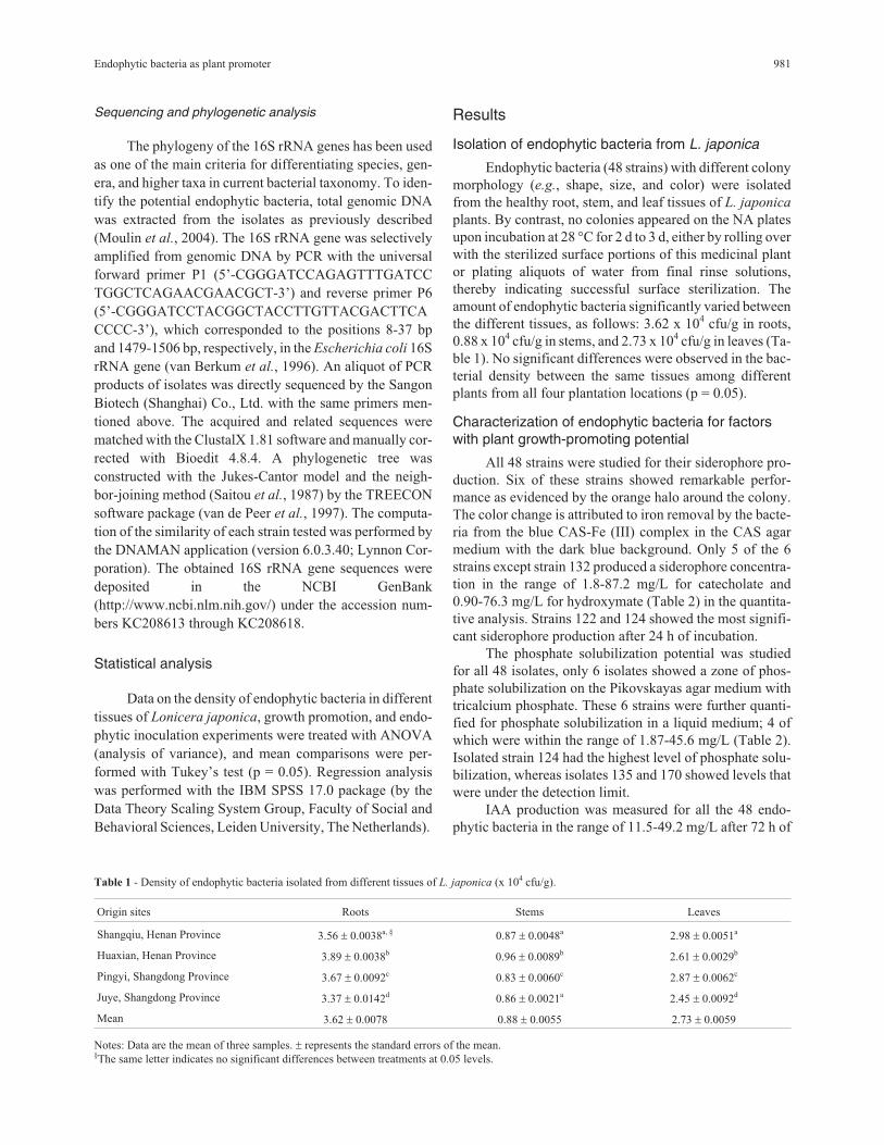
hibition rate of F. oxysporum (Figure 1A-C) for each strain
tested.
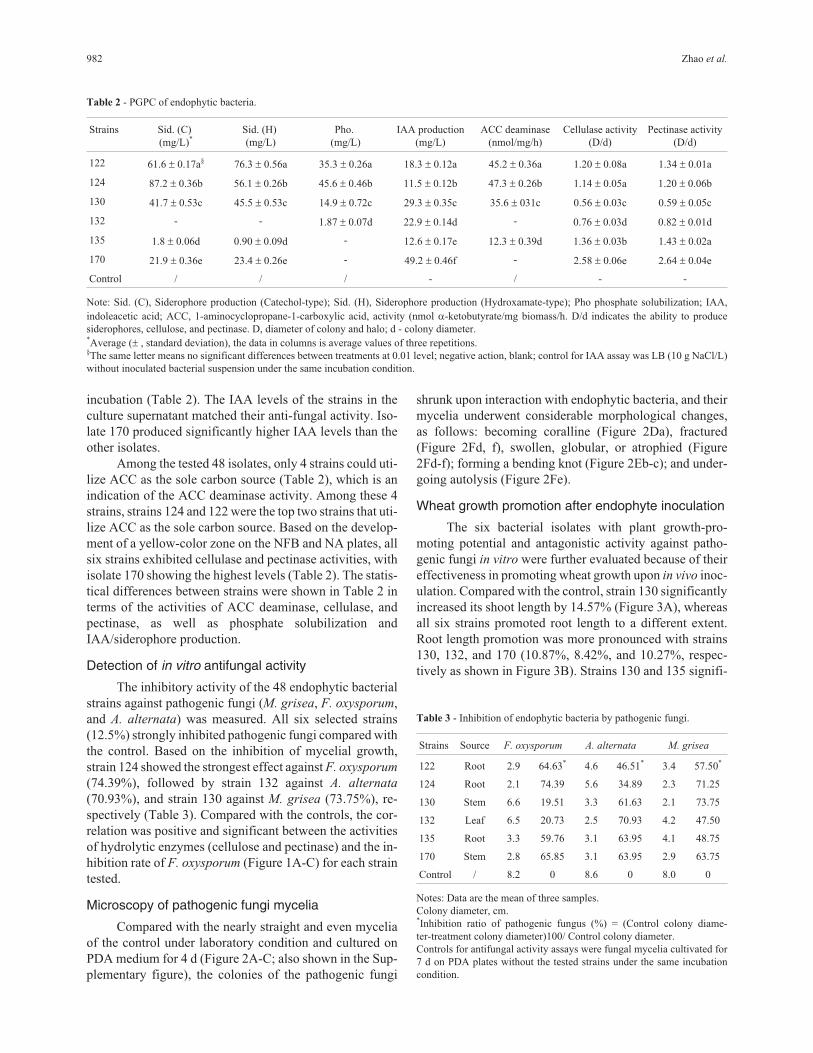
Microscopy of pathogenic fungi mycelia
Compared with the nearly straight and even mycelia
of the control under laboratory condition and cultured on
PDA medium for 4 d (Figure 2A-C; also shown in the Sup-
plementary figure), the colonies of the pathogenic fungi
shrunk upon interaction with endophytic bacteria, and their
mycelia underwent considerable morphological changes,
as follows: becoming coralline (Figure 2Da), fractured
(Figure 2Fd, f), swollen, globular, or atrophied (Figure
2Fd-f); forming a bending knot (Figure 2Eb-c); and under-
going autolysis (Figure 2Fe).
Wheat growth promotion after endophyte inoculation
The six bacterial isolates with plant growth-pro-
moting potential and antagonistic activity against patho-
genic fungi in vitro were further evaluated because of their
effectiveness in promoting wheat growth upon in vivo inoc-
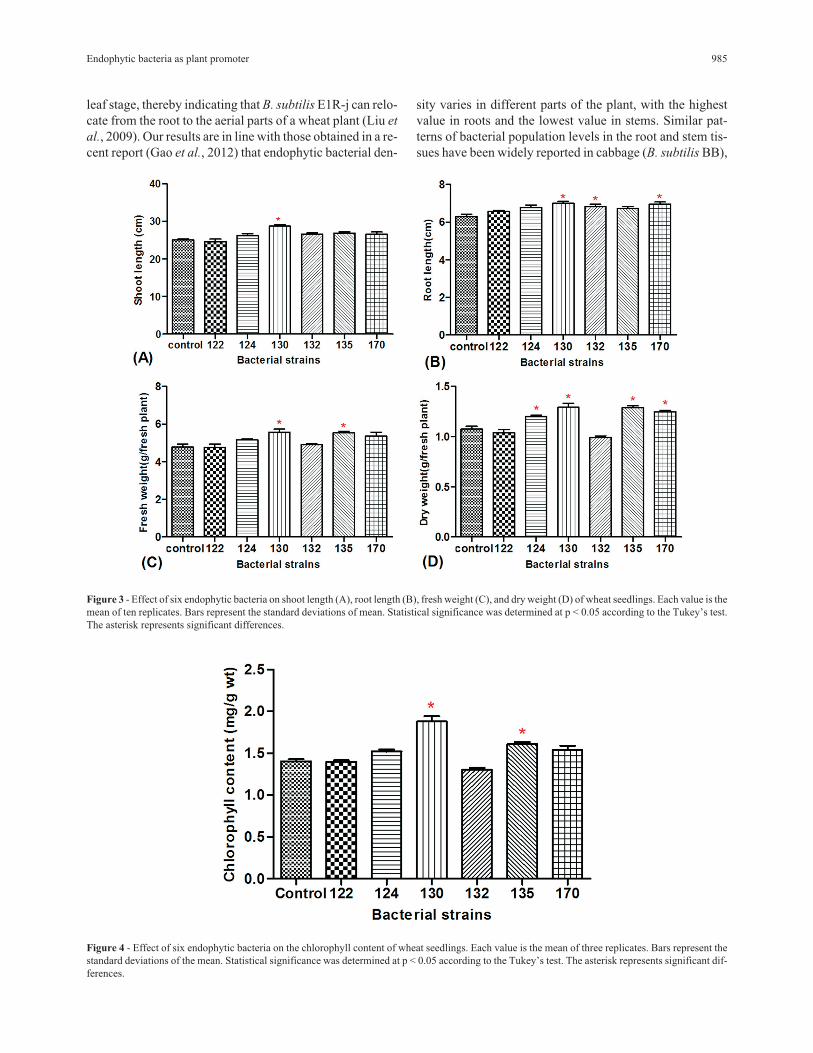
ulation. Compared with the control, strain 130 significantly
increased its shoot length by 14.57% (Figure 3A), whereas
all six strains promoted root length to a different extent.
Root length promotion was more pronounced with strains
130, 132, and 170 (10.87%, 8.42%, and 10.27%, respec-
tively as shown in Figure 3B). Strains 130 and 135 signifi-
982 Zhao et al.
Table 2 - PGPC of endophytic bacteria.
Strains Sid. (C)
(mg/L)*
Sid. (H)
(mg/L)
Pho.
(mg/L)
IAA production
(mg/L)
ACC deaminase
(nmol/mg/h)
Cellulase activity
(D/d)
Pectinase activity
(D/d)
122 61.6 � 0.17a§ 76.3 � 0.56a 35.3 � 0.26a 18.3 � 0.12a 45.2 � 0.36a 1.20 � 0.08a 1.34 � 0.01a
124 87.2 � 0.36b 56.1 � 0.26b 45.6 � 0.46b 11.5 � 0.12b 47.3 � 0.26b 1.14 � 0.05a 1.20 � 0.06b
130 41.7 � 0.53c 45.5 � 0.53c 14.9 � 0.72c 29.3 � 0.35c 35.6 � 031c 0.56 � 0.03c 0.59 � 0.05c
132 - - 1.87 � 0.07d 22.9 � 0.14d - 0.76 � 0.03d 0.82 � 0.01d
135 1.8 � 0.06d 0.90 � 0.09d - 12.6 � 0.17e 12.3 � 0.39d 1.36 � 0.03b 1.43 � 0.02a
170 21.9 � 0.36e 23.4 � 0.26e - 49.2 � 0.46f - 2.58 � 0.06e 2.64 � 0.04e
Control / / / - / - -
Note: Sid. (C), Siderophore production (Catechol-type); Sid. (H), Siderophore production (Hydroxamate-type); Pho phosphate solubilization; IAA,
indoleacetic acid; ACC, 1-aminocyclopropane-1-carboxylic acid, activity (nmol �-ketobutyrate/mg biomass/h. D/d indicates the ability to produce
siderophores, cellulose, and pectinase. D, diameter of colony and halo; d - colony diameter.*Average (� , standard deviation), the data in columns is average values of three repetitions.§The same letter means no significant differences between treatments at 0.01 level; negative action, blank; control for IAA assay was LB (10 g NaCl/L)
without inoculated bacterial suspension under the same incubation condition.
Table 3 - Inhibition of endophytic bacteria by pathogenic fungi.
Strains Source F. oxysporum A. alternata M. grisea
122 Root 2.9 64.63* 4.6 46.51* 3.4 57.50*
124 Root 2.1 74.39 5.6 34.89 2.3 71.25
130 Stem 6.6 19.51 3.3 61.63 2.1 73.75
132 Leaf 6.5 20.73 2.5 70.93 4.2 47.50
135 Root 3.3 59.76 3.1 63.95 4.1 48.75
170 Stem 2.8 65.85 3.1 63.95 2.9 63.75
Control / 8.2 0 8.6 0 8.0 0
Notes: Data are the mean of three samples.
Colony diameter, cm.*Inhibition ratio of pathogenic fungus (%) = (Control colony diame-
ter-treatment colony diameter)100/ Control colony diameter.
Controls for antifungal activity assays were fungal mycelia cultivated for
7 d on PDA plates without the tested strains under the same incubation
condition.
cantly increased wheat fresh weight (16.48% and 15.78%,
Figure 3C) and dry weight (20.07% and 19.65%, Figu-
re 3D). Regression analysis (Figure 1A) showed a signifi-
cant positive correlation between IAA production and
increase in root length of the wheat seedlings inoculated
with endophytic bacteria.
Compared with the uninoculated control (Figure 4),
the chlorophyll content of the endopyhte-inoculated wheat
Endophytic bacteria as plant promoter 983
Figure 1 - Regression analysis of the effect of endophytic bacteria on root length and the pathogenic fungus F. oxysporum. (A) Regression analysis of
bacterial IAA and root lengths of seedlings inoculated with endopyhtic bacteria. (B) Regression analysis of bacterial cellulase activity and inhibition rate
of F. oxysporum. (C) Regression analysis of bacterial pectinase activity and inhibition rate of F. oxysporum. Names of strains are shown on different data
dots.
increased to a range of 8.4% to 33.98%, with the strain
130-treated group showing the highest content (33.98%).
The inoculation of strain 130 induced the highest increase
in shoot length, root length, fresh weight, dry weight, and
chlorophyll content of wheat.
Identification of the six plant growth-promotingendophytic bacteria
Physiological and biochemical tests were conducted,
including measurements of catalase, V-P test, utilization of
carbohydrate, the production of enzymes, growth tempera-
ture, salt tolerance, and bacterial morphology (i.e., size,
shape, and Gram staining; Supplementary Table 1). Based
on these results and the sequencing of 16S rRNA gene and
phylogeny analysis (Figure 5 and Table 4), our endophytic
isolates belonged to two genera, namely, Bacillus and
Paenibacillus. Strains 122 and 130 showed high identity
with Paenibacillus and were most closely related to P.
polymyxa IAM 13419T (D16276) and P. ehimensis
KCTC3748 (AY116665) with 98.7% and 100% similarity,
respectively. Therefore, a Paenibacillus sub-clade was
formed. Strains 170, 124, 132, and 135 had high sequence
similarities to B. atrophaeus NRRLNRS-213T (EU138516)
(99.6%), B. megaterium IAM13418T (D16273) (99.1%),
and B. subtilis FL (EU221673) with a similarity of 99.6%
and 99.7%.
Discussion
This work is the first report on the isolation and popu-
lation density of endophytic bacteria from the medicinal
plant L. japonica, which is widely planted in the Henan and
Shandong provinces in eastern China. As summarized in
Table 1, the endophytic bacterial load varied significantly
in different tissues, but it remained relatively stable without
significant differences in the same tissues of different
plants from different planting locations (p = 0.05), thereby
implying that the prevalence of endophytic bacteria de-
pended on the plant tissues being colonized and the micro-
environment they lived in. The endophytic bacterial
number in roots is the highest probably because the root soil
environment is fairly complex under the effect of both bi-
otic and abiotic factors. Previous studies have reported that
the population of endophytic B. subtilis E1R-j in roots in-
creases when the bacterial cell number reisolated from the
leaf tissue distinctly decreases from the second to fourth
984 Zhao et al.
Figure 2 - Morphological changes of the mycelia of plant pathogenic fungi upon interaction with Lonicera japonica endophytes. Images in A, B, and C
were representative of normal mycelia of M. grisea MG01, F. oxysporum FO02, and A. alternata AA03. Images in D, E, F were the atrophy and deformity
of M. grisea MG01 mycelia (by strain 130), bending knot of F. oxysporum FO02 mycelia (by strain 124), autolysis, fracture, and atrophy of A. alternata
AA03 mycelia (by strain 132).
leaf stage, thereby indicating that B. subtilis E1R-j can relo-
cate from the root to the aerial parts of a wheat plant (Liu et
al., 2009). Our results are in line with those obtained in a re-
cent report (Gao et al., 2012) that endophytic bacterial den-
sity varies in different parts of the plant, with the highest
value in roots and the lowest value in stems. Similar pat-
terns of bacterial population levels in the root and stem tis-
sues have been widely reported in cabbage (B. subtilis BB),
Endophytic bacteria as plant promoter 985
Figure 3 - Effect of six endophytic bacteria on shoot length (A), root length (B), fresh weight (C), and dry weight (D) of wheat seedlings. Each value is the
mean of ten replicates. Bars represent the standard deviations of mean. Statistical significance was determined at p < 0.05 according to the Tukey’s test.
The asterisk represents significant differences.
Figure 4 - Effect of six endophytic bacteria on the chlorophyll content of wheat seedlings. Each value is the mean of three replicates. Bars represent the
standard deviations of the mean. Statistical significance was determined at p < 0.05 according to the Tukey’s test. The asterisk represents significant dif-
ferences.
cacao (B. subtilis), rose, and crops such as maize, wheat,
rice (B. subtilis strain NR-64), soybean, sweet corn, sugar
beet, and potato, or in various medicinal plants such as
Glycyrrhiza spp., Pinellia ternate, Lycium chinense, Digi-
talis purpurae, Leonurus heterophyllus, Bletilla striata,
Belamcanda chinases, P. pedatisecta, and Taxus
yunnanensis (Venieraki et al., 2011; Gao et al., 2012;
Wulff et al., 2003; Bahig et al., 2012; Li et al., 2012; Miller
et al., 2012a; Leite et al., 2013).
Siderophore production could confer competitive ad-
vantages in bacteria for colonizing plant tissues, excluding
other microorganisms from the same ecological niche
986 Zhao et al.
Figure 5 - Neighbor-joining tree based on the alignment of nucleotide sequences of the 16S rRNA gene from the tested strains (shown in bold) and refer-
ence strains. GenBank accession numbers were placed in parentheses. Bootstrap values greater than 50% were indicated. Scale bar represents the number
of substitutions per site.
Table 4 - Identification and classification of the tested strains.
Strains Genus affiliation Accession No. of the 16S rDNA sequence Best closest match Similarity (%)
122 Paenibacillus KC208613 Paenibacillus polymyxa IAM 13419T (D16276) 98.7
124 Bacillus KC208614 Bacillus megaterium IAM13418T (D16273) 99.1
130 Paenibacillus KC208617 Paenibacillus ehimensis IFO15659T (AB021184) 100
132 Bacillus KC208615 Bacillus subtilis FL (EU221673) 99.6
135 Bacillus KC208616 Bacillus subtilis FL (EU221673) 99.7
170 Bacillus KC208618 Bacillus atrophaeus NRRLNRS-213T (EU138516) 99.6
(Loaces et al., 2011), competing for nutrients, and protect-
ing plant from phytopathogens (Compant et al., 2005).
Metagenomic analysis (Sessitsch et al., 2012) revealed that
the presence of a high number of genes involved in sidero-
phore production in an endophyte community that colo-
nizes rice roots indicates a strong biocontrol capacity
because endophytes compete with other pathogens for iron.
Among the 6 endophytic bacteria (3 strains from roots, 2
strains from stems, and 1 strain from leaves; 4 Bacillus and
2 Paenibacillus strains), strains 122 and 124 showed a
higher capacity for siderophore production.
Several phosphate-solubilizing microorganisms are
able to convert insoluble phosphorus to a soluble form
through acidification, secretion of organic acids or protons
(Richardson et al., 2009), or chelation and exchange reac-
tions (Hameeda et al., 2008; Bhattacharyya et al., 2012),
thereby representing a possible mechanism of direct plant
growth promotion under field conditions (Verma et al.,
2001). These microorganisms are important for plant nutri-
tion because they increase phosphate uptake and act as
biofertilization promoters of wheat crops. Bacillus is re-
portedly one of the most significant phosphate-solubilizing
bacteria (Mehnaz et al., 2006). Four of our six strains are
capable of phosphate solubilization. Strains 124 (Bacillus)
and 122 (Paenibacillus) showed relatively higher phos-
phate solubilization than the others, and these strains signif-
icantly increased the dry weight and fresh weight of the
inoculated wheat, respectively.
In this report, six endophytic strains from the medici-
nal plant L. japonica exhibited inhibitory activity (Table 3)
against phytopathogenic fungi (F. oxysporum, M. grisea, A.
alternata), which are useful model organisms for studying
various aspects of host-pathogen interactions. These patho-
gens are chosen as test targets in the antifungal activity ex-
periment because of their capacity to cause epiphytic
disease and major damage in crops and plants, including L.
japonica and wheat. Previous works have verified that pro-
moting plant growth and inhibiting phytopathogen growth
may involve a large number of bacterial endophytes. A re-
cent report also suggests that the cellulase and pectinase
produced by Klebsiella oxytoca GR-3 might play an impor-
tant role in plant-microbe interactions and the intercellular
colonization of roots (Ma et al., 2011). Our findings re-
vealed that all 6 of the selected strains (Bacillus and
Paenibacillus) exhibited cellulose and pectinase activities.
Growth-promoting agents can enhance plant growth
and have the potential to replace the use of chemical fertil-
izers, pesticides, and other supplements (Kim et al., 2011;
Bhattacharyya et al., 2012). Endophytes that are associated
with medicinal plants are of interest as producers of com-
pounds responsible for the observed plant bioactivity with
biosynthetic potential (Miller et al., 2012b). This finding is
in agreement with our result, i.e., strain 130 induced the
largest increase in root length, stem length, flesh weight,
dry weight, and the chlorophyll content of wheat in vivo, al-
though its in vitro ACC deaminase activity is lower than
that of strains 122 and 124. One explanation for this phe-
nomenon is that strain 130 contains ACC deaminase but its
activity was not induced very much in the in vitro study. Al-
ternatively, strain 130 showed a higher IAA production,
thereby directly promoting wheat growth via hormonal
stimulation. Furthermore, plant growth is the overall result
of all growth-promoting molecules produced by root endo-
phytes and ectophytes (Patten et al., 2002).
In summary, our work showed that some of the endo-
phytic bacteria (strains 130, 135, and 170) isolated from the
medicinal plant L. japonica can produce wheat growth-
promoting molecules in vitro and increase wheat growth
(i.e., root length, stem length, flesh weight, dry weight, and
chlorophyll content) in vivo. Therefore, these endophytic
bacteria have the potential to be used as plant growth-
promoting agents in agriculture to increase crop growth.
Acknowledgments
This work was supported by projects from the Na-
tional Science Foundation of China (U1204301), the Henan
Provincial Education Department of Science and Technol-
ogy Research Key Research Project (12A210019), and the
Foundation for University Key Teacher by the Ministry of
Education of Henan Province (2012GGJS166). The Au-
thors are grateful to Dr. Guihong Yin for providing the
wheat seeds (cv. `Zhoumai18’). We thank Prof. Yi Ren of
the Department of Biomedical Sciences, Florida State Uni-
versity College of Medicine, Tallahassee, FL, USA, Dr.
Jane Kelly, of the Medical Research Service, RD-33, De-
partment of Veterans Affairs Medical Center, 3710 SW
U.S. Veterans Hospital Road, Portland, OR, USA, and an
anonymous reviewer from the USA for proofreading and
polishing the final draft of our manuscript.
References
Bahig ED, Salih B, Youssuf G et al. (2012) Characterization of
endophytic bacteria associated with rose plant (Rosa
damascena trigintipeta) during flowering stage and their
plant growth promoting traits. J Plant Interact 7:248-225.
Belimov AA, Hontzeas N, Safronova VI et al. (2005) Cadmium-
tolerant plant growth-promoting bacteria associated with the
roots of Indian mustard (Brassica juncea L. Czern.). Soil
Biol Biochem 37:241-250.
Bhattacharyya PN, Jha DK (2012) Plant growth-promoting rhizo-
bacteria (PGPR): emergence in agriculture. World J Microb
Biot 28:1327-1350.
Bianco A, Daffonchio FQ, Lorenzo B et al. (2011) Restructuring
of Endophytic Bacterial Communities in Grapevine Yel-
lows-Diseased and Recovered Vitis vinifera L. Plants. Appl
Environ Microbiol 77:5018-5022.
Broggi L, González HHL, Resnik S et al. (2007) Alternaria
alternata prevalence in cereal grains and soybean seeds
from Entre Ríos, Argentina. Rev Iberoam Micol 24:47-51.
Chen Y, Yan F, Chai Y et al. (2013) Biocontrol of tomato wilt dis-
ease by Bacillus subtilis isolates from natural environments
Endophytic bacteria as plant promoter 987
depends on conserved genes mediating biofilm formation.
Environ Microbiol 15:916-927.
Compant S, Duffy B, Nowak J et al. (2005) Use of plant growth-
promoting bacteria for biocontrol of plant diseases: princi-
ples, mechanisms of action, and future prospects. Appl En-
viron Microb 71:4951-4959.
Deng ZS, Zhao LF, Kong ZY et al. (2011) Diversity of endophytic
bacteria within nodules of the Sphaerophysa salsula in dif-
ferent regions of Loess Plateau in China. FEMS Microbiol
Ecol 76:463-475.
Fiske CH, Subbarow Y (1925) A colorimetric determination of
phosphorus. J Biol Chem 66:375-400.
Gao LL, Chen XL, Jian T et al. (2012) Isolation and identification
of endophytic nitrogen-fixing bacteria in rice with antipa-
thogenic functions. J Huazhong Agri University 31:553-
557.
Geetha R, Falguni S, Anjana JD et al. (2008) Enhanced growth
and nodulation of pigeon pea by co-inoculation of Bacillus
strains with Rhizobium spp.. Biores Technol 99:4544-4550.
González HHL, Pacin A, Resnik SL et al. (1996) Deoxynivalenol
and contaminant mycoflora in freshly harvested Argentin-
ean wheat in 1993. Mycopathologia 135:129-134.
Gordon AS, Weber RP (1951) Colorimetric estimation of indo-
leacetic acid. Plant Physiol 26:192-195.
Hameeda B, Harini G, Rupela OP et al. (2008) Growth promotion
of maize by phosphate-solubilizing bacteria isolated from
composts and macrofauna. Microbiol Res 163:234-242.
Inés L, Lucía F, Ana Fernández S (2011) Dynamics, diversity and
function of endophytic Siderophore-producing bacteria in
rice. Microb Ecol 61:606-618.
Khan Z, Kim SG, Jeon YH et al. (2008) A plant growth promoting
Paenibacillus polymyxa strain GBR-1, suppresses root-knot
nematode. Biores Technol 99:3016-3023.
Kim YC, Johan L, Brian B et al. (2011) The multifactorial basis
for plant health promotion by plant- associated bacteria.
Appl Environ Microbiol 77:1548-1555.
Lai KP, Chen SH, Hu MY et al. (2012) Control of postharvest
green mold of citrus fruit by application of endophytic
Paenibacillus polymyxa strain SG-6. Postharv Biol Technol
69:40-48.
Leite HA, Silva AB, Gomes FP et al. (2013) Bacillus subtilis and
Enterobacter cloacae endophytes from healthy Theobroma
cacao L. trees can systemically colonize seedlings and pro-
mote growth. Appl Microbiol Biotechnol 97:2639-2651.
Li HJ, Li P, Ye WC (2003) Determination of five major iridoid
glucosides in Flos Lonicerae by high-performance liquid
chromatography coupled with evaporative light scattering
detection. J Chromatogr A 1008:167-172.
Li J, Zhang HR, Liu NY et al. (2010) Study on isolation and iden-
tification endophytic fungi and antibacterial activity of Flos
Lonicerae. Chin J Antibiot 35:236-238.
Li L, Sinkko H, Montonen L et al. (2012) Biogeography of symbi-
otic and other endophytic bacteria isolated from medicinal
Glycyrrhiza species in China. FEMS Microbiol Ecol 79:46-
68.
Liu B, Qiao HP, Huang LL et al. (2009) Biological control of
take-all in wheat by endophytic Bacillus subtilis E1R-j and
potential mode of action. Biol Control 49:277-285.
Loaces I, Lucía F, Ana FS (2011) Dynamics, diversity and func-
tion of endophytic siderophore-producing bacteria in rice.
Microb Ecol 61:606-618.
Ma Y, Mani R, Luo YM et al. (2011) Inoculation of endophytic
bacteria on host and non-host plants-effects on plant growth
and Ni uptake. J Hazard Mater 195:230-237.
María SO, María ES, María MR et al. (2013) Toxigenic profile
and AFLP variability of Alternaria alternata and Alternaria
infectoria occurring on wheat. Braz J Microbiol 44:447-455.
Mehnaz S, Lazarovits G (2006) Inoculation effects of Pseudomo-
nas putida, Gluconacetobacter azotocaptans, and
Azospirillum lipoferum on corn plant growth under green-
house conditions. Microb Ecol 51:326-335.
Miller KI, Qing C, Sze DM et al. (2012a) Culturable endophytes
of medicinal plants and the genetic basis for their bio-
activity. Microb Ecol 64:431-449.
Miller KI, Qing C, Sze DMY et al. (2012b) Investigation of the
biosynthetic potential of endophytes in traditional Chinese
anticancer herbs. Plos One 7:e35953.
Moulin L, Béna G, Boivin-Masson C et al. (2004) Phylogenetic
analyses of symbiotic nodulation genes support vertical and
lateral gene co-transfer within the Bradyrhizobium genus.
Mol Phylogenet Evol 30:720-732.
Mphprc (2000) Pharmacopoeia of the People’s Republic of China.
Chemical Industry Press, Beijing, 177 p.
Patten CL, Glick BR (2002) The role of bacterial indoleacetic acid
in the development of the host plant root system. Appl Envi-
ron Microbiol 68:3795-3801.
Ramirez ML, Sturm ME, Oviedo MS et al. (2005) Alternaria spe-
cies isolated from wheat in Argentina. Proceeding of Myco-
Globe: Reducing Impact of Mycotoxins in Tropical Agricul-
ture. Acra, Ghana, pp 35.
Raza W, Yang XM, Wu HS et al. (2009) Isolation and character-
ization of fusaricidin-type compound-producing strain of
Paenibacillu polymyxa SQR-21 active against Fusarium
oxysporum f. sp. nevium. Eur J Plant Pathol 125:471-483.
Richardson AE, Barea JM, McNeill AM et al. (2009) Acquisition
of phosphorus and nitrogen in the rhizosphere and plant
growth promotion by microorganisms. Plant Soil 321:305-
339.
Saitou N, Nei M (1987) The neighbor-joining method: a new
method for reconstructing phylogenetic trees. Mol Biol Evol
4:406-425.
Schwyn B, Neilands JB (1987) Universal chemical assay for the
detection and determination of siderophores. Anal Biochem
160:47-56.
Sessitsch A, Hardoim PJ, Döring WA et al. (2012) Functional
characteristics of an endophyte community colonizing rice
roots as revealed by metagenomic analysis. MPMI 25:28-
36.
Sundara-Rao WVB, Sinha MK (1963) Phosphate dissolving mi-
croorganisms in the soil and rhizosphere. Indian J Agr Sci
33:272-278.
Van Berkum P, Beyene D, Eardly BD (1996) Phylogenetic rela-
tionships among Rhizobium species nodulating the common
bean (Phaseolus vulgaris L.). Int J Syst Evol Microbiol
46:240-244.
Van de Peer, Wachter YDR (1997) Construction of evolutionary
distance trees with TREECON for Windows: accounting for
variation in nucleotide substitution rate among sites.
Comput Appl Biosci 13:227-230.
Venieraki A, Dimou M, Pergalis P et al. (2011) The genetic diver-
sity of culturable nitrogen-fixing bacteria in the rhizosphere
of wheat. Microb Ecol 61:277-285.
988 Zhao et al.
Verma SC, Ladha JK, Tripathi AK (2001) Evaluation of plant
growth promoting and colonization ability of endophytic
diazotrophs from deep water rice. J Biotechnol 91:127-141.
Vincent JM (1970) The cultivation, isolation and maintenance of
Rhizobia. A Manual for the practical study of the root-
nodule bacteria international biological programme hand-
book. Blackwell Scientific, Oxford, pp 1-13.
Wu L (2007) Effect of chlorogenic acid on antioxidant activity of
Flos Lonicerae extracts. J Zhejiang Univ Sci B 8:673-679.
Wulff EG, van Vuurde JWL, Hockenhull J (2003) The ability of
the biological control agent Bacillus subtilis, strain BB, to
colonise vegetable brassicas endophytically following seed
inoculation. Plant and Soil 255:463-474.
Xu YJ, Zhao LF, Dai M et al. (2013) Isolation and characteristic
of endophytic bacteria in medicinal plant Flos Lonicerae.
Guandong Agr Sci 40:121-124.
Zhao LF, Xu YJ, Sun R et al. (2011) Identification and character-
ization of the endophytic plant growth prompter Bacillus ce-
reus strain MQ23 isolated from Sophora alopecuroides root
nodules. Braz J Microbiol 42:567-575.
Supplementary MaterialFigure S1. Antagonistic activity of endophytes against phyto-
pathogenic fungi after 3 d. (A) Inhibition of M. grisea MG01
by endophyte strain 130; (B0 inhibition of F. oxysporum
FO02 by endophyte strain 124; C. inhibition of A. alternata
AA03 by endophyte strain 132. A’, B’, and C’ were control
pathogenic fungusfungal colonies without endophytes.
Table S1. A Physiological and biochemical test results and cell
characteristics of strains 122, 124, 130, 132, 135, and 170.
Associate Editor: Lara Durães Sette
All the content of the journal, except where otherwise noted, is licensed under a
Creative Commons License CC BY-NC.
Endophytic bacteria as plant promoter 989