molecular carcinogenesis 9999:1–10 (2006) mesenchymal
TRANSCRIPT
Author Proof
AMesenchymal Transformation in EpithelialOvarian Tumor Cells Expressing EpidermalGrowth Factor Receptor Variant III
Reema Zeineldin,1 Martina Rosenberg,1 Dominic Ortega,1 Christian Buhr,1
Miquella G. Chavez,1 M. Sharon Stack,2 Donna F. Kusewitt,3 and Laurie G. Hudson1*1Program in Toxicology & Pharmacology, College of Pharmacy,University of New Mexico Health Sciences Center, Albuquerque, New Mexico2Department of Cell and Molecular Biology, Northwestern University, Chicago, Illinois3Department of Veterinary Biosciences, The Ohio State University, Columbus, Ohio
Overexpression of the epidermal growth factor (EGF) receptor occurs frequently in ovarian cancer and is associated
with poor patient prognosis. A constitutively active mutant EGF receptor termed variant III (EGFRvIII) has been detectedat a high frequency in many human tumors, including those of the ovary. To identify the consequences of EGFRvIIIexpression in ovarian tumor cells, we introduced EGFRvIII into the epithelial ovarian cancer cell line (OVCA 433). TheEGFRvIII-transfected cells displayed a dissociated, motile phenotype and fibroblastic morphology. The EGFRvIII-
dependent phenotype was comparable to that observed in EGF-stimulated parental OVCA 433 cultures and requiredthe catalytic activity of the mutant receptor. Disruption of adherens and desmosomal junctions in EGFRvIII expressingcells was evident by immunofluorescent detection of specific junctional components. In addition, Western blot analysis
confirmed decreased levels of cellular plakoglobin and b-catenin in EGFRvIII-expressing cells, and E-cadherin proteinand mRNA were nearly absent. The loss of E-cadherin was accompanied by decreased expression of additional ovarianepithelial markers, including keratins 7, 8, and 18 and mucins 1 and 4. In contrast, the mesenchymal markers N-
cadherin and vimentin were elevated in EGFRvIII expressing cells. Overall, the switch in cadherins from E-cadherin to N-cadherin, coupled with gain of vimentin expression and loss of the epithelial keratins and mucins typically expressed inwell-differentiated epithelial ovarian carcinomas, are consistent with transition to a mesenchymal phenotype as an
outcome of EGFRvIII expression. These findings suggest that EGFRvIII expression may regulate phenotypic plasticity inovarian cancer and thereby contribute to more aggressive disease. � 2006 Wiley-Liss, Inc.
Key words: epidermal growth factor receptor; EGFR; EGFRvIII; signal transduction; ovarian cancer; adherens junctions;epithelial-mesenchymal transition
INTRODUCTION
Ovarian carcinoma is the leading cause of deathfrom gynecologic malignancy, resulting in approxi-mately 16 210 deaths in 2005 [1], with epithelialtumors accounting for �90% of ovarian malignan-cies [2]. In the adult female, the normal ovarianepithelium is a single cell layer separated by asubepithelial basement membrane from an under-lying stroma, the tunica albuginea, which comprisesdense collagenous connective tissue [2]. The meso-dermally derived ovarian surface epithelium (OSE)is a simple epithelium that displays epithelialand mesenchymal characteristics and containsboth keratin and vimentin intermediate filaments.In tissue culture, normal ovarian epithelial cellsexpress epithelial markers including keratins 7, 8,and 18, desmoplakin and mucin 1, as well asmesenchymal markers such as vimentin and neural(N)-cadherin [2,4]. Ovarian epithelial cells exhibitphenotypic plasticity; reversible modulation ofovarian epithelium to a fibroblastic form occurs
during postovulatory repair and is also observed inovarian tumor progression [2,3].
In the early stages of neoplastic transformation,OSE undergoes Mullerian differentiation so thatnew epithelial features appear, while the mesench-ymal characteristics of OSE diminish [2]. The newepithelial features include altered cell shape [5],appearance of epithelial (E)-cadherin, formation ofjunctional complexes [2,6,7], expression of epithe-lial membrane antigens, and increased production of
MOLECULAR CARCINOGENESIS 9999:1–10 (2006)
� 2005 WILEY-LISS, INC. MC-05-0104.R2
Abbreviations: OSE, ovarian surface epithelium; EGF, epidermalgrowth factor; EGFRvIII, epidermal growth factor receptor variant III;PBS, phosphate buffered saline; EMT, epithelial to mesenchymaltransformation.
*Correspondence to: University of New Mexico Health SciencesCenter, College of Pharmacy, MSC09 5360, 1 University of NewMexico, Albuquerque, NM 87131-0001.
Received 18 July 2005; Revised 12 February 2006; Accepted 17March 2006
DOI 10.1002/mc.00000
Published online 00 Month 2006 in Wiley InterScience(www.interscience.wiley.com)
Author Proof
Asecretory products including mucins (mucin1-4) andCA125 [2,5]. At later stages of tumor progression,some of these specialized epithelial characteristics,such as E-cadherin expression, may diminish ascells de-differentiate [6–8] and acquire mesenchy-mal properties associated with a more aggressivephenotype.
The mechanisms regulating phenotypic transi-tions in ovarian cancer are unknown; however,reversible modulation of ovarian epithelial cellsduring postovulatory repair has been linked toexpression and activity of the epidermal growthfactor (EGF) receptor [2,3]. Overexpression of theEGF receptor and its ligands is often detected inovarian tumors [9–13] and is associated with a lessfavorable prognosis. Aberrant EGF receptor activa-tion may play a critical role in ovarian tumorprogression and metastasis through control of pro-liferation [14–17], regulation of cell:cell and cell:matrix interactions [18–20], stimulation of cellmigration [18,21], and induction of many matrixdegrading proteases including those belonging tothe plasminogen activator and matrix metallopro-teinase families [20]. Soluble ligand-binding variantsof the EGF receptor (extracellular domain) arealso linked to disease progression in ovarian cancer[13]. Although the role of these soluble receptors inovarian pathophysiology is unclear, it has beensuggested that truncated ectodomain variants maymodulate EGF receptor autophosphorylation [13].Importantly, decreased expression of EGF receptor inovarian carcinoma cells in culture reduces theirmalignant character [22,23]. Together, these find-ings suggest that EGF receptor activation is a factor innumerous aspects of ovarian cancer biology.
Epidermal growth factor receptor can be activatedby ligand binding or receptor mutation. Naturallyoccurring EGF receptor mutations occur in humantumors. Of particular interest is a mutant EGFreceptor designated DEGFR, de2-7 EGFR or EGFRvariant III (EGFRvIII). The EGFRvIII is a 145-kDa EGFreceptor with a deletion in the extracellular domainof the receptor corresponding to nucleotides 275–1075 (exons 2 through 7) of the EGF receptor cDNA[24,25]. This mutant EGF receptor has been fre-quently detected in a variety of human tumorsincluding glioblastoma, breast, lung, prostate, andovarian cancers [26–30]. This mutant receptor doesnot bind EGF [31]; nevertheless, it is constitutivelyactive as detected by receptor dimerization [31],autophosphorylation [32], and activation of keysignal transduction cascades [28,33]. EGFRvIII isfunctionally active without ligand in models oftumorigenesis; it stimulates DNA synthesis, trans-formation of fibroblasts, and potentiates tumorgrowth in nude mice [28,33]. In addition, expressionof EGFRvIII in a glioblastoma or small cell lungcancer cell line renders the cells more invasive[34,35].
The potential impact of EGFRvIII expressionon the development or progression of ovariancancer is currently unknown. In this study, we stablyexpressed EGFRvIII in an epithelial ovarian carci-noma cell line (OVCA 433). We found that EGFRvIIIexpression resulted in a dispersed phenotype char-acterized by increased cell migration, dissolution ofadherens and desmosomal junctions, and down-regulation of certain junctional proteins. Interest-ingly, protein and mRNA levels of the epithelialmarker E-cadherin were significantly decreased inthe EGFRvIII-expressing cells when compared toligand-stimulated parental or vector control cells.The decrease in E-cadherin was accompanied by anincrease in N-cadherin expression. Other epithelialmarkers were diminished in EGFRvIII-expressingcells including keratins 7, 8, and 18, and mucins 1and 4, whereas vimentin expression was elevated.These results suggest that the molecular changesassociated with EGFRvIII expression may driveepithelial-mesenchymal transition, and contributeto metastatic dissemination of ovarian cancer.
MATERIALS AND METHODS
Cell Culture and Treatment
Ovarian carcinoma cell lines OVCA 433, andOVCA 429 were generously provided by Dr. RobertBast, Jr., M. D. Anderson Cancer Center, Houston, TXand grown as described previously [18,19,36].Briefly, cells were grown in minimum essentialmedium (MEM) supplemented with 10% (v/v) fetalcalf serum (FCS), 1 mM sodium pyruvate, 2 mM L-glutamine, 0.5 units/mL penicillin, 0.5 mg/mL strep-tomycin (this medium is referred to later as completegrowth medium). Cells were maintained at 378Cunder 5% carbon dioxide. The OVCA 433 cellline was selected for transfection based on low tomoderate expression of endogenous wild-type EGFreceptor compared to other OVCA lines, as deter-mined by Western blot analysis and evidence ofintact EGF-dependent signaling pathways (data notshown). The EGFRvIII construct was a generousgift of Dr. David Moscatello, Thomas JeffersonUniversity, Philadelphia, PA. OVCA 433 cells werecotransfected with a vector containing a neomycinresistance gene and either the EGFRvIII construct inPLTR2 vector or the PLTR2 vector at a 1:10 ratio bycalcium phosphate method. Clones were selected inpresence of 300 mg/mL G418 (GibcoQ1/BRL). TenEGFRvIII-transfected clones were isolated from twoindependent transfections. All clones displayedthe same phenotype, and were similar. Two EGFR-vIII-transfected clones, designated EGFRvIIIA1 andEGFRvIIIA2, were selected for extensive character-ization. For experiments involving EGF (BiomedicalTechnologies, Stoughton, MA), OVCA 433 cell lineswere placed into MEM containing 0.1% (w/v) bovineserum albumin (BSA) for 24 h prior to growth factor
2 ZEINELDIN ET AL.
Molecular Carcinogenesis DOI 10.1002/mc

Author Proof
Aaddition, as described previously [18,19]. Treatmentwith the EGF receptor catalytic inhibitor AG1478(CalBiochem, LaJolla CA) was conducted in com-plete growth medium unless otherwise noted in thefigure legends.
Immunofluorescence
An antibody against the novel epitope in EGFRvIII(Ab 1825) was raised in chickens by Aves Laboratory(Tigard, OR) as described elsewhere [25] with thefollowing modifications: the anti-peptide antibodywas produced for the peptide LEEKKGNYVVTDHCafter adding an amino caproic acid just upstream ofthe C-terminal cysteine, which was used for con-jugation to keyhole limphet hemacyanin. Antibo-dies used in immunofluorescence included mouseanti-E-cadherin antibody #C20820 (TransductionLaboratory, Lexington, KY), mouse anti-b-cateninantibody #MAB2081 (Chemicon, Temecula, CA),and a chicken anti-plakoglobin antibody (generousgift of Dr. K. Green, Northwestern University,Chicago, IL). Cells were seeded in a Lab Tek IIchamber slide system (Nalge Nunc Int., Naperville,IL). For detection of EGFRvIII, cells were fixed for 7min at room temperature in freshly prepared 3.7%(w/v) paraformaldehyde in phosphate buffered sal-ine (PBS: 0.137 M sodium chloride, 27 mM potassiumchloride, 43 mM dibasic sodium phosphate, 15 mMmonobasic potassium phosphate). Cells were incu-bated in blocking buffer (10% nonfat dry milk in PBS)for 1 h at 378C, washed three times with PBS,incubated with chicken anti-EGFRvIII antibody1825 at 1:800 dilution prepared in the blockingsolution, for 1 h at 378C, washed with PBS, incubatedwith anti-chicken-fluoresceinated antibody (1:900dilution in blocking buffer) for 20 min at 378C, andmounted with Vectorshield mounting media. For allother antibodies, cells were fixed for 2 min in ice-cold, dehydrated methanol. For detection of E-cadherin, b-catenin, and plakoglobin, cells werewashed with PBS containing 0.8 mM magnesiumsulfate, and 0.18 mM calcium chloride. For detectionof b-catenin only, cells were permeabilized byincubating in cold 0.1% Triton X-100 in PBScontaining calcium and magnesium on ice for5 min, then cells were washed three times with PBScontaining calcium and magnesium. Cells wereincubated in blocking buffer (3% BSA in PBS contain-ing calcium and magnesium) and processed asdescribed above with primary antibodies directedagainst E-cadherin, b-catenin, and plakoglobin at1:100 dilution in blocking buffer. Cells were thenincubated for 1 h at 378C with secondary antibodies(anti-mouse-FITC conjugated antibody #1034-02,Southern Biotechnology Associates, Birmingham,AL or anti-chicken-FITC conjugated antibody #F-1005, Aves Labs) at 1:500 dilution prior to washingand mounting. Slides were examined with anOlympus BH2-RFCA microscope (Melville, NY) and
images were obtained with an Omegafire digitalcamera (Optronix, Goleta, CA).
Western Blot Analysis
Control and treated cells were washed with ice-cold PBS and harvested in lysis buffer (10 mM Tris-HCl, pH 7.4, 1% SDS, 5 mM EDTA, 0.1 mMdithiothreitol and 1 mM PMSF). Ten micrograms oftotal cell lysate was resolved by electrophoresisthrough 10% SDS–polyacrylamide, transferred topolyvinylidene difluoride (PVDF) membranes (Milli-pore Corp., Bedford, MA) and probed with theindicated antibodies. These included antibodyfor intracellular domain of EGFR (sc-03, SantaCruz, Santa Cruz, CA), and anti-phosphorylatedERK (#9101 Cell Signaling, Beverly, MA) at 1:1000dilutions, a cocktail of antibodies against phospho-EGFR (#2237, 2234, 2235, 2231 Cell Signaling) at1:1000 each, anti-E-cadherin (Ab H108, Santa Cruz),anti-plakoglobin (AbH80, Santa Cruz), anti-b-cate-nin (E5, Santa Cruz), and anti-desmoglein (Trans-duction Labs) at 1:500 dilutions; anti-N-cadherin(Zymed Laboratories, Inc., San Francisco, CA) at1:250 dilution; and anti-vimentin, anti-mucin-1(Chemicon) and anti-keratins 7, 8, and 18 (#K0199-07, K0199-10, and K0199-21, respectively, US Biolo-gical, Swampscott, MA) at 1:100 dilution. Secondaryantibodies were obtained form Promega (Madison,WI). Quantitation was performed by direct lumines-cence detection with SuperSignal Pico chemilumi-nescent substrate (Pierce, Rockford, IL) and theKodak Image Station 440 (NEN Life Science Pro-ductsQ2). Quantitation was performed on samplesobtained from three independent experiments andvalues represent the mean� standard deviation.
PCR Detection of mRNA
RNA was extracted from cells growing in 10 cmtissue culture plates with Trizol reagent (InvitrogenLife Technologies, Carlsbad, CA) as recommended bythe vendor. Reagents for PCR were obtained fromInvitrogen Life Technologies. To generate first strandcDNA, 250 ng of random primers was annealed to2 mg of total RNA in a 12 mL reaction, which washeated to 708C for 10 min, then quick chilled on ice.Then 10 nmol of each dNTP, 200 nmol DTT, 30 URNase inhibitor, and 5� first strand buffer wereadded, so the total volume was 19 mL. The tubes wereincubated at 428C for 2 min, followed by addition of200 U of SuperScript II RNase H� reverse transcriptaseand incubation at 258C for 10 min, at 428C for 50min, and at 708C for 15 min. The PCR reactionmixture contained 2 mL of cDNA, 25 mL of Taq PCRmaster mix, 5 pmol of each primer and the volumewas brought to 50 mL. The E-cadherin primers were50GGGTGACTACAAAATCAATC30 and 50GGGGGC-AGTAAGGGCTCTTT30 [37]. Thermal cycling startedwith an initial denaturation step at 948C for 4 minand ended with a final extension step at 728C for 7
EGFRvIII EXPRESSION IN OVARIAN TUMOR CELLS 3
Molecular Carcinogenesis DOI 10.1002/mc
Author Proof
Amin. For E-cadherin primers, thermal cycling con-sisted of 40 cycles of 948C for 60 s, 608C for 90 s, and728C for 120 s. PCR amplifications were performedon a PTC-200 Peltier Thermal Cycler (MJ Research,Inc. Watertown, MA). PCR samples were thenelectrophoresed through 2% agarose gel and visua-lized by ethidium bromide staining.
For real-time PCR, the first strand cDNA wasgenerated as described above. The real-time PCRwas performed with TaqMan universal conditions(Applied Biosystems, Foster City, CA), in which900 nM of each primer and 250 nM of the probewere used for E-cadherin. The E-cadherin primerswere 50AGGTGACAGAGCCTCTGGATAGA30 and50CATTCCCGTTGGATGACACA30, and the E-cad-herin probe was FAM-CGCATTGCCACATACACTC-TCTTC-TAMRA. In addition, real-time PCR wasperformed for GAPDH with the Applied BiosystemsGAPDH control reagent kit according to the manu-facturer’s protocol with 250 nM of each primer and100 nM of the probe. Real-time PCR reactions werecarried out on 200 ng of cDNA in a total volume of45 mL containing TaqMan 2�master mix with an ABIprism 7000 sequence detection system. Evaluation ofamplification efficiency for E-cadherin and GAPDHwas performed according to Applied Biosystemsrecommendations, and the efficiency of the twoamplification reactions was found to be equivalent.The transcripts were quantified with the DDCt
method according to Applied Biosystems recom-mendations with GAPDH as the normalizer. Forquantitation of E-cadherin, EGFRvIIIA1 was used asthe calibrator.
Transplantation
Cells were resuspended at 107/mL in culturemedium without fetal bovine serum, then mixedwith an equal volume of Matrigel. The mixturewas chilled to prevent polymerization of the Matri-gel. One milliliter of the mixture was injectedsubcutaneously in the flank region of each ofseveral nude mice. After 2 mo, animals were killedby CO2 inhalation and masses were identified grosslyat 1/4 injection sites for vector control cells, 2/4injection sites for clone A1 of EGFRvIII-expressingcells, and 1/3 injection sites for clone A2 of EGFRvIII-expressing cells. Each mass was approximately1�0.5 mm in size; masses were translucent, with awaxy texture.
Immunohistochemistry
Tumor sections were stained for cytokeratin,vimentin, E-cadherin, and N-cadherin. After decera-tion (removal of wax), antigen retrieval was per-formed with Dako Target Retrieval Solution assuggested by the manufacturer (Dako, Carpinteria,CA). Staining was performed with a Dako Autostai-ner. The primary antibodies included mouse mono-clonals raised against human epidermal keratin
(Clones AE1 and AE3, Dako, a fragment of humanE-cadherin (Clone 36, BD Biosciences, FranklinLakes, NJ), and N-cadherin (Clone 3B9, Zymed) anda rabbit polyclonal anti-vimentin antibody (Bio-meda, Foster City, CA). Briefly, endogenous perox-idase activity was blocked for 5 min with Dakoperoxidase block, protein block (Dako was appliedfor 5 min or PowerBlock (BioGenex, San Ramon,CA) for 10 min, slides were incubated with primaryantibody diluted 1:50 (anti-keratin), 1:100 (anti-N-cadherin, anti-E-cadherin), or 1:750 (anti-vimentin)for 30 min and with appropriate LSAB2 (Dakoor Vectastain Elite (Vector, Burlingame, CA) second-ary reagents for 30 min. Diaminobenzidine wasused for color development, and sections werecounterstained with hematoxylin, dehydrated, andmounted.
Statistical Analysis
For all analyses, differences between each two celllines being compared were evaluated with a Welch’stwo sample t-test. P-values below 0.05 were consid-ered statistically significant.
RESULTS
EGFRvIII Expression in OVCA 433 Cells Promotes a
Dissociated Cell Phenotype
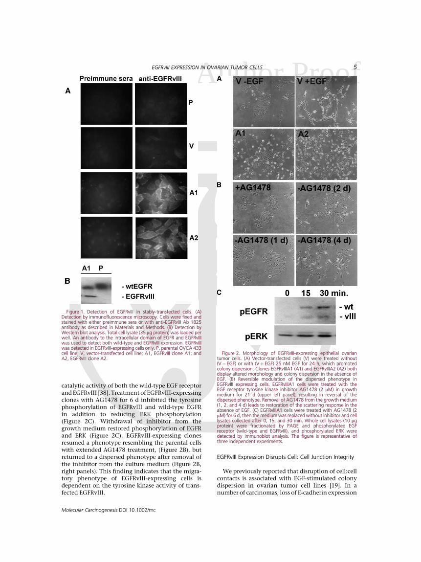
EGF receptor termed variant III was stablyexpressed in an ovarian tumor cell line (OVCA433), as described in Materials and Methods. Expres-sion of EGFRvIII in transfected cells was confirmedby immunofluorescence microscopy, with an anti-body raised against the unique epitope formed by theexon 2-7 deletion characteristic of this EGF receptormutation. Staining for EGFRvIII was observed in thetransfected cell lines EGFRvIIIA1 (A1) and EGFR-vIIIA2 (A2), while the parental OVCA 433 (P) andcontrol transfected (V) cells were not immunoreac-tive with the EGFRvIII antibody (Figure 1A). Further-more, expression of EGFRvIII was evident in thetransfected cells as detected by an antibody recogniz-ing the intracellular domain of EGFR and EGFRvIII(Figure 1B). In Figure 2A, the phenotype of twoEGFRvIII transfected clones is compared to that ofligand-activated parental OVCA 433 cells. In theabsence of EGF, OVCA 433 (Figure 2A) and OVCA429 [19] colonies displayed an epithelioid morphol-ogy and cells were tightly associated. Activationof EGF receptor by EGF promoted a migratoryresponse as detected by increased colony dispersion(Figure 2A) and enhanced in vitro wound closure(data not shown) in both cell lines. The EGFRvIIIexpressing clones EGFRvIIIA1 and EGFRvIIIA2, inthe absence of exogenous ligand, recapitulatedthe phenotype of EGF-stimulated parental OVCA433 cells (Figure 2A). Importantly, the phenotypewas reversed when cells were treated with AG1478(Figure 2B), a highly selective inhibitor of the
4 ZEINELDIN ET AL.
Molecular Carcinogenesis DOI 10.1002/mc
Author Proof
Acatalytic activity of both the wild-type EGF receptorand EGFRvIII [38]. Treatment of EGFRvIII-expressingclones with AG1478 for 6 d inhibited the tyrosinephosphorylation of EGFRvIII and wild-type EGFRin addition to reducing ERK phosphorylation(Figure 2C). Withdrawal of inhibitor from thegrowth medium restored phosphorylation of EGFRand ERK (Figure 2C). EGFRvIII-expressing clonesresumed a phenotype resembling the parental cellswith extended AG1478 treatment, (Figure 2B), butreturned to a dispersed phenotype after removal ofthe inhibitor from the culture medium (Figure 2B,right panels). This finding indicates that the migra-tory phenotype of EGFRvIII-expressing cells isdependent on the tyrosine kinase activity of trans-fected EGFRvIII.
EGFRvIII Expression Disrupts Cell: Cell Junction Integrity
We previously reported that disruption of cell:cellcontacts is associated with EGF-stimulated colonydispersion in ovarian tumor cell lines [19]. In anumber of carcinomas, loss of E-cadherin expression
Figure 1. Detection of EGFRvIII in stably-transfected cells. (A)Detection by immunofluorescence microscopy. Cells were fixed andstained with either preimmune sera or with anti-EGFRvIII Ab 1825antibody as described in Materials and Methods. (B) Detection byWestern blot analysis. Total cell lysate (35 mg protein) was loaded perwell. An antibody to the intracellular domain of EGFR and EGFRvIIIwas used to detect both wild-type and EGFRvIII expression. EGFRvIIIwas detected in EGFRvIII-expressing cells only. P, parental OVCA 433cell line; V, vector-transfected cell line; A1, EGFRvIII clone A1; andA2, EGFRvIII clone A2.
Figure 2. Morphology of EGFRvIII-expressing epithelial ovariantumor cells. (A) Vector-transfected cells (V) were treated without(V� EGF) or with (Vþ EGF) 25 nM EGF for 24 h, which promotedcolony dispersion. Clones EGFRvIIIA1 (A1) and EGFRvIIIA2 (A2) bothdisplay altered morphology and colony dispersion in the absence ofEGF. (B) Reversible modulation of the dispersed phenotype inEGFRvIII expressing cells. EGFRvIIIA1 cells were treated with theEGF receptor tyrosine kinase inhibitor AG1478 (2 mM) in growthmedium for 21 d (upper left panel), resulting in reversal of thedispersed phenotype. Removal of AG1478 from the growth medium(1, 2, and 4 d) leads to restoration of the scattering response in theabsence of EGF. (C) EGFRvIIIA1 cells were treated with AG1478 (2mM) for 6 d, then the medium was replaced without inhibitor and celllysates collected after 0, 15, and 30 min. Whole cell lysates (10 mgprotein) were fractionated by PAGE and phosphorylated EGFreceptor (wild-type and EGFRvIII), and phosphorylated ERK weredetected by immunoblot analysis. The figure is representative ofthree independent experiments.
EGFRvIII EXPRESSION IN OVARIAN TUMOR CELLS 5
Molecular Carcinogenesis DOI 10.1002/mc
Author Proof
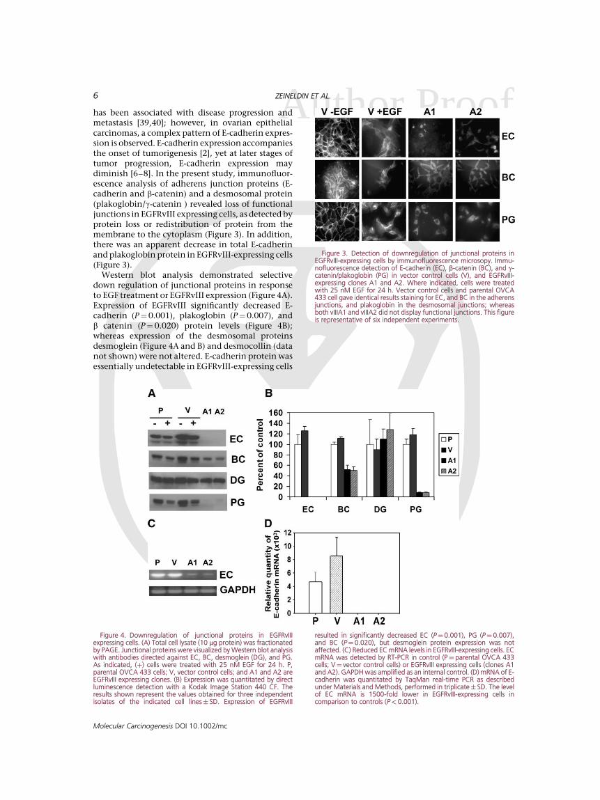
Ahas been associated with disease progression andmetastasis [39,40]; however, in ovarian epithelialcarcinomas, a complex pattern of E-cadherin expres-sion is observed. E-cadherin expression accompaniesthe onset of tumorigenesis [2], yet at later stages oftumor progression, E-cadherin expression maydiminish [6–8]. In the present study, immunofluor-escence analysis of adherens junction proteins (E-cadherin and b-catenin) and a desmosomal protein(plakoglobin/g-catenin ) revealed loss of functionaljunctions in EGFRvIII expressing cells, as detected byprotein loss or redistribution of protein from themembrane to the cytoplasm (Figure 3). In addition,there was an apparent decrease in total E-cadherinand plakoglobin protein in EGFRvIII-expressing cells(Figure 3).
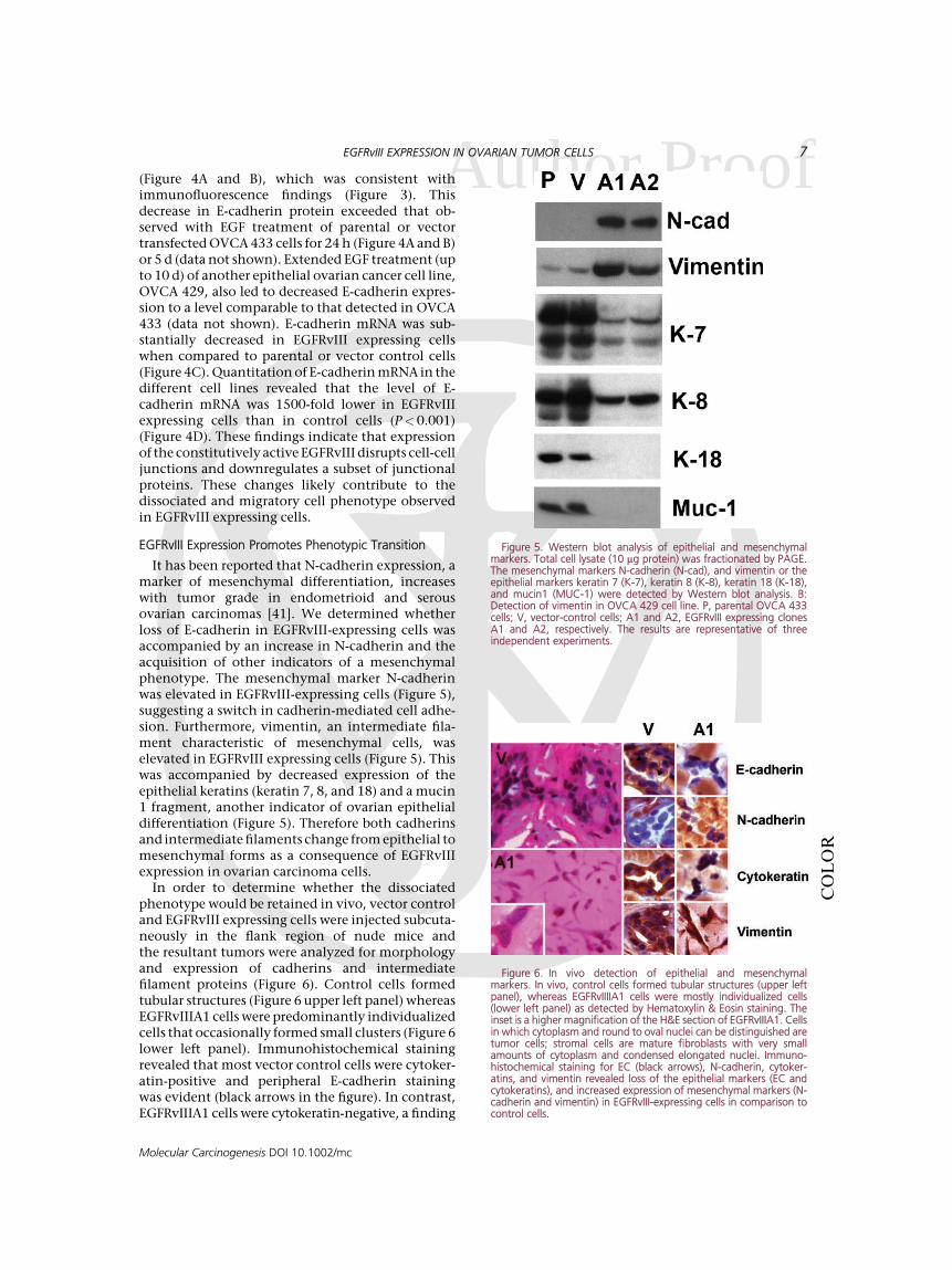
Western blot analysis demonstrated selectivedown regulation of junctional proteins in responseto EGF treatment or EGFRvIII expression (Figure 4A).Expression of EGFRvIII significantly decreased E-cadherin (P¼0.001), plakoglobin (P¼0.007), andb catenin (P¼0.020) protein levels (Figure 4B);whereas expression of the desmosomal proteinsdesmoglein (Figure 4A and B) and desmocollin (datanot shown) were not altered. E-cadherin protein wasessentially undetectable in EGFRvIII-expressing cells
Figure 3. Detection of downregulation of junctional proteins inEGFRvIII-expressing cells by immunofluorescence microsopy. Immu-nofluorescence detection of E-cadherin (EC), b-catenin (BC), and g-catenin/plakoglobin (PG) in vector control cells (V), and EGFRvIII-expressing clones A1 and A2. Where indicated, cells were treatedwith 25 nM EGF for 24 h. Vector control cells and parental OVCA433 cell gave identical results staining for EC, and BC in the adherensjunctions, and plakoglobin in the desmosomal junctions; whereasboth vIIIA1 and vIIIA2 did not display functional junctions. This figureis representative of six independent experiments.
Figure 4. Downregulation of junctional proteins in EGFRvIIIexpressing cells. (A) Total cell lysate (10 mg protein) was fractionatedby PAGE. Junctional proteins were visualized by Western blot analysiswith antibodies directed against EC, BC, desmoglein (DG), and PG.As indicated, (þ) cells were treated with 25 nM EGF for 24 h. P,parental OVCA 433 cells; V, vector control cells; and A1 and A2 areEGFRvIII expressing clones. (B) Expression was quantitated by directluminescence detection with a Kodak Image Station 440 CF. Theresults shown represent the values obtained for three independentisolates of the indicated cell lines� SD. Expression of EGFRvIII
resulted in significantly decreased EC (P¼0.001), PG (P¼ 0.007),and BC (P¼0.020), but desmoglein protein expression was notaffected. (C) Reduced EC mRNA levels in EGFRvIII-expressing cells. ECmRNA was detected by RT-PCR in control (P¼ parental OVCA 433cells; V¼ vector control cells) or EGFRvIII expressing cells (clones A1and A2). GAPDH was amplified as an internal control. (D) mRNA of E-cadherin was quantitated by TaqMan real-time PCR as describedunder Materials and Methods, performed in triplicate� SD. The levelof EC mRNA is 1500-fold lower in EGFRvIII-expressing cells incomparison to controls (P<0.001).
6 ZEINELDIN ET AL.
Molecular Carcinogenesis DOI 10.1002/mc
Author Proof
A(Figure 4A and B), which was consistent withimmunofluorescence findings (Figure 3). Thisdecrease in E-cadherin protein exceeded that ob-served with EGF treatment of parental or vectortransfected OVCA 433 cells for 24 h (Figure 4A and B)or 5 d (data not shown). Extended EGF treatment (upto 10 d) of another epithelial ovarian cancer cell line,OVCA 429, also led to decreased E-cadherin expres-sion to a level comparable to that detected in OVCA433 (data not shown). E-cadherin mRNA was sub-stantially decreased in EGFRvIII expressing cellswhen compared to parental or vector control cells(Figure 4C). Quantitation of E-cadherin mRNA in thedifferent cell lines revealed that the level of E-cadherin mRNA was 1500-fold lower in EGFRvIIIexpressing cells than in control cells (P<0.001)(Figure 4D). These findings indicate that expressionof the constitutively active EGFRvIII disrupts cell-celljunctions and downregulates a subset of junctionalproteins. These changes likely contribute to thedissociated and migratory cell phenotype observedin EGFRvIII expressing cells.
EGFRvIII Expression Promotes Phenotypic Transition
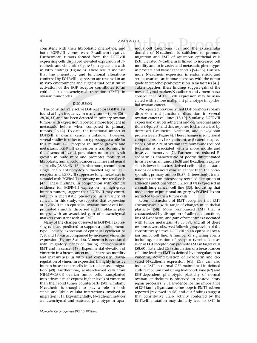
It has been reported that N-cadherin expression, amarker of mesenchymal differentiation, increaseswith tumor grade in endometrioid and serousovarian carcinomas [41]. We determined whetherloss of E-cadherin in EGFRvIII-expressing cells wasaccompanied by an increase in N-cadherin and theacquisition of other indicators of a mesenchymalphenotype. The mesenchymal marker N-cadherinwas elevated in EGFRvIII-expressing cells (Figure 5),suggesting a switch in cadherin-mediated cell adhe-sion. Furthermore, vimentin, an intermediate fila-ment characteristic of mesenchymal cells, waselevated in EGFRvIII expressing cells (Figure 5). Thiswas accompanied by decreased expression of theepithelial keratins (keratin 7, 8, and 18) and a mucin1 fragment, another indicator of ovarian epithelialdifferentiation (Figure 5). Therefore both cadherinsand intermediate filaments change from epithelial tomesenchymal forms as a consequence of EGFRvIIIexpression in ovarian carcinoma cells.
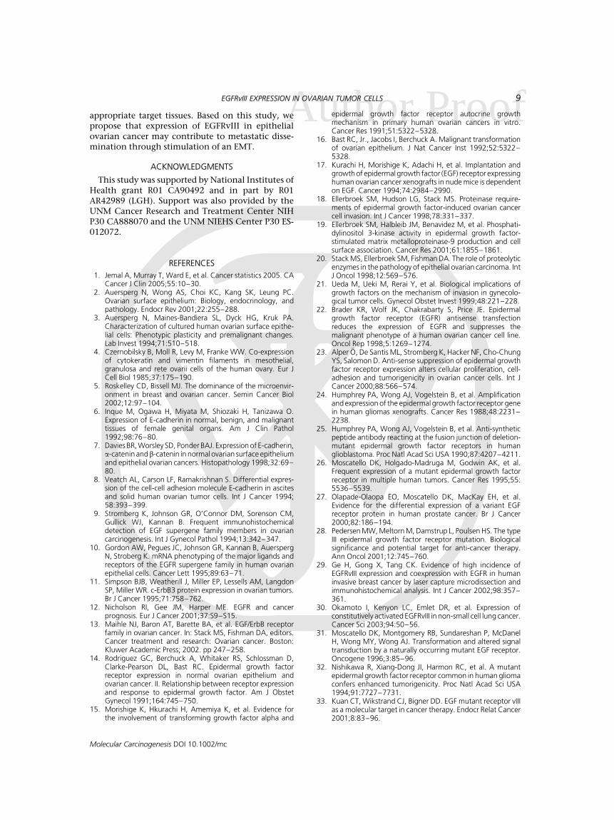
In order to determine whether the dissociatedphenotype would be retained in vivo, vector controland EGFRvIII expressing cells were injected subcuta-neously in the flank region of nude mice andthe resultant tumors were analyzed for morphologyand expression of cadherins and intermediatefilament proteins (Figure 6). Control cells formedtubular structures (Figure 6 upper left panel) whereasEGFRvIIIA1 cells were predominantly individualizedcells that occasionally formed small clusters (Figure 6lower left panel). Immunohistochemical stainingrevealed that most vector control cells were cytoker-atin-positive and peripheral E-cadherin stainingwas evident (black arrows in the figure). In contrast,EGFRvIIIA1 cells were cytokeratin-negative, a finding
Figure 5. Western blot analysis of epithelial and mesenchymalmarkers. Total cell lysate (10 mg protein) was fractionated by PAGE.The mesenchymal markers N-cadherin (N-cad), and vimentin or theepithelial markers keratin 7 (K-7), keratin 8 (K-8), keratin 18 (K-18),and mucin1 (MUC-1) were detected by Western blot analysis. B:Detection of vimentin in OVCA 429 cell line. P, parental OVCA 433cells; V, vector-control cells; A1 and A2, EGFRvIII expressing clonesA1 and A2, respectively. The results are representative of threeindependent experiments.
Figure 6. In vivo detection of epithelial and mesenchymalmarkers. In vivo, control cells formed tubular structures (upper leftpanel), whereas EGFRvIIIIA1 cells were mostly individualized cells(lower left panel) as detected by Hematoxylin & Eosin staining. Theinset is a higher magnification of the H&E section of EGFRvIIIA1. Cellsin which cytoplasm and round to oval nuclei can be distinguished aretumor cells; stromal cells are mature fibroblasts with very smallamounts of cytoplasm and condensed elongated nuclei. Immuno-histochemical staining for EC (black arrows), N-cadherin, cytoker-atins, and vimentin revealed loss of the epithelial markers (EC andcytokeratins), and increased expression of mesenchymal markers (N-cadherin and vimentin) in EGFRvIII-expressing cells in comparison tocontrol cells.
COLOR
EGFRvIII EXPRESSION IN OVARIAN TUMOR CELLS 7
Molecular Carcinogenesis DOI 10.1002/mc
Author Proof
Aconsistent with their fibroblastic phenotype, andboth EGFRvIII clones were E-cadherin-negative.Furthermore, tumors formed from the EGFRvIIIexpressing cells displayed elevated expression of N-cadherin and vimentin (Figure 6), in agreement within vitro findings (Figure 5). These results indicatethat the phenotypic and functional alterationsconferred by EGFRvIII expression are retained in anin vivo environment and suggest that constitutiveactivation of the EGF receptor contributes to anepithelial to mesenchymal transition (EMT) inovarian tumor cells.
DISCUSSION
The constitutively active EGF receptor EGFRvIII isfound at high frequency in many tumor types [26–28,30,33] and has been detected in primary ovariantumors with expression reportedly more frequent inmetastatic lesions when compared to primarytumors [26,42]. To date, the functional impact ofEGFRvIII in ovarian cancer is unknown; however,several studies in other tumor types suggest a role forthis mutant EGF receptor in tumor growth andmetastasis. EGFRvIII expression is transforming inthe absence of ligand, potentiates tumor xenograftgrowth in nude mice and promotes motility offibroblasts, human colon cancer cell lines and neuralstem cells [28,33,43–46]. Furthermore, recombinantsingle chain antibody-toxin directed against EGFreceptor and EGFRvIII suppresses lung metastases ina model with EGFRvIII expressing murine renal cells[47]. These findings, in conjunction with clinicalevidence for EGFRvIII expression in high-gradehuman tumors, suggest that EGFRvIII may contri-bute to a metastatic phenotype in a variety ofcancers. In this study, we reported that expressionof EGFRvIII in an epithelial ovarian tumor cell linepromoted a motile, dispersed and fibroblastic phe-notype with an associated gain of mesenchymalmarkers consistent with an EMT.
Many of the changes observed in EGFRvIII expres-sing cells are predicted to support a motile pheno-type. Reduced expression of epithelial cytokeratins7, 8, and 18 was accompanied by increased vimentinexpression (Figures 5 and 6). Vimentin is associatedwith migratory behavior during developmentalEMT and in cancer [48]. Experimental elevation ofvimentin in a breast cancer model increases motilityand invasiveness in vitro and conversely, down-regulation of vimentin expression in highly invasivehuman breast cancer cells leads to decreased migra-tion [49]. Furthermore, acsites-derived cells fromNIH:OVCAR-3 ovarian tumor cells transplantedinto athymic mice express higher levels of vimentinthan their solid tumor counterparts [50]. Similarly,N-cadherin is thought to play a role in bothstable and labile cellular interactions involved inmigration [51]. Experimentally, N-cadherin inducesa mesenchymal and scattered phenotype in squa-
mous cell carcinoma [52] and the extracellulardomain of N-cadherin is sufficient to promotemigration and EMT of squamous epithelial cells[53]. Elevated N-cadherin is linked to increased cellmotility and to invasive and metastatic phenotypesin prostate and breast cancer cells [54–56]. Further-more, N-cadherin expression in endometrioid andserous ovarian carcinomas increases with the tumorgrade and reaches peak expression in metastases [41].Taken together, these findings suggest gain of themesenchymal markers N-cadherin and vimentin as aconsequence of EGFRvIII expression may be asso-ciated with a more malignant phenotype in epithe-lial ovarian cancer.
We reported previously that EGF promotes colonydispersion and junctional disruption in severalovarian cancer cell lines [18,19]. Similarly, EGFRvIIIexpression disrupts adherens and desmosomal junc-tions (Figure 3) and this response is characterized bydecreased E-cadherin, b-catenin, and plakoglobinprotein levels (Figure 4). These changes in junctionalcomponents may be significant, as b-catenin expres-sion is lost in 21% of ovarian carcinomas and reducedb-catenin is associated with a more motile andinvasive phenotype [7]. Furthermore, reduced E-cadherin is characteristic of poorly differentiatedinvasive ovarian tumors [6,8] and E-cadherin expres-sion is lower in ascites-derived cells and metastaticlesions of advanced ovarian cancer than the corre-sponding primary tumors [8,57]. Interestingly, trans-mission electron microscopy revealed disruption ofadherens junctions when EGFRvIII was expressed ina small lung cancer cell line [35], indicating thatmodulation of junctional integrity by EGFRvIII is notrestricted to ovarian tumor cells.
Recent discussions of EMT recognize that EMTencompasses a wide range of changes in epithelialplasticity [58]. More pronounced EMT that ischaracterized by disruption of adherens junctions,loss of E-cadherin, and gain of vimentin is associatedwith tumor metastasis [48,58,59], and all of theseresponses were observed following expression of theconstitutively active EGFRvIII in an epithelial ovar-ian tumor cell line. A number of signaling eventsincluding, activation of receptor tyrosine kinasessuch as EGF receptor, can promote EMT in target cells[58,60]. Extended EGF stimulation of a breast cancercell line leads to EMT as defined by upregulation ofvimentin, downregulation of E-cadherin and ele-vated N-cadherin expression [61]. EGF can alsoinduce EMT in normal OSE maintained in definedculture medium containing hydrocortisone [62] andEGF-dependent phenotypic plasticity of normalovarian epithelium is observed in postovulatoryrepair processes [2,3]. Evidence for the importanceof EGF family ligand autocrine loops in EMT has beenreported [reviewed in 58] and our findings suggestthat constitutive EGFR activity conferred by theEGFRvIII mutation may similarly lead to EMT in
8 ZEINELDIN ET AL.
Molecular Carcinogenesis DOI 10.1002/mc
Author Proof
Aappropriate target tissues. Based on this study, wepropose that expression of EGFRvIII in epithelialovarian cancer may contribute to metastatic disse-mination through stimulation of an EMT.
ACKNOWLEDGMENTS
This study was supported by National Institutes ofHealth grant R01 CA90492 and in part by R01AR42989 (LGH). Support was also provided by theUNM Cancer Research and Treatment Center NIHP30 CA888070 and the UNM NIEHS Center P30 ES-012072.
REFERENCES
1. Jemal A, Murray T, Ward E, et al. Cancer statistics 2005. CACancer J Clin 2005;55:10–30.
2. Auersperg N, Wong AS, Choi KC, Kang SK, Leung PC.Ovarian surface epithelium: Biology, endocrinology, andpathology. Endocr Rev 2001;22:255–288.
3. Auersperg N, Maines-Bandiera SL, Dyck HG, Kruk PA.Characterization of cultured human ovarian surface epithe-lial cells: Phenotypic plasticity and premalignant changes.Lab Invest 1994;71:510–518.
4. Czernobilsky B, Moll R, Levy M, Franke WW. Co-expressionof cytokeratin and vimentin filaments in mesothelial,granulosa and rete ovarii cells of the human ovary. Eur JCell Biol 1985;37:175–190.
5. Roskelley CD, Bissell MJ. The dominance of the microenvir-onment in breast and ovarian cancer. Semin Cancer Biol2002;12:97–104.
6. Inque M, Ogawa H, Miyata M, Shiozaki H, Tanizawa O.Expression of E-cadherin in normal, benign, and malignanttissues of female genital organs. Am J Clin Pathol1992;98:76–80.
7. Davies BR, Worsley SD, Ponder BAJ. Expression of E-cadherin,a-catenin andb-catenin in normal ovarian surface epitheliumand epithelial ovarian cancers. Histopathology 1998;32:69–80.
8. Veatch AL, Carson LF, Ramakrishnan S. Differential expres-sion of the cell-cell adhesion molecule E-cadherin in ascitesand solid human ovarian tumor cells. Int J Cancer 1994;58:393–399.
9. Stromberg K, Johnson GR, O’Connor DM, Sorenson CM,Gullick WJ, Kannan B. Frequent immunohistochemicaldetection of EGF supergene family members in ovariancarcinogenesis. Int J Gynecol Pathol 1994;13:342–347.
10. Gordon AW, Pegues JC, Johnson GR, Kannan B, AuerspergN, Stroberg K. mRNA phenotyping of the major ligands andreceptors of the EGFR supergene family in human ovarianepithelial cells. Cancer Lett 1995;89:63–71.
11. Simpson BJB, Weatherill J, Miller EP, Lessells AM, LangdonSP, Miller WR. c-ErbB3 protein expression in ovarian tumors.Br J Cancer 1995;71:758–762.
12. Nicholson RI, Gee JM, Harper ME. EGFR and cancerprognosis. Eur J Cancer 2001;37:S9–S15.
13. Maihle NJ, Baron AT, Barette BA, et al. EGF/ErbB receptorfamily in ovarian cancer. In: Stack MS, Fishman DA, editors.Cancer treatment and research: Ovarian cancer. Boston:Kluwer Academic Press; 2002. pp 247–258.
14. Rodriguez GC, Berchuck A, Whitaker RS, Schlossman D,Clarke-Pearson DL, Bast RC. Epidermal growth factorreceptor expression in normal ovarian epithelium andovarian cancer. II. Relationship between receptor expressionand response to epidermal growth factor. Am J ObstetGynecol 1991;164:745–750.
15. Morishige K, Hkurachi H, Amemiya K, et al. Evidence forthe involvement of transforming growth factor alpha and
epidermal growth factor receptor autocrine growthmechanism in primary human ovarian cancers in vitro.Cancer Res 1991;51:5322–5328.
16. Bast RC, Jr., Jacobs I, Berchuck A. Malignant transformationof ovarian epithelium. J Nat Cancer Inst 1992;52:5322–5328.
17. Kurachi H, Morishige K, Adachi H, et al. Implantation andgrowthof epidermalgrowth factor (EGF) receptor expressinghuman ovarian cancer xenografts in nude mice is dependenton EGF. Cancer 1994;74:2984–2990.
18. Ellerbroek SM, Hudson LG, Stack MS. Proteinase require-ments of epidermal growth factor-induced ovarian cancercell invasion. Int J Cancer 1998;78:331–337.
19. Ellerbroek SM, Halbleib JM, Benavidez M, et al. Phosphati-dylinositol 3-kinase activity in epidermal growth factor-stimulated matrix metalloproteinase-9 production and cellsurface association. Cancer Res 2001;61:1855–1861.
20. Stack MS, Ellerbroek SM, Fishman DA. The role of proteolyticenzymes in the pathology of epithelial ovarian carcinoma. IntJ Oncol 1998;12:569–576.
21. Ueda M, Ueki M, Rerai Y, et al. Biological implications ofgrowth factors on the mechanism of invasion in gynecolo-gical tumor cells. Gynecol Obstet Invest 1999;48:221–228.
22. Brader KR, Wolf JK, Chakrabarty S, Price JE. Epidermalgrowth factor receptor (EGFR) antisense transfectionreduces the expression of EGFR and suppresses themalignant phenotype of a human ovarian cancer cell line.Oncol Rep 1998;5:1269–1274.
23. Alper O, De Santis ML, Stromberg K, Hacker NF, Cho-ChungYS, Salomon D. Anti-sense suppression of epidermal growthfactor receptor expression alters cellular proliferation, cell-adhesion and tumorigenicity in ovarian cancer cells. Int JCancer 2000;88:566–574.
24. Humphrey PA, Wong AJ, Vogelstein B, et al. Amplificationand expression of the epidermal growth factor receptor genein human gliomas xenografts. Cancer Res 1988;48:2231–2238.
25. Humphrey PA, Wong AJ, Vogelstein B, et al. Anti-syntheticpeptide antibody reacting at the fusion junction of deletion-mutant epidermal growth factor receptors in humanglioblastoma. Proc Natl Acad Sci USA 1990;87:4207–4211.
26. Moscatello DK, Holgado-Madruga M, Godwin AK, et al.Frequent expression of a mutant epidermal growth factorreceptor in multiple human tumors. Cancer Res 1995;55:5536–5539.
27. Olapade-Olaopa EO, Moscatello DK, MacKay EH, et al.Evidence for the differential expression of a variant EGFreceptor protein in human prostate cancer. Br J Cancer2000;82:186–194.
28. Pedersen MW, Meltorn M, Damstrup L, Poulsen HS. The typeIII epidermal growth factor receptor mutation. Biologicalsignificance and potential target for anti-cancer therapy.Ann Oncol 2001;12:745–760.
29. Ge H, Gong X, Tang CK. Evidence of high incidence ofEGFRvIII expression and coexpression with EGFR in humaninvasive breast cancer by laser capture microdissection andimmunohistochemical analysis. Int J Cancer 2002;98:357–361.
30. Okamoto I, Kenyon LC, Emlet DR, et al. Expression ofconstitutively activated EGFRvIII in non-small cell lung cancer.Cancer Sci 2003;94:50–56.
31. Moscatello DK, Montgomery RB, Sundareshan P, McDanelH, Wong MY, Wong AJ. Transformation and altered signaltransduction by a naturally occurring mutant EGF receptor.Oncogene 1996;3:85–96.
32. Nishikawa R, Xiang-Dong JI, Harmon RC, et al. A mutantepidermal growth factor receptor common in human gliomaconfers enhanced tumorigenicity. Proc Natl Acad Sci USA1994;91:7727–7731.
33. Kuan CT, Wikstrand CJ, Bigner DD. EGF mutant receptor vIIIas a molecular target in cancer therapy. Endocr Relat Cancer2001;8:83–96.
EGFRvIII EXPRESSION IN OVARIAN TUMOR CELLS 9
Molecular Carcinogenesis DOI 10.1002/mc
Author Proof
A34. Lal A, Glazer CA, Martinson HM, et al. Mutant epidermal
growth factor receptor up-regulates molecular effectors oftumor invasion. Cancer Res 2002;62:3335–3339.
35. Damstrup L, Wandahl M, Bastholm L, Elling F, SkovgaardPoulsen H. Epidermal growth factor receptor mutation typeIII transfected into a small cell lung cancer cell line ispredominantly localized at the cell surface and enhances themalignant phenotype. Int J Cancer 2002;97:7–14.
36. Masuho Y, Zalutsky M, Knapp RC, Bast RC, Jr. Interactionof monoclonal antibodies with cell surface antigens ofhuman ovarian carcinomas. Cancer Res 1984;44:2813–2819.
37. Wong AST, Maines-Bandiera SL, Rosen B, et al. Constitutiveand conditional cadherin expression in cultured humanovarian surface epithelium: Influence of family history ofovarian cancer. Int J Cancer 1999;81:180–188.
38. Han Y, Caday CG, Nanda A, Cavenee WK, Huang HJS.Tyrphostin AG 1478 preferentially inhibits human gliomacells expressing truncated rather than wild-type epidermalgrowth factor receptors. Cancer Res 1996;56:3859–3861.
39. Behrens J, Birchmeier W. The role of E-cadherin mediated celladhesion in tumor metastasis. In: CitiQ3 S, editor. Molecularmechanisms of epithelial cell junctions: From development todisease. Austin: R.G. Landes Co.; 1994. pp 229–241.
40. Perl AK, Wilgenbus P, Dahl U, Semb H, Christofori G. A causalrole for E-cadherin in the transition from adenoma tocarcinoma. Nature 1998;392:190–193.
41. Houle CD, Ding X, Foley JF, Afshari CA, Barrett C, Davis BJ.Loss of expression and altered localization of KAI1 and CD9proteins are associated with epithelial ovarian cancerprogression. Gynec Oncol 2002;86:69–78.
42. Montgomery RB, Klein-Szanto A, Ross E, Godwin A.Expression of mutant EGFR in ovarian cancer. Proc Am AssocCancer Res 2004;45:1800.
43. Montgomery RB. Antagonistic and agonistic effects ofquinazoline tyrosine kinase inhibitors on mutant EGFreceptor function. Int J Cancer 2002;101:111–117.
44. Pedersen MW, Tkach V, Pedersen N, Berezin V, Poulsen HS.Expression of a naturally occurring constitutively activevariant of the epidermal growth factor receptor in mousefibroblasts increases motility. Int J Cancer 2004;108:643–653.
45. Rodrigues S, Attoub S, Nguyen QD, et al. Selectiveabrogation of the proinvasive activity of the trefoil peptidespS2 and spasmolytic polypeptide by disruption of the EGFreceptor signaling pathways in kidney and colonic cancercells. Oncogene 2003;22:4488–4497.
46. Boockvar JA, KapitonovD, Kapoor G, et al. Constitutive EGFRsignaling confers a motile phenotype to neural stem cells.Mol Cell Neurosci 2003;24:1116–1130.
47. Schmidt M, Maurer-Gebhard M, Groner B, Kohler G,Brochmann-Santos G, Wels W. Suppression of metastasisformation by a recombinant single chain antibody-toxin
targeted to full-length and oncogenic variant EGF receptors.Oncogene 1999;18:1711–1721.
48. Hay ED. Extracellular matrix, cell skeletons, and embryonicdevelopment. Am J Med Genet 1989;34:14–29.
49. Hendrix MJ, Seftor EA, Chu YW, Trevor KT, Seftor RE. Role ofintermediate filaments in migration, invasion and metastasis.Cancer Metastasis Rev 1996;15:507–525.
50. Veatch AL, Carson LF, Ramakrishnan S. Phenotypic variationsand differential migration of NIH:OVCAR-3 ovarian carci-noma cells isolated from athymic mice. Clin Exp Metastasis1995;13:165–172.
51. Conacci-Sorrell M, Zhurinsky J, Ben-Ze’ev A. The cadherin-catenin adhesion system in signaling and cancer. J Clin Invest2002;109:987–991.
52. Islam S, Carey TE, Wolf GT, Wheelock MJ, Hojnson KR.Expression of N-cadherin by human squamous carcinamacells induces a scattered fibroblastic phenotype withdisrupted cell-cell adhesion. J Cell Biol 1996;135:1643–1654.
53. Kim JB, Islam S, Kim YJ, et al. N-Cadherin extracellular repeat4 mediates epithelial to mesenchymal transition andincreased motility. J Cell Biol 2000;151:1193–1206.
54. Bussemakers MJG, Bokhoven AV, Tomita K, Jansen CFI,Schalken JA. Complex cadherin expression in humanprostate cancer cells. Int J Cancer 2000;85:446–450.
55. Nieman MT, Prudoff RS, Johnson KR, Wheelock MJ. N-cadherin promotes motility in human breast cancer cellsregardless of their E-cadherin expression. J Cell Biol1999;147:631–643.
56. Hazan RB, Phillips GR, Qiao RF, Norton L, Aaronson SA.Exogenous expression of N-cadherin in breast cancer cellsinduces cell migration, invasion, and metastasis. J Cell Biol2000;148:779–790.
57. Fujimoto J, Ichigo S, Hirose R, Sakaguchi H, Tamaya T.Expression of E-cadherin and alpha- and beta-catenin mRNAin ovarian cancers. Cancer Lett 1997;115:207–212.
58. Huber MA, Kraut N, Beug H. Molecular requirements forepithelial–mesenchymal transition during tumor progres-sion. Curr Opinion Cell Biol 2005;17:548–558.
59. Savagner P. Leaving the neighborhood: Molecular mechan-isms involved during epithelial-mesenchymal transition.Bioessays 2001;23:912–923.
60. Thiery JP. Epithelial–mesenchymal transitions in develop-ment and pathologies. Curr Opinion Cell Biol 2004;15:740–746.
61. Ackland ML, Newgreen DF, Fridman M, et al. Epidermalgrowth factor-induced epithelio-mesenchymal transition inhuman breast carcinoma cells. Lab Invest 2003;83:435–448.
62. Salamanca CM, Maines-Bandiera SL, Leung PC, Hu YL,Auersperg N. Effects of epidermal growth factor/hydrocorti-sone on the growth and differentiation of human ovariansurface epithelium. J Soc Gynecol Investig 2004;11:241–251.
Q1: Please provide the complete location.
Q2: Please provide the complete location.
Q3: Please check the change made.
10 ZEINELDIN ET AL.
Molecular Carcinogenesis DOI 10.1002/mc

1 1 1 R I V E R S T R E E T, H O B O K E N, N J 0 7 0 3 0
ELECTRONIC PROOF CHECKLIST, MOLECULAR C A R C I N O G E N E S I S
***IMMEDIATE RESPONSE REQUIRED***Please follow these instructions to avoid delay of publication.
READ PROOFS CAREFULLY• This will be your only chance to review these proofs.• Please note that the volume and page numbers shown on the proofs are for position only.
ANSWER ALL QUERIES ON PROOFS (Queries for you to answer are attached as the last page of your proof.)• Mark all corrections directly on the proofs. Note that excessive author alterations may ultimately result in delay of
publication and extra costs may be charged to you.
CHECK FIGURES AND TABLES CAREFULLY (Color figures will be sent under separate cover.)• Check size, numbering, and orientation of figures.• All images in the PDF are downsampled (reduced to lower resolution and file size) to facilitate Internet delivery.
These images will appear at higher resolution and sharpness in the printed article.• Review figure legends to ensure that they are complete.• Check all tables. Review layout, title, and footnotes.
COMPLETE REPRINT ORDER FORM• Fill out the attached reprint order form. It is important to return the form even if you are not ordering reprints. You
may, if you wish, pay for the reprints with a credit card. Reprints will be mailed only after your article appears inprint. This is the most opportune time to order reprints. If you wait until after your article comes off press, thereprints will be considerably more expensive.
RETURN PROOFSREPRINT ORDER FORMCTA (If you have not already signed one)
RETURN WITHIN 48 HOURS OF RECEIPT VIA FAX TO Mary Beth Puccio AT 516-437-3532
QUESTIONS? Mary Beth Puccio, Production EditorPhone: 201-748-8873E-mail: [email protected] to journal acronym and article production number(i.e., MC 00-001 for Molecular Carcinogenesis ms 00-001).
111 River Street Hoboken, NJ 07030, USA
201-748-8864FAX: 201-748-6825
CCOOPPYYRRIIGGHHTT TTRRAANNSSFFEERR AAGGRREEEEMMEENNTT
Date:
To:
Production/ContributionID#______________Publisher/Editorial office use only
Re: Manuscript entitled _______________________________________________________________________________________________________________________________________________________________________ (the "Contribution")
for publication in _______________________________________________________________________ (the "Journal")published by Wiley-Liss, Inc., a subsidiary of John Wiley & Sons, Inc. ( "Wiley").
Dear Contributor(s):
Thank you for submitting your Contribution for publication. In order to expedite the publishing process and enable Wiley todisseminate your work to the fullest extent, we need to have this Copyright Transfer Agreement signed and returned to us as soon aspossible. If the Contribution is not accepted for publication this Agreement shall be null and void.
A. COPYRIGHT
1. The Contributor assigns to Wiley, during the full term of copyright and any extensions or renewals of that term, allcopyright in and to the Contribution, including but not limited to the right to publish, republish, transmit, sell, distributeand otherwise use the Contribution and the material contained therein in electronic and print editions of the Journal and inderivative works throughout the world, in all languages and in all media of expression now known or later developed, andto license or permit others to do so.
2. Reproduction, posting, transmission or other distribution or use of the Contribution or any material contained therein, inany medium as permitted hereunder, requires a citation to the Journal and an appropriate credit to Wiley as Publisher,suitable in form and content as follows: (Title of Article, Author, Journal Title and Volume/Issue Copyright [year]Wiley-Liss, Inc. or copyright owner as specified in the Journal.)
B. RETAINED RIGHTS
Notwithstanding the above, the Contributor or, if applicable, the Contributor's Employer, retains all proprietary rights otherthan copyright, such as patent rights, in any process, procedure or article of manufacture described in the Contribution, and theright to make oral presentations of material from the Contribution.
C. OTHER RIGHTS OF CONTRIBUTOR
Wiley grants back to the Contributor the following:
1. The right to share with colleagues print or electronic "preprints" of the unpublished Contribution, in form and content asaccepted by Wiley for publication in the Journal. Such preprints may be posted as electronic files on the Contributor'sown website for personal or professional use, or on the Contributor's internal university or corporate networks/intranet, orsecure external website at the Contributor’s institution, but not for commercial sale or for any systematic externaldistribution by a third party (e.g., a listserve or database connected to a public access server). Prior to publication, theContributor must include the following notice on the preprint: "This is a preprint of an article accepted for publication in[Journal title] copyright (year) (copyright owner as specified in the Journal)". After publication of the Contribution byWiley, the preprint notice should be amended to read as follows: "This is a preprint of an article published in [include thecomplete citation information for the final version of the Contribution as published in the print edition of the Journal]",and should provide an electronic link to the Journal's WWW site, located at the following Wiley URL:http://www.interscience.Wiley.com/. The Contributor agrees not to update the preprint or replace it with the publishedversion of the Contribution.
2. The right, without charge, to photocopy or to transmit online or to download, print out and distribute to a colleague a copyof the published Contribution in whole or in part, for the Contributor's personal or professional use, for the advancementof scholarly or scientific research or study, or for corporate informational purposes in accordance with Paragraph D.2below.
3. The right to republish, without charge, in print format, all or part of the material from the published Contribution in a book
written or edited by the Contributor.
4. The right to use selected figures and tables, and selected text (up to 250 words, exclusive of the abstract) from theContribution, for the Contributor's own teaching purposes, or for incorporation within another work by the Contributorthat is made part of an edited work published (in print or electronic format) by a third party, or for presentation inelectronic format on an internal computer network or external website of the Contributor or the Contributor's employer.
5. The right to include the Contribution in a compilation for classroom use (course packs) to be distributed to students at the
Contributor’s institution free of charge or to be stored in electronic format in datarooms for access by students at theContributor’s institution as part of their course work (sometimes called “electronic reserve rooms”) and for in-housetraining programs at the Contributor’s employer.
D. CONTRIBUTIONS OWNED BY EMPLOYER
1. If the Contribution was written by the Contributor in the course of the Contributor's employment (as a "work-made-for-hire" in the course of employment), the Contribution is owned by the company/employer which must sign this Agreement(in addition to the Contributor’s signature), in the space provided below. In such case, the company/employer herebyassigns to Wiley, during the full term of copyright, all copyright in and to the Contribution for the full term of copyrightthroughout the world as specified in paragraph A above.
2. In addition to the rights specified as retained in paragraph B above and the rights granted back to the Contributor pursuantto paragraph C above, Wiley hereby grants back, without charge, to such company/employer, its subsidiaries anddivisions, the right to make copies of and distribute the published Contribution internally in print format or electronicallyon the Company's internal network. Upon payment of the Publisher's reprint fee, the institution may distribute (but notresell) print copies of the published Contribution externally. Although copies so made shall not be available for individualre-sale, they may be included by the company/employer as part of an information package included with software or otherproducts offered for sale or license. Posting of the published Contribution by the institution on a public access websitemay only be done with Wiley's written permission, and payment of any applicable fee(s).
E. GOVERNMENT CONTRACTS
In the case of a Contribution prepared under U.S. Government contract or grant, the U.S. Government may reproduce, withoutcharge, all or portions of the Contribution and may authorize others to do so, for official U.S. Government purposes only, if theU.S. Government contract or grant so requires. (U.S. Government Employees: see note at end).
F. COPYRIGHT NOTICE
The Contributor and the company/employer agree that any and all copies of the Contribution or any part thereof distributed orposted by them in print or electronic format as permitted herein will include the notice of copyright as stipulated in the Journaland a full citation to the Journal as published by Wiley.
G. CONTRIBUTOR'S REPRESENTATIONS
The Contributor represents that the Contribution is the Contributor's original work. If the Contribution was prepared jointly,the Contributor agrees to inform the co-Contributors of the terms of this Agreement and to obtain their signature to thisAgreement or their written permission to sign on their behalf. The Contribution is submitted only to this Journal and has notbeen published before, except for "preprints" as permitted above. (If excerpts from copyrighted works owned by third partiesare included, the Contributor will obtain written permission from the copyright owners for all uses as set forth in Wiley'spermissions form or in the Journal's Instructions for Contributors, and show credit to the sources in the Contribution.) TheContributor also warrants that the Contribution contains no libelous or unlawful statements, does not infringe on the rights orprivacy of others, or contain material or instructions that might cause harm or injury.

CHECK ONE:_____________________________________ ______________________
[____]Contributor-owned work Contributor's signature Date
______________________________________________________________Type or print name and title
_____________________________________ ______________________Co-contributor's signature Date
______________________________________________________________Type or print name and title
ATTACH ADDITIONAL SIGNATURE PAGE AS NECESSARY
_____________________________________ ______________________[____]Company/Institution-owned work Company or Institution (Employer-for-Hire) Date
(made-for-hire in thecourse of employment) _____________________________________ ______________________
Authorized signature of Employer Date
[____]U.S. Government work
NNoottee ttoo UU..SS.. GGoovveerrnnmmeenntt EEmmppllooyyeeeess
A Contribution prepared by a U.S. federal government employee as part of the employee's official duties, or which is an officialU.S. Government publication is called a "U.S. Government work," and is in the public domain in the United States. In such case,the employee may cross out Paragraph A.1 but must sign and return this Agreement. If the Contribution was not prepared as part ofthe employee's duties or is not an official U.S. Government publication, it is not a U.S. Government work.
[____]U.K. Government work (Crown Copyright)
Note to U.K. Government Employees
The rights in a Contribution prepared by an employee of a U.K. government department, agency or other Crown body as part ofhis/her official duties, or which is an official government publication, belong to the Crown. In such case, the Publisher willforward the relevant form to the Employee for signature.
�������������������
Telephone Number: • Facsimile Number:
To: Mary Beth Puccio At FAX #: 516-437-3532
From: Dr.
Date:
Re: Molecular Carcinogenesis, ms #
Dear Ms. Puccio,
Attached please find corrections to ms# __________. Please contact me shouldyou have any difficulty reading this fax at the numbers listed below.
Office phone:Email:Fax:Lab phone:
Thank you,
Dr.
E-proofing feedback comments:

C1
REPRINT BILLING DEPARTMENT •• 111 RIVER STREET • HOBOKEN, NJ 07030, USAPHONE: (201) 748-8864; FAX: (201) 748-6825
E-MAIL: [email protected] REPRINT ORDER FORM
Please complete this form even if you are not ordering reprints. This form MUST be returned with your corrected proofsand original manuscript. Your reprints will be shipped approximately 4 weeks after publication. Reprints ordered after printingwill be substantially more expensive.
JOURNAL Molecular Carcinogenesis VOLUME ISSUE
TITLE OF MANUSCRIPT
MS. NO. NO. OF PAGES AUTHOR(S)
No. of Pages 100 Reprints 200 Reprints 300 Reprints 400 Reprints 500 Reprints$ $ $ $ $
1-4 336 501 694 890 10525-8 469 703 987 1251 14779-12 594 923 1234 1565 185013-16 714 1156 1527 1901 227317-20 794 1340 1775 2212 264821-24 911 1529 2031 2536 303725-28 1004 1707 2267 2828 338829-32 1108 1894 2515 3135 375533-36 1219 2092 2773 3456 414337-40 1329 2290 3033 3776 4528
**REPRINTS ARE ONLY AVAILABLE IN LOTS OF 100. IF YOU WISH TO ORDER MORE THAN 500 REPRINTS, PLEASE CONTACT OUR REPRINTSDEPARTMENT AT (201) 748-8864 FOR A PRICE QUOTE.
Please send me _____________________ reprints of the above article at $
Please add appropriate State and Local Tax (Tax Exempt No.____________________) $for United States orders only.
Please add 5% Postage and Handling $
TOTAL AMOUNT OF ORDER** $**International orders must be paid in currency and drawn on a U.S. bankPlease check one: Check enclosed Bill me Credit CardIf credit card order, charge to: American Express Visa MasterCard
Credit Card No Signature Exp. Date
BILL TO: SHIP TO: (Please, no P.O. Box numbers)Name Name
Institution Institution
Address Address
Purchase Order No. Phone Fax

Softproofing for advanced Adobe Acrobat Users - NOTES toolNOTE: ACROBAT READER FROM THE INTERNET DOES NOT CONTAIN THE NOTES TOOL USED IN THIS PROCEDURE.
Acrobat annotation tools can be very useful for indicating changes to the PDF proof of your article.By using Acrobat annotation tools, a full digital pathway can be maintained for your page proofs.
The NOTES annotation tool can be used with either Adobe Acrobat 4.0, 5.0 or 6.0. Other annotation tools are also available in Acrobat 4.0, but this instruction sheet will concentrateon how to use the NOTES tool. Acrobat Reader, the free Internet download software from Adobe,DOES NOT contain the NOTES tool. In order to softproof using the NOTES tool you must havethe full software suite Adobe Acrobat 4.0, 5.0 or 6.0 installed on your computer.
Steps for Softproofing using Adobe Acrobat NOTES tool:
1. Open the PDF page proof of your article using either Adobe Acrobat 4.0, 5.0 or 6.0. Proofyour article on-screen or print a copy for markup of changes.
2. Go to File/Preferences/Annotations (in Acrobat 4.0) or Document/Add a Comment (in Acrobat6.0 and enter your name into the “default user” or “author” field. Also, set the font size at 9 or 10point.
3. When you have decided on the corrections to your article, select the NOTES tool from theAcrobat toolbox and click in the margin next to the text to be changed.
4. Enter your corrections into the NOTES text box window. Be sure to clearly indicate where thecorrection is to be placed and what text it will effect. If necessary to avoid confusion, you canuse your TEXT SELECTION tool to copy the text to be corrected and paste it into the NOTEStext box window. At this point, you can type the corrections directly into the NOTES textbox window. DO NOT correct the text by typing directly on the PDF page.
5. Go through your entire article using the NOTES tool as described in Step 4.
6. When you have completed the corrections to your article, go to File/Export/Annotations (inAcrobat 4.0) or Document/Add a Comment (in Acrobat 6.0).
7. When closing your article PDF be sure NOT to save changes to original file.
8. To make changes to a NOTES file you have exported, simply re-open the original PDFproof file, go to File/Import/Notes and import the NOTES file you saved. Make changes and re-export NOTES file keeping the same file name.
9. When complete, attach your NOTES file to a reply e-mail message. Be sure to include yourname, the date, and the title of the journal your article will be printed in.