molecular characterisation of three novel photosynthetic ... · molecular characterisation of three...
TRANSCRIPT

Molecular Characterisation of three Novel
Photosynthetic Proteins in Arabidopsis
thaliana
Dissertation
zur Erlangung des Doktorgrades der Fakultät für Biologie
der Ludwig-Maximilians-Universität München
Vorgelegt von
Ute Armbruster
aus Kleve
München
Mai 2008
Erstgutachter: Prof. Dr. Dario Leister Zweitgutachter: Prof. Dr. Jörg Nickelsen Tag der mündlichen Prüfung: 26.05.2008

Summary
i
Summary
Photosynthesis is the biological process, in which organisms utilize light energy in order to
synthesize organic matter. In plants this process takes place in the chloroplast. Nuclear
photosynthetic genes and nuclear genes involved in chloroplast gene expression are co-regulated at
the transcriptional level. Therefore, the working hypothesis was put forth that the physiological
function of genes with so-far unknown functions, which show a similar transcriptional regulation,
resides in photosynthesis or chloroplast gene expression. To test this hypothesis, three co-regulated
genes with so-far unknown physiological functions were analysed employing a reverse genetics
strategy and biochemical analyses of the encoded proteins. Insertional mutants for the genes
encoding the Putative Photosynthetic Protein 1 (PPP1), 4 (PPP4) and the Thylakoid Membrane
Phosphoprotein of 14 kDa (TMP14) were characterised with respect to putative defects in the
processes of photosynthesis or chloroplast gene expression. Both PPP4 and TMP14 belong to a
novel, nucleus encoded und chloroplast targeted family of four membrane proteins in Arabidopsis.
They functionally and physically interact in a previously unknown complex in the stromal lamellae
of thylakoid membranes, which can be solubilised with the detergent digitonin. Several lines of
biochemical evidence suggest that both proteins interact with other thylakoid proteins of unknown
identity. Electron transport measurements of the single and the double mutants of PPP4 and TMP14
support that this new complex is indeed involved in the regulation of photosynthesis, especially
under low light conditions. The assumed function for the stromal localised PPP1 as an RNA
binding protein involved in chloroplast gene expression was investigated by analyses of plastid
gene expression in ppp1 mutant plants. Here, decreased levels of chloroplast proteins and changes
in plastid translation rate could be demonstrated. This was accompanied by alterations in the
photosynthetic electron transport. Additionally, it was shown that plants lacking PPP1 show
delayed germination in response to the exogenous application of the phytohormone ABA, which
indicates a function of this protein in the ABA signalling network.

Zusammenfassung
ii
Zusammenfassung
Photosynthese beschreibt den biologischen Prozess der Umwandlung von Licht in chemische
Energie, und deren Nutzung zur Synthese organischen Materials. Bei Pflanzen findet dieser Prozess
im Chloroplasten statt. Nukleäre Gene, welche Komponenten der Photosynthese oder der
plastidären Genexpression kodieren, werden auf transkriptioneller Ebene ko-reguliert. Daher
wurden drei als unbekannt annotierte Gene mit ähnlicher transkriptioneller Regulation und ihre
Genprodukte, „Putative Photosynthetic Protein“ 1 (PPP1) und 4 (PPP4) und „Thylakoidmembran
Phosphoprotein 14 kDa“ (TMP14) auf eine mögliche Funktion in der Photosynthese und der
plastidären Genexpression hin untersucht. Die Charakterisierung dieser putativen neuen
Photosynthese-Komponenten erfolgte mittels reverser Genetik und biochemischer Analysen. PPP4
und TMP14 gehören zu einer kernkodierten und chloroplastidär lokalisierten Familie von
Membranproteinen mit vier Mitgliedern in Arabidopsis. Beide Proteine interagieren in einem
bislang unbekannten Komplex in den stromalen Lamellen der Thylakoidmembran sowohl auf
Proteinebene als auch funktionell. Mehrere unabhängige biochemische Untersuchungen
implizieren, dass PPP4 und TMP14 mit weiteren Thylakoidproteinen bislang unbekannter Identität
interagieren. Messungen des photosynthetischen Elektronentransports bei Einfach- und
Doppelmutanten weisen vor allem unter niedrigen Lichtbedingungen auf eine Funktion dieses
neuen Komplexes in der Photosynthese hin. Das dritte Protein PPP1 ist im Stroma lokalisiert und
wurde mittels Analyse der plastidären Genexpression in der ppp1 Mutante auf eine putative
Funktion in diesem Prozess hin untersucht. Dabei konnte gezeigt werden, dass die Mutanten
verringerte Mengen an Chloroplasten-Proteinen und veränderte plastidäre Translationsraten
aufweisen. Damit einhergehend weisen die ppp1 Mutanten Veränderungen im photosynthetischen
Elektronenfluss auf. Zusätzlich konnte gezeigt werden, dass bei exogener Zugabe des
Phytohormons ABA die ppp1 Mutanten verzögerte Keimung aufweisen, welches auf eine Funktion
des Proteins in dem ABA Signal-Netzwerk hindeutet.

Contents
iii
Contents
Summary ............................................................................................................................................. i
Zusammenfassung ..............................................................................................................................ii
Contents.............................................................................................................................................iii
Abbreviations ...................................................................................................................................vii
1 Introduction...................................................................................................................1
1.1 Photosynthesis................................................................................................................................ 1
1.1.1 Photosynthetic electron transport .............................................................................................. 1
1.1.1.1 Linear electron transport ..................................................................................................... 1
1.1.1.2 Cyclic electron transport around PSI................................................................................... 2
1.1.1.3 Physical measurements of electron transport ...................................................................... 4
1.1.2 Carbon fixation.......................................................................................................................... 4
1.1.3 The thylakoid membrane........................................................................................................... 5
1.1.3.1 The thylakoid polypeptide composition .............................................................................. 6
1.1.4 Regulation of photosynthetic processes .................................................................................... 8
1.1.4.1 Energisation dependent non-photochemical quenching (qE).............................................. 8
1.1.4.2 State transitions ................................................................................................................... 9
1.2 Expression of photosynthetic genes ............................................................................................. 9
1.2.1 Nuclear gene expression.......................................................................................................... 10
1.2.1 Chloroplast gene expression.................................................................................................... 11
1.3 Abscisic acid (ABA) biosynthesis in plants ............................................................................... 11
1.4 Aims of thesis ............................................................................................................................... 12
2 Materials and Methods ..............................................................................................14
2.1 Materials....................................................................................................................................... 14
2.1.1 Chemicals ................................................................................................................................ 14
2.1.2 Antibiotics ............................................................................................................................... 14
2.1.3 Enzymes, kits and biochemical agents .................................................................................... 14
2.1.4 Membranes .............................................................................................................................. 14
2.1.5 Antibodies ............................................................................................................................... 14
2.2 Methods ........................................................................................................................................ 15
2.2.1 Plant lines and propagation ..................................................................................................... 15

Contents
iv
2.2.2 Nucleic acid analyis................................................................................................................. 15
2.2.2.1 DNA analysis .................................................................................................................... 15
2.2.2.2 RNA analysis..................................................................................................................... 17
2.2.2.3 Analysis of mRNAs associated with polysomes............................................................... 17
2.2.2.4 Transcript end mapping of rbcL using circular RT-PCR.................................................. 18
2.2.3 Transformation of Arabidopsis ............................................................................................... 18
2.2.3.1. Bacterial strains ................................................................................................................ 18
2.2.3.2 Agrobacterium binary vectors ........................................................................................... 18
2.2.3.3 Agrobacterium-mediated transformation of A. thaliana ................................................... 19
2.2.4 Biochemical Analysis.............................................................................................................. 19
2.2.4.1 Antibody production.......................................................................................................... 19
2.2.4.2 SDS-PAGE........................................................................................................................ 19
2.2.4.3 Immunoblot analysis ......................................................................................................... 19
2.2.4.4 Total protein isolation ....................................................................................................... 20
2.2.4.5 Isolation of intact chloroplasts .......................................................................................... 20
2.2.4.6 Fractionation of chloroplasts ............................................................................................. 20
2.2.4.7 Preparation of thylakoid membranes................................................................................. 20
2.2.4.8 Fractionation of thylakoids................................................................................................ 21
2.2.4.9 PSI isolation ...................................................................................................................... 21
2.2.4.10 Blue native and second dimension gels........................................................................... 21
2.2.4.11 Bis (Sulfosuccinimidyl) suberate (BS3) crosslinking...................................................... 22
2.2.4.12 Co-Immunoprecipitation ................................................................................................. 22
2.2.4.13 Salt treatment of thylakoid membranes........................................................................... 22
2.2.4.14 Trypsin treatment of thylakoid membranes..................................................................... 23
2.2.4.15 In vivo labelling with 35S-Methionine ............................................................................. 23
2.2.5 Mass spectrometry................................................................................................................... 23
2.2.5.1 Tryptic in gel digestion of proteins ................................................................................... 23
2.2.5.2 LC-ESI MS/MS................................................................................................................. 24
2.2.5.3 Protein identification ......................................................................................................... 24
2.2.6 Pigment analysis...................................................................................................................... 24
2.2.7 Database analysis, Digital Northern, prediction of subcellular targeting and protein
modelling................................................................................................................................. 25
2.2.8 Intracellular localization of dsRED fusions ............................................................................ 25

Contents
v
2.2.9 Germination assay ................................................................................................................... 26
2.2.10 Determination of photosynthetic parameters using the PAM fluorometer ........................... 26
2.2.10.1 Chlorophyll fluorescence measurements......................................................................... 26
2.2.10.2 Measurements of the redox state of P700 ....................................................................... 26
2.2.10.3 Cyclic electron flow measurements ................................................................................ 27
3 Results ..........................................................................................................................28
3.1 Characterisation of PPP4 and TMP14 ...................................................................................... 28
3.1.1 Description of a novel protein family in photosynthetic organisms ....................................... 28
3.1.2 Localisation of PPP4 and TMP14 in the thylakoid membrane ............................................... 31
3.1.3 Knock-out mutants of PPP4 and TMP14................................................................................ 32
3.1.4 Localisation of PPP4 in BN-PAGE......................................................................................... 34
3.1.5 TMP14 protein levels are decreased in ppp4 mutants............................................................. 37
3.1.6 Investigating the interaction between PPP4 and TMP14 ........................................................ 37
3.1.6.1 Epitope tagging of PPP4.................................................................................................... 37
3.1.6.2 Crosslinking....................................................................................................................... 38
3.1.6.3 Co-immunoprecipitation (Co-IP) ...................................................................................... 41
3.1.7 TMP14 and PPP4 are not stably associated with any of the main photosynthetic complexes 43
3.1.8 Photosynthetic parameter of WT, ppp4, tmp14, ppp4 tmp14 and PPP4:c-myc
overexpressors ......................................................................................................................... 45
3.1.9 Topology of PPP4 and TMP14 ............................................................................................... 49
3.1.10 Analysis of thylakoid protein composition ........................................................................... 52
3.1.11 Location of thylakoid proteins in WT and ppp4 tmp14 ........................................................ 53
3.1.12 Investigating phosphorylation of thylakoid proteins in WT and ppp4 tmp14....................... 55
3.1.13 Characterisation of PPP4-like and TMP14-like .................................................................... 55
3.2 Characterisation of PPP1 ........................................................................................................... 58
3.2.1 Gene expression, protein structure and localisation................................................................ 58
3.2.2 Phenotypical analysis of knock-out mutants of PPP1 and CSP41 ......................................... 60
3.2.3 Leaf pigments and photosynthetic parameters ........................................................................ 62
3.2.4 Analysis of the abundance of photosynthetic proteins............................................................ 65
3.2.5 Chloroplast expression analysis .............................................................................................. 67
3.2.6 Analysis of transcript processing ............................................................................................ 68
3.2.7 Effect of the absence of photosynthetic complexes on PPP1 abundance................................ 70
Contents
vi
3.2.8 A PPP1:CFP fusion complements the ppp1 mutant phenotype .............................................. 70
3.2.9 Germination assay ................................................................................................................... 71
4 Discussion ....................................................................................................................73
4.1 PPP4/TMP14................................................................................................................................ 73
4.1.1 Novel family of chloroplast localised proteins........................................................................ 73
4.1.2 A novel thylakoid complex predominantly localised in the stromal lamellae ........................ 73
4.1.3 Phenotype of ppp4 and ppp4 tmp14 mutants in relation to specific processes of
photosynthesis ......................................................................................................................... 75
4.1.3.1 Linear electron flow .......................................................................................................... 75
4.1.3.2 Regulatory processes in photosynthesis ............................................................................ 76
4.1.3.3 Cyclic electron flow around PSI (CEF) ............................................................................ 77
4.1.3.3.1 In-depth analysis of CEF in ppp4 and ppp4 tmp14 mutants ....................................... 79
4.1.3.3.2 Speculation about an involvement of PPP4 and TMP14 in CEF processes................ 80
4.1.4 Conclusions ............................................................................................................................. 81
4.2 The putative RNA-binding protein PPP1 ................................................................................. 82
4.2.1 Physiological role: Chloroplast gene expression..................................................................... 82
4.2.2 The homologue AtCSP41........................................................................................................ 84
4.2.3 Involvement of PPP1 in ABA signalling ................................................................................ 84
5 References....................................................................................................................87
6 Supplementary Data.................................................................................................101
6.1 Mass spectrometry fragmentation spectra.............................................................................. 101
6.1.1 PPP4 ...................................................................................................................................... 101
6.1.2 TMP14................................................................................................................................... 103
6.1.3 TMP14-like ........................................................................................................................... 104
6.2 Plastome expression analysis of ppp1 mutants........................................................................ 105
Acknowledgements ......................................................................................................................... 106
Curriculum vitae............................................................................................................................. 107
Ehrenwörtliche Versicherung......................................................................................................... 109

Abbreviations
vii
Abbreviations
ΦII Effective quantum yield of PSII °C Degree celsius ABA Abscisic acid ABI4 ABA insensitive 4 ATP Adenine triphosphate Auto Autofluorescence ß-Car ß-carotene ß-DM ß-dodecylmaltoside BN gel Blue native gel BS3 Bis (Sulfosuccinimidyl) suberate C-terminus Carboxy terminus Ci Curie cDNA Complementary deoxyribonucleic acid CEF Cyclic electron flow CFP Cyan fluorescent protein Chl Chlorophyll Co-IP Co-immunoprecipitation Col-0 Arabidopsis ecotype Columbia CSP41 Chloroplast stem loop binding protein 41 kDa Cyt b6f Cytochrome b6f complex D Dark Da Dalton DNA Deoxyribonucleic acid EDTA Ethylene diamin tetraacetic acid EGTA Ethylene glycol tetraacetic acid ETR Electron transport rate Fd Ferredoxin FNR Ferredoxin NADPH dehydrogenase FR Far-red light HA Hemagglutinin HL High light g Gram g Times the force of gravity h Hour HEPES 4-(2-hydroxyethyl)-1-piperazineethanesulfonic acid HPLC High performance liquid chromatography IgG Immunoglobulin G k Kilo LB left T-DNA border LEF Linear electron flow Ler Arabidopsis ecotype Landsberg erecta LHC Light harvesting complex LL Low light µ Micro m Meter M Mole(s) per litre min Minute(s)

Abbreviations
viii
mol Mole MS Mass spectrometry N-terminus Amino terminus NADP(H) Nicotinamide adenine dinucleotide phosphate NDH NAD(P)H dehydrogenase complex No-0 Arabidopsis ecotype Noessen-0 NPQ Non-photochemical quenching Nx Neoxanthin P680 Reaction center of PSII P700 Reaction center of PSI PAGE Polyacrylamide gel electrophorese PAM Pulse amplitude modulation PAR Photosynthetic active radiation PCR Polymerase chain reaction pers. com. Personal communication PGR5 Proton gradient defective 5 PGRL1 PGR5 like 1 PMSF Phenylmethanesulphonylfluoride PPP1/4 Putative photosynthetic protein 1/4 PSI Photosystem I PSII Photosystem II PQ Plastoquinone PQH2 Plastohydroquinone PVDF Polyvinylidene difluoride qE Δ pH-dependent NPQ qP Photochemical quenching RB Right T-DNA border RFP Red fluorescent protein RNA Ribonucleic acid Rubisco Ribulose biphosphate carboxylase/ oxygenase r.u. Relative units s Second(s) SD Standard deviation SDR Short chain dehydrogenase reductase SDS Sodium dodecyl sulphate TMP14 Thylakoid membrane protein 14 kDa Tris Tris(hydroxymethyl)-aminomethane v/v Volume per volume VAZ Violaxanthin, antheraxanthin, zeaxanthin (Xanthophyll cycle pigments) VDE Violaxanthin-deepoxidase w/v Weight per volume WS Arabidopsis ecotype wassilewskija WT Wild-type ZEP Zeaxanthin epoxidase

1 Introduction
1
1 Introduction
1.1 Photosynthesis
During photosynthesis light energy is transformed into chemical energy in form of NADPH and
ATP, which are then employed by the Calvin-Benson cycle to incorporate atmospheric carbon into
the biosphere. The highly optimised light reaction of photosynthesis, as it has evolved in plants,
takes place in the thylakoid membranes of the chloroplast. The concerted action of four large
membrane complexes, the Photosystems I and II (PSI and PSII), the Cytochrome b6f complex (Cyt
b6f) and an ATP-synthase to conduct the transformation of light into chemical energy has been
studied thoroughly by spectroscopy, molecular genetics, biochemistry and structural analyses
(reviewed by Jensen et al., 2007; Dekker and Boekema, 2005; Richter et al., 2005). Important for
optimal photosynthetic activity are the spatial organisation of the photosynthetic apparatus and
various regulatory processes fine-tuning and adjusting this process.
1.1.1 Photosynthetic electron transport
Electron flow from water to the final electron acceptor NADPH is referred to as linear electron
transport. This process also leads to the phosphorylation of ADP to ATP via the generation of a
proton gradient across the thylakoid membrane. The cyclic electron flow around PSI, which
generates ATP without accumulation of the final electron acceptor NADPH can adjust the
ATP:NADPH ratio according to the requirements of the Calvin-Benson cycle and other chloroplast
localised physiological processes for.
1.1.1.1 Linear electron transport
The primary step in oxygenic photosynthesis consists of light driven charge separations, which are
catalysed by two large transmembrane protein complexes, PSI and PSII. These two photosystems
synergistically interact by a number of redox components including plastoquinone, the Cyt b6/f
complex and plastocyanin. Thereby, light energy is converted into chemical energy in form of
NADPH and ATP. Synthesis of the latter occurs by the ATP-synthase, which employs physical
energy conserved by the transmembrane proton gradient, a product of light driven proton electron
symport. Linear electron flow from PSII to PSI occurs according to the so-called Z-scheme, which
refers to the midpoint redox potentials of the redox carriers within this electron transport chain
(Figure 1.1). In detail, light-induced charge separations in PSII, which acts as a water-
plastoquinone oxidoreductase (Renger and Govindjee, 1985), cause the reduction of a stromal side

1 Introduction
2
bound quinone acceptors (QA) via rapid oxido-reductions of a phaeophytin molecule. The resulting
electron gaps in the reaction centre special pair chlorophylls lead to the oxidation of H2O molecules
by the lumenal oxygen evolving complex producing molecular oxygen and protons as by-products
(Cruz et al., 2005). Two electrons from the PSII acceptor QA and two stromal localised protons are
then transferred to a free membrane soluble plastoquinone molecule (PQ) to form
plastohydroquinol (PQH2). Electron flow from the PQH2 pool to PSI occurs via the Cyt b6f complex
by reduction of the Rieske iron-sulphur protein of this complex and release of protons into the
lumen (Joliot and Joliot, 1986). Optimisation of proton translocation occurs via the Cyt b6f localised
Q-cycle, in which one electron from PQH2 is employed to again reduce a molecule of PQ on the
stromal side of the Cyt b6f complex (Joliot and Joliot, 1994). The second electron is transported to
the lumen-localised electron carrier plastocyanin, which then transfers the electrons to the stromal
acceptor site of PSI. Here, light energy drives a further charge separation that leads to the reduction
of ferredoxin and then, catalysed by the ferredoxin: NADPH oxidoreductase (FNR), of NADP+ to
NADPH (Taiz and Zeiger, 1998).
1.1.1.2 Cyclic electron transport around PSI
In this pathway stromal electron acceptors from PSI are employed to reduce plastoquinone resulting
in the generation of ΔpH without the accumulation of NADPH (dashed line, Figure 1.1; reviewed
by Shikanai, 2007). There are two pathways carrying out this process: the antimycin A sensitive,
which requires PGR5 and PGRL1 (Munekage et al., 2002; DalCorso et al., 2008) and the NADH
dehydrogenase (NDH) complex dependent one. Cyclic electron flow (CEF) participates in
regulatory processes of photosynthesis, which require additional acidification of the lumen or ATP
by this pathway. The activation of the Calvin-Benson cycle enzymes by ATP after a dark period for
example is accelerated by CEF (Joliot and Joliot, 2002). The extent of non-photochemical
quenching (NPQ) of excess light energy also depends on additional acidification of the lumen by
cyclic electron transport. Both processes, as measured by the NPQ capacity of the plant under the
respective conditions, are impaired in the pgr5 and pgrl1 mutants (Munekage et al., 2002; Dal
Corso et al., 2008). However, cyclic electron flow around PSI might not be restricted to carry out
fine-tunning functions, because if plants lack both the PGR5/ PGRL1 and NDH dependent
pathways, they are severely affected in growth and photosynthetic parameters in all conditions
tested. This led to the assumption that CEF is mandatory for plant fitness (Munekage et al., 2004),
probably because the ATP:NADPH ratio produced by linear electron flow does not meet the
requirements for carbon fixation by the Benson-Calvin-cycle and other chloroplast localised
physiological processes. The antimycin A sensitive CEF seems to carry out the majority of Fd-PQ

1 Introduction
3
reduction, because mutants in this pathway contrary to those impaired in NDH activity, show
significant changes in photosynthetic parameters (Munekage et al., 2002; DalCorso et al., 2008;
Muraoka et al., 2006; Munshi et al., 2006; Kamruzzaman et al., 2005). However, the executing
components of the antimycin A sensitive pathway are not completely deciphered, as the involved
proteins lack binding domains for electron transferring co-factors (DalCorso et al., 2008).
Fig. 1.1 Z-scheme of linear electron flow in oxygenic photosynthesis Redox carriers are placed at their midpoint redox potentials (at pH 7). The vertical arrows display the light induced decrease of redoxpotential of the reaction centre chlorophylls. Z, electron donor to P680; Ph a, pheophytin a, electron acceptor of P680; QA, plastoquinone tightly bound to PSII; QB, pool made up of PQ and PQH2; Ao, chlorophyll a, the primary electron acceptor of PSI; A1, phylloquinone; FX, FB and FA, iron sulphur clusters; Fd, soluble ferredoxin; NADP+, oxidised nicotinamide adenine dinucleotide phosphate. Dashed line incidates cyclic electron flow around PSI (CEF). Because both linear electron flow (LEF) and the CEF around PSI employ identical components
except for PGR5 and PGRL1 (Munekage et al., 2002; DalCorso et al., 2008), the specific dissection
of both pathways remains difficult to achieve (Breyton et al., 2006). Different models of the mode
of action and regulation of CEF have been postulated, which are in part supported by experimental
evidences (Okegawa et al., 2005; Joliot and Joliot, 2002; DalCorso et al., 2008). Highly
controversial remains the question whether Fd directly reduces PQ or indirectly through reduction
of component of the Cyt b6f complex. The FNR has been found associated with the Cyt b6f complex
in flowering plants and is thought to reduce Fd in order to activate CEF via the Q-cycle of this
complex (Zhang et al., 2001; Joliot and Joliot, 2002). However Okegawa and co-workers showed
that an impaired Q-cycle does not interfere with PGR5 mediated CEF (2005). The latest results

1 Introduction
4
stem from the identification of PGRL1 as a mandatory component of CEF, which interacts with
PSI, Cyt b6f, FNR and Fd and is indispensable for the accumulation of PGR5 (DalCorso et al.,
2008).
Whereas in the process of CEF around PSI, where electrons fed into the PQ pool are then employed
for reduction of PSI, they can also be used to reduce oxygen in a process referred to as chloro-
respiration, which is postulated to occur especially at higher light intensities and to function as a
safety valve to prevent over-reduction of the stroma (Rosso et al., 2006).
1.1.1.3 Physical measurements of electron transport
Chlorophyll a (Chl a) fluorescence is widely used as an indicator for light-driven electron transport.
Because the oxidised state of the reaction centre of PSI (P700+) is much longer lived than that of
PSII (P680+), with the oxidised states of the photosystems being quenchers of excitons, most
measurable fluorescence is coming from the PSII associated Chl a molecules. The energy of the
light induced excitation states of chlorophyll molecules can be transferred to three competing
pathways, which are the photochemical charge separation, the release of heat and fluorescence. As
such the efficiency of energy consumption by photochemistry and heat at a given state can be
measured reciprocally by determining Chl a fluorescence (Clayton, 1980). Maximum fluorescence
can be measured during a very strong light pulse that saturates reduction of PSII acceptors after
dark adaption of the plant, as heat dissipation of energy is then negligible. Light induced changes in
photochemistry and heat dissipation can then be estimated from measuring Chl a fluorescence
during the application of further saturating light pulses to light adapted plants. To quantify this
fluorescence, the Pulse Amplitude Modulation (PAM) fluorometer system has been designed
(Schreiber et al., 1986). This instrument can additionally be used for recording P700 oxidation
(Schreiber et al., 1988). Thereby, the activity of both photosystems as well as the
reduction/oxidation state of inter-chain electron carriers and stromal acceptors can be estimated.
1.1.2 Carbon fixation
Carbon dioxide fixation occurs in the Calvin-Benson cycle, which consists of three stages, (i) the
carboxylation of ribulose-1,5-bisphosphate, forming two molecules of 3-phosphoglycerate, (ii) their
reduction to glyceraldehyde-3-phosphate, the carbohydrates forming module and (iii) the
regeneration of ribulose-1,5-bisphosphate. In total, this reaction requires 2 NADPH and 3 ATP for
the fixation of one carbon dioxide (Taiz and Zeiger, 1998). In C3 plants, the carboxylation step is
catalysed by the enzyme ribulose biphosphate carboxlase/ oxygenase (Rubisco), which represents
the most abundant enzyme on earth. The functional enzyme is composed of eight large subunits

1 Introduction
5
encoded in the chloroplast genome and eight small subunits encoded by a small multigene (RBCS)
family in the nucleus (Allahverdiyeva et al., 2005; Miziorko and Lorimer, 1983). Besides the
carboxylation of ribulose 1,5-bisphosphate, it also catalyses the oxygenation, a competing process,
which especially predominates at high temperatures or low carbon dioxide concentrations.
1.1.3 The thylakoid membrane
The thylakoid membrane is composed of a lipid bilayer, which embeds a high amount of proteins.
Besides representing the location of photosynthetic electron transport reactions, it leads to the
separation of the aqueous content of the chloroplast into the external stroma and the enclosed
lumen, this partitioning being required for the generation of the transmembrane proton gradient.
The thylakoid membranes themselves are structurally heterogeneous. They consist of two main
domains: the grana, which are stacks of thylakoids, and the stroma lamellae connecting the grana
stacks (Figure 1.2; reviewed by Dekker and Boekema, 2005). Protein composition and biochemical
properties differ in the two domains (Albertsson, 1990). The grana are enriched in Photosystem II
(PSII), whereby the stacking of the grana membranes mainly originates from van-der-Waals
attractive forces between chlorophylls of the PSII light-harvesting complex (LHCII) and the cation-
mediated electric interaction of the proteins (Allen et al., 1988; Chow et al., 1981; Barber et al.,
1982). This tight packing of grana stacks leads to the exclusion of complexes with stromal side
protrusions such as PSI and the ATPase, which are localised in the stromal lamellae (Figure 1.2).
Whereas most experiments have located the Cyt b6f complex in both grana and stromal lamellae
(Anderson 1982, Vallon et al., 1991), others have shown the existence of grana devoid of Cyt b6f
(van Roon et al., 2000). It is disputed that under severe stacking conditions the Cyt b6f complex
might be displaced to the margins of PSII-LHCII supercomplexes, due to steric hindrances of the
protruding loop of the subunit IV (Dekker and Boekema, 2005). The spatial organisation of the
different photosynthetic complexes ensures sequential action of the linear electron transport, as it
prevents spill over of excitation energy from PSII to PSI (Trissl and Wilhelm, 1993) and it has been
proposed that it physically separates the cyclic from the linear electron flow (Joliot et al., 2004).
Moreover, the thylakoid membrane organization can also be rapidly and dynamically modified
according to environmental cues, a process of which the major contributing effector is thought to be
the phosphorylation of LHCII molecules (Pesaresi et al., 2002).

1 Introduction
6
Fig 1.2 Compartmentalisation of the chloroplast and spatial arrangement of the thylakoid membrane The chloroplast consists of the aqueous stroma enclosed by two lipid bilayers, the outer and the inner envelope. Imbedded in the stroma is an interconnected membrane system, the thylakoid membrane, which encloses the lumen. The thylakoid membrane is structured into stromal lamellae connecting stacks of thylakoid membranes, the grana. The distribution of thylakoid protein complexes is highly organised. ATPase and PSI are localised in the stromal lamellae and regions exposed to the stroma. PSII is mainly localised in the stacked thylakoids (adapted from Allen and Forsberg, 2001).
1.1.3.1 The thylakoid polypeptide composition
As the majority of processes performed by the thylakoid membrane relate to the harvesting and
conversion of light energy, most components of the thylakoid proteome belong to one of the four
large photosynthetic complexes or associate temporarily for regulatory purposes, as assembly or
stability factors (reviewed by Nelson and Yocum, 2006). In flowering plants, the protein
compositions of the four main photosynthetic complexes are principally understood. Crystallisation
of the respective complexes and additional biochemical evidences lead to the allocation of 21
different subunits to PSII (Barber, 2006; Shi and Schröder, 2004), eight to Cyt b6f (Stroebel et al.,
2003), 16 to PSI (Jensen et al., 2007) and nine to the ATPase (Seelert et al., 2000). The functional
units for the complexes are dimers for PSII and Cyt b6f and monomers for PSI and ATPase, but also
the presence of supercomplexes comprising more functional entities of one or of different kinds
have been reported (Boekema and Dekker, 2005). Association of antenna complexes to the two
photosystems is prerequisite for efficient harvesting of light energy. The PSII antenna consists of
six different subunits. Three Lhcb proteins, Lhcb4 (CP29), Lhcb5 (CP26), and Lhcb6 (CP24),
which form the minor antenna, are monomeric and directly associated with PSII. They act in the
transfer of excitation energy from the major light harvesting complex of PSII (LHCII) to the PSII
core (Yakushevska et al., 2001). The major light harvesting complex consists of three other Lhcb
polypeptides, Lhcb1-3 and is in its functional state trimeric. The ratio of LHCII trimers to PSII

1 Introduction
7
dimeric core complexes is about 8:1, but in Arabidopsis one PSII functional unit binds two to four
trimers, which leaves the rest unbound or loosely bound (Kouril et al., 2005). PSI light harvesting is
carried out by a complex consisting of the four polypeptides Lhca1-4, which is stably associated
with PSI. All light harvesting polypeptides bind carotenoides and chlorophyll a and b as prosthetic
groups.
Both the nuclear and the plastid genome encode subunits of photosynthetic complexes, but all
essential core subunits of PSII and PSI binding the cofactors necessary for electron transport are
encoded by the plastome. Figure 1.3 depicts the location of the subunit encoding genes. Plastid
encoded subunits are coloured in green and nucleus encoded ones in red.
Figure 1.3 Schematic view of the thylakoid polypeptide composition in flowering plants PSII, photosystem II; Cyt b6/f, cytochrome b6/f; PSI, photosystem I; LHCI/ LHCII, light harvesting complex I/II, PC plastocyanin, FD, Ferredoxin, FNR, Ferredoxin NADP Reductase. Nuclear encoded subunits are depicted in red, plastome encoded subunits in green. Those subunits, of which knock-out mutants have been characterised are coloured in a darker shade (from Leister, 2003). However, knowledge about the thylakoid polypeptide composition remains incomplete. Therefore,
the quest for novel photosynthetic proteins persists and extensive research is carried out in order to
identify the function of previously undescribed subunits. In the past two years, knock-out mutants
of the plastid-encoded low molecular weight subunits PetL,-N and G of the Cyt b6f complex, and,
PsbM and -I of PSII have been characterised in tobacco (Schwenkert et al., 2007; Umate et al.,
2007; Schwenkert et al., 2006) and phenotypes of Arabidopsis mutants lacking the nuclear encoded
PSII minor antenna Lhcb6 (Kovacs et al., 2006) and the small subunits PsbQ and –R (Yi et al.,
2006; Suorsa et al., 2006) have been published. Even previously unknown components of
photosystems have been identified by proteomic approaches. The Thylakoid Membrane Phospho-
protein of 14 kDa (TMP14), has been designated a novel subunit of PSI (PsaP), on the basis that it

1 Introduction
8
co-migrates with PSI in Blue native gels and because PSI accumulation is prerequisite for its
stability. Additionally, it was demonstrated that PsaP abundance depends on the presence of the PSI
subunits L, H and G (Khrouchtchova et al., 2005). Another approach to identify novel
photosynthetic components is the characterisation of unknown proteins, of which the encoding
genes are co-regulated on transcriptional level with nuclear photosynthetic genes. With this method
PGRL1 has been identified, which represents an indispensable component of cyclic electron flow
around PSI (Chapter 1.1.1.2; DalCorso et al., 2008). The basis to this approach will further be
described in Chapter 1.2.1.
1.1.4 Regulation of photosynthetic processes
Photosynthesis is not a statical situation, as the adjustment of physiological processes to
environmental conditions is crucial for survival and fitness of the plant, due to its sessile nature.
Therefore modes of regulation of photosynthesis have evolved in plants in order to cope with
environmental changes. Here those will be described, which adapt the plant to changing light
conditions by either protecting them from an excess of highly energetic intermediates produced by
the absorption of light energy, or by ensuring optimum photosynthetic performance in a variety of
conditions. Regulatory processes as short term responses include the dissipation of excess light
energy (qE; Holt et al., 2004) and the balancing of excitation energy between the two photosystems
by the mechanism of state transitions (qT; Wollman, 2001; Allen and Forsberg, 2001). These two
components and photoinhibition (qI) amount for the short-term response process of non-
photochemical quenching as measured by quenching of Chl a fluorescence.
1.1.4.1 Energisation dependent non-photochemical quenching (qE)
The majority of NPQ stems from the dissipation of excess light energy as heat by the rapidly
inducible non-photochemical quenching (qE). Exciton pressure is thereby diverted from PSII
protecting it from photoinactivation. This process is also known as feed-back de-excitation, because
the thermal dissipation of energy is stimulated by the light driven proton translocation into the
lumen (Szabo et al., 2005). The resulting low luminal pH has two roles in the induction of qE. One
role is the activation of the violaxanthin de-epoxidase (VDE), the enzyme that catalyses the
conversion of violaxanthin to antheraxanthin and zeaxanthin, the latter being the pigment required
for qE realisation (Demmig-Adams and Adams 1993, Niyogi et al., 1999). The second role is the
protonation of PSII proteins involved in this process (Ruban et al., 1992). In addition to de-
epoxidised xanthophylls and protonation of specific proteins, the Lhcb-related PsbS protein is
required for qE (Niyogi et al., 2005). If light energy exceeds the capacity of qE, reactive oxygen

1 Introduction
9
species originating from the un-funnelled transfer of light energy to oxygen, damage PSII proteins,
thus leading to the inhibition of PSII activity, a process also referred to as photoinhibition
(Nishiyama et al., 2006).
1.1.4.2 State transitions
To avoid a preferential excitation of one of the two photosystems, plants can adjust the size of their
relative PSI and PSII antenna accordingly by state transitions. This regulatory mechanism occurs,
because of the different light absorption properties of the PSI and PSII light harvesting systems.
Whereas the PSI antenna preferentially absorbs light of 700 nm, the PSII antenna has an optimum at
650 nm (Rochaix, 2007). The sensor for an imbalance in excitation energy between the two
photosystems is the reduction state of the plastoquinone pool and the balance restoration is executed
by a mobile pool of LHCII (Larsson et al., 1983; Kyle et al., 1984).
If the rate of charge separation is increased in PSII as compared to PSI, the plastoquinone reduction
state increases. This leads to the occupancy of the QO site on the lumenal side of the Cyt b6f
complex with plastohydroquinol, an event that activates the LHCII kinase (Zito et al., 1999). The
kinase STN7 has been shown to be obligatory for the phosphorylation of LHCII (Bellafiore et al.,
2005; Bonardi et al., 2005). However, a direct interaction between STN7 and LHCII has not been
demonstrated and therefore the presence of one or more further kinases downstream of STN7
cannot be excluded. At higher light intensities, when LHCII is required at PSII for non-
photochemical quenching, the kinase is deactivated and the dephosporylated mobile LHCII pool re-
associates with PSII (Rintamäki et al., 2000). A persisting imbalance of excitation energy between
the two photosystems can be counteracted by changes in plastid gene expression resulting in the
alteration of the PSII/PSI ratio (Allen, 1995; Pfannschmidt et al., 2001). This long-term acclimation
is impaired in stn7 mutants, indicating a function not only in the short term response of state
transitions but also in the signalling process, which subsequently leads to the changes in PSI and
PSII stoichiometry (Bonardi et al., 2005).
1.2 Expression of photosynthetic genes
Prerequisite for the acclimation of plants to long-term changes in growth conditions are adjustments
in the amounts and stoichiometries of photosynthetic components. Respective regulatory
mechanisms occur by changes in gene expression in both the chloroplast and the nucleus. During
the co-evolution of chloroplasts and their host cells a co-ordination of gene expression had to be
achieved in order to synchronise these adaptive changes, but also to accommodate the chloroplast in
its functional state within the cell. This required the development of mutual communication

1 Introduction
10
pathways resulting in the control of gene expression by mediated signals. Both, plastid and nuclear
gene expressions respond to the redox state of the chloroplast as an intrinsic marker for
photosynthetic performances. The pools of reduced plastoquinone, thioredoxin and glutathione, but
also the levels of reactive oxygen species (Pfannschmidt et al., 2003) amount for redox state
signals, which act within the chloroplast, but are also mediated to the nucleus. Additionally,
tetrapyrrole derivates and organellar gene expression are implicated in the chloroplast to nucleus
(retrograde) signalling pathways influencing the expression of nuclear organelle genes (reviewed by
Pesaresi et al., 2007). Recent evidence indicates that the transcriptional repressor ABI4 (Abscisic
Acid Insensitive 4) is involved in the nuclear execution of chloroplast deriving signals
(Koussevitzky et al., 2007).
1.2.1 Nuclear gene expression
Most nuclear chloroplast genes are the final outcome of complex events, following the initial
endosymbiosis of a cyanobacterial-like prokaryote by a eukaryotic cell and subsequent gene
transfer to the host nucleus. Several thousands of genes of the endosymbiont were gradually
relocated during this process (Timmis et al., 2004). Additionally, nuclear encoded proteins evolved
with new, plant specific functions in the chloroplast. The control of photosynthetic gene expression
results from the integration of light, developmental, plastidial, redox and carbohydrate signals
(Terzaghi and Cashmore, 1995; Smeekens, 2000; Rolland et al., 2002; Pfannschmidt, 2003; Strand,
2004). All of these signals affect nuclear gene expression in a highly organised regulatory pattern.
This was demonstrated by analysing the expression of 3292 nuclear genes encoding chloroplast
proteins under 101 different genetic or environmental conditions (Richly et al., 2003, Biehl et al.,
2005). Firstly, a “master switch” acting in a binary mode by either inducing or repressing sets of
genes encoding chloroplast proteins was observed in more than half of the conditions tested.
Secondly, a “mixed response”, with about the same amount of up- and down-regulated genes was
induced by the other half of conditions. Mutants involved in retrograde signalling pathways acted in
a binary mode, indicating a global effect on the expression of analysed genes (Richly et al., 2003;
Biehl et al., 2005). Two of the 23 groups of co-regulated genes (regulons), containing mostly genes
coding for proteins involved in photosynthesis or plastome gene expression, escaped these two
responses (Biehl et al., 2005). The expressional control especially of these two subsets of
chloroplast proteins indicates a co-ordination of the expression of plastome- and nucleus-encoded
proteins involved in photosynthesis. Some genes of unknown function are also present in these two
regulons, which suggests a putative photosynthetic function of the respective gene products.

1 Introduction
11
1.2.1 Chloroplast gene expression
As expected from the eubacterial origin of the chloroplast, the structure of its genes and the
expressing apparatus principally resemble those of eubacteria. This includes the organisation of
genes in operons, their polycistronic expression and posttranscriptional processing (reviewed by
Lopez-Juez and Pyke, 2005). Contrary to bacteria, some chloroplast genes include introns and also
editing of the encoded RNA takes place. Accordingly, both inherited and newly acquired
characteristics of the chloroplast expressional apparatus require regulatory proteins. These proteins
are encoded by the nucleus and thus enable nuclear regulation of chloroplast gene expression,
which is primarily regulated at post-transcriptional level (Deng et al., 1989). However, transcription
of chloroplast genes also involves nuclear encoded gene products. One RNA-polymerase is nuclear
encoded (nuclear encoded RNA-polymerase; NEP) (Barkan and Goldschmidt-Clermont, 2000) and
the other one (plastid encoded RNA-polymerase; PEP) requires assembly with nuclear encoded
sigma factors to gain promoter specificity (reviewed by Kanamaru and Tanaka, 2004).
Concomitantly, the chloroplast translational process requires nuclear factors as the chloroplast
ribosome consists of subunits encoded by the chloroplast as well as by the nucleus (Yamaguchi et
al., 2003). Another characteristic of the nuclear control of chloroplast gene expression is the
evolution and divergence of the eukaryotic pentatricopeptide repeat (PPR) family of proteins. These
are involved in multiple gene expressional processes of the chloroplast (Shikanai, 2006). The
formation of 3’- stem loops is one characteristic of bacterial transcripts preserved in the chloroplast,
where they are critical for stability by impeding degradation by exonucleases (Drager et al., 1996).
Additionally, targeted cleavage of the stem loop is assumed to be a mechanism of regulating mRNA
accumulation (Monde et al., 2000). One protein implicated in this mechanism is CSP41, which was
first described as a component binding the 3’- stem loop of the petD transcript in spinach (Yang et
al., 1995). It was shown that it acts as an endonuclease and specifically cleaves the 3’- stem loop
common to chloroplast transcripts (Yang et al., 1997, Bollenbach and Stern, 2003).
1.3 Abscisic acid (ABA) biosynthesis in plants
The plant hormone ABA is synthesized in the cytosol from carotenoid precursors stemming from
the chloroplast and is involved in many plant regulatory pathways, such as onset of seed dormancy
and acquisition of desiccation tolerance in seeds as well as the response to drought and high salinity
conditions in vegetative tissue (Seo and Koshiba, 2002). The biosynthesis of the C15 compound
ABA occurs in multiple enzymatic steps from the C5 compound isopentenyl pyrophosphate via the
C40 carotenoids zeaxanthin, antheraxanthin, violaxanthin (xanthophyll-cycle pigments) and
neoxanthin (Figure 1.4; Seo and Koshiba, 2002). Some genes encoding photosynthetic components

1 Introduction
12
respond to ABA (Seki et al., 2002) and it has been demonstrated that sugar and ABA
responsiveness of a minimal RBCS light-responsive unit is mediated by direct binding of ABI4
(Acevedo-Hernandez et al., 2005). Recently, this ABA induced repressor ABI4 has been implicated
in the execution of chloroplast originating (retrograde) signals in the nucleus (Koussevitzky et al.,
2007). Additionally, it is known that manipulation of enzymes involved in carotenoid biosynthesis
can influence ABA levels in the plant (Estevez et al., 2001, Frey et al., 1999, Lindgren et al., 2003).
Several environmental stress conditions either modulate the activity of enzymes involved in the
xanthophyll cycle (Golding and Johnson 2003) or increase the amount of these pigments in the
chloroplast (Demmig-Adams and Adams, 1993).
Fig 1.4 Biosynthetic pathway of ABA in the chloroplast and cytosol (Seo and Koshiba, 2002) (a) Pathway leading to the synthesis of ß-Carotene. (b) Xanthophyll-cycle pigments and neoxanthin, which are bound to the thylakoid light harvesting complexes. (c) Cytosolic pathways leading to the synthesis of ABA. Zeaxanthin epoxidase (ZEP), 1-deoxy-D-xylolose-5-phosphate synthetase (DXS), phytoene synthetase (PSY), phytoene desaturase (PDS) 9-cis-epoxycarotenoid dioxygenase (NCED), short-chain-dehydrogenase/ reductase (SDR), aldehyde oxidase (AO). Over-expression of the enzymes DXS, ZEP and PSY leads to higher endogenous ABA levels within the plant (Estevez et al., 2001; Frey et al., 1999; Lindgren et al., 2003).
1.4 Aims of thesis
The basic idea to this thesis relies on the hypothesis that unknown genes co-regulated tightly with
genes encoding components involved in a specific physiological activity likely code for proteins, of
which the function also resides in this process (guilt by association; Walker et al., 1999). Here, the

1 Introduction
13
physiological process is photosynthesis (Biehl et al., 2005) and the identification of the function of
three unknown proteins was approached in the scope of this thesis. The characterisation of the
Putative Photosynthetic Proteins 1, 4 and 5 (PPP1, PPP4, PPP5) was approached by reverse
genetics combined with physiological dissection of knock-out plants employing physical
measurements of electron transport and biochemical analyses. The as unknown annotated protein
PPP5 was shown to be phosphorylated and therefore was termed Thylakoid Membrane Phospho
protein of 14 kDa (TMP14; Hansson and Vener, 2002). Subsequently, this protein will be referred
to by this name.

2 Materials and Methods
14
2 Materials and Methods
2.1 Materials 2.1.1 Chemicals All chemicals were purchased from Sigma-Aldrich (Munich, Germany), Roth (Karlsruhe,
Germany), Applichem (Darmstadt, Germany), Merck (Darmstadt, Germany), Serva (Heidelberg,
Germany) and Biomol (Hamburg, Germany), except were stated otherwise. All chemicals were
analytical grade.
2.1.2 Antibiotics Antibiotics were provided by Duchefa (Haarlem, Netherlands) and by Sigma-Aldrich (Munich,
Germany).
2.1.3 Enzymes, kits and biochemical agents Enzymes used for cloning were obtained from New England Biolabs (Frankfurt, Germany), Roche
(Penzberg, Germany) and Qiagen (Hildesheim, Germany). Those enzymes employed for the
synthesis of cDNA were purchased from Invitrogen (Karlsruhe, Germany). For DNA purifications
kits from Qiagen (Hildesheim, Germany) were used. Western-detection was carried out with the
Enhanced Chemiluminescence kit (ECL; Pierce, Rockford, USA). Immunopure® Protein-A Agarose
and BS3 were purchased from Pierce (Rockford, USA).
2.1.4 Membranes Nitrocellulose membranes were acquired from Millipore (Eschborn, Germany) and positively
charged Nylon membranes from Roche (Penzberg, Germany).
2.1.5 Antibodies Peptide synthesis, generation of antibodies in rabbits and their monospecific purification was
performed by Biogenes (Berlin, Germany). Commercially available primary antibodies against
photosynthetic polypeptides were purchased from Agrisera (Vänas, Sweden). Tag antibodies were
obtained from Sigma (c-myc, HA), Roche (c-myc) and Invitrogen (GFP), Actin antibodies from
Dianova (Hamburg, Germany), Phospho-threonine antibodies from New England Biolabs
(Frankfurt, Germany) and secondary antibodies from GE Healthcare and Sigma- Aldrich (both
Munich, Germany). AtpD antibody was kindly provided by J. Meurer (Department of Botany,
LMU, Munich), Rieske antibody by F. Ossenbühl (Department of Botany, LMU, Munich),
antibodies against FNR, PsaD, PsaF by V. Scheller (Copenhagen, Denmark), TIC 110 by U.
Vothknecht (Department of Botany, LMU, Munich), PsbS by K. Niyogi (Berkeley, USA).

2 Materials and Methods
15
2.2 Methods
2.2.1 Plant lines and propagation
Arabidopsis seeds were stratified for 2 days at 2-5°C in the dark to break dormancy and then sown
out on plastic trays with soil. Plants were grown in a growth chamber illuminated with a 12h-light
(20°C)/12h-dark (18°C) cycle with a PFD of 120 μmol photons m-2s-1 or under controlled
greenhouse conditions (PDF 70-90 μmol photons m-2s-1, 16h light/ 8h dark cycles). Fertilisation
with “Osmocote Plus” (Scotts Deutschland GMBH, Hildesheim, Germany) was performed
according to manufacturer’s instructions.
The insertion mutant lines carried either T-DNA insertions or Dissociation (Ds) element insertions
and were identified by searching the insertion flanking database SIGNAL
(http://signal.salk.edu/cgi-bin/tdna express). The ppp1-1 and the tmp14-like mutants derive from the
SALK T-DNA collection (http://signal.salk.edu/; Alonso et al., 2003), the ppp1-2 mutant from the
GABI-KAT collection (Li et al., 2003) and the ppp4–like from the SAIL collection (Session et al.,
2002), with all four of them in the Columbia-0 (Col-0) background. The tmp14-2 mutant originates
from the FLAG collection of T-DNA insertion lines in the Wassilevskija (WS) background
(Samson et al., 2002). Dissociation (Ds) element insertions are the ppp4-1 mutant from IMA
(Parinov et al., 1999), the ppp4-2 mutant from the Exotic collection (Exotic Handbook by J. Clarke
2000), both in Landsberg erecta (Ler) background and tmp14-1 from the RIKEN collection in
Noessen (No-0) background.
2.2.2 Nucleic acid analyis
2.2.2.1 DNA analysis
Arabidopsis DNA was isolated by disruption of leaf material frozen in liquid nitrogen with metal
beads and addition of isolation buffer (200 mM Tris/ HCl pH 7.5, 250 mM NaCl, 25 mM EDTA,
0.5% SDS). DNA in the supernatant was precipitated by addition of 0.8 volumes of isopropanol and
centrifugation at 16000g, RT for 20 min. The insertion flanking sites were identified by sequencing
after PCR-amplifications using a combination of gene- and insertion-specific primers. T-DNA
primers specific for ROK2 (SALK-collection) were LBa1 and RBb1; for Ac106 (GABI-KAT
collection) LbGK1 and o2588; for CSA110 (SAIL-collection) LB1 and RB1, for GKB5 (FLAG-
collection) TAG5 and TAG3, for IMA-DS Ds3´-1 and Ds5´-1; for Exotic-DS Ds3´-1 and Ds5´-3
and for the Koncz collection Fish1 (Table 2.1).
2 Materials and Methods
16
Oligonucleotides Sequence 5’-3’
At1g09340-1176s AGAAGTTGAGCCCATACTAGA
At1g09340-1851as CAGTCAGTGGGTCCC TATAA
At1g09340-2281s TCGGTCACGTTAAGGTCAGT
At1g09340-2779as TTACCTGATCACGGAAAGGG
At3g63140--114s GCGACCGTTGGATGTTGTTA
At3g63140-581as AAGCAGACCTAACAGTATCCA
At4g01150--154s AACACCACTGAGTTGGATTTC
At4g01150-410as GACGAGGTCTCTTCTGAAGAA
At4g01150--25s AAGAAGAAGTTCTCGGCGTG
At4g01150-916as CTCAGCCAATTCCTTTCTGC
At2g46820--222s ACTCCAAGATCGTCTCTACG
At2g46820-521as: GGGGTTCTGCTGGAATGATT
At2g46820-950s TGGTTCACTTACAAGAACCTG
At2g46820-1427as CATGCATGGTTAGCTTAGCTT
At1g52220-20s CAACTTTGCCTTCGCCATTGT
At1g52220-817as CTGGCCAAGTATATCCGCTA
At4g38100-10s TGCACCAGGTCTACCACAAT
At4g38100-530as TTGAGTTTCCTCATCTTCAGC
Lba1 TGGTTCACGTAGTGGGCCATCG
Rbb1 TCAGTGACAACGTCGAGCAC
LbGK1 CCCATTTGGACGTGAATGTAGACAC
o2588 CGCCAGGGTTTTCCCAGTCACGACG
Lb1 CTATTGGTAATAGGACACTGG
Rb1 GTTAAAACTGCCTGGCAC
Ds3´-1 CGATTACCGTATTTATCCCGTTCG
Ds5´-1 CCGTTTACCGTTTTGTATATCCCG
Ds5´-3 CGGTCGGTACGGGATTTTCC
Fish1 CTGGGAATGGCGAAATCAAGGCATC
TAG5 CTACAAATTGCCTTTTCTTATCGAC
TAG3 CTGATACCAGACGTTGCCCGCATAA
Table 2.1 Oligonucleotides employed for identification of gene insertion lines
Primers specific for confirmation of the knock-out allele ppp1-1 were At1g09340-1176s and
At1g09340-1851as, for ppp1-2 At1g09340-2281s and At1g09340-2779as; for atcsp41 At3g63140-

2 Materials and Methods
17
114s and At3g63140-581as, for ppp4-1 At4g01150--154s, and At4g01150-410as, for ppp4-2
At4g01150--25s and At4g01150-916as, for tmp14-1 At2g46820--222s and At2g46820-521as, for
tmp14-2 At2g46820-950s and At2g46820-1427as, for tmp14-like At1g52220-20s and At1g52220-
817as, for ppp4-like At4g38100-10s and At4g38100-530as (Table 2.1).
2.2.2.2 RNA analysis
For RNA analysis, total leaf RNA was extracted from fresh tissue using the TRIzol reagent
(Invitrogen, Karlsruhe, Germany). Reverse transcriptase-mediated PCRs (RT-PCR) were carried
out by synthesizing first-strand cDNA using the SuperScriptTM Reverse Transcriptase (Invitrogen,
Karlsruhe, Germany) and dT oligomers, followed by PCR with specific primers for TMP14-like
(upstream of insertion: 5’-CAACTTTGCCTTCGCCATTGT-3’, 5’-GCTGCTAGGGTTTCG
AGTAA-3’; downstream of insertion: 5’-TGGGCATCATTGAATCTCATC-3’, 5’-CTGGC
CAAGTATATCCGCTA-3’), PPP4-like (upstream of insertion: 5’-TGCACCAGGTCTACCA
CAAT-3’, 5’-GCAACAACTACTCCATCACG-3’; downstream of insertion: 5’-CTCGAATAGTG
AAGCTCCTC-3’, 5’-CACTCACTATCAGACCCAAG-3’), AtCSP41 (upstream of insertion: 5’-A
TGGCGGCTTTATCATCCTC-3’, 5’-TTTCACCTCCGACCACATTG-3’) and ACTIN1 (5’-TGC
GACAATGGAACTGGAATG-3’; 5’-GGATAGCATGTGGAAGTGCATACC-3’) as control.
Northern analyses were performed under stringent conditions, according to Sambrook et al. (1989).
Probes complementary to nuclear and chloroplast genes were used for the hybridizations. Table 2.2
lists the genes analyzed and the primers used to amplify the probes. All the probes were cDNA
fragments labeled with 32P. Signals were quantified by using a phosphoimager (Storm 860;
Molecular Dynamics) and the program IMAGE QUANT for Macintosh (version 1.2; Molecular
Dynamics).
Genes Forward primer (5’-3’) Reverse primer (5’-3’)
ACTIN1 TGCGACAATGGAACTGGAATG GGATAGCATGTGGAAGTGCATACC
psbA TGCATCCGTTGATGAATGGC TCGGCCAAAATAACCGTGAG
psaA GATTATTCGTTCGCCGGAAC TGGAGCTGCTTTGTGATAATG
rbcL CGTTGGAGAGACCGTTTCTT CAAAGCCCAAAGTTGACTCC Table 2.2 Oligonucleotides employed for Northern probe generation
2.2.2.3 Analysis of mRNAs associated with polysomes
Polysomes were isolated as described by Barkan (1988). Leaf tissue (200 mg) was ground with
mortar and pestle in liquid nitrogen. Subsequently, the microsomal membranes were solubilized

2 Materials and Methods
18
with 1% Triton X-100 and 0.5% sodium deoxycholate. The solubilized material was layered onto
15 to 55% step sucrose gradients and centrifuged in a Beckman L7-55 ultracentrifuge (Kontron
TST 60.4 rotor) at 45000 rpm for 65 min at 4 °C. The sucrose gradient was fractionated and the
mRNA associated with polysomes was then extracted with phenol/chloroform/isoamyl alcohol
(25:24:1) followed by precipitation at room temperature with 95% ethanol. All samples were then
subjected to Northern analyses.
2.2.2.4 Transcript end mapping of rbcL using circular RT-PCR
Transcripts were ligated using T4 RNA ligase and cDNA synthesis of rbcL transcript ends was
performed using primers 5’-CGATCAAGGCTGGTAAGC-3’ and 5’-TCACTACCTGGTGTT
CTGC-3’ followed by nested PCR using primers 5’-CTTGCTTTAGTCTCTGTTTGTGGTG A-3’
and 5’-GACGTGATCTTGCAGTCGAG-3’. PCR products were separated by agarose gel, excised
and sequenced (Perrin et al., 2004). This experiment was performed in collaboration with Agata
Kazmierczak (Department of Botany, LMU, Munich).
2.2.3 Transformation of Arabidopsis
2.2.3.1. Bacterial strains
The bacterial strains used were: E. coli DH5α (Bethesda Res. Lab., 1986) and Agrobacterium
tumefaciens GV3101 (pMP90RK) (Koncz et al., 1990).
2.2.3.2 Agrobacterium binary vectors
For expression of C-terminal tagged PPP4 cDNAs in plants the vectors pPCV812ΔNotI-Pily
(Hemagglutinin; HA) and pPCV812ΔNotI-Lola (c-myc) were used (kindly provided by C. Koncz,
Max-Planck Institute for Plant Breeding Research, Germany). Both vectors derive from the same
binary backbone vector (pPCV812), which carries a double 35S promoter upstream of the multiple
cloning site and contains a ß-lactamase gene conferring resistance of bacteria to ampicillin/
carbenicillin and a hygromycin resistance gene for plant selection. The complete coding region of
PPP4 (primers: PPP4:c-myc-s: CGTCCCGGGATGGCGATATCA; PPP4:c-myc-as: GCCAGA
TCTTTCGCTTCCTGC) was ligated into both vectors using the BglII and SmaI restriction enzyme
sites. For complementation of the ppp1-1 mutant, plants were transformed with AtCSP41b:CFP-
pBA002, which carries the complete PPP1 coding region upstream of a CFP encoding sequence
(kindly provided by S. Hoth, University of Erlangen- Nürnberg; Raab et al., 2006).

2 Materials and Methods
19
2.2.3.3 Agrobacterium-mediated transformation of A. thaliana
Arabidopsis mutant plants were transformed as reported (Clough and Bent, 1998). Budding plants
were dipped for 15 s in an Agrobacterium suspension containing 2.5% sucrose and the surfactant
Silwet L-77 (0.02%). After dipping, plants were covered with clear plastic for two days to sustain
high humidity levels, which facilitates transformation. Subsequently the plants were transferred to
the greenhouse and grown to full maturity until seeds could be harvested.
2.2.4 Biochemical Analysis
2.2.4.1 Antibody production
Antibodies against epitopes of PPP1, PPP4 and TMP14 were produced in rabbits. Epitope
synthesis, injection into rabbits, collection of serum and subsequent monospecific purification of
IGs was carried out by Biogenes (Berlin, Germany). The epitopes ranging in size from 11 to 14
amino acids were designed in such way that they were specific for the respective proteins (fasta
search: www.arabidopsis.org/cgi-bin/fasta/nph-TAIRfasta.pl) and covered a hydrophile stretch of
amino acids with high antigenicity. For analysis of hydrophility the hydrophobicity plot of Clone
manager was employed. To evaluate antigenicity the JaMBW Chapter 3.1.7 plot was used
(http://bioinformatics.org/JaMBW/3/1/7/). The amino acid sequences of the epitopes in one letter
code were KILHLKGDRKDYDF for PPP1, VKTAQEAWEKVDDK for TMP14 and
LITDLKEKWDG for PPP4.
2.2.4.2 SDS-PAGE
Identical amounts of proteins equivalent to 2- 5 μg of chlorophylls calculated as described in Porra
et al. (2002) were solubilised in SDS loading buffer (50 mM Tris/ HCl pH 6.8, 4% w/v SDS, 12%
v/v glycerol, 50 mM DTT, 0.01% bromophenol blue), loaded and separated by SDS-PAGE (10% -
16% acrylamide) as described by Schägger and von Jagow (1987). After an overnight run at 30 mA
per gel (150 mm x 180 mm x 15 mm separating gel; anode buffer: 0.2 M Tris/ HCl (pH 8.9),
cathode buffer: 0.1 M Tris, 0.1 M Tricine, 0.1% SDS), Gels were either stained with silver or
Coomassie Brilliant Blue to visualise proteins according to standard protocols or specific proteins
were detected using the method of immunoblot analysis.
2.2.4.3 Immunoblot analysis
Proteins separated by native or SDS-PAGE were transferred to polyvinylidene difluoride (PVDF)
membranes according to Towbin et al. (1979) by a semi-dry blotting system using a current

2 Materials and Methods
20
corresponding to 1 mA cm-2 in transfer buffer (96 mM glycine, 10 mM Tris, 10% (v/v) methanol).
Replica filters were incubated with antibodies specific for the proteins of interest. Signals were
detected using the Enhanced Chemiluminescence Western Blotting Kit (Pierce, Rockford, USA).
2.2.4.4 Total protein isolation
Leaves were disrupted in the presence of liquid nitrogen and total proteins were isolated by adding
extraction buffer (100 mM Tris/ HCl pH 8.0, 50 mM EDTA pH 8.0, 0.25 M NaCl, and 1 mM DTT,
0.75% (w/v) SDS) and incubation at 68°C for 10 min. After centrifugation at 15000g for 10 min,
the chlorophyll concentration of the supernatant was determined as described in Porra et al., (2002)
and total protein according to 5 µg of chlorophyll were loaded onto an SDS gel.
2.2.4.5 Isolation of intact chloroplasts
Leaves of 4- to 5- week-old plants were homogenized in homogenization buffer (330 mM sorbitol,
50 mM HEPES/ KOH pH 7.6, 20 mM EDTA) and the filtrate was collected after passing through
two layers of Miracloth (Calbiochem through VWR International GmbH, Darmstadt, Germany).
Chloroplasts were collected by centrifugation for 5 min at 2000g, 4°C. The pellet was carefully
resuspended in the homogenization buffer. Chloroplasts were loaded on a two step Percoll gradient
as described in Aronsson and Jarvis (2002). Intact chloroplasts at the interface between the two
Percoll phases were broken by incubation for 30 min on ice in four volumes lysis buffer (20 mM
HEPES/ KOH pH 7.5, 10 mM EDTA). To separate thylakoids and stroma phases, ruptured
chloroplasts were centrifuged at 42000g, 30 min at 4°C.
2.2.4.6 Fractionation of chloroplasts
Intact chloroplasts were re-suspended in TE buffer (10 mM Tris/ HCl pH 8.0, 1 mM EDTA pH 8.0)
at a chlorophyll concentration of 2 mg/ml chlorophyll and loaded onto a three step sucrose gradient
consisting of, from the bottom to the top, 1.2 M, 1 M, and 0.46 M sucrose in TE. After
centrifugation for 2 h at 30000g, 4°C, the upper phase containing the stroma, the two interphases
containing inner and outer envelopes and the pellet consisting of thylakoids were collected, the
three membrane fractions were washed with TE and proteins were separated by SDS-PAGE.
2.2.4.7 Preparation of thylakoid membranes
Leaves from 4-week-old plants were harvested in the middle of the light period and thylakoids were
prepared as described by Bassi et al. (1985). In detail, leaf material was homogenized in ice-cold
buffer containing 0.4 M sorbitol, 0.1 M Tricine/ KOH pH 7.8 and 1 mM PMSF, the resulting
homogenate was filtered through nylon mesh and centrifuged at 4°C, 3000g for 10 min.

2 Materials and Methods
21
Subsequently, chloroplast were broken in 20 mM HEPES/ KOH pH 7.8, 10 mM EDTA pH 8.0 and
thylakoids were collected by centrifugation at 12000g for 10 min and resuspended in a buffer
containing 50% glycerol, 10 mM HEPES/ KOH pH 7.5, 1 mM EDTA pH 8.0).
2.2.4.8 Fractionation of thylakoids
Isolated chloroplasts were lysed in 25 mM HEPES/ KOH pH 7.5, 5 mM MgCl2 and thylakoids were
fractionated into grana, intermediate membranes and stroma lamellae by digitonin treatment
followed by differential centrifugation as modified from Ossenbuehl et al. (2002). In brief,
thylakoid membranes were incubated with 0.2% digitonin in 15 mM Tricine/ KOH pH 7.9, 0.1 M
sorbitol, 10 mM NaCl, 5 mM MgCl2, for 1 min at room temperature. The incubation was stopped
by 10 fold dilution in the same buffer. The suspension was centrifuged four times at 4 °C. Each
supernatant was used for the next centrifugation step. The relative acceleration rates were 1000g for
10 min, 10000g for 30 min, 40000g for 60 min and 150000 g for 90 min. The different pellets grana
thylakoids (10000g), intermediate membranes (40000g) and stroma thylakoids (150000g) according
to 5 µg of chlorophyll were loaded onto an SDS gel.
2.2.4.9 PSI isolation
Thylakoids were prepared as described, then washed twice with 5 mM EDTA (pH 7.8) and diluted
in the same solution to a chlorophyll concentration of 2 mg/ml. Solubilisation of membrane
complexes was carried out by addition of 2% n-dodecyl-ß-D-maltoside (ß-DM) and incubation on
ice for 10 min. Afterwards, centrifugation at 16000g for 5 min at 4°C was performed in order to
remove un-solubilised membranes. The supernatant was loaded onto a sucrose gradient, originating
from a freeze-thawing cycle of a 0.4 M sucrose, 20 mM Tricine/ KOH (pH 7.5) and 0.06% (w/v) ß-
DM containing solution, followed by centrifugation at 191000g for 21 h at 4°C. The PSI migrated
as a distinct green band at the bottom of the centrifuge tube. The purity of the PSI isolation was
analysed by separation of the proteins in a 16% to 23% acrylamide Tris-Glycine SDS-PAGE
following standard protocols (Sambrook et al., 1989).
2.2.4.10 Blue native and second dimension gels
Leaves from 4- to 5-week-old plants were harvested and thylakoids were prepared as already
described (Bassi et al., 1985). For the native PAGE analysis, protein amounts equivalent to 100 μg
of chlorophyll were washed twice with 20 mM HEPES/ KOH pH 7.8, 10 mM EDTA pH 8.0, and
subsequently solubilised in 750 mM ε-aminocaproic acid, 50 mM Bis-Tris/ HCl pH 7.0, 5 mM
EDTA pH 7.0, 50 mM NaCl for 1 h with 2.8% (w/v) digitonin at 4°C on a wheel, or for 20 min

2 Materials and Methods
22
with 1.25% (w/v) n-dodecyl-ß-D-maltoside (ß-DM) on ice. Solubilised protein complexes were
separated from unsolubilised by centrifugation for 1 h at 16000g, 4°C (digitonin) or for 20 min (ß-
DM). The supernatant was supplemented with 5% (w/v) Coomassie Brilliant Blue in 750 mM
aminocaproic acid, and loaded onto polyacrylamides gel (4-12% acrylamide). One–dimensional
BN-PAGE and 2D BN/SDS-PAGE were carried out as described by Schägger and von Jagow
(1991)
2.2.4.11 Bis (Sulfosuccinimidyl) suberate (BS3) crosslinking
Isolated thylakoids were washed 5 times with 20 mM HEPES/ KOH pH 7.5 to remove the EDTA of
the lysis buffer. Crosslinking was carried out with a thylakoid suspension containing 30 µg/ml
chlorophyll by adding 400 µM BS3 and subsequent incubation for 1h on ice. The reaction was
quenched by the addition of 150 mM Tris/ HCl pH 7.5.
2.2.4.12 Co-Immunoprecipitation
Isolated thylakoids were washed twice in 20 mM HEPES/ KOH pH 7.8, 10 mM EDTA pH 8.0 and
then resuspended in Co-IP buffer (50 mM HEPES/ KOH pH 8.0, 330 mM sorbitol, 150 mM NaCl,
0.5% (w/v) BSA, 1 mM PMSF) at a chlorophyll concentration of 1.5 mg/ml. ProteinA-agarose was
washed five times in Co-IP buffer, specific antibodies (1/6 volume) were added and the binding
reaction was carried out for 2 h on a wheel at 4°C. Afterwards, beads were washed five times in Co-
IP buffer to remove unbound antibodies. Meanwhile, solubilisation of thylakoid protein complexes
was carried out by adding 2.5% digitonin to the thylakoid suspension and subsequent incubation for
30 min on a wheel at 4°C. Solubilised complexes were separated from unsolubilised thylakoid
membrane by centrifugation for 30 min at 16000g, 4°C. Solubilised thylakoid membranes were
diluted 1:5 in Co-IP buffer and subsequently combined with the antibody binding agarose beads.
The immune-reaction was carried out on a wheel over night at 4°C. Subsequently the beads were
collected by centrifugation for 2 min at 3000g and washed six times with the Co-IP buffer
containing 0.5% digitonin. Samples were kept on ice at all times. Proteins were eluted from the
matrix by incubation with SDS loading dye buffer for 10 min at 60°C, beads were pelleted by
centrifugation for 5 min at 3000g, RT.
2.2.4.13 Salt treatment of thylakoid membranes
According to Karnauchov et al. (1997) isolated thylakoids were resuspended in 50 mM HEPES/
KOH pH 7.5 at a chlorophyll concentration of 0.5 mg/ml. Salts were added to a final concentration
of 2 M NaCl, 0.1 M Na2CO3, 2 M NaSCN and 0.1 M NaOH. Extraction was carried out for 30 min

2 Materials and Methods
23
on ice, soluble and membrane proteins were separated by centrifugation for 10 min at 10000g, 4°C
and Western analysis was performed on both fractions using specific antibodies.
2.2.4.14 Trypsin treatment of thylakoid membranes
Thylakoid membranes were isolated as described omitting the addition of PMSF and then
resuspended in 50 mM HEPES/ KOH pH 8, 300 mM sorbitol at a chlorophyll concentration of 1
mg/ml. Trypsin was added to a concentration of 10 µg/ml. Samples were taken at 0, 10 and 20 min
after the addition of trypsin, proteins were precipitated with 10 volumes of 100% acetone and
resuspended in SDS loading dye containing 5 mM of the serine endopeptidase inhibitor PMSF.
2.2.4.15 In vivo labelling with 35S-Methionine
For the radioactive labelling of chloroplast proteins according to Pesaresi et al. (2001), leaves of 3-
week-old Arabidopsis WT and ppp1-1 plants grown in the greenhouse were vacuum-infiltrated in a
syringe containing 1 mCi of 35S-L-methionine in 10 ml of 1 mM KH2PO4 (pH 6.3), 0.1% Tween-20
and illuminated with 50 µmol photons m-2s-1 for 1h. Afterwards, leaves were disrupted in the
presence of 10 mM Tris/ HCl pH 6.8, 10 mM MgCl2 and 20 mM KCl and soluble protein was
separated from membrane protein by centrifugation at 7500g, 4°C for 10 min. Both fractions were
loaded onto an SDS-PAGE, which was subsequently dried and signals were quantified by using a
phosphoimager (FLA 3000; FujiFilm Europe GMBH, Düsseldorf, Germany).
2.2.5 Mass spectrometry
Mass spectrometry analyses of protein samples were carried out in collaboration with B. Müller
(Department of Botany, LMU, Munich).
2.2.5.1 Tryptic in gel digestion of proteins
The desired protein containing gel slice was excised and washed twice in ddH2O for 10 min.
Digestion was then carried out with trypsin in a basic buffer (100 mM NH4HCO3). The peptides
were eluted by shrinking the gel with one volume of CH3CN. After short centrifugation the peptide
containing supernatant was supplemented with one quarter volume of H2CO2 to stop digestion.
Peptides were dried and then resuspended in solvents used for analysis.
2 Materials and Methods
24
2.2.5.2 LC-ESI MS/MS
A quarternary HPLC pump (Flux, Basel, Switzerland) including a CTC auto sampler was connected
to a Thermo Fisher Scientific LTQ-Orbitrap mass spectrometer (linear quadruple ion trap coupled
to a FT-analyzer) (Thermo Electron Corp., San Jose, CA).
The LTQ-Orbitrap was operated via Instrument Method files of Xcalibur to acquire a full high
resolution MS scan between 400 and 2000 m/z (resolution was set to 7500) followed by full
MS/MS scans of the six most intensive ions from the preceding MS scan. The heated desolvation
capillary was set to 200°C. The relative collision energy for collision induced dissociation was set
to 35%, dynamic exclusion was enabled with a repeat count of two, a repeat duration of 0.5 min,
and a three minutes exclusion duration window. Samples were loaded onto a 10 cm fused silica
column.
The fritless 100 μm capillary was packed in house with ProntoSIL C18 ace-EPS (ProntoSIL C18
ace-EPS, Bischoff Analysentechnik und -geräte GmbH, Leonberg, Germany). The column flow rate
was set to 0.15-0.25 μL / min and a spray voltage of 1.3 kV was used. The buffer solutions for
chromatography were 5% ACN (acetonitrile); 0.1% formic acid, 80% ACN; 0.1% formic acid.
After equilibration for 5 min with buffer A, a linear gradient was generated within 80 min.
2.2.5.3 Protein identification
The SEQUEST algorithm was used to interpret MS/MS spectra. Results were interpreted on the
basis of a conservative criteria set, i.e. only results with dCn (delta normalized correlation) scores
greater than 0.2 were accepted, all fragments had to be at least partially tryptic and the cross-
correlation scores (Xcorr) of single charged, double charged or triple charged ions had to be greater
than 2, 2.8, or 3.5. Spectra were manually evaluated to match the following criteria: Distinct peaks
with signals clearly above noise levels, differences of fragment ion masses in the mass range of
amino acids, and fulfilment of consecutive b and y ion series.
2.2.6 Pigment analysis
Pigments were analyzed by reverse-phase HPLC as described previously by Färber et al. (1997).
For pigment extraction, leaf discs were frozen in liquid nitrogen and disrupted with beads in
microcentrifuge tubes in the presence of acetone. After a short centrifugation, pigment extracts were
filtered through a 0.2 μm membrane filter and either used directly for HPLC analysis or stored for
up to 2 days at -20°C (the pigment analysis was performed in collaboration with Peter Jahns,
Düsseldorf, Germany).

2 Materials and Methods
25
2.2.7 Database analysis, Digital Northern, prediction of subcellular targeting and
protein modelling
For the analysis of gene models and their coverage by full-length mRNAs or ESTs, the NCBI
(www.ncbi.nlm.nih.gov), MIPS (http://mips.gsf.de/proj/thal/db/index.html), and TAIR
(www.arabidopsis.org) databases were used. To identify orthologues, the protein sequences were
blasted against the non-redundant protein sequence (nr) database using NCBI blastp
(www.ncbi.nlm.nih.gov/ blast/Blast.cgi). For the confirmation of Chlamydomonas reinhardtii and
cyanobacterial homologues, the JGI Chlamy v2.0 Blast (http://genome.jgi-psf.org/cgi-bin) and the
Cyanobase similarity search (http://bacteria.kazusa.or.jp /cyano/ cgi-bin) online programs were
applied.
Digital Northerns were performed by using the Genevestigator site (www.genevestigator.ethz.ch;
Zimmermann et al., 2004). Sequence data were analysed with Clone Manager 5. Amino acid
sequences were aligned using the CLUSTAL-W program (www.ebi.ac.uk/clustalw/; Chenna et al.,
2003) and alignments were shaded according to sequence similarity using the Boxshade server 3.21
(www.ch.embnet.org/software/ BOX_form.html). Sequence identities and similarities were
calculated using NCBI Blast 2 sequences (Tatusova and Madden, 1999) Chloroplast transit peptide
predictions were done using the programs TargetP (version 1.1; http://www.cbs.dtu.dk/
services/TargetP/; Emanuelsson et al., 2000) and ChloroP (version 1.1; http://www.cbs.dtu.dk
/services/ChloroP/; Emanuelsson et al., 1999). Protein molecular weights were calculated using
Protparam (http://www.expasy.org/ tools/protparam.html) and transmembrane domains were
predicted using the TMHMM Server v. 2.0 (www.cbs.dtu.dk/services/ TMHMM-2.0). Protein
structures were calculated using the 3D-Jigsaw server (www.bmm.icnet.uk/~3djigsaw) and protein
models were visualised by Swiss PDB Viewer (http://expasy.org/spdbv)
2.2.8 Intracellular localization of dsRED fusions
The red fluorescent protein from the reef coral Discosoma (dsRED) (Jach et al., 2001) was used as
a reporter to determine the intracellular localization of TMP14-like and PPP4-like in transient gene
expression assays. The coding regions of the analyzed genes were amplified using primers 5’-
CGCCATGGCTTCAATTTCTGCA-3’ and 5’-CGCCATGGCCTGGCCAAGTATATCC-3’ for
TMP14-like, 5’-CGCCATGGAGCTCTGCACCA-3’ and 5’-CGCCATGGCCTCACTATCAGAC
CC-3’ for PPP4-like and cloned upstream of the dsRed sequence using the NcoI restriction enzyme
site. Sterile cotyledons of 2 week-old plants (ecotype ColGl-1) were cut into small pieces and
incubated for 16 h at 24°C in the dark in a protoplasting solution (10 mM MES, 20 mM CaCl2, 0.5

2 Materials and Methods
26
M mannitol pH 5.8, 0.1 g/ml macerozyme (Duchefa), 0.1 g/ml cellulase (Duchefa)) followed by the
isolation of protoplasts as described in Dovzhenko et al. (2003). Plasmid DNA (40 µg) was
introduced into protoplasts by PEG transfection as previously described (Koop et al., 1996).
Microscopy analysis (with Fluorescence Axio Imager microscope in ApoTome mode (Zeiss)) was
conducted after 16 h of incubation at 23°C in the dark. Fluorescence was excited with the X-Cite
Series 120 fluorescence lamp (EXFO) and images were collected in the 565–620 nm (dsRED
fluorescence) and 670–750 nm (chlorophyll autofluorescence) ranges.
2.2.9 Germination assay
Seeds from identically grown WT and mutant plants were surface sterilised with 7.5% (v/v)
hypochloride and 0.5% (v/v) Triton-X and grown on MS plates containing 1% sucrose and various
concentration of abscisic acid (ABA). Seeds were stratified for 4 days at 4°C, then plates were
transferred to 16 h/ 8 h light dark cycle at 22°C and amount of germinated seedlings was monitored
after 48, 60, 72 and 96 h after the shift. The germination assay was performed in collaboration with
Sabine Raab (University of Erlangen, Germany).
2.2.10 Determination of photosynthetic parameters using the PAM fluorometer
2.2.10.1 Chlorophyll fluorescence measurements
In vivo chlorophyll a fluorescence of single leaves was measured using either the Pulse Amplitude
Modulation 101/103 (PAM 101/103) as already described in Varotto et al. (2000) or the Dual-
PAM-100 fluorometer (Walz, Effeltrich, Germany). Plants were dark adapted for 30 min and
minimal fluorescence (F0) was measured. Then pulses (0.8 s) of white light (5000 μmol photons m-2
s-1) were used to determine the maximum fluorescence (Fm) and the ratio (Fm - F0)/Fm = Fv/Fm
(maximum quantum yield of PSII) was calculated. A 15 min illumination with actinic light of
varying intensities was supplied to drive electron transport between PSII and PSI. Then firstly
steady state fluorescence (Fs) and then by further saturation pulses (0.8 s, 5000 μmol photons m-2 s-
1) Fm’ were determined and the effective quantum yield of PSII (ΦII ) was calculated as (Fm’-Fs)/
Fm’ Additionally the photosynthetic parameters qP (photo-chemical quenching (Fm’ – Fs)/(Fm
’ –
F0)) and NPQ (non-photochemical quenching (Fm-Fm’)/Fm’) were determined.
2.2.10.2 Measurements of the redox state of P700
Redox changes in P700 were measured by monitoring the absorbance at 810 nm and 860 nm with a
PAM 101/103 chlorophyll fluorometer (Walz) connected to a Dual Wavelength ED_P700DW
emitter detector unit as described by Schreiber et al. 1988. Oxidised P700 level (ΔA) was recorded

2 Materials and Methods
27
in vivo during actinic light illumination at different light intensities (from 70 to 1200 µmol photons
m-2s-1). The maximum level of oxidised P700 (ΔAmax) was determined with far red light (720 nm,
50 µmol photons m-2s-1) illumination. The P700 oxidation state was then calculated as (ΔA/ΔAmax).
2.2.10.3 Cyclic electron flow measurements
Ferredoxin-dependent plastoquinone reduction was measured in ruptured chloroplasts diluted in
lysis buffer (see Chapter 2.2.4.5) to 10 μg chlorophyll ml-1 and immediately used for the
measurements of chlorophyll fluorescence with a PAM fluorometer 101/103 (Walz, Germany). The
fluorescence increase after the addition of 5 μM spinach ferredoxin (Sigma) and 0.25 mM NADPH
(Sigma) was recorded under measuring light corresponding to 1 μmol photons m-2s-1.

3. Results
28
3 Results
3.1 Characterisation of PPP4 and TMP14 The presented work is based on a macroarray analysis, which was conducted in order to uncover the
transcriptional regulation of nuclear genes encoding chloroplast proteins (Biehl et al., 2005). Here,
the Arabidopsis thaliana (Arabidopsis) genes encoding the Putative Photosynthetic Protein 4
(PPP4, At4g01150) and the Thylakoid Membrane Phosphoprotein of 14 kDa (TMP14, At2g46820;
Hansson and Vener, 2003) were found to be co-regulated with groups enriched with photosynthetic
genes and therefore were assumed to be involved in photosynthesis. TMP14 has been described as a
novel subunit of PSI, probably interacting with the LHCII binding interface of this complex
(Khrouchtchova et al., 2005). The proteins encoded by PPP4 and TMP14 show sequence
similarities, also with two other Arabidopsis proteins and their functional interaction has been
investigated in this work.
3.1.1 Description of a novel protein family in photosynthetic organisms
Expression analysis of PPP4 and TMP14 with Genevestigator (www.genevestigator. ethz.ch;
Zimmermann et al., 2004) underlines their putative involvement in photosynthesis as both genes are
preferentially expressed in green tissue and in response to light. PPP4 encodes a protein of 164
amino acids, of which the N-terminal 62 amino acids are predicted by ChloroP to be a chloroplast
targeting sequence (cTP). The resulting mature protein is calculated to have a molecular weight of
11.4 kDa. TMP14 encodes a protein of 174 amino acids, of which the first 49 are predicted to be a
cTP. The calculated molecular weight of TMP 14 is 13.9 kDa. Both proteins are predicted by the
TMHMM server (www.cbs.dtu.dk/ services/TMHMM-2.0) and most other computer programs for
prediction of protein secondary structure (www.expasy.org) to contain two transmembrane helices
(TMHMM: PPP4: amino acids 91-113 and 123-142; TMP14: amino acids 103-125 and 130-52).
Within these two transmembrane regions they exibit their highest sequence similaritiy (Figure
3.1.1; 40% identity and 66% similarity of mature protein sequences, as calculated by NCBI Blast 2
sequences).
Further two proteins of Arabidopsis exhibit sequence similarities to PPP4 and TMP14. One of the
two proteins shows a higher homology to TMP14 and is thus named TMP14-like (At1g52220; 50%
identity, 70% similarity to TMP14, 36% identity, 61% similarity to PPP4; sequences without cTP).
The other one shows higher homology to PPP4 and is thus named PPP4-like (At4g38100; 45%
identity, 68% similarity to PPP4, 34% identity, 56% similarity to TMP14; sequences without cTP).

3. Results
29
Both of these proteins are also predicted by the TMHMM server to contain two transmembrane
helices each (TMP14-like: amino acids 85-107 and 112-134; PPP4-like: amino acids 118-140 and
150-169).
Fig 3.1.1 Sequence comparison of mature proteins of all four homologous Arabidopsis proteins Sequences of PPP4, TMP14, PPP4-like and TMP14-like without cTP were aligned using ClustalW (Thompson et al., 1994) and Boxshade. Conserved amino acids are highlighted by black boxes, whereas grey ones indicate closely related amino acids. To investigate orthologous proteins of this protein family in other plant species, the mature PPP4
amino acid sequence was blasted against the non-redundant protein sequence (nr) database of NCBI
(Chapter 2.2.7). This showed that proteins of this novel Arabidopsis family are conserved in all
photosynthetic organisms. The protein sequences of the resulting homologous proteins from grape-
wine (Vitis vinifera), populus (Populus trichocarpa), rice (Oryza sativa), the moos Physcomitrella
patens, the alga Chlamydomonas reinhardtii and cyanobacteria (Nostoc sp. PCC 7120 and
Synechocystis sp. PCC 6803) were aligned using ClustalW (default settings, gap extension: 1). An
unrooted phylogram was built from the C-terminal 120 amino acids of each protein, which show
highest similarity, in order to avoid unspecific comparison of amino acids. For this the programs
Phylip version 3.67, Protmlk (default settings; http://evolution.genetics.washington.
edu/phylip.html) and Phylodraw version 0.8 (http://pearl.cs. pusan.ac.kr/ phylodraw) were
employed. As demonstrated in Figure 3.1.2 depicting the resulting phylogram, five major branches
can be distinguished, with four of them containing one of the Arabidopsis homologous proteins: (i)
PPP4 orthologues, (ii) PPP4-like orthologues, (iii) TMP14 orthologues, (iv) TMP14-like
orthologues and (v) cyanobacterial proteins. This suggests that all four eukaryotic proteins are
ancient and have evolved from one cyanobacterial ancestor. Some interesting aspects are put forth
by this bioinformatical analysis. Firstly, the PPP4 clade contains the highest number of orthologues,
with a lower level of divergence of the flowering plant proteins as compared to the other clades.
Moreover, the only alga homologue also clusters with PPP4. The moss Physcomitrella has one

3. Results
30
PPP4 orthologue a well, but interestingly its proteome contains three different proteins most
homologous to TMP14-like.
For TMP14 a sequence alignment with orthologues has been published (Hansson and Vener, 2003)
and the alignment of the PPP4 orthologues is depicted in Figure 3.1.3.
Fig 3.1.2 Unrooted Phylogram of the PPP4/TMP14 family of proteins
Protein sequences from Arabidopsis, grape-wine (Vitis vinifera), populus (Populus trichocarpa), rice (Oryza sativa), Physcomitrella patens, Chlamydomonas reinhardtii and cyanobacteria (Nostoc sp. PCC 7120 and Synechocystis sp. PCC 6803) were obtained from the non-redundant (nr) protein database of NCBI (www.ncbi.nlm.nih.gov) and aligned using ClustalW (default settings; gap extensions:1; Thompson et al., 1994). The unrooted Phylogram was built from the C-terminal 120 amino acids of each mature protein using the programs phylip version 3.67, Protmlk (default settings; http://evolution.genetics.washington.edu/phylip.html) and Phylodraw version 0.8 (http://pearl.cs. pusan.ac.kr/ phylodraw).

3. Results
31
Fig 3.1.3 Sequence comparison of the mature PPP4 proteins in Arabidopsis, Oryza, Vitis, Populus, Physcomitrella
and Chlamydomonas The mature amino acid sequence without cTP of the Arabidopsis thaliana PPP4 (At4g01150) was compared with related sequences from Oryza sativa (Os08g0430600, Os06g0264800), Vitis vinifera (IDs GI:147805130, GI:157349856), Populus trichocarpa (ID GI:118487811), Physcomitrella patens (ID GI:168048886) and Chlamydomonas reinhardtii (ID 1860020) using ClustalW (Thompson et al., 1994) and Boxshade. Black boxes indicate strictly conserved amino acids, and gray boxes closely related ones. Sequences were obtained from NCBI (www.ncbi.nlm.nih.gov and http://genome.jgi-psf.org/cgi-bin).
3.1.2 Localisation of PPP4 and TMP14 in the thylakoid membrane
Analysis of the Arabidopsis chloroplast proteome (Kleffmann et al., 2004), indicated that PPP4 and
TMP14 are localised in the chloroplast and also the analyses of the thylakoid (Friso et al., 2004)
and the envelope (Fröhlich et al., 2003) proteome found these proteins in the respective fractions.
To verify the localisation within the chloroplast, Arabidopsis chloroplasts were isolated and then
fractionated with a sucrose density gradient into thylakoids, stroma and two envelope containing
fractions. Immuno-detection using specific antibodies against PPP4 and TMP14 (Chapter 2.2.4.1)
demonstrated that both proteins are enriched in the thylakoid membrane (Figure 3.1.4A). A weak
signal of the TMP14 antibody can be found in the envelope fractions, but the detected protein seems
to have a slightly higher molecular weight, which indicates that the obtained signals is unspecific.
To verify the transmembrane prediction of the TMHMM server, WT thylakoid membranes were
treated with solutions of chaotropic salts and alkaline pH and the resulting fractions were assayed
with Western analysis in order to obtain information about the stability of membrane protein
interactions (Karnauchov et al., 1997). After treatment with NaCl and Na2CO3, which extract
membrane associated proteins, as seen with the stromal exposed ß-subunit of the ATPase, both
PPP4 and TMP14 can be found in the membrane fraction, which verifies the bioinformatically
predicted transmembrane topology (Figure 3.1.4B). However, despite their similar protein
sequence, anchorage in the membrane differs. Whereas only slight amounts of PPP4 are extracted
by NaSCN and NaOH, most TMP14 protein can be found in the soluble fraction. The release of

3. Results
32
TMP14 with NaOH (pH 13) indicates that the majority of interactions anchoring the protein are of
electrostatic nature (Karnauchov et al., 1997) and thus executed by other proteins. That
electrostatics might play an important role for TMP14 interaction with other proteins, is supported
by the amino acid sequence of the N-terminal sequence of the mature TMP14 protein, which
contains one third (19/57) charged amino acids (5 lysines, 3 arginines, 8 glutamic acids and 2
aspartic acids). Whereas the very N-terminus contains 5 basic amino acids, the following stretch
before the predicted transmembrane helices contains all 10 acidic amino acids. In the acidic amino
acid stretch there are also 8 threonines, of which at least one is phosphorylated (T65, T66; Hansson
and Vener, 2003), adding to the negativity of this stretch. The extraction of TMP14 by 2 M NaSCN,
a salt which at this concentration destroys the integrity of the membrane, but not by 2 M NaCl
suggests that extraction of the TMP14 protein by chaotropic salts, which disrupt hydrophobic
interactions, can only take place under conditions in which the integrity of the thylakoids is
destroyed.
Fig 3.1.4 Localisation and topology of PPP4 and TMP14
(A) Fractionation of chloroplasts. Chloroplasts were isolated via Percoll gradient and after lysis further fractionated on a sucrose gradient. Phases were collected and proteins of half of the two envelope fractions and 1/10th of the stroma were precipitated with TCA. Chloroplasts corresponding to 2.5 µg and thylakoids corresponding to 5 µg of chlorophyll and precipitated fractions were loaded onto SDS-PAGE, transferred to PDVF membrane and immunodetection was carried out using antibodies against fraction specific proteins, Lhcb3 (thylakoids), Tic 110 (envelope), RbcL (stroma) as well as PPP4 and TMP14. (B) Extraction of PPP4 and TMP14 by solutions of chaotropic salts or alkaline pH. Isolated Arabidopsis thylakoids were resuspended at 0.5 mg chlorophyll/ml in buffer (10 mM HEPES/ KOH, pH 7.5) containing either 2 M NaCl, 2 M NaSCN, 0.1 M Na2CO3, 0.1 M NaOH or no additive. After incubation for 30 min on ice, the assays were separated into membrane fractions (P) and supernatants (S), proteins were separated by SDS-PAGE and immuno-labelling was carried out with specific antibodies against AtpB as control for membrane associated proteins, Lhcb1 as control for membrane integral proteins, PPP4 and TMP14.
3.1.3 Knock-out mutants of PPP4 and TMP14
Gene structures of PPP4 and TMP14 are depicted in Figure 3.1.5A. The predicted intron-exon
organizations were confirmed by RT-PCR for both genes and the sequences obtained were

3. Results
33
compared with the coding sequences available on public websites (MIPS:
http://mips.gsf.de/proj/plant/jsf/athal/searchjsp/index.jsp and TAIR: http://www.arabidopsis.org).
For TMP14 only the coding sequence annotated as At2g46820.1 could be supported. Insertion lines
for PPP4 and TMP14 were identified by searching T-DNA insertion flanking databases. The
presence of the insertion and the exact insertion point were confirmed by PCR with gene and T-
DNA border specific primers and subsequent sequencing of the amplified product. For each, PPP4
and TMP14 two independent knock-out alleles were obtained and analysed. Both ppp4 alleles are in
the Landsberg erecta (Ler) ecotype, whereas tmp14-1 is an insertion in Noessen (No-0) and tmp14-
2 in Wassilewskija (WS) background. The lack of PPP4 and TMP14 proteins in the respective
mutants was confirmed by Western analysis of total leaf protein extract using monospecific epitope
antibodies (Figure 3.1.5B). However, no significant growth phenotype could be observed in the
mutants neither under greenhouse nor under climate chamber conditions.
A
B
Fig 3.1.5 Gene models, T-DNA insertions and their effects on gene product accumulation
(A) Exons are depicted as open numbered boxes, introns as connecting black line. For each T-DNA mutant used in this study, the site and the orientation of the insertion is provided. Except for tmp14-2 all mutant alleles stem from the insertion of Dissociation (DS) transposable elements. Both ppp4 alleles are in the Landsberg erecta (Ler) background and were found in the IMA DS (ppp4-1; SGT_3_4785) and in the Exotic DS collection (ppp4-2; GT_5_18961). The tmp14-1 allele was discovered in the RIKEN DS collection and has Noessen (No-0) background (RATM15-2546-1_H) and tmp14-2 has originated from a T-DNA insertion of the pGKB5 T-DNA in the gene of the Wassilewskija (WS) ecotype (FLAG_218A11). (B) Western analysis was conducted on total leaf protein extract with PPP4, TMP14 specific antibodies and antibodies against Actin as loading control.
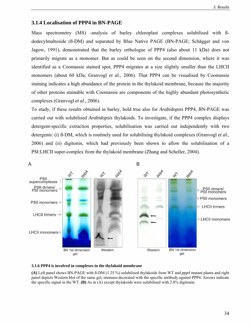
3. Results
34
3.1.4 Localisation of PPP4 in BN-PAGE
Mass spectrometry (MS) -analysis of barley chloroplast complexes solubilised with ß-
dodecylmaltoside (ß-DM) and separated by Blue Native PAGE (BN-PAGE; Schägger and von
Jagow, 1991), demonstrated that the barley orthologue of PPP4 (also about 11 kDa) does not
primarily migrate as a monomer. But as could be seen on the second dimension, where it was
identified as a Coomassie stained spot, PPP4 migrates at a size slightly smaller than the LHCII
monomers (about 60 kDa; Granvogl et al., 2006). That PPP4 can be visualised by Coomassie
staining indicates a high abundance of the protein in the thylakoid membrane, because the majority
of other proteins stainable with Coomassie are components of the highly abundant photosynthetic
complexes (Granvogl et al., 2006).
To study, if these results obtained in barley, hold true also for Arabidopsis PPP4, BN-PAGE was
carried out with solubilised Arabidopsis thylakoids. To investigate, if the PPP4 complex displays
detergent-specific extraction properties, solubilisation was carried out independently with two
detergents: (i) ß-DM, which is routinely used for solubilising thylakoid complexes (Granvogl et al.,
2006) and (ii) digitonin, which had previously been shown to allow the solubilisation of a
PSI:LHCII super-complex from the thylakoid membrane (Zhang and Scheller, 2004).
3.1.6 PPP4 is involved in complexes in the thylakoid membrane
(A) Left panel shows BN-PAGE with ß-DM (1.25 %) solubilised thylakoids from WT and ppp4 mutant plants and right panel depicts Western blot of the same gel, immuno-decorated with the specific antibody against PPP4. Arrows indicate the specific signal in the WT. (B) As in (A) except thylakoids were solubilised with 2.8% digitonin.

3. Results
35
Complex separation by native first dimensions following solubilisation with ß-DM, demonstrated
that also Arabidopsis PPP4 migrates just below the LHCII monomers, which runs at about 60 kDa
(Granvogl et al., 2006). However, as can be seen in Figure 3.1.6B solubilisation with the detergent
digitonin does result in the extraction of a larger PPP4-containing complex from the thylakoid
membrane, as PPP4 co-migrates with two complexes on the BN-PAGE. The bigger of the two
complexes migrates at a size between monomers and trimers (about 140 kDa) of LHCII, whereas
the smaller one runs just below the LHCII monomers. In the subsequent steps of this thesis all
solubilisation steps were performed by using digitonin in order to identify further components of
the two PPP4-containing complexes.
Consequently, analysis of the second, denaturing SDS-PAGE was carried out and it could be shown
that unlike in barley, Arabidopsis thylakoids do not contain sufficient PPP4 to be stained with
colloidal Coomassie, as no spot became visible at the expected size and location. However, by
comparing the protein spot patterns of silver stained BN second dimensions from WT and ppp4
thylakoid complexes (Figure 3.1.7) it becomes evident that the protein can be visualised by silver
staining. The identity of the respective spot in the WT second dimension as PPP4 was verified by
excision and subsequent MS-analysis of the trypsin digested protein. Moreover, MS analysis
identified TMP14 as a protein of slightly larger size (Figure 3.1.7, black arrow) than PPP4 (red
arrow) co-migrating with the larger PPP4 containing complex. The spot co-migrating with the
smaller complex of PPP4 was identified as TMP14-like (green arrow). For each respective spot the
identified peptides yielded 46% of sequence coverage for the mature PPP4, 40% for TMP14 and
41% for TMP14-like (for fragmentation spectra of identified peptides see Chapter 6.1). Subsequent
Western analyses of the WT second dimension with the available antibodies against PPP4 and
TMP14 show that both can be found co-localised in four distinct positions. The two lower
molecular weight complexes are according to the sizes of PPP4-containing complexes as also seen
in the first dimension and contain the majority of both proteins. TMP14 is mostly co-localised with
the higher molecular weight complex of the two. The two larger complexes, in which only small
fractions of PPP4 and TMP14 co-migrate, are at the size of the Cyt b6f monomer and at a size
slightly smaller than PSII dimers and PSI monomers. Analyses of second dimensions of solubilised
thylakoids from mutant plants indicate that PPP4 forms a complex of smaller size, if TMP14 is
absent, whereas TMP14 complex assembly is impaired in the absence of PPP4. However, some
TMP14 still forms a complex, as detectable by MS analysis of the excised spot at the same position
as in the WT. Altogether, it can be concluded from analysing thylakoid complexes of WT, ppp4 and
tmp14 plants that absence of neither PPP4 nor TMP14 affects the abundances and structures of the

3. Results
36
major photosynthetic thylakoid complexes (neither does the double mutant; data not shown).
However, the existence of a novel complex probably containing both PPP4 and TMP14 could be
demonstrated by solubilising thylakoid membranes with digitonin.
Fig 3.1.7 PPP4 co-migrates with TMP14 and TMP14-like
Two-dimensional separation of digitonin solubilised thylakoid protein complexes by BN/SDS-PAGE. After separation of the protein complexes of WT (A), ppp4 (B) and tmp14 (C) on a Blue-Native gel, the composition of their subunits was further analysed by subsequent denaturing Tris-Tricine SDS-PAGE. The gels were either stained with silver (A-C) or blotted onto PVDF membrane and immunodecorated with TMP14 and PPP4 specific antibodies (A). Silver-stained spots as indicated by arrows were excised and analysed by MS-analysis. Red, black and green arrows indicate spots of which the highest abundant protein is PPP4, TMP14 and TMP14-like, respectively. (D) Peptides identified by MS in each respective spot are indicated in red. Identification of the macromolecular protein complexes of thylakoid membranes is given on the top of gel (A).

3. Results
37
3.1.5 TMP14 protein levels are decreased in ppp4 mutants
Because in the previous experiments immuno-detection of TMP14 in the ppp4 mutants (KO
analysis and BN second dimension) indicated a severe reduction in protein amounts, quantification
of relative TMP14 levels in total protein extract and in thylakoids was performed by Western
analysis. Antibodies against Actin were used as loading controls for total protein extracts. The
application of antibodies against Lhca3 to ensure equal loading of thylakoid proteins was employed,
because this antibody was used by Khrouchtchova et al., 2005 in their work on TMP14 for the same
purpose. As can be seen in Figure 3.1.8, TMP14 protein accumulates up to about 75% of WT in
ppp4 total protein extract. However, the amount of TMP14 integrated in the thylakoid membrane is
only about 50% in comparison to WT. This indicates that the reduction of TMP14 levels is probably
due to stabilizing or integrating effects of PPP4 and does not result from changes in TMP14
expression.
Fig 3.1.8 TMP14 protein abundance in the thylakoid membrane depends on presence of PPP4
Total protein extract (left panel) and thylakoid proteins (right panel) of Ler and ppp4 plants corresponding to 5 µg of chlorophyll and decreasing amounts as indicated in the legend were separated by SDS-PAGE and immuno-detection was carried out with specific antibodies against TMP14, PPP4 and Actin, Lhca3 as loading control.
3.1.6 Investigating the interaction between PPP4 and TMP14
To verify the interaction of PPP4 and TMP14 two independent approaches were taken. Firstly,
crosslinking with Bis (Sulfosuccinimidyl) suberate (BS3), a membrane-impermeable crosslinking
agent with an 11.4 Å spacer arm was carried out and secondly, Co-IP was performed with digitonin
solubilised thylakoids.
3.1.6.1 Epitope tagging of PPP4
Tagged versions of the PPP4 protein were created because of two reasons. Firstly, tagged proteins
are routinely used for immuno-precipitation experiments, because of the specificity of the

3. Results
38
commercially available tag antibodies. Secondly, indications for multimerisation of PPP4
necessitated the creation of a PPP4 of differing size. Therefore, the nucleotide sequences encoding
the c-myc and the HA epitopes were fused to the 3’ of the PPP4 cDNA sequence. For this, full
length PPP4 cDNA was inserted into the binary vectors pPCV812ΔNotI-Pily (HA) and
pPCV812ΔNotI-Lola (c-myc; Chapter 2.2.3.2), which both contain a double 35S promoter upstream
of the insertion region. Agrobacterium mediated transformation of ppp4-2 plants was carried out
according to Chapter 2.2.3.3 and transformants were selected with hygromycin. Homozygous T3
lines were analysed for expression of the transgene by Western analysis of total protein extract,
using specific antibodies against Actin as loading control, PPP4, HA and c-myc (Figure 3.1.9).
Fig 3.1.9 Transgenic plants expressing epitope tagged PPP4
The PPP4 cDNA was inserted into the binary vectors pPCV812ΔNotI-Pily (Hemagglutinin; HA) and pPCV812ΔNotI-Lola (c-myc) flanked by a 35S promoter at the 5’and the respective tag coding sequence at the 3’. (A) Four week old WT, ppp4-2 and ppp4-2 plants transformed with both constructs. (B) Transgene expression analysis was carried out on total protein extract. PPP4 is overexpressed in all four lines and tags are recognised by the respective antibodies as depicted on the right As can be observed in Figure 3.1.9B the tagged proteins are expressed at higher levels in the
transgenic plants than the native PPP4 in WT. The resulting tagged PPP4 has an apparent molecular
weight of about 16 kDa. Because detection with the HA antibody yields some slight unspecific
bands upon long exposure, work was continued with the 35S::PPP4:c-myc lines # 6 and # 8. To
obtain plants expressing both native and tagged protein the line #6 was crossed with the Ler WT.
3.1.6.2 Crosslinking
Crosslinking is a powerful tool to dissect protein interactions. If putative interaction partners are
known, the existence of a complex comprising these components can easily be demonstrated by
crosslinking of proteins in a mutant lacking a specific protein, followed by Western detection using
antibodies against the expected interaction partner protein and vice versa. Depending on the

3. Results
39
structure and sequence of the interacting proteins, different crosslinkers are required to detect the
interaction by covalently linking the interaction partners. Here, Bis (Sulfosuccinimidyl) suberate
(BS3), a membrane-impermeable, non-cleavable crosslinking agent with an 11.4 Å spacer arm and
its cleavable analog 3,3´-Dithiobis[sulfosuccinimidylpropionate] (DTSSP), which has a 12 Å spacer
arm, were successfully employed to yield immuno-detectable PPP4 crosslinking products.
To investigate the existence of a complex containing TMP14 and PPP4, thylakoid membranes were
crosslinked with BS3 according to the method described in Chapter 2.2.4.11. The Coomassie-
stained gel shows that protein content and patterns are equal between the different samples (Figure
3.1.10A). Furthermore, it demonstrates that no unspecific crosslinking events have occurred as the
protein patterns still resemble those of the untreated thylakoid proteome. In Western analyses the
monomeric PPP4 can be detected in WT and tmp14 as a strong band of about 11 kDa, in accordance
with its deduced molecular mass of 11.4 kDa. A further strong band is visible in both samples at
about 23 kDa and two weaker bands at about 34 kDa and 45 kDa, the sizes indicating that these
PPP4 containing bands might be originating from crosslinking of components of a PPP4 tetramer.
Additionally, to these four crosslinking products, four more immuno-reactive bands are present in
the WT but not in the tmp14 sample. The lack of these extra WT bands in tmp14 demonstrates that
they stem from a PPP4 TMP14 interaction. Immunodecoration of the same membrane with TMP14
antibodies shows that monomeric TMP14 can be detected in WT and ppp4 as a weak double band
of the expected molecular weight of 14 kDa (Hansson and Vener, 2003). A further, strong band is
visible in both samples at about 28 kDa, suggesting the detection of a crosslinked TMP14 dimer.
According to the results obtained with the PPP4 antibody, the TMP14 antibody recognises
additional bands in WT crosslinked thylakoids, which are absent in the ppp4 mutant sample.
Merging the signals derived from immunodetection with the PPP4 and the TMP14 antibody, the
bands missing in the mutants overlap in the WT, further indicating that they originate from a
crosslinked interaction of both PPP4 and TMP14 (Figure 3.1.10A; right panel). That two immuno-
detectable bands appear in WT at about 25 kDa might result from a post-translational modification
of one of the two interaction partners leading to a slight increase of the size of the crosslinked
product. In the ppp4 sample two further clear bands are detected by the TMP14 antibody, one at
about 40 kDa and one at about 45 kDa. Both of the two could be due to the detection of a TMP14
trimer of a predicted 42 kDa, which might preferentially form in the absence of PPP4. The other of
the two bands might originate from the interaction of TMP14 with a further unknown protein. As
this band shows exclusively in the ppp4 mutant, the presence of the interaction might only occur in
the absence of PPP4.

3. Results
40
Fig 3.1.10 PPP4:TMP14 and PPP4:PPP4c-myc interactions can be crosslinked with BS3
(A) Thylakoid proteins of WT, tmp14 and ppp4 mutant plants were crosslinked with BS3, separated by SDS PAGE, subsequently either stained with colloidal Coomassie or transferred to PVDF and then immuno-decorated using specific PPP4 and TMP 14 antibodies. The signals resulting from detection with the TMP14 and PPP4 antibodies were converted into coloured bands (PPP4: red; TMP14: blue) and merged using Adobe Photoshop. (B,C) Western analysis of thylakoid extract from WT, ppp4-2, 35S::PPP4:c-myc in ppp4-2 #6, #8, 35S::PPP4:c-myc#6 in Ler in order to quantify the relation of TMP14 and PPP4 levels (D) Thylakoid proteins from Ler and ppp4-2 plants expressing PPP4:c-myc from a 35S promoter, tmp14 and WT were crosslinked with BS3, separated by SDS-PAGE and immuno-decorated with the TMP14 and PPP4 antibodies. Numbers on left of TMP14 and on right of PPP4 hybridised blots indicate nature of crosslinking products (1: TMP14; 2:PPP4; 3:PPP4:c-myc).
3. Results
41
When comparing the strength of the monomeric signals with the strength of signals of crosslinked
products, it becomes obvious that crosslinking of TMP14 has progressed further than that of PPP4.
Firstly, this could be due to the differences in amino acid sequence between PPP4 and TMP14 and
the chemical properties of the crosslinker, which reacts with two amino groups on both sides of a
11.4 Å spacer, and covalently links them. TMP14 contains 12 and therefore five more basic amino
acid residues than PPP4 in non-membrane spanning regions, which can be employed in the
crosslinking reaction. Secondly, the higher rate of TMP14 crosslinking could be due to a better
accessibility of the protein to the crosslinker. TMP14 might be part of the periphery of a bigger
complex and thus be more exposed to the reactive agent.
To verify the existence of PPP4 multimers, the crosslinking reaction was carried out with Ler and
ppp4 plants expressing PPP4:c-myc (Figure 3.1.10D). Here, crosslinking of the overexpressed
tagged PPP4 occurred at an increased rate as compared to the native PPP4. Accordingly, to allow
visual separation of crosslinking products, incubation of thylakoids from PPP4:c-myc expressing
plants with BS3 was reduced from 1 h to 30 min. Detection of PPP4 from PPP4:c-myc expressing
Ler plants shows that besides the monomers of both PPP4 (Figure 3.1.10D: 2) and PPP4:c-myc (3)
and the dimer of PPP4:cmyc (3:3) a specific crosslinking product is present at a size smaller than
the PPP4:c-myc dimer. This probably presents a crosslinked PPP4, PPP4:c-myc interaction (3:2).
The dimer of the untagged PPP4 cannot be detected here, probably because of the the lower rate of
crosslinking reactions within the 30 min of incubation time. Crosslinked interactions between
TMP14 and PPP4 are severely decreased in both PPP4:c-myc expressing backgrounds, as shown by
immuno-decoration using the TMP14 antibody.
3.1.6.3 Co-immunoprecipitation (Co-IP)
To identify interaction partners of membrane proteins by co-immunoprecipitation (Co-IP) it is
prerequisite to solubilise the membranes in such a way that the protein complexes stay intact.
Because treatment with digitonin resulted in the detection of two complexes containing PPP4 and
TMP14 in BN-gels (Figure 3.1.7), solubilisation was carried out with this detergent in order to
verify the interaction of both proteins and to identify further interactors. As a preliminary
experiment, Co-IP was carried out with the specific antibodies against PPP4 and TMP14 according
to the method described in Chapter 2.2.4.12. However, only the TMP14 antibody was successfully
employed for precipitation of the respective protein. Therefore, co-immunoprecipitation was
performed with thylakoids of WT and tmp14 mutant plants with this antibody in order to
investigate, if also PPP4 can be precipitated and to ensure that precipitation of PPP4 depends on the
presence of TMP14 and not on an unspecific interaction of the TMP14 antibody with the similar

3. Results
42
PPP4 protein. Although Western analyses of tmp14 total protein extracts (3.1.1.3 and 3.1.1.5) had
not indicated any cross reactions of the specific antibodies, the absence of a cross reaction of the
TMP14 antibody with the native PPP4 protein had to be established. According to Figure 3.1.11A
immunodecoration with the TMP14 antibody yields a signal in the WT input and Co-IP lane,
demonstrating that the TMP14 antibody was successfully employed for precipitation of the native
protein. Indeed, using the PPP4 antibody a signal is visible in the all lanes except for the tmp14 Co-
IP lane, demonstrating that precipitation of PPP4 with the TMP14 antibody occurs only in the
presence of TMP14. A control immuno-decoration with the PSI-F antibody results solely in
detectable signals in the input and not in the Co-IP lanes, showing that the detection of PPP4 in the
WT immunoprecipitate is not due to contamination with other thylakoid proteins.
To identify interaction partners besides PPP4, the co-immunoprecipitate was analysed by MS. As
MS analysis is very sensitive and thus prone to measure the slightest contaminations, the precipitate
from tmp14 thylakoids was used as a negative control. Additionally, the co-immunoprecipitate of
TMP14 from the ppp4 thylakoids was analysed, because additional TMP14 crosslinking signals had
appeared in this mutant, which were not present in the WT (Figure 3.1.10A). However, MS analysis
only detected TMP14 in this precipitate, which might be due to the disbandment of the interaction
of TMP14 with the unknown protein by the solubilisation with digitonin. As expected from Western
analysis the WT sample contained TMP14 and the co-precipitated PPP4.
In order to test if also the reciprocal precipitation of TMP14 with PPP4 is possible, thylakoid
membranes of WT, Ler and ppp4 expressing PPP4:c-myc were isolated and Co-IP was performed
with either antibodies against TMP14 or c-myc (Figure 3.1.11B). Using the TMP14 antibody both
PPP4 forms can be co-precipitated, however with a bias to the native protein, when both forms are
present. This underlines the results obtained from crosslinking of thylakoid proteins in the PPP4:c-
myc overexpressing backgrounds, where interaction of TMP14 with PPP4 seemed to be suppressed
(Figure 31.1.10D). With the c-myc antibody the native PPP4 can be precipitated in the presence of
PPP4:c-myc. However, precipitation of detectable amounts of TMP14 with the c-myc antibody was
not possible. This could be due to a displacement of the interacting TMP14 protein by this antibody.

3. Results
43
Fig 3.1.11 PPP4 co-precipitates with TMP14 and PPP4:c-myc
(A) Thylakoid membranes from WT (No-0) and tmp14 mutants at a concentration of 1.5 mg chlorophyll ml-1 were incubated with 1/6th volume of specific antibodies against TMP14 for 2h at 4°C and then treated with 2.5% digitonin for 1h at 4°C. Solubilised complexes were precipitated with ProteinA-Agarose by o/n end over end mixing at 4°C and following centrifugation. The pellet was washed five times and bound proteins were eluted by adding boiling SDS-PAGE loading dye. Thylakoid membrane (T) and immunoprecipitated (P) proteins were separated by SDS-PAGE, transferred to PDVF membrane and immuno-decorated with specific antibodies against TMP14, PPP4 and PSI-F. (B) Co-IP was carried out with thylakoid proteins from WT, Ler and ppp4-2 plants expressing PPP4:c-myc as described in (A) with antibodies against TMP14 and c-myc.
3.1.7 TMP14 and PPP4 are not stably associated with any of the main photosynthetic
complexes
In earlier work, TMP14 was reported to be a novel subunit of PSI based on the results that it co-
migrated solely with PSI in both BN gels and sucrose gradient fractionations (Khrouchtchova et al.,
2005). It was therefore named PSI-P. This work also showed that TMP14/ PSI-P protein was absent
in plants lacking the L and G subunits of PSI and that its levels were increased in a psao RNAi line.
Because the results from BN gels, crosslinking and Co-IP experiments demonstrate that TMP14 is
forming a complex with PPP4 and is not associated with any of the four major thylakoid complexes,
two further experiments from the work of Khrouchtchova et al. were repeated to confirm that
TMP14 is not associated with PSI and to investigate, if its levels as stated in the previous work
depend on the presence of L and O subunits of PSI. Therefore, PSI was isolated by sucrose gradient

3. Results
44
fractionation and Western analysis was carried out with antibodies against TMP14, PPP4 and PsaF.
Figure 3.1.12A demonstrates that TMP14 and PPP4 can be detected in the thylakoids, but not in the
PSI isolation, thereby confirming the results gained from BN gel analysis in this thesis (Chapter
3.1.4). For the second experiment, thylakoid proteins were isolated from psal (SALK_000637),
psao (SALK_021621), tmp14, ppp4 mutant plants and the corresponding WT ecotypes, separated
by SDS-PAGE and Western analysis was carried out with specific antibodies against TMP14,
PPP4, PsaL and PsaO (Figure 3.1.12B). Lhca3 hybridisation was used as loading control as in
Khrouchtchova et al. (2005). However, contrary to their results, TMP14 levels are neither changed
in the psal nor the psao, but are altered in the ppp4 mutant background.
Fig 3.1.12 TMP14 and PPP4 are not constitutive components of one of the thylakoid photosynthesis complexes (A) PSI was isolated from WT thylakoids via sucrose gradient centrifugation. Thylakoid and PSI proteins according to 5 µg of chlorophyll were separated by SDS-PAGE and Western analysis was carried out using specific antibodies against PsaF, TMP14 and PPP4. (B) Thylakoid proteins according to 5 µg of chlorophyll of the three ecotypes Col-0, Ler, No-0 and the four mutant lines psal, psao, ppp4 and tmp14 were separated by SDS-PAGE and Western analysis was performed with antibodies depicted on the right. (C) Immuno-detection of TMP14 and PPP4 in Col-0, Ler, hcf136, petC, psad1/d2, atpd, ppp4 and tmp14. The applied antibodies are depicted on the right. Total protein extract corresponding to 40 µg of protein was loaded each.

3. Results
45
Additionally, it was tested, wether PPP4 and TMP14 belong to one of the other three main
photosynthetic complexes. Therefore, total protein extracts were isolated from the PSII deficient
hcf136 mutant (Plücken et al., 2002), the Cyt b6f deficient petc mutant (Maiwald et al., 2003), the
PSI deficient psad1 psad2 double mutant (Ihnatowicz et al., 2003) and the ATP-synthase deficient
atpd mutant (Maiwald et al., 2003), all four grown heterotrophically on sucrose containing medium.
Additionally, total protein was extracted from tmp14, ppp4 and the two ecotypes Col-0 and Ler,
which are the respective ecotypic backgrounds of hcf136, psad1 psad2, atpd and petc, ppp4. These
plants were grown under the same conditions as the complex knock-out mutants.
If TMP14 and PPP4 were constitutively associated with one of the complexes, their lack would
cause a destabilisation of these proteins, then leading to a severe decrease in TMP14 and PPP4
protein levels. However, Western analysis with the respective antibodies reveals that PPP4 is only
slightly reduced in the mutants lacking PSII, the Cyt b6f complex and PSI and that the apparent
reduction can be due to less protein loaded, as also the Actin control gives a slightly weaker signal
(Figure 3.1.12C). TMP14 levels show also a decrease in the same three mutant backgrounds, but
more significantly than PPP4, which could indicate a differential regulation of both proteins
3.1.8 Photosynthetic parameter of WT, ppp4, tmp14, ppp4 tmp14 and PPP4:c-myc overexpressors
Both mutant alleles ppp4-1 and ppp4-2 exhibit a decrease in the effective quantum yield of PSII
(ΦII, (Fm’-F0)/Fm’) and an increase in PQ pool reduction as indicated by the parameter 1-qP (1-
[(Fm’-Fs)/(Fm’-F0)]) (Table 3.1.1A). The measurement was carried out with the PAM 101/103
chlorophyll fluorometer and plants were illuminated with 80 µmol photons m-2 s-1 for 15 min before
determining photosynthetic parameters by giving an 800 ms saturating light pulse of 4000 µmol
photons m-2s-1. Under these conditions the tmp14 mutant showed no differences in parameters
compared to WT.
To investigate the effect of the tmp14 mutant background on photosynthetic parameters in the ppp4
mutant and additionally to test complementation of the PPP4:c-myc construct, Chl a measurements
and determination of P700 oxidation rate were carried out with tmp14-1, ppp4-1, a cross of both
lines and the PPP4:c-myc overexpressing plants using the Dual-PAM-100 fluorometer. To exclude
the influence of ecotypical differences in photosynthetic parameters between Ler (background of
ppp4-1) and No-0 (background of tmp14-1) on the results obtained with the double mutant, WT
plants of both genetic backgrounds and two double mutants as well as one WT segregating from
one heterozygous double mutant were measured. Ecotypical differences between Ler and No-0
have already been described for the phytochrome-mediated response (Magliano et al., 2005).

3. Results
46
However, all three WT showed identical values and no differences were detected between the two
double mutants. Therefore, ecotypical differences between Ler and No-0 regarding the
photosynthetic parameters as measured in this thesis, could be excluded. Photosynthetic parameters
were determined after light adaption of the plants to two different actinic light intensities. To
investigate photosynthetic activity of mutants and overexpressors under low light, plants were
illuminated with actinic light of 77 µmol photons m-2 s-1 (Table 3.1.1B) before determining Chl a
fluorescence and P700 absorption parameters. For high light 1052 µmol photons m-2 s-1 was applied
(Table 3.1.1C). In order to determine Chl a fluorescence parameters, plant were light adapted for
15 min. P700 oxidation rate was measured after 5 min of light adaption.
The results indicate that changes in photosynthetic parameters of ppp4 are exacerbated in the ppp4
tmp14 double mutant. In detail, after low actinic light illumination the values for 1-qP and
ΔA/ΔAmax are slightly increased whereas NPQ ((Fm-Fm’)/Fm’) andΦII are slightly decreased in both
ppp4 and the double mutant. In comparison to WT parameters measured in the tmp14 mutant show
the same trend as for ppp4 and the double mutant, but for this mutant values are not significantly
changed. When illuminated with high actinic light, only the ΔA/ΔAmax parameter remains
significantly changed in ppp4 and the double mutant as compared to the WT, but contrary to the
increase observed in low light this parameter is decreased. Also the NPQ appears to be lower, but
differences from WT are not significant. The 35S::PPP4:c-myc complements all significant
changes, but values remain below WT at lower actinic light intensities for ΦII and 1-qP. That values
are different from WT in these plants might be due to two possibilities. (i) The higher abundance of
PPP4 in the 35S::PPP4:c-myc lines could have a direct effect on photosynthetic parameters. (ii)
The tagged protein is not fully functional, because of differing properties caused by the tag. That
PPP4:c-myc at least shows a different binding affinity to TMP14 in comparison tothe native PPP4
has been shown by crosslinking and Co-IP.
To exclude that differences in pigment levels are responsible for the NPQ phenotype of ppp4 plants,
a quantitative analysis of leaf pigments was conducted, but only slight, significant increases could
be observed in violaxanthin (WT: 33 ± 2 µmol per mmol chl; ppp4: 36 ± 1), lutein (WT: 123 ± 2;
ppp4: 131 ± 2) and ß-carotene levels (WT: 79 ± 1; ppp4: 84 ± 3). The lumenal localised
violaxanthin de-epoxidase is activated by low pH (Hager, 1969) and therefore the increase in
violaxanthin suggests that the decrease in NPQ might be due to a defect in lumen acidification.

3. Results
47
Table 3.1.1 Photosynthetic parameters of tmp14, ppp4, ppp4 tmp14 and 35S::PPP4:c-myc plants (A) Chl a fluorescence parameters of Ler, ppp4-1 and ppp4-2 as determined by measurement with the PAM 101/103 chlorophyll fluorometer after 15 min of 80 µmol photons m-2 s-1 actinic light. (B) Chl a fluorescence and P700 absorption parameters of WT, tmp14-1, ppp4-1, ppp4 tmp14 plants and in two lines overexpressing PPP4:c-myc as measured with Dual-PAM 100 after 5 min (P700) or 15 min (Chl a) of 77 µmol photons m-2 s-1 actinic light (settings: actinic light, 5; measuring light, 5; saturation pulse: 5) (C) Chl a fluorescence and P700 absorption parameters in WT, tmp14-1, ppp4-1, ppp4 tmp14 plants and in two lines overexpressing PPP4:c-myc as measured with Dual-PAM 100 after 5/ 15 min of 1052 µmol photons m-2 s-1 actinic light (settings: actinic light, 17; measuring light, 5). Values are means (± SD) of 15 independent measurements for WT (Ler: 5, No-0: 5,Ler x No-0: 5), 10 for ppp4 tmp14 and 5 for the other genotypes.

3. Results
48
A decrease in the ΔA/ΔAmax and NPQ parameters are indicative for a defect in cyclic electron flow
(CEF; Munekage et al.; 2002, DalCorso et al., 2008) and as at least ΔA/ΔAmax is significantly
reduced in ppp4 and the double mutant, two assays were performed in order to elucidate a function
of PPP4 and TMP14 in the cyclic electron transport. Firstly, an intact leaf assay for determining
CEF was carried out by measuring the transient increase in NPQ after onset of low light. This
measurement relates to the hypothesis that NPQ induction during the activation period of
photosynthesis is caused by the transient acidification of the thylakoid lumen when CEF activity is
higher than the activity of the ATP-synthase (DalCorso et al., 2008). Secondly, to assess electron
transfer from ferredoxin (Fd) to PQ during CEF around PSI, Fd-dependent reduction of PQ was
monitored in situ as an increase in chlorophyll fluorescence under low measuring light. In WT, the
addition of NADPH and Fd to ruptured chloroplasts induces a marked and rapid increase in
chlorophyll fluorescence (Munekage et al., 2002; DalCorso et al., 2008). In this in situ assay
NADPH is thought to transfer electrons to Fd via the reverse reaction of the Fd:NADPH
oxidoreductase (FNR). In the intact leaf assay however, ecotypic differences could be observed, as
No-0 has significantly lower transient NPQ than Ler. Therefore, a phenotype of the double mutant
could at least partly be due to the genetic ecotypic background. As the ppp4 mutant alone already
shows a phenotype in the described changes of photosynthetic parameters, the transient increase in
NPQ was measured with ppp4-2 and Ler. When grown in the climate chamber at a 12h/12h light
dark cycle, ppp4 plants only show a slight decrease in NPQ in the steady state phase (Figure
3.1.13A). However, if measurements are performed with plants grown in continuous light, the
transient increase in NPQ is significantly lower in ppp4 as compared to WT (Figure 3.1.13B). This
could indicate that acclimation of the plant to continuous light includes PPP4-dependent non-
photochemical quenching.
In the in situ assay, all three WTs (Ler, No-0, Ler x No-0) show the same extent and kinetics of the
increase in chlorophyll fluorescence. Therefore, only one WT curve is depicted in Figure 3.1.13C.
The kinetic of the chlorophyll fluorescence increase is slower in ppp4 tmp14 double mutant, yet no
significant changes in the final level of chlorophyll fluorescence can be detected. However, Chl a
fluorescence already increases in response to the addition of NADPH.

3. Results
49
Fig 3.1.13 Cyclic electron flow measurements
(A+B) Time courses of induction and relaxation of NPQ monitored during dark-to-light (80 µmol photons m-2 s-1, white bar) transition of Ler (filled circles) and ppp4-2 (open squares) grown in a 12 h/12 h dark light cycle (A) or in continuous light (B). The 3 min light period (white bar) was followed by a 2 min dark period (black bar). Values are means (± SD) of five independent measuremts each per time point (C) Quantification of CEF in situ. Increases in chlorophyll fluorescence were measured in ruptured chloroplasts under low measuring light (1 µmol photons m-2 s-1), after the addition of NADPH and Fd. At this light intensity, the fluorescence level should predominately reflect the reduction of plastoquinone by cyclic electron transport from ferredoxin, not by PSII photochemistry (Munekage et al., 2002).
3.1.9 Topology of PPP4 and TMP14
As mentioned afore, TMP14 was first described, because of its posttranslational modification by
phosphorylation of the N-terminus (Hansson and Vener, 2003), which was found by trypsin
digestion of all stromal side exposed proteins and subsequent enrichment of phosphopeptides.
Because TMP14 has two predicted transmembrane domains, this indicates that both termini are
stromal exposed. To determine PPP4 and to verify TMP14 topology, thylakoids were subjected to
mild digestion with trypsin for 10 min with the aim to solely target tryptic sites on the stromal side
of the thylakoid membrane. Both proteins contain multiple trypsin cleavage sites in their N-
terminus, which also bears the sequence of the epitopes, against which specific antibodies were
made. Therefore, stromal side treatment with trypsin of a topology in which the N- and C-termini of

3. Results
50
the proteins were exposed to the stromal side (topology 1, Figure 3.1.14B), would result in
destruction of the epitopes and absence of detectability. Because PPP4 has one tryptic site in the
transmembane regions connecting loop, treatment with trypsin on a stromal exposed loop topology
(topology 2) would result in detectable product of 5 kDa for PPP4 with the specific antibody. The
tryptic digest was additionally performed with thylakoids of tmp14 and ppp4 mutants in order to
evaluate if the absence of one interaction partner would change accessibility of the other to the
protease, and with PPP4:c-myc expressing plants as a confirmation for PPP4 topology. Because the
TMP14 antibody reacts unspecifically with chlorophyll on the PVDF membrane, thylakoid proteins
were precipitated with acetone after the trypsin treatment to avoid this unspecific interaction during
Western detection. The reaction of the antibody with the low molecular chlorophyll could interfere
with the detection of smaller TMP14 degradation products. As an indication that tryptic proteolysis
took place only at the stromal side of the membrane and no unspecific cleavage of lumenal proteins
occurred, Western analysis of the treated thylakoid extracts was performed with an antibody against
the lumenal protein PsbO, a component of the PSII oxygen evolving complex (Figure 3.1.14A).
This shows that after 10 min of digestion PsbO remains intact. Hybridisation with an antibody
against the stromal side localised AtpB confirms activity of trypsin. Western detection with the
specific antibodies against TMP14 and PPP4 clearly indicates that both proteins are protected from
trypsin treatment, because their amount does not decrease, but even seems to increase after the
protease treatment. This result could be due to a negative effect of associated proteins on the
accessibility of PPP4 and TMP14 to the detergent SDS in the loading buffer after acetone
precipitation. If the associated proteins, which together with PPP4 and TMP14 precipitate in
acetone as a partly insoluble fraction, are partially removed by proteolytic cleavage before the
precipitation, higher amounts of PPP4 and TMP14 would be soluble. A 10 min trypsin treatment of
TMP14 results in a detectable fragment of smaller size, which indicates that the TMP14 N-terminus
with its 8 tryptic sites is heavily protected and cleavage might have occurred at an arginine residue
two amino acids upstream of the predicted transmembrane domain or at the only lumenal exposed
tryptic site. Digested PPP4 exhibits one degradation product after 10 min of slightly smaller size.
Therefore topology 1 with stromal localised N- and C-terminus appears likely. Verification of this
result arises from PPP4:c-myc digestion. The amount of detectable c-myc is decreasing and an
increasing amount of normal sized PPP4 indicates that the c-myc tag is cleaved off.

3. Results
51
Fig 3.1.14 Determination of PPP4 topology by stromal side tryptic digest of thylakoid proteins
(A) Thylakoids were isolated from WT, 35S::PPP4:c-myc, ppp4 and tmp14 and incubated with 10 µg/ml trypsin. Samples were taken at 0, 10 and 20 min after the addition of trypsin and proteins were separated by SDS-PAGE. Immuno-detection was carried out with specific antibodies against AtpB, as a control for trypsin activity, PsbO as a control for absence of trypsin in the lumen, TMP14, PPP4 and c-myc. Asterisks indicate degradation products. (B) Schematic drawings of probable topologies. Scissors indicate possible luminal accessible tryptic sites. Red boxes indicate location of epitopes used for antibody production. Dashed green line stands for c-myc tag fused to PPP4. For PPP4 topology 1 could be supported.
Additionally, lack of PPP4 does not result in enhanced degradation of TMP14 or vice versa. Both
proteins even seem to be more protected in the absence of the interaction partner, as no degradation
product can be detected in the respective other mutant. Interestingly, the increase by trypsin
treatment of Western detectable TMP14 is even higher in the ppp4 mutant background as compared
to WT, indicating that more TMP14 precipitates in SDS-loading dye in the absence of PPP4,

3. Results
52
speculatively because of an increased association with other hydrophobic proteins. Two results
obtained by this experiment suggest that TMP14 and PPP4 are associated with other proteins in the
thylakoid membrane: (i) hardly any digestion of PPP4 and TMP14 takes place despite the high
number of tryptic sites in the experimentally shown stromal localised N- and C- termini (TMP14: 8
N-terminal, 3 C-terminal; PPP4: 3 N-terminal, 3 C-terminal). (ii) PPP4 and TMP14 from untreated
thylakoids seem to only be partly solubilised in Laemmli buffer, whereas solubilisation is markedly
increased if samples are treated with trypsin.
3.1.10 Analysis of thylakoid protein composition
The analysis of thylakoid proteome
composition of WT, ppp4, tmp14 and
ppp4 tmp14 does not show any highly
significant differences between WT
and mutants as can be seen in Figure
3.1.15A. However, PGR5 content is
lower in the No-0 background, which
could account for the differences
observed in the transient increase in
NPQ after light onset between the
different ecotypes, as described in
Chapter 3.1.8. Additionally, a slight
increase in PGRL1 levels can be
observed in all mutant backgrounds,
especially in the double mutants. To
investigate, whether PPP4 and TMP14
protein levels are altered in the
pgrl1ab mutant, chloroplast proteins
were isolated from WT and pgrl1ab
double mutants. Immunodetection with
specific antibodies against PPP4 and
TMP14 showed no differences in
protein levels as compared to WT,
which can be seen in Figure 3.1.15B.
Fig 3.1.15 Thylakoid proteome analysis
(A) Thylakoid protein composition in Ler, No-0, Ler x No-0, ppp4-1, tmp14-1 and two independent homozygous ppp4 tmp14 lines. Thylakoids were extracted and protein according to 5 µg of chlorophyll was separated by SDS-PAGE. Replicate filters were hybridised with specific antibodies as depicted on the right. (B) Chloroplasts were isolated from WT and pgrl1ab mutant plants and proteins according to 5 µg of Chl were separated by SDS-PAGE and replicate filters were hybridised with PPP4 and TMP14.

3. Results
53
3.1.11 Location of thylakoid proteins in WT and ppp4 tmp14
To identify the specific localisation of PPP4 and TMP14 within the thylakoid membrane and to
investigate, if a lack of PPP4 and TMP14 causes changes in the spatial arrangement of thylakoid
complexes, thylakoids of WT and the ppp4 tmp14 double mutant were fractionated into grana,
intermediate membranes and stromal lamellae by a short treatment with the detergent digitonin and
subsequent differential centrifugation (Chapter 2.2.4.8). The rational of this experiment is that the
low concentration of digitonin and the short solubilisation time, followed by dilution, leads to the
detachment of vesicles from exposed regions of thylakoid membranes. With differential
centrifugation different sized vesicles can be precipitated. The 10000g (10K) fraction mainly
consists of the granal thylakoids, the 40000g (40K) of intermediate/ margin regions and the
150000g (150K) of stromal lamellae. In this work, additionally the proteins from the supernatant
after centrifugation at 150000g for 90 min were isolated by acetone precipitation, to see if PPP4 and
TMP14 were present. The membrane proteins contained within in this fraction are due to a
preferential extraction by digitonin. The Coomassie stained gel and the Western results in Figure
3.1.16 demonstrate that the analysed subunits of photosynthetic complexes are primarily found
according to their known localisation in both WT and ppp4 tmp14. As such no significant
difference in the spatial organisation can be assigned. However, the chlorophyll a/b ratio, which
was measured from all precipitated fractions in three independent fractionation experiments,
appears to be significantly lower in the stromal fraction of the ppp4 tmp14 mutant, indicating that
more PSII antenna is present. This is underlined by the coomassie staining of the gel, which
demonstrates a higher amount of LHCII in the stromal lamellae fraction of the ppp4 tmp14 mutant.
Western analysis of the different thylakoid fractions with the specific antibodies against PPP4 and
TMP14 demonstrate a localisation of both proteins in the stroma lamella regions of the thylakoid
membrane, whereby the exclusion of TMP14 from the other regions is more evident than for PPP4.
The supernatant, which contains lumenal, contaminating stromal and solubilised membrane proteins
as well as thylakoid membrane threads, also includes a substantial amount of PPP4 and TMP14 as
can be seen in the Coomassie stained gel, which shows two bands of the size of PPP4 and TMP14
in the WT, which are lacking in the double mutant (Figure 3.1.16, asterisks). Interestingly, two
further proteins can be identified by immunodetection to be present in the supernatant, the
ferredoxin:NADPH oxidoreductase (FNR) and PGRL1, a protein involved in the cyclic electron
flow around PSI (Dal Corso et al., 2008), which functions in the PGR5 dependent pathway.

3. Results
54
Coomassie stained bands from the supernatant were analysed by MS and the identity of the putative
PPP4 and TMP14 bands was verified. Additionally, the Coomassie stained protein bands running
just above the 34 kDa marker band, contained both forms of FNR, the thylakoid associated
(At5g66190) and the soluble protein (At1g20020) as well as PGRL1. Results obtained from
analysing the supernatant indicate that the complex of PPP4 and TMP14 is easily extracted by
digitonin. This could be due to a localisation in exposed regions with high lipid to protein ratio.
Moreover, also PGRL1 and thylakoid associated FNR are located in thylakoid regions with such
properties.
Fig 3.1.16
Fractionation of thylakoids
Thylakoids were extracted from WT and ppp4 tmp14 double mutants and treated with 0.1 % digitonin for 1 min, followed by dilution in 10 volumes and differential centrifugation at 10000g (10K; grana), 40000g (40K; intermediate membranes) and 150000g (150K; stroma lamellae). Soluble protein was precipitated with 5 volumes of 100% acetone. Proteins according to 5 µg of chlorophyll from untreated thylakoids (T), grana (10 K), intermediate membranes (40 K), stroma lamellae (150 K) and precipitated supernatant protein originating from 40 µg of treated thylakoids (sn) were separated by SDS-PAGE and replicate filter were hybridised with specific antibodies as depicted on the right. Numbers underneath state Chl a/b ratio of fractions of three independent experiments.

3. Results
55
3.1.12 Investigating phosphorylation of thylakoid proteins in WT and ppp4 tmp14
Because TMP14 has previously been described as a phospho-protein phosphorylated at a threonine
residue (Hansson and Vener, 2003) and detection with a phospho-threonine antibody has been
published (Khrouchtchova et al., 2005), WT
and ppp4 tmp14 thylakoids were isolated from
dark (D), low light (LL) and far-red (FR)
adapted plants in the presence of the
phosphatase inhibitor NaF to investigate
phosphorylation. Western analysis was
performed with phospho-threonine antibodies
(Figure 3.1.17), but no immuno-detectable
band of the size of TMP14 could be identified
that was present in WT, but absent in the
double mutant. However, the specificity of
phospho-threonine antibodies highly varies
between commercially available batches.
Instead of detecting phosphorylated TMP14,
this experiment led to the identification of a
slightly higher phosphorylation state of LHCII
and the PSII components CP43, D1 and D2.
This is in line with the slightly higher
reduction rate of the plastoquinone pool in the
ppp4 tmp14 double mutant, as determined by
the PAM fluorometer (Chapter 3.1.8), because
thylakoid kinase activity is known to be
induced by reduction of the plastoquinone
pool (Ihnatowicz et al., 2008).
3.1.13 Characterisation of PPP4-like and TMP14-like
Both TMP14-like and PPP4-like are predicted by ChloroP to have an N-terminal chloroplast transit
peptide, which contains for TMP14-like i 55 amino acids and for PPP4-like is 51 amino acids. Both
proteins have been identified in the chloroplast by shot-gun proteomics (Kleffman et al., 2004) and
the presence of TMP14-like in the thylakoid membrane was shown by Friso et al., 2004 by the
Fig 3.1.17 Phosphorylation pattern in WT and ppp4
tmp14
Thylakoids were isolated from WT and tmp14 ppp4 plants adapted to dark (D), low light (LL) and far-red light (FR) in the presence of 10 mM NaF. Proteins were separated by SDS-PAGE and either stained with Coomassie or transferred to PVDF membrane followed by hybridisation with phospho-threonine antibodies from Cell Signaling (New England Biolabs; Frankfurt, Germany).

3. Results
56
same method. Localisation of both proteins in the chloroplast was verified with transient expression
of a C-terminal RFP fusion in Arabidopsis protoplasts (Figure 3.1.18A). Here, the auto-
fluorescence of the chlorophyll (Auto) and the RFP-fluorescence of the fusion proteins overlap as
can be seen in the merged pictures. Expression analysis using Genevestigator shows that the gene
encoding TMP14-like has a similar expression pattern as PPP4 and TMP14 and also two other
genes encoding photosynthetic proteins PetC and Lhca1 (Figure 3.1.18B). It is also highest
expressed in green tissue, whereas PPP4-like expression peaks in seeds and in root tips. The
calculated molecular weight of the mature protein is 11 kDa for TMP14-like and 15.7 kDa for
PPP4-like.
Fig 3.1.18 Gene expression and gene product localisation of TMP14-like and PPP4-like (A) Arabidopsis protoplasts were transiently transfected by polyethylene glycol-mediated DNA uptake with the fusion constructs of the full coding sequence with dsRED and analyzed by using fluorescence microscopy. In the upper panels, the Differential Interference Contrast (DIC) view of the protoplasts is displayed. The second and third from the top show auto-fluorescence of the chlorophyll and RFP fluorescence, respectively and the lower panel shows a merged image of the latter. Scale bar, 20 μm. Organ and growth stage specific gene expression of PPP4, TMP14, TMP14-like and PPP4-like was analysed with Genevestigator. PetC encoding the Rieske subunit of Cyt b6f and Lhca1 were used as control for photosynthetic gene expression. All gene-level profiles were normalized for colouring in such way that for each gene the highest signal intensity obtains value 100% (black) and absence of signal obtains value 0 % (white).
The gene structures of PPP4-like and TMP14-like are depicted in Figure 3.1.19A and identification
of intron-exon structure was performed as described in Chapter 3.1.3 for PPP4 and TMP14
insertion lines. One T-DNA insertion line each for TMP14-like and PPP4-like could be identified
by screening T-DNA insertion flanking databases. The presence of the insertion and the exact
insertion point were confirmed by PCR with gene and T-DNA border specific primers and

3. Results
57
subsequent sequencing of the amplified product. The absence of transcript was verified by RT-PCR
using primer combinations up- and downstream of the insertions (Figure 3.1.19B). Furthermore,
photosynthetic parameters were determined in tmp14-like and ppp4-like, but no significant
difference to WT could be detected (data not shown). Because the TMP14-like protein migrates
with the smaller PPP4 containing complex as shown by MS analysis of second dimension silver
stained protein spots (Figure 3.1.7), crosslinking with BS3 was carried out with thylakoids from
WT, tmp14, tmp14-like and ppp4-like plants (Figure 3.1.19C), but changes in PPP4 crosslinking
products are only visible in the tmp14, but not in the other two mutant backgrounds.
Fig 3.1.19 T-DNA insertion lines of TMP14-
like, PPP4-like/ Pattern of PPP4 in crosslinked
thylakoids of these mutants
(A) Exons are depicted as open numbered boxes, introns as connecting black line. For each T-DNA mutant used in this study, the site and the orientation of the insertion is provided. In tmp14-like the concerned gene is disrupted by insertion of the ROK2 T-DNA. This line was identified in the SALK T-DNA collection as SALK_052057. The ppp4-like mutant carries an insertion of the pCSA110 T-DNA and was identified in the Syngenta SAIL-collection as SAIL_1240_C05. The T-DNA insertions are not drawn to scale. (B) RT-PCR analysis was performed on the transcripts of both WT and mutant plants with gene specific primers upstream (1) and downstream (2) of the insertion and ACTIN1 primers as positive control. (C) Thylakoid proteins from WT, tmp14, tmp14-like and ppp4-like were isolated and crosslinked with BS3, separated by SDS-PAGE and immunodetection was carried out with the PPP4 antibody.

3. Results
58
3.2 Characterisation of PPP1
The Arabidopsis protein Putative Photosynthetic Protein 1 (PPP1) is homologous to AtCSP41, of
which the spinach orthologue was described to be a member of the complex that binds the 3’ stem
loop of the petD transcript and cleaves specifically within this structure (Yang et al., 1996; Yang
and Stern, 1997; Bollenbach and Stern, 2003). Orthologues of both PPP1 and CSP41 were co-
purified with the plastid encoded RNA-polymerase (PEP) in Sinapis alba (Pfannschmidt et al.,
2000) and the plastid 70S ribosome in Chlamydomonas reinhardtii (Yamaguchi et al., 2003).
Additionally, in Arabidopsis both proteins have been found co-migrating with the ribosomal 50S
proteins L31 and L5 in native gels (Peltier et al., 2006). Taken together, these results strongly
suggest a putative function of PPP1 in plastid gene expression.
3.2.1 Gene expression, protein structure and localisation
It was shown that the expression of PPP1 (At1g09340), but not of the homologous AtCSP41
(At3g63140) is co-regulated with those of nuclear photosynthetic genes (supplementary data, Biehl
et al., 2005). However, the Digital Northern analysis employing Genevestigator states that the
expression of both genes is highest in green tissue (https://www.genevestigator.ethz.ch). PPP1 and
AtCSP41 derive from a common ancestor of cyanobacterial origin and the encoded proteins have
diverged to such extent that in Arabidopsis the homology of PPP1 to AtCSP41 is relatively
moderate (35% identity and 53% similarity; Yamaguchi et al., 2003). The three dimensional protein
structures of PPP1 and AtCSP41 can be modelled, as the proteins belong to the short chain
dehydrogenase/ reductase family of archaea, eubacteria, and eukarya (Baker et al., 1998), for which
the three dimensional structures of several members are known. Proteins of this family employ
NAD(P)(H) as cofactors for their enzymatic activity and thus a binding domain can be defined
(Jörnvall et al., 1995; Figure 3.2.1). Prediction of the three-dimensional structures using the 3D-
jigsaw server, which builds thee-dimensional protein models based on homologues of known
structure (http://www.bmm.icnet.uk/~3djigsaw) and subsequent visualisation by Swiss PDB Viewer
(http://expasy.org/spdbv/) demonstrates that the structural scaffold is highly similar (Figure
3.2.1.A). However, PPP1 carries two extensive stretches lacking secondary structure, which are not
present in AtCSP41. The first one is located downstream of the N-terminal nucleotide binding
domain (blue) and the second one at the C-terminus preceding two α- helices. The predicted
molecular weight of the mature PPP1 protein is 37 kDa and of AtCSP41 36 kDa (ProtParam,
www.expasy.org).

3. Results
59
Fig 3.2.1 Predicted three dimensional structure of mature PPP1 and AtCSP41
(A) Atom coordinates of PPP1 and AtCSP41 without cTP were predicted by the 3D-JIGSAW (version 2.0, www.bmm.icnet.uk/servers/3djigsaw) server, which builds three dimensional structures based on homologous proteins of known structure. The three dimensional protein models were visualised using Deepview Swiss-Pdb Viewer (http://expasy.org/spdbv). The N-terminus of the protein is depicted in purple, the C-terminusin green. The putative NAD(P) binding domain (NBD) is shown in light blue. (B) Alignment of PPP1, AtCSP41 protein sequences without cTP and sequence of the homologous Synechocystis sp. PCC 6803 protein slr1540 (http://bacteria.kazusa.or.jp/cyano/) using Clustal W (Thompson et al., 1994) and Boxshade. Conserved amino acids are highlighted by black boxes, whereas grey ones indicate closely related amino acids.

3. Results
60
PPP1 and CSP41 have both been experimentally localised in the chloroplast by proteomic studies
(Kleffmann et al., 2004). Stable expression of a PPP1:CFP fusion in Arabidopsis confirmed the
chloroplast localisation and additionally demonstrated a localisation of PPP1 in the stromules,
which are stroma filled tubules extending from and occasionally connecting plastids (Raab et al.,
2006). Fractionation of Arabidopsis chloroplasts and followed by immuno-detection of PPP1, RbcL
and Lhcb3 as stromal and thylakoid control respectively, shows that PPP1 is indeed localised in the
stroma and does not associate with the thylakoid membrane (Figure 3.2.2).
3.2.2 Phenotypical analysis of knock-out mutants of PPP1 and CSP41
Two mutant alleles were identified for PPP1 by screening the T-DNA Express database. The
ppp1-1 mutant line has already been described previously (Hassidim et al., 2007), showing that
transcript is present upstream of the insertion, but not downstream. It was also demonstrated that
transcript levels of the truncated transcript were higher. Analysis of transcript levels upstream of the
insertion site yielded the same result, when repeated within the scope of this thesis (data not
shown). However, the lack of PPP1 protein in both ppp1-1 and ppp1-2 was proven by using a
specific PPP1 antibody against a peptide sequence of the N-terminus downstream of the predicted
nucleotide binding domain (Chapter 2.2.4.1; Figure 3.2.3B). For AtCSP41 no publicly available
mutant allele exists. Thus, the Csaba Koncz (MPIZ Cologne) library of genomic DNA from
insertion mutants was screened with gene specific and T-DNA specific primers (Rios et al., 2002).
The line 61.888 was identified to contain an insertion in the second exon of AtCSP41. In this line
the T-DNA has inserted as a tandem with the left borders facing the genomic flanking regions on
both sides. The absence of AtCSP41 transcript was verified by RT-PCR using primers upstream of
the insertion site (Figure 3.2.3C).As can be seen in Figure 3.2.3D both mutant alleles for PPP1
display a reduction in leave size and show pale-green pigmentation, when grown in the greenhouse
with long-day illumination (16h light, 8h dark). When grown in the climate chamber with a 12h
light 12h dark regime (Figure 3.2.3E), the mutants are only slightly smaller and have WT
pigmentation. The growth and appearance of atcsp41 is WT-like under all conditions tested,
whereas a ppp1 atcsp41 double mutant displays the ppp1 mutant phenotype (Figure 5.2.3D).
Fig 3.2.2 PPP1 is a stromal protein
Arabidopsis chloroplasts were isolated, followed by separation of stromal contents from thylakoid membranes. Chloroplast and thylakoid proteins corresponding to 5 µg of Chlorophyll and stromal proteins deriving from lysed chloroplast corresponding to 15 µg of chlorophyll were loaded. Immuno-detection was carried out using the PPP1 antibody (Chapter 2.2.4.1), RbcL and Lhcb3 antibodies.
Chlor
oplas
t
Stro
ma
Thyla
koids
PPP1a
RbcL
Lhcb3

3. Results
61
Fig 3.2.3 Location of insertion in the mutant alleles and mutant phenotype
(A) The translated exons are shown as white numbered boxes and the introns as black lines. Both, orientation and site of insertions are depicted. All three mutant alleles originate from T-DNA insertions: ppp1-1 from the SALK T-DNA collection as SALK_021748, ppp1-2 from the GABI-KAT collection as GABI_452H11 and the atcsp41 allele from screening the T-DNA collection of C Koncz (Max-Planck-Institute for Plant Breeding Research, Cologne, Germany) as 61.888. (B) Protein analysis of PPP1 in WT and both mutant alleles. Total leaf protein was extracted and 40 µg of protein each lane was loaded on an SDS-gel. The filter was immuno-decorated with a PPP1 specific antibody (Chapter 2.2.4.1) and an antibody against Actin as loading control. (C) Expression analysis of AtCSP41 in WT and mutant. RT-PCR analysis was performed on the transcripts of both WT and mutant plants with gene specific primers upstream of the insertion and actin primers as positive control. (D) Growth of WT, ppp1-1, ppp1-2, atcsp41 and ppp1 atcsp41 plants in the greenhouse (16 h/ 8 h light dark cycle of natural sun-light supplemented with HQI light; Osram). (E) Growth of WT, ppp1-1 and ppp1-2 in the climate chamber (12 h/ 12h light dark cycle of 120 µmol photons m-2s-1).

3. Results
62
3.2.3 Leaf pigments and photosynthetic parameters
Because leaf pigmentation was altered in the ppp1 mutants when grown in the greenhouse, a
quantitative analysis of leaf pigments of these plants and WT was carried out by HPLC (Chapter
2.2.6). In the greenhousegrown plants the chlorophyll a/b ratio remains unchanged in the mutant,
but total chlorophyll content is slightly decreased as compared to WT (Table 3.2.1). The major
difference of the ppp1 mutant lies in an increased level of the xanthophyll-cycle pigments
Violaxanthin, Antheraxanthin and Zeaxanthin (VAZ), which indicates that mutant chloroplasts
suffer enhanced photo-oxidative stress (Demmig-Adams and Adams, 1992).
Table 3.2.1 Pigment analysis of ppp1-1 and ppp1-2 grown at 100 µmol photons m-2s-1
Leaf pigments analysis was performed with WT and the two mutant alleles grown under low-light conditions (LL) in the greenhouse. Pigment content was determined by HPLC of five plants for each genotype. The carotenoid content is given in mmol per mol Chl (a + b), and the Chl content is expressed as nmol Chl (a + b) per g fresh weight. Mean values ± SD are shown. Nx, neoxanthin; VAZ, xanthophyll cycle pigments (violaxanthin + antheraxanthin + zeaxanthin); β-Car, β-carotene. To investigate, if a more pronounced phenotype can be obtained by elevated stress conditions, WT,
ppp1-1, atcsp41 and the respective double mutant plants were grown for four weeks under higher
light intensities (400 µmol photons m-2 s-1) and pigment analysis was carried out. Only ppp1 and
ppp1 atcsp41 plants show differences to WT (Table 3.2.2). As expected, plants lacking PPP1
exhibit even more enhanced VAZ pigment levels when grown at higher light intensities.
Additionally, the decrease in the levels of chlorophylls per fresh weight is more evident, but also a
decrease in the Chl a/b ratio can be observed. This is indicative for an increase either in the PSII/
PSI, or in the antenna/ reaction centre ratio. The decrease in ß-carotene levels in the mutant
supports an increase in the PSII/PSI ratio, because 22 molecules of this pigment are associated with
PSI, while PSII only binds seven (Nelson and Yocum, 2006). The atcsp41 mutant shows WT
pigment levels, whereas the double mutant behaves exactly like the ppp1 mutant.
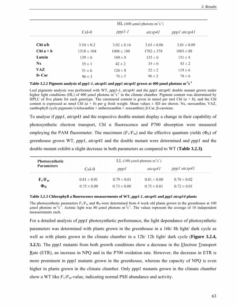
3. Results
63
Table 3.2.2 Pigment analysis of ppp1-1, atcsp41 and ppp1 atcsp41 grown at 400 µmol photons m-2s-1
Leaf pigments analysis was performed with WT, ppp1-1, atcsp41 and the ppp1 atcsp41 double mutant grown under higher light conditions (HL) of 400 µmol photons m-2s-1 in the climate chamber. Pigment content was determined by HPLC of five plants for each genotype. The carotenoid content is given in mmol per mol Chl (a + b), and the Chl content is expressed as nmol Chl (a + b) per g fresh weight. Mean values ± SD are shown. Nx, neoxanthin; VAZ, xanthophyll cycle pigments (violaxanthin + antheraxanthin + zeaxanthin); β-Car, β-carotene. To analyse if ppp1, atcsp41 and the respective double mutant display a change in their capability of
photosynthetic electron transport, Chl a fluorescence and P700 absorption were measured
employing the PAM fluorometer. The maximum (Fv/Fm) and the effective quantum yields (ΦII) of
greenhouse grown WT, ppp1, atcsp41 and the double mutant were determined and ppp1 and the
double mutant exhibit a slight decrease in both parameters as compared to WT (Table 3.2.3).
Table 3.2.3 Chlorophyll a fluorescence measurements of WT, ppp1-1, atcsp41 and ppp1 atcsp14 plants
The photosynthetic parameters Fv/Fm and ΦII were determined from 4 week old plants grown in the greenhouse at 100 µmol photons m-2s-1. Actinic light was 80 µmol photons m-2s-1. The values represent the average of 10 independent measurements each. For a detailed analysis of ppp1 photosynthetic performance, the light dependance of photosynthetic
parameters was determined with plants grown in the greenhouse in a 16h/ 8h light/ dark cycle as
well as with plants grown in the climate chamber in a 12h/ 12h light/ dark cycle (Figure 3.2.4,
3.2.5). The ppp1 mutants from both growth conditions show a decrease in the Electron Transport
Rate (ETR), an increase in NPQ and in the P700 oxidation rate. However, the decrease in ETR is
more prominent in ppp1 mutants grown in the greenhouse, whereas the capacity of NPQ is even
higher in plants grown in the climate chamber. Only ppp1 mutants grown in the climate chamber
show a WT like Fv/Fm-value, indicating normal PSII abundance and activity.

3. Results
64
Altogether, ppp1 plants grown in the climate chamber (Figure 3.2.4.B/D; Figure 3.2.5.B/D) show a
phenotype indicative for a constraint in carbon fixation at higher actinic light intensities (Golding
and Johnson, 2003). In detail, if carbon fixation is limited, PSI acceptors are over-reduced and
alternative pathways such as cyclic electron flow around PSI (Shikanai, 2006) or the water-water
cycle (Rizhsky et al., 2003) are favoured. These alternative pathways reduce plastoquinone, leading
to an enhanced acidification of the lumen, which is exacerbated by the reduction in ATP net
consumption through carbon fixation. Besides causing an increase in non-photochemical
quenching, the low lumenal pH is thought to lead to an increase in P700 oxidation and a decrease in
ETR by slowing down the electron proton symport of the plastoquinone pool.
Fig 3.2.4 Chlorophyll a fluorescence measurements
WT (filled circles) and ppp1 (white squares) plants grown in the greenhouse (A, C) or in the climate chamber (B, D) were analysed with the PAM-fluorometer. In (A) and (B) the Electron Transport Rate (ETR) and in (C) and (D) the non-photochemical quenching of PSII were determined by measuring Chl a fluorescence. Leaves were illuminated for 15 Min with actinic light of different light intensities, steady state fluorescence was determined as Fs and a saturating light pulse of 800 ms and 5000 µmol photons m-2s-1 was given yielding Fm. ETR was calculated as PAR x ΦII x 0.5 x 0.84, ΦII as (Fm-Fs)/Fm and NPQ as (Fm-Fm’)/Fm’.

3. Results
65
Fig 3.2.5 P700 oxidation rate and transient NPQ measurements
(A+B) The P700 oxidation rate was determined of WT (filled circles) and ppp1 (white squares) plants grown in the greenhouse (A) or in the climate chamber (B). Absorbance changes at 830 nm were recorded, while illuminating the leaves for 5 min with actinic light of different light intensities giving ΔA and far-red light to yield ΔAmax. P700 oxidation ratio was calculated as ΔA/ ΔAmax. Each value represents the average of 5 independent measurements (± SD). (C+D) Time courses of induction and relaxation of NPQ monitored during dark-to-light (80 µmol photons m-2 s-1, white bar) transition of WT and ppp1 plants grown in the greenhouse. NPQ was calculated as (Fm-Fm’)/Fm’ (C) or the climate chamber (D). The 3 min light period (white bar) was followed by a 2 min dark period (black bar). Note that NPQ induction during the activation period of photosynthesis is thought to be caused by the transient acidification of the thylakoid lumen when CEF activity is higher than the activity of the ATP synthase.
3.2.4 Analysis of the abundance of photosynthetic proteins
Abundances of chloroplast proteins were investigated by Western analysis of total protein extract of
WT and ppp1-1 mutant plants grown in the greenhouse or the climate chamber using antibodies
against one subunit of each multi-protein complex. Protein levels were normalised with Actin
(Figure 3.2.6). The Western analysis demonstrates that in mutant plants grown in the greenhouse
levels of most examined proteins are decreased to 50% of WT. Exceptions are the nucleus encoded
Lhcb1 protein of the light harvesting complex of PSII (LHCII), which shows slightly increased
levels and the large subunit of the Ribulose-biphosphate carboxylase/ oxygenase (Rubisco, RbcL),
of which levels are ~ 35% of WT. The chloroplast protein levels of the ppp1 plants grown in the

3. Results
66
climate chamber equals WT, except for a decrease in the large and the small subunit of Rubisco
(80% of WT).
Additionally, thylakoid complexes of WT and ppp1-1 mutant plants grown in the greenhouse were
analysed by Blue native (BN)-PAGE (Figure 3.2.6C). Here, protein samples according to same
amounts of chlorophyll were loaded. In the ppp1-1 mutant inter-complex stoichiometry remains
largely unchanged, except for an increase in free LHCII monomers and a decrease in PSII
supercomplexes.
Fig 3.2.6 Chloroplast protein
composition
Levels of thylakoid membrane polypeptides in mutant and WT plants. 40 μg of total protein of ppp1-1 plants and 40 μg and decreasing amounts of WT plants grown in the climate chamber (A) or in the greenhouse (B) were separated by SDS-PAGE and subsequently transferred to PVDF membranes. Replicate filters were immunolabelled with antibodies. Three independent experiments were performed, and representative results are shown. (C) Thylakoids from WT and ppp1-1 plants grown in the greenhouse were solubilised with 1.25 % ß-DM and separated by Blue native (BN)-PAGE. Identity of complexes is depicted on the right.

3. Results
67
3.2.5 Chloroplast expression analysis
To investigate if the decrease in protein levels from plants grown in the greenhouse was due to
changes in transcript levels, total leaf RNA was analysed by Northern hybridisation with probes
specific for petD, psaB, psbA and ACTIN1 as loading control (Chapter 2.2.2.2; Figure 3.2.7B).
Whereas psaA transcript is increased more than two fold in the ppp1-1 mutant, rbcL is slightly
decreased and psbA transcripts show WT levels. These results are supported by an expression
analysis of all chloroplast genes by qRT-PCR (supplementary data, Chapter 6.2), which shows a
significant up-regulation of psaA in ppp1-1 and no significant changes for rbcL and psbA.
Additionally, the chloroplast and cytoplasmatic translational rates were examined by a 35S
methionine in vivo translation assay (Chapter 2.2.4.15; Figure 3.2.7A). The results of this
experiment show that radioactive labelling of both subunits of the Rubisco, RbcL and RbcS is
decreased in the ppp1-1 mutant background. That disruption of expression of one of the Rubisco
subunits effects expression of the other has been repetitively observed and therefore the co-
regulation of Rubisco subunit expression is in accordance with previous publications (Schmidt and
Mishkind, 1983; Rodermel et al., 1996; Pesaresi et al., 2001). A further difference can be observed
in the translation of the D1 protein of PSII (PSII-D1), which seem to be increased marginally.
To investigate if the changes seen in the in vivo translation assay can be correlated with an
alteration of the polysome association of rbcL and psbA transcript, polysome formation on both
transcripts was assessed by sucrose density-gradient fractionation. Transcripts associated with
polysomes have higher sedimentation rates than those associated with singular ribosomes or
ribosome free mRNA. The proportion of transcripts, which are polysome-associated compared to
unassociated allows to estimate the efficiency of translation initiation and elongation (Barkan,
1993). Significant differences can neither be detected in the association of rbcL transcript, nor of
psaA and psbA (Figure 3.2.7C). Altogether, these results indicate that in the ppp1-1 mutant
background less RbcL protein is stably expressed. However, defects leading to this phenotype are
neither related to differential transcription, because transcript levels remain relatively unchanged,
nor to defects in the translational apparatus, because initiation, elongation and termination of
translation seem to be functional in ppp1.

3. Results
68
Fig 3.2.7 Effect of the ppp1 mutation on chloroplast gene expression
(A) In vivo translation assay. 35S Methionine was applied to leaves of WT and ppp1 plants grown in the greenhouse (16 h/8 h light / dark cycles) for 1h, illuminated with 50 µmol photons m-2s-1. Soluble and membrane proteins were isolated from WT and mutant leaves, separated by SDS-PAGE and analysed by fluorography. The most intensely labelled band corresponds to the large subunit of Rubisco. (B) Northern analysis of plastid transcripts. Total leaf RNA (20 µg) of WT and ppp1 mutant plants were hybridised with gene specific probes against ACTIN1 as loading control, psbA, rbcL and psaA. (C) Association of chloroplast mRNA with polysomes. Total extracts from 4-week-old WT and ppp1 mutant plants were fractionated on sucrose gradients. Eleven fractions of equal volume were collected from the top to the bottom of the sucrose gradients. An equal proportion of the RNA purified from each fraction was analysed by gel-blot hybridisation. The membrane was stained with methylene-blue as loading control and transcripts of psbA, rbcL and psaA were detected with gene-specific probes.
3.2.6 Analysis of transcript processing
CSP41 was reported to be involved in cleavage of the 3’ stem loop of chloroplast transcripts
(Bollenbach et al., 2003). Because similarities in primary amino acid sequence, as well as in the
predicted three dimensional structures of CSP41 and PPP1, are present, it was investigated, whether
the phenotype of the decrease in RbcL protein abundance could be correlated with a defect in rbcL
transcript processing in the ppp1 mutant. Additionally, because all results indicating a 3’ stem-loop

3. Results
69
endonuclease activity of CSP41 stem from in vitro experiments, 5’ and 3’ transcript end mapping
from atcsp41 and the ppp1 atcsp41 double mutant transcripts was carried out. This experiment was
performed together with Agata Kazmierczak employing the method of circular RT-PCR. This
involves the ligation the 3’ and 5’ ends of the same transcript, followed by RT-PCR and further
amplification. Agarose gel separation of the PCR-product shows that in WT and ppp1 one band
predominates, whereas in atcsp41 and the ppp1 atcsp41 double mutant two major bands can be
amplified (Figure 3.2.8). A PCR product of smaller size can also be detected in the ppp1 mutant,
but amounts are low.
Fig 3.2.8 Transcript end mapping of rbcL
Transcript end mapping. (A) Products of mRNA ligation followed by RT-PCR and specific amplification of rbcL transcript ends. (B) Depicted is the structure of the rbcL 3’ stem loop. Numbers indicate cleavage sites. (C) Schematic drawing of the rbcL transcripts. Numbers on top of transcript indicate processing sites in all genotypes, numbers below transcript indicate processing sites only in ppp1-1, atcsp41 and the ppp1 atcsp41 double mutant.
Sequencing of the PCR products led to the identification of the higher molecular band in all four
genotypes as rbcL transcript starting at bases -70 to -64 upstream and terminating at bases 1527 to
1531 downstream of the start-codon, which is also downstream of the 3’ stem loop. The smaller

3. Results
70
PCR products probably stem from degradation products, because they either start at base 1, the
start-codon or terminate at base 1440, the stop-codon, with at least the respective 5’ end found in
the mutants impeding with binding of the ribosome and thus translation. These results suggest that
transcript degradation is enhanced in all mutant backgrounds.
3.2.7 Effect of the absence of photosynthetic complexes on PPP1 abundance
The effect of the absence of each of the four photosynthetic complexes on PPP1 and RbcL protein
levels was examined. PPP1 and RbcL abundance is greatly reduced in PSII, Cyt b6f and PSI knock-
outs (Figure 3.2.9; for further information see Chapter 3.1.7). Whereas RbcL levels are also slightly
reduced in the atpd mutants, PPP1 protein accumulates to levels even higher than in WT. Besides
increased degradation of the RbcL protein because of oxidative stress, the highly reduced amounts
of PPP1 might represent one of the mechanisms leading to the down-regulation of RbcL protein.
However, other mechanisms in the regulation of RbcL abundance must exist to explain its altered
levels for instance in plants lacking the ATP-synthase complex. One of these mechanisms is the
autoregulation of Rubisco large-subunit translation according to its assembly state, which has
recently been described for tobacco (Wostrikoff and Stern, 2007) and Chlamydomonas chloroplasts,
where this process is known to be modulated by oxidative stress (Irihimovitch et al., 2000; Cohen et
al., 2005)
3.2.8 A PPP1:CFP fusion complements the ppp1 mutant phenotype
To investigate the putative RNA-binding function of PPP1, RNA Co-Immunoprecipitation Chips
(RIP-Chip) analysis has been initiated (Schmitz-Linneweber et al., 2005). In this method, immuno-
precipitated RNA and supernatant RNA are coupled to two different fluorescent dyes and are then
hybridised to an oligonucleotide Chip with probes covering all plastid transcripts. However, the
peptide antibody against PPP1 did not recognise the protein in its native state and thus was not
suitable for co-immunoprecipitation. An alternative for RIP-Chip analysis were plants expressing
the PPP1:CFP fusion under the control of a 35S promoter (Raab et al., 2006). To confirm complete
functionality of this fusion protein, ppp1-1 mutant plants were transformed with the construct to test
Fig 3.2.9 The abundance of PPP1 and RbcL in the
absence of photosynthetic complexes
Immunodetection of PPP1 and RbcL in Col-0, Ler, hcf136, petC, psad1/d2, atpd. The applied antibodies are depicted on the right. Total protein extract corresponding to 40 µg of protein was loaded each.

3. Results
71
for complementation. Homozygous T3 were grown in the greenhouse to investigate the
complementation of the growth phenotype by the introduced fusion protein. In 80% of analysed
lines the growth phenotype was reverted (two lines shown: Figure 3.2.10A). Total protein was
extracted from complemented mutant plants and it was shown by hybridisation with PPP1 and CFP
(Invitrogen) specific antibodies that they contained the bigger 64 kDa fusion protein (PPP1: 37 kDa,
CFP: 27 kDa), but not the native PPP1 protein (Figure 3.2.10B). Both antibodies yielded a further
signal at approximately 70 kDa, which probably represents the immature form of the PPP1:CFP
fusion protein before import into the chloroplast. Additionally, the Coomassie staining demonstrates
that RbcL protein levels are WT-like in the CFP:PPP1 overexpressors, further verifying the
complete physiological functionality of the fusion protein.
Fig 3.2.10 Transgenic plants expressing CFP tagged PPP1 in a ppp1-1 mutant background
(A) Four week old WT, ppp1-1 and ppp1-1 plants grown in the greenhouse (16 h light/ 8 h dark cycles) transformed with the PPP1:CFP construct. (B) Transgene expression analysis was carried out on total protein extract. PPP1:CFP is over-expressed in the two lines and CFP is recognised by the specific antibody against CFP and PPP1. The growth phenotype and the reduction in RbcL protein levels are complemented in both lines.
3.2.9 Germination assay
The expression rates of some photosynthetic genes are regulated by the phytohormone abscisic acid
(ABA) (Seki et al., 2002). Also the transcription of PPP1 responds negatively to ABA (Hoth et al.,
2002). Because PPP1 is a putative RNA-binding protein, it was assumed that it might carry out
regulatory functions in the chloroplast in response to this phytohormone (Raab et al., 2006). As
ABA levels within the plant directly correlate with the degree of seed germination, an assay was
performed to elucidate a possible function of PPP1 in the complex ABA signalling network. The

3. Results
72
germination was carried out with both ppp1 mutant alleles. Homozygous mutants and WT plants
were identified as progeny from one heterozygous plant and the germination assay was performed
on their progeny, which had been harvested simultaneously in order to avoid genetic and
environmental background specific differences in germination rates. This experiment was carried
out in collaboration with S. Raab (University of Nürnberg-Erlangen, Germany). Both alleles show a
significant reduction in germination rate in comparison to WT, when exogenous ABA is applied
(Figure 3.2.11). For ppp1-1, a concentration of 1µM ABA yielded the highest difference in
germination rate, whereas for ppp1-2 0.5 µM was sufficient to inhibit germination.
Fig 3.2.11 The ppp1 mutants display a delayed germination rates in response to ABA
Seed dormancy of wild-type, ppp1-1 (B) and ppp1-2 (A) seeds. In both independent experiments freshly harvested Seeds were plated on ABA-containing medium (A: 0.5 µM ABA; B: 1µM ABA) and maintained at 22°C. The degree of germination of WT (closed circles) and ppp1 (open squares) seeds was monitored after 2, 2.5, 3 and 5 days.

4 Discussion
73
4 Discussion
4.1 PPP4/TMP14
4.1.1 Novel family of chloroplast localised proteins
Within this thesis a novel protein family consisting of four Arabidopsis proteins with unknown
functions has been characterised. All four members have sequence similarities, especially in the two
predicted transmembrane helices. The location of these four proteins in the chloroplast as suggested
before by proteomic analyses (Kleffmann et al., 2004), was verified by transient expression of RFP-
fusions in Arabidopsis protoplasts for TMP14-like and PPP4-like, by MS analysis of thylakoid
proteins from BN second dimensions for PPP4, TMP14 and TMP14-like and by Western analysis
for PPP4 and TMP14. The latter demonstrated that both PPP4 and TMP14 are primarily localised in
the stroma lamellae. Here, PPP4 and TMP14 interact as shown by crosslinking and Co-IP, in a
previously un-described complex.
4.1.2 A novel thylakoid complex predominantly localised in the stromal lamellae
This work demonstrates the existence of a novel thylakoid complex, which as minimal constituents
contains PPP4 and TMP14. Each of both proteins seems to be able to form homomeric complexes
in the absence of the other interaction partner as shown by BN page analysis and crosslinking for
both and for PPP4 additionally by Co-IP. Whereas PPP4 accumulates to the same levels in tmp14 as
in WT, TMP14 levels are decreased in the ppp4 background. Photosynthetic measurements
demonstrated that this novel complex is indeed involved in photosynthesis, as parameters are
changed in ppp4 and ppp4 tmp14 mutants. Differences to WT present in the ppp4 single mutants are
exacerbated in the double mutant background. Considering the lack of phenotype in the tmp14
mutant, where PPP4 protein accumulates to WT levels, it can be concluded that presence of PPP4 is
sufficient to carry out the physiological function. Conversely, in ppp4 mutants an effect on
photosynthetic parameters is evident, indicating that TMP14 can only partly carry out the function
of PPP4, which could be due to its reduced amounts present in the thylakoid membrane. In the
double mutant, where both proteins are absent, the phenotype is enhanced, which supports the idea
that TMP14 can to some extent carry out the physiological function of PPP4. As such, it can be
speculated that heteromeric and homomeric complexes of both proteins at least partially act
redundant and that the physiological function carried out by PPP4 and TMP14 requires a certain
amount of complexes, which are not present in the ppp4 mutant. However, despite the evidence for
these complexes, it remains unclear, if complex formation is necessary for the proteins’ functions.

4 Discussion
74
As will be described in the following section, some other properties of both proteins do differ and
therefore it appears likely that both proteins carry out further protein specific functions.
Both crosslinking experiments and protease protection assays indicate that TMP14 interacts with
other proteins especially in the absence of PPP4. Electrostatic interactions with additional proteins
seem to be important for the anchorage of TMP14 in the thylakoid membrane. In the case of PPP4
only the protease protection assay is indicative for the existence of associated proteins. The MS
analysis of the co-immunoprecipitate derived from digitonin-solubilised thylakoids did not identify
any other interaction partners of this complex. Therefore, it can be assumed that digitonin destroys
the association of other proteins. Accordingly, the complex containing PPP4 and TMP14, migrating
at the size of the Cyt b6f monomer in BN gels most probably also represents a multimeric form of
both components. The lack of co-migration of PPP4 and TMP14 with the Cyt b6f dimer represents
one further indication against the interaction of PPP4 and TMP14 with this essential photosynthetic
complex. Moreover, it was shown, that presence of the Cyt b6f complex is not mandatory for
accumulation of PPP4 and TMP14, because both proteins can be found in plants lacking this
complex. Concomitantly, in mutants devoid of the other major thylakoid complexes, PSII, PSI and
ATP-synthase, PPP4 and TMP14 protein levels are relatively stable. Therefore it can be concluded
that the complex comprising PPP4 and TMP14 is not stably associated with any of the four major
thylakoid complexes. Interestingly, TMP14 accumulates to lower levels than PPP4 in theplants
lacking one of the respective complexes (exception is atpd, where both proteins are present at WT
levels), suggesting that differential regulatory mechanisms are involved.
The obtained data allow speculation about the functional composition of the heteromeric complex.
Because no further components of this complex could be precipitated by Co-IP, it can be assumed
that the heteromeric complex solubilised with digitonin solely consists of PPP4 and TMP14
subunits. If one now postulates that complex formation is indispensable for the function of PPP4
and TMP14 and as a consequence according to the photosynthetic parameters measured in the
mutants that homomeric and heteromeric complexes act redundant, it can be estimated that all three
complexes capable of performing the so-far unknown photosynthetic activity should contain the
same amount of subunits. To speculate about a putative size and composition of the heteromere
complex containing PPP4 and TMP14 the following experimental result have to be taken into
consideration: (i) the molecular size of the complex solubilised with digitonin, (ii) the ratio of PPP4
and TMP14 in this complex and (iii) the crosslinking pattern observed in the respective other
mutants to estimate number of subunits composing the functional complex.

4 Discussion
75
The digitonin solubilised, heteromeric complex has a slightly higher molecular weight than the
LHCII monomer, which runs at about 60 kDa (Granvogl et al., 2006) and contains as visualised by
silver staining of the second dimension slightly more TMP14 than PPP4 (reaction was quenched
before saturation of signal strength). In crosslinked thylakoids of ppp4 mutant plants, the TMP14
pattern can be interpreted as the result of five proteins interacting, whereas for PPP4 the biggest
crosslinking product seems to represent a tetramer. However, TMP14 crosslinking is always further
progressed and therefore it cannot be excluded that amounts of crosslinked PPP4 pentamers
deriving from the applied experimental conditions, are below the detection level. Conclusively, the
heteromeric complex is bigger than 60 kDa, the highest molecular crosslinking product seems to be
a pentamer and it contains more TMP14 than PPP4. Therefore, the existence of a heteromeric
complex consisting of three TMP14 and two PPP4, with a molecular weigh of 65 kDa appears
possible. Further experiments need to be conducted to clarify the composition of this new complex
4.1.3 Phenotype of ppp4 and ppp4 tmp14 mutants in relation to specific processes of
photosynthesis
Measurements of photosynthetic parameters employing the PAM fluorometer showed a slight, but
significant phenotype in the ppp4 mutant, which was exacerbated in the ppp4 tmp14 double mutant.
Parameters changed in these mutants are a decrease in NPQ under both light and high light, an
increase in 1-qP under low light and an increase in ΔA/ ΔAmax in low and a decrease in high light.
The WT-like Fv/Fm indicates that PSII abundance and function is unchanged in these mutant plants.
Additionally, the lower NPQ excludes that PPP4 and TMP14 are components of carbon fixation,
because an impairement in this process should cause higher NPQ (Johnson and Golding, 2003).
However, defects in the linear electron flow, in regulatory processes of photosynthesis or in the
cyclic electron flow around PSI could at least partially account for the changes in photosynthetic
parameters observed in the ppp4 and ppp4 tmp14 mutants. To investigate the physiological function
of both proteins, the photosynthetic phenotypes of ppp4 and ppp4 tmp14 will be compared with
phenotypes of mutants impaired in the respective processes of photosynthesis. Then further results
obtained in this work will be put into context in the attempt to survey possible functions of PPP4
and TMP14 in this process.
4.1.3.1 Linear electron flow
The increase of 1-qP, ΔA/ΔAmax and the decrease of NPQ in ppp4 and ppp4 tmp14 plants under low
light illumination could point to a defect in linear electron flow downstream of PSII. Firstly,
because of the co-migration of PPP4 and TMP14 with the Cyt b6f complex, a possible involvement

4 Discussion
76
at the level of Cyt b6f will be discussed. Concurrently, similar changes in photosynthetic parameters
as stated above are present in the partially Cyt b6f defective mutants petL and pgr1, when measured
with low actinic light. However, PAM measurements of these mutants at higher light intensities
result in increasing differences of NPQ in comparison to WT, whereas the ppp4 and the ppp4 tmp14
double mutations lead only to a marginal reduction in NPQ and the ΔA/ΔAmax value, of which the
latter is even slightly increased in petL and pgr1 under high light. Furthermore, these partially Cyt
b6f defective mutants do not exhibit the changes in the kinetics of in situ cyclic electron flow (CEF)
measurement, which occur in the ppp4 tmp14 mutant (Schwenkert et al., 2007; Munekage et al.,
2001; Okegawa et al., 2005). Therefore, a defect in the linear electron transport chain at the Cyt b6f
complex appears unlikely.
Mutants with impairments in PSI function, such as psad1 (Ihnatowicz et al., 2004), psae1
(Ihnatowicz et al., 2007) and psag (Zygadlo et al., 2005) show a marked increase in 1-qP, because
electron flow is partially blocked at PSI. They also show deficiencies in PSI function, a feature that
can not be found in ppp4 and ppp4 tmp14 mutant plants. Further evidence against a role of PPP4
and TMP14 in PSI stems from the biochemical analyses, which exclude an interaction of PPP4 and
TMP14 with this thylakoid complex.
4.1.3.2 Regulatory processes in photosynthesis
Conversely, regulatory processes of photosynthesis might be affected in the ppp4, ppp4 tmp14
mutants. Under low light state transition balances the excitation energy between the two
photosystems by the formation of a PSI-LHCII complex (for a review, see Allen and Forsberg 2001).
Plants lacking the kinase STN7 cannot perform this process (Bellafiore et al., 2005; Bonardi et al.,
2005). Therefore, they show an increased 1-qP and a slightly decreased NPQ, when measured under
low light illumination. However, the ΔA/ΔAmax value remains unchanged in stn7 plants (E. Aseeva,
Botany, LMU, Munich; pers. com.), whereas in the ppp4 and ppp4 tmp14 mutants the P700
oxidation rate is higher than in WT. This, together with the increased phosphorylation rate of LHCII
and the decreased Chl a/b ratio in the stromal lamellae fraction of the ppp4 tmp14 double mutant,
suggests that the mutant plants show increased LHCII phosphorylation and that the changes in 1-qP
and NPQ cannot be due to a defect in this process.
One further regulatory mechanism is the conversion of excess light energy into heat (qE), involving
the PSII protein PsbS, low lumenal pH and xanthopyll cycle pigments (Li et al., 2000; Li et al.,
2004). Defects in qE only slightly affect photosynthetic parameters when measured with low actinic
light, as can be seen in the psbs mutant, which is impaired in qE. Here, the photosynthetic
parameters NPQ and 1-qP also resemble those of ppp4 and the ppp4 tmp14 double mutant (Li et al.,

4 Discussion
77
2002). However, as the NPQ value measured with high actinic light consists substantially of qE,
psbs mutants display a largely decreased NPQ under these conditions. The ΔA/ΔAmax value of psbs
mutants has not been published. However, another mutant defective in qE (que1), of which the
specific location of the mutation has not been mapped yet, shows an increase in the ΔA/ΔAmax
value, when measured with low light and a slight decrease when measured with high light (Kalituho
et al., 2006). Conclusively, photosynthetic parameters of mutants with defects in qE resemble those
of ppp4 and ppp4 tmp14 mutants, except for their immense decrease in NPQ under high light.
Further experimental evidence against a direct involvement of PPP4 and TMP14 in the process of
qE is the the predominant localisation of both proteins in the stromal lamellae, whereas qE is
executed by LHCII (Ruban et al., 2007), of which the majority is localised in the thylakoid grana.
Additionally, neither levels nor thylakoid localisation of the PsbS protein is altered in the ppp4
tmp14 mutants. That ppp4 is not defective in the xanthophyll cyle can be concluded from the only
slight increase in violaxanthin pigments as compared to WT, which rather supports that the decrease
in NPQ might be due to lower acidification of the lumen (Hieber et al., 2000). As such it can be
excluded that the phenotype of mutants lacking PPP4 and TMP14 is due to a defect in the execution
of the qE process, which therefore indicates a defect in modulating the ΔpH across the thylakoid
membrane. Two mechanisms are thought to be part of this modulation (Kramer et al., 2004): (i) The
cyclic electron flow around PSI, which will be further discussed in the next chapter and (ii) changes
in conductivity of the ATP-synthase to protons. The latter mechanism is not likely to be affected in
the ppp4 and ppp4 tmp14 mutant backgrounds, because only some of their photosynthetic
parameters could be explained by changes in the ATP-synthase conductivity. In detail, the
measured differences at low light could be due to a higher conductivity of the ATP-synthase,
resulting in decreased low lumenal pH, but the high light mutant parameter ΔA/ΔAmax cannot be
explained by this. Additionally, little is known about the mechanism of changes in ATP-synthase
conductivity (Kanazawa and Kramer, 2002; Takizawa et al., 2008) and the presented work does not
support an interaction of PPP4 and TMP14 with the ATP-synthase, which would be expected, if
proteins had influence on its conductivity.
4.1.3.3 Cyclic electron flow around PSI (CEF)
Cyclic electron flow around PSI (CEF) is a mechanism that can modulate acidification of the lumen
(Munekage et al., 2002). The pgrl1ab mutant is impaired in the antimycin A sensitive cyclic
electron flow or the switch between LEF and this pathway (DalCorso et al., 2008). When
photosynthetic parameters are measured with low actinic light, these mutants display a reduction in
NPQ and an increase in 1-qP in about as much as the ppp4 tmp14 double mutant (G. DalCorso,

4 Discussion
78
Department of Botany, LMU, Munich; pers. com). This phenotype can be explained considering
that with impairments in cyclic electron flow, fewer protons are translocated into the lumen, this
leading to a decrease in heat dissipation of light energy, which is proportional to the acidification of
the lumen. The NPQ is therefore lower. If light energy conversion into heat is reduced, more of this
energy will be used for charge separation at PSII leading to a higher reduction of the plastoquinone
pool by PSII (higher 1-qP). However, the ΔA/ΔAmax ratio is also increased in ppp4 and ppp4 tmp14
plants, a phenotype not observed in the pgrl1ab mutant. Yet, this phenotype in the ppp4 and ppp4
tmp14 plants can be explained by the higher reduction of the plastoquinone pool under low light
growth conditions, which leads to an increased amount of phosphorylated LHCII at PSI, possibly
resulting in higher oxidation rates of P700 due to an increase in antenna size. That pgrl1ab does not
show this phenotype might be due to a direct function in PSI, with which the PGRL1 protein has
been shown to be associated (DalCorso et al., 2008).
Differences between the phenotypes of ppp4 and ppp4 tmp14 compared to pgrl1ab become evident,
when photosynthetic parameters obtained from high light illumination are compared. Here the
ΔA/ΔAmax value approaches zero and NPQ is reduced to half of WT in the pgrl1ab mutant. In ppp4
and ppp4 tmp14 NPQ is only slightly reduced and ΔA/ΔAmax is significantly lower, but still about
80% of WT. Mutants of ppp4 and ppp4 tmp14 lack the strong NPQ phenotype indicating that
proton translocation by CEF is only marginally impaired. But together with the decrease in P700
oxidation rate, it can be proposed that CEF indeed is slightly affected and therefore leads to the
over-reduction of the stroma and consequently to an indirect decrease of the ΔA/ΔAmax value by
charge recombination (DalCorso et al., 2008; Endo et al., 2005, Munekage et al., 2002).
Besides the antimycin A-sensitive CEF pathway there is also the NdH dependent CEF pathway
(Munekage et al., 2004), which is thought to be minor in comparison to the antimycin A-sensitive
CEF. Mutants in this pathway do not exhibit the changes in the photosynthetic parameters as
measured in this thesis for ppp4 and ppp4 tmp14 mutant plants (Muraoka et al., 2006; Munshi et
al., 2006; Kamruzzaman et al., 2005), however it cannot be excluded that growth conditions of
reported mutants differed. Electrons from the PQ pool can also be transferred to oxygen, a process
referred to as chloro-respiration (Cournac et al., 2000). It has been proposed that plants lacking the
capability to reduce oxygen should have a higher 1-qP and a lower ΔA/ΔAmax value. This process is
thought to be of special importance under high light (Rosso et al., 2006) and therefore no changes
in photosynthetic parameters would be expected under low light. As ppp4 and ppp4 tmp14 mutant
plants exhibit changes especially under low light, an involvement of PPP4 and TMP14 in this
process appears unlikely.

4 Discussion
79
4.1.3.3.1 In-depth analysis of CEF in ppp4 and ppp4 tmp14 mutants
From this comparison of photosynthetic phenotypes observed in the ppp4 mutant and in the ppp4
tmp14 double mutant with those of plants lacking or being impaired in physiological processes of
photosynthesis, it can be concluded that PPP4 and TMP14 are not essential constituents of one of
these processes. However, PPP4 and TMP14 might be involved in their regulation or partially in
their functionality. The preceding discussion of possible pathways involving PPP4 and TMP14
suggests that they function in the modulation of the pH gradient and thereby in qE.
Accordingly, the most likely candidate for a process that involves the PPP4 TMP14 complex is the
cyclic electron flow around PSI, as the Chl a fluorescence and P700 absorption phenotypes of ppp4
and ppp4 tmp14 can be explained by a mostly low light and not high light dependent impairment in
CEF downstream of PSI. To further investigate the hypothesis of an involvement of PPP4 and
TMP14 in CEF, attention has to be paid to the phenotypes in CEF observed with either the ppp4
mutant in the in vivo measurement or the ppp4 tmp14 double mutants in the in situ measurement.
The in vivo experiment was performed by measuring the transient increase of NPQ after light onset.
This measurement relates to the hypothesis that NPQ induction during the activation period of
photosynthesis is caused by the transient acidification of the thylakoid lumen when CEF activity is
higher than the activity of the ATP-synthase (DalCorso et al., 2008). In this experiment the ppp4
mutants behave like WT and only in the steady state show a slight reduction in NPQ. However,
when plants are grown in continuous light, they exhibit a significant decrease in the transient NPQ.
Accordingly, it can be concluded that normally PPP4 is not involved in the enhanced CEF during
light onset, but that the process of acclimation of the plant to continuous light involves PPP4-
regulated transient NPQ.
In the in situ measurements the ppp4 tmp14 double mutants display an increase in Chl a
fluorescence already after addition of NADPH, indicating that more Fd is stably bound to thylakoid
complexes, as has been shown for psad1 and psae1. These mutants exhibit increased cyclic electron
flow (DalCorso et al., 2008). A further phenotype that psad1, psae1 and ppp4 tmp14 have in
common is the increased abundance of the PGRL1 protein in the thylakoid membrane. This
suggests that when PPP4 and TMP14 are missing, the plant senses a disturbtion and accordingly
increases PGRL1 protein levels and the amount of thylakoid membrane bound Fd. But contrary to
psad1 and psae1, the ppp4 tmp14 mutant plants do not exhibit enhanced cyclic electron flow in the
in situ measurement, as fluorescence reaches WT steady state levels. This demonstrates that an
increase of cyclic electron flow as measured in the psae or psad mutants does not occur in this
double mutant, despite the increase in PGRL1 levels and membrane bound Fd, which is thought to

4 Discussion
80
cause enhanced CEF (DalCorso et al., 2008). A further phenotype of ppp4 tmp14 mutants in the in
situ experiment, are the decreased kinetics of the increase in Chl a fluorescence following the
addition of Fd. This indicates changes in the electron transport from NADPH via Fd to
plastoquinone in the double mutant. One possible explanation is the impairment in FNR function.
However, levels and localisation of this protein are not changed in the ppp4 tmp14 double mutants.
The second possible explanation is a decreased capability of the proteins involved in PQ reduction
to carry out this process in the ppp4 tmp14 double mutant, which would implicate a direct function
of PPP4 and TMP14 in CEF.
A further indication for a possible functional interaction of PPP4 and TMP14 with components of
cyclic electron flow, is the susceptibility of PPP4 and TMP14 to extraction by digitonin, which was
also shown for PGRL1 and thylakoid associated FNR, but not for other thylakoid proteins. From
this experimental result a localisation of all four proteins in the same region of stromal thylakoid
membranes can be hypothesised. To investigate if PPP4 and TMP4 transiently interact with these
essential components of CEF, split-ubiquitin analysis in yeast will be carried out in future
experiments.
4.1.3.3.2 Speculation about an involvement of PPP4 and TMP14 in CEF processes
If one assumes that PPP4 and TMP14 carry out a function in CEF, the lack of which is especially
measurable in low light, one firstly has to speculate about low light CEF, the occurrence of which is
disputed (Laisk et al., 2005). However, the hypothesis has been put forth that, as has been shown in
Chlamydomonas (Finazzi and Forti, 2004), association of LHCII with PSI (in state 2), which occurs
in vascular plants in PSII favouring low light, might result in an increase in CEF. One indication for
this hypothesis is the co-ordinated upregulation of both state 2 transition and CEF in the psad1
mutant (Pesaresi et al., 2002; DalCorso et al., 2008). If indeed state 2 transition results in increased
cyclic electron flow in plants, there should be substantial amounts of CEF under PSII favouring low
light. That low light CEF plays a physiological role, is supported by the visible phenotypes of
pgrl1ab and pgr5 plants grown at such light intensities (Munekage et al., 2002, DalCorso et al.,
2008). A lack of antimycin A-sensitive CEF should account for the changes of photosynthetic
parameters in the pgrl1ab mutant, when measured with low light. As ppp4 and ppp4 tmp14 exhibit
similar changes and as also parameters measured at high light have the same trend as in pgrl1ab,
there is the possibility that the complex comprised of PPP4 and TMP14 might have a function in the
same process. The nature of the changes in photosynthetic parameters in ppp4 and ppp4 tmp14 as
compared to WT and pgrl1ab suggests that the physiological function of PPP4 and TMP14 occurs
throughout light conditions and is not enhanced by environmental stimuli, which increase CEF,

4 Discussion
81
such as high light. Concomitantly, CEF at transition of dark to light is not influenced by a lack of
PPP4. Therefore, if PPP4 and TMP14 are involved in antimycin A-sensitive CEF, the co-existence
of an additional process, which is upregulated in response to high light or dark to light transition,
represents a crucial requirement.
But, if one now postulates an involvement of PPP4 and TMP14 in CEF, what could be the functions
of PPP4 and TMP14 in this process? There are two possibilities. Firstly, both proteins might be
components of the postulated enzyme Fd-PQ oxidoreductase (FQR). Secondly, they might regulate
CEF. Some results obtained in this thesis support a structural function. Firstly, PPP4 and TMP14
form a heteromeric complex comprised of several subunits, which cannot be found stably
associated with any other complex and thus seems to represent a structural entity. Secondly, this
complex is highly abundant in the thylakoid membrane, as shown by silver-staining of BN second
dimensions.
4.1.4 Conclusions
Many questions about the function of this novel complex remain open. Prerequisite for further
speculations are an in-depth analysis of other interaction partners, of which the presence but not the
identity has been shown by several biochemical approaches. Furthermore, application of inhibitors
of CEF should be able to strengthen or weaken the involvement of PPP4 and TMP14 in this
pathway. Antimycin A is known to impair the pathway also involving PGR5 (Munekage et al.,
2002, Munekage et al., 2004). Additionally, the possibility has to be taken into account that the
other two members of this novel protein family PPP4-like and TMP14-like carry out the same
function. Single mutants of ppp4-like and tmp14-like do not exhibit changes in photosynthetic
parameters. Therefore, the analysis of further double, triple and ultimately the quadruple mutant
should yield additional information about the function of the complex. Furthermore, the silencing of
the gene encoding the only member of this novel protein family in the single cell alga
Chlamydomonas reinhardtii, in which CEF is a crucial mechanism under oxygen depleted
conditions (Finazzi and Forti, 2004) might clarify if this protein is essential for optimal
photosynthesis and CEF.

4 Discussion
82
4.2 The putative RNA-binding protein PPP1
4.2.1 Physiological role: Chloroplast gene expression
The nuclear co-regulation of genes encoding photosynthetic proteins and those encoding the
chloroplast expressional apparatus (Biehl et al., 2005) led to the postulation of a mechanism co-
ordinating the expression of plastome and nuclear encoded components of the photosynthetic
machinery (Leister, 2005). Because of the stromal localisation of PPP1 and its published co-
localisation with components of the plastid expressional apparatus (Yamaguchi et al., 2003;
Pfannschmidt et al., 2001: Peltier et al., 2006), a function in the expression of chloroplast genes
appears likely. Furthermore, as PPP1 expression is co-regulated with photosynthetic genes, the
encoded protein might represent one of the components executing nuclear control on the expression
of chloroplast photosynthetic genes in order to achieve co-ordination of plastome and nuclear
encoded protein levels in the chloroplast. This work was performed with the aim to elucidate the
effect of PPP1 on chloroplast gene expression and grasp its mode of function.
Under optimum growth conditions as prevailing in the climate chamber, ppp1 plant appearance
resembles WT, except for a slight retardation in growth. However, regarding photosynthetic
parameters recorded by illumination with higher actinic light intensities, ppp1 plants show a clear
phenotype. They exhibit increased non-photochemical quenching, a higher P700 oxidation state and
a decrease in the electron transport rate, all three combined being indicative for a defect in carbon
fixation (Golding and Johnson, 2003; Golding et al., 2004). In line with the results obtained by
PAM measurement, the analysis of the chloroplast proteome shows that protein levels of both
subunit of the carbon fixing enzyme Rubisco, RbcL and RbcS are slightly reduced, whereas the
levels of components of the electron transport chain remain unchanged. Therefore, it can be
assumed that in climate chamber conditions, lack of PPP1 solely causes a reduction in Rubisco
levels. Because of its plastid stromal localisation and its predicted function in gene expression, a
role of PPP1 in rbcL expression can be assumed. However, two results obtained from the
chloroplast proteome analysis of ppp1 plants grown in the greenhouse suggests that PPP1 might
influence levels of more chloroplast proteins: (i) although light intensity in average is about the
same as in the climate chamber, levels of Rubisco are half in comparison to plants grown in the
climate chamber, indicating that downregulation of Rubisco levels might be partially of secondary
nature; (ii) no increase can be observed in the PSII/PSI ratio of plants grown in the greenhouse, as
would be expected from a limitation in terminal electron acceptors of PSI (Allahverdiyeva et al.,
2005). The unchanged PSII/PSI ratio in ppp1 plants grown in the greenhouse is caused by an equal

4 Discussion
83
downregulation of both complexes to 50% of WT. If the reduction of rbcL expression was the
primary effect in the ppp1 plants, their phenotype should be comparable to the tobacco rbcS
antisense lines published by Jiang and Rodermel (1995), which contain about 30-40% of Rubisco.
Here, Cyt f levels are slightly reduced and LHCII levels are slightly increased, these results being in
line with the ppp1 phenotype. However, the Chl a/b ratio is decreased, a change in line with an
enhanced PSII/PSI ratio as expected from Allahverdiyeva et al. (2005), which cannot be observed
in ppp1 plants grown in the greenhouse. Yet, comparing these two results, one has to consider that
plant-specific differences might exist. The question if PPP1 could be involved more globally in
regulating the abundance of chloroplast proteins and thereby execute a general nuclear control can
easily be speculated on, when the extensive demand for the expression of the high abundant protein,
Rubisco is considered. Results obtained in this thesis by radioactive labelling of chloroplast
proteins, shows that after one hour RbcL represents the most labelled protein, followed by LHCII
and PSII-D1 (Chapter 3.2.5). Therefore, it can be postulated that in optimal environmental
conditions as present in the climate chamber, a normal expressional set without PPP1 is sufficient
for the expression of all plastid genes except for rbcL, because of its high demand. Sub-optimal
conditions in the more variable light and temperature regimes of the greenhouse should result in
increased oxidative stress for chloroplast proteins, and therefore probably require an enhanced
expression of photosynthetic proteins encoded in the nucleus, as well as in the chloroplast.
Assuming that PPP1 is a crucial component of regulating chloroplast gene expression, the higher
demand of protein turn-over would be impaired in plants lacking this protein and thus lead to a
decrease in functional complexes. However, for the preceding speculations, we have to exclude a
regulation of the psbA expression by PPP1, because the turn-over of the encoded protein PSII-D1 is
higher than of any other thylakoid peptides (Matteo et al., 1981) and therefore would be a second
component highly influenced by sub-optimal expressional activity in the chloroplast. The
experimental results show that translation of PSII-D1 is even slightly increased in the ppp1 mutant.
Therefore, psbA expression cannot be controlled by PPP1.
Despite all collected experimental evidences, the mode of action of the control of gene expression
by PPP1 remains elusive. Indications that PPP1 acts post-transcriptionally, derive from chloroplast
transcriptome (supplementary data) and gel blot analyses, which demonstrate that rbcL transcript
levels are not significantly changed in the mutant, although a decrease in RbcL protein levels can be
observed by Western analysis. The in vivo translation-assay shows that translation of rbcL is
lowered, but polysome assembly of the transcript is unchanged in the mutant, indicating that neither
initiation of translation, elongation nor termination are significantly affected (Barkan, 1993). These

4 Discussion
84
results suggest that PPP1 might be involved in posttranslational processes such as stabilising and
folding of the nascent polypeptide chain. However, PPP1 was shown to bind petD mRNA in vitro
by Raab et al., which together with the co-migration with the ribosomal subunits (Peltier et al.,
2006) suggests regulation at an earlier step of gene expression. To further dissect the function of
PPP1, interactions with either mRNAs or other proteins need to be experimentally tested. RNA
immuno-precipitation Chips (RIP-Chips) are carried out in co-operation with Christian Schmitz-
Linneweber (Berlin). Preliminary results indicate that PPP1 binds the majority of chloroplast
mRNAs, which further supports firstly a more global function of PPP1 in chloroplast gene
expression and secondly regulation of chloroplast gene expression by PPP1 at transcript level.
4.2.2 The homologue AtCSP41
Because of protein sequence homologies, similarities in the three dimensional structures and
reported co-localisations with the 70S ribosome or some of its components (Yamaguchi et al.,
2003; Peltier et al., 2006), it was of interest to investigate, if PPP1 and AtCSP41 have redundant
functions. The phenotypic analyses of the respective knock-out mutants show that AtCSP41 does
not seem to carry out the same physiological function as PPP1, because the atcsp41 ppp1 double
mutant lacks a synthetic phenotype. However, if PPP1 and AtCSP41 interact in vivo, AtCSP41
stability could be dependent on the presence of PPP1 and therefore the ppp1 mutant could already
resemble the double mutant, lacking both PPP1 and AtCSP41 protein. Therefore it cannot be
excluded that PPP1 and AtCSP41 carry out the same enzymatic activity. Since it has been reported
that CSP41 (Yang and Stern, 1997, Bollenbach et al., 2003) is involved in 3’ stem-loop processing,
transcript end mapping was performed in ppp1, atcsp41 and ppp1 atcsp41 double mutant
background on rbcL, psbA and petD (data of latter two not shown). This analysis demonstrated
differences in transcript ends in all three mutant backgrounds with bias to shorter mRNAs as
compared to WT, but never in the 3’ stem loop of these transcripts. However, cleavage of the stem
loop is thought to result in degradation of the transcript (Rott et al., 1998) and therefore these ends
should be short-lived. These results do not exclude that PPP1 and AtCSP41 are involved in
transcript processing, but they rather indicate that PPP1 and AtCSP41 impede degradation.
4.2.3 Involvement of PPP1 in ABA signalling
Because it was reported that the expression of PPP1 is highly downregulated by ABA (Hoth et al.,
2002), it was investigated, if a lack of PPP1 causes a differential response of the plant to this
phytohormone. Raab et al. (2006) speculated that the signal of the phytohormone ABA might be
transferred into the chloroplast via RNA binding proteins to change gene expression. If PPP1
4 Discussion
85
enhances chloroplast gene expression, downregulation of its expression by ABA in the nucleus
should result in downregulation of its function in chloroplast gene expression. Because lack of
PPP1 seems to primarily target rbcL expression, a decrease in Rubisco would be expected. This is
in line with the negative regulation of a minimal light-responsive unit found in several RBCS
promoters by ABA (Acevedo-Hernandez et al., 2005), posing the prospect that ABA downregulates
carbon-fixation by repressing expression of Rubisco. However, a recent publication indicates that at
least in tobacco, lower amounts of RbcS in the chloroplast lead to an auto-downregulation of rbcL
expression by unassembled RbcL protein (Wostrikoff and Stern, 2007), making PPP1 dispensable
for a coordination of the expression of both Rubisco subunits.
The germination assays carried out with WT and both ppp1 mutant alleles allowed the
investigation, if lack of PPP1 changes germination behaviour. This experiment was solely carried
out with plants grown in the climate chamber, where the ppp1 mutant shows a reduction in Rubisco
levels. Interestingly, the mutant seeds showed an enhanced sensitivity in their germination rate to
ABA, indicating higher endogenous levels of this phytohormone or perturbations in ABA
signalling. To answer the question of how a lack of PPP1 can influence ABA signalling, the
biosynthesis pathway of ABA has to be taken into consideration. ABA originates from the same
biosynthetic pathway as the carotenoids, the 2-C-methyl-D-erythritol-4-phosphate (MEP) pathway,
of which the last enzymatic steps occur in the cytosol (Nambara and Marion-Poll, 2005). Direct
plastidic precursors of ABA are the Xanthophyll-cycle pigments (violaxanthin, antheroxanthin and
zeaxanthin) and neoxanthin (Schwartz et al., 2003). Overexpression of the zeaxanthin epoxidase
(ZEP) causes an enhanced accumulation of ABA in seeds (Lindgren et al., 2003). Therefore, it can
be concluded that modulation of the VAZ-pool can influence ABA levels. One phenotype of ppp1
plants grown in the climate chamber is the capacity for enhanced non-photochemical quenching, as
demonstrated by analysing the transient increase in NPQ. For non-photochemical quenching,
violaxanthin needs to be converted into zeaxanthin, which occurs by activation of the violaxanthin
de-epoxidase (VDE) by low lumenal pH (Niyogi et al., 1998; Bugos and Yamamoto, 1996). In line
with the phenotype of the ZEP-overexpressing plants, the increased enzymatic reversion of
zeaxanthin by ZEP after the NPQ-spike could be a signal activating ABA-biosynthesis at least in
the differentiated chloroplasts of green seeds. Because the ppp1 mutant grown in the climate
chamber mimics a limitation in carbon fixation- which in nature occurs under environmental
conditions of low CO2, as caused by drought (Golding and Johnson, 2003)- the increase in ABA
levels might have physiological relevance. As amounts of ABA in the seed directly correlate with
the dormancy length (Karssen et al., 1983), an increase in ABA levels as response to limitations in

4 Discussion
86
carbon fixation would postpone germination, which might be advantageous for the survival of the
plant. To allow further characterisation of the influence of VAZ pool modulations on ABA
production, additional mutants with changes in NPQ and VAZ pigments will be analysed regarding
ABA dependent germination behaviour. Additionally, to dissect ABA biosynthesis in response to
modulation of its plastidic carotenoid precursors, ppp1 has been crossed into an ABA luciferase
reporter line (Christmann et al., 2005). This will facilitate to analyse if modulation of the VAZ pool
results in differential production of ABA and when, if this mechanism is restricted to seeds or can
be observed also in leaves.

5. References
87
5 References
A Acevedo-Hernandez, G.J., Leon, P., and Herrera-Estrella, L.R. (2005). Sugar and ABA
responsiveness of a minimal RBCS light-responsive unit is mediated by direct binding of ABI4. Plant J 43, 506-519.
Albertsson, P.A., Andreasson, E., and Svensson, P. (1990). The domain organization of the plant
thylakoid membrane. FEBS Lett 273, 36-40. Allahverdiyeva, Y., Mamedov, F., Maenpaa, P., Vass, I., and Aro, E.M. (2005). Modulation of
photosynthetic electron transport in the absence of terminal electron acceptors: characterization of the rbcL deletion mutant of tobacco. Biochim Biophys Acta 1709, 69-83.
Allen, J.F., and Forsberg, J. (2001). Molecular recognition in thylakoid structure and function.
Trends Plant Sci 6, 317-326. Allen, J.F., Alexciev, K., and Hakansson, G. (1995). Photosynthesis. Regulation by redox
signalling. Curr Biol 5, 869-872. Allen, K.D., Duysen, M.E., and Staehelin, L.A. (1988). Biogenesis of thylakoid membranes is
controlled by light intensity in the conditional chlorophyll b-deficient CD3 mutant of wheat. J Cell Biol 107, 907-919.
Alonso, J.M., Stepanova, A.N., Leisse, T.J., Kim, C.J., Chen, H., Shinn, P., Stevenson, D.K., Zimmerman, J., Barajas, P., Cheuk, R., Gadrinab, C., Heller, C., Jeske, A., Koesema, E., Meyers, C.C., Parker, H., Prednis, L., Ansari, Y., Choy, N., Deen, H., Geralt, M., Hazari, N., Hom, E., Karnes, M., Mulholland, C., Ndubaku, R., Schmidt, I., Guzman, P., Aguilar-Henonin, L., Schmid, M., Weigel, D., Carter, D.E., Marchand, T., Risseeuw, E., Brogden, D., Zeko, A., Crosby, W.L., Berry, C.C., and Ecker, J.R. (2003). Genome-wide insertional mutagenesis of Arabidopsis thaliana. Science 301, 653-657.
Anderson, J.M. (1982). The role of chlorophyll-protein complexes in the function and structure of
chloroplast thylakoids. Mol Cell Biochem 46, 161-172. Aro, E.M., Suorsa, M., Rokka, A., Allahverdiyeva, Y., Paakkarinen, V., Saleem, A.,
Battchikova, N., and Rintamaki, E. (2005). Dynamics of photosystem II: a proteomic approach to thylakoid protein complexes. J Exp Bot 56, 347-356.
Aronsson, H., and Jarvis, P. (2002). A simple method for isolating import-competent Arabidopsis
chloroplasts. FEBS Lett 529, 215-220.
B Baker, M.E., Grundy, W.N., and Elkan, C.P. (1998). Spinach CSP41, an mRNA-binding protein
and ribonuclease, is homologous to nucleotide-sugar epimerases and hydroxysteroid dehydrogenases. Biochem Biophys Res Commun 248, 250-254.
Barber, J. (1982). The control of membrane organization by electrostatic forces. Biosci Rep 2, 1-
13.

5. References
88
Barber, J. (2006). Photosystem II: an enzyme of global significance. Biochem Soc Trans 34, 619-631.
Barkan, A. (1988). Proteins encoded by a complex chloroplast transcription unit are each translated from both monocistronic and polycistronic mRNAs. Embo J 7, 2637-2644.
Barkan, A., and Goldschmidt-Clermont, M. (2000). Participation of nuclear genes in chloroplast
gene expression. Biochimie 82, 559-572. Bassi, R., dal Belin Peruffo, A., Barbato, R., and Ghisi, R. (1985). Differences in chlorophyll-
protein complexes and composition of polypeptides between thylakoids from bundle sheaths and mesophyll cells in maize. Eur J Biochem 146, 589-595.
Bellafiore, S., Barneche, F., Peltier, G., and Rochaix, J.D. (2005). State transitions and light
adaptation require chloroplast thylakoid protein kinase STN7. Nature 433, 892-895. Biehl, A., Richly, E., Noutsos, C., Salamini, F., and Leister, D. (2005). Analysis of 101 nuclear
transcriptomes reveals 23 distinct regulons and their relationship to metabolism, chromosomal gene distribution and co-ordination of nuclear and plastid gene expression. Gene 344, 33-41.
Bollenbach, T.J., and Stern, D.B. (2003). Secondary structures common to chloroplast mRNA 3'-
untranslated regions direct cleavage by CSP41, an endoribonuclease belonging to the short chain dehydrogenase/reductase superfamily. J Biol Chem 278, 25832-25838.
Bollenbach, T.J., Tatman, D.A., and Stern, D.B. (2003). CSP41a, a multifunctional RNA-binding
protein, initiates mRNA turnover in tobacco chloroplasts. Plant J 36, 842-852. Bonardi, V., Pesaresi, P., Becker, T., Schleiff, E., Wagner, R., Pfannschmidt, T., Jahns, P.,
and Leister, D. (2005). Photosystem II core phosphorylation and photosynthetic acclimation require two different protein kinases. Nature 437, 1179-1182.
Breyton, C., Nandha, B., Johnson, G.N., Joliot, P., and Finazzi, G. (2006). Redox modulation of
cyclic electron flow around photosystem I in C3 plants. Biochemistry 45, 13465-13475. Bugos, R.C., and Yamamoto, H.Y. (1996). Molecular cloning of violaxanthin de-epoxidase from
romaine lettuce and expression in Escherichia coli. Proc Natl Acad Sci 93, 6320-6325. C
Chenna, R., Sugawara, H., Koike, T., Lopez, R., Gibson, T.J., Higgins, D.G., and Thompson, J.D. (2003). Multiple sequence alignment with the Clustal series of programs. Nucleic Acids Res 31, 3497-3500.
Chow, W.S., Ford, R.C., and Barber, J. (1981). Possible effects of the detachment of stromal
lamellae from granal stacks on salt-induced changes in spillover. A study by sonication of chloroplasts. Biochim Biophys Acta 635, 317-326.
Christmann, A., Moes, D., Himmelbach, A., Yang, Y., Tang, Y., and Grill, E. (2006).
Integration of abscisic acid signalling into plant responses. Plant Biol (Stuttg) 8, 314-325. Clayton, R.K. (1980). Photosynthesis: Physical mechanisms and chemical patterns. (Cambridge,
England: Cambridge University Press). Clough, S.J., and Bent, A.F. (1998). Floral dip: a simplified method for Agrobacterium-mediated
transformation of Arabidopsis thaliana. Plant J 16, 735-743.

5. References
89
Cohen, I., Knopf, J.A., Irihimovitch, V., and Shapira, M. (2005). A proposed mechanism for the inhibitory effects of oxidative stress on Rubisco assembly and its subunit expression. Plant Physiol 137, 738-746.
Cournac, L., Redding, K., Ravenel, J., Rumeau, D., Josse, E.M., Kuntz, M., and Peltier, G.
(2000). Electron flow between photosystem II and oxygen in chloroplasts of photosystem I-deficient algae is mediated by a quinol oxidase involved in chlororespiration. J Biol Chem 275, 17256-17262.
Cruz, J.A., Avenson, T.J., Kanazawa, A., Takizawa, K., Edwards, G.E., and Kramer, D.M.
(2005). Plasticity in light reactions of photosynthesis for energy production and photoprotection. J Exp Bot 56, 395-406.
D DalCorso, G., Pesaresi, P., Masiero, S., Aseeva, E., Schunemann, D., Finazzi, G., Joliot, P.,
Barbato, R., and Leister, D. (2008). A Complex Containing PGRL1 and PGR5 Is Involved in the Switch between Linear and Cyclic Electron Flow in Arabidopsis. Cell 132, 273-285.
Dekker, J.P., and Boekema, E.J. (2005). Supramolecular organization of thylakoid membrane
proteins in green plants. Biochim Biophys Acta 1706, 12-39. Demmig-Adams, B., and Adams, W.W., 3rd. (1993). The Xanthophyll Cycle, Protein Turnover,
and the High Light Tolerance of Sun-Acclimated Leaves. Plant Physiol 103, 1413-1420. Deng, X.W., Tonkyn, J.C., Peter, G.F., Thornber, J.P., and Gruissem, W. (1989). Post-
transcriptional control of plastid mRNA accumulation during adaptation of chloroplasts to different light quality environments. Plant Cell 1, 645-654.
Drager, R.G., Zeidler, M., Simpson, C.L., and Stern, D.B. (1996). A chloroplast transcript
lacking the 3' inverted repeat is degraded by 3'-->5' exoribonuclease activity. RNA 2, 652-663.
E Emanuelsson, O., Nielsen, H., and von Heijne, G. (1999). ChloroP, a neural network-based
method for predicting chloroplast transit peptides and their cleavage sites. Protein Sci 8, 978-984.
Emanuelsson, O., Nielsen, H., Brunak, S., and von Heijne, G. (2000). Predicting subcellular
localization of proteins based on their N-terminal amino acid sequence. J Mol Biol 300, 1005-1016.
Endo, T., Kawase, D., and Sato, F. (2005). Stromal over-reduction by high-light stress as
measured by decreases in P700 oxidation by far-red light and its physiological relevance. Plant Cell Physiol 46, 775-781.
Estevez, J.M., Cantero, A., Reindl, A., Reichler, S., and Leon, P. (2001). 1-Deoxy-D-xylulose-5-
phosphate synthase, a limiting enzyme for plastidic isoprenoid biosynthesis in plants. J Biol Chem 276, 22901-22909.

5. References
90
F Farber, A., Young, A.J., Ruban, A.V., Horton, P., and Jahns, P. (1997). Dynamics of
Xanthophyll-Cycle Activity in Different Antenna Subcomplexes in the Photosynthetic Membranes of Higher Plants (The Relationship between Zeaxanthin Conversion and Nonphotochemical Fluorescence Quenching). Plant Physiol 115, 1609-1618.
Finazzi, G., and Forti, G. (2004). Metabolic Flexibility of the Green Alga Chlamydomonas
reinhardtii as Revealed by the Link between State Transitions and Cyclic Electron Flow. Photosynth Res 82, 327-338.
Frey, A., Audran, C., Marin, E., Sotta, B., and Marion-Poll, A. (1999). Engineering seed
dormancy by the modification of zeaxanthin epoxidase gene expression. Plant Mol Biol 39, 1267-1274.
Friso, G., Giacomelli, L., Ytterberg, A.J., Peltier, J.B., Rudella, A., Sun, Q., and Wijk, K.J. (2004). In-depth analysis of the thylakoid membrane proteome of Arabidopsis thaliana chloroplasts: new proteins, new functions, and a plastid proteome database. Plant Cell 16, 478-499.
Froehlich, J.E., Wilkerson, C.G., Ray, W.K., McAndrew, R.S., Osteryoung, K.W., Gage, D.A., and Phinney, B.S. (2003). Proteomic study of the Arabidopsis thaliana chloroplastic envelope membrane utilizing alternatives to traditional two-dimensional electrophoresis. J Proteome Res 2, 413-425.
G Golding, A.J., and Johnson, G.N. (2003). Down-regulation of linear and activation of cyclic
electron transport during drought. Planta 218, 107-114. Golding, A.J., Finazzi, G., and Johnson, G.N. (2004). Reduction of the thylakoid electron
transport chain by stromal reductants--evidence for activation of cyclic electron transport upon dark adaptation or under drought. Planta 220, 356-363.
Granvogl, B., Reisinger, V., and Eichacker, L.A. (2006). Mapping the proteome of thylakoid
membranes by de novo sequencing of intermembrane peptide domains. Proteomics 6, 3681-3695.
H Hansson, M., and Vener, A.V. (2003). Identification of three previously unknown in vivo protein
phosphorylation sites in thylakoid membranes of Arabidopsis thaliana. Mol Cell Proteomics 2, 550-559.
Hassidim, M., Yakir, E., Fradkin, D., Hilman, D., Kron, I., Keren, N., Harir, Y., Yerushalmi, S., and Green, R.M. (2007). Mutations in CHLOROPLAST RNA BINDING provide evidence for the involvement of the chloroplast in the regulation of the circadian clock in Arabidopsis. Plant J 51, 551-562.
Hieber, A.D., Bugos, R.C., and Yamamoto, H.Y. (2000). Plant lipocalins: violaxanthin de-
epoxidase and zeaxanthin epoxidase. Biochim Biophys Acta 1482, 84-91. Holt, N.E., Fleming, G.R., and Niyogi, K.K. (2004). Toward an understanding of the mechanism
of nonphotochemical quenching in green plants. Biochemistry 43, 8281-8289.

5. References
91
Hoth, S., Morgante, M., Sanchez, J.P., Hanafey, M.K., Tingey, S.V., and Chua, N.H. (2002). Genome-wide gene expression profiling in Arabidopsis thaliana reveals new targets of abscisic acid and largely impaired gene regulation in the abi1-1 mutant. J Cell Sci 115, 4891-4900.
I Ihnatowicz, A., Pesaresi, P., and Leister, D. (2007). The E subunit of photosystem I is not
essential for linear electron flow and photoautotrophic growth in Arabidopsis thaliana. Planta 226, 889-895.
Ihnatowicz, A., Pesaresi, P., Lohrig, K., Wolters, D., Muller, B., and Leister, D. (2008).
Impaired photosystem I oxidation induces STN7-dependent phosphorylation of the light-harvesting complex I protein Lhca4 in Arabidopsis thaliana. Planta 227, 717-722.
Ihnatowicz, A., Pesaresi, P., Varotto, C., Richly, E., Schneider, A., Jahns, P., Salamini, F., and Leister, D. (2004). Mutants for photosystem I subunit D of Arabidopsis thaliana: effects on photosynthesis, photosystem I stability and expression of nuclear genes for chloroplast functions. Plant J 37, 839-852.
Irihimovitch, V., and Shapira, M. (2000). Glutathione redox potential modulated by reactive
oxygen species regulates translation of Rubisco large subunit in the chloroplast. J Biol Chem 275, 16289-16295.
J Jach, G., Binot, E., Frings, S., Luxa, K., and Schell, J. (2001). Use of red fluorescent protein
from Discosoma sp. (dsRED) as a reporter for plant gene expression. Plant J 28, 483-491. Jensen, P.E., Bassi, R., Boekema, E.J., Dekker, J.P., Jansson, S., Leister, D., Robinson, C., and
Scheller, H.V. (2007). Structure, function and regulation of plant photosystem I. Biochim Biophys Acta 1767, 335-352.
Jiang, C.Z., and Rodermel, S.R. (1995). Regulation of Photosynthesis during Leaf Development
in RbcS Antisense DNA Mutants of Tobacco. Plant Physiol 107, 215-224. Joliot, P., and Joliot, A. (1994). Mechanism of electron transfer in the cytochrome b/f complex of
algae: evidence for a semiquinone cycle. Proc Natl Acad Sci 91, 1034-1038. Joliot, P., Beal, D., and Joliot, A. (2004). Cyclic electron flow under saturating excitation of dark-
adapted Arabidopsis leaves. Biochim Biophys Acta 1656, 166-176.
Jornvall, H., Persson, B., Krook, M., Atrian, S., Gonzalez-Duarte, R., Jeffery, J., and Ghosh, D. (1995). Short-chain dehydrogenases/reductases (SDR). Biochemistry 34, 6003-6013.
K
Kalituho, L., Grasses, T., Graf, M., Rech, J., and Jahns, P. (2006). Characterization of a nonphotochemical quenching-deficient Arabidopsis mutant possessing an intact PsbS protein, xanthophyll cycle and lumen acidification. Planta 223, 532-541.
Kamruzzaman Munshi, M., Kobayashi, Y., and Shikanai, T. (2005). Identification of a novel
protein, CRR7, required for the stabilization of the chloroplast NAD(P)H dehydrogenase complex in Arabidopsis. Plant J 44, 1036-1044.
5. References
92
Kanamaru, K., and Tanaka, K. (2004). Roles of chloroplast RNA polymerase sigma factors in chloroplast development and stress response in higher plants. Biosci Biotechnol Biochem 68, 2215-2223.
Kanazawa, A., and Kramer, D.M. (2002). In vivo modulation of nonphotochemical exciton
quenching (NPQ) by regulation of the chloroplast ATP synthase. Proc Natl Acad Sci 99, 12789-12794.
Karnauchov, I., Herrmann, R.G., and Klosgen, R.B. (1997). Transmembrane topology of the
Rieske Fe/S protein of the cytochrome b6/f complex from spinach chloroplasts. FEBS Lett 408, 206-210.
Karssen, C.M., Brinckhorst-van der Swan, D.L.C., Breekland, A.E., and Koornneef, M.
(1983). Induction of dormancy during seed developmentby endogenous abscisic acid: studies on abscisic acid deficient genotype of Arabidopsis thaliana (L.) Heynh. Planta 157, 158-165.
Khrouchtchova, A., Hansson, M., Paakkarinen, V., Vainonen, J.P., Zhang, S., Jensen, P.E., Scheller, H.V., Vener, A.V., Aro, E.M., and Haldrup, A. (2005). A previously found thylakoid membrane protein of 14kDa (TMP14) is a novel subunit of plant photosystem I and is designated PSI-P. FEBS Lett 579, 4808-4812.
Kleffmann, T., Russenberger, D., von Zychlinski, A., Christopher, W., Sjolander, K.,
Gruissem, W., and Baginsky, S. (2004). The Arabidopsis thaliana chloroplast proteome reveals pathway abundance and novel protein functions. Curr Biol 14, 354-362.
Koncz, C., Mayerhofer, R., Koncz-Kalman, Z., Nawrath, C., Reiss, B., Redei, G.P., and Schell,
J. (1990). Isolation of a gene encoding a novel chloroplast protein by T-DNA tagging in Arabidopsis thaliana. Embo J 9, 1337-1346.
Koop, H.U., Steinmuller, K., Wagner, H., Rossler, C., Eibl, C., and Sacher, L. (1996).
Integration of foreign sequences into the tobacco plastome via polyethylene glycol-mediated protoplast transformation. Planta 199, 193-201.
Kouril, R., Zygadlo, A., Arteni, A.A., de Wit, C.D., Dekker, J.P., Jensen, P.E., Scheller, H.V., and Boekema, E.J. (2005). Structural characterization of a complex of photosystem I and light-harvesting complex II of Arabidopsis thaliana. Biochemistry 44, 10935-10940.
Koussevitzky, S., Nott, A., Mockler, T.C., Hong, F., Sachetto-Martins, G., Surpin, M., Lim, J.,
Mittler, R., and Chory, J. (2007). Signals from chloroplasts converge to regulate nuclear gene expression. Science 316, 715-719.
Kovacs, L., Damkjaer, J., Kereiche, S., Ilioaia, C., Ruban, A.V., Boekema, E.J., Jansson, S.,
and Horton, P. (2006). Lack of the light-harvesting complex CP24 affects the structure and function of the grana membranes of higher plant chloroplasts. Plant Cell 18, 3106-3120.
Kramer, D.M., Avenson, T.J., and Edwards, G.E. (2004). Dynamic flexibility in the light
reactions of photosynthesis governed by both electron and proton transfer reactions. Trends Plant Sci 9, 349-357.
Kyle, D.J., Ohad, I., and Arntzen, C.J. (1984). Membrane protein damage and repair: Selective
loss of a quinone-protein function in chloroplast membranes. Proc Natl Acad Sci 81, 4070-4074.

5. References
93
L Laisk, A., Eichelmann, H., Oja, V., and Peterson, R.B. (2005). Control of cytochrome b6f at low
and high light intensity and cyclic electron transport in leaves. Biochim Biophys Acta 1708, 79-90.
Larsson, U.K., Jergil, B., and Andersson, B. (1983). Changes in the lateral distribution of the light-harvesting chlorophyll-a/b--protein complex induced by its phosphorylation. Eur J Biochem 136, 25-29.
Leister, D. (2003). Chloroplast research in the genomic age. Trends Genet 19, 47-56. Leister, D. (2005). Genomics-based dissection of the cross-talk of chloroplasts with the nucleus
and mitochondria in Arabidopsis. Gene 354, 110-116. Leister, D., and Schneider, A. (2003). From genes to photosynthesis in Arabidopsis thaliana. Int
Rev Cytol 228, 31-83. Li, X.P., Muller-Moule, P., Gilmore, A.M., and Niyogi, K.K. (2002). PsbS-dependent
enhancement of feedback de-excitation protects photosystem II from photoinhibition. Proc Natl Acad Sci 99, 15222-15227.
Li, X.P., Bjorkman, O., Shih, C., Grossman, A.R., Rosenquist, M., Jansson, S., and Niyogi,
K.K. (2000). A pigment-binding protein essential for regulation of photosynthetic light harvesting. Nature 403, 391-395.
Li, X.P., Gilmore, A.M., Caffarri, S., Bassi, R., Golan, T., Kramer, D., and Niyogi, K.K. (2004). Regulation of photosynthetic light harvesting involves intrathylakoid lumen pH sensing by the PsbS protein. J Biol Chem 279, 22866-22874.
Li, Y., Rosso, M.G., Strizhov, N., Viehoever, P., and Weisshaar, B. (2003). GABI-Kat
SimpleSearch: a flanking sequence tag (FST) database for the identification of T-DNA insertion mutants in Arabidopsis thaliana. Bioinformatics 19, 1441-1442.
Lindgren, L.O., Stalberg, K.G., and Hoglund, A.S. (2003). Seed-specific overexpression of an
endogenous Arabidopsis phytoene synthase gene results in delayed germination and increased levels of carotenoids, chlorophyll, and abscisic acid. Plant Physiol 132, 779-785.
Lopez-Juez, E., and Pyke, K.A. (2005). Plastids unleashed: their development and their
integration in plant development. Int J Dev Biol 49, 557-577.
M Magliano, T.M., Botto, J.F., Godoy, A.V., Symonds, V.V., Lloyd, A.M., and Casal, J.J. (2005).
New Arabidopsis recombinant inbred lines (Landsberg erecta x Nossen) reveal natural variation in phytochrome-mediated responses. Plant Physiol 138, 1126-1135.
Maiwald, D., Dietzmann, A., Jahns, P., Pesaresi, P., Joliot, P., Joliot, A., Levin, J.Z., Salamini,
F., and Leister, D. (2003). Knock-out of the genes coding for the Rieske protein and the ATP-synthase delta-subunit of Arabidopsis. Effects on photosynthesis, thylakoid protein composition, and nuclear chloroplast gene expression. Plant Physiol 133, 191-202.
Miziorko, H.M., and Lorimer, G.H. (1983). Ribulose-1,5-bisphosphate carboxylase-oxygenase.
Annu Rev Biochem 52, 507-535.

5. References
94
Monde, R.A., Schuster, G., and Stern, D.B. (2000). Processing and degradation of chloroplast mRNA. Biochimie 82, 573-582.
Munekage, Y., Hojo, M., Meurer, J., Endo, T., Tasaka, M., and Shikanai, T. (2002). PGR5 is
involved in cyclic electron flow around photosystem I and is essential for photoprotection in Arabidopsis. Cell 110, 361-371.
Munekage, Y., Hashimoto, M., Miyake, C., Tomizawa, K., Endo, T., Tasaka, M., and Shikanai, T. (2004). Cyclic electron flow around photosystem I is essential for photosynthesis. Nature 429, 579-582.
Munshi, M.K., Kobayashi, Y., and Shikanai, T. (2006). Chlororespiratory reduction 6 is a novel
factor required for accumulation of the chloroplast NAD(P)H dehydrogenase complex in Arabidopsis. Plant Physiol 141, 737-744.
Muraoka, R., Okuda, K., Kobayashi, Y., and Shikanai, T. (2006). A eukaryotic factor required for accumulation of the chloroplast NAD(P)H dehydrogenase complex in Arabidopsis. Plant Physiol 142, 1683-1689.
N
Nambara, E., and Marion-Poll, A. (2005). Abscisic acid biosynthesis and catabolism. Annu Rev Plant Biol 56, 165-185.
Nandha, B., Finazzi, G., Joliot, P., Hald, S., and Johnson, G.N. (2007). The role of PGR5 in the
redox poising of photosynthetic electron transport. Biochim Biophys Acta 1767, 1252-1259. Nelson, N., and Yocum, C.F. (2006). Structure and function of photosystems I and II. Annu Rev
Plant Biol 57, 521-565. Nishiyama, Y., Allakhverdiev, S.I., and Murata, N. (2006). A new paradigm for the action of
reactive oxygen species in the photoinhibition of photosystem II. Biochim Biophys Acta 1757, 742-749.
Niyogi, K.K. (1999). PHOTOPROTECTION REVISITED: Genetic and Molecular Approaches.
Annu Rev Plant Physiol Plant Mol Biol 50, 333-359. Niyogi, K.K., Grossman, A.R., and Bjorkman, O. (1998). Arabidopsis mutants define a central
role for the xanthophyll cycle in the regulation of photosynthetic energy conversion. Plant Cell 10, 1121-1134.
Niyogi, K.K., Li, X.P., Rosenberg, V., and Jung, H.S. (2005). Is PsbS the site of non-
photochemical quenching in photosynthesis? J Exp Bot 56, 375-382.
O Okegawa, Y., Tsuyama, M., Kobayashi, Y., and Shikanai, T. (2005). The pgr1 mutation in the
Rieske subunit of the cytochrome b6f complex does not affect PGR5-dependent cyclic electron transport around photosystem I. J Biol Chem 280, 28332-28336.
Ossenbühl, F., Hartmann, K., and Nickelsen, J. (2002). A chloroplast RNA binding protein from
stromal thylakoid membranes specifically binds to the 5' untranslated region of the psbA mRNA. Eur J Biochem 269, 3912-3919.

5. References
95
P Parinov, S., Sevugan, M., Ye, D., Yang, W.C., Kumaran, M., and Sundaresan, V. (1999).
Analysis of flanking sequences from dissociation insertion lines: a database for reverse genetics in Arabidopsis. Plant Cell 11, 2263-2270.
Peltier, J.B., Cai, Y., Sun, Q., Zabrouskov, V., Giacomelli, L., Rudella, A., Ytterberg, A.J.,
Rutschow, H., and van Wijk, K.J. (2006). The oligomeric stromal proteome of Arabidopsis thaliana chloroplasts. Mol Cell Proteomics 5, 114-133.
Perrin, R., Lange, H., Grienenberger, J.M., and Gagliardi, D. (2004). AtmtPNPase is required
for multiple aspects of the 18S rRNA metabolism in Arabidopsis thaliana mitochondria. Nucleic Acids Res 32, 5174-5182.
Pesaresi, P., Schneider, A., Kleine, T., and Leister, D. (2007). Interorganellar communication.
Curr Opin Plant Biol 10, 600-606. Pesaresi, P., Varotto, C., Meurer, J., Jahns, P., Salamini, F., and Leister, D. (2001). Knock-out
of the plastid ribosomal protein L11 in Arabidopsis: effects on mRNA translation and photosynthesis. Plant J 27, 179-189.
Pesaresi, P., Lunde, C., Jahns, P., Tarantino, D., Meurer, J., Varotto, C., Hirtz, R.D., Soave,
C., Scheller, H.V., Salamini, F., and Leister, D. (2002). A stable LHCII-PSI aggregate and suppression of photosynthetic state transitions in the psae1-1 mutant of Arabidopsis thaliana. Planta 215, 940-948.
Pfannschmidt, T., Schutze, K., Brost, M., and Oelmüller, R. (2001). A novel mechanism of
nuclear photosynthesis gene regulation by redox signals from the chloroplast during photosystem stoichiometry adjustment. J Biol Chem 276, 36125-36130.
Pfannschmidt, T., Schutze, K., Fey, V., Sherameti, I., and Oelmüller, R. (2003). Chloroplast
redox control of nuclear gene expression--a new class of plastid signals in interorganellar communication. Antioxid Redox Signal 5, 95-101.
Pfannschmidt, T., Ogrzewalla, K., Baginsky, S., Sickmann, A., Meyer, H.E., and Link, G.
(2000). The multisubunit chloroplast RNA polymerase A from mustard (Sinapis alba L.). Integration of a prokaryotic core into a larger complex with organelle-specific functions. Eur J Biochem 267, 253-261.
Plücken, H., Müller, B., Grohmann, D., Westhoff, P., and Eichacker, L.A. (2002). The HCF136
protein is essential for assembly of the photosystem II reaction center in Arabidopsis thaliana. FEBS Lett 532, 85-90.
Porra, R.J. (2002). The chequered history of the development and use of simultaneous equations
for the accurate determination of chlorophylls a and b. Photosynth Res 73, 149-156.
R Raab, S., Toth, Z., de Groot, C., Stamminger, T., and Hoth, S. (2006). ABA-responsive RNA-
binding proteins are involved in chloroplast and stromule function in Arabidopsis seedlings. Planta 224, 900-914.
Renger, G., and Govindjee, M. (1985). The mechanism of photosynthetic water oxidation.
Photosynthesis Research 6, 33-55.

5. References
96
Richly, E., Dietzmann, A., Biehl, A., Kurth, J., Laloi, C., Apel, K., Salamini, F., and Leister, D. (2003). Covariations in the nuclear chloroplast transcriptome reveal a regulatory master-switch. EMBO Rep 4, 491-498.
Richter, M.L., Samra, H.S., He, F., Giessel, A.J., and Kuczera, K.K. (2005). Coupling proton
movement to ATP synthesis in the chloroplast ATP synthase. J Bioenerg Biomembr 37, 467-473.
Rintamäki, E., Martinsuo, P., Pursiheimo, S., and Aro, E.M. (2000). Cooperative regulation of
light-harvesting complex II phosphorylation via the plastoquinol and ferredoxin-thioredoxin system in chloroplasts. Proc Natl Acad Sci 97, 11644-11649.
Rios, G., Lossow, A., Hertel, B., Breuer, F., Schaefer, S., Broich, M., Kleinow, T., Jasik, J.,
Winter, J., Ferrando, A., Farras, R., Panicot, M., Henriques, R., Mariaux, J.B., Oberschall, A., Molnar, G., Berendzen, K., Shukla, V., Lafos, M., Koncz, Z., Redei, G.P., Schell, J., and Koncz, C. (2002). Rapid identification of Arabidopsis insertion mutants by non-radioactive detection of T-DNA tagged genes. Plant J 32, 243-253.
Rochaix, J.D. (2007). Role of thylakoid protein kinases in photosynthetic acclimation. FEBS Lett
581, 2768-2775. Rodermel, S., Haley, J., Jiang, C.Z., Tsai, C.H., and Bogorad, L. (1996). A mechanism for
intergenomic integration: abundance of ribulose bisphosphate carboxylase small-subunit protein influences the translation of the large-subunit mRNA. Proc Natl Acad Sci 93, 3881-3885.
Rolland, F., Moore, B., and Sheen, J. (2002). Sugar sensing and signaling in plants. Plant Cell 14
Suppl, S185-205. Rosso, D., Ivanov, A.G., Fu, A., Geisler-Lee, J., Hendrickson, L., Geisler, M., Stewart, G.,
Krol, M., Hurry, V., Rodermel, S.R., Maxwell, D.P., and Huner, N.P. (2006). IMMUTANS does not act as a stress-induced safety valve in the protection of the photosynthetic apparatus of Arabidopsis during steady-state photosynthesis. Plant Physiol 142, 574-585.
Rott, R., Liveanu, V., Drager, R.G., Stern, D.B., and Schuster, G. (1998). The sequence and
structure of the 3'-untranslated regions of chloroplast transcripts are important determinants of mRNA accumulation and stability. Plant Mol Biol 36, 307-314.
Ruban, A.V., Walters, R.G., and Horton, P. (1992). The molecular mechanism of the control of
excitation energy dissipation in chloroplast membranes. Inhibition of delta pH-dependent quenching of chlorophyll fluorescence by dicyclohexylcarbodiimide. FEBS Lett 309, 175-179.
Ruban, A.V., Berera, R., Ilioaia, C., van Stokkum, I.H., Kennis, J.T., Pascal, A.A., van Amerongen, H., Robert, B., Horton, P., and van Grondelle, R. (2007). Identification of a mechanism of photoprotective energy dissipation in higher plants. Nature 450, 575-578.
S
Sambrook, J., Fritsch, E.F., and Maniatis, T. (1989). Molecular Cloning. (New York: Cold Spring Harbor Laboratory Press).

5. References
97
Samson, F., Brunaud, V., Balzergue, S., Dubreucq, B., Lepiniec, L., Pelletier, G., Caboche, M., and Lecharny, A. (2002). FLAGdb/FST: a database of mapped flanking insertion sites (FSTs) of Arabidopsis thaliana T-DNA transformants. Nucleic Acids Res 30, 94-97.
Schagger, H., and von Jagow, G. (1987). Tricine-sodium dodecyl sulfate-polyacrylamide gel
electrophoresis for the separation of proteins in the range from 1 to 100 kDa. Anal Biochem 166, 368-379.
Schagger, H., and von Jagow, G. (1991). Blue native electrophoresis for isolation of membrane
protein complexes in enzymatically active form. Anal Biochem 199, 223-231. Schmidt, G.W., and Mishkind, M.L. (1983). Rapid degradation of unassembled ribulose 1,5-
bisphosphate carboxylase small subunits in chloroplasts. Proc Natl Acad Sci 80, 2632-2636. Schmitz-Linneweber, C., Williams-Carrier, R., and Barkan, A. (2005). RNA
immunoprecipitation and microarray analysis show a chloroplast Pentatricopeptide repeat protein to be associated with the 5' region of mRNAs whose translation it activates. Plant Cell 17, 2791-2804.
Schreiber, U., Schliewa, U., and Bilger, W. (1986). Continuous recording of photochemical and
non-photochemical chlorophyll fluorescence quenching with a new type of modulation fluorometer. Photosynth Res 10, 51-62.
Schreiber, U., Klughammer, C., and Neubauer, C. (1988). Measuring P700 absorbance changes
around 830 nm with a new type of pulse modulation system. Z. Naturforsch. 43c, 686-698. Schwartz, S.H., Tan, B.C., McCarty, D.R., Welch, W., and Zeevaart, J.A. (2003). Substrate
specificity and kinetics for VP14, a carotenoid cleavage dioxygenase in the ABA biosynthetic pathway. Biochim Biophys Acta 1619, 9-14.
Schwenkert, S., Legen, J., Takami, T., Shikanai, T., Herrmann, R.G., and Meurer, J. (2007).
Role of the low-molecular-weight subunits PetL, PetG, and PetN in assembly, stability, and dimerization of the cytochrome b6f complex in tobacco. Plant Physiol 144, 1924-1935.
Schwenkert, S., Umate, P., Dal Bosco, C., Volz, S., Mlcochova, L., Zoryan, M., Eichacker,
L.A., Ohad, I., Herrmann, R.G., and Meurer, J. (2006). PsbI affects the stability, function, and phosphorylation patterns of photosystem II assemblies in tobacco. J Biol Chem 281, 34227-34238.
Seelert, H., Poetsch, A., Rohlfs, M., and Dencher, N.A. (2000). Dye-ligand chromatographic purification of intact multisubunit membrane protein complexes: application to the chloroplast H+-FoF1-ATP synthase. Biochem J 346 Pt 1, 41-44.
Seki, M., Ishida, J., Narusaka, M., Fujita, M., Nanjo, T., Umezawa, T., Kamiya, A., Nakajima,
M., Enju, A., Sakurai, T., Satou, M., Akiyama, K., Yamaguchi-Shinozaki, K., Carninci, P., Kawai, J., Hayashizaki, Y., and Shinozaki, K. (2002). Monitoring the expression pattern of around 7,000 Arabidopsis genes under ABA treatments using a full-length cDNA microarray. Funct Integr Genomics 2, 282-291.
Seo, M., and Koshiba, T. (2002). Complex regulation of ABA biosynthesis in plants. Trends Plant
Sci 7, 41-48.

5. References
98
Sessions, A., Burke, E., Presting, G., Aux, G., McElver, J., Patton, D., Dietrich, B., Ho, P., Bacwaden, J., Ko, C., Clarke, J.D., Cotton, D., Bullis, D., Snell, J., Miguel, T., Hutchison, D., Kimmerly, B., Mitzel, T., Katagiri, F., Glazebrook, J., Law, M., and Goff, S.A. (2002). A high-throughput Arabidopsis reverse genetics system. Plant Cell 14, 2985-2994.
Shi, L.X., and Schroder, W.P. (2004). The low molecular mass subunits of the photosynthetic
supracomplex, photosystem II. Biochim Biophys Acta 1608, 75-96. Shikanai, T. (2006). RNA editing in plant organelles: machinery, physiological function and
evolution. Cell Mol Life Sci 63, 698-708. Shikanai, T. (2007). Cyclic electron transport around photosystem I: genetic approaches. Annu Rev
Plant Biol 58, 199-217. Smeekens, S. (2000). Sugar-Induced Signal Transduction in Plants. Annu Rev Plant Physiol Plant
Mol Biol 51, 49-81. Strand, A. (2004). Plastid-to-nucleus signalling. Curr Opin Plant Biol 7, 621-625. Stroebel, D., Choquet, Y., Popot, J.L., and Picot, D. (2003). An atypical haem in the cytochrome
b(6)f complex. Nature 426, 413-418. Suorsa, M., Sirpio, S., Allahverdiyeva, Y., Paakkarinen, V., Mamedov, F., Styring, S., and
Aro, E.M. (2006). PsbR, a missing link in the assembly of the oxygen-evolving complex of plant photosystem II. J Biol Chem 281, 145-150.
Szabo, I., Bergantino, E., and Giacometti, G.M. (2005). Light and oxygenic photosynthesis:
energy dissipation as a protection mechanism against photo-oxidation. EMBO Rep 6, 629-634.
T Taiz, L., and Zeiger, E. (1998). Plant Physiology. (Sinauer Associates, Inc). Takizawa, K., Kanazawa, A., and Kramer, D.M. (2008). Depletion of stromal P(i) induces high
'energy-dependent' antenna exciton quenching (q(E)) by decreasing proton conductivity at CF(O)-CF(1) ATP synthase. Plant Cell Environ 31, 235-243.
Tatusova, T.A., and Madden, T.L. (1999). BLAST 2 Sequences, a new tool for comparing protein
and nucleotide sequences. FEMS Microbiol Lett 174, 247-250. Terzaghi, W.B., and Cashmore, A.R. (1995). Photomorphogenesis. Seeing the light in plant
development. Curr Biol 5, 466-468. Timmis, J.N., Ayliffe, M.A., Huang, C.Y., and Martin, W. (2004). Endosymbiotic gene transfer:
organelle genomes forge eukaryotic chromosomes. Nat Rev Genet 5, 123-135. Towbin, H., Staehelin, T., and Gordon, J. (1979). Electrophoretic transfer of proteins from
polyacrylamide gels to nitrocellulose sheets: procedure and some applications. Proc Natl Acad Sci U S A 76, 4350-4354.
Trissl, H.W., and Wilhelm, C. (1993). Why do thylakoid membranes from higher plants form
grana stacks? Trends Biochem Sci 18, 415-419.

5. References
99
U Umate, P., Schwenkert, S., Karbat, I., Dal Bosco, C., Mlcochova, L., Volz, S., Zer, H.,
Herrmann, R.G., Ohad, I., and Meurer, J. (2007). Deletion of PsbM in tobacco alters the QB site properties and the electron flow within photosystem II. J Biol Chem 282, 9758-9767.
V Vallon, O., Bulte, L., Dainese, P., Olive, J., Bassi, R., and Wollman, F.A. (1991). Lateral
redistribution of cytochrome b6/f complexes along thylakoid membranes upon state transitions. Proc Natl Acad Sci 88, 8262-8266.
van Roon, H., van Breemen, J.F., de Weerd, F.L., Dekker, J.P., and Boekema, E.J. (2000).
Solubilization of green plant thylakoid membranes with n-dodecyl-alpha,D-maltoside. Implications for the structural organization of the Photosystem II, Photosystem I, ATP synthase and cytochrome b6 f complexes. Photosynth Res 64, 155-166.
Varotto, C., Pesaresi, P., Meurer, J., Oelmuller, R., Steiner-Lange, S., Salamini, F., and
Leister, D. (2000). Disruption of the Arabidopsis photosystem I gene psaE1 affects photosynthesis and impairs growth. Plant J 22, 115-124.
W Walker, M.G., Volkmuth, G., Sprinzak, E., Hodgson, D., and Klingler, T. (1999). Prediction of
gene function by genome-scale expression analysis: prostate cancer-associated genes. Genome Research 9, 1198-1203.
Wollman, F.A. (2001). State transitions reveal the dynamics and flexibility of the photosynthetic apparatus. Embo J 20, 3623-3630.
Wostrikoff, K., and Stern, D. (2007). Rubisco large-subunit translation is autoregulated in
response to its assembly state in tobacco chloroplasts. Proc Natl Acad Sci 104, 6466-6471. Y
Yakushevska, A.E., Jensen, P.E., Keegstra, W., van Roon, H., Scheller, H.V., Boekema, E.J., and Dekker, J.P. (2001). Supermolecular organization of photosystem II and its associated light-harvesting antenna in Arabidopsis thaliana. Eur J Biochem 268, 6020-6028.
Yamaguchi, K., Beligni, M.V., Prieto, S., Haynes, P.A., McDonald, W.H., Yates, J.R., 3rd, and Mayfield, S.P. (2003). Proteomic characterization of the Chlamydomonas reinhardtii chloroplast ribosome. Identification of proteins unique to the70 S ribosome. J Biol Chem 278, 33774-33785.
Yang, J., and Stern, D.B. (1997). The spinach chloroplast endoribonuclease CSP41 cleaves the 3'-
untranslated region of petD mRNA primarily within its terminal stem-loop structure. J Biol Chem 272, 12874-12880.
Yang, J., Usack, L., Monde, R.A., and Stern, D.B. (1995). The 41 kDa protein component of the
spinach chloroplast petD mRNA 3' stem-loop:protein complex is a nuclear encoded chloroplast RNA-binding protein. Nucleic Acids Symp Ser, 237-239.
Yi, X., Hargett, S.R., Frankel, L.K., and Bricker, T.M. (2006). The PsbQ protein is required in
Arabidopsis for photosystem II assembly/stability and photoautotrophy under low light conditions. J Biol Chem 281, 26260-26267.

5. References
100
Ytterberg, A.J., Peltier, J.B., and van Wijk, K.J. (2006). Protein profiling of plastoglobules in chloroplasts and chromoplasts. A surprising site for differential accumulation of metabolic enzymes. Plant Physiol 140, 984-997.
Z
Zhang, H., Whitelegge, J.P., and Cramer, W.A. (2001). Ferredoxin:NADP+ oxidoreductase is a subunit of the chloroplast cytochrome b6f complex. J Biol Chem 276, 38159-38165.
Zhang, S., and Scheller, H.V. (2004). Light-harvesting complex II binds to several small subunits
of photosystem I. J Biol Chem 279, 3180-3187. Zimmermann, P., Hirsch-Hoffmann, M., Hennig, L., and Gruissem, W. (2004).
GENEVESTIGATOR. Arabidopsis microarray database and analysis toolbox. Plant Physiol 136, 2621-2632.
Zito, F., Finazzi, G., Joliot, P., and Wollman, F.A. (1998). Glu78, from the conserved PEWY
sequence of subunit IV, has a key function in cytochrome b6f turnover. Biochemistry 37, 10395-10403.
Zygadlo, A., Jensen, P.E., Leister, D., and Scheller, H.V. (2005). Photosystem I lacking the PSI-
G subunit has a higher affinity for plastocyanin and is sensitive to photodamage. Biochim Biophys Acta 1708, 154-163.

6 Supplementary Data
101
6 Supplementary Data
6.1 Mass spectrometry fragmentation spectra
6.1.1 PPP4
Fig 6.1.1 Fragmentation spectrum of the identified tryptic peptide ASSEETSSID of PPP4
Fig 6.1.2 Fragmentation spectrum of the identified tryptic peptide KELAEDIESLK of PPP4
#10751-10751 RT:63.65-63.65 NL: 8.43E3
0 200 400 600 800 1000 1200m/z
0
5
10
15
20
25
30
35
40
45
50
55
60
65
70
75
80
85
90
95
100
Rel
ativ
e A
bund
ance
yy10+1
1146.4
yy5+1
589.3yy9
+1
1017.5
yy8+1
904.3bb 10
+1
1128.4yy7+1
833.4bb 6
+1
686.3yy3+1
347.3yy6
+1
704.3bb 8
+1
928.3bb 5
+1
571.2yy4
+1
476.3bb 2
+1
258.2bb 7
+1
799.3bb 4+1
442.3bb 4
+2
222.1
bb 3+1
371.3
bb 9+1
1015.2yy5+2
295.3
#12750-12750 RT:75.32-75.32 NL: 2.11E4
0 200 400 600 800 1000 1200 1400 1600 1800m/z
0
5
10
15
20
25
30
35
40
45
50
55
60
65
70
75
80
85
90
95
100
Rel
ativ
e A
bund
ance
yy10+1
1161.5
yy4+1
476.3
yy12+1
1361.5 bb 15+1
1577.4yy11+1
1274.5 yy13+1
1448.6yy5+1
589.3 yy6+1
702.4yy8
+1
945.5
bb 9+1
892.3
yy7+1
831.5
yy3+1
375.2bb 17
+1
1793.5bb 16
+1
1678.5
bb 7+1
692.3
bb 8+1
779.2
bb 11+1
1108.3
bb 12+1
1222.8
bb 5+1
504.3bb 7+2
346.2

6 Supplementary Data
102
Fig 6.1.3 Fragmentation spectrum of the identified tryptic peptide ELAEDIESLK of PPP4
Fig 6.1.4 Fragmentation spectrum of the identified tryptic peptideVMELVGLGYTGWFVYR of PPP4
#14734-14734 RT:89.97-89.97 NL: 7.67E3
0 200 400 600 800 1000 1200 1400 1600 1800m/z
0
5
10
15
20
25
30
35
40
45
50
55
60
65
70
75
80
85
90
95
100
Rel
ativ
e A
bund
ance
yy9+1
1148.4yy11
+1
1318.5bb 7+1
758.2yy4+1
584.3yy8
+1
1091.5
yy7+1
928.6
bb 14+1
1568.5yy12
+1
1417.5yy10
+1
1261.7yy2+1
338.3bb 4
+1
489.3
bobo 13+2
726.5
bb 15+1
1731.6
bobo 4+1
471.3
#11634-11634 RT:68.56-68.56 NL: 5.90E4
0 100 200 300 400 500 600 700 800 900 1000 1100m/z
0
5
10
15
20
25
30
35
40
45
50
55
60
65
70
75
80
85
90
95
100R
elat
ive
Abu
ndan
ce
yy6+1
704.3
yy4+1
476.3
yy3+1
347.2 yy8+2
452.9bobo 6
+1
653.1
yy7+1
833.3bobo 5+1
540.1bobo 4+1
425.1yy8
+1
904.4yy5
+1
589.4 bobo 7+1
782.2
bobo 3+1
296.0yy2+1
260.3bobo 9
+1
982.1
bobo 8+1
869.2yy7
+2
417.3 bb 9+1
1000.4

6 Supplementary Data
103
6.1.2 TMP14
Fig 6.1.5 Fragmentation spectrum of the identified tryptic peptide ATTEVGEAPATTTEAETTELPEIVK of
TMP14
Fig 6.1.6 Fragmentation spectrum of the identified tryptic peptideAGVVALWGSAGMISAIDR of TMP14
#12560-12560 RT:79.23-79.23 NL: 4.81E3
0 200 400 600 800 1000 1200 1400 1600m/z
0
5
10
15
20
25
30
35
40
45
50
55
60
65
70
75
80
85
90
95
100
Rel
ativ
e A
bund
ance
yy11+1
1093.4yy8+1
878.4
yy5+1
561.3
yy4+1
474.4
bb 7+1
697.3 bb 16+1
1500.6yy12
+1
1279.5bb 13+1
1229.4yy10+1
1036.4yy6
+1
674.3yy3
+1
403.3yy9
+1
949.4 bb 12+1
1116.4yy7
+1
821.4 bb 15+1
1387.4yy2+1
290.2
bb 5+1
398.3bb 17
+1
1615.5
#11428-11428 RT:72.15-72.15 NL: 4.05E4
0 200 400 600 800 1000 1200 1400 1600 1800 2000m/z
0
5
10
15
20
25
30
35
40
45
50
55
60
65
70
75
80
85
90
95
100
Rel
ativ
e A
bund
ance
yy17+1
1829.7
yy5+1
585.4
yy18+1
1900.8bb 8
+1
759.2yy6+1
698.4 yy11+1
1229.5yy13
+1
1459.7yy15
+1
1661.6yy9
+1
1029.5yy8+1
928.4yy7+1
827.5bb 14
+1
1359.5bb 18
+1
1761.6
bb 17+1
1660.8
bb 5+1
502.0

6 Supplementary Data
104
6.1.3 TMP14-like
Fig 6.1.7 Fragmentation spectrum of the identified tryptic peptide YLLFKPDR of TMP14-like
Fig 6.1.9 Fragmentation spectrum of the identified tryptic peptide KSVADILGQ of TMP14-like
#10995-10995 RT:64.99-64.99 NL: 9.98E2
0 100 200 300 400 500 600 700 800 900m/z
0
5
10
15
20
25
30
35
40
45
50
55
60
65
70
75
80
85
90
95
100
Rel
ativ
e A
bund
ance
bb 7+1
727.3
yoyo 8+1
784.3bb 8
+1
784.3bb 6
+1
614.2yy2
+1
204.0 yy4+1
430.3
bb 5+1
501.3yy3
+1
317.2bb 4
+1
386.6
yy5+1
545.2 yy6+1
616.3yy1+1
147.1
bb 3+1
315.4
bb 2+1
216.3
bobo 6+1
596.2
yoyo 2+1
186.0
yy7+1
715.5
#10455-10455 RT:62.11-62.11 NL: 1.48E4
0 100 200 300 400 500 600 700 800 900 1000m/z
0
5
10
15
20
25
30
35
40
45
50
55
60
65
70
75
80
85
90
95
100
Rel
ativ
e A
bund
ance
yy6+2
388.4
yy5+1
662.3
yy3+1
387.2 yy6+1
775.4yy4+1
515.2
yoyo 4+2
249.1 bb 2+1
277.1
yy7+2
444.9bb 7
+1
877.3yy1+1
175.1
bb 3+1
390.1
bb 4+1
537.1yoyo 4
+1
497.3yy5+2
331.9bb 5
+1
665.2
yoyo 6+2
379.2yoyo 5
+1
644.5 yy7+1
888.3yoyo 1
+1
157.3

6 Supplementary Data
105
6.2 Plastome expression analysis of ppp1 mutants
Fig 6.2.1 Plastome expression
analysis by qRT-PCR
Carried out by Andreol Falcon De Longvialle (ARC Centre of Excellence, Perth, Australia) with RNA from ppp1-1 and Col-0 plants grown in the greenhouse using gene specific primers. Two independent experiments were performedt and are plotted. For each experiment three repetitions were made and error bars represent standard deviation.

Acknowledgements
106
Acknowledgements First of all, I would like to thank Prof. Dr. Dario Leister for giving me the opportunity to carry out
my Ph.D. research under his supervision.
Additionally, I would like to thank everyone, who contributed to the experimental outcome of this
thesis: Dr. Bernd Müller and Youlia Davidova for performing the MS analyses, Prof. Dr. Peter
Jahns for the pigment analyses, Agata Kazmierczak for the transcript end mappings, Sabine Raab
and Dr. Peter Hoth for the germination assays, Prof. Dr. Christian Schmitz-Linneweber for trial
RIP-Chip analyses and Dr. Andreol Falcon De Longevialle for plastome expression analyses.
Moreover I would like to thank Alexander Hertle, Dr. Elena Aseeva and Dr. Tatjana Kleine for
critically reading and correcting my thesis.
I would like to express my gratefulness to the other members of the team, who were always willing
to discuss experimental problems or novel hypotheses.
Last but not least, I would like to thank my family and friends, especially my parents and Alex for
supporting and helping me throughout stressfull times.

Curriculum vitae
107
Curriculum vitae PERSONAL DATA Date of birth: 19.08.1978 Place of Birth: Kleve, NRW, Germany Nationality: German RESEARCH EXPERIENCES October 2005 - now Ludwig-Maximilians-Universität München Department Biology I, Botany - PhD continuation. Supervisor Prof. Dr. Dario Leister July 2004 – September 2005 Max Planck Institut für Züchtungsforschung (Köln) - PhD in “Characterization of three novel photosynthetic proteins in Arabidopsis thaliana”.
Supervisor Prof. Dr. Dario Leister February 2004 - April 2004 School of Biological Sciences, University of Auckland (New Zealand) - Technical Assistant November 2002 – December 2003 School of Biological Sciences, University of Auckland (New Zealand) - Diploma thesis “Epitope tagging of the flowering time gene GIGANTEA” September 2001 - March 2002 Scion (formerly Forest Research Institute), Rotorua (New Zealand) - Internship at the department of Molecular Biology EDUCATION 2000 - 2004 Universität zu Köln - Main diploma studies in Biology with graduation in March 2004 (Grade: 1.0) 1998 - 2000 Rheinische Friedrich-Wilhelms-Universität Bonn -Pre-diploma studies in Biology (Grade: 1.0) August 1995- February 1996 Eastlake High school, Redmond, Washington, USA 1989 - 1998 Konrad-Adenauer Gymnasium in Kleve - Abitur (Grade: 2.6)

Curriculum vitae
108
PUBLICATIONS Noutsos, C., Kleine, T., Armbruster, U., DalCorso, G., and Leister, D. (2007). Nuclear insertions of organellar DNA can create novel patches of functional exon sequences. Trends Genet 23, 597-601. David, K.M., Armbruster, U., Tama, N., and Putterill, J. (2006). Arabidopsis GIGANTEA protein is post-transcriptionally regulated by light and dark. FEBS Lett 580, 1193-1197. POSTERS Armbruster, U. and Leister, D.: PPP4: Identifizierung von photosynthetischen Proteinen mittels Expressionsanalysen. 21. Tagung Molekularbiologie der Pflanzen, Dabringhausen 2008 Armbruster, U. and Leister, D.: PPP4: A putative photosynthetic protein. 10th International Colloquium on Endocytobiology and Symbiosis 2007, Gmunden, Austria Armbruster, U., David, K.M. and Putterill, J.: Epitope tagging of the Arabidopsis flowering time gene GIGANTEA. Queenstown Molecular Biology Meeting 2003 (New Zealand) SCHOLARSHIPS and PRIZES International Colloquium on Endocytobiology and Symbiosis 2007, Gmunden, Austria - Poster prize Queenstown Molecular Biology Meeting 2003, New Zealand -Poster prize 2001-2004 - Scholarship of the „Studienstiftung des deutschen Volkes“, additionally financing internship and diploma
thesis in New Zealand LANGUAGES German, mother tongue English, fluent French, basic
Ehrenwörtliche Versicherung
109
Ehrenwörtliche Versicherung
Hiermit versichere ich, dass ich die vorliegende Arbeit selbständig und nur unter Verwendung
der angegebenen Hilfsmittel und Quellen angefertigt habe. Die Autorin hat zuvor nicht
versucht, anderweitig eine Dissertation einzureichen oder sich einer Doktorprüfung zu
unterziehen. Die Dissertation wurde keiner weiteren Prüfungskommission weder in Teilen noch
als Ganzes vorgelegt.
München,
Mai 2008
Ute Armbruster