penpen accesscc e s s isolation and characterisation of a novel bohle ... · isolation and...
TRANSCRIPT
DISEASES OF AQUATIC ORGANISMSDis Aquat Org
Vol. 99: 169–177, 2012doi: 10.3354/dao02472
Published July 25
INTRODUCTION
The family Iridoviridae comprises 5 genera — Iridovirus, Chloriridovirus, Ranavirus, Lymphocys-tivirus and Megalocytivirus. Virions are enveloped,120 to 200 nm in diameter and display icosahedralsymmetry with an electron-dense core, containing asingle linear, circularly permuted and terminallyredundant double-stranded DNA molecule of be -tween 140 and 303 kbp. Virions are stable in water at4°C for extended periods and are able to resist desic-cation at 42°C for up to 6 wk (Fauquet et al. 2005).
Ranaviruses have been isolated from amphibians,fish and reptiles primarily located in North and SouthAmerica, Europe and SE Asia (Zupanovic et al. 1998,Hyatt et al. 2000, Fauquet et al. 2005, Fox et al. 2006,Densmore & Green 2007, Chinchar et al. 2009). Virusisolates show serological and genetic relatedness
within the genus Ranavirus but are serologically andgenetically distinct from members of other generawithin the Iridoviridae (Fauquet et al. 2005). In Aus-tralia, 2 ranaviruses have been identified: epizootichaematopoietic necrosis virus (EHNV) from redfinperch Perca fluviatilis (Langdon et al. 1986) and rain-bow trout Onco rhyn chus mykiss (formerly Salmogairdneri) (Langdon et al. 1988) and Bohle iridovirus(BIV) from metamorphs of the ornate burrowing frogLimnodynastes ornatus (Gray) (Speare & Smith 1992,Marsh et al. 2002).
Studies conducted by Langdon et al. (1988),Moody & Owens (1994) and Cullen & Owens (2002)demonstrated the lethal potential of ranaviruses tonative and introduced Australian amphibians andfinfish using laboratory-based challenge trials. Theability of ranaviruses to infect a wide range of hostspecies, including finfish, suggested a major risk
© Inter-Research 2012 · www.int-res.com*Email: [email protected]
Isolation and characterisation of a novel Bohle-likevirus from two frog species in the Darwin rural
area, Australia
R. P. Weir1,*, N. J. G. Moody2, A. D. Hyatt2, S. Crameri2, R. Voysey2, J. Pallister2, I. V. Jerrett1
1Department of Resources, PO Box 3000, Darwin, Northern Territory 0800, Australia2Australian Animal Health Laboratory, Private Bag 24, Geelong, Victoria 3220, Australia
ABSTRACT: Twelve captive magnificent tree frogs Litoria splendida and 2 green tree frogs L.caerulea on a property in the Darwin rural area (Northern Territory, Australia) either died or wereeuthanased after becoming lethargic or developing skin lesions. Samples from both species of frogwere submitted for histopathology and virus isolation. An irido-like virus was cultured from tissuesamples taken from both species and was characterised using electron microscopy, restrictionenzyme digests and nucleic acid amplification and sequencing. The isolates were determined tobelong to the genus Ranavirus, were indistinguishable from each other and shared a 98.62%nucleotide similarity and a 97.32% deduced amino acid homology with the Bohle iridovirus overa 1161 bp region of the major capsid gene. This is the first isolation of a ranavirus from amphibiansin the Northern Territory and the first report of natural infection in these 2 species of native frog.The virus is tentatively named Mahaffey Road virus (MHRV).
KEY WORDS: Ranavirus · Bohle virus · Iridovirus · Amphibians · Mahaffey Road virus · Litoria spp.
Resale or republication not permitted without written consent of the publisher
OPENPEN ACCESSCCESS
Dis Aquat Org 99: 169–177, 2012
associated with national and international trade inwildlife and translocation of pathogens (Hyatt et al.2002).
In March 2008, mortalities occurred in a group ofmagnificent tree frogs Litoria splendida and 2 greentree frogs L. caerulea on a property in the Darwinrural area (Northern Territory, Australia). The facilitywhere the mortalities occurred bred native frogs forthe commercial pet trade. The affected group com-prised brood stock collected from the Bullo Riverarea of the Northern Territory and their progeny.Eight of a group of 12 magnificent tree frogs diedover a 10 d period, and the remaining 4 frogs wereeuthanased after appearing lethargic or developingskin lesions. One week later, 2 of 13 green tree frogsin an adjacent pen were found dead. A virus was iso-lated from the affected animals and determined by avariety of techniques to be a ranavirus closely relatedto BIV. This is the first isolation of a ranavirus from anatural infection in L. splendida and L. caerulea, andthe first in Australia from affected adult frogs.
MATERIALS AND METHODS
Histopathology
A wide range of tissues taken from the cadavers ofboth Litoria splendida and L. caerulea at necropsywere used for histopathological examination. Tissueswere fixed in 10% neutral buffered formalin anddehydrated through graded alcohols and xylol beforebeing embedded in paraffin wax. Sections of 4 µmthickness were cut from each sample and stainedwith haematoxylin and eosin. Spleen, liver, lip, kid-ney, heart, lung, stomach and intestine were studied.
Virus isolation
Upon post-mortem examination, samples of liver,spleen and lip lesions from both frog species wereprocessed for virus isolation. Samples were homo -genised individually in brain heart infusion broth con-taining penicillin G (6 mg ml−1), streptomycin sulphate(20 mg ml−1) and amphotericin B (2.5 mg ml−1). Eachhomogenate was then transferred to a 10 ml centrifugetube containing 5 ml of brain heart infusion broth andantibiotics and held overnight at 4°C prior to clarifica-tion by centrifugation (2000 × g for 15 min at 4°C).
The following day, confluent monolayers of bluegillfry (BF-2, ATCC-CCL-91) cells, cultured in Eagleminimum essential medium containing 5% foetal
bovine serum and antibiotics in 25 cm2 cell cultureflasks, were inoculated (each with 200 µl) and exam-ined daily for the presence of cytopathic effect (CPE).Any grossly contaminated homogenates were pro-cessed last after filtration through a 0.22 µm sterilemembrane filter (Sartorius or Millipore). Sampleswere also inoculated onto BSR (Sato et al. 1977),African green monkey (VERO; ATCC-CCL-81) madindarby bovine kidney (MDBK; ATCC-CCL-22), por -cine kidney (PSEK), Aedes albopictus (C6/36; ATCC-CRL-1660) and primary crocodile cell cultures. Toamplify virus for molecular analyses, 24 h old BF-2cell cultures were inoculated with virus isolates at di-lutions of 1/20, 1/200 and 1/2000, incubated at 25°Cand monitored daily for the appearance of CPE.
Electron microscopy
Clarified homogenates were inoculated onto BF2cells as described above. When CPE was apparent,supernatant was adsorbed onto formvar-filmed, car-bon-coated copper grids and stained with 2% (w/v)phosphotungstic acid (pH 6.8) for 1 min. Scrapedcells were pelleted by low speed centrifugation (2000× g for 3 min), fixed in 2.5% (v/v) glutaraldehyde in0.1 M cacodylate buffer (pH 7.2, 300 mOsm l−1) for40 min, rinsed in 0.1 M cacodylate buffer (3 × 20 min),post fixed with 1% (w/v) osmium tetroxide 0.1 Mcacodylate buffer (1 h) and rinsed with milli-Q water(3 × 5 min). The cells were dehydrated throughgraded ethanol (70 to 100%) and infiltrated andembedded in Spurr’s epoxy resin. Ultrathin sectionswere double-stained in uranyl acetate and lead cit-rate and examined in a Philips CM120 transmissionelectron microscope at 120 kV.
Laser dissection
Haematoxylin and eosin stained sections of identi-fied lesions (see ‘Histopathology’) from Litoria cae ru -lea were examined with a Leica laser dissectionmicro scope (LMD6500). Lesions were excised, col-lected into sterile Eppendorf tubes and examined viaconventional PCR, sequencing and phylogeneticanalysis for the presence of ranavirus.
Restriction enzyme digests
To compare nucleic acid profiles, DNA from theisolate (472 ng), BIV (1047 ng) and EHNV (240 ng)
170
Weir et al.: Novel Bohle-like virus in Australian frogs
were digested at 37°C for 2 h in a 30 µl single digestreaction containing 40 units of the restriction en -zymes Xba I (New England Biolabs), Kpn I (Pro mega)and Hin dIII (New England Biolabs). Digests wereanalysed on a 0.8% agarose gel and visualised usingSYBR Safe DNA gel stain (Invitrogen) and a blue-light transilluminator.
Conventional PCR, sequencing and phylogenetic analysis
Infected cell cultures were clarified by centrifuga-tion at 10 000 × g for 10 min, and nucleic acid wasextracted from 140 µl supernatant using a commer-cial spin column kit according to the manufacturer’sinstructions (QIAamp Viral RNA Kit, Qiagen). Inaddition to the test sample, nucleic acid was alsoextracted from 140 µl aliquots of clarified tissue cul-ture supernatant from uninfected cell cultures, andcell cultures containing BIV, EHNV (EHNVrt: rain-bow trout isolate; EHNVrp: redfin perch isolate) andred seabream iridovirus (RSIV).
For diagnostic purposes, the 151F/152R primer set(based on 2 primer sets recommended by the WorldOrganisation for Animal Health for the detection ofEHNV (Manual of Diagnostic Tests for Aquatic Ani-mals 2009, www. oie. int/ fileadmin/ Home/ eng/ Health_standards/ aahm/ 2010/ 2.3.01_EHN.pdf) was used toproduce a 321 bp amplicon and the 153F/154R primerset to produce a separate 625 bp amplicon of theranavirus major capsid protein open reading framewere used. The re gions amplified correspond to bases191 to 511 and 767 to 1391 of the EHNV capsid genesequence (AY187045). For phylogenetic analysis, the151F/ 154R primer combination was used to produce a1161 bp amplicon, corresponding to bases 191 to 1391of the EHNV capsid gene sequence (AY187045). Reac-tion mixtures of 25 µl final volume containing 100 ngtemplate, 12.5 µl Hot Start Taq Master Mix (Qiagen)and 0.36 µM of each primer were incubated at 95°C for15 min followed by 35 cycles of 95°C for 30 s, 55°C for30 s and 72°C for 60 s. Final extension was at 72°C for7 min. Products were electrophoresed through 2%agarose gels in TAE (40 mM Tris, 29 mM acetic acid,1 mM EDTA), and amplicons were visualised by stain-ing with SYBR Safe DNA gel stain (Invitrogen) and ablue-light transilluminator. To further discriminate themagnificent tree frog isolate from other ranaviruses,the intergenic region was amplified using the 16R and16F primers and se quencing conditions described byJancovich et al. (2005). Reaction mixtures of 50 µl finalvolume containing 40 ng template, 45 µl Platinum PCR
SuperMix (Invitrogen) and 0.2 µM of each primer wereincubated at 94°C for 2 min, followed by 40 cycles of94°C for 30 s, 48°C for 30 s and 72°C for 60 s. Final ex-tension was at 75°C for 5 min. Products were elec-trophoresed through 1% agarose gels in TAE, and am-plicons were visualised by staining with SYBR SafeDNA gel stain (Invitrogen).
Amplicons of the expected size were cut from thegel using sterile scalpel blades, purified from theagarose using a commercial spin column kit accord-ing to the manufacturer’s instructions (QIAamp PCRProduct Purification Kit, Qiagen) and sequenced bydirect product sequencing using the BigDye Termi-nator v3.1 Cycle Sequencing chemistry and 3130xlGenetic Analyzer (Applied Biosystems) according tothe manufacturers’ instructions. Each amplicon wassequenced using the forward and reverse primer.Consensus sequences were produced and comparedto sequences in the public domain by BlastNsearches on the National Center for BiotechnologyInformation (NCBI) web site (www. ncbi. nlm. nih. gov/blast/Blast.cgi). Sequences were aligned usingClustalW, and phylogenetic and molecular evolu-tionary analyses were conducted using MEGA4(Tamura et al. 2007). Briefly, the evolutionary historywas inferred using the neighbour-joining method.The evolutionary distances were computed using themaximum composite likelihood method and are inthe units of the number of base substitutions per site.All positions containing gaps and missing data wereeliminated from the dataset using the complete dele-tion option. There were a total of 1108 positions in thefinal dataset. Comparative nucleotide sequenceanalysis was undertaken using GeneDoc (www.nrbsc. org/gfx/ genedoc).
Real-time PCR detection
DNA was extracted from 100 µl of infected cell cul-ture supernatant using PrepMan Ultra Reagent(Applied BioSystems), according to the manufac-turer’s instructions. Four real-time assays (Pallister etal. 2007) were undertaken using primers and probestargeting the major capsid protein of ranaviruses,European catfish virus (ECV), EHNV and the inter-genic variable region of BIV (Table 1). The test relieson the differential binding pattern of a set of probesto differentiate BIV (BIV probe) from EHNV (EHNVprobe), and BIV together with EHNV from the Euro-pean viruses, European sheatfish virus (ESV) andECV (EURO probe). The CON probe detects a regionconserved in all ranaviruses sequenced so far.
171
Dis Aquat Org 99: 169–177, 2012
The 25 µl real-time PCR reaction contained 12.5 µlPlatinum Quantitative PCR SuperMix-UDG (Invitro-gen), 0.625 µl of each primer (3.6 µM stock), 0.625 µlof probe (1 µM stock), 0.05 µl of ROX reference dye(Invi trogen), 1 µl template and sterile water. PCR wasundertaken in a 7500 Fast Real-time PCR Mach ine(Applied BioSystems) with cyc ling conditions of 50°Cfor 2 min, 95°C for 10 min and 45 cycles of 95°C for15 s and 60°C for 1 min. EHNV, ECV and BIV wereused as positive controls.
RESULTS
Histopathology
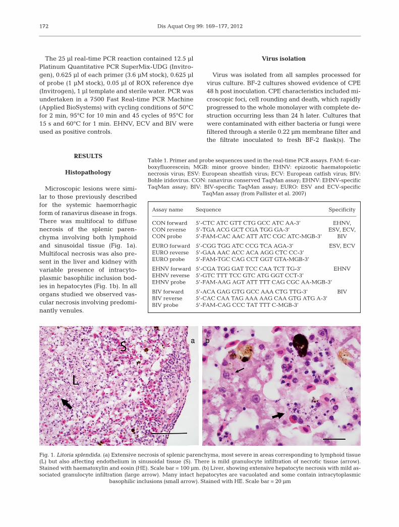
Microscopic lesions were simi-lar to those previously describedfor the systemic haemorrhagicform of ranavirus disease in frogs.There was multifocal to diffusenecrosis of the splenic paren -chyma involving both lymphoidand sinusoidal tissue (Fig. 1a).Multifocal necrosis was also pre-sent in the liver and kidney withvariable presence of intracyto-plasmic baso philic inclusion bod-ies in hepatocytes (Fig. 1b). In allorgans studied we observed vas-cular necrosis involving predomi-nantly venules.
Virus isolation
Virus was isolated from all samples processed forvirus culture. BF-2 cultures showed evidence of CPE48 h post inoculation. CPE characteristics included mi-croscopic foci, cell rounding and death, which rapidlyprogressed to the whole monolayer with complete de-struction occurring less than 24 h later. Cultures thatwere contaminated with either bacteria or fungi werefiltered through a sterile 0.22 µm membrane filter andthe filtrate inoculated to fresh BF-2 flask(s). The
172
Assay name Sequence Specificity
CON forward 5’-CTC ATC GTT CTG GCC ATC AA-3’ EHNV, CON reverse 5’-TGA ACG GCT CGA TGG GA-3’ ESV, ECV, CON probe 5’-FAM-CAC AAC ATT ATC CGC ATC-MGB-3’ BIV
EURO forward 5’-CGG TGG ATC CCG TCA AGA-3’ ESV, ECVEURO reverse 5’-GAA AAC ACC ACA AGG CTC CC-3’ EURO probe 5’-FAM-TGC CAG CCT GGT GTA-MGB-3’
EHNV forward 5’-CGA TGG GAT TCC CAA TCT TG-3’ EHNVEHNV reverse 5’-GTC TTT TCC GTC ATG GGT CCT-3’ EHNV probe 5’-FAM-AAG AGT ATT TTT CAG CGC AA-MGB-3’
BIV forward 5’-ACA GAG GTG GCC AAA CTG TTG-3’ BIVBIV reverse 5’-CAC CAA TAG AAA AAG CAA GTG ATG A-3’ BIV probe 5’-FAM-CAG CCC TAT TTT C-MGB-3’
Table 1. Primer and probe sequences used in the real-time PCR assays. FAM: 6-car-boxyfluorescein; MGB: minor groove binder; EHNV: epizootic haematopoieticnecrosis virus; ESV: European sheatfish virus; ECV: European catfish virus; BIV:Bohle iridovirus. CON: ranavirus conserved TaqMan assay; EHNV: EHNV-specificTaqMan assay; BIV: BIV-specific TaqMan assay; EURO: ESV and ECV-specific
TaqMan assay (from Pallister et al. 2007)
Fig. 1. Litoria splendida. (a) Extensive necrosis of splenic parenchyma, most severe in areas corresponding to lymphoid tissue(L) but also affecting endothelium in sinusoidal tissue (S). There is mild granulocyte infiltration of necrotic tissue (arrow).Stained with haematoxylin and eosin (HE). Scale bar = 100 µm. (b) Liver, showing extensive hepatocyte necrosis with mild as-sociated granulocyte infiltration (large arrow). Many intact hepatocytes are vacuolated and some contain intracytoplasmic
basophilic inclusions (small arrow). Stained with HE. Scale bar = 20 µm

Weir et al.: Novel Bohle-like virus in Australian frogs
filtered samples exhibited similar CPE characteristicsand timing as the samples that were not filtered,demonstrating a filterable infectious agent. Evidencefor the presence of an infectious virus was confirmedby subsequent passages in cell cultures where CPEwas consistently and repeatedly observed. The virusisolate was provisionally named Mahaffey Road virus(MHRV) and assigned the isolate number DPP7200.
Electron microscopy and laser dissection
Icosahedral particles of 136 ± 7 nm (mean ± SD),(n = 7) were observed by negative contrast electronmicroscopy (Fig. 2). Examination of ultrathin sectionsshowed the presence of cytoplasmic viruses whichappeared singularly, in paracrystalline arrays and/oraggregates (Fig. 3a). Viruses were present in differ-
ent stages of assembly and were observed buddingfrom the plasma membrane (Fig. 3b).
Cytoplasmic inclusion bodies as identified by elec-tron microscopy and histopathology examinationwere dissected from the fixed tissue, and sequencingof the PCR products confirmed the presence of rana -virus in the hepatic and splenic lesions.
Restriction enzyme digests, conventional PCR,sequencing and phylogenetic analysis
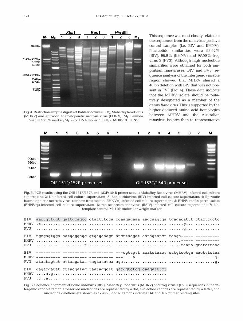
Fig. 4 shows restriction enzyme (RE) digestion pro-files for MHRV, BIV and EHNV. All 3 RE digest pro-files were different for each of the REs. Using theclassification of Hyatt et al. (2000), MHRV cannot beassigned to either the BIV or EHNV species.
In less than 24 h, all BF-2 cell cultures were dis-playing CPE at dilutions in excess of 1/20 and 1/200but not 1/2000. However, between 24 and 48 h, allcell cultures exhibited CPE in all dilutions. Materialwas harvested for PCR analysis from the BF-2 cul-tures after 4 d.
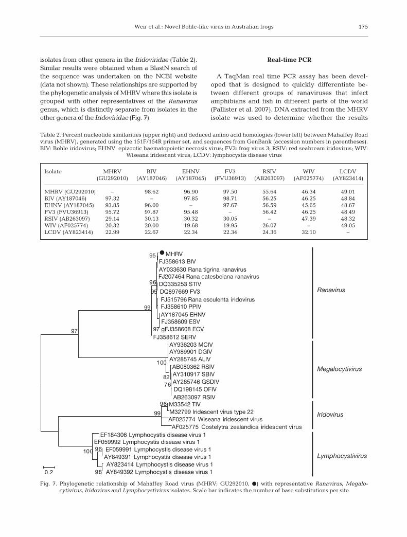
Amplicons of the expected size (321 bp for the 151F/152R primer set; 625 bp for the 153F/ 154R primer set)were produced for the isolate amplified in cell cultureafter inoculation with MHRV for both primer sets(Fig. 5). Positive reactions were also produced for BIVand both EHNV isolates. No amplicon was producedfor template extracted from uninfected cell culture su-pernatant, and larger amplicons were produced fromtemplate extracted from RSIV-in fected cell cultures.
Sequencing of the amplicons generated using the151F/154R primer set resulted in a 1161 bp con sensussequence of MHRV (GenBank accession GU292010).
173
Fig. 2. Transmission electron micrograph of negatively con-trasted ranaviruses. The outer capsid is apparent (arrow).
Scale bar = 150 nm
Fig. 3. Transmission electron micrographs of ultrathin sections of cells infected with Mahaffey Road virus (MHRV; DPP7200).(a) Intracytoplasmic aggregation of viruses. Empty and full viruses are apparent; an aberrant form (arrow) is also present.
Scale bar = 200 nm. (b) Viruses budding (arrow) from the plasma membrane. Scale bar = 200 nm
Dis Aquat Org 99: 169–177, 2012
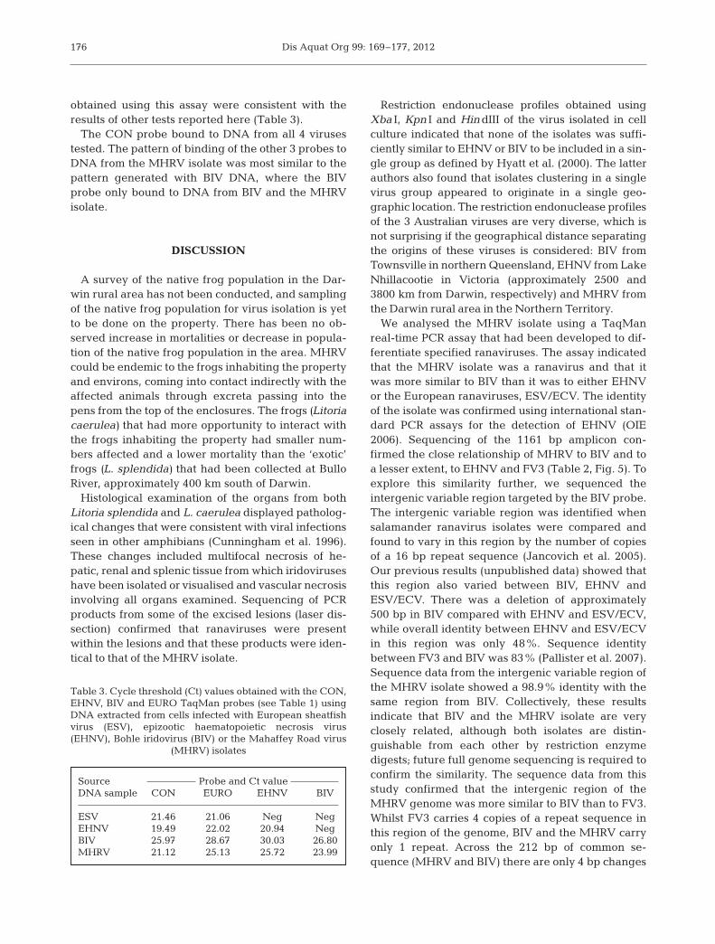
This sequence was most closely related tothe se quen ces from the ranavirus-positivecontrol samples (i.e. BIV and EHNV).Nucleo tide similarities were 98.62%(BIV), 96.9% (EHNV) and 97.50% frogvirus 3 (FV3). Although high nucleotidesimilarities were obtained for both am-phibian rana viruses, BIV and FV3, se-quence analysis of the intergenic variablere gion showed that MHRV shared a48 bp deletion with BIV that was not pre-sent in FV3 (Fig. 6). These data indicatethat the MHRV isolate should be puta-tively designated as a member of thegenus Ranavirus. This is supported by thehigher deduced amino acid homologiesbetween MHRV and the Australianranavirus isolates than to representative
174
Fig. 4. Restriction enzyme digests of Bohle iridovirus (BIV), Mahaffey Road virus(MHRV) and epizootic haematopoietic necrosis virus (EHNV). M1: Lambda
Hin dIII Eco RV marker; M2: 2-log DNA ladder; 1: BIV; 2: MHRV; 3: EHNV
Fig. 5. PCR results using the OIE 151F/152R and 153F/154R primer sets. 1: Mahaffey Road virus (MHRV)-infected cell culturesupernatant; 2: Uninfected cell culture supernatant; 3: Bohle iridovirus (BIV)-infected cell culture supernatant; 4: Epizootichaematopoietic necrosis virus, rainbow trout isolate (EHNVrt)-infected cell culture supernatant; 5: EHNV redfin perch isolate(EHNVrp)-infected cell culture supernatant; 6: red seabream iridovirus (RSIV)-infected cell culture supernatant; 7: No-
template control; M: 1 kb molecular weight marker
BIV aactgttggt gattgcagcc ctattttcca ccaagagaaa aagcaagtga tgagacattt ctactcgctcMHRV .t........ .......... .......... .......... .......... ......g... ..........FV3 .......... .......... .......... .......... .......... ......g... ..........
BIV tgcgagtgga aatgaggagc gtgagaaagt atcttaagat aatagtatct taaga----- ----------MHRV .......... .......... .......... .......... .......... .....----- ----------FV3 .......... .........t .......... .......... .......... .....taata gtatcttaag
BIV ---------- ---------- ---------- ---cgttgtt acatctaact cttgtcctga aactttctaaMHRV ---------- ---------- ---------- ---....a.. .......... .......... ........g.FV3 ataatagtat cttaagataa tagtatctca aga....... .......... .......... ........g.
BIV gagacgatat cttacgatag taataggctt gacggtctcg caagatttctMHRV ....a.g... .......... .......... .......... ..........FV3 .c..a..... .......... .......... .......... ..........
Fig. 6. Sequence alignment of Bohle iridovirus (BIV), Mahaffey Road virus (MHRV) and frog virus 3 (FV3) sequences in the in-tergenic variable region. Conserved nucleotides are represented by a dot, nucleotide changes are represented by a letter, and
nucleotide deletions are shown as a dash. Shaded regions indicate 16F and 16R primer binding sites
Weir et al.: Novel Bohle-like virus in Australian frogs
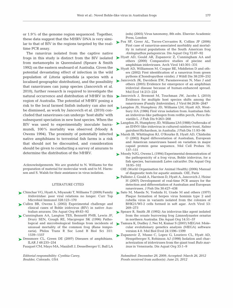
isolates from other genera in the Irido viridae (Table 2).Similar results were obtained when a BlastN search ofthe sequence was undertaken on the NCBI website(data not shown). These relationships are supported bythe phylogenetic analysis of MHRV where this isolate isgrouped with other representatives of the Ranavirusgenus, which is distinctly separate from isolates in theother genera of the Iridoviridae (Fig. 7).
Real-time PCR
A TaqMan real time PCR assay has been devel-oped that is designed to quickly differentiate be -tween different groups of ranaviruses that infectamphibians and fish in different parts of the world(Pallister et al. 2007). DNA extracted from the MHRVisolate was used to determine whether the results
175
Isolate MHRV BIV EHNV FV3 RSIV WIV LCDV (GU292010) (AY187046) (AY187045) (FVU36913) (AB263097) (AF025774) (AY823414)
MHRV (GU292010) – 98.62 96.90 97.50 55.64 46.34 49.01BIV (AY187046) 97.32 – 97.85 98.71 56.25 46.25 48.84EHNV (AY187045) 93.85 96.00 – 97.67 56.59 45.65 48.67FV3 (FVU36913) 95.72 97.87 95.48 – 56.42 46.25 48.49RSIV (AB263097) 29.14 30.13 30.32 30.05 – 47.39 48.32WIV (AF025774) 20.32 20.00 19.68 19.95 26.07 – 49.05LCDV (AY823414) 22.99 22.67 22.34 22.34 24.36 32.10 –
Table 2. Percent nucleotide similarities (upper right) and deduced amino acid homologies (lower left) between Mahaffey Roadvirus (MHRV), generated using the 151F/154R primer set, and sequences from GenBank (accession numbers in parentheses).BIV: Bohle iridovirus; EHNV: epizootic haematopoietic necrosis virus; FV3: frog virus 3; RSIV: red seabream iridovirus; WIV:
Wiseana iridescent virus; LCDV: lymphocystis disease virus
MHRVFJ358613 BIVAY033630 Rana tigrina ranavirusFJ207464 Rana catesbeiana ranavirusDQ335253 STIVDQ897669 FV3FJ515796 Rana esculenta iridovirusFJ358610 PPIVAY187045 EHNVFJ358609 ESVgFJ358608 ECV
FJ358612 SERVAY936203 MCIVAY989901 DGIVAY285745 ALIVAB080362 RSIVAY310917 SBIVAY285746 GSDIVDQ198145 OFIVAB263097 RSIV
M33542 TIVM32799 Iridescent virus type 22AF025774 Wiseana iridescent virus
AF025775 Costelytra zealandica iridescent virusEF184306 Lymphocystis disease virus 1
EF059992 Lymphocystis disease virus 1EF059991 Lymphocystis disease virus 1AY849391 Lymphocystis disease virus 1AY823414 Lymphocystis disease virus 1AY849392 Lymphocystis disease virus 198
96100
97
95
95
96
99
96
99
97
7682
100
0.2
Ranavirus
Megalocytivirus
Iridovirus
Lymphocystivirus
Fig. 7. Phylogenetic relationship of Mahaffey Road virus (MHRV; GU292010, d) with representative Ranavirus, Megalo-cytivirus, Iridovirus and Lymphocystivirus isolates. Scale bar indicates the number of base substitutions per site
Dis Aquat Org 99: 169–177, 2012
obtained using this assay were consistent with theresults of other tests reported here (Table 3).
The CON probe bound to DNA from all 4 virusestested. The pattern of binding of the other 3 probes toDNA from the MHRV isolate was most similar to thepattern generated with BIV DNA, where the BIVprobe only bound to DNA from BIV and the MHRVisolate.
DISCUSSION
A survey of the native frog population in the Dar-win rural area has not been conducted, and samplingof the native frog population for virus isolation is yetto be done on the property. There has been no ob -served increase in mortalities or decrease in popula-tion of the native frog population in the area. MHRVcould be endemic to the frogs inhabiting the propertyand environs, coming into contact indirectly with theaffected animals through excreta passing into thepens from the top of the enclosures. The frogs (Litoriacaerulea) that had more opportunity to interact withthe frogs inhabiting the property had smaller num-bers affected and a lower mortality than the ‘exotic’frogs (L. splendida) that had been collected at BulloRiver, approximately 400 km south of Darwin.
Histological examination of the organs from bothLitoria splendida and L. caerulea displayed patholog-ical changes that were consistent with viral infectionsseen in other amphibians (Cunningham et al. 1996).These changes included multifocal necrosis of he-patic, renal and splenic tissue from which iridoviruseshave been isolated or visualised and vascular necrosisinvolving all organs examined. Sequencing of PCRproducts from some of the excised lesions (laser dis-section) confirmed that ranaviruses were presentwithin the lesions and that these products were iden-tical to that of the MHRV isolate.
Restriction endonuclease profiles obtained usingXba I, Kpn I and Hin dIII of the virus isolated in cellculture indicated that none of the isolates was suffi-ciently similar to EHNV or BIV to be included in a sin-gle group as defined by Hyatt et al. (2000). The latterauthors also found that isolates clustering in a singlevirus group appeared to originate in a single geo-graphic location. The restriction endonuclease profilesof the 3 Australian viruses are very diverse, which isnot surprising if the geographical distance separatingthe origins of these viruses is considered: BIV fromTownsville in northern Queensland, EHNV from LakeNhillacootie in Victoria (approximately 2500 and3800 km from Darwin, respectively) and MHRV fromthe Darwin rural area in the Northern Territory.
We analysed the MHRV isolate using a TaqManreal-time PCR assay that had been developed to dif-ferentiate specified ranaviruses. The assay indicatedthat the MHRV isolate was a ranavirus and that itwas more similar to BIV than it was to either EHNVor the European ranaviruses, ESV/ECV. The identityof the isolate was confirmed using international stan-dard PCR assays for the detection of EHNV (OIE2006). Sequencing of the 1161 bp amplicon con-firmed the close relationship of MHRV to BIV and toa lesser extent, to EHNV and FV3 (Table 2, Fig. 5). Toexplore this similarity further, we sequenced theintergenic variable region targeted by the BIV probe.The intergenic variable region was identified whensalamander ranavirus isolates were compared andfound to vary in this region by the number of copiesof a 16 bp repeat sequence (Jancovich et al. 2005).Our previous results (unpublished data) showed thatthis region also varied between BIV, EHNV andESV/ECV. There was a deletion of approximately500 bp in BIV compared with EHNV and ESV/ECV,while overall identity between EHNV and ESV/ECVin this region was only 48%. Sequence identitybetween FV3 and BIV was 83% (Pallister et al. 2007).Sequence data from the intergenic variable region ofthe MHRV isolate showed a 98.9% identity with thesame region from BIV. Collectively, these resultsindicate that BIV and the MHRV isolate are veryclosely related, although both isolates are distin-guishable from each other by restriction enzymedigests; future full genome sequencing is required toconfirm the similarity. The sequence data from thisstudy confirmed that the intergenic region of theMHRV genome was more similar to BIV than to FV3.Whilst FV3 carries 4 copies of a repeat sequence inthis region of the genome, BIV and the MHRV carryonly 1 repeat. Across the 212 bp of common se -quence (MHRV and BIV) there are only 4 bp changes
176
Source Probe and Ct valueDNA sample CON EURO EHNV BIV
ESV 21.46 21.06 Neg NegEHNV 19.49 22.02 20.94 NegBIV 25.97 28.67 30.03 26.80MHRV 21.12 25.13 25.72 23.99
Table 3. Cycle threshold (Ct) values obtained with the CON,EHNV, BIV and EURO TaqMan probes (see Table 1) usingDNA extracted from cells infected with European sheatfishvirus (ESV), epizootic haematopoietic necrosis virus(EHNV), Bohle iridovirus (BIV) or the Mahaffey Road virus
(MHRV) isolates
Weir et al.: Novel Bohle-like virus in Australian frogs
or 1.9% of the genome region sequenced. Together,these data suggest that the MHRV DNA is very simi-lar to that of BIV in the regions targeted by the real-time PCR assay.
The ranavirus isolated from the captive nativefrogs in this study is distinct from the BIV isolatedfrom metamorphs in Queensland (Speare & Smith1992) on the eastern seaboard of Australia. Given thepotential devastating effect of infection in the wildpopulation of Litoria splendida (a species with alocalised geographic distribution), and the possibilitythat ranaviruses can jump species (Jancovich et al.2010), further research is required to investigate thenatural occurrence and distribution of MHRV in thisregion of Australia. The potential of MHRV posing arisk to the local farmed finfish industry can also notbe dismissed, as work by Jancovich et al. (2010) con-cluded that ranaviruses can undergo ‘host shifts’ withsubsequent speciation in new host species. When theBIV was used to experimentally inoculate barra-mundi, 100% mortality was observed (Moody &Owens 1994). The proximity of potentially infectednative amphibians to terrestrial fish farms is a riskthat should not be discounted, and considerationshould be given to conducting a survey of anurans todetermine the distribution of the MHRV.
Acknowledgements. We are grateful to N. Williams for thepreparation of material for molecular work and to M. Harm-sen and S. Walsh for their assistance in virus isolation.
LITERATURE CITED
Chinchar VG, Hyatt A, Miyazaki T, Williams T (2009) FamilyIridoviridae: poor viral relations no longer. Curr TopMicrobiol Immunol 328: 123−170
Cullen BR, Owens L (2002) Experimental challenge andclinical cases of Bohle iridovirus (BIV) in native Aus-tralian anurans. Dis Aquat Org 49: 83−92
Cunningham AA, Langton TES, Bennettt PMB, Lewin JF,Drury SEN, Gough RE, Macgregor SK (1996) Patho-logical and microbiological findings from incidents ofunusual mortality of the common frog (Rana tempo-raria). Philos Trans R Soc Lond B Biol Sci 351: 1539−1557
Densmore CL, Green DE (2007) Diseases of amphibians.ILAR J 48: 235−254
Fauquet CM, Mayo MA, Maniloff J, Desselberger U, Ball LA
(eds) (2005) Virus taxonomy, 8th edn. Elsevier AcademicPress, London
Fox SF, Greer AL, Torres-Cervantes R, Collins JP (2006)First case of ranavirus-associated morbidity and mortal-ity in natural populations of the South American frogAtelognathus patagonicus. Dis Aquat Org 72: 87−92
Hyatt AD, Gould AR, Zupanovic Z, Cunningham AA andothers (2000) Comparative studies of piscine andamphibian iridoviruses. Arch Virol 145: 301−331
Hyatt AD, Williamson M, Coupar BE, Middleton D and oth-ers (2002) First identification of a ranavirus from greenpythons (Chondropython viridis). J Wildl Dis 38: 239−252
Jancovich JK, Davidson EW, Parameswaran N, Mao J andothers (2005) Evidence for emergence of an amphibianiridoviral disease because of human-enhanced spread.Mol Ecol 14: 213−224
Jancovich J, Bremont M, Touchman JW, Jacobs L (2010)Evidence for multiple host species shifts among theranaviruses (Family Iridoviridae). J Virol 84: 2636−2647
Langdon JS, Humphrey JD, Williams LM, Hyatt AD, West-bury HA (1986) First virus isolation from Australian fish: an iridovirus-like pathogen from redfin perch, Perca flu-viatilis L. J Fish Dis 9: 263−268
Langdon JS, Humphrey JD, Williams LM (1988) Outbreaks ofan EHNV-like iridovirus in cultured rainbow trout, Salmogairdneri Richardson, in Australia. J Fish Dis 11: 93−96
Marsh IB, Whittington RJ, O’Rourke B, Hyatt AD, ChisholmO (2002) Rapid differentiation of Australian, Europeanand American ranaviruses based on variation in majorcapsid protein gene sequence. Mol Cell Probes 16: 137−151
Moody NJG, Owens L (1994) Experimental demonstration ofthe pathogenicity of a frog virus, Bohle iridovirus, for afish species, barramundi Lates calcarifer. Dis Aquat Org18: 95−102
OIE (World Organisation for Animal Health) (2006) Manualof diagnostic tests for aquatic animals. OIE, Paris
Pallister J, Gould A, Harrison D, Hyatt A, Jancovich J, HeineH (2007) Development of real-time PCR assays for thedetection and differentiation of Australian and Europeanranaviruses. J Fish Dis 30: 427−438
Sato M, Maeda N, Yoshida H, Urade M and others (1977)Plaque formation of herpes virus hominis type 2 andrubella virus in variants isolated from the colonies ofBHK21/WI-2 cells formed in soft agar. Arch Virol 53: 269−273
Speare R, Smith JR (1992) An iridovirus-like agent isolatedfrom the ornate burrowing frog Limnodynastes ornatusin northern Australia. Dis Aquat Org 14: 51−57
Tamura K, Dudley J, Nei M, Kumar S (2007) MEGA4: Mole-cular evolutionary genetics analysis (MEGA) softwareversion 4.0. Mol Biol Evol 24: 1596−1599
Zupanovic Z, Musso C, Lopez G, Louriero CL, Hyatt AD,Hengstberger S, Robinson AJ (1998) Isolation and char-acterization of iridoviruses from the giant toad Bufo mar-inus in Venezuela. Dis Aquat Org 33: 1−9
177
Editorial responsibility: Cynthia Carey, Boulder, Colorado, USA
Submitted: December 29, 2009; Accepted: March 26, 2012Proofs received from author(s): June 25, 2012