molecular characterization of a novel segmented dsrna ... · 17 december 2010 mr. omar darissa...
TRANSCRIPT

Molecular characterization of a novel segmented dsRNA mycovirus and its association with hypovirulence of Fusarium graminearum.
Dissertation
A thesis submitted to the
Fachbereich Biologie, Universität Hamburg
for the degree of
doctor rerum naturalium
By
Darissa Omar
Bethlehem, Palestine
Hamburg, 2011


17 December 2010 Mr. Omar Darissa Bramfelder Chaussee 9 22177 Hamburg, Germany RE: Review of thesis entitled “Molecular characterization of a novel segmented dsRNA mycovirus and its association with hypovirulence of Fusarium graminearum” by Mr. Omar Darissa I confirm that the thesis of Mr. Omar Darissa was reviewed by me, a native English speaker, for English language accuracy. The thesis is well written, and therefore I would recommend that the dissertation be accepted in its current form. During my 42 years as a professor, I have read many theses and Mr. Darissa’s thesis meets the standards for the University of Wisconsin-Madison.
Phone numbers: University of Wisconsin: 608-262-1410 Home: 608-845-7717 ([email protected] or [email protected])

To my parents Mousa and Meriam Darissa
To my wife Laila Darissa
To my children Mousa, Mahmoud, and Mamoun
Contents
i
Contents
Contents ..................................................................................................................... i
List of Figures ......................................................................................................... v
List of Tables ......................................................................................................... vii
ABBREVIATIONS .................................................................................................. viii
1. Introduction ..................................................................................................... 1
1.1. Mycoviruses .......................................................................................................... 1
1.1.1. dsRNA mycoviruses ........................................................................................... 2
1.1.1.1. Family Totiviridae ............................................................................................ 3
1.1.1.2. Family Partitiviridae ........................................................................................ 3
1.1.1.3. Family Chrysoviridae ...................................................................................... 4
1.1.1.4. Family Reoviridae ............................................................................................ 5
1.1.2. Positive-strand RNA mycoviruses ...................................................................... 6
1.1.3. DNA mycoviruses ............................................................................................... 6
1.2. Mycovirus associated hypovirulence. ................................................................... 6
1.3. Mycoviruses of F. graminearum ............................................................................ 8
1.4. Fusarium head blight ............................................................................................ 8
1.4.1. The fungus Fusarium graminearum .................................................................... 8
1.4.2. The disease cycle of F. graminearum in wheat. .................................................. 9
1.5. Methods for the sequence determination of dsRNA templates. ........................... 10
1.5.1. Random PCR (rPCR). ........................................................................................ 10
1.5.2. SPAT and FLAC methods. ................................................................................ 12
1.5.3. Direct cloning of dsRNA into dsDNA vectors. .................................................. 14
1.6. Aims of this study ............................................................................................... 15
2. Material and Methods ................................................................................ 16
2.1 Material ............................................................................................................. 16
2.1.1 Enzymes and chemicals .................................................................................... 16
2.1.2. Microbial strains and culture conditions. ......................................................... 16
2.1.3. Media and buffers ............................................................................................ 17
2.1.4. Oligonucleotides (primers) ............................................................................... 18
2.2 Methods ............................................................................................................ 21
2.2.1 Isolation and purification of dsRNA .................................................................. 21
2.2.2 DNA extraction using the CTAB method ............................................................ 22

Contents
ii
2.2.3 Phenol extraction method of total nucleic acids ............................................... 22
2.2.3 Random PCR (rPCR) .......................................................................................... 22
2.2.4 Single Primer Amplification Technique (SPAT) ................................................. 23
2.2.5 Full length Amplification of cDNA (FLAC) ......................................................... 24
2.2.6 Direct ligation of dsRNA into pJET1.2 and pGEM®-T vectors: ........................... 24
2.2.7 Cloning and sequencing: .................................................................................. 25
2.2.7.1 Preparation of electrocompetent cells. .......................................................... 25
2.2.7.2 Preparation of chemical competent cells. ...................................................... 25
2.2.7.3 Transformation of competent cells. ................................................................ 26
2.2.7.4 MiniPreps and restriction digestion. .............................................................. 26
2.2.8 Molecular identification of China 9 isolate. ....................................................... 26
2.2.9 Purification of Virus-Like Particles ................................................................... 26
2.2.9.1 Transmission Electron Microscope (TEM). ..................................................... 27
2.2.10. Hyper immunization of rabbits. ...................................................................... 27
2.2.11. Purification of the antibodies. ........................................................................ 28
2.2.12. Ultrastructural studies. ................................................................................. 28
2.2.12.1 Primary and secondary Fixations ................................................................. 28
2.2.12.2 Dehydration, infiltration, and embedding ..................................................... 28
2.2.12.3 Sectioning and TEM. ..................................................................................... 29
2.2.12.4 Immunohistology. ......................................................................................... 29
2.2.13 Northern Blot analysis. ................................................................................... 30
2.2.14 Southern Blot analysis. ................................................................................... 30
2.2.15 Protein sequence analysis. ............................................................................. 30
2.2.16 Labeling of virus surface proteins. ................................................................. 31
2.2.17 Western blot .................................................................................................... 31
2.2.18 Relative quantification PCR. ............................................................................ 31
2.2.19 Virulence assay on wheat heads. .................................................................... 32
2.2.20 Virulence assay on maize cobs. ...................................................................... 32
2.2.21 Growth assays ................................................................................................. 33
2.2.21.1 Production of Perithecia. .............................................................................. 33
2.2.21.2 Transmission of FgV-ch 9 through conidia ................................................... 33
2.2.21.2.1 Reverse transcription: ............................................................................... 33
2.2.21.2.2 PCR: .......................................................................................................... 34
2.2.22 Expression of FgV-ch9 in F. graminearum PH-1 .............................................. 34
2.2.22.1 Semi-quantitative PCR. ................................................................................. 35
2.2.23 Dicer 2 gene disruption by double homologous recombination ...................... 35
2.2.24. Preparation of F. graminearum protoplasts. ................................................... 35
2.2.24.1. Transformation of F. graminearum protoplasts with plasmid constructs. ... 36

Contents
iii
2.2.24.2 Protoplast transfection with purified VLPs. .................................................. 36
2.2.25 Data analysis and accession numbers. ........................................................... 37
3. Results ............................................................................................................. 38
3.1. Optimization of the methods for the sequence-determination of dsRNA templates.
.................................................................................................................................. 38
3.1.1. Random PCR (rPCR). ........................................................................................ 38
3.1.2. Single Primer Amplification Technique (SPAT) ................................................ 39
3.1.3. Full Length Amplification of cDNA (FLAC) ....................................................... 41
3.1.4. Direct cloning of dsRNA into DNA vector ......................................................... 42
3.2. Identification of the fungus isolate ..................................................................... 43
3.3. Molecular characterization of FgV-ch9 ................................................................ 44
3.3.1. Virus purification and dsRNA isolation ............................................................ 44
3.3.2. SDS-PAGE and peptide sequencing ................................................................. 46
3.3.3. Specificity of the produced polyclonal antibodies for FgV-ch9 ........................ 46
3.3.4. Quantitative PCR .............................................................................................. 47
3.3.5. Nucleotide sequencing ..................................................................................... 47
3.3.5.1. dsRNA1 ......................................................................................................... 48
3.3.5.2. dsRNAs 2 and 4 ............................................................................................. 52
3.3.5.3. dsRNA3 ......................................................................................................... 53
3.3.5.4. dsRNA5 ......................................................................................................... 54
3.3.5.5. The 5`and 3`UTRs .......................................................................................... 55
3.6. Association of the virus with hypovirulence-traits of F. graminearum China 9. .. 57
3.6.1. Ultrastructural properties of China 9 isolate. ................................................... 58
3.6.2. Effect of virus titer on the growth rate of F. graminearum China 9. ................. 62
3.6.3. Colony morphology of F. graminearum China 9 isolate .................................... 62
3.6.4. Effect of virus titer on the conidiation of F. graminearum China 9. ................. 63
3.6.4.1. Virus transmission through conidia .............................................................. 64
3.6.5. Effect of virus titer on the pathogenicity of F. graminearum China 9 for wheat
plants. ....................................................................................................................... 65
3.6.6. Effect of virus titer on the pathogenicity of F. graminearum China 9 on maize
plants. ....................................................................................................................... 68
3.7. Transfection of F. graminearum PH-1 with particles of FgV-ch9. ........................ 69
3.7.1 Effect of FgV-ch9 on the conidiation of F. graminearum PH-1. .......................... 70
3.7.2 Effect of FgV-ch9 on perithecia development of F. graminearum PH-1. ............ 71
3.7.3. Pathogenicity of virus-transfected F. graminearum PH-1 on wheat plants. ...... 73
3.7.4. Pathogenicity of virus-transfected F. graminearum PH-1 on maize plants. ...... 76
3.8. Co-infection of maize with F. graminearum China9 and PH-1 .............................. 77

Contents
iv
3.9. Consequences of the over expression of FgV-ch9 putative genes in F.
graminearum PH-1. .................................................................................................... 78
3.9.1. The conidiation capacity of F. graminearum PH-1 expressing genes encoded by
FgV-ch9. .................................................................................................................... 79
3.9.1. Effect of the expression of genes encoded by FgV-ch9 on the pathogenicity of
F. graminearum PH-1 on wheat. ................................................................................. 80
3.9.1. Effect of the expression of genes encoded by FgV-ch9 on the pathogenicity of
F. graminearum PH-1 for maize. ................................................................................. 82
3.10. Effect of disruption of Dicer 2 gene on F. graminearum PH-1 and China 9
isolates. ..................................................................................................................... 84
4. Discussion ..................................................................................................... 85
4.1. Optimization of the methods for the sequence determination of dsRNA templates.
.................................................................................................................................. 85
4.2. Molecular characterization of FgV-ch9 ................................................................ 88
4.3. Association of FgV-ch9 with hypovirulence of its host. ...................................... 91
5. Summary ............................................................................................................ 97
6. Zusammenfassung ........................................................................................... 99
7. References ....................................................................................................... 101
8. Acknowledgements ........................................................................................ 117
9. Curriculum Vitae ............................................................................................. 118

List of Figures
v
List of Figures
FIG. 1 PROPERTIES OF THE MAJOR VIRAL FAMILIES ENCOMPASSING MYCOVIRUS MEMBERS. ........................................... 2 FIG. 2 THE LIFE CYCLE OF F. GRAMINEARUM (SEXUAL PHASE, G. ZEAE), CAUSAL AGENT OF FUSARIUM HEAD BLIGHT ON
WHEAT. .................................................................................................................................................. 10 FIG. 3 A SCHEMATIC ILLUSTRATION OF THE RPCR METHOD FOR THE SEQUENCE DETERMINATION OF DSRNA TEMPLATES. . 12 FIG. 4 A SCHEMATIC ILLUSTRATION OF THE MAJOR STEPS OF THE SPAT METHOD. ....................................................... 13 FIG. 5 AN ILLUSTRATION OF THE MAJOR STEPS OF THE FLAC METHOD. ..................................................................... 14 FIG. 6 PAN 7.1 OVER-EXPRESSION VECTOR. ......................................................................................................... 35 FIG. 7 AGAROSE GEL ELECTROPHORESIS SHOWING THE SENSITIVITY OF THE RPCR FOR THE AMPLIFICATION OF DSRNA
TEMPLATES. ............................................................................................................................................ 38 FIG. 8 AGAROSE GEL ELECTROPHORESIS OF RPCR PRODUCTS AMPLIFIED UNDER DIFFERENT CONDITIONS. ....................... 39 FIG. 9 ELECTROPHORESIS PATTERN OF RT-PCR PRODUCTS OBTAINED BY THE SINGLE PRIMER AMPLIFICATION TECHNIQUE.
............................................................................................................................................................. 40 FIG. 10 AGAROSE GEL ELECTROPHORESIS OF RT-PCR PRODUCTS OBTAINED BY THE FLAC METHOD. .............................. 41 FIG. 11 AGAROSE GEL ELECTROPHORESIS OF THE PCR PRODUCTS OBTAINED AFTER DIRECT CLONING OF DSRNA INTO
PJET1.2 VECTOR. ..................................................................................................................................... 42 FIG. 12 RESTRICTION DIGESTION PROFILES OF PGEM®-T VECTORS CLONED WITH DSRNA SEGMENTS. ............................ 42 FIG. 13 PHYLOGENETIC IDENTIFICATION OF CHINA9 FUNGAL ISOLATE BASED ON THE 28S RDNA GENE. ......................... 43 FIG. 14 VIRUS-LIKE PARTICLES OF FGV-CH9.......................................................................................................... 44 FIG. 15 AGAROSE GEL ELECTROPHORESIS OF DSRNAS ISOLATED FROM THE FUNGUS F. GRAMINEARUM CHINA 9 OR FROM
PURIFIED VLPS OF FGV-CH9. ..................................................................................................................... 45 FIG. 16 NORTHERN BLOT ANALYSIS OF FGV-CH9 DSRNA SEGMENTS. ....................................................................... 45 FIG. 17 SDS-PAGE AND WESTERN BLOT ANALYSIS OF FGV-CH9 STRUCTURAL PROTEINS. ........................................... 46 FIG. 18 SPECIFICITY OF THE FGV-CH9 POLYCLONAL ANTIBODIES............................................................................... 47 FIG. 19 PARTIAL NUCLEOTIDE SEQUENCES OF FGV-CH9 DSRNA1 (18A), DSRNA2 (18B), AND DSRNA3 (18C). ............. 49 FIG. 20 COMPARISON OF THE CONSERVED RDRPS MOTIFS OF SEVERAL DSRNA MYCOVIRUSES INCLUDING FGV-CH9. ....... 50 FIG. 21 PHYLOGRAMS OF THE RDRP (A) AND THE CP (B) OF FGV-CH9. .................................................................... 52 FIG. 22 VERIFICATION OF THE TERMINAL REPEATS AT THE 3` TERMINUS OF DSRNA4 OF FGV-CH9. ............................... 53 FIG. 23 MULTIPLE-SEQUENCE ALIGNMENT OF THE 12 C2H2 ZINC FINGER DOMAIN PRESENT AT THE C-TERMINUS OF THE
PROTEIN ENCODED BY DSRNA5 OF FGV-CH9. ............................................................................................... 55 FIG. 24 COMPARISON OF THE 5` (A) AND 3`(B) UTRS OF THE 5 DSRNA SEGMENTS OF FGV-CH9. ................................ 56 FIG. 25 A DIAGRAMMATIC REPRESENTATION OF THE GENOMIC STRUCTURE OF FGV-CH9. ............................................ 57 FIG. 26 TRANSMISSION ELECTRON MICROGRAPHS OF F. GRAMINEARUM CHINA 9 ASSOCIATED WITH DIFFERENT TITERS OF
FGV-CH9. ............................................................................................................................................... 59 FIG. 27 ULTRASTRUCTURAL PHENOTYPES OF F. GRAMINEARUM CHINA 9 ASSOCIATED WITH HIGH TIERS OF FGV-CH9. ...... 60 FIG. 28 IMMUNO-DETECTION OF VLPS IN CHINA 9 CELLS ASSOCIATED WITH HIGH VIRUS TITER. .................................... 61 FIG. 29: EFFECT OF FGV-CH9 TITER ON THE GROWTH RATE OF F. GRAMINEARUM CHINA 9 ISOLATE. ............................. 62 FIG. 30. GROWTH RATE AND COLONY MORPHOLOGY OF F. GRAMINEARUM CHINA 9 CULTURES ASSOCIATED WITH DIFFERENT
VIRUS TITERS. .......................................................................................................................................... 63 FIG. 31: PRODUCTION OF CONIDIA IN F. GRAMINEARUM CHINA 9 IN CORRELATION WITH THE TITER OF FGV-CH9. ........... 64 FIG. 32 DIFFERENCES IN THE COLONY GROWTH RATE OF MYCELIA ASSOCIATED WITH MODERATE AND HIGH VIRUS TITER ON
SNA MEDIUM. ........................................................................................................................................ 64 FIG. 33 RT-PCR PRODUCTS OF THE DETECTION OF FGV-CH9 IN SINGLE CONIDIA ORIGINATING CULTURES OF F.
GRAMINEARUM CHINA 9 ISOLATE. .............................................................................................................. 65 FIG. 34. REDUCED PATHOGENICITY ON WHEAT PLANTS OF F. GRAMINEARUM CHINA 9 ISOLATE IS ASSOCIATED WITH THE
PRESENCE OF FGV-CH9. ............................................................................................................................ 66

List of Figures
vi
FIG. 35 EFFECT OF THE ASSOCIATION OF F. GRAMINEARUM CHINA 9 WITH DIFFERENT TITERS OF FGV-CH9 ON ITS
PATHOGENICITY ON WHEAT. ...................................................................................................................... 67 FIG. 36 ASSOCIATION OF FGV-CH9 OF HYPOVIRULENCE OF CHINA 9 FUNGAL ISOLATE ON MAIZE. .................................. 68 FIG. 37 MAIZE COBS INOCULATED WITH F. GRAMINEARUM CHINA 9 ASSOCIATED WITH DIFFERENT TITERS OF FGV-CH9. .. 69 FIG. 38 DUPLEX RT-PCR OF VIRUS-TRANSFECTED COLONIES OF F. GRAMINEARUM PH-1. ........................................... 70 FIG. 39: TRANSFECTION OF F. GRAMINEARUM PH-1 WITH FGV-CH9 NEGATIVELY AFFECTS ITS CONIDIATION CAPACITY. .... 71 FIG. 40. PERITHECIA DEVELOPMENT OF F. GRAMINEARUM PH-1 BEFORE AND AFTER TRANSFECTION WITH FGV-CH9. ...... 72 FIG. 41 PERITHECIA OF F. GRAMINEARUM PH-1 AND CHINA 9 ISOLATES. .................................................................. 72 FIG. 43. SYMPTOM DEVELOPMENT ON WHEAT SPIKES AFTER 3 WEEKS OF INOCULATION WITH CONIDIA OF PH-1VLP1. .... 74 FIG. 44. SYMPTOM DEVELOPMENT ON WHEAT SPIKES AFTER 3 WEEKS OF INOCULATION WITH CONIDIA OF PH-1VLP11. .. 75 FIG. 46 PATHOGENICITY OF F. GRAMINEARUM PH-1 TRANSFECTED WITH FGV-CH9 FOR MAIZE. ................................... 76 MAIZE COBS WERE INFECTED WITH CONIDIA OF PH-1 TRANSFECTED WITH FGV-CH9. A: PH-1VLP1, B: PH-1VLP11, C:
WT PH-1. THE COBS WERE INJECTED WITH THE FUNGAL CONIDIA AT THE STAGE OF EARLY KERNEL FORMATION. IN
EACH TREATMENT, 8-10 COBS WERE INJECTED. COBS WERE PHOTOGRAPHED 5 WEEKS POST INFECTION. ............... 76 FIG. 45: EFFECT OF TRANSFECTION OF F. GRAMINEARUM PH-1WITH VLPS OF FGV-CH9 ON ITS PATHOGENICITY ON MAIZE
PLANTS. .................................................................................................................................................. 76 FIG. 47: SIMULTANEOUS INOCULATION OF CONIDIA OF F. GRAMINEARUM CHINA 9 AND PH-1 ISOLATES INTO MAIZE. ...... 77 FIG. 48 CO-INFECTION OF MAIZE WITH F. GRAMINEARUM CHINA 9 AND PH-1 ISOLATES. ............................................ 78 FIG. 49 SEMI-QUANTITATIVE PCR OF F. GRAMINEARUM PH-1 MUTANTS EXPRESSING GENES ENCODED BY FGV-CH9. ...... 79 FIG. 50: SOUTHERN BLOT ANALYSIS OF F. GRAMINEARUM PH-1 MUTANTS EXPRESSING GENES ENCODED BY FGV-CH9. .... 79 FIG. 51: EFFECT OF FGV-CH9 GENES ON THE CONIDIATION OF F. GRAMINEARUM PH-1. ............................................. 80 FIG. 52: EFFECT OF THE EXPRESSION OF FGV-CH9 GENES ON THE PATHOGENICITY OF F. GRAMINEARUM PH-1 FOR WHEAT.
............................................................................................................................................................. 80 FIG. 53: THE EFFECT OF THE EXPRESSION OF FGV-CH9 GENES ON THE VIRULENCE OF F. GRAMINEARUM PH-1 FOR WHEAT.
............................................................................................................................................................. 81 FIG. 54: EFFECT OF THE EXPRESSION OF FGV-CH9 GENES ON THE PATHOGENICITY OF F. GRAMINEARUM PH-1 FOR MAIZE.82 FIG. 55: SYMPTOM DEVELOPMENT ON MAIZE COBS AFTER 5 WEEKS OF INOCULATION WITH CONIDIA OF F. GRAMINEARUM
PH-1 EXPRESSING GENES OF FGV-CH9. ....................................................................................................... 83 FIG. 56 GROWTH RATE DIFFERENCES BETWEEN PH-1 CELLS AFTER TRANSFECTION WITH FGV-CH9. ............................... 93 FIG. 57 SINGLE CONIDIA ORIGINATING CULTURES OF VIRUS TRANSFECTED PH-1 CELLS DISPLAY GROWTH RATE DIFFERENCES.
............................................................................................................................................................. 94

List of Tables
vii
List of Tables
TABLE 1: LIST OF THE PRIMERS USED IN THIS STUDY .................................................................................... 19
TABLE 2. THE EFFICIENCY OF DIFFERENT DNA POLYMERASES IN THE SPAT AND THE FLAC METHODS. ................. 40
TABLE 3. THE TRANSFORMATION EFFICIENCY OF DSRNA-LIGATED PJET1.2 AND PGEM®-T VECTORS................... 43
TABLE 4: SIZE AND FUNCTION OF THE GENOMIC DSRNA SEGMENTS AND THE DEDUCED PROTEINS OF FGV-CH9. .... 48
TABLE 5: LIST OF THE VIRUSES USED IN CONSTRUCTING THE PHYLOGRAMS IN FIG. 21 A,B. .................................. 51

Abbreviations
viii
ABBREVIATIONS
% Percentage
°C centigrade
µ micro (10-6
)
aa amino acids
BLAST Basic Local Alignment Search Tool
bp base pairs
cDNA complementary Deoxyribonucleic Acid
CP Coat protein
cv. Cultivated variety; cultivar
DIG Digoxygenin
DMSO Dimethzlsulfoxide
DNA Deoxyribonucleic Acid
dNTPs Desoxynucleotide triphosphate (s)
DON Deoxynivalenol
dsRNA double-stranded RNA
DTT Dithiotreitol
dUTP Desoxyuracil triphosphate
EDTA Ethzlenediaminetetraacetic acid
et al. et alii = and others
EtBr Ethidium bromide
FHB Fusarium Head Blight
Fig. Figure
FLAC Full Length Amplification of cDNA
h hour
HCl Hzdrochloride
hph Hygromycin B phosphotransferase
IPTG Isopropzlthio-ß-D-galactoside
kb kilo bases (= 1000 bp)
kDa kilo Dalton (= 1000 Da)

Abbreviations
ix
LB Luria-Bertani medium
M Molar (mol/L)
mg milligram
min minutes
ml milliliter
mRNA messenger RNA
NCBI National Center for Biotechnology Information
ng nanogram
nm nanometer
nt nucleotide
OD Optical Density
ORF Open Reading Frame
PBS phosphate-buffered saline buffer
PBS-T PBS Tween
PCR Polymerase Chain Reaction
PEG polyethylene glycol
pH Potentia Hydrogenii
qPCR quantitative PCR
RdRP RNA dependent RNA Polymerase
RNA Ribonucleic acid
rPCR random PCR
rpm round per minute
RT-PCR Reverse transcriptase PCR
SDS Sodium Dodecylsulfate
sec seconds
SNA Synthetic Nutrient Agar
SPAT Single Primer Amplification Technique
TE Tris-EDTA
Tm Annealing Temperature
Tris Tris-(hydroxymethyl) aminomethane
U unit
UV Ultra violet

Abbreviations
x
v/v Volume per volume
vol Volume
w/v Weight per volume
WT Wild type
X-gal 5-Bromo-4-chloro-3-indolyl-ß-D-galactoside

1. Introduction
1
1. Introduction
The association of several viruses with hypovirulence of their fungal plant pathogenic
hosts has motivated scientists to explore the world of mycoviruses or fungal viruses. As the
majority of mycoviruses have dsRNA genomes, several methods for the handling and sequence
determination of such genomes have been established. Molecular characterization and
phylogenetic analysis of many of the so far reported mycoviruses have led to the initiation of new
taxonomic virus families and genera to accommodate the diversity of accumulating mycovirus
members.
The fungus Fusarium graminearum is the major causal agent of a worldwide disease of
cereals called fusarium head blight. F. graminearum infections can lead to sever losses in the
yield and quality of important crops like wheat and barley. The reported association of several
isolates of F. graminearum with mycovirus infection might help in developing an effective and
safe control method of the disease.
In this introduction, a brief description of each of the mycovirus families, including
mycovirus members that infect F. graminearum and those that are associated with hypovirulence
of their hosts is presented. Moreover, a short background about the fungus F. graminearum and
its disease life cycle in wheat is included. Finally, a description of the molecular approaches for
the sequence determination of dsRNA templates is illustrated.
1.1. Mycoviruses
Mycoviruses (fungal viruses) have been described in many fungal species including
phytopathogenic fungi (Pearson et al., 2009). Since the first report of a fungal virus which was in
1962 in diseased mushroom, Agaricus bisporus (Hollings), more than 200 mycoviruses classified
into 10 families have been reported (Ghabrial and Suzuki, 2009). Although the majority of the
mycoviruses are associated with dsRNA and to a lesser extent with ss (+) RNA genomes, few
mycoviruses with ssDNA, or dsDNA genomes have been reported (Yu et al., 2010). With the
exception of few cases, most of the reported mycoviruses have been associated with cryptic or
latent infections of their hosts (Buck, 1998). In figure 1, some properties of the major taxonomic
families with mycovirus members are shown. Mycoviruses have limited routes of transmission.
These include the intercellular routes such as hyphal anastomosis and heterokaryosis or via
sexual and asexual spores (Xie et al., 2006; Chu et al., 2004; Buck, 1998). These transmission

1. Introduction
2
limitations are also reflected on the natural host range of mycoviruses, which is restricted to
fungal individuals who are vegetatively compatible.
Fig. 1 Properties of the major viral families encompassing mycovirus members.
1.1.1. dsRNA mycoviruses
Mycoviruses with dsRNA genomes are classified into 4 major families based on the
number and sequence of their genomic segments. These families are Totiviridae, Partitiviridae,
Chrysoviridae, and Reoviridae. In addition to the dsRNA mycoviruses, these families encompass
members that infect organism other than fungi such as protozoa and plants. The genomes of
mycoviruses belonging to theses families are encapsidated usually in isometric particles with a
diameter of 25-50 nm except for mycoreoviruses which have spherical double-shelled particles
with a diameter of about 80 nm (Ghabrial and Suzuki, 2009; Pearson et al., 2009). Interestingly,
the proteins encoded by dsRNA mycoviruses, belonging to different genera, share little sequence
similarities. Moreover, phylogenetic studies of the most conserved protein among dsRNA
mycoviruses, their RNA-dependent-RNA-Polymerase (RdRp), indicate that these viruses are
most likely polyphyletic (Ghabrial and Suzuki, 2009).

1. Introduction
3
1.1.1.1. Family Totiviridae
Viruses belonging to this family have non-segmented dsRNA genomes (4.6 - 7 kb) and
are encapsidated in isometric particles (single molecule per particle) of ~40 nm in diameter.
Totiviridae genome encompasses two ORFs encoding the CP and RdRp, Virions have a buoyant
density in CsCl of 1.40-1.43gcm-3
. Members of Totiviridae that infect phytopathogenic fungi are
classified into two genera; Totivirus reported in yeasts (e.i Saccharomyces cerevisiae) as well as
the smut fungus Ustilago maydis and Victorivirus which infect filamentous fungi. Those
members of Totiviridae that infect parasitic protozoa belong to either genus Giardiavirus or
Leishmaniavirus.
The amino acid sequences of the RdRp of totivirus share 8 conserved motifs and an
overall relatively significant sequence similarity. Diverse RdRp expression strategies have been
reported for the different members of Totiviridae. For example, Saccharomyces cerevisiae
viruses; ScV-L-A and ScV-L-BC express their RdRp as a fusion with the CP (gag-pol-like) due
to a ribosomal frame-shifting. Ustilago maydis virus H1 (UmV-HI) on the other hand, produce
such a fusion protein without a ribosomal frame-shifting. Proteins that expressed separately as a
result of coupled translation have been reported for members of the Victorivirus such as
Helminthosporium victoriae virus 190S (HvV190S).
Totiviruses of yeasts and smut fungi are usually associated with a satellite dsRNA (M-
dsRNA) that encodes a toxin protein. The yeast and smut fungal isolates that host the M-dsRNA
are immune against the encoded toxin. On the other hand, the isolates that are not associated with
the M-dsRNA are sensitive to the produced toxin and are eliminated. This phenomenon is known
as the yeast or smut killer system. Well-characterized mycoviral dsRNAs coding for the killer
toxins include those associated with Ustilago maydis (Park et al., 1996) and Saccharomyces
cerevisiae (Bostian et al., 1980). Interestingly, the multiplication of the helper totivirus and its
satellite dsRNA are regulated by host genes. The overall result of such regulation is support of
virus replication to a limit where the viral infection is not harmful to the host. Satellites and
defective dsRNA of victorivirus, on the other hand, are not associated with the production of the
killer toxin.
1.1.1.2. Family Partitiviridae
The viruses classified in this family have bipartite dsRNA genomes (1.4 - 2.3 kb)
separately encapsidated in isometric particle of about 30-40 nm in diameter. The two dsRNA

1. Introduction
4
segments are usually similar in size, one encoding the RdRP while the other encoding the CP.
Association with a satellite dsRNA is common among members of the this family. Virions have a
buoyant density in the range of 1.34-1.39 gcm-3
.
Besides Partitivirus, this family contains two other genera Alphacryptoviruses and
Betacryptovirus. However, only members of the Partitivirus have been reported to infect fungi
(fungal partitiviruses). Members of the Alpha- and Betacryptovirus on the other hand, infect
plants (plant partitiviruses). Fungal partitiviruses are transmitted mainly by asexual spores and
hyphal anastomosis, while plant partitiviruses are transmitted only by pollen and ovule to the
seed embryo.
In vitro transcription assays showed that the virions of all fungal and plant partitiviruses
are associated with RdRp activity. The RdRp can function as a transcriptase and as a replicase. In
the virions, the RdRp transcribe the parental positive strand RNA to produce ssRNA. The
produced ssRNA displace the parental positive strand RNA and the later is released out of the
virions and serve a template for protein synthesis by the host machinery or packaged in an
assembling virion. The replicase activity of the RdRp produces a negative-strand RNA on the
packaged positive-strand RNA to make a dsRNA genome.
The complete genomes of many fungal partitiviruses including Atkinsonella hypoxylon
virus, Fusarium solani virus 1, and Fusarium poae virus 1 have been sequenced. Phylogenetic
analysis based on the amino acid sequences of the RdRPs showed that members of the family
Partitiviridae are clustered into 4 groups. Most of the partitvirus members were grouped in either
of two large clusters called subgroup 1 and 2. The average molecular weight of the CPs for the
members of subgroup 1 and 2 was 47 KDa and 74 KDA, respectively. Interestingly, the third
cluster encompasses fungal and plant parttiviruses together. The hosts of some of these viruses
are phytopathogenic fungi. This evokes the probable horizontal transfer of partitivirus members
between plants and fungi (Ghabrial et al., 2008).
1.1.1.3. Family Chrysoviridae
Multipartite linear monocistronic dsRNA genomes (4 segments of 2.4 - 3.6 kb) separately
encapsidated in non-enveloped isometric particles (35-40 nm in diameter) are characteristics for
members of the family Chrysoviridae. The virions of chrysoviruses have buoyant densities in the
range of 1.34-1.39 gcm-3
. Besides the main components (virus particles, each encapsidating one

1. Introduction
5
of the four genomic dsRNAs), the appearance of extra components, representing empty particles
and replication intermediates with distinct densities, is frequent in purified chrysoviruses.
Whereas the proteins encoded by dsRNA1 and 2 operate as the RdRp and CP,
respectively, the role of those encoded by dsRNA3 and 4 is not obvious yet. Genomes of
chrysoviruses that infect fungi have relatively long 5`and 3`UTRs with regions of high sequence
similarity. Moreover, a (CAA)n sequence repeat upstream of the start codon exists probably in
most of the characterized chrysoviruses. Although the (CAA)n repeat was reported in
tobamoviruses to act as a translational enhancer, the role of the (CAA)n repeat in chrysoviruses is
not clear yet.
Although chrysoviruses were classified previously as members of the family
Partitiviridae, phylogenetic studies of the RdRp indicated that they are more related to toti- than
to partitiviruses. There is not much known about the replication of chrysoviruses. However, in
vitro transcription studies showed that the virus particles are associated with RdRp activity and
that a full length mRNA is transcribed from each dsRNA in a conservative mechanism.
Many chrysoviruses occur in mixed infections with other mycoviruses or plant viruses
and are associated with symptomatic infections of their hosts. Examples include Hv145SV and
Hv190SV that infect the phytopathogenic fungus Helminthosporium victoriae, ACDACV and
CCRSACV that co-infect cherry trees in combination with a partitvirus and a suspected totivirus
and the obscure chrysovirus AbV-1 and MBV (member of the family Barnaviridae) which are
associated with the La France disease of cultivated mushroom (Agaricus bisporus). In most of the
cases, the contribution of each virus in the disease development and the nature of the interactions
between the co-infecting viruses are not clear yet.
1.1.1.4. Family Reoviridae
Members of the family Reoviridae that infect fungi are classified in the genus
Mycoreovirus. Mycoreoviruses have 11-12 dsRNA monocistronic genomic segments between
0.7 - 5 kbp in size encapsidated in double-shelled particles about 80 nm in diameter. Except for
several RdRp motifs, the functions of the proteins encoded by mycoreoviruses are not well
characterized. To date, only three mycoreoviruses have been identified. Two of these have been
reported in C. parasitica and one in the root-rot fungus, Rosellinia necatrix. The three
mycoreoviruses are associated with hypovirulence of their fungal hosts.

1. Introduction
6
1.1.2. Positive-strand RNA mycoviruses
Mycoviruses with positive-strand RNA genomes (9-17 kb) and no true virions belong to
the families Hypoviridae, Endornaviridae, and Narnaviridae (Ghabrial and Suzuki, 2009). Some
of these viruses were classified originally as dsRNA mycoviruses probably because they exist
mostly as dsRNA replicative form (RF) in their hosts (Ghabrial and Suzuki, 2009; Pearson et al.,
2009; Nuss, 2005).
Members of the family Hypoviridae have linear RNA genomes in the size range of 9-13
kb. Their RdRp is closely related to those of Bymovirus in the family Potyviridae. Cryphonectria
hypovirus 1 (CHV1) is one of the mycoviruses that has been intensively studied for its potential
use as a biocontrol agent of the chestnut blight fungus Cryphonectria parasitica.
The genomes of Endornaviruses consists of a large RNA (14-17 kbp as estimated form
the dsRNA RF encoding a single protein with RdRp and an RNA helicase motifs. To date, only
two Endornaviruses infecting a fungal host have been reported. These viruses are Phytophthora
endornavirus 1 and Helicobasidium mompa endornavirus 1.
The family Narnaviridae encompasses two genera; Narnavirus and Mitovirus. Members
of this family have RNA genomes coding merely for RdRp and the viruses present as RNA-RdRp
nucleoprotein complexes. Mitovirus infections associated with hypovirulence in phytopathogenic
fungi were reported for Cryphonectria mitovirus 1 and Ophiostoma mitoviruses 3a, 4, 5, and 6.
1.1.3. DNA mycoviruses
Sclerotinia sclerotiorum hypovirulence-associated DNA virus 1 (SsHADV-1) is the only
reported mycovirus with a DNA genome. The virus has a circular ssDNA segment of 2166 nt
encapsidated in isometric particles of 22 nm in diameter. The ssDNA genome encodes two
proteins; a replication initiation protein (Rep) and the coat protein (CP). Interestingly, SsHADV-
1 is not only associated with hypovirulence of its host but also is transmissible between
vegetatively incompatible strains of S. sclerotiorum.
1.2. Mycovirus associated hypovirulence.
Reduced or debilitated fungal virulence (hypovirulence) due to mycovirus infections have
been reported for several phytopathogenic fungal isolates. This phenomenon has attracted the
consideration of the prospective utilization of hypovirulent strains of phytopathogenic fungi and
their associated mycoviruses as biocontrol agents as well as models to understand the

1. Introduction
7
mechanisms of fungal pathogenesis. For example, hypovirulent strains of the chestnut blight
fungus Cryphonectria parasitica were used effectively to control the disease in Europe
(Anagnostakis, 1982). When hypovirulent fungal strains of C. parasitica were applied onto
chestnut, the resident virulent strains were converted to the hypovirulent phenotype (Nuss D. L,
1992). Indeed, the interactions between a fungal host and its mycovirus and their utilization as
biocontrol agents are best studied and described for C. parasitica and its associated hypoviruses
(Dawe, A. L. & Nuss, D. L. 2001).
Mycovirus-associated hypovirulence was also reported in F. graminearum (Chu et al.,
2002), Botrytis cinerea (Castro et al., 2003), Helminthosporium victoriae (Ghabrial, S. A.
2001), Chalara elegans (Park et al., 2006), Sclerotinia sclerotiorum (Zhang et al., 2009; Boland
G.J, 1992; Yu et al., 2010), Sclerotinia homoeocarpa (Deng et al., 2003; Zhou, T. & Boland, G.
J., 1997), Sclerotinia minor (Melzer & Boland, 1996), Ophiostoma
novo-ulmi (Hong et al., 1999),
the white root rot Rosellinia necatrix (Chiba et al., 2009), Leucostoma persoonii (Hammar S.
1989), and Diaporthe ambigua (Preisig et al., 2000).
In most of the above examples, several aspects have hindered our understanding of the
biology of hypovirulence and restricted the implementation of mycoviruses or their dsRNAs in
biological control or genetic engineering programs. First, the absence of extracellular routes of
infection for mycoviruses is the main barrier for their implementation as biological control
agents. Second, the unavailability of effective gene transfer systems to utilize the power of
hypovirulence-inducing dsRNAs for the control of plant pathogenic fungi or to study their
interactions with their hosts. Fortunately, the successful use of Agrobacterium tumefasciens to
transform yeast and several filamentous fungi has been achieved recently. The availability of
agrotransformation protocols for the various species of phytopathogenic fungi would help to
understand the interactions between mycoviruses and their fungal host. Finally, the generation of
dsRNA from a cDNA clone is technically challenging. When this technical limitation was
overcome for the hypovirus of C. parasitica as an example, tantalizing findings have been
uncovered. The viral and host genes involved in the hypovirulence of C. parasitica are now well
characterized. Moreover, all the spores produced by a transfected strain of C. parasitica have the
integrated cDNA as well as the equivalent dsRNA. Natural hypovirulent strains on the other
hand, produce dsRNA-free ascospores and many dsRNA free conidia, which would limit the
spread of hypovirulence effects.

1. Introduction
8
1.3. Mycoviruses of F. graminearum
To date, only few mycoviruses have been isolated from the phytopathogenic fungus F.
graminearum (Yu et al., 2009; Chu et al., 2002; Theisen et al., 2001). These viruses are
Fusarium graminearum virus 1, virus 2, virus 3 and virus 4 (FgV1, FgV2, FgV3, FgV4).
Whereas the complete nucleotide sequence for FgV1, FgV3 and FgV4 were reported, only a
partial sequence with an RdRP motif was published for FgV2. The dsRNA virus reported by
Theisen et al., (2001) has not been fully characterized yet. While these viruses are still
unassigned to a specific genus, genome organization and phylogenetic analysis showed that FgV1
belongs most likely to the ss (+) RNA mycoviruses while FgV4 belongs to the family
Partitiviridae. The deduced amino acid sequence of the RdRP of FgV3 showed close relatedness
to members of the families Chrysoviridae and Totiviridae (Yu et al., 2009).
1.4. Fusarium head blight
Fusarium head blight (scab, FHB) is an important fungal disease of small-grain cereals
like wheat and barley. Several species of the genus Fusarium can cause the disease of which F.
graminearum is the most common. Other species includes F. poae, F. culmorum, and F.
avenaceum. (Osborne and Stein, 2007). The disease epidemics have caused great yield and
quality losses worldwide. The reduction of the kernels weight and the contamination of the
infected grains with mycotoxins make them unacceptable and not safe for the animal and human
consume. In North America, losses due to FHB were estimated to be more than $1 billion per
year (McMullen et al., 1997). Moreover, the germination rate of infected seeds is highly reduced
which contribute further to the yield loss (Argyris et al., 2003).
1.4.1. The fungus Fusarium graminearum
The major causal agent of head blight in the United States, Canada, and Europe is F.
graminearum Schwabe [teleomorph: Gibberella zeae (Schwein.) Petch] (Goswami and Kistler,
2004). In addition to the FHB, the fungus is the main causal agent of the ear rot disease of maize
(Tamburic-Ilincic and Schaafsma, 2009). Although phylogenetic studies of a worldwide
collection of F. graminearum isolates showed that they could be classified into 11 distinct
species, the original name F. graminearum is still used to describe all of these isolates (Starkey et
al., 2007; Leslie and Bowden, 2008). The economic losses and health consequences associated

1. Introduction
9
with FHB are mainly because of the capability of F. graminearum to produce mycotoxins such as
deoxynivalenol (DON), nivalenol (NIV), zearalenone, fusarin C and aurofusarin (Trail 2009;
Bennett and Klich, 2003; Gilbert and Tekauz, 1995). For example, when DON is ingested in
ample amounts, it can inhibit the protein biosynthesis in human and mammals, which may lead to
death.
1.4.2. The disease cycle of F. graminearum in wheat.
As shown in Fig. 2, the disease cycle of F. graminearum starts when airborne spores
settle on the wheat flower. At temperatures around 25°C and highly humid conditions, the spores
germinate and enter the plant through natural openings or degenerating anther tissues. In the early
stages of the infection, the fungus spreads intercellularly through the xylem and pith and the
plants show no symptoms. At a later stage, the fungus spreads intracellularly and colonizes the
tissues. In this phase symptoms such as necrosis, water soaking of the chlorenchyma, and tissue
bleaching start to appear. The fungus produces a mycotoxin called DON which acts as virulence
factor for wheat and most likely also maize. It causes tissue necrosis and facilitates the fungal
spread into the rachis. Due to the accumulation of the DON and the colonization of the fungus in
the developing seeds, the emerging wheat grains on an infected plant are small and shrunken. F.
graminearum produces asexual (conidia) and sexual spores (ascospores) that allow the spread of
the fungus to other plant individuals as well as infection inocula for the next seasons.
The sexual life cycle of F. graminearum starts when hyphal cells with two distinct nuclei
are formed. These binucleate cells develop into the fruiting body initials (perithecium initials)
filled with tubular sacs (asci) containing the ascospores. The ascospores are released and initiate
the infection of other flowering wheat plants. Conidia, on the other hand, are produced from the
sporodochia, which are small masses of hyphae bearing specialized stalks called the
conidiophores, where the asexual spores or conidia are formed. Conidia are produced on the
surface of infected plants. They serve mainly in the short distance spread of the fungus.

1. Introduction
10
Fig. 2 The life cycle of F. graminearum (sexual phase, G. zeae), causal agent of
Fusarium head blight on wheat.
Figure courtesy from Trail F. 2009.
1.5. Methods for the sequence determination of dsRNA templates.
Several methods have been described for the sequence determination of dsRNA templates
(Attoui et al., (2000); Potgieter et al., (2002) & (2009); Vreede et al., (1998), Lambden et al.,
(1992); Imai et al., (1983); Coutts & Livieratos (2003)). These methods include the random PCR
(rPCR), Single Primer Amplification Technique (SPAT), and Full Length Amplification of
cDNAs (FLAC).
1.5.1. Random PCR (rPCR).
The random PCR (rPCR) involves the use of a primer with a random hexamer at its 3` end
(Froussard 1992). This method was established for the random amplification of ssRNAs and was

1. Introduction
11
applied thereafter for genomic dsRNA viral templates (Márquez et al., 2007). An illustration of
the basic steps of a rPCR on a dsRNA template is shown in Fig 3a, b. In brief, 1st strand cDNA is
synthesized randomly using the hexamer sequences at the 3` of the primer. The 2nd
strand cDNA
is synthesized with the Klenow Fragment reaction as shown in Fig. 3a. Alternatively, this step
might be omitted since the production of the 1st cDNA from a dsRNA template will result in
several overlapping cDNAs that will anneal to form a dscDNA as shown in Fig. 3b. Finally, the
dscDNA is PCR amplified using the primer employed in the cDNA synthesis but lacks the 3`
hexamer sequence.
The sensitivity of rPCR was tested for ssRNA templates by Froussard (1992, 1998). He
got intense amplification products starting from template amounts as little as 1 pg. So far, the
sensitivity of this method for dsRNA templates was not tested. The need for cloning and
sequencing of many PCR products, the filling of gaps, the use of other methods such as RACE
(Rapid Amplification of cDNA Ends) for the terminal sequence determination and the
requirement for highly pure template to avoid non-specific amplification make the rPCR methods
time-consuming, laborious and costly.

1. Introduction
12
Fig. 3 A schematic illustration of the rPCR method for the sequence determination of
dsRNA templates.
a: with, b: without the involvement of the Klenow Fragment reaction.
1.5.2. SPAT and FLAC methods.
A remarkable progress in dsRNA cloning was the establishment of sequence-independent
single-primer amplification Technique (SPAT) by Lambden et al., (1992). The method was
employed either solely or with modifications to clone dsRNA of different sizes up to 2.5 kb
(Bigot et al., 1995; James et al., 1999; Attoui et al., 2000; Zhang & Rowhani, 2000; Chen et al.,
2002). The method was further modified to clone larger dsRNAs (>3 kb) (Vreede et al., (1998);
Potgieter et al., (2002) and (2009); Mann et al., (2007)). These modifications include enrichment
of the longer dsRNA segments by means of sucrose gradients or purification from electrophoresis
gels, labeling and size fractionation of cDNA, ligation of primers with extended lengths, the use
of anchor primer, which prime themselves for full-length amplification of cDNAs (FLAC), the
use of highly toxic chemicals like MMOH for the efficient denaturation of dsRNA. Although
such modifications might be useful for the sequence determination of large dsRNA genomes,
many of them might not be available in every laboratory e.g. ultracentrifugation, radioactive
labeling of cDNA, MMOH’s handling and disposal regulations.

1. Introduction
13
As shown in Figures 4 and 5, the SPAT and FLAC methods involve the use of T4 RNA
ligase to ligate a DNA oligonucleotide to both 3` ends of a dsRNA template. In the SPAT method
the ligated primer is 3`-blocked to prevent concatemer while in the FLAC method the primer is
designed to prime itself for cDNA synthesis, where it has two complementary halves separated
by a spacer that has a loop structure. In the SPAT method, cDNA from each strand is synthesized
using a primer complementary to the 3` part of the ligated primer. In both methods, excess RNA
is removed by treatment with NaOH and the complementary cDNAs are annealed for few hours
at 65-68°C. A primer complementary to the 5` part of the ligated oligonucleotide is used for PCR
amplification of the template.
Fig. 4 A schematic illustration of the major steps of the SPAT method.

1. Introduction
14
Fig. 5 An illustration of the major steps of the FLAC method.
1.5.3. Direct cloning of dsRNA into dsDNA vectors.
The cloning of viral dsRNAs directly into dsDNA vectors without any previous
transcription and amplification steps was never reported so far except for one patent application
by Skotnicki et al., (1985). In the current study, the conditions, results, and evaluation of several
attempts to clone genomic dsRNA into DNA vectors are presented.

1. Introduction
15
1.6. Aims of this study
The first aim of this study was to characterize a novel mycovirus with segmented dsRNA
genomes infecting a F. graminearum isolate from China. The characterizations involve the
determination of the full-length nucleotide sequence of the virus. Furthermore, it includes
molecular characterization and phylogenetic studies of the virus.
Second, the study aims at exploring possible association of the virus with hypovirulence
of its fungal host. This involves a comparative phenotypic study of a virus-positive and a virus-
free F. graminearum. These phenotypes include the growth rate of the fungus, conidiation,
formation of perithecia, and infectivity on cereal plants such as wheat and maize.

2. Materials and Methods
16
2. Material and Methods
2.1 Material
2.1.1 Enzymes and chemicals
Restriction enzymes, T4 DNA and T4 RNA ligases, DNaseI, S1 nuclease, Klenow
Fragment, RevertAid™ Premium Reverse Transcriptase, and Long PCR Enzyme Mix were
obtained from Fermentas (St. Leon Roth, Germany). The Phusion® High-Fidelity DNA
Polymerase, Platinium DNA polymerase, Go Taq DNA polymerase, and 5PRIME Taq
polymerase were purchased from Finnzyme (Espoo, Finland), Invitrogen (Darmstadt, Germany),
Promega (Mannheim, Germany), and 5PRIME (Hamburg, Germany) respectively. Chemicals
used in the dsRNA purification, culture media and buffers were obtained from Sigma-Aldrich
(Dorset, England), Duchefa (Haarlem, Netherland), Merck (Darmstadt, Germany), Roth
(Karlsruhe, Germany), Serva (Heidelberg, Germany), New England Biolab (Frankfurt am Main,
Germany). Chemicals used in Southern, Northern, and Western blots were purchased from Roche
(Mannheim, Germany).
2.1.2. Microbial strains and culture conditions.
Ten Fusarium graminearum strains were isolated from China By the group of Dr. Zhang
Deyong, Institute of Plant Protection of the Hunan Academy of Agricultural Sciences in
Changsha, China. The isolates were named China 1 to China 10, respectively and grown on
complete medium CM for one week at 25°C in the dark. Agar blocks containing fungal mycelia
were transferred to 100 ml of CM broth and incubated under shaking (110 rpm) for 4 to 7 days
at 25°C. The mycovirus characterized in this study was purified from F. graminearum China 9
isolate and named F. graminearum mycovirus China 9 (FgV-ch9).
The wild type (WT) F. graminearum isolate PH-1 was obtained from T. Miedaner
(Miedaner et al., 2000). All of the fungal isolates were preserved on SNA plates as described by
Nirenberg (1981) or stored in water at -70°C. For the cultivation of the WT F. graminearum, the
fungus was grown on CM medium at 28°C under shaking (180 rpm) in the dark.
To induce conidiation, either a mycelial plug (0.5 cm2) or a drop of conidial suspension
was place on SNA plates for 2 weeks at 18°C under near-UV light (TLD 36 W-08; Philips,
Eindhoven, The Netherlands) and white light (TL 40 W-33 RS; Philips) with a 12h photoperiod.

2. Materials and Methods
17
All of the plasmid clones were propagated in Escherichia coli strains XL-1 blue (Stratagene) or
NM522 (Pharmacia). Bacterial clones were stored in 50% glycerol at -70C°.
2.1.3. Media and buffers
CM medium (Leach et al., 1982):
Solution A (100x): 100 g/l Ca(NO3)2 x 4 H2O.
Solution B (100x): 20 g/l KH2PO4; 25 g/l MgSO4 x 7H2O; 10 g/l NaCl.
Solution C (100x): 20% (w/v) Glucose.
Suspension D (100x): 60 g/l H3BO3; 390 mg/l CuSO4 x 5H2O;13 mg/l KI; 60 mg/l MnSO4 x H2O;
51 mg/l (NH4)6Mo7O24 x 4H2O; 5.48 g/l ZnSO4 x 7H2O; 932 mg/l FeCl3 x 6 H2O.
Solution E: 1 g Yeast extract; 0.5 g Casein, hydrolyzed by enzymatic cleavage;
0.5 g Casein, hydrolyzed by acid degradation.
Solution A, B and C are sterilized by filtration through 0.2 µm filters, while suspension D is
sterilized by the addition of 2 ml chloroform. To prepare one liter of CM, solution E is dissolved
in ddH2O to a final volume of 929 ml and autoclaved. Solid CM is prepared by the addition of
16 g/l granulated agar. After autoclaving for 20 min at 121°C, 5 0 ml of solution C, 1 ml of
solution D, and 10 ml of each solution A, and B are added. In cases where Hygromycin B is to
be added for selection purposes, it was supplemented at a concentration of 250 µg/ml.
SNA synthetic nutrient poor medium (Nirenberg, 1981):
To prepare 1 l of SNA medium the following were dissolved in ddH2O and autoclaved:
1 g KNO3; 1 g KH2PO4; 0.5 g KCl; 0.5 g MgSO4 x 7 H2O; 0.2 g Sucrose; 0.2 g Glucose; 16 g
granulated agar. Sterilize by autoclaving as described above.
Carrot Agar medium (Klittich and Leslie, 1988 with some modifications).
To prepare 1 l of Carrot agar medium, 400 g of fresh carrots were washed, peeled, cut
into small pieces, and boiled in 400 ml of H2O in a microwave for 10 min. The carrot was
further blend in a blender and the juice was filtered through cheesecloth. Before autoclaving for
50 min at 121°C, 20 g of granulated agar were added and the medium volume was brought to 1
l with ddH2O.
The media and buffers used for the preparation and transformation of F. graminearum
protoplasts are (amounts per l):
YEPD: 3 g yeast extract, 10 g Bacto peptone, 20 g D-Glucose in ddH2O.
STC: 20% Sucrose, 10 mM Tris-HCl pH 8.0, and 50 mM CaCl2.
2. Materials and Methods
18
PEG: 40% PEG4000 and 60% STC.
TB3: 200 g Sucrose, 3 g yeast extract, 3 g casamino acids in ddH2O.
1.5% agar-TB3: 1.5% granulated agar in TB3.
The media used in the E. coli transformation and Blue-White screening of the E. coli are
prepared according to Sambrook et al. (2001) as follows:
TFB buffer:
(100 mM RbCl; 45 mM MnCl2. 4 H2O; 10 mM CaCl2. 2H2O; 3 mM Cl3CoH18N6; 10 mM
MES-KOH, pH 6.3)
DND:
12.5 M DMSO; 1 M DTT; 10 mM KAcO, pH 7.5
SOB-Medium (per liter):
20 g Tryptone; 5 g Yeast extract; 0.5 g NaCl; 0.2 g KCl. Adjust pH to 7.5.
SOC-Medium:
SOB medium; 20 mM Glucose; 20 mM MgCl2.
LB-Medium (per liter):
10 g Tryptone; 5 g Yeast extract; 10 g NaCl.
LB-Agar (per liter):
add 15 g Micro-agar to the LB medium.
AIX-Agar (per liter):
Add to the LB-agar medium 150 mg Ampicillin; 47 mg IPTG; 40 mg X-Gal dissolved in 1 ml
Dimethylformamid.
LB-amp: is LB-Agar supplemented with 150 mg/l Ampicillin.
All media were autoclave for 20 min at 121°C. Ampicillin, IPTG, and X-Gal were added to the
media after autoclaving.
STE buffer (10x)
0.5M Tris-HCl, 1M NaCl and 10 mM EDTA, pH 7.0
2.1.4. Oligonucleotides (primers)
The primers used in this study were designed using the PerlPrimer v1.1.18 software and
were synthesized by Eurofins MWG Operon (Ebersberg, Germany). The sequences of primers,
listed in 5´-3´-direction, are shown in the table below:

2. Materials and Methods
19
Table 1: List of the primers used in this study:
Name Sequence and modifications
Primers used for the sequence determination of the mycovirus
UP-dN6 CCTGAATTCGGATCCTCCNNNNNN
UP CCTGAATTCGGATCCTCC
PC3 PO4-GGATCCCGGGAATTCGG(A)17-NH2
PC3-T7 loop p-GGATCCCGGGAATTCGGTAATACGACTCACTATATTTTTATAGT
GAGTCGTATTA-OH
PC2 CCGAATTCCCGGGATCC
Primers used for the screening of E.cloi transformants and sequencing of the clones
M13F GTAAAACGACGGCCAG
M13R CAGGAAACAGCTATGAC
T3 ATTAACCCTCACTAAAG
T7 AATACGACTCACTAT
Primers used for the identification of F. graminearum
CH1 GATAGCGAACAAGTAGAGTGA
CH2 GTCCGTGTTTCAAGACGGGC
Primers used to screen fungal transformants
HygF GAATTCAGCGAGAGCCTGAC
HygR GATGTTGGCGACCTCGTATT
gpdaF TCCGAAGTAGGTAGAGCGAGT
Primers used in the real-time PCR
rtRDRPF GAGTATTACCAGCAACAACCA
rtRDRPR CCAGTGCCTTATTGTAACCC
Rt3116F ATGAACTGATACGAAACGGTG
Rt3116R AGGTGCATACACAAAGTTGAG
rtFF GCAGCTACACCAGTTAACAG
rtFR AAAGTGCCGATTCTATACATGG
rt21F GCCTCTCATTCTATAACGCC
rt21R ACATCAATCGAATGTCCTCAG
rtzincF AGTAGTTATGACGATGATGCAC

2. Materials and Methods
20
Name Sequence and modifications
rtzincR TCAGTTATCGGGTAGGTGTC
Primers used to construct the Dicer 2 knockout vector
dic up F GGGAGTTGCATGATGCAAGGAT
dic up R agatgccgaccgaacaagagctgtcccccGCAAGATAGGTCGCAAGAATGGA
dic dow F caatgctacatcacccacctcgctcccccCGGGAGTTGGTAAAGATGGCA
dic dow R TCAAGCGGGAAAACCACTCT
dic nes F caagcttCAGCTTCGTAGCGTGAAT
dic nes R caagcttAGACCTGAGAATGAGTATGC
Primers used to construct the overexpression vector
startFBamHI cgcggatccATAGGTGCGCGGGGAGAAA
capFBamHI cgcggatccATGGCATCGAACGCATTGT
zincFBamHI cgcggatccACATACCAGCAATTCGCCGAT
zincRBamHI cgcggatccCATACTGCCTGGTGCCAAAACA
FRBamHI cgcggatccGCATATGCCCATTACGCGTTGA
21RBamHI cgcggatccACAAGCATTTCCGAACCAAA
capRBamHI cgcggatccGTTGCTACTGGCGCCAATTT
Primers used in the duplex PCR, virus transmission, and the semi-Q-PCR
Start F ACATAGGTGCGCGGGGAGAAA
124 REV CTGCAAACCGCTCTGATTCACT
3116RR CAGTTAGCGGTGTGGTTGGC
4REV ACAATGACGTTTCAAGCGCC
3100 REV AAATTAGGCGTCCACTACAAGG
4mid3for AGTCGGCATGGCAAACAGA
54for GCAGCAGGAGGATAAGCAT
-TubF TGCTGTTCTGGTCGATCTTG
-TubR ATGAAGAAGTGAAGTCGGGG
3116F TGGGTCGTGCGCAAGGAAA
21DR CTGTACTGGCCATGGCATATTGT
21DI TTGTCGTGCCTGGGTGCTTAT

2. Materials and Methods
21
Name Sequence and modifications
3346up F TGTTACGGCGAGCTAATGTACC
3346up R CTGGAGGCGTAGCGTTTACT
zincF GATTTGCTTCGAGCCACAACG
zincR TGCTAGCTCCGGTGGTTCAAAT
2.2 Methods:
2.2.1 Isolation and purification of dsRNA
The fungus F. graminearum China 9 was grown on CM medium for 4-7 days under
shaking (100 rpm) at 25°C in the dark. The mycelium was collected by filtration through two
Whatman No. I filter papers, washed with distilled water, dried by blotting with paper towels
and frozen at -70°C till use.
Double-stranded RNA (dsRNA) was isolated using the CF-11 fibrous cellulose
chromatography (Sigma-Aldrich) as described by Preisig et al., (1998) with some modifications.
Briefly, 5g of mycelium were put into a 50 ml reaction vessel with one stainless steel grinding
ball (25 mm in diameter), frozen in liquid nitrogen and pulverized in a Mixer Mill MM 400
(Retsch, Haan, Germany) at a frequency of 30 Hz for 30 sec. The ground powder was then
suspended in 10 ml of 2x STE buffer with 5mg/ml Bentonite and 1.5% (w/v) SDS at 60°C for 5
min. Ten ml of phenol:chloroform:isoamyl alcohol (5:1:1, pH 4.5) and 100 µl of β-
mercaptoethanol were added and the mixture was shaken for 30 min at 37°C. After
centrifugation at 7,818 x g for 10 min at RT, the supernatant was mixed with 1g of CF11 and
ethanol was added to a final concentration of 17% (v/v). The mixture was shaken for 10 min at
80 rpm and applied into a 15 ml syringe blocked with glass wool. The mixture was pressed into
the column and the collected flow through was reapplied to the column. The column was
washed with 50 ml of 2x STE containing 17% ethanol (v/v), the bound dsRNA was eluted with
10 ml of 1x STE buffer and finally precipitated with 1 vol of isopropanol for 1 h at -70°C. The
pellet was collected by centrifugation, washed with 75% (v/v) ethanol, dried and resuspended in
60-100 µl of distilled water. Part of the extracted dsRNA was treated with DNaseI followed by
S1 nuclease (Fermentas) for 30 min each as recommended by the supplier. Aliquots (10 µl) of
digested and non-digested dsRNA were separated on 1% Agarose gel containing 0.5 µg/ml
ethidium bromide for 1 h at 120 V in 1x TAE buffer and then visualized under ultraviolet light.

2. Materials and Methods
22
The dsRNAs were purified from the agarose gel using the NucleoSpin® Extract II
(MACHEREY-NAGEL) and served as a template in the methods described below.
2.2.2 DNA extraction using the CTAB method
Grind fungal mycelia in liquid nitrogen with a pestle. Transfer 50 mg of ground powder to
a 2 ml tube and add 900 µl of CTAB buffer. Mix the contents of the tube and incubate at 65°C for
1 h. Centrifuge the tube at 10,000 x g for 10 min at RT. Transfer the supernatant to a new tube,
and add 900 µl of chloroform and mix by inverting the tube 10 times. Centrifuge as above and
transfer the supernatant to a new tube. Precipitate the nucleic acids with 1 vol isopropanol for 30
min at -20°C. Centrifuge the tube at 12,000 x g for 20 min at 4°C. Wash the pellet with 70%
ethanol and dissolve the N.A. in 50-100 µl TE buffer containing 1-2 µg RNase A for 1 h at 37°C.
CTAB Buffer: Prepare the CTAB buffer by mixing the following:
100 ml of 1 M Tris HCl pH 8.0
280 ml of 5 M NaCl
40 ml of 0.5 M EDTA
20 g of cetyltrimethyl ammonium bromide (CTAB)
Bring total volume to 1 L with ddH2O and sterilize by autoclaving.
2.2.3 Phenol extraction method of total nucleic acids
About 100-400 mg mycelium was ground in liquid nitrogen using a mortar and pestle. To
the ground mycelia, 0.5 ml of 1x STE buffer containing 1.5% SDS and 20 mg/ml Bentonite was
added. The tubes were incubated at 60ºC for 10 min. One vol of Phenol : chloroform : isoamyl
alcohol (25:24:1) was added and the tubes were incubated at RT for 20 min with shaking. The
tubes were centrifuged for 10 min at 7,818 x g, and the supernatant was transferred to a new tube
and re-extracted with Phenol: chloroform: isoamyl alcohol as described above. Nucleic acids
were precipitated from the supernatant with 1 vol of isopropanol for 30 min at -70°C. The pellet
was washed with 0.5 ml 70% ethanol, dried for 5 min at 50°C, and dissolved in 100-300 µl
ddH2O.
2.2.3 Random PCR (rPCR)
Up to 100 ng of a mixture of the 5 dsRNA segments were mixed with 0.25, 0.5, 1 or 2 µM
of the up-dN6 primer, incubated at 99ºC for 2 min and quenched on ice for 5 min. Two hundred U

2. Materials and Methods
23
of RevertAid™ Reverse Transcriptase, 50 mM Tris-HCl (pH 8.3 at 25°C), 50 mM KCl, 4 mM
MgCl2, 10 mM DTT, 1 mM dNTPs and 20 U of RiboLock™ RNase Inhibitor were added, and
the mixture was incubated at 43ºC for 1 h. At this stage, the cDNA was used either directly in the
subsequent PCR or for the synthesis of second strand cDNA as follows. The cDNA was heated at
99°C for 2 min then quenched on ice for 5 min. Ten U of the Klenow Fragment, 50 mM Tris-HCl
(pH 8.0 at 25°C), 5 mM MgCl2, 1 mM DTT, 0.5 mM dNTPs and ddH2O to a final volume of 50
µl were added. The reaction was incubated at 37ºC for 30 min. The dscDNA was purified with
the NucleoSpin® Extract II, eluted in 30 µl ddH2O and stored at -20°C till use. To test the
sensitivity of the rPCR, 10-8
-10-1
µg were reverse transcribed in the presence of 2 µM of the
universal primer-dN6 and directly amplified as described above without the Klenow Fragment
reaction.
Amplification of the dscDNA took place in a reaction mixture containing; 1 µl of cDNA, 1x
Taq Buffer advanced, 1.5 U of Taq DNA Polymerase (5 PRIME), 2 mM MgCl2, 0.25 mM
dNTPs and 1 µM of the UP primer. The thermal cycling was performed in a Biometra T1
thermo cycler as follows: one cycle at 94ºC for 2 min, 65°C for 1 min and 72°C for 1 min, then
35 cycles of 94ºC for 40 sec, 52ºC for 30 sec and 72ºC for 3 min followed by a final extension
step at 72ºC for 8 min.
2.2.4 Single Primer Amplification Technique (SPAT)
Primer PC3 described by Potgieter et al. (2002) was ligated to the 3` ends of the dsRNA
as follows. About 250 ng of PC3 primer were ligated to 200 ng of a mixture of dsRNAs at a
molar ration of >40:1. The ligation mixture included: 50 mM HEPES/NaOH, pH 8.0, 20 mM
MgCl2, 0.01% BSA, 1 mM ATP, 3 mM DTT, 10% (v/v) DMSO, 20% (w/v) (PEG)6000, 20 U of
Ribolock Rnase inhibitor and 30 U of T4 RNA ligase in a final volume of 30 µl. The ligation
components were incubated at 37°C for 6 h then at 18°C descending at a rate of 2°C per h down
to 12°C. The dsRNA was purified with the NucleoSpin® Extract II kit, eluted in 40 µl ddH2O,
and concentrated in the SpeedVac vacuum concentrator (Savant Instruments Inc., USA) for 10-
15 min.
In another treatment, the PEG6000, DMSO, BSA, and Ribolock RNase inhibitor were
omitted from the ligation mixture and the reaction was incubated overnight at 16°C. The primer-
ligated dsRNA was purified from excess primer with the NucleoSpin® Extract II and used in the
subsequent RT-PCR.

2. Materials and Methods
24
The reverse transcription, removal of the RNA and annealing of the cDNAs were carried
out basically as described for the FLAC method below with one exception: that is about 100 ng
of the Oligo (dT)18 were used to prime the PC3-dsRNA in the cDNA synthesis reaction.
2.2.5 Full length Amplification of cDNA (FLAC)
About 250 ng of PC3-T7 loop primer described by Potgieter et al., (2009) were ligated to
200 ng of a mixture of dsRNAs as described for the SPAT method above. The purified primer-
ligated dsRNA was denatured at 98°C for 2 min in the presence of 1M betaine and 2.5% (v/v)
DMSO then quenched on ice for 5 min. The cDNA synthesis reaction contained: 50 mM Tris-
HCl (pH 8.3 at 25°C), 75 mM KCl, 3 mM MgCl2, 10 mM DTT, 1 mM dNTPs, 20 U of Ribolock
RNase inhibitor and 400 U of RevertAid™ Premium Reverse Transcriptase. The reaction was
incubated at 50°C for 1 h followed by 15 min at 55°C. RNA was digested with 0.1 M NaOH at
70°C for 20 min, followed by the addition of 0.1M Tris-HCl pH 7.5 and 0.1 M HCl to neutralize
the reaction. The cDNA was then incubated at 68°C for one h followed by 1-2 h at 65°C.
The amplification mixture, calculated for a final volume of 25 µl, contained: 5 µl of
cDNA, 1x of the provided DNA polymerase buffer, 320 µM of each dNTP, 2 mM MgCl2 and
1.25 µM of PC2 primer and 2.5 U of one of the following DNA polymerases. Phusion® High-
Fidelity DNA Polymerase with Phusion GC Buffer, Platinium DNA polymerase, Go Taq DNA
polymerase with the colorless buffer, 5PRIME Taq polymerase with advanced buffer set, or
Long PCR Enzyme Mix with the long PCR buffer. The mixtures were incubated in a Biometra T
professional thermo cycler at 72°C for 2 min followed by 95°C for 2 min and then subjected to
35 cycles of 95°C for 25 sec with an increment of 1 sec per cycle, 65°C for 30 sec and 68°C or
72°C (as recommended by the manufacturer) for 5 min followed by a final step of 72°C for 10
min.
2.2.6 Direct ligation of dsRNA into pJET1.2 and pGEM®-T vectors:
About 200 ng of a mixture of the dsRNAs were ligated into the E. coli cloning vector
pJET1.2 (Fermentas) or into pGEM®-T (Promega) at a molar ratio of about 4:1 (insert:vector).
The ligation mixture contained 2.5 weiss U of T4 DNA ligase and 25 U of T4 RNA ligase, 1 mM
ATP, 5% (w/v) PEG6000 and 40 mM Tris-HCl, 10 mM MgCl2, 10 mM DTT in a final volume
of 15 µl. The reaction was incubated at 14ºC for 24 h. In a second treatment, the reaction was

2. Materials and Methods
25
incubated at 14°C for 24 h then at 4°C for extra 24 h. Ligated plasmids were transformed into
XL-1 blue E. coli competent cells by means of heat shock at 42°C for 1 min. The obtained clones
were screened by means of either PCR or restriction digestion. Moreover, part of the positive
clones was sequenced. The experiment was repeated 3 times.
2.2.7 Cloning and sequencing:
PCR products were purified from the agarose gel or directly from the PCR tube with the
NucleoSpin® Extract II, cloned into pGEM
®-T cloning vector or pJET1.2 and transformed into
E. coli competent cells either by heat shock or by electroporation as described in the following
sections. The sequences were determined using the Sanger sequencing with an ABI 3730XL
sequencer (Eurofins MWG Operon) and assembled into contigs using the DNA Baser V2.90
RC.
2.2.7.1 Preparation of electrocompetent cells.
About 1-2 ml of E. coli XL-1 blue cells were cultured in 500 ml LB medium under
shaking at 37ºC until the optical density of the culture at wavelength of 600 measures 0.5- 0.6.
The culture was incubated on ice for 20 min and then the bacterial cells were pelleted at 2,000 x g
at 0-2°C for 15 min. The pellet was resuspended and washed two times with 250 ml and a third
wash with 10 ml of ice-cooled ddH2O. Each of the washing steps was performed at 3,000 x g for
15 min. The pellet was resuspended in 800 µl of 7% DMSO, divided into 50 µl aliquots, frozen in
liquid nitrogen, and stored at -80ºC
2.2.7.2 Preparation of chemical competent cells.
E. coli NM522 was cultured on LB-agar overnight at 37°C. Several colonies were
transferred into 1 l Erlenmeyer flask with 30 ml of SOB supplemented with 20 mM MgCl2 and
cultured until the OD550 reaches ~0.5. The culture was transferred into a sterile glass-tube and
incubated on ice for 15 min. The tube was centrifuged at 1500 x g at 4°C for 10 min, and then
the pellet was resuspended in 10 ml TFB buffer, and incubated on ice for 10 min. The
suspension was centrifuged as described above, and then the pellet was resuspended in 4 ml
TFB buffer, and incubated on ice for 10 min. DND solution (140 µl) was added to the
suspension, mixed gently, and incubated on ice for 15 min. The last step was repeated once

2. Materials and Methods
26
more. The competent cells were transferred (200 µl aliquots) into Eppendorf tubes and used
immediately for transformation.
2.2.7.3 Transformation of competent cells.
Chemical competent cells (50 µl) were mixed with the cloning vector an Eppendorf tube
and heat-shocked at 42°C for 1 min in a water bath. In case of electrocompetent cells, the cloning
vector was purified from salts after the ligation reaction by ethanol precipitation. About 20-30 µl
of the competent cells were mixed with the purified vector and electroporated at 1250 V for 4-6
msec.
After the heat- or electric-shock, the tubes were incubated on ice for 2 min, then 700 µl of
SOC medium were added, and the cells were cultured for 1 h at 37°C. The bacteria were cultured
overnight on AIX-LB agar (100 – 150 µl/plate) at 37°C. White colonies were screened by PCR,
using vector-based primers flanking the cloning site. Colonies with positive PCR results were
cultured in LB-amp overnight at 37°C and plasmids were purified as described in the following
section.
2.2.7.4 MiniPreps and restriction digestion.
Plasmids were purified from transformed bacterial cultures using the E.Z.N.A. Plasmid
Miniprep Kit I (PEQLAB Biotechnologie GMBH, Erlangen, Germany). Restriction digestions
with the appropriate enzymes were performed according to the manufacturer instructions.
2.2.8 Molecular identification of China 9 isolate.
DNA from F. graminearum China 9 isolate was extracted with the CTAB method and
subjected to PCR using primers CH1 and CH2 to amplify part of the 28S rRNA gene as
described by Ninet el al., (2005). The amplified products were purified with the
NucleoSpin® Extract II and cloned into pGEM
®-T vector according to the manufacturer
instructions. Positive clones were sequenced by Eurofins MWG BmbH.
2.2.9 Purification of Virus-Like Particles
Virus-Like-Particles (VLPs) were purified from China 9 isolate as described by Aoki et
al., (2009) with some modification. Briefly, 6 grams of frozen mycelium were pulverized using
a Mixer Mill MM 400 (Retsch, Haan, Germany) at a frequency of 25 Hz for 1 min and
suspended in 60 ml of 0.1 sodium-phosphate buffer (pH 7.0). The suspension was shaken for 30

2. Materials and Methods
27
min at 4°C, mixed with 20% (v/v) of (1:1) chloroform:n-butanol and centrifuged at 8000xg for
10 min (Rotor SS34 Sorvall). The chloroform:n-butanol step was repeated 2-3 times until the
supernatant was free of the red pigment from F. graminearum. To precipitate the virus,
polyethelene glycol (PEG6000) and NaCl were added to the supernatant at a final concentration of
8% and 1%, respectively. After 1 h of incubation at 4°C, the precipitate was collected by
centrifugation at 10,000 x g for 15 min at 4°C (rotor SS34 Sorvall) and the pellet was
resuspended in 10 ml of 0.05 sodium-phosphate buffer (pH 7.0) at 4°C for 2-4 h. The suspension
was layered onto 20% (w/v) sucrose cushion and ultra-centrifuged at 105,000 x g in a 50.2Ti
rotor (Beckman) for 2 h at 4°C. The pellet was resuspended in 1 ml of 0.05 M sodium-phosphate
buffer (pH 7.0) and purified by CsCl density equilibrium centrifugation (120,000 x g) at 5°C for
22 h in a SW 55Ti rotor (Beckman). The gradient was fractionated and VLPs were collected by
pelleting at 120,000 x g for 2 h at 4°C in a 50.2Ti rotor (Beckman). The pellet was resuspended
in 100 µl of 0.01 M sodium-phosphate buffer (pH 7.0)
2.2.9.1 Transmission Electron Microscope (TEM).
A copper grid was placed on a drop of purified virus particles on a hydrophobic surface (a
piece of parafilm), incubated at RT for 5-10 min, then washed with 40 drops of water, and dried
with a piece of filter paper. The virus particles were stained with 2% (w/v) of uranium acetate
for 2 min, dried as above, and observed using a transmission electron microscope (LEO 906E,
Zeiss, Germany).
2.2.10. Hyper immunization of rabbits.
Production of polyclonal antibodies for FgV-ch9 was carried out in the laboratory of Prof.
F. Rabenstein, Julius Kühn-Institute, Quedlinburg, Germany. Every 2-3 days, a 100 µl suspension
of purified VLPs in 0.9% NaCl was injected into a rabbit intravenously (5 injections). The
amount of injected VLPs was 50 µg in each of the first two injections, 60 µg in each of the third
and fourth injections, and 80 µg in the fifth injection. Blood samples (bleedings), 6- 9 ml each,
were collected from the rabbit directly before the first injection (Null serum) as well as 20, 27,
and 33 days after the first injection (sera 2- 4). Sera were obtained by spinning the collected
blood samples after aggregation of erythrocytes at 8,000 x g at 4°C for 15 min to sediment the
cellular debris. The supernatant (Serum) was stored aliquoted at -20°C until use.
2. Materials and Methods
28
2.2.11. Purification of the antibodies.
Virus-free F. graminearum PH-1 mycelium was ground in liquid Nitrogen using a mortar
and pestle. About 50 mg of the ground mycelium was mixed with 750 µl of PBS buffer and 250
µl of serum in an Eppendorf tube. The tube was incubated at 37°C for 1 h under gentle shaking.
Precipitated proteins (non-specific Antibodies bound to non-viral antigens) were pelleted by
centrifugation at 10,000 x g at RT for 15 min. The supernatant (supposed to contain the viral-
specific antibodies) was collected and stored at -20°C until use. The purity and sensitivity of the
antibodies were tested by means of Western blot analysis as described in section 2.2.17. For this
purpose, dilutions of the antibodies (1:500 and 1:1000) were employed to bind to partially
purified viral proteins. Anti-rabbit alkaline phosphatase conjugate (Sigma) was used as the
second antibody.
2.2.12. Ultrastructural studies.
Fresh 3-days old mycelia cultures on CM-plates of F. graminearum China 9 originating
from single conidia and exhibiting different growth rates (restricted, moderate, normal) were used
for the ultrastructural studies. In addition, as a control the WT PH-1 isolate was included in the
study.
2.2.12.1 Primary and secondary Fixations
Fixation of the mycelia were carried out by covering the plates with the fixation solution
(2% Glutaraldehyde in 75 mM Cacodylate buffer pH 7,0) for 30 min at RT, then for 3 h on ice.
This will preserve the tissues by means of protein crosslinking. Excess glutraraldehyde was
removed by 3 times washing with 75 mM Cacodylate buffer. For the ease of handling aerial
mycelia, 2% Agar in 75 mM Cacodylate buffer pH 7,0 was poured onto the plates. Small agar
pieces containing mycelia tissues were cut out and used in the next steps. To preserve lipid
saturated membranes, a second fixation, using 1% OsO4 in 75 mM Cacodylate buffer pH 7.0
overnight at 4°C was performed. Excess osmium tetroxide was removed by 3 times washing with
75 mM Cacodylate buffer (1x 1 min, 1x 10 min, 1x 20 min).
2.2.12.2 Dehydration, infiltration, and embedding
The water content of the tissues was gradually decreased as follows. The tissues were
incubated successively in 30, 50, 70, 90, and 100% acetone at 4°C for 10 min each, followed by

2. Materials and Methods
29
additional two incubations in 100% acetone for 10 min each, at RT. Infiltration of the embedding
medium (Spurr) was performed by incubating the dehydrated tissues successively in:
1 part Spurr : 3 parts acetone for 1.5 h, 1 part Spurr : 1 part acetone for 3 h, 3 parts Spurr : 1 part
acetone overnight, 100% Spurr for 4 h, then the Spurr was exchanged and the tissues were
incubated in the fresh embedding medium overnight. The tissues were embedded in silicone
molds and incubated at 70°C for 16 h to let the Spurr polymerize.
The Spurr medium: 5g Embedding Medium ERL 4221D (Serva 21041), 3g D.E.R. 736 (Serva
18247), 13g Nonenylsuccinic anhydrid (Serva 30812), 0.2g Dimethylaminoethanol (Serva
20130).
2.2.12.3 Sectioning and TEM.
Using an Ultramicrotome (Leica) with a diamond knife, ultrathin sections (~70 nm) were
cut and mounted on a copper grid (150 mesh), filmed with Mowital. For contrasting, the grids
were incubated on a drop of 2% Uranyl acetate for 10 min followed by 10 min on 2% lead citrate.
The sections were observed in a TEM (LEO 906E, 100kV) with a CCD Gatan-Camera and the
software Digital Micrograph 3.3.4.
2.2.12.4 Immunohistology.
To localize the particles of FgV-ch9 in its fungal host, fresh mycelia tissues were fixed,
dehydrated, and sectioned as described above with little modifications. Briefly, the primary
fixation was performed with 2% PFA (paraformaldehyde) in 50 mM MSB buffer pH 6.8 (100
mM PIPES, Sigma P-6757; 10 mM EGTA, Sigma E-4378; 5 mM MgSO4; pH 6.8 with KOH).
The secondary fixation was achieved with 0.5% OsO4 for 1 h on ice. The washings after each
fixation were done with 50 mM MSB buffer. Ethanol was used instead of acetone for the
dehydration and infiltration. Embedding was carried out with LR-White medium and
polymerized in gelatin capsules for 2 h at RT then for 36 h at 50°C in O2 free atmosphere. The
ultrathin sections (70-80 nm) were mounted on Mowital-coated nickel grids.
The grids were incubated on a drop of 50 mM MSB for 30 min and then were blocked with 3%
BSA + 0.2% BSA-C in MSB. The grids were incubated on a drop of the purified antibodies
(section 2.2.11) at a dilution of 1:800 for 1 h at RT and then washed 3 times with 1% BSA +
0,07% BSA-C in MSB buffer. The grids were incubated on a drop of the gold-conjugated
antibody (1:40) for 1 h at RT, washed with 50 mM MSB, and then incubated on 1%

2. Materials and Methods
30
Glutaraldehyde in H2O for 10 min. The girds were washed 3 times, 5 min each with H2O and
contrasted with 2% Uranyl acetate for 10 s and 0.2% lead citrate for 15 s.
2.2.13 Northern Blot analysis.
Purified dsRNAs were separated in 1% agarose gel containing 2% formaldehyde in TAE
buffer (40 mM Tris-acetate, and 1 mM EDTA, pH 8.0) for 1 h at 120 V. The gel was soaked in
0.1 M NaOH for 30 min before neutralization with 1.5 M Tris-HCl pH 7.5 containing 0.5 M
NaCl for 20 min at room temperature. The RNAs were capillary transferred onto Hybond-N+
nylon membrane (Amersham Pharmacia Biotech) in 10x SSC buffer overnight. The RNAs were
fixed on the membrane by UV cross-linking (Herolab) for 1 min then the membrane was
blocked with DIG Easy Hyb (Roche) for 30 min and the RNAs were hybridized with DNA
probes at 50°C overnight. Five DNA probes, P1-P5 specific for dsRNAs 1-5 respectively (P1:
2122-3021, P2: 1679-2850, P3: 36-1016, P4: 29-1226, P5: 884-2074) were digoxigenin (Dig)-
11- dUTP-labeled by PCR amplification as recommended by the manufacturer (Roche
Diagnostics, Manheim, Germany). Chemiluminescent signals of RNA-probe hybrids were
detected with a DIG detection kit and a CDP-Star kit (Roche) using LAS camera.
2.2.14 Southern Blot analysis.
Genomic DNA of the WT and transformed strains of F. graminearum were isolated using
the CTAB method and digested overnight with the appropriate restriction enzymes. Digested
DNA was then resolved in 1% agarose gel by electrophoresis for 1 h at 100 V. Denaturation,
neutralization, capillary transfer, hybridization, and detection were carried out as described in
section 2.2.13 with some modifications. Briefly, the 2% formaldehyde was omitted from the
agarose gel and the denaturation with 0.1 M NaOH and the neutralization with 1.5 M Tris-HCL
were performed for 10 min each. The capillary transfer was performed in 20x SSC buffer.
Finally, the blocking of the membrane with DIG Easy Hyb was extended to 2 h. Signals were
detected with the Fast Red Reagent (Sigma).
2.2.15 Protein sequence analysis.
Proteins of purified virus particles were separated in a 12% SDS-PAGE for 2 h at 50 mA
and stained with 0.1% (w/v) Coomassie Blue R-250 (CBB, Bio-Rad). To identify the sequence
of the viral structural proteins, the stacking and separation gels were prepared using fresh

2. Materials and Methods
31
reagents and were polymerized for 18 - 24 h. Viral proteins were separated as described above
and electro-blotted to a Roti®-PVDF membrane (ROTH, Germany) for 1 h at 35 mA. The
membrane was stained for 10 sec with Coomassie Blue and then rinsed in ddH2O. The protein
bands were cut out with a sterile razor blade and incubated at 4°C till processing. The partial
identification of the protein sequences was carried out at the Institut für Klinische Chemie at the
Universitäts-Klinikum Hamburg-Eppendorf (UKE, Germany) with the Q-TOF mass
spectrophotometry using the reverse-phase HPLC.
2.2.16 Labeling of virus surface proteins.
About 10 – 15 µg of purified virus particles were mixed with 5 mM freshly-prepared EZ-
Link Sulfo-NHS-Biotin (Thermo scientific) and incubated for 25 min at RT. The reaction was
stopped by the addition of 1.5 µl of 1M Tris-HCl pH 7.5. Labeled proteins were denatured,
separated in SDS-PAGE, blotted, and detected as described in the following section.
2.2.17 Western blot
Proteins were denatured by adding one vol of 2x Laemmli buffer (Laemmli, 1970), heating
for 5 min at 95°C, and then quenching on ice. Denatured proteins were separated in 12.5% SDS-
PAGE for 1.5 - 2 h at 30 mA, blotted to a PVDF membrane for 1 h at 100 V, which was then
blocked with 5% of skimmed milk in 1x PBS buffer. Depending on the nature of the protein
under study (Biotin-labeled, bound to Rabbit-IgGs), the membranes were incubated with either
ExtrAvidin®- or antirabbit-alkaline phosphatase conjugate diluted as recommended (Sigma) in
1% (w/v) skimmed milk for 1 h at 37°C. Labeled proteins were finally detected with the CDP-
Star (Roche) and LAS camera.or with the Fast Red Reagent and photographed
2.2.18 Relative quantification PCR.
Double-stranded RNA was phenol-extracted from 50 µg of purified virus particles treated
with DNAse I for 30 min at 37ºC. In a second treatment, the dsRNA was purified from the
infected mycelium by means of cellulose chromatography as described above. For each reverse
transcription reaction, 200 ng of the extracted dsRNAs was mixed with 0.5 µM of a segment-
specific primer (Table 1) in the presence of 10% (v/v) DMSO and incubated at 98ºC for 2 min
then quenched on ice for 5 min. The cDNA was synthesized with 200 U of RevertAid™
Premium Reverse Transcriptase (Fermentas) in a mixture containing 50 mM Tris-HCl (pH 8.3 at

2. Materials and Methods
32
25°C), 50 mM KCl, 4 mM MgCl2, 10 mM DTT, 0.5 mM dNTPs and 20 U of RiboLock™
RNase Inhibitor (Fermentas) at 50ºC for 45 min followed by 55ºC for 10 min. Serial dilutions of
the synthesized cDNA (10-1
, 10-2
and 10-3
) were made. Two µl cDNA from each dilution were
amplified in a reaction mixture containing; 1x of SYBR Green I Master (Roche) and 0.4 µM of
each primer (forward and reverse primer per each segment, see Table 1) in a final volume of 20
µl. The thermal cycling was performed in a LightCycler® 480 Multiwell Plate 96 (Roche) as
follows: one cycle at 95ºC for 10 min, then 35 cycles of 95ºC for 10 sec, 60ºC for 15 sec, and
72ºC for 20 sec followed by one cycle of melting at 95°C for 5 sec, 60°C for 1 min and 97°C at
a continuous acquisition mode of 5 per °C. The products were cooled to 40°C for 10 sec. The
denaturation and extension steps were carried out at a ramp rate of 4.4°C/sec while the annealing
and cooling steps at 2.2 and 1.5°C/sec respectively.
2.2.19 Virulence assay on wheat heads.
Wheat plants Nandu c.v. (Lochow-Petkus, Bergen-Wohlde, Germany) were grown under
greenhouse conditions till the flowering stage. Infection of the plants took place in an infection
chamber that provides a 12h photoperiod, 20°C, and 70% humidity. At the anthesis stage, each
of two alternate central spikelets was inoculated with a 10-µl suspension containing 500-1000
conidia. Inoculated spikes were covered with plastic bags sprayed with water for the first 3 days.
Spikes were collected 3 weeks post-infection. As positive and negative controls, spikes were
infected with the wild-type strain and water, respectively. For each treatment, three independent
positive transformants were used to inoculate 30 spikes.
2.2.20 Virulence assay on maize cobs.
Maize inbred line A188 (Green and Philips, 1975) was used for the virulence assay. The
plants were grown and infected in a greenhouse at 26°C-30°C, 60-80% humidity, and natural
daily photoperiod. In short-day periods, artificial light was supplemented for 12 h. Pollination of
the maize silks was manually performed three days before inoculation. Using a syringe and
cannula, 4 ml conidia suspension (4 x 10⁴ conidia/ml) was injected into the silk channel of the
primary ears of each maize cob. Inoculated cobs were covered with plastic bags misted with
water for the first 3 days. As positive and negative controls, several maize cobs were inoculated
with the wild-type F. graminearum PH-1 strain and water, respectively. Disease severity

2. Materials and Methods
33
measurements were determined 5 weeks after inoculation as described by Reid and Hamilton
(1995).
2.2.21 Growth assays
The growth rate of F. graminearum China 9 isolate harboring various amounts of FgV-
ch9 as well as of the wild-type transformants was determined by placing a mycelial plug (0.5
cm2) on agar plates of CM and SNA media. The cultures were incubated at 25-28°C in the dark.
The diameters of the growing mycelia colonies were measured with a ruler daily for 1 week.
2.2.21.1 Production of Perithecia.
Carrot agar plates were inoculated with 10 µl of F. graminearum conidia suspension
(~100 conidia) and were incubated at 25°C in the dark up to one week until the plate is covered
with aerial hyphae. The mycelia were vigorously pressed down with 1 ml of sterile 2.5% (v/v)
Tween 60 solution using a sterile glass rod while spreading the solution all over the plate.
Remaining mycelia clumps were removed and the plates were incubated at 25°C in the dark or
under a mixture of near UV and white light with a 12 h photoperiod up to three weeks. Produced
Perithecia were counted under a stereoscope.
2.2.21.2 Transmission of FgV-ch 9 through conidia
Mycelial plugs of F. graminearum China 9 isolate with undefined, high, moderate, or low
virus titers were cultured on SNA plates for 2 weeks to induce conidiation. Conidia were
collected from the SNA plate by pouring 50 µl ddH2O across the plate, pipetting the water out,
and streaking it on 1.6% water agar plates. Using a stereoscope, a single conidium was cut out
from the water agar plate under sterile conditions and cultured on a CM plate layered with a
sterile cellophane membrane. Twenty single conidia were isolated as described above. The CM
plates were then incubated in the dark at RT for 4-5 days. Mycelia was harvested and ground in
liquid nitrogen. Total nucleic acids were extracted with the standard phenol-chloroform method
and serve as a template in RT-PCR to detect FgV-ch9. The RT-PCR conditions are described in
the following subsections.
2.2.21.2.1 Reverse transcription:
Phenol-extracted total nucleic acids (1 - 2 µg) were denatured in the presence of 1 µM of
either of the antisense primers 4REV, or 3100REV and 10% DMSO in a final vol. of 10 µl at

2. Materials and Methods
34
98ºC for 2 min and then quenched on ice for 3 min. To the denatured N.A., 10 µl of the RT mix
were added and the reaction was incubated at 42ºC for 45 min, then at 70°C for 10 min. The RT
mix contained; 250 mM Tris-HCl (pH 8.3 at 25°C), 250 mM KCl, 20 mM MgCl2, 50 mM DTT,
RiboLock™ RNase Inhibitor, 200 µM dNTPs, and 40 U of M-MuLV Reverse Transcriptase
(Fermentas).
2.2.21.2.2 PCR:
The PCR reaction contained; 10% of the RT reaction (2 µl), 1x PCR buffer, 2.5 mM
MgCl2, 200 µM dNTPs, 0.4 µM of the primer 4REV, 0.4 µM of the primer 54for (alternatively,
primers 3100REV and 4mid3for were used), and 1 U of FirePol Taq polymerase (Espoo, Finland)
in a final volume of 25 µl.
Cycling conditions: an initial denaturation step at 94ºC for 3 min, then 30 cycles at 94ºC for 30
sec., 57ºC for 30 sec, and 72ºC for 1 min. A final extension step was performed at 72ºC for 5
min.
2.2.22 Expression of FgV-ch9 in F. graminearum PH-1
The ORFs of dsRNA 2, 3, 4, and 5 of FgV-ch9 were amplified by PCR with specific
primers (Table 1). The PCR products were digested with BamHI and ligated into PAN 7.1 over-
expression vector (Fig. 6) at the BamHI restriction site. The vector was excised with the
appropriate enzyme and transformed into protoplasts of F. graminearum PH-1. Positive
transformants were determined by means of PCR and Southern blot analysis. The expression of
FgV-ch9 genes in the transformants was determined by means of semi-quantitative PCR as
described in the following section.
gpdA promoter 1...2129
transcription starting site 2130...2130
trpC 2308...3071
HPH promoter 3072...3811
trpC terminator 4849...5608
HPH gene 3812...4831
PUC18 sequence 5609...8260
PAN7.1
8260 bp
BamHI 2302...2307
2. Materials and Methods
35
Fig. 6 PAN 7.1 over-expression vector.
2.2.22.1 Semi-quantitative PCR.
Total nucleic acids were extracted from the positive transformants (section 2.2.22) with
the phenol-chloroform method, treated with DNase I for 30 min at 37°C, and the RNA was
purified with phenol-chloroform extraction. Reverse transcription was performed using Oligo
(dT)18 primer and 200 U of RevertAid™ Premium Reverse Transcriptase as described by the
manufacturer. The cDNA was amplified in the PCR (29 cycles, Tm 55°C) using β–tubulin
specific primers (β -TubF and β–TubR, Table 1). PCR products were resolved in agarose gel
electrophoresis, photographed, and analyzed using the Image software. A new set of PCRs were
performed using a set of specific primers (dsRNA2; 21DI/21DR, dsRNA3; 3116F/3116RR,
dsRNA4; 3346up F/3346up R, dsRNA5; zincF/zincR, Table1) for the correspondent transgenes.
Based on the Image software calculations, the amount of cDNAs introduced into each PCR were
supposed to be equal.
2.2.23 Dicer 2 gene disruption by double homologous recombination
Dicer 2 gene of F. graminearum was knocked out by the double homologous
recombination method. A construct was synthesized so that a hygromycine cassette (selection
marker) was flanked by two homologous fragments. The homologous fragments were produced
by PCR (amplicon size 850-1000bp), gel purified and mixed with a double amount of SmaI-
excised hygromycine fragment in a fusion PCR. A nested PCR was carried out using 100 ng of
the fusion PCR product to amplify the desired product and to reduce the smear. The nested PCR
product was ligated into pGEM-T vector, propagated in E. coli, and purified with a MiniPrep kit.
The disruption construct was release from the plasmid by digestion with the appropriate enzymes
and used for F. graminearum transformation.
2.2.24. Preparation of F. graminearum protoplasts.
About 1 x 10⁶ conidia were inoculated in 100 ml YEPD medium and incubated overnight
in the dark at 150 rpm at RT. Grown mycelia was harvested by filtration through a 200 µm
Wilson-sieve, washed with 200 ml sterile water, and dried on a stack of sterile filter paper.
Meanwhile the enzyme solution was prepared (2.5% (w/v) Driselase, 0.5% (w/v) Lysing
enzymes, and 1.2 M KCl), stirred for 30 min, centrifuged at 3,000 x g at RT, and the supernatant
was filtrated through 0.2 µm sterile filters. About 1 g of the dried mycelia were mixed with 20 ml

2. Materials and Methods
36
of the enzyme solution and incubated for 90 min at 30°C with gentle shaking. The protoplasts
were filtered twice through a 100 µm and 40 µm Wilson-sieves, respectively. The protoplasts
were pelleted at 2000 x g for 10 min at RT and washed with 10 ml STC medium. Protoplasts
were finally resuspended in STC solution at a concentration of 1 x 10⁷ protoplasts per ml.
2.2.24.1. Transformation of F. graminearum protoplasts with plasmid constructs.
In a 50 ml Falcon tube, 200 µl protoplasts (2 x 10⁷) were gently mixed with 10 µg of
linearized plasmid and incubated at RT for 20 min. To induce transformation, 1 ml of the PEG
solution was added and the tube was incubated further at RT for 20 min. Cell wall regeneration
was induced by adding 5 ml TB3 medium and incubating the tube overnight at RT at 100 rpm.
Protoplasts were pelleted at 5000 rpm for 10 min at RT and resuspended in 300-500 µl of TB3
medium. Protoplasts were mixed with 50 ml of 1.5% agar-TB3 medium (cooled to 50°C)
containing 250 µg/ml Hygromycin B and poured equally into 5 petridishes (96 mm). The plates
were incubated overnight at 28°C in the dark and then overlaid with 10 ml/plate of 1.5% agar-
TB3 medium containing 250 µg/ml of hygromycin B. The plates were incubated as above for few
days and the emerging colonies were transferred onto CM plates supplemented with 250 µg/ml
Hygromycin B.
2.2.24.2 Protoplast transfection with purified VLPs.
Protoplast transfection was carried out as described by Kanematsu et al., (2004) and
Sasaki et al., (2006) with some modifications. CsCl-purified VLPs were filtrated through a 0.2
µm Millipore sterile filter and eluted in 300 µl of 0.01 Na-phosphate buffer, pH 7.0. The purified
VLPs contained 60 ng/µl of dsRNA as was measured with a pico-drop spectrophotometer. In a 15
ml Falcon tube, 100 µl protoplasts (1 x 10⁷) were gently mixed with 3-5 µg of VLPs and
incubated on ice for 30 min. To induce transformation, 500 µl of sterile 60% (w/v) PEG4000
supplemented with 10 mM of MOPS pH 7 and 10 mM of CaCl2 were added, the contents were
mixed gently and incubated at 25°C for 25 min. Cell wall regeneration was induced by adding 2
ml TB3 medium and incubating the tube overnight at RT with no shaking. Protoplasts were
pelleted at 4000 rpm for 10 min at RT and resuspended in 300 µl of TB3 medium. Protoplasts
were cultured on CM plates (100 µl/plate) at 25°C in the dark for one week until the mycelia
cover the whole plate. From each plate, 3-4 mycelial plugs (~0.5 cm2) were cut out; each was

2. Materials and Methods
37
transferred to a cellophane-CM plate, and incubated for 4-5 days at 25°C in the dark. Part of the
mycelia was used for Nucleic acid isolation and RT-PCR to check for positive transfectants.
2.2.25 Data analysis and accession numbers.
Sequence similarity searches were performed in the National Center for Biotechnology
Information (NCBI), Swissprot, and EMBL databases using the BLAST program. The presented
E values of the BLAST results indicate the reliability of the alignment results where it is defined
as the probability due to chance that there is another alignment with a similarity greater than the
scored result. Typically, a good E value from a BLAST search is e−5 or lower depending on the
query size. Multiple alignments of nucleic and amino acid sequences were carried out using the
CLUSTALX2 with the default parameters. The shading of multiple alignments was performed
using the BOXSHADE Server (http://www.ch.embnet.org/software/BOX_form.html).
Phylogenetic trees were constructed with the Molecular Evolutionary Genetic Analysis MEGA4
program (Tamura K. et al., 2007). A bootstrap test was conducted with 100 replicates for the
neighbor-joining (NJ) tree. Fungal and viral sequences, other than those isolated from F.
graminearum China 9 isolate, were retrieved from the GenBank and EMBL databases, (Table 5).
Primers were designed using the PerlPrimer v1.1.18 software. Relative quantification analysis
was carried out using the LightCycler® 480 Software release 1.5.0 (Roche). The zinc fingers
were detected according to Persikov et al., (2009) using the online program
(http://compbio.cs.princeton.edu/zf/form.html). FgV-ch9 genomic sequences were deposited in
GenBank with accession numbers HQ228213-HQ228217 for dsRNA1 to dsRNA5, respectively.
Statistical analysis of the results was performed using the Student’s t-test of the Excel software.
Values less than 0.05 (P < 0.05) were considered statistically significant.

3. Results
38
3. Results
3.1. Optimization of the methods for the sequence-determination of dsRNA templates.
Several methods have been optimized in this study for achieving the full-length sequence
of dsRNA templates. These methods include a set of PCR-based approaches such as the random
PCR (rPCR), Single Primer Amplification Technique (SPAT), and the Full Length Amplification
of cDNA (FLAC). Moreover, the direct cloning of dsRNA templates into a DNA vectors without
any transcription of amplification steps was introduced and evaluated.
3.1.1. Random PCR (rPCR).
The rPCR products created different banding patterns after agarose gel electrophoresis
depending on the conditions employed. In the presence of 2µM of the primer-dN6, several
distinct bands were amplified from 100 pg of dsRNA (Fig. 7, lane 4). When more dsRNA served
as a template, the size range and the intensity of the bands increased (Fig. 7, lanes 1-3). A single
or no amplification products were obtained when 10 pg or less were applied into the rPCR (Fig.
7, lanes 5-8).
Fig. 7 Agarose gel electrophoresis showing the sensitivity of the rPCR for the
amplification of dsRNA templates.
The dsRNAs were reverse transcribed with 2µM of the primer-dN6 and the cDNA was introduced directly
into the PCR without a Klenow Fragment reaction. Different amount of the dsRNAs were employed: 1: 100
ng, 2: 10 ng, 3: 1 ng, 4: 100 pg, 5: 10 pg, 6: 1 pg, 7: 0.1 pg, 8: 0.01 pg, 9: no dsRNA negative control, M:
λ-PstI marker.
The use of the Klenow Fragment for the second strand cDNA synthesis produced a
banding profile with a size range of 0.4 - 2.2 kb compared to 0.4-3 kb when the cDNA was
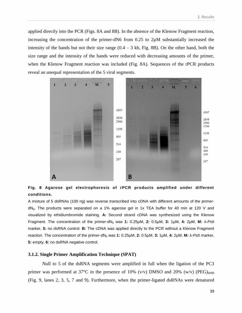
3. Results
39
applied directly into the PCR (Figs. 8A and 8B). In the absence of the Klenow Fragment reaction,
increasing the concentration of the primer-dN6 from 0.25 to 2µM substantially increased the
intensity of the bands but not their size range (0.4 – 3 kb, Fig. 8B). On the other hand, both the
size range and the intensity of the bands were reduced with decreasing amounts of the primer,
when the Klenow Fragment reaction was included (Fig. 8A). Sequences of the rPCR products
reveal an unequal representation of the 5 viral segments.
Fig. 8 Agarose gel electrophoresis of rPCR products amplified under different
conditions.
A mixture of 5 dsRNAs (100 ng) was reverse transcribed into cDNA with different amounts of the primer-
dN6. The products were separated on a 1% agarose gel in 1x TEA buffer for 40 min at 120 V and
visualized by ethidiumbromide staining. A: Second strand cDNA was synthesized using the Klenow
Fragment. The concentration of the primer-dN6 was 1: 0.25µM, 2: 0.5µM, 3: 1µM, 4: 2µM, M: λ-PstI
marker, 5: no dsRNA control. B: The cDNA was applied directly to the PCR without a Klenow Fragment
reaction. The concentration of the primer-dN6 was 1: 0.25µM, 2: 0.5µM, 3: 1µM, 4: 2µM, M: λ-PstI marker,
5: empty, 6: no dsRNA negative control.
3.1.2. Single Primer Amplification Technique (SPAT)
Null to 5 of the dsRNA segments were amplified in full when the ligation of the PC3
primer was performed at 37°C in the presence of 10% (v/v) DMSO and 20% (w/v) (PEG)6000
(Fig. 9, lanes 2, 3, 5, 7 and 9). Furthermore, when the primer-ligated dsRNAs were denatured

3. Results
40
with DMSO alone, full-length cDNAs were not obtained as was reflected in the PCR results (data
not shown). The type of the DNA polymerase employed in the amplification played a key role in
the number of full-length products obtained (Fig 9, lanes 2, 3, 5, 7 and 9). For each DNA
polymerase, the number of amplified segments is presented in Table 2. When the primer ligation
reaction was carried out at 16°C in the absence of DMSO and (PEG)6000, several truncated PCR
products with sizes less than 1 kb were obtained (data not shown).
Table 2. The efficiency of different DNA polymerases in the SPAT and the FLAC methods.
DNA polymerase Producer SPAT1 FLAC
1
Platinium DNA polymerase Invitrogen 0 0
Taq Polymerase 5 PRIME 2-3 4-5
GoTaq Promega 4-5 1-2
Phusion® High-Fidelity DNA
Polymerase
Finnzyme 2-4 5
Long PCR Enzyme mix Fermentas 0 1-2
1The values represent the numbers of full-length PCR products obtained.
Fig. 9 Electrophoresis pattern of RT-PCR products obtained by the Single Primer
Amplification Technique.
The ligation of the PC3 primer was performed at 37°C in the presence of 10% (v/v) DMSO and 20% (w/v)
(PEG)6000. Different DNA polymerases were used in the PCR: A: Finzyme, B: GoTaq, C: Platinium, D: 5
PRIME, E: Long PCR Enzyme Mix. Lanes 2, 3, 5, 7 and 9: amplified products. Lanes 1, 4, 6, 8 and 10:
negative control. M: λ-PstI marker. Each polymerase was tested 3 times.

3. Results
41
3.1.3. Full Length Amplification of cDNA (FLAC)
When the PC3-T7 loop primer was ligated to the mixture of the dsRNAs at 37°C in the
presence of 10% (v/v) DMSO and 20% (w/v) PEG6000 as described above, full-length amplicons
representing all or part of the segments were obtained depending on the DNA polymerase
employed (Fig. 10). The use of the Phusion® High-Fidelity DNA Polymerase and the supplied
Phusion GC Buffer resulted in the amplification of the 5 dsRNAs in one RT-PCR reaction (Fig.
10, lane 2). Similar results were obtained for the DNA polymerase supplied by 5 PRIME (Fig.
10, lane 7). Restriction digestion of the cloned full-length PCR products resulted in 5 different
profiles representing all of the segments (data not shown). Clones with different digestion
profiles were sequenced and analyzed. The 5 segments have conserved 5`and 3`terminal
sequences, a known property of most segmented viral genomes.
Fig. 10 Agarose gel electrophoresis of RT-PCR products obtained by the FLAC
method.
The ligation of the PC3-T7 loop primer was performed at 37°C in the presence of 10% (v/v) DMSO and
20% (w/v) (PEG)6000. Different DNA polymerases were used in the PCR: A: Finzyme, B: Platinium, C:
GoTaq, D: 5 PRIME, E: Long PCR Enzyme Mix. Lanes 2, 3, 5, 7 and 9: amplified products. Lanes 1, 4, 6,
8 and 10: negative control. M: λ-PstI marker.

3. Results
42
3.1.4. Direct cloning of dsRNA into DNA vector
The ligation of 200 ng of dsRNA mixture into 50 ng of the pJET1.2 vector produced as few as 5-9
transformants of which 2-4 had an insert longer than 150 bp regardless of the ligation conditions
employed. PCR screening of the transformants showed that the longest insert length obtained under the
conditions employed is 1 kb (Fig. 11).
When the dsRNA was ligated into pGEM®-T vector for 24 h about 7 transformants were obtained
3 of which contain an insert of 150 – 2100 bp in length. Restriction digestions of the clones with enzymes
flanking the insert (NdeI and SacII) produced variable profiles. While in some clones a complete release
of the insert was observed (Fig. 12, lanes 2, 5), in other clones the insert was not released indicating that
one of the restriction sites might be missing (Fig. 12, lane 3).
Although increasing the ligation time to 48 h resulted in improved transformation efficiency
(Table 3), the size range of the cloned fragments remains similar to that obtained after 24 h of ligation.
The identities of part of the clones were further checked by sequencing (data not shown). The calculated
transformation efficiencies for the direct cloning experiment are summarized in Table 3.
Fig. 12 Restriction digestion profiles of
pGEM®-T vectors cloned with dsRNA
segments.
The ligation reaction was carried out for 24 h. M: λ-
PstI marker, 1-7: clones digested with SacII and
NdeI.
Fig. 11 Agarose gel electrophoresis
of the PCR products obtained after
direct cloning of dsRNA into pJET1.2
vector.
Bacterial colonies were transformed with
pJET1.2 vector after ligation with dsRNA. The
inserts were amplified with primers flanking the
cloning site. M: λ-PstI marker, 1-13: each
represents a single colony.

3. Results
43
Table 3. The transformation efficiency of dsRNA-ligated pJET1.2 and pGEM®-T vectors.
Cloning vector Transformation efficiencya & insert size
Ligation time 24 h Ligation time 48 h
pJET1.2 1.22e1
(up to 1 kp)
3.60e1
(up to 1 kp)
pGEM®-T 2.80e
1 (up to 2.1 kp) 1.60e
2 (up to 2.2 kp)
a Calculated according to http://www.sciencegateway.org/tools/transform.htm.
The results represent the average of 3 independent experiments.
3.2. Identification of the fungus isolate
The fungus isolate China 9 showed morphological properties similar to those described
for F. graminearum including the size, shape, and septation of conidia, as well as the colony
color and morphology in agar (Data not shown). To support these results, molecular identification
was carried out based on sequence analysis of the ribosomal RNA genes (rDNA) which are
commonly used in identification and taxonomic studies at the species level (Ninet. et al., 2005).
Multiple alignment and phylogenetic results (Fig. 13) revealed China 9 isolate to be closely
related to the F. graminearum species complex.
Fig. 13 Phylogenetic identification of China9 fungal isolate based on the 28S rDNA
gene.
The fungal sequences were obtained from the NCBI gene bank database. Multiple-sequence alignment
was performed using CLUSTALX (2.0). The tree was constructed based on the NJ method with 1000
bootstrap replicates. Bootstrap values of 50% or more are shown, F. verticillioides served as an out-group.

3. Results
44
3.3. Molecular characterization of FgV-ch9
In this part of the study, I present the results on the molecular characterization of a novel
mycovirus from F. graminearum China 9 isolate (FgV-ch9). This includes determination of the
number and nature of the virus genomic segments, purification of the virus particles, the
complete nucleotide sequence of the virus, the encapsidation pattern of the virus segments,
production of polyclonal antibodies for the virus, sequence analysis of the virus genome and
phylogenetic investigations.
3.3.1. Virus purification and dsRNA isolation
Isometric VLPs of 35-40 nm in diameter were purified from the mycelium of F.
graminearum China 9 isolate (Fig. 14) and thereafter named tentatively FgV-ch9. The buoyant
density of the virions in cesium chloride is in the range 1.42-1.45 g/cm3.
Fig. 14 Virus-like particles of FgV-ch9.
The particles were purified from China 9 isolate by CsCl-gradient ultracentrifugation, negatively stained
with 2% (w/v) uranyl acetate and observed under transmission electron microscope.
Depending on the virus titer in the fungus and the electrophoresis conditions, the number
of the visibly detectable dsRNA segments ranged from 2 to 5 (Fig. 15). However, Northern Blot
analysis (Fig. 16) and nucleotide sequence analysis showed that always five unique segments are
associated with the purified VLPs.

3. Results
45
Several purifications of the viral dsRNA from the mycelium by means of cellulose
chromatography followed by S1 nuclease and DNase I digestions showed that the distribution of
the segments in the virus population is not equal giving that 2-3 of the dsRNAs present below the
detection threshold by agarose electrophoresis. Indeed, it was always possible to observe 4-5
distinct dsRNA segments only from mycelium containing a high titer of the virus (Fig. 15 lane 2)
or from virions purified from such a mycelium (Fig. 15, compare lanes 3 & 4). Fungal cultures
with various titers of FgV-ch9 were obtained by single conidiation. Furthermore, the resolution of
the extracted dsRNA segments in 1.2% agarose gel electrophoresis into 4-5 distinct bands was
only possible by extending the separation time to 90 min or more at 110 V (Fig. 15 compare lanes
1 & 2 ). The sizes of the five dsRNA segments and of the encoded proteins are summarized in
Table 4.
Fig. 16 Northern Blot analysis of FgV-ch9 dsRNA segments.
Purified dsRNAs were resolved in 1% agarose gel, blotted onto Hybond-N+ nylon membrane, and
hybridized with Dig-labeled probes prepared by PCR amplification of cloned cDNAs. Lanes 1-5
correspond to dsRNAs1-5, respectively. Lane A: Purified dsRNAs separated on a 1.2% agarose gel and
stained with ethidium bromide.
Fig. 15 Agarose gel electrophoresis of
dsRNAs isolated from the fungus F.
graminearum China 9 or from purified VLPs
of FgV-ch9.
The dsRNAs were purified either from the mycelium
or from CsCl-purified VLPs by CF-11 chromatography
and standard phenol method, respectively. The
dsRNAs were treated with DNase I and S1 nuclease
and separated in 1.2% agarose gel (arrows). 1:
dsRNAs purified from mycelium with high titer of FgV-
ch9 and electrophoresed for 30-40 min. 2: as in 1 but
electrophoresed for 90 min. 3: dsRNAs extracted from
viral particles purified from mycelium with high titer of
FgV-ch9. 4: as in 3 but with lower virus titer. M: λ DNA
digested with PstI

3. Results
46
3.3.2. SDS-PAGE and peptide sequencing
Proteins from purified VLPs were resolved into three distinct protein bands (upper-faint
~130 kDa, middle-double ~ 70 kDa, and lower ~ 60 kDa) after electrophoresis in denaturing
polyacrylamide gel (Fig. 17a lane 1). Tryptic peptide sequences from the upper-faint and the
lower bands were identical to the deduced amino acid sequences of dsRNA1 and dsRNA2,
respectively (Fig. 19a and b, boxed). The irresolvable middle-double bands (Fig. 17a, lane 1)
have peptide sequences identical to the protein deduced from dsRNA3 (Fig 19c, boxed) and
therefore were considered as a single band. With the exception of the band representing dsRNA1,
the sizes of the other two bands were slightly different from the corresponding sizes calculated
from the amino acid sequence deduced from DNA clones (compare Fig 17 to Table 4).
3.3.3. Specificity of the produced polyclonal antibodies for FgV-ch9
Immunoblot experiment using serum of the FgV-ch9-immunized rabbit (4th
bleeding)
against SDS-PAGE resolved FgV-ch9 structural proteins resulted in the detection of 2 of the viral
proteins (RdRP, upper band and CP, lower double band) in the presence of a light background
(Fig. 18, lane 1). In the control where Null serum was used, no signals were detected (Fig. 18,
lane 2).
Fig. 17 SDS-PAGE and Western Blot
analysis of FgV-ch9 structural proteins.
CsCl gradient-purified particles were denatured and
subjected to SDS-PAGE (a, 1). The surface proteins of
the purified VLPs were labeled with NHS-Biotin,
separated in SDS-PAGE, blotted, and detected with
the Fast Red reagent (b, 2). The preferential labeling
of the protein encoded by dsRNA3 (arrow), which
appears as a double band probably due to protein
degradation or modification, indicated that this protein
might be the outer capsid protein. M: Prestained protein
marker.

3. Results
47
.
3.3.4. Quantitative PCR
The various intensities of the 5 dsRNA-segments isolated from purified virions or from
the fungal mycelium as shown in agarose gel electrophoresis (Fig. 15) might be correlated to
unequal encapsidation and/or expression of the dsRNA segments. Relative quantitative PCR of
the five dsRNAs showed that they are encapsidated unequally. In relation to dsRNA4 (was set as
a reference because it has the lowest cross point value), dsRNAs1, 2, 3 and 5 isolated from
purified virions, were present at ratios of 0.63 : 0.27 : 0.78 : 0.87, respectively. A similar trend
was observed when the dsRNAs were purified from fungal host mycelia. Since the size of the
purified isometric particles does not allow the encapsidation of the 5 dsRNA segments in one
particle at the ratios determined by the real-time PCR calculations, I conclude that the dsRNAs
are encapsidated separately.
3.3.5. Nucleotide sequencing
The complete nucleotide sequences of the 5 dsRNA-segments were determined from at
least three full-length clones for each segment, originating from different virus purifications or
dsRNAs purified from infected mycelium. The sequences were submitted to the Genbank and the
accession numbers are shown in Table 4. Sequence analysis showed that each dsRNA segment is
monocistronic with a single open reading frame (OFR).
Fig. 18 Specificity of the FgV-ch9
polyclonal antibodies.
Null serum and serum of the 4th bleeding were
diluted 1000x and applied in a immunoblot for the
detection of FgV-ch9 structural proteins purified by
means of CsCl gradient ultracentrifugation. The
serum was purified by precipitating non-specific Abs
with healthy Fusarium extract. 1: serum of the 4th
bleeding, arrows indicate FgV-ch9 specific signals,
2: Null serum.

3. Results
48
Table 4: Size and function of the genomic dsRNA segments and the deduced proteins of FgV-ch9.
Segment (accession no.) Size (bp) Size of deduced
protein (kDa)
Function
dsRNA1 (HQ228213) 3581 127 640 (1137 aa) RDRP
dsRNA2 (HQ228214) 2850 94 872 (875 aa) Unknown
dsRNA3 (HQ228215) 2830 93 606 (856 aa) Putative capsid protein
dsRNA4 (HQ228216) 2746-3786* 91 303 (835 aa) Unknown
dsRNA5 (HQ228217) 2423 79 872 (712 aa) Contains a C2H2 zinc
finger domain
*Due to the presence of 3`terminal repeats, the size of dsRNA4 is variable.
3.3.5.1. dsRNA1
The complete nucleotide sequence of dsRNA1 revealed that it is 3581 bp and contains a
single large ORF from nt positions 83 to 3496 coding for 1137 aa (Table 4). The calculated
molecular weight of the encoded protein (127 kDa) is in agreement with the estimated size of the
corresponding band in the SDS-PAGE (Fig. 17a, lane1; upper-faint band). In addition, peptide
sequences of this band match perfectly with the deduced amino acid sequence of the ORF on
dsRNA1 as shown in (Fig.19a).
Since sequence analysis of the deduced protein encoded on dsRNA1 showed the
presence of the 8 conserved RdRP motifs characteristic for RNA viruses (Bruenn, 1993) shown
in (Fig. 20), I propose that RNA1 codes for the RdRP of the mycovirus.
This was further corroborated by BLAST searches of the deduced amino acid sequence
of dsRNA1 which revealed sequence similarities to several viral RdRPs (virus full names and
accession numbers are in Table 5). For example: to AsV1816 with 37% identity, 53% similarity,
and an E value of zero, MoCV1 with 33% identity, 50% similarity, and by AbV-1 with 29%
identity, 46% similarity. Sequence similarities (≤27% identity and ≤43% similarity, e-32
and
lesser) were also obtained for viral RdRPs belonging to the family Chrysoviridae. These include
CnV-1, Hv145SV, CCRS-CV, FuoxV1, PcV and others. Less significant similarities were
obtained for RdRPs belonging to the family Totiviridae (BLAST hits of e-09
or lesser).

3. Results
49
Fig. 19 Partial nucleotide
sequences of FgV-ch9 dsRNA1
(18a), dsRNA2 (18b), and dsRNA3
(18c).
The deduced amino acid sequences are
written in the one letter code under the
nucleotide sequences. The translation
start and termination codons are
indicated with an underline and an
asterisk, respectively. The tryptic
peptide-derived amino acid sequences,
isolated by reverse-phase HPLC, are
boxed.

3. Results
50
Fig. 20 Comparison of the conserved RdRPs motifs of several dsRNA mycoviruses
including FgV-ch9.
Numbers 1-8 stand for the eight conserved motifs typical of RdRPs of RNA viruses. Multiple sequence
alignments were carried out with the CLUSTALX (2.0). Dark shading and asterisks imply identical residues
at the indicated position while light gray shading and single dots signify columns with similar residues.
Numbers in parentheses relate to the number of amino acid residues between the motifs. The full names
of the viruses are shown in Table 5.
Interestingly, an identity of 100% was found to the partial RdRP aa sequence (73 aa)
available for Fusarium graminearum dsRNA mycovirus-2 (FgV2; No AAR17790). A phylogram
presenting/summarizing the data is shown in Fig. 21a. Although AsV1816 is assigned as an
unclassified member in the Totiviridae and the classification of AbV-1 as a chrysovirus is not
finalized Ghabrial and Suzuki, 2009, the results in Fig. 21 indicate that these two viruses together
with FgV-ch9 form a new cluster within the family Chrysoviridae.

3. Results
51
Table 5: List of the viruses used in constructing the phylograms in Fig. 21a,b.
Virus Acronyma Accession no. Family/
genusb RdRP cp
Ustilago maydis virus H1 UmV-HI NP_620728.1 NP_620728.1 TT
Saccharomyces cerevisiae virus L-A (L1) Sc V-L-A NP_620495.1 NP_620494.1 TT
Saccharomyces cerevisiae virus L-BC (La) ScV-L-BC NP_042581.1 NP_042580.1 TT
Gremmeniella abetina RNA virus L1 GaRV-L1 AAK11656.1 NP_624332.2 TV
Helminthosporium victoriae 190SV Hv190SV NP_619670.2 NP_619669.2 TV
Coniothyrium minitans mycovirus CmV YP_392467.1 YP_392466.1 TV
Epichloe festucae virus 1 EfV1 CAK02788.1 CAK02787.1 TV
Magnaporthe oryzae virus1 MoV1 YP_122352.1 YP_122351.1 TV
Magnaporthe oryzae virus 2 MoV2 NP_047560.1 YP_001649205.1 TV
Sphaeropsis sapinea RNA virus 1 SsRV1 NP_047558.1 NP_047557.1 TV
Sphaeropsis sapinea RNA virus 2 SsRV2 YP_001649206.1 NP_047559.1 TV
Botryotinia fuckeliana totivirus 1 BfTV1 YP_001109580.1 YP_001109579.1 TV
Atkinsonella hypoxylon virus AhV NP_604475.1 NP_604476.1 P
Aspergillus ochraceus virus 1 AoV1 ABV30675.1 ABV30676.1 P
Ceratocystis resinifera virus 1 CrV1 YP_001936016.1 YP_001936015.1 P
Discula destructiva virus 1 DdV-1 NP_116716.1 NP_116742.1 P
Discula destructiva virus 2 DdV-2 NP_620301.1 NP_620302.1 P
Fusarium poae virus 1 FUPO-1 NP_624349.1 NP_624348.2 P
Fusarium solani virus 1 FsV-1 NP_624350.1 NP_624351.1 P
Gremmeniella abietina RNA virus MS1 GaRV-MS1 NP_659027.1 NP_659028.1 P
Aspergillus mycovirus 1816 AsV1816 ABX79996.1 NAc uT
Heterobasidion annosum partitivirus HaV AAL79540.1 NAc P
Ophiostoma himal-ulmi partitivirus 1 OPV1 CAJ31886.1 CAJ31887.1 P
Penicillium stoloniferum virus S PsV-S YP_052856.2 YP_052857.1 P
Rhizoctonia solani virus 717 RsV-717 NP_620659.1 NP_620660.1 P
Penicillium chrysogenum virus PcV YP_392482.1 YP_392483.1 C
Helminthosporium victoriae 145S Hv145SV YP_052858.1 YP_052859.1 C
Agaricus bisporus virus 1 AbV-1 CAA64144.1 BAA01612.1 ?C
Cryphonectria nitschkei chrysovirus 1 CnV-1 ACT79255.1 ACT79251.1 C
Cherry chlorotic rusty spot associated
chrysovirus
CCRS-CV CAH03664.1 CAH03665.1 C
Amasya cherry disease associated
chrysovirus
ACD-CV YP_001531163.1 YP_001531162.1 C
Fusarium oxysporum chrysovirus 1 FuoxV1 ABQ53134.1 ABQ58816.1 C
Magnaporthe oryzae chrysovirus 1 MoCV1 YP_003858286.1 YP_003858288.1 uC a Provisional acronym
bTT: Totiviridae, genus Totivirus; TV: Totiviridae, genus Victorivirus; P: Partitiviridae; C:
Chrysoviridae; ?: obscure; uT: unclassified. cNA: not available.

3. Results
52
Fig. 21 Phylograms of the RdRP (a) and the CP (b) of FgV-ch9.
Multiple-sequence alignments of the deduced amino acid sequences of FgV-ch9 and selected
chrysoviruses, totiviruses, and partitiviruses were done using CLUSTALX (2.0) with default parameters.
The trees were analyzed by the NJ method with 100 bootstrap replicates. The numbers near to the
branches refer to the percentage of bootstrap replicates. Only bootstrap percentages over 50 are shown.
The trees were outgroup-rooted to the Totivirus UmV-HI. The full names of the viruses are shown in Table
5.
3.3.5.2. dsRNAs 2 and 4
Sequence analysis of the DNA clones of FgV-ch9 dsRNA2 and 4 showed that they are
2850 and 2746 bp, respectively. Each segment has a single ORF with deduced proteins of
molecular weights of 94 and 91 kDa for dsRNA2 and 4, respectively. From SDS-PAGE, the
estimated molecular mass of the protein encoded by dsRNA2 is ~62 kDa (Fig.17, lane 1, lower
band). Interestingly, the last 146-208 nts of the 3`-end of dsRNA4 were present in 6 out of 8
DNA clones as multiple tandem repeats. In the presence of these tandem repeats, various lengths
of dsRNA4 were measured for the different clones (2746-3786 bp). We have further confirmed
the presence of such tandem repeats in dsRNA4 by a PCR-based approach to exclude the

3. Results
53
possibility of cloning and sequencing artifacts. Using a forward primer (F1, 5`-
GCAGCTACACCAGTTAACAG-3`) that anneals outside the repeat region and a reverse primer
(R1, 5`-CGGATAAAACGTCAGTGGCAT-3`) complementary to part of the repeat sequence,
PCR amplification resulted in several amplicons with size differences corresponding to ~200
nucleotides (Fig.22, Lanes 1-3). In a control treatment, the F1 primer and a reverse primer (R2,
5`-AAAGTGCCGATTCTATACATGG-3`), designed to anneal outside the repeat region, were
employed (Fig.22, lane 5).
Purified dsRNAs from independent mycelia cultures or VLPs were employed as templates
in the described PCRs. BLAST searches of the deduced aa sequences of dsRNA2 resulted in
29% identities and 50% similarity (E value of 1e-49
) with the hypothetical protein encoded by
dsRNA3 of MoCV1. The protein encoded by dsRNA4 showed no significant similarities to any
protein in the GenBank and EMBL databases.
3.3.5.3. dsRNA3
Sequence analysis of full-length DNA clones of dsRNA3 showed that it consists of 2830
bp coding for one large ORF of 856 aa and a calculated molecular mass of 93 kDa. The estimated
size of the protein in SDS-PAGE is ~70 kDa (Fig. 17a, lane 1; middle-double band). Surface
protein labeling of purified virions with NHS-Biotin showed a labeling preference for the middle-
Fig. 22 Verification of the
terminal repeats at the 3`
terminus of dsRNA4 of FgV-ch9.
PCR products (brace) of dsRNAs
templates extracted from purified VLPs
(lane 1) or purified from independent
mycelia cultures (lanes 2 and 3) and
using a forward primer (F1) outside the
repeated region and a reverse primer
that anneal in the repeated region (R1).
4: PCR negative control (water), 5: PCR
product (arrow) using F1 primer and a
reverse primer (R2) that anneal outside
the repeated region, M: λ DNA digested
with PstI.

3. Results
54
double band (Fig. 17b, Lane 2; arrow). Peptide sequences of this band match perfectly with the
deduced aa encoded on dsRNA3 (Fig. 19b). BLAST searches showed significant sequence
similarity of the deduced aa of dsRNA3 to the L3 protein (CP) of AbV-1 (22% identity and 39%
similarity at a BLAST match of 5e-13
). One other significant match to the hypothetical protein
encoded by segment 4 of MoCV1 was observed (23% identity, 39% similarity, and an E value of
1e-13
). These results indicate that dsRNA3 codes most likely for the capsid protein.
Phylogenetic analysis of the capsid protein encoded by dsRNA3 showed that although
FgV-ch9, AbV-1, and MoCV1 cluster as a distinct clade, they are more related to members of the
Chrysoviridae than to those of the other families (Fig. 21b). Unfortunately, the capsid protein of
AsV1816 is not identified yet.
3.3.5.4. dsRNA5
This segment has a length of 2424 bp with a single ORF (nt 97 to 2139) coding for a
protein of 712 aa with calculated molecular mass of 79 kDa. BLAST searches of the deduced
amino acid sequence of dsRNA5 showed that while the N-terminus of the protein encoded by
dsRNA5 has no significant similarity to any published protein, the C-terminus of the protein (aa
positions 326 to 711) shares high similarities to zinc finger proteins (BLAST matches of 2e-13
).
Using the online software developed by Persikov et al., we have identified 12 fingers in this
region. Multiple alignment of the 12 fingers showed that all of them share the typical
characteristics of C2H2 zinc fingers (Fig. 23). Such fingers are composed usually of 2-3 strands
in their N-terminus and one α helix in the C-terminus. The specific binding affinity of zinc
fingers to nucleic acids or proteins is conferred by some of the amino acid residues present in
their α helix.

3. Results
55
Fig. 23 Multiple-sequence alignment of the 12 C2H2 zinc finger domain present at the
C-terminus of the protein encoded by dsRNA5 of FgV-ch9.
C, H, and number above the aligned sequences refer to the residues, Cys and His, and residue number in
the alpha helix, respectively. Bold indicates conserved amino acid residues. Multiple sequence alignment
was done using CLUSTALX with some manual adjustments.
3.3.5.5. The 5`and 3`UTRs
The lengths of the 5`UTRs of the different FgV-ch9 dsRNA segments lay between 78 and
105 nt. Multiple alignment of the 5`UTRs of the five dsRNA segments showed regions of high
sequence similarities (Fig. 24a). Stretches of identical sequences of 10 and 19 nt were found at
positions (in regard to the consensus line) 1 to 10 and from 23 to 42, respectively. Sixty-one of
the 5`-terminal 72 nts are identical in at least three of the five dsRNAs. Sequence similarities in
the 5`UTR dramatically decreases in the region located directly upstream of the ATG initiator
codon (specified in bold, Fig. 24a). The average GC content of the 5`UTRs of the five dsRNAs is
41% indicating a TA rich area. The CAA repeats, characteristics of the 5`UTR of chrysoviruses
(Jiang and Ghabrial, 2004) were observed in FgV-ch9, with a lesser frequency though. Whereas
in PcV and CCRS-CV the CAA sequence is repeated 7-14 times per dsRNA, in FgV-ch9 they
occur 2-6 times only (Fig 24a, in italics). In the 5`UTRs of tobamoviruses similar repeats were
described to function as translational enhancer elements (Gallie and Walbot, 1992). The 3`UTRs
of FgV-ch9 are relatively long, between 85 and 188 nt in length (Fig. 24b). In addition to the 6 nt

3. Results
56
at 3`- terminal which are identical among the 5 dsRNAs, other regions with high similarity are
distributed along the last 80 nt of the 3`UTR of the 5 dsRNAs (Fig. 24b).
Fig. 24 Comparison of the 5` (a) and 3`(b) UTRs of the 5 dsRNA segments of FgV-ch9.
Multiple-sequence alignments were done with CLUSTAL X. Gray shading and asterisks specify columns
with identical bases while dots indicate that three or four out of the five bases are identical in that column.
The (CAA) repeats in (a) are underlined and the start codons are shown in bold.
A summary of the genomic organization of FgV-ch9 is illustrated in Fig. 25.

3. Results
57
Fig. 25 A diagrammatic representation of the genomic structure of FgV-ch9.
Colored rectangles represent the ORF of each segment. P2 & P4 are proteins with unidentified function.
RdRP: RNA dependent RNA Polymerase. CP: capsid protein. Conserved terminal motifs are indicated
with blue boxes at the 5`and 3`UTRs. The numbers represent (from left to right) the first nucleotide, the
first nucleotide of the ORF, the last nucleotide in the ORF, and the length of the segment, respectively.
3.6. Association of the virus with hypovirulence-traits of F. graminearum China 9.
This part of the results presents several hypovirulent features of F. graminearum China 9 in
comparison to the WT isolate PH-1. These features are presumably due to the association of
China 9 isolate with various titers of FgV-ch9. Evidences supporting this assumption are
presented in the following sections of the results.
Cultures of F. graminearum China 9 originating from a conidium were found to be associated
with different amounts of FgV-ch9 dsRNAs. These cultures were classified into three major
categories (high, moderate, and low) depending on the amount of the viral dsRNA per gram of
fresh mycelia. Mycelia of the high and moderate categories contained 50-150 and 8-30 µg viral

3. Results
58
dsRNA/g fresh weight mycelia, respectively. The amount of viral dsRNA in mycelia with low
virus titers was not visible in agarose gels but was still detectable by means of RT-PCR. In
correlation to the amount of the viral dsRNA (virus titer), the three culture types of China 9
exhibited also differences in:
cellular ultrastructure
growth rate
colony morphology
conidiation capacity, and
virulence on wheat and maize plants.
These differences are explained in details in the following sections.
3.6.1. Ultrastructural properties of China 9 isolate.
Ultrastructural examination of F. graminearum China 9 showed the presence of VLPs in
membrane-bound vesicles, in vacuole-like structures, and in the abnormal looking cytoplasm
(Figs. 26 A, 27 A-D, and 28 A and B). Severe cellular degeneration and regions with decreased
electron density in the cytoplasm (Fig. 27, A), the presence of many large vacuole-like structures
(Fig. 26, A), accumulation of membranous structures or vesicles (Fig.27, B), and disintegration of
mitochondrial and nuclear membranes (Fig.27, B and C, respectively) are the major abnormal
features observed for F. graminearum China 9 with high virus titer and severely restricted
growth.
Although most of the examined cells of China 9 with moderate virus titer have a normal
nucleus and abundance of ribosomes, many of them have partially disorganized cytoplasm (Fig.
26, B). On the other hand, the majority of the cells of China 9 that have low virus titer appeared
normal with organized cytoplasm of usual electron density (Fig. 26, C). To compare the cell
structure of China 9 isolate to another F. graminearum isolate, the cells of the WT PH-1 were
also examined (Fig. 26, D). The use of polyclonal antibodies to allocate FgV-ch9 in the fungal
cells showed that virus is scattered in the cytoplasm and the vacuole-like structures (Fig. 28, A
and B).

3. Results
59
Fig. 26 Transmission electron micrographs of F. graminearum China 9 associated with different titers of FgV-ch9.
A: China 9 with high virus titers possesses abnormal cytoplasm with many large vacuoles that lack electron density.
B: China 9 with moderate virus titer showing normal nucleus (long arrow), abundance of ribosomes, but also a disorganized part of the cytoplasm
(short arrow).
C: China 9 with low virus titer has normal cytoplasm and nucleus (arrow).
D: Wild type strain PH-1.
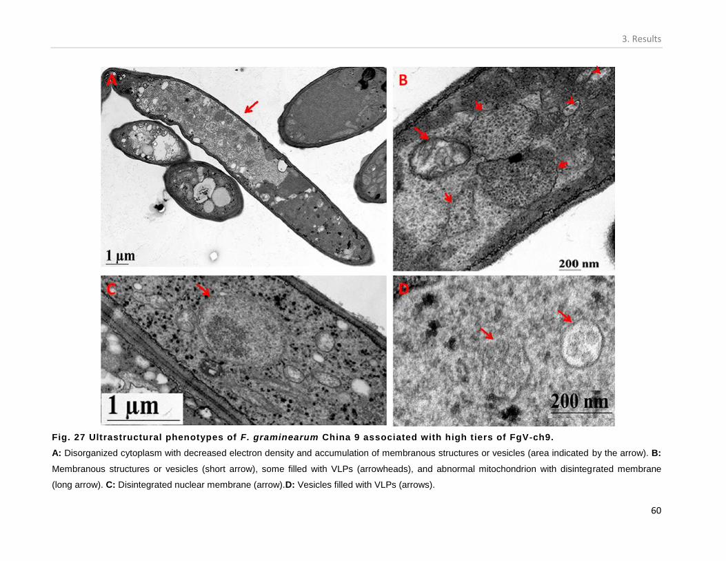
3. Results
60
Fig. 27 Ultrastructural phenotypes of F. graminearum China 9 associated with high tiers of FgV-ch9.
A: Disorganized cytoplasm with decreased electron density and accumulation of membranous structures or vesicles (area indicated by the arrow). B:
Membranous structures or vesicles (short arrow), some filled with VLPs (arrowheads), and abnormal mitochondrion with disintegrated membrane
(long arrow). C: Disintegrated nuclear membrane (arrow).D: Vesicles filled with VLPs (arrows).

3. Results
61
Fig. 28 Immuno-detection of VLPs in China 9 cells associated with high virus titer.
A: The VLPs of FgV-ch9 were mainly detected within the vacuole-like structures and scattered in the cytoplasm. Gold particles conjugated to the
second antibody appear as black dots.
B: A high magnification micrograph of a section of the vacuole with gold particles (arrows).

3. Results
62
3.6.2. Effect of virus titer on the growth rate of F. graminearum China 9.
The growth rate of F. graminearum China 9 on CM was significantly reduced in cultures
associated with high and moderated virus titers (Figs. 29 and 30). The colony area of 6 days-old
cultures associated with high or moderate virus titers were 23.6 and 60.6 cm2, respectively. At
low virus titer, the mycelia of 4 days-old cultures of F. graminearum China 9 cover the whole
Petri-dishes (81 cm2).
Fig. 29: Effect of FgV-ch9 titer on the growth rate of F. graminearum China 9 isolate.
The fungus was grown on solid CM medium at 25°C in the dark. The colony area was measured 4 and 6
days after inoculation. Values are the average area of 6 colonies from 2 independent experiments.
Different letters indicate statistically significant differences.
3.6.3. Colony morphology of F. graminearum China 9 isolate
Besides the restricted growth rate in cultures associated with high virus titers, such
cultures also exhibited abnormal growth phenotypes including dark pigmentation and altered
(uneven) colony morphology with the frequent formation of ear-like structures at the colony
margin (Fig. 30).

3. Results
63
Fig. 30. Growth rate and colony morphology of F. graminearum China 9 cultures
associated with different virus titers.
1: China + low virus titer, 2: China 9 + moderate virus titer, 3: China 9 + high virus titer. The cultures were
photographed after 4 days of growth on CM medium at 25°C in the dark.
3.6.4. Effect of virus titer on the conidiation of F. graminearum China 9.
The capacity of F. graminearum China 9 to produce conidia was significantly reduced in
cultures associated with high to moderate titers of viral dsRNAs. In correlation to the culture with
low virus titers, the production of conidia per plate SNA medium in cultures with high and
moderate virus titers was reduced to 16% and 60%, respectively (Fig.30). As a trend, the capacity
for conidiation in all of the F. graminearum China 9 cultures is relatively lower than that of the
F. graminearum PH-1 isolate (0.3-2.4 million in China 9 in comparison to 7-10 million conidia
per plate in PH-1).
The reduced production of conidia was also associated with slower growth rate of the
fungal mycelium on the SNA medium. For example, the area of one-week-old fungal colonies
with high virus titer was less than half the area of those associated with moderate virus titers (Fig.
32).

3. Results
64
Fig. 31: Production of conidia in F. graminearum China 9 in correlation with the titer
of FgV-ch9.
Conidiation was initiated in SNA medium at 18°C for 2 weeks. The results are the average of 5
independent experiments. Different letters indicates statistically significant differences.
3.6.4.1. Virus transmission through conidia
The transmission of FgV-ch9 through conidia was determined by means of RT-PCR using
RdRP specific primers. Total nucleic acids from mycelia originating from single conidia cultures
served as template in RT-PCR. Transmission of FgV-ch9 through conidia is dependent on the
titer of the virus in the parent fungal inocula grown on SNA medium. When inocula with high
Fig. 32 Differences in the colony
growth rate of mycelia associated
with moderate and high virus titer
on SNA medium.
1: China 9 + moderate virus titer, 2: China 9
+ high virus titer. The cultures were
photographed 7 days after inoculation.

3. Results
65
and moderate virus titers were used for conidiation, the virus transmitted into 100% of the
emerging conidia (Fig. 33, A, B). The percent of virus-containing conidia produced from inocula
of low virus titer was 20- 45 (Fig. 33, E, F). The use of inocula containing variable undefined
virus titers for conidiation resulted in a virus transmission of 85-100% (Fig. 33, C & D).
Fig. 33 RT-PCR products of the detection of FgV-ch9 in single conidia originating
cultures of F. graminearum China 9 isolate.
The presence of the virus in conidia was tested by means of RT-PCR using primer specific to dsRNA1
(RdRP) of the virus. The conidia were produced on SNA using initial fungal inocula harboring titers of FgV-
ch9 that are A: high, B: moderate, C and D: variable undefined, E and F: low. The expected sizes of the
PCR products are indicated by arrows. L: λ DNA digested with PstI, +: PCR positive control (a viral
plasmid clone serve as a template in the PCR, -: PCR negative control (water).
3.6.5. Effect of virus titer on the pathogenicity of F. graminearum China 9 for wheat plants.
In addition to the reduced growth rate and conidiation of F. graminearum China 9
cultures associated with high and moderate titers of FgV-ch9, the virus is probably also
associated with hypovirulence of its fungal host. Infection of wheat spikes with conidia
originated from cultures associated with high and moderate virus titers resulted in typical
Fusarium-blight symptoms that are restricted to the inoculated spikelet (10% of total kernels).
However, the symptoms were visible in about 27% of kernels when conidia with low virus titers

3. Results
66
were inoculated (Figs. 34 and 35). Inoculations with the WT PH-1 conidia and water were
included as positive and negative controls, respectively.
Fig. 34. Reduced pathogenicity on wheat plants of F. graminearum China 9 isolate is
associated with the presence of FgV-ch9.
Results are the percent of infected kernels of 30 spikes per treatment except for PH-1 and water controls
(8 and 6 spikes, respectively). The results were collected 3 weeks after inoculation. Different letters
indicate statistically significant differences.

3. Results
67
Fig. 35 Effect of the association of F. graminearum China 9 with different titers of
FgV-ch9 on its pathogenicity on wheat.
Two central spikelets of wheat spikes were each inoculated with 500 conidia of the WT PH-1 (d) or China 9 harboring a: high virus titer, b: moderate virus titer, c: low virus titer. The infection was monitored for 3 weeks post inoculation. Arrowheads indicate inoculation sites.

3. Results
68
3.6.6. Effect of virus titer on the pathogenicity of F. graminearum China 9 on maize plants.
Less than 6% of the maize kernels developed symptoms 5 weeks after of inoculation with
F. graminearum China 9 cultures with high and moderate virus titers (Fig. 36). On the other
hand, infection of maize cobs with conidia harboring low virus titers increase the percentage of
infected kernels to 28% (Figs. 36 and 37). While cobs inoculated with water, (negative control)
showed no disease symptoms, those infected with the WT PH-1 produced kernels with severe
symptoms (Figs. 36 and 37).
Fig. 36 Association of FgV-ch9 of hypovirulence of China 9 fungal isolate on maize.
Results are the percentage of infected kernels of 10 maize cobs per treatment. Each maize cob was
inoculated with 4 ml of conidia suspension with a concentration of 40 conidia per µl or with purified water
(control). Different letters indicate statistically significant differences.

3. Results
69
Fig. 37 Maize cobs inoculated with F. graminearum China 9 associated with different
titers of FgV-ch9.
a: China 9 with low virus titer, b: China 9 with high and moderate virus titer, c: WT PH-1, d: water
(negative control). The cobs were injected with the fungal conidia at the stage of early kernel formation. In
each treatment, 10 cobs were injected. Cobs were Photographed 5 weeks post infection.
3.7. Transfection of F. graminearum PH-1 with particles of FgV-ch9.
In order to further proof that the hypovirulent features of China 9 isolate are due to its
association with FgV-ch9, several attempts to cure the fungus completely from the virus were
carried out. Unfortunately, all of these attempts failed. Alternatively, purified VLPs of FgV-ch9
were transfected successfully into the protoplasts of the WT isolate PH-1. Characterizations of
the transfected cultures for possible hypovirulent traits are presented subsequent sections below.
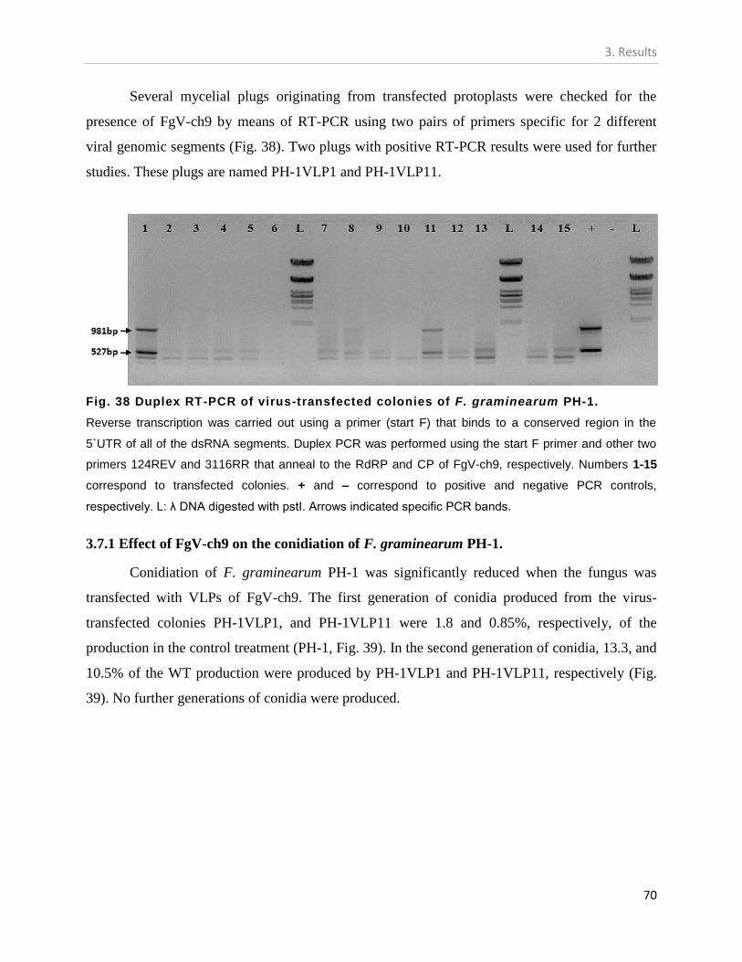
3. Results
70
Several mycelial plugs originating from transfected protoplasts were checked for the
presence of FgV-ch9 by means of RT-PCR using two pairs of primers specific for 2 different
viral genomic segments (Fig. 38). Two plugs with positive RT-PCR results were used for further
studies. These plugs are named PH-1VLP1 and PH-1VLP11.
Fig. 38 Duplex RT-PCR of virus-transfected colonies of F. graminearum PH-1.
Reverse transcription was carried out using a primer (start F) that binds to a conserved region in the
5`UTR of all of the dsRNA segments. Duplex PCR was performed using the start F primer and other two
primers 124REV and 3116RR that anneal to the RdRP and CP of FgV-ch9, respectively. Numbers 1-15
correspond to transfected colonies. + and – correspond to positive and negative PCR controls,
respectively. L: λ DNA digested with pstI. Arrows indicated specific PCR bands.
3.7.1 Effect of FgV-ch9 on the conidiation of F. graminearum PH-1.
Conidiation of F. graminearum PH-1 was significantly reduced when the fungus was
transfected with VLPs of FgV-ch9. The first generation of conidia produced from the virus-
transfected colonies PH-1VLP1, and PH-1VLP11 were 1.8 and 0.85%, respectively, of the
production in the control treatment (PH-1, Fig. 39). In the second generation of conidia, 13.3, and
10.5% of the WT production were produced by PH-1VLP1 and PH-1VLP11, respectively (Fig.
39). No further generations of conidia were produced.

3. Results
71
Fig. 39: Transfection of F. graminearum PH-1 with FgV-ch9 negatively affects its
conidiation capacity.
The conidiation capacities of the strains are presented as percentage in relation to that of the WT PH-1.
The results are shown for the first and the second generations of conidia produced from an FgV-ch9-
transfected parent protoplast. Conidiation was initiated in SNA medium at 18°C for 2 weeks. The results
are the average of 4 independent experiments. Different letters indicate statistically significant differences.
3.7.2 Effect of FgV-ch9 on perithecia development of F. graminearum PH-1.
Three F. graminearum PH-1 cultures originating from RT-PCR positive virus-transfected
colonies were tested for their capacities to produce perithecia. For each culture, the number of
perithecia is the average of perithecia counts of three plates. The capacity of the three virus-
transfected cultures to produce perithecia was significantly reduced in comparison to the WT PH-
1. The average number of produced perithecia in 9 plates representing 3 independent experiments
was 45 in comparison to 220 in the WT PH-1 (Figs. 40 and 41). F. graminearum China 9 isolate
did not produce any perithecia (Fig. 41, C).

3. Results
72
Fig. 40. Perithecia development of F. graminearum PH-1 before and after transfection
with FgV-ch9.
Values are the average number of developed perithecia in 9 plates of carrot-agar medium representing 3
transfected-colonies. Produced Perithecia were counted under a stereoscope. Different letters indicated
statistically significant differences.
Fig. 41 Perithecia of F. graminearum PH-1 and China 9 isolates.
Perithecia were produced on carrot-agar medium and incubation at 25°C under a mixture of near UV and
white light with a 12 h photoperiod up to three weeks. A: PH-1 isolate transfected with VLPs of FgV-ch9,
B: WT PH-1, C: China 9 isolate.

3. Results
73
3.7.3. Pathogenicity of virus-transfected F. graminearum PH-1 on wheat plants.
When wheat plants were inoculated with the first generation of conidia produced from
PH-1VLP1 and PH-1VLP11, the percent of symptomatic kernels were reduced in comparison
with untranfected WT PH-1. While the reduction in diseased kernels reaches 50% in case of PH-
1VLP1, it was less than 20% for PH-1VLP11 (Figs. 42, 43, and 44). Symptoms developed due to
infections with the second generation of conidia were comparable to those produced by the WT
(Figs. 42, 43, and 44).
Fig. 42: Effect of transfection of F. graminearum PH-1 with VLPs of FgV-ch9 on its pathogenicity
on wheat plants.
Results are the percent of infected kernels of 30-60 spikes per treatment except for the WT PH-1 (6
spikes). The pathogenicity was tested for two successive generations of conidia produced from two
transfected cells (PH-1VLP1 and PH-1VLP11). The results were collected 3 weeks post infection. Different
letters indicate statistically significant differences.
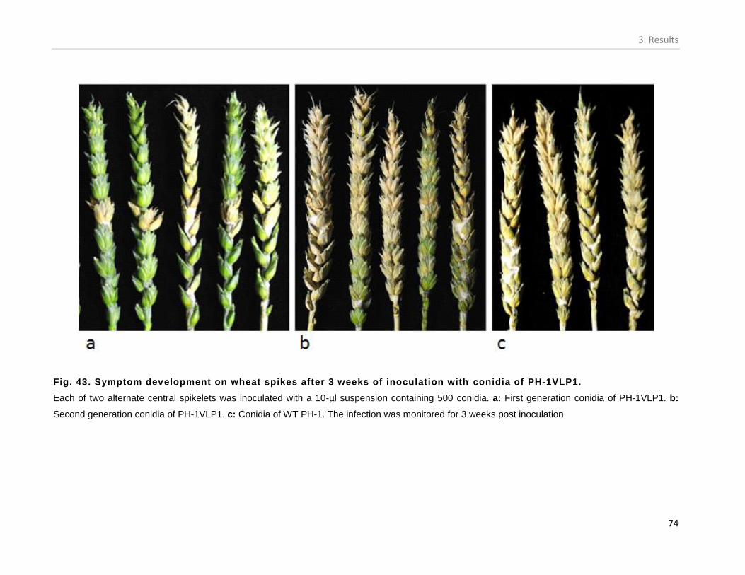
3. Results
74
Fig. 43. Symptom development on wheat spikes after 3 weeks of inoculation with conidia of PH-1VLP1.
Each of two alternate central spikelets was inoculated with a 10-µl suspension containing 500 conidia. a: First generation conidia of PH-1VLP1. b:
Second generation conidia of PH-1VLP1. c: Conidia of WT PH-1. The infection was monitored for 3 weeks post inoculation.

3. Results
75
Fig. 44. Symptom development on wheat spikes after 3 weeks of inoculation with conidia of PH-1VLP11.
Each of two alternate central spikelets was inoculated with a 10-µl suspension containing 500 conidia. a: First generation conidia of PH-1VLP11, b:
Second generation conidia of PH-1VLP11 and c: Conidia of the WT PH-1. The infection was monitored for 3 weeks post inoculation.

3. Results
76
3.7.4. Pathogenicity of virus-transfected F. graminearum PH-1 on maize plants.
Infection of maize with PH-1VLP1 and PH-1VLP11 conidia resulted in 68 and 82% of
diseased kernels, respectively (Fig. 45 and 45). In comparison to PH-1, these results are not
significantly different.
Fig. 46 Pathogenicity of F. graminearum PH-1 transfected with FgV-ch9 for maize.
Maize cobs were infected with conidia of PH-1 transfected with FgV-ch9. A: PH-1VLP1, B: PH-1VLP11,
C: WT PH-1. The cobs were injected with the fungal conidia at the stage of early kernel formation. In each
treatment, 8-10 cobs were injected. Cobs were Photographed 5 weeks post infection.
Fig. 45: Effect of transfection
of F. graminearum PH-1with
VLPs of FgV-ch9 on its
pathogenicity on maize plants.
Results are the percentage of infected
kernels of 8-10 maize cobs per
treatment. Each cob was inoculated
with 4 ml of conidia suspension with a
concentration of 40 conidia per µl.
Different letters indicate statistically
significant differences.

3. Results
77
3.8. Co-infection of maize with F. graminearum China9 and PH-1
Simultaneous introduction of plant pathogenic fungi to the host could either decrease or
increase the threshold of the disease. Following this concept, I have co-applied the virulent PH-1
and the hypovirulent China9 isolates into maize cobs to evaluate the outcome of such an
interaction on the disease development.
The results showed that simultaneous inoculation of maize cobs with conidia of F.
graminearum China 9 and PH-1 isolates significantly reduces the percentage of infected kernels
in comparison to infections with PH-1 alone (Figs. 47 and 48). While infection due to China 9 or
PH-1 resulted in 6 and 92% of diseased kernels, respectively, those resulting from a mixed
infection of both isolates were 29% (Figs. 47 and 48).
Fig. 47: Simultaneous inoculation of conidia of F. graminearum China 9 and PH-1
isolates into maize.
Results are the percent of infected kernels of 6-10 cobs per treatment. Each maize cob was inoculated
with 4 ml of conidia suspension with a concentration of 40 conidia per µl. In case of simultaneous
infection, 2 ml of conidia suspension from each isolate were mixed and injected into the maize cobs.
Different letters indicate statistically significant differences.

3. Results
78
Fig. 48 Co-infection of maize with F. graminearum China 9 and PH-1 isolates.
Maize cobs were inoculated with conidia of A: China 9, B: China 9 and PH-1, C: PH-1 and D: water
(negative control). In each treatment, 6-10 cobs were injected with the fungal conidia at the stage of early
kernel formation. Maize cobs were photographed 5 weeks after inoculation with the conidia.
3.9. Consequences of the over expression of FgV-ch9 putative genes in F. graminearum PH-
1.
In order to reveal the role of each of the genes encoded by the dsRNAs of FgV-ch9 in
hypovirulence, the genes were cloned into a plasmid vector under a constitutive fungal promoter
(gpdA) and transformed into protoplasts of the WT isolate PH-1. The effects of each
constitutively expressed gene on the virulence of PH-1 isolate are presented.
The transformation of F. graminearum PH-1 protoplasts with Pan 7.1 vector cloned with
the genes encoded by dsRNA2, 3, 4, or 5 resulted in several positive transformants checked by
PCR. In each case, the expression vigor of 5 positive transformants was determined by means of
semi quantitative PCR (Fig. 49). Colonies with the highest expression levels (Fig. 49, arrows)
were double-checked for successful integration of the transgene by means of Southern blot
analysis (Fig. 50). These colonies were used for conidiation as well as for pathogenicity assays on
wheat and maize plants.
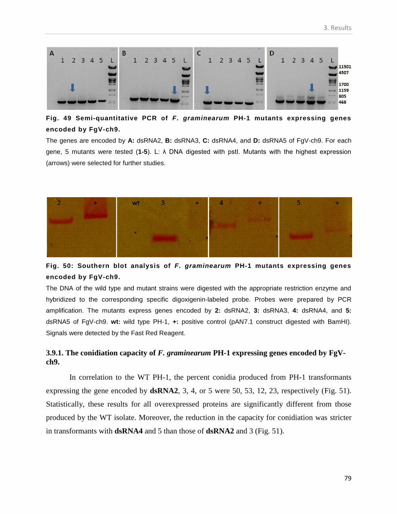
3. Results
79
Fig. 49 Semi-quantitative PCR of F. graminearum PH-1 mutants expressing genes
encoded by FgV-ch9.
The genes are encoded by A: dsRNA2, B: dsRNA3, C: dsRNA4, and D: dsRNA5 of FgV-ch9. For each
gene, 5 mutants were tested (1-5). L: λ DNA digested with pstI. Mutants with the highest expression
(arrows) were selected for further studies.
Fig. 50: Southern blot analysis of F. graminearum PH-1 mutants expressing genes
encoded by FgV-ch9.
The DNA of the wild type and mutant strains were digested with the appropriate restriction enzyme and
hybridized to the corresponding specific digoxigenin-labeled probe. Probes were prepared by PCR
amplification. The mutants express genes encoded by 2: dsRNA2, 3: dsRNA3, 4: dsRNA4, and 5:
dsRNA5 of FgV-ch9. wt: wild type PH-1, +: positive control (pAN7.1 construct digested with BamHI).
Signals were detected by the Fast Red Reagent.
3.9.1. The conidiation capacity of F. graminearum PH-1 expressing genes encoded by FgV-
ch9.
In correlation to the WT PH-1, the percent conidia produced from PH-1 transformants
expressing the gene encoded by dsRNA2, 3, 4, or 5 were 50, 53, 12, 23, respectively (Fig. 51).
Statistically, these results for all overexpressed proteins are significantly different from those
produced by the WT isolate. Moreover, the reduction in the capacity for conidiation was stricter
in transformants with dsRNA4 and 5 than those of dsRNA2 and 3 (Fig. 51).
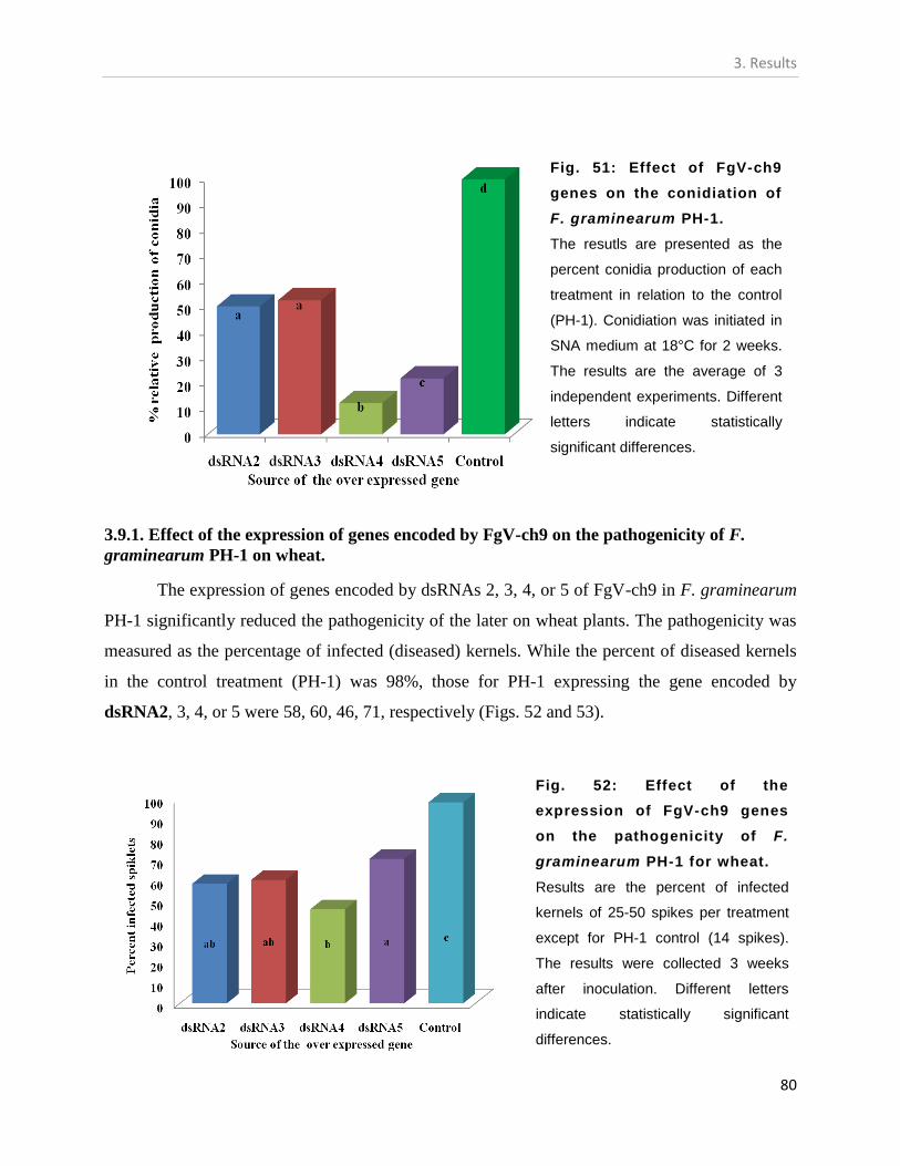
3. Results
80
3.9.1. Effect of the expression of genes encoded by FgV-ch9 on the pathogenicity of F.
graminearum PH-1 on wheat.
The expression of genes encoded by dsRNAs 2, 3, 4, or 5 of FgV-ch9 in F. graminearum
PH-1 significantly reduced the pathogenicity of the later on wheat plants. The pathogenicity was
measured as the percentage of infected (diseased) kernels. While the percent of diseased kernels
in the control treatment (PH-1) was 98%, those for PH-1 expressing the gene encoded by
dsRNA2, 3, 4, or 5 were 58, 60, 46, 71, respectively (Figs. 52 and 53).
Fig. 51: Effect of FgV-ch9
genes on the conidiation of
F. graminearum PH-1.
The resutls are presented as the
percent conidia production of each
treatment in relation to the control
(PH-1). Conidiation was initiated in
SNA medium at 18°C for 2 weeks.
The results are the average of 3
independent experiments. Different
letters indicate statistically
significant differences.
Fig. 52: Effect of the
expression of FgV-ch9 genes
on the pathogenicity of F.
graminearum PH-1 for wheat.
Results are the percent of infected
kernels of 25-50 spikes per treatment
except for PH-1 control (14 spikes).
The results were collected 3 weeks
after inoculation. Different letters
indicate statistically significant
differences.

3. Results
81
Fig. 53: The effect of the expression of FgV-ch9 genes on the virulence of F.
graminearum PH-1 for wheat.
Two central spikelets of wheat spikes were each inoculated with 500 conidia expressing the gene
encoded by a: dsRNA2, b: dsRNA3, c: dsRNA4, and d: dsRNA5 of FgV-ch9. e: water (negative control),
f: WT PH-1. The infection was monitored for 3 weeks post inoculation.

3. Results
82
3.9.1. Effect of the expression of genes encoded by FgV-ch9 on the pathogenicity of F.
graminearum PH-1 for maize.
The percentages of infected kernels in maize plants after 5-weeks of inoculation with F.
graminearum PH-1 expressing the gene encoded by dsRNAs 2, 3, 4, or 5 of FgV-ch9 were 54,
56, 62, 79, respectively (Figs. 54 and 55). Except for the data collected for dsRNA5, the above
results are significantly different from the percent of infected kernels in the WT treatment, PH-1,
(92%).
Fig. 54: Effect of the expression of FgV-ch9 genes on the pathogenicity of F.
graminearum PH-1 for maize.
Results are the percentage of infected kernels of 10-20 cobs in each treatment except for the PH-1control
(6 cobs). Each maize cob was inoculated with 4 ml of conidia suspension with a concentration of 40
conidia per µl. Different letters indicate statistically significant differences.

3. Results
83
Fig. 55: Symptom development on maize cobs after 5 weeks of inoculation with
conidia of F. graminearum PH-1 expressing genes of FgV-ch9.
The genes are encoded by a: dsRNA2, b: dsRNA3, c: dsRNA4, and d: dsRNA5 of FgV-ch9. e: water, f:
WT PH-1. The cobs were injected with the fungal conidia at the stage of early kernel formation. In each
treatment, 10-20 cobs were injected. Cobs were Photographed 5 weeks post infection.

3. Results
84
In conclusion, all of the PH-1 mutants that express one of the genes of FgV-ch9 are
hypovirulent in comparison to the WT PH-1 isolate. However, the threshold of the hypovirulence
seems to be dependent on the expressed gene and the nature of the host plant (wheat or maize).
3.10. Effect of disruption of Dicer 2 gene on F. graminearum PH-1 and China 9 isolates.
To check if the levels of FgV-ch9 dsRNAs are regulated by the fungal host through
quelling, I have knocked out the Dicer 2 gene of China 9 isolate. This experiment aimed also at
evaluating the outcomes of eliminating the Dicer 2 gene on the vigor of the fungus. As a control
experiment, the Dicer 2 gene of the WT PH-1 isolate was knocked out.
The results showed that the disruption of the Dicer 2 gene has no significant effect on the
growth rate or on the pathogenicity of either F. graminearum PH-1 or China 9 isolates on wheat
or maize plants. Furthermore, F. graminearum China 9 dicer 2-mutant cultures behave like WT
cultures with low virus titers in term of the mycelium growth rate and virus titer in the mycelium.

4. Discussion
85
4. Discussion
4.1. Optimization of the methods for the sequence determination of dsRNA templates.
The isolation of dsRNA from plant tissues has been established by Morris & Dodds
(1979) as a tool to generally detect virus infections. Thereafter, the method, which requires two
working days, has been frequently employed to purify dsRNA from plants and fungi as a source
for the subsequent molecular biology techniques such as RT-PCR, cloning and sequence
determination of viral genomes. I have optimized the method enabling better isolation of intact
dsRNA in one day. First, the use of Bentonite in the extraction buffer as an RNase inhibitor
helped to protect the dsRNA before and after the phenol treatment. Although dsRNA is more
resistant to RNase A than ssRNA, it could be partially or fully degraded by this enzyme at low
salt concentration. (Nosek et al., 1993). Also, we observed that a combination of acidic
phenol:chloroform:isoamyl alcohol at a ratio of 5:1:1 is optimal in term of the amount of dsRNA
recovered in the aqueous phase (Sambrook et al., 1989). Acidic phenol mixed with chloroform at
the 5:1 ratio probably prevents the loss of RNA in the interphase due to insoluble protein:RNA
complexes. Moreover, applying the phenol for 30 min at 37°C might substitute the incubation for
1 hour at room temperature. Finally, precipitation of the dsRNA in 1 volume of isopropanol for 1
hour at –80°C is equivalent in our hands to overnight precipitation with ethanol at -20°C.
Alternatively, we have precipitated the dsRNA with isopropanol by 3 times freeze-thawing in
liquid nitrogen.
The rPCR method has been established by Froussard (1992) as a tool to amplify whole
RNA sequences. Several publications appeared later reporting the use of the method for the
random amplification of viral dsRNA genomes (Marquez et al., 2007 & Willenborg et al., 2009).
I have tested the sensitivity of this method for amplifying a mixture of dsRNA templates and
optimized the conditions to produce distinct amplicons of considerable sizes. Such optimization
reduces the time and costs for cloning and sequencing of many clones. In the range of 0.1 – 100
ng of dsRNA, the optimum concentration of the universal primer-dN6 for achieving intense
products suitable for cloning lies between 1-2 µM. The use of the Klenow Fragment for the
second strand cDNA synthesis followed by the removal of excess universal primer-dN6 has
increased the number of smaller amplicons (Fig 2A). Using dsRNA as a template for reverse
transcription provides dscDNA for the PCR amplification in the first step without any need for
the Klenow Fragment reaction. In theory, the production of a second round of dscDNA would

4. Discussion
86
result in shorter DNA templates for the subsequent PCR amplification. Although Froussard
(1992) was able to amplify from 1 pg of ssRNA using the rPCR method, in our hands only
amounts equal to 100 pg or more of the dsRNA templates resulted in considerable amplification
products. However, we were still able to obtain few faint bands when 10 pg of dsRNA served as
a template for the rPCR. Using a mixture of dsRNAs of 2.5 - 3.6 kb has resulted in a maximum
rPCR product of about 3 kb under the conditions described in this study. Probably bigger sized
PCR products could be achieved when longer templates are used. These results indicate that the
rPCR is a powerful and sensitive method for the random amplification of dsRNA templates. The
uneven representation of the 5 dsRNA segments among the rPCR products as revealed from their
sequences, might be mainly due to their unequal abundance in the host.
The SPAT and FLAC methods constitute powerful means for the full-length amplification
of long dsRNA templates when performed under optimum conditions. First, incubating the
ligation reaction at 37°C in the presence of 20% PEG6000 and 10% DMSO highly improved the
ligation of the primer to the 3`ends of the dsRNA. T4 RNA ligase is in principle an ssRNA ligase
but was shown to ligate DNA oligos to dsRNA (Imai et al., 1983) with a lesser efficiency though
(Higgins et al. 1979). The ligation reaction can be inhibited by RNA secondary structures and can
be enhanced with the addition of 10% DMSO and also by PEG which act by macromolecule
crowding (Harrison and Zimmerman 1984). Probably, under the conditions described above,
partial denaturation of the dsRNA termini occurs, providing single stranded 3`ends to the ligation
enzyme. Second, denaturing the dsRNA with 1M betaine and 2.5% DMSO has resulted in full-
length cDNAs. This treatment replaces the use of the highly toxic chemical MMOH, which is
used frequently for such purposes (Potgieter et al., 2009). The employment of DMSO alone at
various concentrations for the denaturation of dsRNA was not sufficient for the production of
full-length cDNAs. Whereas low DMSO concentrations (up to 15%) might not be enough to
denature relatively long dsRNAs, the use of higher concentrations of DMSO might inhibit the
reverse transcriptase (Jucà and Aoyama, 1995). Third, the ligations of linkers, which loop back to
prime themselves, make the introduction of primers in the reverse transcription step superfluous.
This eliminates the chances for initiation of cDNA synthesis from places other than the ends and
reduces the production of truncated cDNAs. Fourth, the use of high annealing temperature in the
PCR prevents non-specific amplification. Although the calculated melting temperature (Tm) for
the primer PC2 is 54 - 56°C, going up to 67°C has not affected the specific annealing of the PC2
primer as revealed by the PCR results (Potgieter et al., 2009). Finally, we have compared 5

4. Discussion
87
commercial DNA polymerases, designed to amplify long DNA templates (4 – 20 kb), of which
only two have produced the complete set of full length amplifiable products with different
intensities, though. The other polymerases produced 0-3 full-length PCR products. We have no
clue to interpret these results since the 5 polymerases were applied under optimal conditions
using identical number of units, the same cDNA preparation, and the same thermocycler
machine.
Our attempts to clone viral genomic dsRNA into DNA vectors without prior transcription
and amplification steps showed that the principle is valid. Although under the conditions applied
in this study we achieved relatively long sequences (up to 2.2 kp), we did not obtain full length
clones. Surprisingly, the cloning of S1 nuclease-treated dsRNA into the pGEM®-T vector
resulted in more transformants and bigger inserts than those obtained for ligation into the blunt
end pJET1.2 vector.To my knowledge, there is no information in the literature explaining the
mechanism by which such direct ligation and transformation take place. Skotnicki et al., (1985)
claimed that it is possible to clone, in full or partially, dsRNAs from RF or replicative
intermediate (RI) stages of several ssRNA viruses such as tobacco mosaic virus (TMV) , alfalfa
mosaic virus (AMV) , Fiji disease virus (FDV), barley yellow dwarf virus (BYDV) and beet
western yellows virus (BWYV).
However, the patent does not clearly state when it was possible to obtain full length
clones, does not present data on the transformation efficiency, and to our knowledge was not
published in any referred journal so far. I think that this method should be further explored to
understand the molecular mechanisms behind it that might enable further optimizations and
eventually lead to cloning of full-length dsRNA segments.
In conclusion, the optimization described in this study resulted in reproducible tools for
the isolation, transcription and amplification of full length products from a mixture of dsRNA
templates without prior need for separation or for the use of noxious chemical and expensive or
time-consuming approaches. Furthermore, the implementation of the direct cloning of dsRNA
into DNA vectors as a laboratory protocol requires more molecular investigations and
evaluations. Such investigations may help to optimize the method for the cloning of full length
RNA molecules, enhance their transformation efficacy and thereby might improve virome
research.

4. Discussion
88
4.2. Molecular characterization of FgV-ch9
We have isolated 10 Fusarium isolates from China and screened them for the presence of
viral dsRNA. In one of the isolates, namely China 9, five dsRNA segments, associated with
isometric VLPs of 35-40 nm in diameter were identified. The China 9 fungus isolate was
phylogenetic-identified as F. graminearum using the method described by Ninet et al. (2005).
The novel virus was named tentatively F. graminearum mycovirus-China9 (FgV-ch9). The
purified dsRNAs were resolved into 2-5 bands following agarose gel electrophoresis depending
on the virus titer in the fungal mycelium and the electrophoresis conditions. At high virus titer
and relatively long electrophoresis times (~2 h), the dsRNAs could be resolved into 4-5 distinct
bands. At low virus titer however, dsRNA 2, 3, and in some cases dsRNA1, were below the
detection threshold of agarose gel electrophoresis. Additional reasons for the inconsistent
resolution of the dsRNAs in agarose gel electrophoresis might be attributed to the small size
differences between dsRNA 2, 3 and 4 (20-84bp), the unequal band intensities, and the
heterogeneous lengths associated with the 3`UTR tandem repeats of dsRNA 4 (discussed below).
Incomplete resolution in agarose gel was also reported for PcV and AsV1816 (Jiang and
Ghabrial, 2004; van Diepeningen et al., 2006). Our ability to detect the 5 dsRNAs segments in
the various single-conidia originated cultures or in the purified virions by means of RT-PCR and
northern hybridisation (data not shown) indicates that FgV-ch9 particles encapsidate 5 genomic
segments.
The complete nucleotide sequence of the five dsRNA-segments was determined. Each of
the segments was found to have a single unique ORF. BLAST searches with the deduced aa
sequences showed that dsRNA1 encodes a putative RdRP, closely related to those of dsRNA
mycoviruses. The function of the proteins encoded by dsRNA2 and dsRNA4 are not known yet
since that encoded by dsRNA2 is only similar to the hypothetical protein encoded by MoCV1,
while that of dsRNA4 has no significant similarity to any published protein. The identification of
the protein encoded by dsRNA2 in SDS-PAGE might indicate that this protein is associated with
virions. Interestingly, our analysis of 8 independent DNA clones of dsRNA4 showed that the 3`-
terminus of this segment contains tandem sequence repeats of 146-208 nt in 6 of the clones.
Although the dominant pattern of the sequence repeats was (146nt)n, in two of the analyzed
clones the repeated sequence was (208nt)n where n = 2-5. The significance of such a 3`terminal
repeat, which to my knowledge has not been reported for another dsRNA mycovirus, is unclear.

4. Discussion
89
Evidence that dsRNA3 encodes the putative CP stems for two sources. First, in vitro
surface protein labeling of CsCl gradient-purified virions followed by SDS-PAGE and
immunoblot analysis showed preferential labeling of the protein band encoded by dsRNA3.
Tryptic peptide sequences of this protein band were identical to those deduced from the DNA
clones of dsRNA3. Second, BLAST searches showed relatively high sequence similarities
between the protein encoded by dsRNA3 and the L3 protein of the AbV-1 associated with La
France disease. The L3 protein was reported by Kuang et al. (2004) to be the AbV-1 CP.
The molecular masses of the proteins encoded by dsRNA2 and dsRNA3 as estimated in
the SDS-PAGE (Fig.5, lane 2) are smaller than those calculated from the deduced aa sequences
(Table 4). These differences might be due to protein processing or immature termination of
translation since none of the tryptic-peptide sequences belonged to the C-terminus of the deduced
aa sequences of dsRNA2 and dsRNA3 (Figs. 6b and 6c). A similar observation was reported for
the protein p58 encoded by the tentative chrysovirus MoCV1 (Urayama et al., 2010).
Furthermore, the appearance of the protein band encoded by dsRNA3 as a double band (Fig.5,
lane 2) might be due to posttranslational modifications. The double band, though encoded by the
same gene, might represent closely related polypeptides associated with the capsid protein. Such
a phenomenon was also described for the totivirus Hv190SV CP (Soldevila et al., 2000).
Whereas, the N-terminus of the protein encoded by dsRNA5 shares no significant
similarity with any known protein, we have identified 12 multiple-adjacent-C2H2 zinc fingers at
its C-terminus. The detected fingers showed high similarity to zinc finger domains of higher
eukaryotic organisms such as Pan troglodytes and Homo sapiens, lower eukaryotes such as the
fungi Neurospora crassa and Penicillium chrysogenum and relatively moderate to low homology
with fingers of few dsDNA viruses such as Emiliania huxleyi virus 99B1 and Neodiprion abietis
nucleopolyhedrovirus (NPV). On the other hand, the zinc fingers of FgV-ch9 share no significant
matches with any of the published proteins in the genus Fusarium. Proteins with C2H2 zinc
fingers are well characterized as regulatory proteins that bind to DNA, ssRNA, dsRNA or
proteins (Finerty and Bass, 1997; Iuchi 2001). In the case of multiple-adjacent-C2H2 zinc
fingers, the binding to multiple different ligands was reported (Tsai and Reed, 1998). In
retroviruses such as the human immunodeficiency virus 1 (HIV-1) and murine leukemia virus
(MuLV), zinc fingers constitute part of the nucleocapsid protein and were reported to have
regulatory functions important for the virus replication and infectivity (Boukhvalova et al., 2010;
Tanchou et al., 1998). The significance of the detected C2H2 zinc finger motifs detected in FgV-

4. Discussion
90
ch9 is still unexplored. To our knowledge, the presence of zinc finger domains has not been
reported for a mycovirus before.
The relative quantitative RT-PCR results are in agreement with the unequal dsRNA
intensities in agarose gel electrophoresis as discussed above. These results indicated that the
dsRNAs, purified directly from the virions or from infected mycelium, must be present in
unequal amounts. If all of the dsRNAs are encapsidated in one particle, which is probably not
possible in this case because of packaging space constraints, they should be present in equimolar
amounts. Moreover, the conserved terminal sequences present at the 5`and 3`UTRs of all five
dsRNAs of FgV-ch9 is a property reported for ssRNA and dsRNA viruses with multipartite and
multicomponent genomes (Ahlquist, 1999; Attoui et al., 1997; Mertens and Sangar, 1985). These
results, together with one unique ORF on each segment, led to the conclusion, that the new virus
indeed possesses five genomic dsRNA segments that are encapsidated separately and in unequal
amounts.
Phylogenetic analysis of the RdRP and the putative CP of FgV-ch9 showed that although
the virus is more related to Chrysoviridae than to other dsRNA mycovirus families, it clusters
distinctly (Figs. 9a and 9b). The new cluster encompasses in addition to FgV-ch9, an unclassified
totivirus (AsV1816), an obscure member of the chrysoviruses (AbV-1), and a Vietnamese isolate
of MoCV1. Although AsV1816 is associated with at least 4 dsRNAs (van Diepeningen et al.,
1998), only the sequence of the segment encoding the RdRP is known. The two smallest
segments were inconsistently detected probably because they were obscured by the rRNA of the
host fungus (Hammond et al., 2008). Further characterization of this virus would be interesting
since its RdRP shares the highest amino acid similarity with that of FgV-ch9 (90%, the sum of
identical and similar aa residues). AbV-1, sometimes called the white button mushroom virus and
La France isometric virus, is the suspected causal agent of the La France disease of cultivated
mushroom (Agaricus bisporus). The disease is associated with isometric virus particles of AbV-1
and several (most common are L1-L5, M1 and M2) dsRNA segments (Kuang et al., 2004;
Romaine and Schlagnhaufer, 1995). The deduced amino acid sequences of segments L1 and L3
encode the virus RdRP and the CP, respectively (Kuang et al., 2004). Recently, AbV-1 was listed
as a tentative chrysovirus in the 8th
report of ICTV (Fauquet et al., 2005). Besides the
phylogenetic relatedness of the RdRP and putative CP encoded by FgV-ch9 and AbV-1, the

4. Discussion
91
proteins encoded by dsRNA4 of FgV-ch9 and L5 dsRNA of AbV-1 share 23% identities and
40% similarities spanning 222 aa of the encoded proteins. The genomic sequences of the
Vietnamese isolate of MoCV1 has been recently deposited in the Genbank and still not subjected
to final classification although the authors related it to chrysoviruses. Although MoCV1 might be
associated with up to 8 dsRNAs, only the 4 most abundant and stable segments were sequenced
(Urayama et al., 2010). In general, FgV-ch9, AbV-1, MoCV1, and AsV1816 have common
features that discriminate them from their relative members of the chrysoviruses. In addition to
the phylogenic differences, these features include the number of genomic dsRNA segments (>4
in comparison to 4 for chrysoviruses), and the point mutations in some of the RdRP conserved
motifs (Fig. 5) as well as the mutations throughout other similar residues (Data not shown).
The phylogenic analysis and genome organization of FgV-ch9 with 5 monocistronic
dsRNAs, the presence of a zinc finger domain in dsRNA5 and a relatively long 3`UTR sequence
repeats in dsRNA4 distinguish the virus from the mycoviruses reported so far. Based on the
results described above, we propose the establishment of a new genus in Chrysoviridae to
accommodate the 4 viruses forming the new phylogenic cluster (FgV-ch9, AsV1816, AbV-1, and
MoCV1).
4.3. Association of FgV-ch9 with hypovirulence of its host.
Fungal viruses have raised the interest of several scientific groups for the prospect
potential use of hypovirulence-associated mycoviruses as biocontrol agents of plant pathogenic
fungi (Nuss, 2005; McCabe et al., 1999; Chiba et al., 2009). Although most of the known
mycoviruses are associated with latent infections with their hosts, several reports on a reduced or
eliminated fungal pathogenicity due to mycoviral infection have been introduced recently (Zhang
et al., 2009; Castro et al., 2003; Yu et al., 2010; Preisig et al., 2000; Mertens, 2004). As part of
this study, the association of FgV-ch9 with hypovirulence-traits of its fungal host is explored and
discussed.
The association of China 9 cultures originated from single conidia with different amounts
(titers) of FgV-ch9 might be due to one of the following scenarios. First, the unequal and separate
encapsidation of the virus dsRNAs might lead to their unequal representation in different single
conidia. Second, different titers of the virus in the parent mycelium or conidial inocula, which

4. Discussion
92
were used for the production of conidia, might result in different titers in the progeny ones.
Finally, the virus replication, transmission, or accumulation might be regulated somehow by an
unknown host mechanism. Possible mechanisms include silencing RNA (siRNA) or quelling.
Due to possible genetic variations among fungal cultures stemming from single conidia, because
of a point mutation as an example, such cultures may have differential accommodation capacity
for the virus. To check this possibility, we have knocked out the Dicer 2 gene of China 9 and PH-
1 isolates. The results showed that Dicer 2 gene has no significant role on the colony
morphology, growth rate, conidiation capacity, and virulence of both isolates on wheat and maize
plants. The titer of FgV-ch9 in Dicer 2 mutants of China 9 isolate was low (PCR detectable)
which is probably because it originated from a single cell (protoplast). Although, Dicer 2 has
most likely no effect on the titer of FgV-ch9, the possibility that the virus is regulated by a host
siRNA is still valid. This is because of the presence of a second Dicer-like gene in the genome of
F. graminearum. This gene is related to the Sms-3 endoribonuclease. Unfortunately, the Sms-3
gene was not subjected to knockout experiment in this study. Evidence that RNA silencing in
fungi can function as virus defense were reported for Aspergillus nidulans (Hammond et al.,
2008) and C. parasitica (Segers et al., 2007). They showed that C. parasitica with a mutation on
its Dicer exhibit sever debilitation in comparison to the WT when it was infected with
Cryphonectria hypovirus 1 (CHV1, Segers et al., 2007). On the other hand, some mycoviruses
like Aspergillus mycoviruses 1816 was reported as a suppressor of RNA silencing (Hammond et
al., 2008). Such a suppression effect was also reported for a protein encoded by CHV1 (Segers et
al., 2006).
The transmission of FgV-ch9 through conidia is dependent on its titer in the mycelium of
the parent culture. The higher the virus titer in the host mycelium, the higher is its chances to be
transmitted into every emerging conidium. Since the viral genome is unequally and separately
encapsidated, the chance to obtain a conidium which lack one or more of the viral particles
(segments) is possible. This is true for conidia originated from mycelium associated with low
virus titers. However, the ability of mycoviruses for intracellular transmission would substitute
for such lack of a certain particle in cultures originating from high number of conidia. The
frequent observation of less than 5 dsRNA segments in agarose gels in some cultures of the
China 9 isolate might be in part due to the lack or inadequate representation of certain virus
particles. In the literature, the transmission of mycoviruses through conidia varied form 1-100%

4. Discussion
93
depending on the mycovirus and its fungal host (Chu et al., 2002; Demarini et al., 1977; Ihrmark
et al., 2002; Hunst et al., 1986; Lima et al., 2010).
We have shown that the fungal isolate China 9 that contain high virus titer exhibits
abnormal and disorganized cytoplasm. Such observation was reported for several mycoviruses
including reports on the accumulation of vacuoles (Kwon et al., 2007), disintegration of nuclei
and mitochondrial membranes (Zhang et al., 2009), cytoplasm of electron light density (Kwon et
al., 2007, Zhang et al., 2009). Moreover, the aggregation of fungal virus particles in membranous
structures or vesicles was observed in Sclerotinia sclerotiorum (Zhang et al., 2009), Penicillium
chrysogenum (Volkoff et al., 1972; Yamashita et al., 1973), P. cyaneofulvum (Border et al.,
1972), P. funiculosum (Border et al., 1972), P. brevicompactum (Hooper et al., 1972), A.
bisporus (Albouy, 1972; Dieleman-van Zaayen, 1972a; Dieleman-van Zaayen, 1972b), P.
stoloniferum (Adler and MacKenzie, 1972; Corbett, 1972; Hooper et al., 1972), Peziza
ostracoderma (Dieleman-van Zaayen, 1970), Saccharomy'ces cerevisiae (Border, 1972),
Thraustochytrium sp. (Kazama and Schornstein, 1972; Kazama and Schornstein, 1973), and A.
foetidus (Banks et al., 1971). Similar pattern of virus particle aggregation was also observed for
several animal viruses such as Herpes virus (Nii et al., 1968; Shipkey et al., 1967) and
adenovirus (Yamamoto, 1969) as well as plant viruses (Weintraub and Ragetli, 1970).
Fig. 56 Growth rate differences between PH-1 cells after transfection with FgV-ch9.
1 and 2: not transfected. 3 and 4: successfully transfected. Transfected cells were grown on CM medium
at 25°C in the dark. Mycelia were photographed after 4 days of culturing.

4. Discussion
94
The reduced growth rate and altered colony morphology of fungal colonies due to the
presence of viruses in their hyphae was reported for many fungal species including F.
graminearum (Kwon et al., 2007). In the case of FgV-ch9, such alterations seem to be correlated
with the virus titer in the hyphal cells. Moreover, the reduction in the growth rate of F.
graminearum cultures was observed for FgV-ch9 transfected colonies (Fig. 56, plates 3, 4), but
not in virus free colonies (Fig. 56, plates1, 2), of the WT isolate PH-1. Furthermore, these
restrictions in the growth rate of FgV-ch9 transfected cells was observed in part of the colonies
originating from the first (Fig. 57), but not the second generation of conidia produced from such
virus-transfected cells. Such reduction in the growth rate of fungi associated with mycoviruses
was reported for F. graminearum (Chu et al., 2002) and many other fungal species (Jiang et al.,
1998; Xie et al., 2006; Zhang et al., 1993; van Diepeningen et al., 2006).
Fig. 57 Single conidia originating cultures of virus transfected PH-1 cells display
growth rate differences.
The cultures originated from the 1st generation conidia. Conidia were grown in CM medium at 25°C in the
dark. Mycelia were photographed after 4 days of culturing.
More important is the association of FgV-ch9 with significant reduction on the
conidiation capacity of its fungal host. This was observed for China 9 isolate with high, moderate
and low titers of FgV-ch9, where the fungal capacity to produce conidia was negatively
correlated with the virus titer. This phenomenon was also observed in FgV-ch9 transfected
cultures of the WT PH-1 isolate, where a severe reduction (~95%) was documented. In addition
to that, transfection of PH-1 isolate with FgV-ch9 significantly reduced the former capacity to
produce perithecia. Furthermore, the expression of the genes encoding by FgV-ch9 in the WT
isolate also reduced the conidiation capacity of the later. Such association of the virus, or its
genes, with reduced conidiation and perithecia formation might be of biological and economical

4. Discussion
95
importance. Conidia and ascospores of F. graminearum play a key role on the spread of the
fungus in the field, which would contribute to the disease catastrophe. In addition, perithecia can
constitute an infectious dose in future culturing seasons of cereals because of their ability to
survive and overwinter. Reduced conidiation capacity of some fungi due to their association with
mycoviruses was reported for several fungal species (Castro et al., 2003; Deng and Nuss, 2008;
van Diepeningen et al., 2006).
In addition to the restricted growth, reduced conidiation capacity, and the disorganized
cytoplasm of China 9 associated with moderate to high virus titers, the association of FgV-ch9
with hypovirulence of its fungal host was observed in wheat and maize plants inoculated with the
fungus conidia. F. graminearum China 9 ability to infect wheat and maize is 3-10 folds
(depending on the virus titer) lower than that of the WT PH-1. Further studies were conducted to
check if this difference in the virulence of the two isolates is due to the association of China 9
with FgV-ch9. First, the percentages of the infected wheat and maize kernels (50% and 73%,
respectively) in the plants inoculated with the first generation of conidia produced from FgV-ch9
transfected PH-1 cells were significantly lower than those observed due to infections of the WT
PH-1 (> 95%). However, the inoculation of the plants with the second generation of conidia
produced from FgV-ch9 transfected PH-1 results in disease symptoms comparable to those of the
WT isolate. Although, this might be an indication of an instable transfection, the results obtained
for the first generation of conidia may indicate that FgV-ch9 is associated with hypovirulence of
its host.
In a second experiment, conidia of both isolates (China 9 and PH-1) were mixed at a ratio
of 1:1 and inoculated into the maize cobs. This resulted in 60% reduction in the number of
diseased kernels. The mechanism by which such an effect took place is not fully understood.
However, it might be due to the transfer of the virus to the WT isolate by anastomosis or due to
competition between the fungal isolates. Decreased ability to colonize cereals, reduced
production of mycotoxins, and reduced virulence because of competition between isolates of F.
graminearum (Xu et al., 2007) as well as between different Fusarium species was described by
several researchers independently (Velluti et al., 2000; Ramakrishna et al., 1996; Miedaner et al.,
2004).
An extra proof of the association of FgV-ch9 with hypovirulence of its fungal host is
supported by the results of expressing the individual genes encoded by the virus in cells of the
WT isolate PH-1. In general, the expression of any of the genomic segments of FgV-ch9
4. Discussion
96
(dsRNA2-5) resulted in reduced virulence of the fungal host (PH-1) either on wheat or on maize.
However, the percentage of diseased wheat kernels due to infections with PH-1 mutant
expressing dsRNA4 was less than those resulted from infections of the other mutants. This
probably indicates an association of the gene encoded by dsRNA2 with hypovirulence of the
fungal host on wheat. In maize however, the results reveal a probable association of the genes
encoded by dsRNA2 and dsRNA3 with hypovirulence of the fungal host. This indicates that
dsRNA3 might possess other functions in addition to the particle formation. Such a phenomenon,
multiple functions of certain viral genomes, was frequently reported (Leupin et al., 2005; Yu and
Summers, 1994; Villordo et al., 2010). The resolution of the CP of FgV-ch9 as a double band in
SDS-PAGE, which might be due to a phosphorylation event, may have a role in the association
of this protein with multiple functions. Phosphorolyation of the CP of the hepatitis B virus was
reported to result in multiple functions of the protein in the virus replication (Yu and Summers,
1994). They also found that phosphorlyation of a certain amino acid of the CP is required for the
initiation of infection.
As a conclusion, the information collected in this study on the novel virus FgV-ch9,
including the complete nucleotide sequence, molecular characterization, and its association with
hypovirulence of its fungal host, might constitute a solid basis for further studies that would
employ the virus or its genes for the control of diseases of Fusarium graminearum.

5. Summary
97
5. Summary
Ten Fusarium graminearum (F. graminearum) isolates from China were screened for the
presence of dsRNA mycoviruses. One of them, namely F. graminearum China 9 showed 5
dsRNA segments after agarose gel electrophoresis with sizes ranging from 2.4 to 3.5 kb.
Isometric Virus-Like particles (VLPs) of about 40 nm were successfully purified by means of
CsCl equilibrium-centrifugation and observed under the Transmission Electron Microscope.
As part of this study, several PCR-based methods for the sequence determination of
dsRNA templates were optimized. These methods include random PCR (rPCR), Single Primer
Amplification Techinque (SPAT), and Full Length Amplification of cDNA (FLAC). Sequences
obtained by the rPCR revealed partial information of 4 of the virus genomic segments. Moreover,
the modified versions of the SPAT and FLAC methods allowed the amplification of full-length
segments representing all or part of the viral dsRNAs in a single RT-PCR. In a less efficient
approach, viral sequences up to 2.2 kb in length were obtained by cloning purified dsRNA
segments directly into DNA vectors.
Using the above methods, the 5 dsRNA segments of F. graminearum mycovirus China 9
(FgV-ch9) were completely sequenced and a single ORF per segment was identified. BLAST
results showed that dsRNA1 possess RdRP conserved motifs, dsRNA2 resembles the
hypothetical protein encoded by dsRNA3 of Magnaporthe oryzae chrysovirus 1, dsRNA4 share
no significant similarity to any published protein, and dsRNA5 has a C2H2 zinc finger domain.
Tandem Mass Spectrophotometry, surface protein labeling of purified VLPs, SDS-PAGE, and
protein BLAST results support that three of the virus segments code for structural proteins of
which dsRNA3 possibly codes for the capsid protein. Relative quantitative PCR studies of the 5
dsRNAs isolated from purified VLPs suggested that the segments are encapsidated separately in
unequal amounts. Genomic structure of the virus and the phylogenic study of its RdRP and
capsid proteins support that FgV-ch9 would possibly be the candidate as a type species for a
novel genus in the family Chrysoviridae.
Single conidia originating cultures of F. graminearum China 9 isolate were classified into
three categories based on their association with various amounts of FgV-ch9 dsRNAs. Moderate
and high virus titers of FgV-ch9 are associated with hypovirulence-related traits of China 9
isolate. These include disorganized cytoplasm with large vacuoles and membrane-bound
structures, abnormal colony morphology with restricted growth rate, low conidiation capacity,
5. Summary
98
inability to form perithecia, and reduced virulence on wheat and maize plants. To check if these
phenotypes are due to the direct association with FgV-ch9, purified Virus-like Particles (VLPs)
were transformed into the WT fungal isolate PH-1. The results showed that FgV-ch9 transfected
cultures of PH-1 exhibit reduced pathogenicity of 10% - 50% for wheat and 20-30% for maize, a
significant reduction in their conidiation capacity (>90%), and 5 folds reduction in perithecia
formation. Simultaneous application of the China 9 and PH-1 isolate on maize plants resulted in 3
folds reduction of the diseased kernels.
When the genes encoded by FgV-ch9 dsRNA2, 3, 4, or 5 were constitutively expressed in
cells of PH-1 isolate, a reduction of 50-90% on the conidiation capacity of these cells was
observed. Moreover, the results indicated that the genes encoded by dsRNA 2, 3, and 4
significantly reduced the pathogenicity of PH-1 for wheat (42, 40, 54%, respectively) as well as
for maize (46, 44, 38%, respectively). The over-expression of the gene encoded by dsRNA5
resulted in a less profound effect in either plant. These results confer that FgV-ch9 is associated
with hypovirulence of F. graminearum and might constitute a prospective material for the control
of FHB and ear rot diseases.

6. Zusammenfassung
99
6. Zusammenfassung
Zehn Fusarium graminearum (F. graminearum) Isolate aus China wurden auf die
Anwesenheit von dsRNA und damit Mycoviren gescreent. Eines von ihnen, nämlich F.
graminearum China 9 enthielt 5 dsRNA Segmente nach Agarosegelelektrophorese mit Größen
von 2,4 bis 3,5 kb. Isometrische Viruspartikel von etwa 40 nm wurden erfolgreich durch CsCl-
Gradienten Zentrifugation gereinigt und konnten unter dem Transmissionselektronenmikroskop
beobachtet werden.
Als Teil dieser Studie, wurden PCR-basierte Methoden für die Bestimmung der
Sequenzen der dsRNA Segmente optimiert. Diese Methoden umfassten random PCR (rPCR),
Single Primer Amplification Techinque (SPAT) und Full Lenght Amplifikation der cDNA
(FLAC). Sequenzen, durch rPCR lieferten Teilinformationen von vier der viralen genomischen
Segmente. Darüber hinaus erlaubten die modifizierten Versionen der SPAT und FLAC Methoden
die Amplifikation voller Länge Segmente für die meisten der viralen dsRNA mit einer einzigen
RT-PCR. In einem weniger effizienten Ansatz wurden virale Sequenzen bis zu 2,2 kb Länge
durch Klonieren gereinigter dsRNA-Segmente direkt in DNA-Vektoren erhalten.
Mit diesen Methoden wurden die fünf dsRNA Segmente von F. graminearum Mycovirus
China 9 (FGV-ch9) vollständig sequenziert. In jedem Segment wurde ein einziger ORF
identifiziert. BLAST Ergebnisse zeigten, dass die dsRNA1 konservierte RdRP Motive beinhaltet.
Das dsRNA2 Segment ähnelt dem hypothetischen Protein von dsRNA3 des Magnaporthe oryzae
chrysovirus 1. Für die dsRNA4 konnten keine signifikanten Ähnlichkeiten zu bekannten
Proteinen nachgewiesen werden. Für das dsRNA5 Segment konnte eine C2H2 Zinkfinger-
Domäne im C-terminalen Bereich identifiziert werden. Tandem Massenspektrometrie, Ober-
flächenprotein-spezifische in vitro-Markierung mit Fluoreszenzfarbstoffen von gereinigten VLPs
und SDS-PAGE, sowie Protein BLAST Ergebnisse belegen, dass drei Segmente des Virus
Strukturproteine sind. Möglicherweise kodiert die dsRNA3 für das Kapsidprotein.
Ergebnisse von relativen quantitativen PCR Untersuchungen der Mengenverhältnisse der
fünf dsRNAs aus gereinigten VLPs führten zur Hypothese, dass die Segmente einzeln in
ungleichen Mengen verpackt werden. Die genomische Organisation des Virus und phylogene-
tische Untersuchung basierend auf der RdRP und dem Kapsidprotein unterstützen in hohem
Maße die Zugehörigkeit von FGV-ch9 zur Familie der Chrysoviridae, und wo es möglicherweise
die Typspezies einer neuen Gattung darstellt.

6. Zusammenfassung
100
Kulturen aus vereinzelten Konidien von F. graminearum China 9 wurden in drei
Kategorien auf der Basis ihre Assoziation mit verschiedenen Mengen von FGV-ch9 dsRNAs
gruppiert. Dabei waren moderate und hohe Virustiter von FGV-ch9 mit Hypovirulenz in China 9
verbunden. Dies umfasste desorganisierte Zytoplasmastrukturen mit großen Vakuolen und
Membran-gebundenen Strukturen, abnorme Koloniemorphologie mit eingeschränktem
Wachstum, reduzierte Konidienproduktion, Unfähigkeit Perithecien zu bilden, und verminderte
Virulenz des Pilzes für Weizen und Maispflanzen.
Um zu überprüfen, ob diese Phänotypen auf direkter Interaktion mit FGV-ch9 beruhen,
wurde ein virusfreies F. graminearum Isolat, PH-1, mit gereinigten Virus-ähnliche Partikel
(VLP) transfiziert. Die Ergebnisse zeigten, dass FGV-ch9 transfizierte Kulturen des PH-1 eine
um 10 – 50% reduzierte Pathogenität für Weizen und 20-30% für Mais, eine deutliche
Verringerung ihrer Kapazität, Konidien zu produzieren (> 90%) und eine fünffache Reduktion
der Perithecienbildung aufwiesen. Co-Inokulation von Maispflanzen mit China9 und PH-1 ergab
eine dreifache Reduktion der Zahl erkrankter Körner.
Wenn die Genprodukte von FGV-ch9 dsRNA2, 3, 4 oder 5 konstitutiv in Zellen des PH-1
exprimiert wurden, war eine Reduktion der Konidienproduktion um 50-90% dieser Zellen zu
beobachtet. Darüber hinaus zeigten die Ergebnisse, dass die Gene, die von dsRNA2, 3 und 4
kodiert werde, die Pathogenität von PH-1 für Weizen um 42, 40, 54% und für Mais um 46, 44,
38% reduzierten. Die Überexpression des Gens von dsRNA5 führte zu einer weniger auffälligen
Wirkung in beiden Pflanzen. Diese Ergebnisse unterstützen die Hypothese, dass FGV-ch9 mit
Hypovirulenz von F. graminearum korreliert ist, und somit eine mögliche Methode für die
Kontrolle der Krankheiten darstellt, die durch Fusarium graminearum an Getreide hervorgerufen
werden.

7. References
101
7. References
Adler J. P. and D. W. MacKenzie (1972) Intrahyphal localization of Penicillium stoloniferum
viruses by fluorescent antibody.Abstr. Annu. Meet. Amer. Soc. Microbiol., p. 68.
Ahlquist P. (1999) Bromoviruses Bromoviridae p. 198–204. In A. Granoff and R. G. Webster
ed., Encyclopedia of Virology, 2nd ed. Academic Press, San Diego.
Albouy J. (1972) etude ultramicroscopique ducomplexe viral de la "goutte sbche" de
carpophoresd'Agaricus bisporus. Ann. Phytopathol.4:39-44.
Anagnostakis S.L. (1982) Biological control of chestnut blight. Science 215: 466–471.
Antoniw J.F. (2002) Alphacryptovirus Partitiviridae. In: Tidona CA and Darai G eds. The
Springer Index of Viruses, pp. 676–679. New York: Springer.
Aoki N., Moriyama H., Kodama M., Arie T., Teraoka T., and T. Fukuhara (2009) A novel
mycovirus associated with four double-stranded RNAs affects host fungal growth in
Alternaria alternate. Virus Res. 140:179-187.
Argyris J., Van Sanford D., and D.TeKrony (2003) Fusarium graminearum infection during
wheat seed development and its effect on seed quality. Crop Sci. 43:1782-1788.
Attoui H., De Micco P., and X. de Lamballerie (1997) Complete nucleotide sequence of
Colorado tick fever virus segments M6, S1 and S2. J Gen Virol 78 Pt 11:2895-2899.
Attoui H., Billoir F., Cantaloube J.F., Biagini P., de Micco P., and X. Lamballerie (2000)
Strategies for the sequence determination of viral dsRNA genomes. J. Virol. Methods 89:
147-158.

7. References
102
Banks G. T., Buck K. W., and A. Fleming (1971) The isolation of viruses and viral
ribonucleic acid from filamentous fungi on a pilot plantscale. Chem. Eng. London 251:259-
261.
Bennett J.W. and M. Klich (2003) Mycotoxins. Clin. Microbiol. Rev. 16: 497-516.
Bigot Y., Drezen J.M., Sizaret P.Y., Rabouille A., Hamelin M.H., and G. Periquet (1995)
The genome segments of DpRV, a commensal reovirus of the wasp Diadromus pulchellus
Hymenoptera. Virology 210: 109–119.
Boland G. J. (1992) Hypovirulence and double stranded RNA in Sclerotinia sclerotiorum.
Can J Plant Pathol 14: 10–17.
Border D. J. (1972) Electron microscopy of cells of Saccharomyces cerevisiae infected with
double stranded RNA viruses from Aspergillus niger and Penicillium stoloniferum. Nature N.
Biol. 236:87-88.
Border D. J., Buck K. W., Chain E. B., Kempson-Jones G. F., Lhoas P., and G. Ratti (1972)
Viruses of Penicillium and Aspergillus species. Biochem. J. 127:4P-6P.
Bostian K.A., Hopper J.E., Rogers D.T., and D.J Tipper (1980) Translation analysis of the
killer-associated virus-like particle dsRNA genome of S. cerevisiae: M dsRNA encodes toxin.
Cell, 19: 403-414.
Boukhvalova MS., Prince GA., and JC. Blanco (2010) Inactivation of respiratory syncytial
virus by zinc finger reactive compounds. Virol J 7:20.
Bruenn J.A. (1993) A closely related group of RNA-dependent RNA polymerases from
double-stranded RNA viruses. Nucleic Acids Res. 21:5667-5669.
7. References
103
Buck K.W. (1998) Molecular variability of viruses of fungi. In: Bridge, P., Couteaudier, Y.,
Clarkson, J. Eds., Molecular variability of fungal pathogens. CAB International, Wallingford,
UK, pp.53–72.
Castro M., Kramer K., Valdivia L., Ortiz S., and A. Castillo (2003) A double-stranded RNA
mycovirus confers hypovirulence-associated traits to Botrytis cinerea. FEMS Microbiol Lett
228:87-91.
Chen Z., Lambden P. R., Lau J., Caul E. O. and L. N. Clarke (2002) Human group C
rotavirus: completion of the genome sequence and gene coding assignments of a non-
cultivatable rotavirus. Virus Res 83: 179–187.
Chiba S., Salaipeth L., Lin Y. H., Sasaki A., Kanematsu S., and N. Suzuki (2009) A Novel
Bipartite Double-Stranded RNA Mycovirus from the White Root Rot Fungus Rosellinia
necatrix: Molecular and Biological Characterization, Taxonomic Considerations, and
Potential for Biological Control. J. Virol. 83:12801-12812.
Chu Y.M., Jeon J.J., Yea S.J., Kim Y.H., Yun S.H., Lee Y.W., and K.H. Kim (2002) Double-
stranded RNA mycovirus from Fusarium graminearum. Appl Environ Microbiol 68:2529-
2534.
Chu Y.M., Lim W.S., Yea S.J., Cho J.D., Lee Y.W.,and K.H. Kim (2004) Complexity of
dsRNA mycovirus isolated from Fusarium graminearum. Virus Genes 28:135-143.
Corbett M. K. (1972) Electron microscopy of virus-like particles in Penicillium stoloniferum.
Phytopathology 62:668.
Coutts R. H. A. and I. C. Livieratos (2003) A rapid method for sequencing the 5`- and 3`-
termini of double-stranded RNA viral templates using RLM-RACE. J. Phytopathology 151:
525-527.

7. References
104
Coutts R.H.A., Covelli L., Di Serio F., Citir A., Acikgoz S., Hernandez C., Ragozzino A., and
R. Flores (2004) Cherry chlorotic rusty spot and Amasya cherry diseases are associated with a
complex pattern of mycoviral-like double-stranded RNAs II. Characterization of a new
species in the genus Partitivirus. J Gen Virol 85:3399–3403.
Demarini D. M., Kurtzman C. P., Fennell D. I., Worden K. A. and R. W. Detroy (1977)
Transmission of PsV-F and PsV-S Mycoviruses during onidiogenesis of Penicillium
stoloniferum. J Gen. Microbiol. 100: 59-6.
Dawe A. L. and D. L. Nuss (2001) Hypoviruses and chestnut blight: exploiting viruses to
understand and modulate fungal pathogenesis. Annu Rev Genet 35: 1–29.
Deng F. and D. L. Nuss (2008) Hypovirus papain-like protease p48 is required for initiation
but not for maintenance of virus RNA propagation in the chestnut blight fungus
Cryphonectria parasitica. J. Virol. 82:6369-6378.
Deng F., Xu R., and G. J. Boland (2003). Hypovirulence-associated double-stranded RNA
from Sclerotinia homoeocarpa is conspecific with Ophiostoma novo-ulmi mitovirus 3a-Ld.
Phytopathology 93:1407-1414.
Dieleman-van Zaayen A. (1972) Electron microscopy of mycelium, fruit-bodies and
basidiospores of virus-diseased mushroom, Agaricus bisporus. Mushroom Sci. 8:425-439.
Dieleman-van Zaayen A. (1972) Intracellular appearance of mushroom virus in fruiting
bodies and basidiospores of Agaricus bisporus.Virology 47:94-104.
Dieleman-van Zaayen A., Igesz 0., and J. T.Finch (1970) Intracellular appearance andsome
morphological features of virus-like particles in an ascomycete fungus. Virology 42:534-537.
Doyle J.J., and J.L. Doyle (1987) A rapid DNA isolation procedure for small quantities of
fresh leaf tissue. Phytochem Bull 19:11-15.
7. References
105
Fauquet C.M., Mayo M.A., Maniloff J., Desselberger U., and L.A. Ball (2005) Virus
Taxonomy, VIIIth Report of the ICTV. Elsevier Academic Press, San Diego, CA.
Finerty P. J., and B. L. Bass (1997) A xenopus zinc finger protein that specifically binds
dsRNA and RNA-DNA hybrids. J. Mol. Biol. 271: 195-208.
Froussard P. (1992) A random-PCR method rPCR to construct whole cDNA library from
low amounts of RNA. Nucleic Acids Res. 20: 11.
Froussard P. (1998) rPCR: A powerful tool for random amplification of whole RNA
sequences. Genome Res. 2: 185-190.
Gallie D.R., and V. Walbot (1992) Identification of the motifs within the tobacco mosaic
virus 5'-leader responsible for enhancing translation. Nucleic Acids Res 20:4631-4638.
Ghabrial S. and N. Suzuki (2009) Viruses of Plant Pathogenic Fungi. Annu. Rev.
Phytopathol. 47: 353-384.
Ghabrial S.A. (2008) Totiviruses. In: Mahy BWJ, Van Regenmortel MHV eds Encyclopedia
of virology vol 5, 3rd edn. Elsevier, Oxford, pp 163–174.
Ghabrial S. A. and M. L. Nibert (2009) Victorivirus, a new genus of fungal viruses in the
family Totiviridae. Archives of Virology 154: 2 373-379.
Ghabrial S.A., Buck K.W., Hillman B.I.,and R.G. Milne (2005) Partitiviridae. In: Fauquet
CM, Mayo MA, Maniloff J, Desselberger U, Ball LA eds Virus taxonomy: eighth report of
the International Committee on Taxonomy of Viruses. Elsevier Academic Press, San Diego,
pp.581–590.
Ghabrial S.A., Ochoa W.F., Baker T.S., and M.L. Nibert (2008) Partitiviruses. In: Mahy
BWJ, Van Regenmortel MHV eds Encyclopedia of virology, vol 4, 3rd edn. Elsevier
Academic Press, San Diego, pp. 68–75.

7. References
106
Ghabrial S. A. (2001) Molecular basis of disease in a virus-infected plant pathogenic fungus.
Phytopathology 91: S149–S150.
Ghabrial S. A. (1986) A transmissible disease of Helminthosporium victoriae: evidence for a
viral etiology, p. 164-176. In K. W. Buck ed., Fungal virology. CRC Press, Boca Raton, FL.
Gilbert J. and A. Tekauz, (1995) Effects of Fusarium head blight and seed treatment on
germination, emergence, and seedling vigour of spring wheat. Can. J. Plant Pathol. 17: 252-
259.
Goswami R. S. and H. C. Kistler (2004) Heading for disaster: Fusarium graminearum on
cereal crops. Molecular Plant Pathology, 5: 515–525.
Green C.E. and R.L. Phillips (1975) Plant regeneration from tissue culture of maize. Crop Sci
15: 417-421.
Hammar S., Fulbright D. W., and G. C. Adams (1989) Association of double-stranded RNA
with low virulence in an isolate of Leucostoma persoonii. Phytopathology79: 568-572.
Hammond T. M., Andrewski M. D., Roossinck M. J., and N. P. Keller (2008) Aspergillus
mycoviruses are targets and suppressors of RNA silencing. Eukaryot. cell. 7: 350–357.
Harrison B. and S. B. Zimmerman (1984) N.A.R. 12: 8235-8251.
Higgins N.P., Geballe A.P., and N.R. Cozzarelli (1979) N.A.R. 6:1013-1024.
Hillman B.I., Supyani S., Kondo H., and N. Suzuki (2004) A reovirus of the fungus
Cryphonectria parasitica that is infectious as particles and related to the coltivirus genus of
animal pathogens. J Virol 78:892–898.

7. References
107
Hong Y., Dover S.L., Cole T.E., Brasier C.M., and K.W. Buck (1999) Multiple mitochondrial
viruses in an isolate of the Dutch elm disease fungus Ophiostoma novo-ulmi. Virology 258:
118–127.
Hooper G. R., Wood H. A., Myers R., and R. F. Bozarth (1972) Virus-like particles in
Penicillium brevicompactum and P. stoloniferum hypahe and spores. Phytopathology 62:823-
826.
Hunst P.L., Latterell F.M., and A.E. Rossi (1986) Variation in double-stranded RNA from
isolates of Pyricularia oryzae. Phytopathology 76: 674–678.
Ihrmark K., Johannesson H., Stenstrom E., and J. Stenlid (2002) Transmission of double-
stranded RNA in Heterobasidion annosum Fungal Genetics and Biology, 36 2, pp. 147-154.
Imai M., Richardson M.A., Ikegami N., Shatkin A.J., and Y. Furuichi (1983) Molecular
cloning of double-stranded RNA virus genomes. Proc. Natl. Acad. Sci. U.S.A. 80: 373-377.
Iuchi S. (2001) Three classes of C2H2 zinc finger proteins. Cell Mol Life Sci 58:625-635.
James V. L. A., Lambden P. R., Deng Y., Caul E. O. and I. N. Clarke (1999) Molecular
characterization of human group C rotavirus genes 6, 7 and 9. J Gen Virol 80: 3181-3187.
Jiang D., and S. A. Ghabrial (2004) Molecular characterization of Penicillium chrysogenum
virus: reconsideration of the taxonomy of the genus Chrysovirus. J. Gen. Virol. 85:2111-
2121.
Jiang D., Li G., Fu Y., Yi X. and D. Wang (1998) Transmissible hypovirulent element in
isolate Ep-1PN of Sclerotinia sclerotiorum. Chin Sci Bull 43: 779–781.
Jucà M.B., and H. Aoyama (1995) Effect of dimethyl sulfoxide on reverse transcriptase
activity. Braz. J. Med. Biol. Res. 28: 285-290.
7. References
108
Kanematsu S., Arakawa M., Oikawa Y., Onoue M., Osaki H., Nakamura H., Ikeda K., Kuga-
Uetake Y., Nitta,H., Sasaki A., Suzaki K., Yoshida,K., and N. Matsumoto (2004) A reovirus
causes hypovirulence of Rosellinia necatrix. Phytopathology 94:561-568.
Kazama F. Y. and K. L. Schornstein (1972) Herpes-type virus particles associated with a
fungus. Science 177:696-697.
Kazama F. Y. and K. L. Schornstein (1973) Ultrastructure of a fungus herpes-type
virus.Virology 52:478-487.
Klittich C. J. R. and J. F. Leslie (1988) Nitrate reduction mutants of Fusarium moniliforme
Gibberella fujikuroi. Genetics 118:417–423.
Kuang M., Goodin M. M., Schlagnhaufer C., Schlagnhaufer B., and C. P. Romaine (2004)
Molecular genetic analysis of double-stranded RNA viruses in Agaricus bisporus. Mush. Sci.
16:507-513.
Kwon S. J., Lim W. S., Park S. H., Park M. R., and K. H. Kim (2007) Molecular
characterization of a dsRNA mycovirus, Fusarium graminearum virus-DK21, which is
phylogenetically related to Hypoviruses but has a genome organization and gene expression
strategy resembling those of plant potex-like viruses. Mol. Cells. 23:304-315.
Laemmli U.K. (1970) Cleavage of structural proteins during the assembly of the head of
bacteriophage T4. Nature 227:680-685.
Lambden P. R., Cooke S. J., Caul E. O., and I. N. Clarke (1992) Cloning of noncultivatable
human rotavirus by single primer amplification. J Virol 66: 1817-1822.
Leach J., Lang B.R., and O.C.Yoder (1982) Methods for selection of mutants and in vitro
culture of Cochliobolus heterostrophus. J. Gen. Microbiol. 128:1719-1729.

7. References
109
Leslie J.F. and R.L. Bowden (2008) Fusarium graminearum: when species concepts collide.
Cereal Research Communications Suppl 36: 609–615.
Leupin O., Bontron S., Schaeffer C., and M. Strubin (2005)
Hepatitis B Virus X Protein Stimulates Viral Genome Replication via a DDB1-Dependent
Pathway Distinct from That Leading to Cell Death J. Virol., 79: 4238 - 4245.
Lima, Swiany S.; Abadio, Ana K.R.; Araújo, E. Fernandes, Kitajima, E. Watanabe,
Sartorato, Aloísio; Vieira de Queiroz, Marisa (2010) Mycovirus in Pseudocercospora
griseola, the causal agent of angular leaf spot in common bean. Canadian Journal of
Microbiology, 56: 359-3657.
Mann S., Rao S., Maan N. S., Anthony S. J., Attoui H., Samuel A. R. and P. P. Mertens
(2007) Rapid cDNA synthesis and sequencing techniques for the genetic study of bluetongue
and other dsRNA viruses. J Virol Methods 143: 132-139.
Márquez L. M., Redman R. S., Rodriguez R. J., and M. J. Roossinck (2007) A Virus in a
Fungus in a Plant: Three-Way Symbiosis Required for Thermal Tolerance. Science 315: 513-
515.
McCabe P.M., Pfeiffer P., and N.K. Van Alfen (1999) The influence of dsRNA viruses on the
biology of plant pathogenic fungi. Trends Microbiol 7:377-381.
McMullen M., Jones R., and D. Gallenberg (1997) Scab of wheat and barley: A re-emerging
disease of devastating impact. Plant Dis. 81:1340-1348.
Melzer M.S. and G.J. Boland (1996) Transmissible hypovirulence in Sclerotinia minor.
Canadian Journal of Plant Pathology 18: 19-28.
Mertens P. (2004) The dsRNA viruses. Virus Research 101: 3-13.

7. References
110
Mertens P. and D.V. Sangar (1985) Analysis of the terminal sequences of the genome
segments of four orbiviruses. Prog Clin Biol Res 178:371-387.
Miedaner T., Schilling A.G., and H.H. Geiger (2004) Competition Effects Among Isolates of
Fusarium culmorum Differing in Aggressiveness and Mycotoxin Production on Heads of
Winter Rye EJPP, 110: 63-708.
Miedaner T., Reinbrecht C., and A.G. Schilling (2000) Association among
aggressiveness, fungal colonization, and mycotoxin production of 26 isolates of Fusarium
graminearum in winter rye head blight. J. Plant Dis. Protect. 107: 124-134.
Morris T. J. and J. A. Dodds (1979) Isolation and analysis of double-stranded RNA from
virus-infected plant and fungal tissue. Phytopahtology 69: 854-858.
Nii S., Morgan C., and H. M. Rose (1968) Electron microscopy of herpes simplex virus.J.
Virol. 2:517-536.
Ninet B., Jan I., Bontems O., Léchenne B., Jousson O., Lew D., Schrenzel J., Panizzon R. G.,
and M. Monod (2005) Molecular identification of Fusarium Species in Onychomycoses.
Dermatology. 210:21-25.
Nirenberg H. (1981) A simpilified method for identifying Fusarium spp. occuring on
wheat. Can. J. Bot. 59, p. 1599-1609.
Nosek J., Filipp D., Bederkova K., and P. Gria (1993) Isolation of a dsRNA virus from
Dipodascus Endomyces magnusii. Current Genetics 23: 219-222.
Nuss D.L. (2005) Hypovirulence: mycoviruses at the fungal-plant interface. Nat Rev
Microbiol 3:632-642.
Nuss D.L. (1992) Biological control of Chestnut blight: An example of virus mediated
attenuation of fungal pathogenesis. Microbiol. Rev. 56: 561-576.

7. References
111
Osborne L.E. and J.M. Stein (2007) Epidemiology of Fusarium Head Blight on Small Grain
Cereals. Intl. J. Food. Microbiol. 119:103-108.
Park C.M., Banerjee N., Koltin Y. and J.A. Bruenn, (1996) The Ustilago maydis virally
encoded KP1 killer toxin. Mol. Microbiol. 20: 957-963.
Park Y., Chen X., and Z.K. Punja (2006) Molecular and biological characterization of a
mitovirus in Chalara elegans Thielaviopsis basicola. Phytopathology 96: 468–479.
Pearson M.N., Beever R.E., Boine B., and K. Arthur (2009) Mycoviruses of filamentous
fungi and their relevance to plant pathology. Mol Plant Pathol 10:115-128.
Persikov A. V., Osada R., and M. Singh (2009) Predicting DNA recognition by Cys2His2
zinc finger proteins. Bioinformatics. 25: 22-29.
Potgieter A. A., Page N. A., Liebenberg J., Wright I. M., Landt O. and A. A. Dijk (2009)
Improved strategies for sequence-independent amplification and sequencing of viral double-
stranded RNA genomes. J Gen Virol 90: 1423-1432.
Potgieter A. C., Steele A. D., and A. A. van Dijk (2002) Cloning of complete genome sets of
six dsRNA viruses using an improved cloning method for large dsRNA genes. J Gen Virol
83: 2215-2223.
Preisig O., Moleleki N., Smit W.A., Wingfield B.D., and M.J. Wingfield (2000) A novel
RNA mycovirus in a hypovirulent isolate of the plant pathogen Diaporthe ambigua. J Gen
Virol 81:3107-3114.
Preisig O., Wingfield B.D., and M.J. Wingfield (1998) Coinfection of a fungal pathogen by
two distinct double-stranded RNA viruses. Virology 252: 399–406.

7. References
112
Ramakrishna N., Lacey J., and J. E. Smith (1996) The effects of fungal competition on
colonization of barley grain by Fusarium sporotrichioides on T-2 toxin formation. 13 : 939 –
948.
Reid L.M. and R.I. Hamilton (1995) Effect of macroconidial suspension volume and
concentration on expression of resistance to Fusarium graminearum in maize. Plant Dis.
79:461-466.
Romaine C.P. and B. Schlagnhaufer (1995) PCR analysis of the viral complex associated
with La France disease of Agaricus bisporus. Appl Environ Microbiol 61:2322-2325.
Ruiz-Roldán M. C., Maier F. J., and W. Schäfer (2001) PTK1, a Mitogen-Activated-Protein
Kinase Gene, Is Required for Conidiation, Appressorium Formation, and Pathogenicity of
Pyrenophora teres on Barley. MPMI. 14:116-125.
Sambrook J., Fritsch E. F., and T. Maniatis (2001) Molecular cloning: a laboratory manual,
3rd Ed. Cold Spring Harbor Laboratory Press, Cold Spring Harbor, N.Y.
Sambrook J., Fritsch E.F., and T. Maniatis (1989) Molecular Cloning: A Laboratory Manual,
2nd
edn. Cold Spring Harbor Laboratory Press, Cold Spring Harbor, New York.
Sasaki A., Kanematsu S., Onoue M., Oyama Y., and K. Yoshida (2006) Infection of
Rosellinia necatrix with purified viral particles of a member of Partitiviridae RnPV1-W8.
Arch. Virol. 151:697-707.
Segers G. C., van Wezel R., Zhang X., Hong Y., and D. L. Nuss (2006) Hypovirus papain-
like protease p29 suppresses RNA silencing in the natural fungal host and in a heterologous
plant system. Eukaryot. Cell 5:896–904.
Segers G. C., Zhang X., Deng F., Sun Q., and D. L. Nuss (2007) Evidence that RNA
silencing functions as an antiviral defense mechanism in fungi. Proc. Natl. Acad. Sci. USA
104:12902–12906.

7. References
113
Sergio M., Villordo1., Diego E., Alvarez1 and Andrea V. Gamarnik (2010)A balance
between circular and linear forms of the dengue virus genome is crucial for viral replication
RNA. 16: 2325-2335.
Shipkey F. H., Erlandson R. A., Bailey R. B., Babcock V.I., and C. M. Southam (1967)
Virusbiographies. II. Growth of herpes simplex intissue culture. Exp. Mol. Pathol. 6:39-67.
Skotnicki M.L., Skotnicki A.H., and A.J. Gibbs (1985) An improved method of cloning
double-stranded RNA, and its use in the production of viral gene clones. World Intellectual
Property Organization WO/1985/004898.
Soldevila A.I., Havens W.H., and S.A. Ghabrial (2000) A cellular protein with an RNA-
Binding activity co-purifies with viral dsRNA form mycovirus-infected Helminthosporium
victoriae. Virology 20: 183-190.
Starkey D.E., Ward T.J., Aoki T., Gale L.R., Kistler H.C., Geiser D.M., Suga H., Toth B.,
Varga J., and K. O’Donnell (2007) Global molecular surveillance reveals novel Fusarium
head blight species and trichothecene toxin diversity. Fungal Genetics and Biology 44: 1191-
1204.
Suzuki N., Supyani S., Maruyama K. and B. I. Hillman (2004) Complete genome sequence
of Mycoreovirus-1/Cp9B21, a member of a novel genus within the family Reoviridae,
isolated from the chestnut blight fungus Cryphonectria parasitica. J Gen Virol 85: 3437–
3448.
Tamburic-Ilincic L. and A. W. Schaafsma 2009. Prevalence of Fusarium spp. colonizing
seed corn stalks in southwestern Ontario, Canada. Can. J. Plant Sci. 89: 103_106.
Tamura K., Dudley J., Nei M., and S. Kumar 2007 MEGA4: Molecular Evolutionary
Genetics Analysis MEGA software version 4.0. Molecular Biology and Evolution 24:1596-
1599.

7. References
114
Tanchou V., Decimo D., Pechoux C., Lener D., Rogemond V., Berthoux L., Ottmann M.,and
J.L. Darlix (1998) Role of the N-terminal zinc finger of human immunodeficiency virus type
1 nucleocapsid protein in virus structure and replication. J Virol 72:4442-4447.
Theisen S., Roeseler S., Berger S., and H. Buchenauer (2001) Analysis of double-stranded
RNA and virus-like particles in trichothecene-producing strains of Fusarium graminearum.
Mycotoxin Res. 17:32-36.
Trail F. (2009) For blighted waves of grain: Fusarium graminearum in the postgenomics era.
Plant Physiol 149: 103–110.
Tsai R.Y. and R.R. Reed (1998) Identification of DNA recognition sequences and protein
interaction domains of the multiple-Zn-finger protein Roaz. Mol Cell Biol 18:6447-6456.
Urayama S., Kato S., Suzuki Y., Aoki N., Le M.T., Arie T., Teraoka T., Fukuhara T., and H.
Moriyama (2010) Mycoviruses related to chrysovirus affect vegetative growth in the rice
blast fungus Magnaporthe oryzae. J Gen Virol 2010, In Press, DOI 10.1099/vir.0.025411-0.
van Diepeningen A.D., Debets A.J., and RF. Hoekstra (1998) Intra- and interspecies virus
transfer in Aspergilli via protoplast fusion. Fungal Genet Biol 25:171-180.
van Diepeningen A. D., Debets A. J. M., and R. F. Hoekstra (2006) Dynamics of dsRNA
mycoviruses in black Aspergillus populations. Fungal Genet. Biol. 43: 446–452.
Velluti A., Marin S., Bettucci L., Ramos A.J., and V. Sanchis( 2000) The effect of fungal
competition on colonization of maize grain by Fusarium moniliforme, F. proliferatum and F.
graminearum on fumonisin B1 and zearalenone formation Int. J Food Microbiol., 59: 59-66.
Volkoff O., Walters T., and R. A. Dejardin (1972) An examination of Penicillium notatum
for the presence of Penicillium chrysogenum type virus particles. Can. J. Microbiol. 18:1352-
1353.
7. References
115
Vreede F. T., Cloete M., Napier G. B., van Dijk A. A. and G. J. Viljoen (1998) Sequence-
independent amplification and cloning of large dsRNA virus genome segments by polydA-
oligonuceotide ligation. J Virol Methods 72: 243-247.
Weintraub M. and H. W. J. Ragetli (1970) Electron microscopy of the bean and cowpeas
trains of southern mosaic viruses within leaf cells. J. Ultrastruct. Res. 32:167-189.
Willenborg J., Menzel W., Vetten H.J. and E. Maiss (2009) Molecular characterization of two
alphacryptovirus dsRNAs isolated from Daucus carota. Archives of Virology 154: 541-543.
Xie J., Wei D., Jiang D., Fu Y., Li G., Ghabrial S.A., and Y. Peng (2006) Characterization of
debilitation-associated mycovirus infecting the plant-pathogenic fungus Sclerotinia
sclerotiorum. J Gen Virol 87:241-249.
Xu X., Nicholson P., and A. Ritieni (2007) Effects of fungal interactions among Fusarium
head blight pathogens on disease development and mycotoxin accumulation International
Journal of Food Microbiology, 119: 67-71.
Yamamoto T. (1969) Sequential study on development of infectious canine laryngotracheitis
adnovirus. J. Gen. Virol. 4:397-401.
Yamashita S., Doi Y., and K. Yora (1973) Intracellular appearance of' Penicillium
chrysogenum virus. Virology 55:445-452.
Yu J., Kwon S.J., Lee K.M., Son M., and K.H. Kim (2009) Complete nucleotide sequence of
double-stranded RNA viruses from Fusarium graminearum strain DK3. Arch Virol
154:1855-1858.
Yu M. and J. Summers (1994) Multiple functions of capsid protein phosphorylation in duck
hepatitis b virus replication J Virol., 68:4341-4348.

7. References
116
Yu X., Li B., Fu Y., Jiang D., Ghabrial S.A., Li G., Peng Y., Xie J., Cheng J., Huang J., and
X. Yi (2010) A geminivirus-related DNA mycovirus that confers hypovirulence to a plant
pathogenic fungus. Proc Natl Acad Sci U S A 107:8387-8392.
Zhang L., Fu Y., Xie J., Jiang D., Li G., and X. Yi (2009) A novel mycovirus that is related to
the human pathogen hepatitis E virus and rubi-like viruses. J Virol 83:1981–1991.
Zhang L., Fu Y., Xie J., Jiang D., Li G., and X. Yi (2009) A novel virus that infecting
hypovirulent strain XG36-1 of plant fungal pathogen Sclerotinia sclerotiorum. Virol J 6:96.
Zhang Y.P. and A. Rowhani (2000) A strategy for rapid cDNA cloning from double-stranded
RNA templates isolated from plants infected with RNA viruses by using Taq DNA
polymerase. J Virol Methods 84: 59-63.
Zhang L., Churchill A. C., Kazmierczak P., Kim D. H., and N. K. Van Alfen (1993).
Hypovirulence-associated traits induced by a mycovirus of Cryphonectria parasitica are
mimicked by targeted inactivation of a host gene. Mol. Cell. Biol. 13:7782–7792.
Zhou T. and G. J. Boland (1997) Hypovirulence and double stranded RNAs in Sclerotinia
homoecocarpa. Phytopathology 87: 147–153.

8. Acknowledgments
117
8. Acknowledgements
I would like to express my special thanks to Prof. Dr. Günter Adam and to Prof. Dr. Wilhelm
Schäfer for their incredible guidance and supervision throughout this study, and for their
encouragement and support.
I am thankful to the Deutscher Akademischer Austausch Dienst (DAAD) for supporting and
funding my PhD study in Germany through a scholarship. Special thanks are due to Mrs.
Andrea Gerecke and Mrs. Karla Barth.
My sincere appreciations are due to Dr. Peter Willingmann for many valuable discussions. It is
also the time to express my thanks to him for his help and encouragement during my stay in
Germany.
I am grateful to Mrs. Elke Wölken for helping with Transmission Electron Microscopy, for Dr.
Friedrich Buck for the help with peptide sequencing, and for Prof. Dr. Christian Voigt for
access to his lab equipment.
I would also like to express my thanks to my colleagues Dr. Cornelia Heinze, Judith
Mehrmann, Dr. Frank Meyer, Birgit Hadeler, Cathrin Kröger, and Sigrid George. I would
also like to thank all colleagues and friends from the Phytomedicine, the Molecular
Phytopathology and Genetics, as well as from Biocenter Klein Flottbek.
My sincere appreciations are due to Prof. Dr. Douglas Maxwell from Wisconsin University for
correcting and reviewing the English language of this dissertation.
I am indebted to Semra Ünsal and Brigitte Doormann for their help and support.
My eternal and cordial gratitude are to my wife Laila Darissa, children, parents, brothers,
sisters, and friends for their continuous encouragement and support.

10. Curriculum Vitae
118
9. Curriculum Vitae
Personal information:
Name: Omar Mousa Mohamad Dar-Issa.
Date and place of birth: May 28, 1975, Bethlehem, Palestine
Email: [email protected]
Education:
Secondary school:
Completed in 1993 at Al-Khader Secondary School, Bethlehem by passing the Tawjihi
Certificate.
Undergraduate:
BSc. 1997: Biological Sciences, Minor chemistry, Bethlehem University, Palestine.
Graduate:
MSc. Completed in 2000 at An-Najah National University, Nablus. Thesis title: Interaction
between the biocontrol agents; entomopathogenic nematodes, Serratia marcescens, and
Beauveria bassiana isolated from Palestine.
PhD. March 2010 at Hamburg University, Germany. Thesis title: Molecular characterization
of a novel segmented dsRNA mycovirus and its association with hypovirulence of Fusarium
graminearum.
Languages:
English and Arabic: conversation, reading, and writing. Deutsch: B2T level.
Conferences and workshops:
1. Attended the VII th
International Plant Virus Epidemiology Symposium: Plant Virus
Epidemiology: Current status and future prospects. Held in Aguadulce (Almeria), Spain,
11-16 April, 1999.
2. Attended a workshop on: Development of a Regional Viral Indexing and Certification
Program for Plant Propagation materials in the Middle East (MERC), 1st workshop, Egypt,
March, 2001.
3. Attended and participated in a workshop entitled: Science for Peace and Regional
Scientific Cooperation in Molecular Biology, Microbiology and Biotechnology. Park Plaza
Hotel, Jerusalem. 29 & 30 November - 1 December 1999.
4. Attended a workshop on Integrated Pest management in Palestine. Planning, Research,
Implementation, Extension, and Implementation of IPM. A program for Palestinian-Israeli
Cooperation. Shepherd's Hotel, Bethlehem, November 3-4, 1999.
5. Participated in a scientific meeting and workshop on molecular marker-assisted plant
breeding. Cyprus, February 2002. (Funded by MERC-USAID)
6. Attended a scientific workshop on entomopathogenic nematodes. Eilat, Israel, November
2003.
10. Curriculum Vitae
119
7. Participated by giving lectures and by lab demonstration in a workshop on: Detection of
grapevine viruses. Aghadeer, Morocco, December 2003. (Funded by MERC-USAID)
8. Participated by giving lectures reporting the last scientific achievements in the UNESCO
BETCEN laboratory at Bethlehem University in two MERC projects related to plant
viruses in the Middle East. 12-21 July 2004, Chania, Crete, Greece.
9. Participated by giving a lecture on Agrobacterium-mediated transformation in the meeting
of the TYLCV project funded by MERC-USAID. February, 2005, Aqaba, Jordan.
10. Participated by giving lectures and by lab demonstration in a workshop on:
Agrobacterium-mediated transformation of tomato for GROEL and GUS genes. Giza,
Egypt July 2005. (Funded by MERC-USAID).
11. Attended the American Phytopathological Society (APS) meeting which was held in
Austin, Texas, USA during the period 30 July – 3 August 2005.
12. Attended the fifth Joint Meeting of the DPG Working Group "Plant Viral Diseases" and
the "Nederlandse Kring voor Plantevirologie". 8 - 9 April, 2009, Hamburg, Germany.
13. Participated in the 42nd
annual meeting of the DPG working group of "Plant Viral
Diseases". 11-12 March, 2010, Goettingen, Germany.
Publications
Omar M. Dar-Issa, Naim M. Iraki , Ralf-Udo Ehlers (2003). Effect of Beauveria bassiana on
the invasion and proliferation of the entomopathogenic nematode Heterorhabditis indica inside
Galleria mellonella larvae. Insect Pathogens and Insect Parasitic Nematodes, IOBC wprs Bulletin
26 (1) pp. 169-172.
Basma Sandouka, Amani Abu-Sa'da, Omar Dar-Issa, Ralf-Udo Ehlers and Naim Iraki
(2003). Effect of heat shock on the penetration and development of infective juveniles of two
entomopathogenic nematodes strains Heterorhabditis indica and H. bacteriophora inside
Galleria mellonella larvae. Insect Pathogens and Insect Parasitic Nematodes, IOBC wprs Bulletin
26 (1) pp. 173-176.
Omar Dar-Issa and Naim Iraki (2005). Technical sheets no: 2, 7, 9, 10, and 11 in the electronic
booklet: Detection methods for viruses of Banana, Citrus, Cucumber, Grape, Potato, Stone Fruits,
and Tomato.
http://www.plantpath.wisc.edu/invirlab/TextSheetsComplete.pdf.
Akad Fouad , Eybishtz Assaf , Edelbaum Dagan , Dar-Issa Omar , Iraki Naim and
Czosnek Henryk (2007). Making a friend from a foe: Expressing a GroEL gene from the
whitefly Bemisia tabaci in the phloem of tomato plants confers resistance to Tomato yellow leaf
curl virus. Arch Virol. V 152 No7: 1323-1339.
Iraki, N., O. Dar-Issa, B. Sandouka, M. Sansour, R. Arafeh, A. Abu-Sa'da, and N. Salah.
2007. The Development of Agricultural Biotechnology Capacities in Palestine through the
UNESCO Biotechnology Educational and Training Center at Bethlehem University, p. 487-490.
In Z. Xu, J. Li, Y. Xue, and W. Yang (ed.), Biotechnology and Sustainable Agriculture 2006 and
Beyond. Springer Netherlands.
Elisabeth Grund, Omar Darissa and Guenter Adam (2010). Application of FTA Cards to
Sample Microbial Plant Pathogens for PCR and RT-PCR. J Phytopathol. doi: 10.1111/j.1439-
0434.2010.01695.x.
Darissa, O., Willingmann, P., and G. Adam (2010). Optimized approaches for the sequence
determination of double-stranded RNA templates. Journal of Virological Methods 169:397-403.
Darissa, O., Willingmann, P., Schaefer W., and G. Adam. A novel double-stranded RNA
mycovirus from Fusarium graminearum; Nucleic acid sequence and genomic structure (2010).
Arch. Virol. Accepted.