molecular characterization of the non-biotin-containing subunit of 3
TRANSCRIPT

Molecular Characterization of the Non-biotin-containing Subunitof 3-Methylcrotonyl-CoA Carboxylase*
(Received for publication, February 2, 1999, and in revised form, October 8, 1999)
Angela L. McKean‡§, Jinshan Ke§¶, Jianping Song‡, Ping Che‡, Sara Achenbach‡,Basil J. Nikolau‡, and Eve Syrkin Wurtele¶i
From the Departments of ‡Biochemistry, Biophysics, and Molecular Biology and ¶Botany, Iowa State University,Ames, Iowa 50011
The biotin enzyme, 3-methylcrotonyl-CoA carboxylase(MCCase) (3-methylcrotonyl-CoA:carbon-dioxide ligase(ADP-forming), EC 6.4.1.4), catalyzes a pivotal reactionrequired for both leucine catabolism and isoprenoid me-tabolism. MCCase is a heteromeric enzyme composed ofbiotin-containing (MCC-A) and non-biotin-containing(MCC-B) subunits. Although the sequence of the MCC-Asubunit was previously determined, the primary struc-ture of the MCC-B subunit is unknown. Based upon se-quences of biotin enzymes that use substrates structur-ally related to 3-methylcrotonyl-CoA, we isolated theMCC-B cDNA and gene of Arabidopsis. Antibodies di-rected against the bacterially produced recombinantprotein encoded by the MCC-B cDNA react solely withthe MCC-B subunit of the purified MCCase and inhibitMCCase activity. The primary structure of the MCC-Bsubunit shows the highest similarity to carboxyltrans-ferase domains of biotin enzymes that use methyl-branched thiol esters as substrate or products. The sin-gle copy MCC-B gene of Arabidopsis is interrupted bynine introns. MCC-A and MCC-B mRNAs accumulate inall cell types and organs, with the highest accumulationoccurring in rapidly growing and metabolically activetissues. In addition, these two mRNAs accumulate coor-dinately in an approximately equal molar ratio, andthey each account for between 0.01 and 0.1 mol % ofcellular mRNA. The sequence of the Arabidopsis MCC-Bgene has enabled the identification of animal paralo-gous MCC-B cDNAs and genes, which may have an im-pact on the molecular understanding of the lethal inher-ited metabolic disorder methylcrotonylglyciuria.
The biotin enzyme, 3-methylcrotonyl-CoA carboxylase (MC-Case)1 (3-methylcrotonyl-CoA:carbon-dioxide ligase (ADP-
forming), EC 6.4.1.4), catalyzes the carboxylation of 3-methyl-crotonyl-CoA to form 3-methylglutaconyl-CoA. The reactioncatalyzed by this enzyme appears to interconnect metabolicpathways of leucine catabolism and isoprenoid metabolism(1–5) (illustrated in Fig. 1). MCCase is not universally distrib-uted, but it occurs in animals, plants, and some bacterial spe-cies (1, 5, 6, 46). In humans, deficiencies in MCCase result inthe lethal condition methylcrotonylglyciuria (7).
Biotin enzymes have three structurally conserved functionaldomains: the biotin carboxylase domain, which catalyzes thecarboxylation of biotin; the biotin carboxyl carrier domain,which carries the biotin prosthetic group; and the carboxyl-transferase domain, which catalyzes the transfer of a carboxylgroup from carboxybiotin to the organic substrate specific foreach biotin enzyme (1). The carboxyltransferase domain ofMCCase catalyzes the transfer of a carboxyl group from car-boxybiotin to methylcrotonoyl-CoA. Presumably because of dif-ferences in substrate specificities, carboxyltransferase domainsare less conserved among biotin enzymes than are the biotincarboxylase or the biotin carboxyl carrier domains.
MCCase is composed of two nonidentical subunits: a larger,biotin-containing subunit of approximately 85 kDa (MCC-A),and a smaller, non-biotin-containing subunit of approximately60 kDa (MCC-B) (1–4, 8–10). The amino acid sequence ofMCC-A has been deduced from the corresponding cDNA clones,and this subunit contains the biotin carboxylase and biotincarboxyl carrier domains (11–13). Despite the metabolic impor-tance of MCCase, the sequence and genetic regulation of theMCC-B subunit had not been characterized from any organism.We report here the primary structure of the MCC-B subunitfrom Arabidopsis, deduced from the isolated cDNA and gene,and that the MCC-A and MCC-B mRNAs accumulate in coor-dinate spatial and temporal patterns.
EXPERIMENTAL PROCEDURES
Reagents—The following materials were obtained from the Arabi-dopsis Biological Resource Center (Ohio State University): the Arabi-dopsis expressed sequence tag cDNA clone 145L1T7; an Arabidopsis(ecotype Landsberg erecta) genomic library in the vector lFIX (14); anda size-selected cDNA library (2–3-kb inserts) prepared from poly(A)RNA isolated from 3-day-old Arabidopsis (ecotype Columbia) seedlinghypocotyls in the vector lZAPII (15). An Arabidopsis (ecotype Colum-bia) cDNA library prepared from poly(A) RNA isolated from developingsiliques in vector lgt10 was a gift from Dr. David W. Meinke (OklahomaState University).
Plant Materials—Arabidopsis plants (ecotype Columbia) were grownunder the conditions described previously (16). To obtain organs from3-day old seedlings, sterile Arabidopsis seeds were imbibed in Petriplates on sterile, moist filter paper, and seedlings were harvested 3 dayslater. All other organs were harvested from plants grown in soil. The
* This work was supported in part by National Science FoundationGrant IBN-9507549 (to E. S. W. and B. J. N.), a Herman Frasch award(to E. S. W.), and an Iowa State University Graduate College researchaward (to E. S. W.). This is Journal Paper J-18155 of the Iowa Agricul-ture and Home Economics Experiment Station (Ames, IA), Project Nos.2997 and 2913; supported by Hatch Act and State of Iowa funds. Thecosts of publication of this article were defrayed in part by the paymentof page charges. This article must therefore be hereby marked “adver-tisement” in accordance with 18 U.S.C. Section 1734 solely to indicatethis fact.
The nucleotide sequence(s) reported in this paper has been submittedto the GenBankTM/EBI Data Bank with accession number(s) AF059510and AF059511.
§ These authors contributed equally to this work and should be con-sidered senior co-authors.
i To whom correspondence should be addressed: Dept. of Botany, 441Bessey Hall, Iowa State University, Ames, IA 50011. Tel.: 515-294-8989; Fax: 515-294-1337; E-mail: [email protected].
1 The abbreviations used are: MCCase, 3-methylcrotonyl-CoA carbox-ylase; MCC-A, biotin-containing subunit of MCCase; MCC-B, non-bi-
otin-containing subunit of MCCase; DAF, day(s) after flowering; PAGE,polyacrylamide gel electrophoresis; kb, kilobase(s); GST, glutathioneS-transferase.
THE JOURNAL OF BIOLOGICAL CHEMISTRY Vol. 275, No. 8, Issue of February 25, pp. 5582–5590, 2000© 2000 by The American Society for Biochemistry and Molecular Biology, Inc. Printed in U.S.A.
This paper is available on line at http://www.jbc.org5582
by guest on January 29, 2018http://w
ww
.jbc.org/D
ownloaded from
first three leaves from 17 day-old plants were harvested as matureleaves. In order to stage the development of siliques, the third andsubsequent flowers were tagged with colored threads at the time offlowering. The resulting siliques were harvested individually at knownintervals (1–15 days) after flowering (DAF). Organs harvested for thepurpose of RNA isolation were immediately frozen in liquid nitrogen.Organs isolated for in situ hybridization were immediately fixed asdescribed previously (16).
Isolation and Manipulation of Nucleic Acids—Arabidopsis genomicDNA was isolated by the method of Scott and Playford (17). DNA wasanalyzed and manipulated with modifying enzymes by standard tech-niques (18). Genomic and cDNA bacteriophage libraries were screenedby standard plaque hybridization protocols (18). The authenticity ofputative MCC-B cDNA clones was confirmed by polymerase chain re-action using a combination of the following primers: lgt10 forward andreverse primers, M13 forward and reverse primers, and MCC-B-specificprimers AM819 (59-GGCAGGAATGTAGGCACCAC-39) and AM0404(59-TAACCGCTTCCTCTCCACCTC-39). Primers were used at a concen-tration of 3 mM (vector-specific primers) or 0.3 mM (MCC-B-specificprimers).
In situ hybridizations to RNA were conducted as detailed by Ke et al.(16). The MCC-A and MCC-B mRNAs were detected by subjectinghistological sections to hybridization with the respective 35S-labeledantisense RNA probes. Control hybridizations were conducted with35S-labeled sense RNA probes. Hybridizations with antisense and senseMCC-A and MCC-B probes were carried out on histological sectionsprepared from the identical tissue block. All in situ hybridization ex-periments were conducted in triplicate, each of which gave similarresults.
RNA was isolated from Arabidopsis plant tissues by a phenol/SDSmethod (19). Northern blot membranes were hybridized (16) with 32P-labeled MCC-A or MCC-B cDNA probes (13). To determine the absoluteamounts of MCC-A and MCC-B mRNAs in each RNA sample, nonra-dioactive MCC-A and MCC-B RNA standards were produced by invitro-transcription using bacteriophage RNA polymerases (18). Theconcentrations of the MCC-A and MCC-B RNA standards were deter-mined by two methods: absorbance at 260 nm, and comparison of theUV-induced fluorescence of ethidium bromide-stained MCC RNAstandards with RNAs of known concentrations (RNA Ladder from LifeTechnologies, Inc.). Arabidopsis RNA samples (20 mg/lane) and MCC-Aand MCC-B RNA standards were subjected to electrophoresis, North-ern blotting, and hybridization with radioactive MCC-A and MCC-Bspecific antisense RNA probes. The radioactivity associated with eachhybridizing band was quantified with a Storm 840 PhosphorImager
(Molecular Dynamics). All Northern blot hybridization experimentswere conducted in triplicate.
Protein Methods—MCC-B was expressed in Escherichia coli as fol-lows. The 1336-base pair SalI-NotI cDNA fragment from the clone145L1T7 was subcloned into the expression vector pGEX-4T-1 (Amer-sham Pharmacia Biotech), in-frame with the GST gene. Cultures of E.coli strain XL1Blue containing this construct (p145GEX) were inducedwith isopropyl-1-thio-b-D-galactopyranoside and analyzed by SDS-PAGE. E. coli cultures containing p145GEX accumulate a 66.5-kDaprotein not present in control cultures containing pGEX-4T-1 (Fig. 2A).The presence of this 66.5-kDa protein and the concomitant absence ofthe nonrecombinant GST protein in p145GEX-containing cultures areconsistent with the addition of 397 amino acids to the 27.5-kDa GSTprotein. This 66.5-kDa recombinant protein was recovered in inclusionbodies, and its purity was determined by SDS-PAGE. These analysesindicated that the 66.5-kDa recombinant protein accounted for over80% of the protein associated with the isolated inclusion bodies. Hence,this protein was purified by preparative SDS-PAGE and used to gen-erate antiserum.
MCCase was purified from soybean seedlings by procedures previ-ously used to purify this enzyme from carrot (8); these proceduresincluded chromatography on Cibacron Blue, Q-Sepharose, and mono-meric-avidin affinity matrices. Purification of the enzyme was moni-tored by determining the specific activity of MCCase in each fraction (8,20, 21). In addition, proteins were analyzed by SDS-PAGE (22), nonde-naturing PAGE (47), and Western blotting (23). Purification of MCCasewas repeated more than four times with similar results. MCCase andacetyl-CoA carboxylase activities were determined as the 3-methyl-crotonyl-CoA- and acetyl-CoA-dependent rates of conversion of radioac-tivity from NaH14CO3 into an acid-stable product (5).
Immunological Methods—Antiserum was generated by immunizingrabbits with 1.25 mg of SDS-PAGE-purified protein antigen emulsifiedin Freund’s complete adjuvant. One month later and at 2-week inter-vals thereafter, rabbits were challenged with intramuscular injection ofprotein antigen emulsified in Freund’s incomplete adjuvant. Serum wascollected 2.5 months after the primary injection.
RESULTS AND DISCUSSION
Isolation of a cDNA Coding for a Subunit of a Biotin En-zyme—To identify the MCC-B cDNA, we searched the Arabi-dopsis expressed sequence tag data base for sequences similarto carboxyltransferase domains of biotin enzymes, specificallythose utilizing substrates chemically similar to methylcrotonyl-CoA. The Arabidopsis partial expressed sequence tag cDNAclone 145L1T7 was identified by the BLAST algorithm becauseof its similarity to the carboxyltransferase domain of propionyl-CoA carboxylases. The 145L1T7 clone contains a 1330-nucleo-tide partial cDNA, which codes for 397-residue polypeptide.The corresponding full-length cDNA was isolated in two steps:first, by screening a lgt110 Arabidopsis cDNA library, whichresulted in the isolation of p5CMB (a 1790-nucleotide partialcDNA clone), and second, by screening a size-selected lZAPIIArabidopsis cDNA library, which resulted in the isolation ofthe near-complete cDNA clone pMCC-B. The 1890-nucleotideMCC-B cDNA encodes a 587-amino acid polypeptide with acalculated molecular mass of 64 kDa. The 59 untranslatedregion is at least 78 nucleotides long, and the 49-nucleotide 39untranslated region contains the eukaryotic polyadenylationsignal sequence AAUAAA 29 nucleotides upstream of thepoly(A) tail (24). The MCC-B cDNA hybridizes to a singlemRNA of approximately 2.0 kb, indicating that it is nearlyfull-length.
Identification of the Enzymatic Function of MCC-B—To iden-tify the biochemical function of the protein encoded by theMCC-B cDNA, we expressed a portion of this cDNA in E. coli asa GST fusion protein (termed 145GEX) (Fig. 2A). The expressedprotein was used to generate anti-145GEX serum.
MCCase was purified from 5-day-old soybean seedlings (Ta-ble I) (20) using a procedure similar to one previously used topurify this enzyme from carrots (8). Typically, this proceduregave a purified MCCase preparation with a specific activityabout 400-fold higher than that found in the crude extract; in
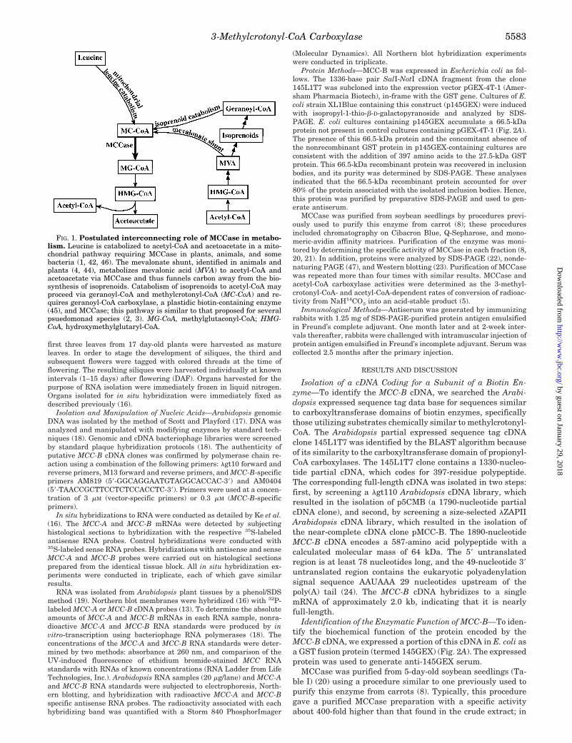
FIG. 1. Postulated interconnecting role of MCCase in metabo-lism. Leucine is catabolized to acetyl-CoA and acetoacetate in a mito-chondrial pathway requiring MCCase in plants, animals, and somebacteria (1, 42, 46). The mevalonate shunt, identified in animals andplants (4, 44), metabolizes mevalonic acid (MVA) to acetyl-CoA andacetoacetate via MCCase and thus funnels carbon away from the bio-synthesis of isoprenoids. Catabolism of isoprenoids to acetyl-CoA mayproceed via geranoyl-CoA and methylcrotonyl-CoA (MC-CoA) and re-quires geranoyl-CoA carboxylase, a plastidic biotin-containing enzyme(45), and MCCase; this pathway is similar to that proposed for severalpsuedomonad species (2, 3). MG-CoA, methylglutaconyl-CoA; HMG-CoA, hydroxymethylglutaryl-CoA.
3-Methylcrotonyl-CoA Carboxylase 5583
by guest on January 29, 2018http://w
ww
.jbc.org/D
ownloaded from

four repetitions of this purification, specific activity rangedbetween 300 and 650 milliunits/mg. These MCCase prepara-tions were judged near homogeneous based upon two criteria.Analysis of these preparations by nondenaturing PAGE (47)revealed the presence of a single protein that migrated with amolecular weight of about 900,000 (Fig. 2D, lane 1). We con-cluded that this protein is MCCase because it contains biotin,which was detected with 125I-streptavidin-Western analyses(Fig. 2D, lane 2), and it reacts with anti-MCC-A serum (datanot shown). Attempts to further purify MCCase by gel-filtra-tion chromatography on Sephacryl S-400 did not increase thespecific activity of the preparation. Furthermore, MCCase ac-tivity eluted from the Sephacryl S-400 column as a single peakcorresponding to a molecular weight of about 900,000. ThisMCCase preparation contains biotin and reacts with anti-MCC-A serum (data not shown). Hence, based upon these anal-yses, we conclude that the MCCase preparation obtained fol-lowing monomeric avidin affinity chromatography is a nearhomogeneous preparation of this enzyme. The specific activityof the purified soybean MCCase (300–650 milliunits/mg) com-pares favorably with previous purifications of this enzyme fromcarrot (700 milliunits/mg) (8) and maize (200–600 milliunits/mg) (11); however, it is an order of magnitude lower than theMCCase purified from animals (25), bacteria (26, 27), and peaand potato (9).
SDS-PAGE analysis of the purified MCCase preparation
identified two polypeptides that were present at an approxi-mately equal-molar ratio (Fig. 2B, lane 5). The larger, 85-kDapolypeptide was identified as the MCC-A subunit because onWestern blot analyses it reacted with 125I-streptavidin (Fig.2C, lane 1) and with antiserum raised against the biotin-con-taining subunit of MCCase (data not shown). Based on thestructure of MCCase from other plant sources (8–10, 46) andfrom animals (21, 25) and bacteria (26, 27), the smaller, 60-kDapolypeptide was tentatively identified as the nonbiotinylatedsubunit of MCCase. Evidence in support of this identificationwas obtained by subjecting the purified MCCase preparationsto nondenaturing PAGE followed by SDS-PAGE. In these anal-yses, the 900-kDa MCCase protein contained both the 85-kDaMCC-A subunit and the 60-kDa polypeptide, and these werethe only polypeptides detected in these preparations. Theseexperiments were conducted with both the monomeric avidinaffinity purified MCCase fraction and the MCCase-containingfraction obtained following a subsequent gel filtration chroma-tography purification step. These findings imply that the 60-kDa polypeptide is the MCC-B subunit.
Evidence in support of the conclusion that the pMCC-B clonecodes for the nonbiotinylated subunit of MCCase was obtainedfrom immunological analyses using the anti-145GEX serum.SDS-PAGE and Western analyses of soybean (Fig. 2C, lane 2)and Arabidopsis (Fig. 2C, lane 4) seedling extracts show thatthe anti-145GEX serum reacts with a single polypeptide in
FIG. 2. Immunological identification of MCC-B. A, expression of the 145L1T7-cDNA as a GST fusion protein. The cDNA, 145L1T7, wassubcloned in-frame with the GST gene in the expression vector pGEX-4T-1. The resulting recombinant plasmid p145-GEX (lane 1), and pGEX-4T-1(lane 2), were propagated in E. coli XL1Blue, and expression of recombinant protein was induced in the presence of 0.1 mM isopropyl-1-thio-b-D-galactopyranoside; protein extracts were fractionated by SDS-PAGE and stained with Coomassie Brilliant Blue. The arrow indicates the expressed66-kDa fusion protein. B, purification of MCCase from soybean seedlings. 10 mg of protein from selected fractions obtained during purification ofMCCase was subjected to SDS-PAGE and stained with Coomassie Brilliant Blue. Lane 1, crude extract; lane 2, polyethylene glycol fraction; lane3, Cibacron Blue fraction; lane 4, Q-Sepharose fraction; lane 5, monomeric avidin fraction. C, Western blot analyses of MCCase. A soybean seedlingextract (50 mg of protein, 0.04 units of activity) (lane 2), monomeric avidin-purified soybean MCCase (1 mg of protein, 0.34 units of activity) (lanes1 and 3), and an Arabidopsis seedling extract (50 mg of protein, 0.03 units of activity) (lane 4) were separated by SDS-PAGE and Western blotted.The blots were probed with 125I-streptavidin, detecting MCC-A (lane 1) or antiserum directed against the145GEX fusion protein, detecting MCC-B(lanes 2–4). D, nondenaturing PAGE of purified soybean MCCase. MCCase (20 mg/lane), purified through the monomeric-avidin affinitychromatography step, was subjected to nondenaturing PAGE, and the resulting gels were stained with Coomassie Brilliant Blue (lane 1) andsubjected to Western analyses that were probed either with 125I-streptavidin (lane 2) or anti-145GEX serum (lane 3).
TABLE IPurification of MCCase from soybean seedlings
350 g of soybean seedlings were used.
Purification step Total protein Total activity Specific activity PurificationSubunit intensitya
MCC-A MCC-B
mg milliunits milliunits/mg fold arbitrary units/mg
Crude extract 714.3 662.9 0.9 1.0 1.0 6 0.08 1.0 6 0.080–18% PEG 594.5 594.5 1.0 1.1 1.25 6 0.08 1.05 6 0.08Cibacron Blue 105.7 724.3 6.8 7.6 7.2 6 0.2 7.8 6 0.3Q-Sepharose 80.1 548.7 6.9 7.7 NDb NDMonomeric avidin 0.9 302.9 336.6 374 350 6 20 380 6 30
a Determined by SDS-PAGE/Western analysis of each fraction, using 125I-streptavidin for the MCC-A subunit and anti-145GEX serum for theMCC-B subunit. The intensity of each subunit-band was quantified with the use of a PhosphorImager.
b ND, not determined.
3-Methylcrotonyl-CoA Carboxylase5584
by guest on January 29, 2018http://w
ww
.jbc.org/D
ownloaded from

each extract that is about 60 kDa. In addition, this antiserumreacts with the 900-kDa purified MCCase protein (Fig. 2D, lane3). To further demonstrate that pMCC-B codes for the nonbi-otinylated subunit of MCCase, the various fractions obtainedduring the purification of the soybean MCCase were subjectedto SDS-PAGE and Western blot analyses with either 125I-streptavidin or anti-145GEX serum. These characterizationsdemonstrate that there is a one-to-one correspondence betweenthe specific activity of MCCase, the relative intensity of theMCC-A subunit band detected with 125I-streptavidin, and therelative intensity of the MCC-B subunit band detected withanti-145GEX serum (Fig. 2C and Table I).
Finally, we tested the effect of anti-145GEX serum on thecatalytic activity of MCCase (Fig. 3). Whereas preimmune con-trol serum did not inhibit MCCase or acetyl-CoA carboxylaseactivity (acetyl-CoA carboxylase is the only other biotin-con-taining enzyme known in Arabidopsis), anti-145GEX serumspecifically inhibited MCCase activity, without affectingacetyl-CoA carboxylase activity. In toto, this series of experi-ments establish that pMCC-A codes for the non-biotin-contain-ing subunit of MCCase.
Isolation and Characterization of the MCC-B Gene of Arabi-dopsis—Southern blot analysis of Arabidopsis (ecotype Colum-bia) DNA digested with EcoRI, HindIII, BamHI, or KpnI andprobed with the MCC-B cDNA reveals a single hybridizingband in each digest (Fig. 4A). Thus, the MCC-B subunit isprobably encoded by a single gene.
An Arabidopsis (ecotype Landsberg) genomic DNA librarywas screened by hybridization with the MCC-B cDNA to iso-late the gene encoding the non-biotin-containing subunit ofMCCase. Twenty-nine hybridizing plaques (out of the 3.0 3 104
plaques that were screened) were identified. These representedoverlapping clones of a single region of the Arabidopsis ge-nome. Two clones, A2041 and A2102, were analyzed in detail. A
4.2-kb SalI fragment, which contained the 39 end of the MCC-Bgene (pMBG), and a 5.2-kb EcoRI fragment, which containedthe 59 end of the MCC-B gene (pMAGP), were subcloned andsequenced. Together, these two overlapping subclones containthe entire MCC-B gene (Fig. 4B).
Comparison of the sequences of the full-length MCC-B cDNAand gene demonstrate that the MCC-B subunit is encoded by a2.78-kb stretch of the genomic sequence and that the tran-scribed region is interrupted by nine introns. There are onlytwo sequence differences between the Landsberg gene andColumbia cDNA: a change of C to T at nucleotide 795 of thecDNA and a change of A to G at nucleotide 855; neither resultsin an amino acid change. The 10 exons of MCC-B range inlength from 56 to 434 nucleotides. The nine intervening intronsare from 77 to 164 nucleotides, larger than the minimum intronlength of 70–73 nucleotides (28). The splice sites of each intronagree with plant consensus splice site sequences (29), and allnine introns contain the highly conserved dinucleotide se-quences GT and AG at the 59 and 39 ends of the introns,respectively. In addition, the AU content of every intron isgreater than 59%, which has been recognized as the minimumAU content for efficient splicing in dicots (28).
Primary Structure of the MCC-B Subunit and Comparison toOther Biotin Enzymes—The MCC-B subunit is a polypeptide of587 amino acid residues. The N-terminal sequence of theMCC-B protein has characteristics of a mitochondrial transitpeptide (30), consistent with the location of MCCase in mito-chondria (9). Analysis of the sequence of the proposed transitpeptide (residues 1–26) with the HELICALWHEEL algorithm
FIG. 3. Immunoinhibition of MCCase activity. Aliquots of Arabi-dopsis extracts were incubated with the indicated amounts of eitherpreimmune control serum (M) or anti-145GEX serum (●). Following a1-h incubation on ice, each aliquot was assayed in triplicate for acetyl-CoA carboxylase (A) and MCCase (B) activity.
FIG. 4. The MCC-B gene of Arabidopsis. A, Southern blot analysisof Arabidopsis DNA probed with the 145L1T7 fragment of the MCC-BcDNA. The endonucleases KpnI, BamHI, HindIII, and EcoRI do nothave restriction sites within the MCC-B gene. B, schematic represen-tation of the structure of the MCC-B gene of Arabidopsis. The nucleo-tide sequence of a 5.28-kb genomic DNA fragment containing theMCC-B gene was determined. Exons are represented as black boxes;introns are represented by solid lines. Positions of the translationalstart (1ATG) and stop (4746TAA) codons are indicated.
3-Methylcrotonyl-CoA Carboxylase 5585
by guest on January 29, 2018http://w
ww
.jbc.org/D
ownloaded from

of the GCG Sequence Analysis package predicts that it willform an amphiphilic a-helix, a common feature of transit pep-tides (31). Proteolytic cleavage of the pre-MCC-B protein ispredicted by the PSORT algorithm (32) to occur at residue 27,within the sequence IRP2GTD. This is consistent with thefinding that an arginine residue is often present at residue 22relative to the cleavage site (33). Cleavage at residue 27 wouldresult in a mature polypeptide with a calculated molecularweight of 60,900, similar to the apparent molecular weight ofthe polypeptide immunologically detected in Arabidopsis leafextracts with anti-145GEX serum (Fig. 2C, lane 4). Further-more, these findings agree with previous determinations of themolecular mass of the MCC-B subunit of MCCase purified frommaize (58 kDa), carrot (65 kDa), pea (54 kDa), and potato (53kDa) (8–10).
Fig. 5 depicts the sequences of the proteins most similar tothe MCC-B subunit; all are carboxyltransferase subunits ofbiotin enzymes (30–39). The three known biotin enzymes mostsimilar to MCC-B, methylmalonyl-CoA decarboxylase (35%identical), propionyl-CoA carboxylase (30% identical), andtranscarboxylase (33% identical), catalyze the conversion ofmethylmalonyl-CoA to propionyl-CoA, or vice versa. This mayreflect the importance of a methyl branch in the molecules thatbind to the carboxyltransferase substrate-binding site of theseenzymes. Comparison of the MCC-B subunit to the carboxyl-transferase subunit of glutaconyl-CoA decarboxylase reinforcesthis hypothesis. The biotin enzyme glutaconyl-CoA decarboxy-lase catalyzes the decarboxylation of glutaconyl-CoA to formcrotonyl-CoA. Glutaconyl-CoA and crotonyl-CoA differ from theMCCase substrate and product only by the absence of themethyl branch, yet the carboxyltransferase subunit of glutaco-nyl-CoA decarboxylase shows lower amino acid identity (23%)
to MCC-B than do the carboxyltransferase subunits of methyl-malonyl-CoA decarboxylase, propionyl-CoA carboxylase, andtranscarboxylase, which all bind shorter, but branched,acyl-CoAs.
Acetyl-CoA carboxylases (40) show ,15% identity to theMCC-B subunit of MCCase (not depicted); the identity is dis-persed throughout the carboxyltransferase domain. The se-quence of the MCC-B subunit of MCCase shows no significantsimilarity to pyruvate carboxylases, biotin enzymes that use ab-keto acid as substrate.
The MCC-B sequence of Arabidopsis has enabled us to iden-tify several animal-derived sequences in the GenBankTM database, the biochemical functions of which had not been defined;these probably represent clones of animal MCC-B. These in-clude a protein (PID g6711) encoded by a hypothetical Caenorh-abditis elegans gene (56% identity) and proteins encoded byexpressed sequence tag cDNAs from Dictyostelium discoideum(GenBankTM accession numbers C90323 and C90323), mouse(GenBankTM accession numbers AA050443, AA463055,AA444444, and AA049241), and human (GenBankTM accessionnumbers R88931 and AA465612).
Spatial and Temporal Patterns of MCC-A and MCC-BmRNA Accumulation—The reaction catalyzed by MCCase isrequired for the catabolism of leucine and of isoprenoids andthe mevalonate shunt (Fig. 1). To begin to comprehend thephysiological roles of these metabolic processes in the growthand development of plants, we examined MCCase expressionby determining the spatial and temporal patterns of MCC-Aand MCC-B mRNA accumulation. This was conducted by RNAblot and in situ hybridization analyses (Figs. 6 and 7). Further-more, because cDNA probes for both MCCase subunits wereavailable (Ref. 13 and this study), these analyses enabled us to
FIG. 5. Comparison of the deduced amino acid sequences of the non-biotin-containing subunit of MCCase. Shown are the MCCBsubunit of Arabidopsis (MCCB.At), carboxyltransferase subunits of the methylmalonyl-CoA decarboxylase of Veillonella parvula (MCDC.Vp) (Ref.34; GenBankTM accession number L22208) and Propionigenium modestum (MCDC.Pm) (Ref. 35; GenBankTM accession number AJ002015), bsubunit of human propionyl-CoA (PCCB.Hs) (Ref. 36; GenBankTM accession number P05166), 12 S subunit of the transcarboxylase of Propionibac-terium shermanii (TC.Ps) (Ref. 37; GenBankTM accession number A48665), and the carboxyltransferase subunits of glutaconyl-CoA decarboxylasefrom Acidaminococcus fermentans (GCDC.Af) (Refs. 38 and 39; GenBankTM accession number G433931). Residues that are identical in MCCB.Atand at least three other sequences are shown on a black background; similar residues are shaded in gray.
3-Methylcrotonyl-CoA Carboxylase5586
by guest on January 29, 2018http://w
ww
.jbc.org/D
ownloaded from
address whether MCC-A and MCC-B mRNAs accumulate co-ordinately during development.
MCC-A and MCC-B mRNAs are detectable in all cell types ofcotyledons, leaves, flower buds, seedling roots, and embryos,but development affects the level of their accumulation (Figs. 6and 7). The ubiquitous accumulation of the MCC-A and MCC-BmRNAs reinforces the concept that MCCase is important for
the metabolic function of all plant cells. Tissues and cells withelevated levels of MCCase mRNAs probably have higher de-mands for metabolic processes that require this enzyme. Sev-eral peaks in MCCase expression are apparent (Figs. 6 and 7).
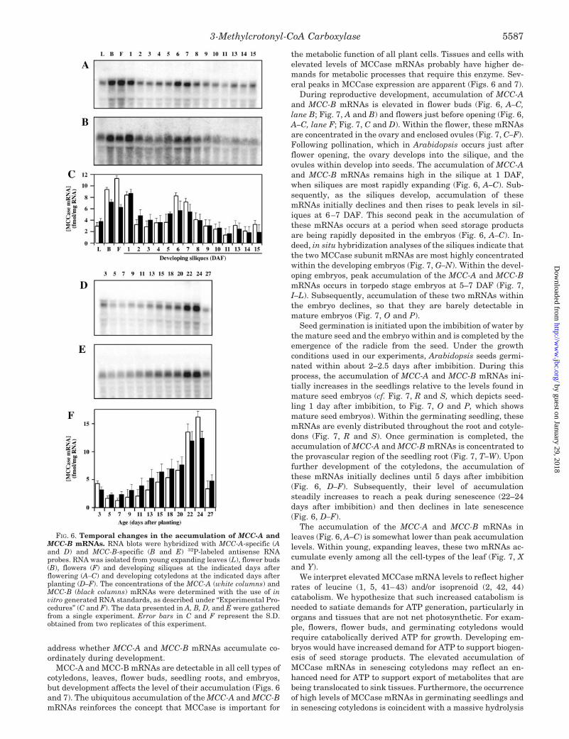
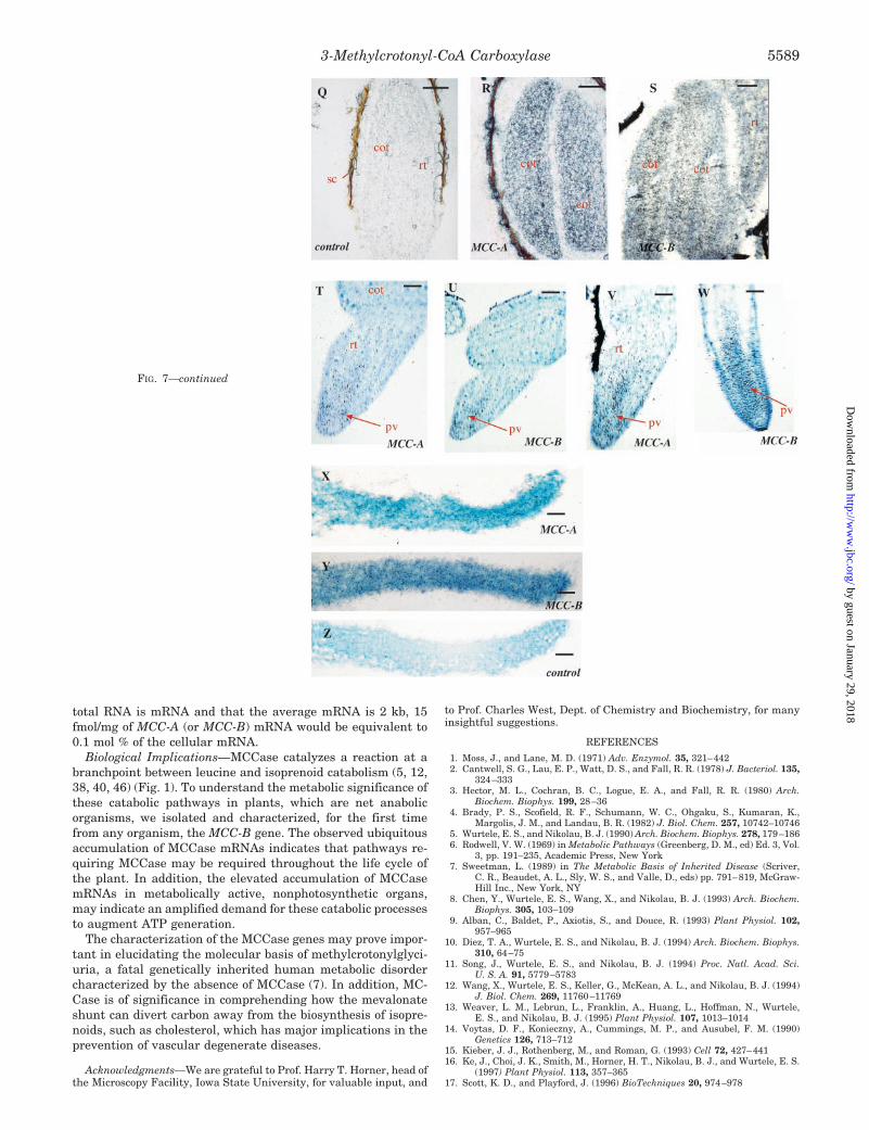
During reproductive development, accumulation of MCC-Aand MCC-B mRNAs is elevated in flower buds (Fig. 6, A–C,lane B; Fig. 7, A and B) and flowers just before opening (Fig. 6,A–C, lane F; Fig. 7, C and D). Within the flower, these mRNAsare concentrated in the ovary and enclosed ovules (Fig. 7, C–F).Following pollination, which in Arabidopsis occurs just afterflower opening, the ovary develops into the silique, and theovules within develop into seeds. The accumulation of MCC-Aand MCC-B mRNAs remains high in the silique at 1 DAF,when siliques are most rapidly expanding (Fig. 6, A–C). Sub-sequently, as the siliques develop, accumulation of thesemRNAs initially declines and then rises to peak levels in sil-iques at 6–7 DAF. This second peak in the accumulation ofthese mRNAs occurs at a period when seed storage productsare being rapidly deposited in the embryos (Fig. 6, A–C). In-deed, in situ hybridization analyses of the siliques indicate thatthe two MCCase subunit mRNAs are most highly concentratedwithin the developing embryos (Fig. 7, G–N). Within the devel-oping embryos, peak accumulation of the MCC-A and MCC-BmRNAs occurs in torpedo stage embryos at 5–7 DAF (Fig. 7,I–L). Subsequently, accumulation of these two mRNAs withinthe embryo declines, so that they are barely detectable inmature embryos (Fig. 7, O and P).
Seed germination is initiated upon the imbibition of water bythe mature seed and the embryo within and is completed by theemergence of the radicle from the seed. Under the growthconditions used in our experiments, Arabidopsis seeds germi-nated within about 2–2.5 days after imbibition. During thisprocess, the accumulation of MCC-A and MCC-B mRNAs ini-tially increases in the seedlings relative to the levels found inmature seed embryos (cf. Fig. 7, R and S, which depicts seed-ling 1 day after imbibition, to Fig. 7, O and P, which showsmature seed embryos). Within the germinating seedling, thesemRNAs are evenly distributed throughout the root and cotyle-dons (Fig. 7, R and S). Once germination is completed, theaccumulation of MCC-A and MCC-B mRNAs is concentrated tothe provascular region of the seedling root (Fig. 7, T–W). Uponfurther development of the cotyledons, the accumulation ofthese mRNAs initially declines until 5 days after imbibition(Fig. 6, D–F). Subsequently, their level of accumulationsteadily increases to reach a peak during senescence (22–24days after imbibition) and then declines in late senescence(Fig. 6, D–F).
The accumulation of the MCC-A and MCC-B mRNAs inleaves (Fig. 6, A–C) is somewhat lower than peak accumulationlevels. Within young, expanding leaves, these two mRNAs ac-cumulate evenly among all the cell-types of the leaf (Fig. 7, Xand Y).
We interpret elevated MCCase mRNA levels to reflect higherrates of leucine (1, 5, 41–43) and/or isoprenoid (2, 42, 44)catabolism. We hypothesize that such increased catabolism isneeded to satiate demands for ATP generation, particularly inorgans and tissues that are not net photosynthetic. For exam-ple, flowers, flower buds, and germinating cotyledons wouldrequire catabolically derived ATP for growth. Developing em-bryos would have increased demand for ATP to support biogen-esis of seed storage products. The elevated accumulation ofMCCase mRNAs in senescing cotyledons may reflect an en-hanced need for ATP to support export of metabolites that arebeing translocated to sink tissues. Furthermore, the occurrenceof high levels of MCCase mRNAs in germinating seedlings andin senescing cotyledons is coincident with a massive hydrolysis
FIG. 6. Temporal changes in the accumulation of MCC-A andMCC-B mRNAs. RNA blots were hybridized with MCC-A-specific (Aand D) and MCC-B-specific (B and E) 32P-labeled antisense RNAprobes. RNA was isolated from young expanding leaves (L), flower buds(B), flowers (F) and developing siliques at the indicated days afterflowering (A–C) and developing cotyledons at the indicated days afterplanting (D–F). The concentrations of the MCC-A (white columns) andMCC-B (black columns) mRNAs were determined with the use of invitro generated RNA standards, as described under “Experimental Pro-cedures” (C and F). The data presented in A, B, D, and E were gatheredfrom a single experiment. Error bars in C and F represent the S.D.obtained from two replicates of this experiment.
3-Methylcrotonyl-CoA Carboxylase 5587
by guest on January 29, 2018http://w
ww
.jbc.org/D
ownloaded from

of proteins in these organs, which would provide free leucine asa substrate for catabolism. These data expand previous studiesin soybean and pea, which indicate that MCCase activity ishigher in metabolically active organs (42) and increases inresponse to carbohydrate starvation (41). The data presentedherein, in combination with these previous studies (41, 42),indicate that changes in MCCase activity are at least partiallyattributable to changes in MCCase mRNA accumulation.
Finally, the changing patterns of MCC-A and MCC-B mRNAaccumulation are similar both temporally and spatially duringdevelopment (Figs. 6 and 7). Indeed, quantitative analysesindicate that these two mRNAs accumulate at approximatelyequal molar ratios (Fig. 6, C and F). These findings imply thatthe expression of the MCC-A and MCC-B genes is coordinatelyregulated. Accumulation of MCC-A (and MCC-B) mRNAsranges from about 2 to 15 fmol/mg RNA. Assuming that 1% of
FIG. 7. Spatial distribution of MCC-A and MCC-B mRNAs in Arabidopsis. Histological tissue sections were hybridized with 35S-labeledantisense RNA probes (A–P and R–Y) or, for controls, with 35S-labeled sense RNA probes (Q and Z), and stained with Toluidine Blue. Black spotsvisualized by autoradiography are silver grains reflecting location of the MCC-A or MCC-B mRNAs. All hybridizations conducted three times withsimilar results. Each type of section was probed with all four probes, and representative results are shown. The distribution of the MCC-A andMCC-B mRNAs (as labeled) is shown in flower buds (A and B), in a flower viewed at lower magnification (C and D) and at higher magnificationto show the ovary (E and F), in siliques at 3 DAF (G and H), in siliques at 5 DAF (I and J), in siliques at 7 DAF (K and L), in siliques at 9 DAF(M and N), in siliques at 12 DAF (O and P), in seedlings at 1 day after imbibition (R and S), in seedlings at 2 days after imbibition (T and U), inseedlings at 3 days after imbibition (V and W), and in young expanding leaves (X and Y). Control hybridizations are shown for seedlings at 2 daysafter imbibition with sense MCC-A probe (Q) and for young leaves with sense MCC-B probe (Z). All control hybridizations show negligible signal.The MCC-A and MCC-B mRNAs accumulate in very similar spatial patterns. r, receptacle; ov, ovary; o, ovule; a, anther; s, sepal; p, petal; oi, outerintegument; ii, inner integument; w, wall of ovary; ge, globular embryo; te, torpedo embryo; cot, cotyledon; rt, root; sc, seed coat (derived from innerand outer integument); pv, provascular cambium. Bars, 585 mm in A–D and G–Z and 41 mm in E and F.
3-Methylcrotonyl-CoA Carboxylase5588
by guest on January 29, 2018http://w
ww
.jbc.org/D
ownloaded from
total RNA is mRNA and that the average mRNA is 2 kb, 15fmol/mg of MCC-A (or MCC-B) mRNA would be equivalent to0.1 mol % of the cellular mRNA.
Biological Implications—MCCase catalyzes a reaction at abranchpoint between leucine and isoprenoid catabolism (5, 12,38, 40, 46) (Fig. 1). To understand the metabolic significance ofthese catabolic pathways in plants, which are net anabolicorganisms, we isolated and characterized, for the first timefrom any organism, the MCC-B gene. The observed ubiquitousaccumulation of MCCase mRNAs indicates that pathways re-quiring MCCase may be required throughout the life cycle ofthe plant. In addition, the elevated accumulation of MCCasemRNAs in metabolically active, nonphotosynthetic organs,may indicate an amplified demand for these catabolic processesto augment ATP generation.
The characterization of the MCCase genes may prove impor-tant in elucidating the molecular basis of methylcrotonylglyci-uria, a fatal genetically inherited human metabolic disordercharacterized by the absence of MCCase (7). In addition, MC-Case is of significance in comprehending how the mevalonateshunt can divert carbon away from the biosynthesis of isopre-noids, such as cholesterol, which has major implications in theprevention of vascular degenerate diseases.
Acknowledgments—We are grateful to Prof. Harry T. Horner, head ofthe Microscopy Facility, Iowa State University, for valuable input, and
to Prof. Charles West, Dept. of Chemistry and Biochemistry, for manyinsightful suggestions.
REFERENCES
1. Moss, J., and Lane, M. D. (1971) Adv. Enzymol. 35, 321–4422. Cantwell, S. G., Lau, E. P., Watt, D. S., and Fall, R. R. (1978) J. Bacteriol. 135,
324–3333. Hector, M. L., Cochran, B. C., Logue, E. A., and Fall, R. R. (1980) Arch.
Biochem. Biophys. 199, 28–364. Brady, P. S., Scofield, R. F., Schumann, W. C., Ohgaku, S., Kumaran, K.,
Margolis, J. M., and Landau, B. R. (1982) J. Biol. Chem. 257, 10742–107465. Wurtele, E. S., and Nikolau, B. J. (1990) Arch. Biochem. Biophys. 278, 179–1866. Rodwell, V. W. (1969) in Metabolic Pathways (Greenberg, D. M., ed) Ed. 3, Vol.
3, pp. 191–235, Academic Press, New York7. Sweetman, L. (1989) in The Metabolic Basis of Inherited Disease (Scriver,
C. R., Beaudet, A. L., Sly, W. S., and Valle, D., eds) pp. 791–819, McGraw-Hill Inc., New York, NY
8. Chen, Y., Wurtele, E. S., Wang, X., and Nikolau, B. J. (1993) Arch. Biochem.Biophys. 305, 103–109
9. Alban, C., Baldet, P., Axiotis, S., and Douce, R. (1993) Plant Physiol. 102,957–965
10. Diez, T. A., Wurtele, E. S., and Nikolau, B. J. (1994) Arch. Biochem. Biophys.310, 64–75
11. Song, J., Wurtele, E. S., and Nikolau, B. J. (1994) Proc. Natl. Acad. Sci.U. S. A. 91, 5779–5783
12. Wang, X., Wurtele, E. S., Keller, G., McKean, A. L., and Nikolau, B. J. (1994)J. Biol. Chem. 269, 11760–11769
13. Weaver, L. M., Lebrun, L., Franklin, A., Huang, L., Hoffman, N., Wurtele,E. S., and Nikolau, B. J. (1995) Plant Physiol. 107, 1013–1014
14. Voytas, D. F., Konieczny, A., Cummings, M. P., and Ausubel, F. M. (1990)Genetics 126, 713–712
15. Kieber, J. J., Rothenberg, M., and Roman, G. (1993) Cell 72, 427–44116. Ke, J., Choi, J. K., Smith, M., Horner, H. T., Nikolau, B. J., and Wurtele, E. S.
(1997) Plant Physiol. 113, 357–36517. Scott, K. D., and Playford, J. (1996) BioTechniques 20, 974–978
FIG. 7—continued
3-Methylcrotonyl-CoA Carboxylase 5589
by guest on January 29, 2018http://w
ww
.jbc.org/D
ownloaded from

18. Sambrook, J., Fritsch, E. F., and Maniatis, T. (1989) Molecular Cloning: ALaboratory Manual, 2nd Ed., Cold Spring Harbor Laboratory, Cold SpringHarbor, New York
19. Dean, C., vandenElzen, P., Tamaki, S., Dunsmuir, P., and Bedbrook, J. (1985)EMBO J. 4, 3055–3061
20. Song, J. (1993) Molecular Cloning and Characterization of 3-Methylcrotonyl-CoA Carboxylase from Soybean. Ph.D. Thesis, Iowa State University
21. Lau, E. P., Cochran, B. C., Munson, L., and Fall, R. R. (1979) Proc. Natl. Acad.Sci. U. S. A. 76, 214–218
22. Laemmli, U. K. (1970) Nature 227, 680–68523. Kyhse-Andersen, J. (1984) J. Biochem. Biophys. Methods 10, 203–20924. Proudfoot, N. J., and Brownlee, G. G. (1976) Nature 263, 211–21425. Lau, E. P., Cochran, B. C., and Fall, R. R. (1980) Arch. Biochem. Biophys. 205,
352–35926. Schiele, U., Niedermeier, R., Sturzer, M., and Lynen, F. (1975) Eur. J. Bio-
chem. 60, 259–26627. Fall, R. R., and Hector, M. L. (1977) Biochemistry 16, 4000–400528. Goodall, G. J., and Filipowicz, W. (1991) EMBO J. 10, 2635–264429. Simpson, C. G., Leader, D. J., and Brown, J. W. S. (1993) in Plant Molecular
Biology (Croy, R. R. D., ed) pp. 183–239, BIOS Scientific Publishers Lim-ited, San Diego, CA
30. Boutry, M., and Chaumont, F. (1993) in Plant Mitochondria with Emphasis onRNA Editing and Cytoplasmic Male Sterility (Brennicke, A., and Kuck, U.,eds) pp. 321–329, VCH Publishers, New York, NY
31. Attardi, G., and Schatz, G. (1988) Annu. Rev. Cell Biol. 4, 289–33332. Nakai, K., and Kanehisa, M. (1992) Genomics 14, 897–91133. Wallace, T. P., and Howe, C. J. (1993) in Plant Molecular Biology Labfax (Croy,
R. R. D., ed) pp. 287–292, BIOS Scientific Publishers Limited, San Diego,CA
34. Huder, J. B., and Dimroth, P. J. (1993) Biol. Chem. 268, 24564–2457135. Bott, M., Pfister, K., Burda, P., Kalbermatter, O., Woehlke, G., and Dimroth,
P. (1997) Eur. J. Biochem. 250, 590–59936. Lamhonwah, A. M., Leclerc, D., Loyer, M., Clarizio, R., and Gravel, R. A.
(1994) Genomics 19, 500–50537. Thornton, C. G., Kumar, G. K., Haase, F. C., Phillips, N. F. B., Woo, S. B., Park,
V. M., Magner, W. J., Shenoy, B. C., Wood, H. G., and Samols, D. J. (1993)Bacteriology 175, 5301–5308
38. Mack, M., Bendrat, K., Zelder, O., Eckel, E., Linder, D., and Buckel, W. (1994)Eur. J. Biochem. 226, 41–51
39. Jacob, U., Mack, M., Clausen, T., Huber, R., Buckel, W., and Messerschmidt,A. (1997) Structure 5, 415–426
40. Yanai, Y., Kawasaki, T., Shimada, H., Wurtele, E. S., Nikolau, B. J., andIchikawa, N. (1995) Plant Cell Physiol. 36, 779–787
41. Aubert, S., Alban, C., Bligny, R., and Douce, R. (1996) FEBS Lett. 383, 175–18042. Anderson, M. D., Che, P., Song, J., Nikolau, B. J., and Wurtele, E. S. (1998)
Plant Physiol. 118, 1121–113843. Wurtele, E. S., and Nikolau, B. J. (1992) Plant Physiol. 99, 1699–170344. Guan, X., Diez, T., Prasad, T., Nikolau, B. J., and Wurtele, E. S. (1999). Arch.
Biochem. Biophys. 362, 12–2145. Nes, W. D., and Bach, T. J. (1985) Proc. R. Soc. Lond. B. Biol. Sci. 225,
425–44446. Wurtele, E. S., and Nikolau, B. J. (2000) Methods Enzymol., in press47. Hedrick, J. L., and Smith, A. J. (1968) Arch. Biochem. Biophys. 126, 155–164
3-Methylcrotonyl-CoA Carboxylase5590
by guest on January 29, 2018http://w
ww
.jbc.org/D
ownloaded from

Nikolau and Eve Syrkin WurteleAngela L. McKean, Jinshan Ke, Jianping Song, Ping Che, Sara Achenbach, Basil J.
3-Methylcrotonyl-CoA CarboxylaseMolecular Characterization of the Non-biotin-containing Subunit of
doi: 10.1074/jbc.275.8.55822000, 275:5582-5590.J. Biol. Chem.
http://www.jbc.org/content/275/8/5582Access the most updated version of this article at
Alerts:
When a correction for this article is posted•
When this article is cited•
to choose from all of JBC's e-mail alertsClick here
http://www.jbc.org/content/275/8/5582.full.html#ref-list-1
This article cites 40 references, 12 of which can be accessed free at
by guest on January 29, 2018http://w
ww
.jbc.org/D
ownloaded from